Is it an orbital foreign body?

DOI: 10.12809/hkmj164952
© Hong Kong Academy of Medicine. CC BY-NC-ND 4.0
 
PICTORIAL MEDICINE
Is it an orbital foreign body?
Mohamed Shaheeda, FRCSEd, MPH; Stacey C Lam, MB, ChB; Noel CY Chan, FRCSEd, FCOphthHK; Hunter KL Yuen, FRCSEd, FCOphthHK
Department of Ophthalmology and Visual Sciences, The Chinese University of Hong Kong, Shatin, Hong Kong
 
Corresponding author: Dr Stacey C Lam (staceylam@gmail.com)
 
 Full paper in PDF
 
A 58-year-old man presented with left globe rupture following blunt orbital trauma. Visual acuity was light perception only. There was lid swelling, ecchymosis, and a deformed globe with scleral laceration. Computed tomography (CT) of the orbit revealed a 3.7-mm hyperdensity at the superomedial aspect of the left globe, interpreted by the radiologist as a possible intra-orbital foreign body (Fig).
 

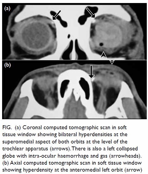
Figure. (a) Coronal computed tomographic scan in soft tissue window showing bilateral hyperdensities at the superomedial aspect of both orbits at the level of the trochlear apparatus (arrows). There is also a left collapsed globe with intra-ocular haemorrhage and gas (arrowheads). (b) Axial computed tomographic scan in soft tissue window showing hyperdensity at the anteromedial left orbit (arrow)
 
Thorough history for the mechanism of trauma, physical examination for a wound site and thin cut CT sections were reviewed. History revealed blunt orbital trauma by a metallic rod rather than a sharp penetrating injury. No cutaneous entry site was evident. Computed tomography revealed bilateral hyperdensities at the level of the trochlea, more pronounced on the left than the right, with no metallic streak artefacts. All of the above supported a diagnosis of trochlear calcification rather than an intra-orbital foreign body, and the patient subsequently underwent emergency repair of scleral laceration without orbital exploration.
 
Discussion
The trochlea is a cartilaginous pulley at the superomedial aspect of the orbit through which the superior oblique muscle tendon passes freely. It has a synovium-lined space, and like other synovial joints of the body, the trochlea can develop calcifications in the cartilage, tendon, or within the bursa-like cleft.1 The reported prevalence of incidental trochlear calcifications on CT is 3-16%, with over 50% being unilateral.2 3 No clear cause has been identified, but prior studies have postulated degenerative, inflammatory, metabolic or traumatic aetiologies.4 Since trochlear calcification is asymptomatic, most cases go unnoticed by radiologists as well as ophthalmologists. In the presence of co-existing orbital trauma, it can be misdiagnosed as an intraorbital foreign body. Surgical exploration around the trochlear region may cause damage and scarring leading to diplopia.
 
Differentiation of the two entities requires history taking, physical examination, and proper imaging. A foreign body is present in one in six cases of orbital trauma, with metallic objects and glass being the most common.5 A review of the history to determine mechanism of injury is useful but may be unreliable. Examination of the skin and conjunctiva, particularly the fornices, may help detect subtle penetrating injuries. Radiological assessment includes plain films, ultrasound, CT, and magnetic resonance imaging. Plain films are limited to detection of metallic foreign bodies, and ultrasound has limited use because foreign bodies can be masked by surrounding highly reflective structures such as bone. Computed tomography is an excellent means to identify high-density objects such as metal or glass, but not organic matter. Magnetic resonance imaging is contra-indicated in metallic foreign bodies, but may be useful for organic foreign matter. Features of trochlear calcification may include symmetrical presentation and typical site at the trochlear apparatus. Its morphology has been described as comma, dot, and inverted U shape.2 In contrast, orbital foreign bodies are often unilateral with no specific size, shape, or location. Metallic foreign bodies may also generate streak artefacts.
 
Orbital calcifications can be incidental or pathological. It is important to recognise trochlear calcifications as distinct from foreign bodies so as to avoid unnecessary surgical exploration in the presence of orbital trauma.
 
References
1. Sobel RK, Goldstein SM. Trochlear calcification: A common entity. Orbit 2012;31:94-6. Crossref
2. Xiao TL, Kalariya NM, Yan ZH, et al. Trochlear calcification and intraorbital foreign body in ocular trauma patients. Chin J Traumatol 2009;12:210-3.
3. Shriver EM, McKeown CA, Johnson TE. Trochlear calcification mimicking an orbital foreign body. Ophthal Plast Reconstr Surg 2011;27:143-4. Crossref
4. Buch K, Nadgir RN, Tannenbaum AD, Ozonoff A, Fujita A, Sakai O. Clinical significance of trochlear calcifications in the orbit. AJNR Am J Neuroradiol 2014;35:573-7. Crossref
5. Nasr AM, Haik BG, Fleming JC, Al-Hussain HM, Karcioglu ZA. Penetrating orbital injury with organic foreign bodies. Ophthalmology 1999;106:523-32. Crossref

Birt-Hogg-Dubé syndrome: a rare cause of familial spontaneous pneumothorax

DOI: 10.12809/hkmj165022
© Hong Kong Academy of Medicine. CC BY-NC-ND 4.0
 
PICTORIAL MEDICINE
Birt-Hogg-Dubé syndrome: a rare cause of familial spontaneous pneumothorax
HM Luk, FHKAM (Paediatrics); Tony MF Tong, MSc; Ivan FM Lo, FHKAM (Paediatrics)
Clinical Genetic Service, Department of Health, 3/F Cheung Sha Wan Jockey Club Clinic, 2 Kwong Lee Road, Shamshuipo, Hong Kong
 
Corresponding author: Dr HM Luk (luksite@gmail.com)
 
 Full paper in PDF
 
A family with a strong history of pneumothorax was referred to our genetic clinic for assessment. There were three siblings who had all developed spontaneous pneumothorax at the age of 30, 58, and 59 years. All were non-smokers with no pre-existing pulmonary disease. High-resolution computed tomography of the thorax for all showed multiple thin-walled pulmonary cysts of variable size on both sides, mainly located at the basal and peripheral lung regions (Fig). Lung biopsy was not informative. Physical examination revealed multiple smooth, dome-shaped papules over the face and ears in one of siblings (Fig b). There were no other features of tuberous sclerosis or history of renal disease in the family. Based on the dermatological findings and diffuse multicystic lung disease, Birt-Hogg-Dubé (BHD) syndrome was suspected. FLCN gene analysis revealed a heterozygous FLCN{NM_144997.5}:c.1285dupC mutation in all affected members. The diagnosis of BHD syndrome was substantiated. Renal imaging was arranged for surveillance of potential renal cell carcinoma.
 

Figure. (a) Chest X-ray showing bilateral multiple pulmonary cysts over the basal region (arrows). (b) Clinical photo of fibrofolliculomas over pinna (arrows). (c and d) High-resolution computed tomography of the thorax showing lower-zone predominant pulmonary cysts of variable shapes and sizes (circles). The walls are thin, sharply demarcated and do not enhance with contrast (arrowhead)
 
The BHD syndrome is a rare autosomal dominant disease characterised by three major organ manifestations1:
(1) Fibrofolliculomas and other benign skin tumours such as trichodiscomas and acrochordons; these skin lesions are predominantly located on the facial, cervical, and upper truncal regions as smooth, dome-shaped, and white to flesh-coloured papules.
(2) Increased susceptibility to renal cell carcinoma of mixed histologies; the most frequent subtype is a hybrid oncocytic tumour with features of renal oncocytoma and chromophobe renal cell carcinoma.
(3) Multiple bilateral pulmonary cysts and spontaneous pneumothorax.
 
The clinical features of BHD syndrome are heterogeneous with wide intra-familial and inter-familial variation. It is caused by mutations of the FLCN gene. Any combination of the cutaneous, renal, and pulmonary features mentioned above present in an individual or multiple family members should alert the clinician to the possibility of BHD syndrome.
 
Bilateral multiple pulmonary cysts are a highly penetrant feature in BHD syndrome. As a result, the risk of pneumothorax in BHD patients is 50 times higher than that of the general population.2 Approximately 80% to 90% of BHD patients develop lung cysts, usually after early mid-adulthood. The BHD-associated lung cysts tend to be located at the basilar and mediastinal regions of the lungs, in contrast to the typically apical location in primary spontaneous pneumothorax and emphysema. Radiologically, the BHD-associated lung cysts are usually irregularly shaped, variable in size and number, and with sharply demarcated thin walls that do not enhance on computed tomographic imaging.
 
Fibrofolliculomas are present in more than 80% of patients with BHD syndrome and typically appear after the age of 20 years. They are dome-shaped, white to flesh-coloured, non-painful and non-pruritic papules located on the facial, cervical, and upper truncal regions.
 
The most threatening complication of BHD syndrome is renal cell carcinoma. It occurs in approximately 15% of BHD patients by the age of 70 years.3 Therefore regular surveillance is mandatory.
 
Physicians should be alert to the possibility of BHD syndrome in a patient who presents with diffuse cystic lung disease, particularly in the presence of a positive family history. Early referral to a clinical genetic service and multidisciplinary management is recommended. Early diagnosis and regular renal surveillance aim to greatly reduce renal cell carcinoma–associated morbidity and mortality.
 
References
1. Menko FH, van Steensel MA, Giraud S, et al. Birt-Hogg-Dubé syndrome: diagnosis and management. Lancet Oncol 2009;10:1199-206. Crossref
2. Gupta N, Seyama K, McCormack FX. Pulmonary manifestations of Birt-Hogg-Dubé syndrome. Fam Cancer 2013;12:387-96. Crossref
3. Stamatakis L, Metwalli AR, Middelton LA, Marston Linehan W. Diagnosis and management of BHD-associated kidney cancer. Fam Cancer 2013;12:397-402. Crossref

A child with giant tumefactive perivascular spaces

DOI: 10.12809/hkmj164918
© Hong Kong Academy of Medicine. CC BY-NC-ND 4.0
 
PICTORIAL MEDICINE
A child with giant tumefactive perivascular spaces
Neeraj R Mahboobani, MB, BS, FRCR; LF Cheng, FRCR, FHKAM (Radiology); Johnny KF Ma, FRCR, FHKAM (Radiology)
Department of Radiology, Princess Margaret Hospital, Laichikok, Hong Kong
 
Corresponding author: Dr Neeraj R Mahboobani (neeraj.mahboobani@gmail.com)
 
 Full paper in PDF
 
A 4-year-old girl was admitted with a 2-day history of a high temperature up to 41°C. She developed sudden-onset generalised tonic-clonic seizure. She had no travel history. Physical examination showed neck stiffness. The working diagnosis was meningitis. Non-contrast computed tomographic (CT) brain prior to lumbar puncture revealed clustered well-defined cystic lesions with no calcifications measuring up to 1.5 cm in the white matter of the right frontal and parietal lobes (Fig 1).
 

Figure 1. Non-contrast computed tomography of the brain on the day of admission showing clustered well-defined cystic lesions with no calcifications measuring up to 1.5 cm in the white matter of the right frontal and parietal lobes (arrows)
 
The child subsequently developed status epilepticus. The neurosurgical unit was consulted and brain abscesses were suspected. In view of her clinical deterioration, burr hole and aspiration of the cystic lesions were planned after magnetic resonance imaging (MRI). The signal intensity of the lesions was identical to that of cerebrospinal fluid (CSF) on all MRI sequences (Figs 2 and 3). There was no rim enhancement around the lesions. Minimal fluid-attenuated inversion recovery (FLAIR) hyperintense signals were noted in the white matter adjacent to the lesions. There was no restricted diffusion on diffusion-weighted imaging.
 

Figure 2. Magnetic resonance imaging (MRI) on day 2 following admission at the level of the right frontal lobe lesions
(a) T1-weighted, (b) T2-weighted, (c) fluid-attenuated inversion recovery (FLAIR) imaging showing the lesions to be identical to cerebrospinal fluid signal on all MRI sequences. Minimal FLAIR hyperintense signals seen around the cysts are suggestive of gliosis (arrows). (d) No enhancement on post-gadolinium scan is shown (arrow). (e) Diffusion-weighted imaging and (f) apparent diffusion coefficient map showing no restricted diffusion (arrows)


Figure 3. Magnetic resonance imaging (MRI) on day 2 following admission at the level of the right parietal lobe lesions
(a) T1-weighted, (b) T2-weighted, (c) fluid-attenuated inversion recovery (FLAIR) imaging showing the lesions to be identical to the cerebrospinal fluid signal on all MRI sequences. Minimal FLAIR hyperintense signals seen around the cysts are suggestive of gliosis (arrows). (d) No enhancement on post-gadolinium scan is shown (arrows). (e) Diffusion-weighted imaging and (f) apparent diffusion coefficient map showing no restricted diffusion (arrows)
 
Imaging features were not compatible with brain abscess so surgical aspiration was withheld. Cerebral hydatid disease was considered a possible diagnosis based on imaging and the child was commenced on oral albendazole 15 mg/kg/day. This diagnosis was subsequently disproved given the lack of an exposure history, absence of Echinococcus granulosus antibody in serum, and absence of cysts in the liver and spleen on ultrasonography.
 
Influenza A virus antigen was subsequently detected in nasopharyngeal aspirate. The child was prescribed antiviral treatment with consequent cessation of seizures and clinical improvement. The clinical diagnosis was influenza virus–associated encephalopathy.
 
Follow-up CT and MRI 3 weeks later showed the non-enhancing cystic lesions to be unchanged in size and signal characteristics. The lesions were classified as giant tumefactive perivascular spaces (GTPVS).
 
The perivascular spaces (PVS) of the brain are pial-lined, interstitial fluid-filled cystic spaces. They accompany penetrating arteries as they enter the brain parenchyma. These are normal structures that can be found in all ages.1 Most PVS are small in size and usually measure less than 5 mm.1 Larger (>1 cm) PVS have been reported and these are called GTPVS, which can occur as single or multiple clustered cysts.2 Such GTPVS in cerebral white matter can have mild FLAIR hyperintensities in the surrounding white matter1 that can be due to gliosis.2
 
Of note, GTPVS are often incidental findings on imaging. They occur in characteristic locations alongside penetrating vessels in the mesencephalothalamic area, cerebral white matter, and cerebellar dentate nuclei.2 They follow the CSF signal on MRI and do not enhance.
 
Sometimes, GTPVS can be misinterpreted as other pathological processes.1 2 3 Lack of a solid component and complete suppression on FLAIR differentiates them from cystic neoplasms.1 Nil to minimal peri-lesional FLAIR signals help to differentiate them from cystic infarction and mucopolysaccharidosis. Parasitic cysts, in particular hydatid cysts, can have identical imaging features and necessitate clinical correlation for exclusion as in this case. Neuroepithelial cysts also have identical imaging features but can only be differentiated from GTPVS by histopathology.1 Knowledge of this entity, its imaging features, and characteristic locations is essential to avoid unnecessary medical treatment and surgical biopsy/intervention.
 
References
1. Kwee RM, Kwee TC. Virchow-Robin spaces at MR imaging. Radiographics 2007;21:1071-86. Crossref
2. Salzman KL, Osborn AG, House P, et al. Giant tumefactive perivascular spaces. AJNR Am J Neuroradiol 2005;26:298-305.
3. Sankararaman S, Velayuthan S, Ambekar S, Gonzalez-Toledo E. Giant tumefactive perivascular spaces: A further case. J Pediatr Neurosci 2013;8:108-10. Crossref

Urothelial carcinoma with paraneoplastic leukocytosis

DOI: 10.12809/hkmj164956
© Hong Kong Academy of Medicine. CC BY-NC-ND 4.0
 
PICTORIAL MEDICINE
Urothelial carcinoma with paraneoplastic leukocytosis
CW Hsu, MD, PhD; HY Su, MD
Department of Emergency Medicine, E-Da Hospital and I-Shou University, Kaohsiung, Taiwan
 
Previously presented at the 8th Asian Conference on Emergency Medicine, Taiwan, 7-10 November 2015.
 
Corresponding author: Dr HY Su (hys927@hotmail.com)
 
 Full paper in PDF
 
An 88-year-old male with no underlying disease presented to our emergency department in March 2015 with a 2-week history of right flank pain but no fever or urinary tract symptoms. He was conscious, alert, and well-oriented. His blood pressure was 145/89 mm Hg, with a heart rate of 82 beats/min and respiratory rate of 16 breaths/min on arrival. Physical examination revealed obvious right flank knocking tenderness. Laboratory testing revealed white blood cell (WBC) count of 34.7 x 10⁹ /L (reference range [RR], 4.5-11.0 x 10⁹ /L), haemoglobin level of 95 g/L (RR, 135-175 g/L), platelet count of 496 x 109 /L (RR, 150-450 x 109 /L), C-reactive protein level of 24.3 mg/L (RR, 0-10 mg/L), and serum calcium level of 3.31 mmol/L (RR, 2.18-2.58 mmol/L). Urinalysis demonstrated no pyuria, but 50 to 75 red blood cells per high-power field. Prothrombin time, activated partial thromboplastin time, and fibrinogen were within normal limits. An abdominal plain film revealed a mass lesion with a concealing right lower psoas shadow (Fig 1). Subsequent abdominal computed tomographic (CT) scan demonstrated a cyst-like lesion with irregular surface, involving adjacent tissues in the retroperitoneal cavity (Fig 2).
 

Figure 1. A plain abdominal film revealing a mass lesion (arrows) with obliteration of right lower psoas shadow (arrowheads)
 

Figure 2. Abdominal computed tomographic scans demonstrating a cyst-like lesion with irregular surface (asterisk) that involves the adjacent psoas muscle, vertebral body (arrowhead), ureter, and great vessels (arrow) in the retroperitoneal cavity
 
A CT-guided biopsy was performed because the discrepancy between clinical presentation and laboratory tests and image study made it difficult for physicians to discriminate psoas muscle abscess from malignancy. Ultimate pathology of biopsy specimens revealed a high-grade urothelial carcinoma with tumour necrosis. The TNM staging was stage IV (cT4N0M0). Blood and urine cultures were all negative. Follow-up 1 month later revealed a WBC count of 34.7 x 10⁹ /L.
 
Urothelial carcinoma originates in the urinary system. Paraneoplastic leukocytosis, defined by a WBC count of >20.0 x 10⁹ /L on more than two occasions 30 days apart, occurs in 0.6% of urothelial carcinoma cases.1 The aetiology of paraneoplastic leukocytosis is considered to be related to the production of granulocyte colony-stimulating factor by tumour cells.2 3 Hypercalcaemia, anaemia and thrombocytosis, as seen in this case, are also frequently seen in paraneoplastic syndrome. It is associated with advanced urothelial cancer and indicates a poor prognosis.1 3 Muscle invasion is also frequently found in cases of urothelial cancer with paraneoplastic leukocytosis.1 The cyst-like pattern of urothelial cancer on abdominal CT scan in combination with the paraneoplastic leukocytosis can mislead physicians into making an incorrect diagnosis, such as pyogenic psoas muscle abscess. We advise physicians to always be aware of urothelial cancer with paraneoplastic leukocytosis while managing a cyst-like lesion in the retroperitoneal cavity.
 
References
1. Izard JP, Gore JL, Mostaghel EA, Wright JL, Yu EY. Persistent, unexplained leukocytosis is a paraneoplastic syndrome associated with a poor prognosis in patients with urothelial carcinoma. Clin Genitourin Cancer 2015;13:e253-8. Crossref
2. Ito N, Matsuda T, Kakehi Y, Takeuchi E, Takahashi T, Yoshida O. Bladder cancer producing granulocyte colony-stimulating factor. N Engl J Med 1990;323:1709-10. Crossref
3. Kumar AK, Satyan MT, Holzbeierlein J, Mirza M, Van Veldhuizen P. Leukemoid reaction and autocrine growth of bladder cancer induced by paraneoplastic production of granulocyte colony-stimulating factor—a potential neoplastic marker: a case report and review of the literature. J Med Case Rep 2014;8:147. Crossref

Extraluminal location of a Foley catheter balloon

DOI: 10.12809/hkmj164938
© Hong Kong Academy of Medicine. CC BY-NC-ND 4.0
 
PICTORIAL MEDICINE
Extraluminal location of a Foley catheter balloon
CL Cho, FRCS Ed (Urol), FHKAM (Surgery); Wayne KW Chan, FRCS Ed (Urol), FHKAM (Surgery); Ringo WH Chu, FRCS Ed (Urol), FHKAM (Surgery); IC Law, FRCS Ed (Urol), FHKAM (Surgery)
Division of Urology, Department of Surgery, Kwong Wah Hospital, Yaumatei, Hong Kong
 
Corresponding author: Dr CL Cho (chochaklam@yahoo.com.hk, ccl296@ha.org.hk)
 
 Full paper in PDF
 
Indwelling urinary catheters are generally safe but may be associated with complications. Although intraperitoneal or extraperitoneal perforation is rare, the condition can be life-threatening.1 Abdominopelvic computed tomography (CT) is commonly performed in hospitalised patients. In many cases the urinary bladder is catheterised and included in the scan; CT scan is a reliable method to evaluate many pathologies of the urinary bladder.2 The apparent extraluminal position of a Foley catheter tip or balloon can be misleading, however.3 We present a case in which a Foley catheter balloon was inflated in a bladder diverticulum mimicking an extraluminal location on a CT scan.
 
Case
A 68-year-old man was admitted with abdominal distension and suprapubic pain in April 2016. He was a visitor to Hong Kong and had a history of recurrent acute urinary retention. A urethral Foley catheter had been inserted in his home country a week before presentation and he travelled to Hong Kong with the catheter in situ. A urological assessment was scheduled on his return home. He had a history of open left nephrectomy performed over 20 years ago for urinary stone disease but otherwise had good medical history.
 
On presentation he was afebrile and normotensive. The abdomen was grossly distended with suprapubic tenderness and a general surgeon was consulted. Digital rectal examination revealed a grossly enlarged non-suspicious prostate. Clear urine and good urine outputs from the Foley catheter were noted. Laboratory tests showed an elevated white blood cell count of 14.5 x 10⁹ /L and normal creatinine level of 125 µmol/L. A nasogastric tube was inserted with a working diagnosis of intestinal obstruction. An urgent contrast CT scan revealed grossly dilated small bowel loops with free fluid in the pelvis and paracolic gutters. The wall of the urinary bladder was thickened with a large prostate and intravesical prostatic protrusion (Fig 1). The tip of the Foley catheter appeared to be at an extravesical location (Fig 2).
 

Figure 1. Coronal computed tomographic scan demonstrating the thickened urinary bladder wall (arrow) and large intravesical prostatic protrusion (arrowhead)
 

Figure 2. Axial computed tomographic scan of the pelvis demonstrating (a) the Foley catheter through the thickened urinary bladder wall (arrow) and extraluminal location of balloon (arrowhead); and (b) the extraluminal location of balloon and Foley catheter tip (arrowhead)
 
A urological opinion was sought and the patient underwent exploratory laparotomy. Intra-operatively, the tip of the Foley catheter and balloon were noted inside a large 5-cm bladder diverticulum at the dome of the urinary bladder. There was a 5-mm concealed perforation at the bladder diverticulum with surrounding dusky tissue covered by slough. Bladder diverticulectomy was performed. The urinary bladder was closed in a two-layer fashion and confirmed water-tight. No bowel injury was evident and an extensive washout was performed. A pelvic drain and 18F Foley catheter were inserted.
 
Postoperatively, the patient progressed well. Diet was resumed on postoperative day 3 and the drain was removed. There were no wound complications and the patient was fit for discharge on day 5. He returned to his home country with the Foley catheter in situ. We advised maintenance of bladder drainage until surgery for benign prostate hyperplasia could be performed.
 
This case concurs with a previous report that extraluminal location of a Foley catheter balloon on imaging can be misleading.3 Exploratory laparotomy based on the radiological findings alone may not be appropriate, especially when the clinical suspicion of bladder perforation is low. Further studies including cystogram should be considered in case of doubt.
 
References
1. White SA, Thompson MM, Boyle JR, Bell PR. Extraperitoneal bladder perforation caused by an indwelling urinary catheter. Br J Surg 1994;81:1212. Crossref
2. Caoili EM, Cohan RH, Korobkin M, et al. Urinary tract abnormalities: initial experience with multi-detector row CT urography. Radiology 2002;222:353-60. Crossref
3. Abadi S, Brook OR, Solomonov E, Fischer D. Misleading positioning of a Foley catheter balloon. Br J Radiol 2006;79:175-6. Crossref

Virilisation in a menopausal woman with a previous kidney transplant

DOI: 10.12809/hkmj164901
© Hong Kong Academy of Medicine. CC BY-NC-ND 4.0
 
PICTORIAL MEDICINE
Virilisation in a menopausal woman with a previous kidney transplant
TM Fung, FRCOG, FHKAM (Obstetrics and Gynaecology)1; WC Wong, FHKCPath, FHKAM (Pathology)2; KW Chan, FHKCP, FHKAM (Medicine)3; KS Fung, FHKCP, FHKAM (Medicine)3
1 Department of Obstetrics and Gynaecology, Princess Margaret Hospital, Laichikok, Hong Kong
2 Department of Clinical Pathology, Pamela Youde Nethersole Eastern Hospital, Chai Wan, Hong Kong
3 Department of Medicine and Geriatrics, Princess Margaret Hospital, Laichikok, Hong Kong
 
Corresponding author: Dr TM Fung (fungtm@ha.org.hk)
 
 
 Full paper in PDF
 
A 53-year-old woman, menopausal for 4 years and with a previous kidney transplant presented with a 3-year history of excessive facial and body hair that required daily shaving in January 2008 (Fig 1). She had had no postmenopausal bleeding or gynaecological disease. She was prescribed immunosuppressants and corticosteroids but no hormones. There was no acne or hoarseness of voice. Examination revealed frontal balding and excessive hair over her chin, both shins, and lower abdomen with mild clitoromegaly. Serum testosterone was 19 nmol/L (reference range, 0.35-2.65 nmol/L); 17-hydroxyprogesterone (17-OHP) and dehydroepiandrosterone sulfate (DHEAS) levels were normal. Adrenocorticotropic hormone and cortisol levels were normal after overnight dexamethasone suppression test. Her follicle-stimulating hormone, luteinising hormone, thyroid-stimulating hormone, and prolactin levels were also normal. Serum oestradiol was in the premenopausal range (298 nmol/L). Transvaginal ultrasound showed a normal uterus with endometrial thickness of 5 mm and no adnexal masses.
 

Figure 1. Facial features of the patient with frontal balding and prominent facial hair requiring regular shaving
 
Questions:
1. What are the differential diagnoses?
2. What further investigations are helpful?
3. How should the patient be managed?
 
Answers:
1. Androgen-secreting tumours of the ovary or adrenal gland.
Hirsutism involves excessive terminal hair growth in a masculine distribution in women. It can occur as a side-effect of immunosuppressants, eg cyclosporin with gingival hyperplasia and hirsutism. Nonetheless virilisation is uncommon. Cyclosporin should be changed to tacrolimus when hirsutism occurs.
Virilisation (development of male characteristics in women) occurs in less than 1% of patients with hirsutism. When it occurs (temporal hair recession and clitoromegaly in this patient) androgen-secreting tumours of the ovaries or adrenals should be suspected, particularly when onset is sudden with rapid progression.
2. Patients with virilisation should have serum testosterone, 17-OHP, and DHEAS checked. Serum testosterone level of >200 ng/dL (ie 6.94 nmol/L or 3 times normal) and/or DHEAS level of >700 µg/dL (ie 24.3 nmol/L) are indicative of virilising tumours.1 2 Computed tomography (CT) of the adrenals and pelvis should be considered. Ultrasonography of the pelvis may be difficult and not diagnostic in menopausal women because the ovaries are small and steroid-secreting tumours can be <1 cm in diameter.3 In this patient, normal levels of DHEAS and 17-OHP made adrenal tumour and congenital adrenal hyperplasia unlikely. Ovarian virilising tumours have to be suspected. Preoperative CT pelvis showed a 1-cm cyst in her right ovary.
3. Endometrial aspirate was performed, there was no hyperplasia or malignancy. She underwent laparotomy and bilateral salpingo-oophorectomy. There was a 1-cm yellowish tumour in the right ovary. The left ovary appeared normal. Hysterectomy was not done as the uterus was densely adhered to the bowel (history of peritoneal dialysis). Histology confirmed stromal luteoma and hyperthecosis in both ovaries without malignancy (Fig 2). After surgery, her testosterone level normalised. Hair re-grew over her frontal region and her body hair reduced.
 

Figure 2. Histology of the right ovary showing stromal luteoma comprising polygonal luteinised cells with abundant eosinophilic cytoplasm (star), surrounded by ovarian stroma with clusters of luteinised cells (arrows) that constitute stromal hyperthecosis (H&E; original magnification, x 100)
 
Ovarian steroid cell tumours account for 0.1% to 0.2% of all ovarian tumours, and are composed of cells resembling steroid-secreting cells.4 Steroid cell tumours of small size and confined to the ovarian stroma are conventionally designated stromal luteomas and are usually associated with stromal hyperthecosis in adjacent stroma. They can have oestrogenic and/or androgenic manifestations with postmenopausal bleeding or virilisation, and are associated with endometrial hyperplasia or carcinoma.5 Hysterectomy and bilateral salpingo-oophorectomy is advised as some of these tumours may have malignant potential.
 
References
1. Somani N, Harrison S, Bergfeld WF. The clinical evaluation of hirsutism. Dermatol Ther 2008;21:376-91. Crossref
2. Hunter MH, Carek PJ. Evaluation and treatment of women with hirsutism. Am Fam Physician 2003;67:2565-72.
3. Outwater EK, Wagner BJ, Mannion C, McLarney JK, Kim B. Sex cord–stromal and steroid cell tumors of the ovary. Radiographics 1998;18:1523-46. Crossref
4. Hayes MC, Scully RE. Stromal luteoma of the ovary: a clinicopathological analysis of 25 cases. Int J Gynecol Pathol 1987;6:313-21. Crossref
5. Yamada S, Tanimoto A, Wang KY, Shimajiri S, Sasaguri Y. Stromal luteoma and nodular hyperthecosis of the bilateral ovaries associated with atypical endometrial hyperplasia of the uterus. Pathol Int 2009;59:831-3. Crossref

Posterior facet talocalcaneal non-osseous coalition: an uncommon but easily missed cause of hindfoot pain

DOI: 10.12809/hkmj164889
© Hong Kong Academy of Medicine. CC BY-NC-ND 4.0
 
PICTORIAL MEDICINE
Posterior facet talocalcaneal non-osseous coalition: an uncommon but easily missed cause of hindfoot pain
Arnold YH Tsang, MB, BS, FRCR; YY Cheuk, FHKCR, FHKAM (Radiology); Andrea WS Au-Yeung, FHKCR, FHKAM (Radiology)
Department of Diagnostic and Interventional Radiology, Kwong Wah Hospital, Yaumatei, Hong Kong
 
Corresponding author: Dr Arnold YH Tsang (arnoldtsang@gmail.com)
 
 
 Full paper in PDF
 
A 50-year-old female presented with chronic hindfoot pain in July 2015. She was treated for plantar fasciitis but the pain remained unresolved. Radiographs were initially interpreted as degenerative changes only. She was referred for further imaging. Review of the radiographs showed an osseous prominence over the posterosuperior aspect of the calcaneus, constituting the abnormal shape of the posterior facet of the subtalar joint with a humpback appearance on lateral view (Fig 1a). Joint space of the medial posterior facet was irregular and narrowed on Harris view (Fig 1b). Magnetic resonance imaging (MRI) showed the medial posterior facet of the subtalar joint to be abnormally oblique on sagittal plane, and the involved joint space was narrowed and irregular with adjacent marrow oedema (Fig 2). No bony bridging was seen. The middle facet was not involved. Included tendons appeared unremarkable and plantar fascia was not thickened. Features were suggestive of fibrocartilaginous coalition at the medial aspect of the posterior facet of the subtalar joint, also referred to as posteromedial talocalcaneal coalition. Computed tomography (CT) was also performed for surgical planning, and demonstrated clearly the irregular medial posterior facet, uninvolved middle facet and lateral posterior facet (Fig 3).
 

Figure 1. (a) The osseous prominence over the posterosuperior aspect of the calcaneus showing a humpback appearance on lateral view (arrow) and (b) an irregular medial posterior facet on Harris view (arrowhead)
 

Figure 2. Magnetic resonance imaging showing (a) the abnormally oblique medial posterior facet (arrow), and (b) an irregular and narrowed joint space with adjacent marrow oedema (arrowhead)
 

Figure 3. Computed tomography showing the irregular medial posterior facet, uninvolved middle facet (arrowhead) and lateral posterior facet (arrow)
 
Tarsal coalition can be osseous or fibrocartilaginous. Calcaneonavicular and talocalcaneal coalition accounts for 90% of hindfoot coalition,1 2 of which 50% are bilateral.1 The talocalcaneal joint, also referred to as the subtalar joint, consists of anterior, middle, and posterior facets. Talocalcaneal coalition usually involves the middle facet at the level of the sustentaculum tali. The incidence of middle facet coalition is less than 1%.1 3 Involvement of the posterior facet is even rarer, and is an easily missed cause of hindfoot pain. Continuous C sign is the classic sign well described for coalition over the middle facet. For posterior facet coalition, the humpback sign as described above is the radiographic finding to be recognised.1 3 4 The medial aspect of the posterior facet is more commonly involved, and Harris view can better demonstrate the medial joint that will be irregular and narrowed in non-osseous coalition. These findings are easily misinterpreted as degenerative changes.
 
Cross-sectional imaging including CT and MRI are valuable to confirm the diagnosis, define the location and extent of the segmentation anomaly, look for associated complications, and aid preoperative planning. In particular, CT is useful in determining the presence of any small bony bridging and is important for surgical planning; MRI is able to provide information about the degree of marrow oedema that may correlate with level of pain. It should be remembered that any unexplained marrow oedema around the subtalar joint, which is an atypical site for simple degenerative changes, should raise the suspicion of coalition. Osseous coalition is less likely to be missed on cross-sectional imaging as it will appear grossly abnormal with bony fusion across the involved facet (Fig 4). Potential complications of posterior facet coalition include peroneus muscle spasms, sinus tarsi syndrome, tarsal tunnel compression giving rise to a distended posterior tibial vein, calcaneal stress fracture, and premature osteoarthritis.1 4 Early recognition of non-osseous posterior facet talocalcaneal coalition is important, particularly in young patients, as surgical treatment can reduce complications later in life.
 

Figure 4. (a) Radiograph and (b) T1-weighted magnetic resonance imaging of another patient in our institution with osseous posterior facet coalition (arrows)
 
References
1. Staser J, Karmazyn B, Lubicky J. Radiographic diagnosis of posterior facet talocalcaneal coalition. Pediatr Radiol 2007;37:79-81. Crossref
2. Newman JS, Newberg AH. Congenital tarsal coalition: multimodality evaluation with emphasis on CT and MR imaging. Radiographics 2000;20:321-32. Crossref
3. McNally EG. Posteromedial subtalar coalition: imaging appearances in three cases. Skeletal Radiol 1999;28:691-5. Crossref
4. Moe DC, Choi JJ, Davis KW. Posterior subtalar facet coalition with calcaneal stress fracture. AJR Am J Roentgenol 2006;186:259-64. Crossref

Neurocysticercosis in a young Indian male

DOI: 10.12809/hkmj164815
© Hong Kong Academy of Medicine. CC BY-NC-ND 4.0
 
PICTORIAL MEDICINE
Neurocysticercosis in a young Indian male
Eugene PL Ng, MB, ChB; Peter YM Woo, MB, BS, FHKAM (Surgery); Alain KS Wong, MB, ChB, FHKAM (Surgery); KY Chan, MB, ChB, FHKAM (Surgery)
Department of Neurosurgery, Kwong Wah Hospital, Yaumatei, Hong Kong
 
Corresponding author: Dr Eugene PL Ng (npl566@ha.org.hk)
 
 Full paper in PDF
 
A 33-year-old Indian male was hospitalised for a 3-day history of headache and left lower limb weakness in December 2014. He had experienced no fever or seizures. He had visited New Delhi, India, the year before. Physical examination revealed the patient to be fully conscious with left lower limb monoparesis. There was no sensory deficit. Computed tomography (CT) revealed a superior parietal intra-axial lesion with a calcified focus (Fig 1a). Magnetic resonance imaging (MRI) delineated a 2 x 1.5 x 1.5 cm circumscribed hypointense cystic lesion with a contrast-enhancing wall and an eccentric intracystic signal with perilesional oedema (Figs 1b to 1d). Differential diagnoses included neurocysticercosis, brain abscess, brain metastasis, and malignant glioma. Craniotomy for excision was performed in view of the possibility of malignancy and the surgically accessible superficial location of the lesion. Intra-operatively, a firm capsular mass containing thick opaque material was seen (Fig 2a). Histology revealed a cysticercus within a fibrous capsule, compatible with the diagnosis of neurocysticercosis (Figs 2b and 2c). A 2-week course of oral albendazole was commenced and the patient was discharged with full recovery.
 

Figure 1. Images of the patient
(a) Computed tomography demonstrating an intra-axial right parietal lesion with a calcific focus (axial, white arrow). (b) Hole-with-dot sign: gadolinium contrast T1-weighted coronal magnetic resonance imaging demonstrating a ring-enhancing hypointense cystic lesion with an eccentric intracystic enhancing signal suggestive of a scolex at the colloidal vesicular stage of infection (white arrowhead, inset). (c) T3-weighted coronal image showing a hyperintense cyst (grey arrow) with a hypointense scolex (inset) and marked perilesional oedema. (d) Fluid-attenuated inversion recovery image depicting a hyperintense lesion (axial, grey arrowhead) with corresponding hypointense scolex (inset)
 

Figure 2. Intra-operative photographs and photomicrographs of the patient
(a) Intra-operative photographs of resected neurocysticercus with a fibrous wall containing thick opaque material. (b) The typical colloid cyst membrane (white arrow; H&E; original magnification, x 40). (c) Taenia solium scolex comprises four suckers (black arrow) and a double row of hooks (grey arrow) for host intestinal wall attachment (H&E; original magnification, x 100)
 
Neurocysticercosis is the most common parasitic infection of the central nervous system (CNS) caused by the larval form of Taenia solium. The peak age of incidence is between 25 and 35 years1 and the condition is endemic to the Indian subcontinent, coastal North Africa, sub-Saharan Africa, Latin America, and China. The main mode of transmission is by faecal-oral ingestion of tapeworm embryos.1 2 Consumption of contaminated poorly cooked pork is a less-frequent alternative source of infection since pigs are intermediate hosts.2 3 Within 72 hours of ingestion, larvae known as oncospheres are released and pass through the intestinal wall into the circulation, subsequently depositing in the CNS, retina, and skeletal muscle as cysticerci.1 2 The parasite can remain viable in the brain for several years after which it undergoes calcific degeneration.3 In endemic areas, the most common presentation is epilepsy, responsible for 30% of cases.1 Focal neurological deficits may occur including cranial nerve palsy due to basal meningitis. Obstructive hydrocephalus develops when lesions occupy the fourth ventricle.1 2
 
Characteristic radiological features include dystrophic calcification on CT imaging, cyst wall contrast enhancement on T1-weighted MRI and identification of the pathognomonic scolex, an eccentric focus of enhancement representing the tapeworm’s head, best delineated with fluid-attenuated inversion recovery sequences (hole-with-dot sign).3 4 Brain abscess or metastases are important differential diagnoses as they are also similarly located at the grey-white matter junction of the middle cerebral artery distribution, associated with disproportionately significant perilesional cerebral oedema and classically exhibit heterogeneous contrast enhancement. Malignant glioma was less likely in our patient since they are morphologically more infiltrative, and the present lesion was well-circumscribed. For this patient, the major distinguishing feature that supported a diagnosis of neurocysticercosis was the presence of dystrophic calcification on CT and, in retrospect, the presence of a scolex on MRI. Absolute criteria for a definitive diagnosis are either histological parasitic proof, imaging demonstration of a scolex, or subretinal parasites on fundoscopy (Table5). Serological enzyme-linked immunoelectrotransfer blot detection of anti-cysticercus antibodies or cerebrospinal fluid enzyme-linked immunosorbent assays are adjunctive investigations.1 6 Management of active neurocysticercosis includes antiepileptic drug administration, anti-inflammatory glucocorticoid therapy, and definitive antiparasitic therapy with albendazole (15 mg/kg per day) or praziquantel (50 mg/kg per day) for 2 to 4 weeks.1 Surgical excision is reserved for cysts that cause mass effect, hydrocephalus, or if the diagnosis is unclear.2
 

Table. Diagnostic criteria of neurocysticercosis5
 
In an era of increasing migration and international travel, patients from developing countries who present with seizures, raised intracranial pressure symptoms, or focal neurological deficit should be suspected of having neurocysticercosis when characteristic imaging findings are identified. In probable cases, a trial of antiparasitic therapy is recommended with serial scans arranged to monitor treatment response.
 
References
1. Garcia HH, Nash TE, Del Brutto OH. Clinical symptoms, diagnosis, and treatment of neurocysticercosis. Lancet Neurol 2014;13:1202-15. Crossref
2. Zymberg ST. Neurocysticercosis. World Neurosurg 2013;79(2 Suppl):S24.e5-8.
3. Dhesi B, Karia SJ, Adab N, Nair S. Imaging in neurocysticercosis. Pract Neurol 2015;15:135-7. Crossref
4. Lerner A, Shiroishi MS, Zee CS, Law M, Go JL. Imaging of neurocysticercosis. Neuroimaging Clin N Am 2012;22:659-76. Crossref
5. Del Brutto OH, Rajshekhar V, White AC Jr, et al. Proposed diagnostic criteria for neurocysticercosis. Neurology 2001;57:177-83. Crossref
6. Gekeler F, Eichenlaub S, Mendoza EG, Sotelo J, Hoelscher M, Löscher T. Sensitivity and specificity of ELISA and immunoblot for diagnosing neurocysticercosis. Eur J Clin Microbiol Infect Dis 2002;21:227-9. Crossref

Crowned dens syndrome: an uncommon cause of cord compression

DOI: 10.12809/hkmj164864
© Hong Kong Academy of Medicine. CC BY-NC-ND 4.0
 
PICTORIAL MEDICINE
Crowned dens syndrome: an uncommon cause of cord compression
Cindy SY Fung, MB, ChB, FRCR; Godfrey KF Tam, FRCR, FHKCR
Department of Radiology, North District Hospital, Sheung Shui, Hong Kong
 
Corresponding author: Dr Cindy SY Fung (sycindy@gmail.com)
 
 Full paper in PDF
 
A 65-year-old man presented to our hospital in June 2015 with a 2-week history of neck pain and progressive weakness in four limbs. There was no recent trauma history. He had a history of cervical myelopathy with decompression performed in 2011. On physical examination, an old scar on his neck was unremarkable with no signs of infection. Neurological examination revealed generalised weakness in all four limbs, more marked in bilateral upper limbs. All limbs were hypertonic with hyperreflexia. There was no sensory loss. His C-reactive protein level was elevated to 39.6 mg/L, white blood cell count was also elevated to 14.8 x 10⁹ /L. His cervical radiograph showed indistinct dens (Fig 1). No abnormal soft tissue thickening was seen. Screws of the previous posterior cervical decompression were in-situ. Computed tomography was performed and revealed erosion of the dens and some mildly hyperdense periodontoid soft tissue (Fig 2). Further study with magnetic resonance imaging showed T1 intermediate, T2 heterogeneously hypointense periodontoid soft tissue with patchy enhancement (Fig 3). The cervicomedullary junction was moderately compressed with internal T2 hyperintense cord signal, signifying cord oedema or myelomalacia. Radiograph of other joints found chondrocalcinosis in the triangular fibrocartilage of the right wrist, which is also a common manifestation of calcium pyrophosphate dihydrate (CPPD) crystal deposition disease (Fig 4). Overall features were compatible with crowned dens syndrome.
 

Figure 1. Lateral cervical spine radiograph demonstrating indistinct dens (arrow)
 

Figure 2. Sagittal reformatting of cervical spine computed tomographic scan revealing erosion of dens and hyperdense periodontoid soft tissue (arrow)
 

Figure 3. Sagittal (a) T1-weighted and (b) T2-weighted magnetic resonance images showing T1 intermediate, T2 heterogenously hypointense periodontoid mass with cord compression
 

Figure 4. Radiograph of other joints found chondrocalcinosis of the triangular fibrocartilage in the right wrist, compatible with calcium pyrophosphate dihydrate crystal deposition disease
 
Crowned dens syndrome was first described by Bouvet et al in 1985.1 It is a rare entity that presents clinically with severe upper neck pain and radiologically a crown-like density surrounding the odontoid process caused by deposition of CPPD crystals, which is now more commonly described, or hydroxyapatite (HA).1 It is more common in elderly patients with no history of trauma. Increased inflammatory indicators, such as an elevated C-reactive protein, are usually seen.2 Diagnosis is not easy as crowned dens syndrome can mimic a wide range of diseases such as meningitis, osteomyelitis, degenerative cervical spine disease, ankylosing spondylitis, gout, rheumatoid arthritis, temporal arteritis, metastatic bone disease, and spinal tumours.3 Computed tomography is the gold standard for identifying crowned dens syndrome, as it is able to depict the shape and site of calcification and any bone erosions. Radiography of other joints (wrist, knee, pubic symphysis) may help to ascertain whether the disease is due to CPPD or HA crystals, and is therefore recommended for routine patient management. Magnetic resonance imaging is indicated for the study of neurological complications as in our patient.4 Prednisolone and non-steroidal anti-inflammatory drugs in combination are the recommended treatment for symptom relief.2
 
Crowned dens syndrome is an under-recognised disease. Familiarity with the clinical and radiological features will help doctors provide prompt and effective treatment.
 
References
1. Bouvet JP, le Parc JM, Michalski B, Benlahrache C, Auquier L. Acute neck pain due to calcifications surrounding the odontoid process: the crowned dens syndrome. Arthritis Rheum 1985;28:1417-20. Crossref
2. Goto S, Umehara J, Aizawa T, Kokubun S. Crowned dens syndrome. J Bone Joint Surg Am 2007;89:2732-6. Crossref
3. Wu DW, Reginato AJ, Torriani M, Robinson DR, Reginato AM. The crowned dens syndrome as a cause of neck pain: report of two new cases and review of the literature. Arthritis Rheum 2005;53:133-7. Crossref
4. Scutellari PN, Galeotti R, Leprotti S, Ridolfi M, Franciosi R, Antinolfi G. The crowned dens syndrome. Evaluation with CT imaging. Radio Med 2007;112:195-207. Crossref

Median arcuate ligament syndrome

DOI: 10.12809/hkmj154821
© Hong Kong Academy of Medicine. CC BY-NC-ND 4.0
 
PICTORIAL MEDICINE
Median arcuate ligament syndrome
FH Ng, MB, ChB, FRCR; Ophelia KH Wai, MB, ChB, FRCR; Agnes WY Wong, MB, ChB, FRCR; SM Yu, MB, ChB, FRCR
Department of Radiology and Organ Imaging, United Christian Hospital, Kwun Tong, Kowloon, Hong Kong
 
Corresponding author: Dr FH Ng (nfh667@ha.org.hk)
 
 
 Full paper in PDF
 
Case
A middle-aged man admitted with abdominal pain and anaemia in December 2015. A computed tomographic (CT) angiogram demonstrated a superior indentation with focal narrowing in the proximal celiac axis (Fig 1). Conventional superior mesenteric arteriogram demonstrated prominent collaterals, and retrograde flow of contrast from the superior mesenteric artery (SMA) to the hepatic arteries (Fig 2). It was likely related to chronic compression of the proximal celiac artery. Low insertion of the median arcuate ligament (MAL) can be found in normal asymptomatic people. In this case, prominent collaterals and the retrograde flow from the SMA supported the diagnosis and may have explained his symptoms. In symptomatic cases, surgical division of the median arcuate ligament is the mainstay of treatment.
 

Figure 1. (a) Reformatted computed tomographic (CT) image showing the characteristic focal narrowing of the celiac artery with a hooked appearance (arrow) which is compressed by the median arcuate ligament (MAL). (b) The CT axial image again demonstrating the compression by the MAL
 

Figure 2. (a) Celiac angiogram showing post-stenotic prominent splenic artery and collaterals. (b) Angiogram of superior mesenteric artery (SMA) showing retrograde flow to celiac axis from the SMA via the pancreaticoduodenal arcade
 
Discussion
The MAL is a fibrous arch that unites the diaphragmatic crura on either side of the aortic hiatus. The crura pass superior and anterior to surround the aortic opening and to join the central tendon of the diaphragm. The ligament usually passes superior to the origin of the celiac axis. The insertion of the ligament may be low and therefore crossover the proximal portion of the celiac axis, causing a characteristic indentation. If it is significantly compressed on the celiac axis, this will compromise vascular flow and produce symptoms.
 
The MAL syndrome was first described in 1963 by Harjola1 and in 1965 by Dunbar et al.2 The definition of the syndrome relies on a combination of both clinical and radiographic features. Clinically, they described a classical triad of chronic postprandial abdominal pain, epigastric bruit, and weight loss.3 4 Extrinsic compression of the celiac trunk by the MAL occurs in 10% to 24% of patients.1 Usually, patients are asymptomatic and the classical triad is not always present, presumably due to collateral supply from the superior mesenteric circulation.1 2 The disease typically occurs in young patients and is more common in thin women who may present with epigastric pain and weight loss.1 The abdominal pain may be associated with eating, but not always.1 On physical examination, an abdominal bruit that varies with respiration may be audible in the mid-epigastric region. Symptoms are thought to arise from compression of the celiac axis with consequent compromised blood flow.
 
The diagnosis of celiac artery compression is traditionally made following conventional angiography. The use of thin-section multidetector CT and three-dimensional imaging techniques has greatly improved the ability to non-invasively obtain detailed images of the mesenteric vessels. Compression of the celiac axis by the MAL produces characteristic findings visible on CT angiography. Computed tomographic angiography can play a role in the diagnosis of this condition by demonstrating the characteristic focal narrowing of the celiac artery (Fig 1) with a hooked appearance that distinguishes this condition from other causes of celiac artery narrowing, such as atherosclerotic disease. Indentation of the origin of coeliac artery is exacerbated during the expiratory phase. Repeating CT on inspiration can often distinguish clinically significant narrowing from transient compression seen only during expiration in some patients, and is how most abdominal CT studies are performed.1
 
The majority of affected patients have no symptoms, thus radiographic finding of celiac axis compression alone may not be significant, unless it is correlated with clinical symptoms. Severe compression occurs in approximately 1% of patients.1 Severe stenosis will result in post-stenotic dilatation, and in some cases, the celiac axis will be fed by the SMA via the pancreaticoduodenal arcade. This was evident on the angiogram of our patient with prominent collaterals and retrograde flow from the SMA (Fig 2). His CT angiogram showed a characteristic hooked focal narrowing at the superior proximal celiac artery (Fig 1).
 
The surgical management of MAL syndrome is controversial.1 2 Surgical treatment in severe cases is advocated, particularly in cases with post-stenotic dilatation and collateral vessels (Fig 2), by division of the ligament. A laparoscopic approach is being increasingly adopted. Celiac angioplasty and stenting by endovascular means may be a future hot topic.2
 
References
1. Harjola PT. A rare obstruction of the coeliac artery: report of a case. Ann Chir Gynaecol Fenn 1963;52:547-50.
2. Dunbar JD, Molnar W, Beman FF, Marable SA. Compression of the celiac artery and abdominal angina. Am J Roentgenol Radium Ther Nucl Med 1965;95:731-44. Crossref
3. Horton KM, Talamini MA, Fishman EK. Median arcuate ligament syndrome: evaluation with CT angiography. Radiographics 2005;25:1177-82. Crossref
4. Duffy AJ, Panait L, Eisenberg D, Bell RL, Roberts KE, Sumpio B. Management of median arcuate ligament syndrome: a new paradigm. Ann Vasc Surg 2009;23:778-84. Crossref

Pages