Primary pelvic retroperitoneal ancient schwannoma—a rare diagnosis of pelvic complex cystic lesion

© Hong Kong Academy of Medicine. CC BY-NC-ND 4.0
 
PICTORIAL MEDICINE
Primary pelvic retroperitoneal ancient schwannoma—a rare diagnosis of pelvic complex cystic lesion
TS Chan, MB, BS, FRCR; T Wong, FRCR, FHKAM (Radiology); NY Pan, FRCR, FHKAM (Radiology)
Department of Radiology, Princess Margaret Hospital, Kwai Chung, Hong Kong
 
Corresponding author: Dr TS Chan (drsunchan@gmail.com)
 
 Full paper in PDF
 
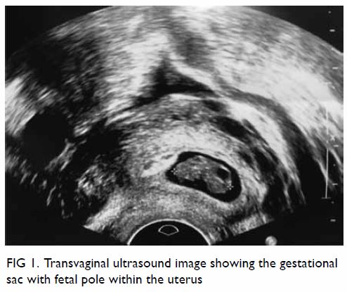
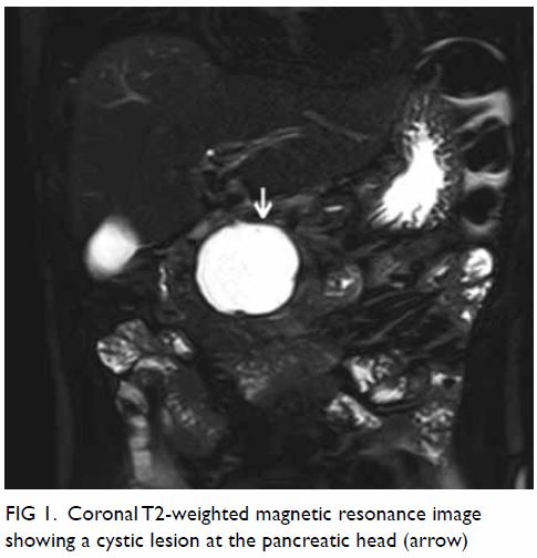
A 42-year-old woman was admitted through our emergency department for subacute onset of lower abdominal pain in September 2014. Bedside ultrasound by a gynaecology specialist showed a left adnexal cyst with fluid interface. Magnetic resonance imaging of the pelvis showed a large well-defined multiloculated cystic lesion measuring 9.2 cm (width) × 8.5 cm (depth) × 8.9 cm (height) with thick T1 and T2 hypointense enhancing wall and septa at the left side of the pelvis (Fig 1). An internal slightly T1 hyperintense component with fluid-fluid level was suspected to be proteinaceous or haemorrhagic content. A 1.6-cm non-enhancing mural nodule was noted at the posterior aspect. Partial 
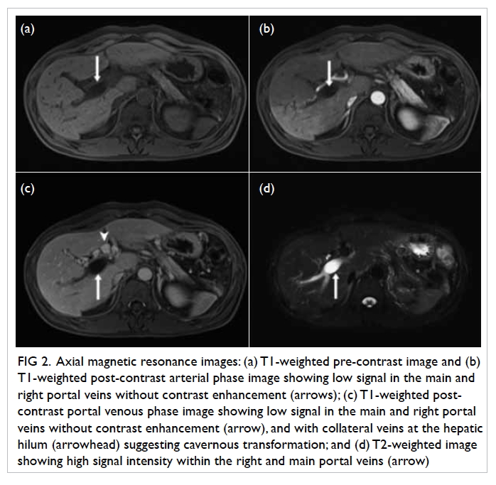
bicornuate uterus was noted. Bilateral ovaries were normal in size with small cysts (Fig 2). Laparoscopy was done, with excision of the lesion. Intra-operative frozen section showed a benign spindle cell tumour. Final histopathological evaluation revealed a retroperitoneal schwannoma.
 

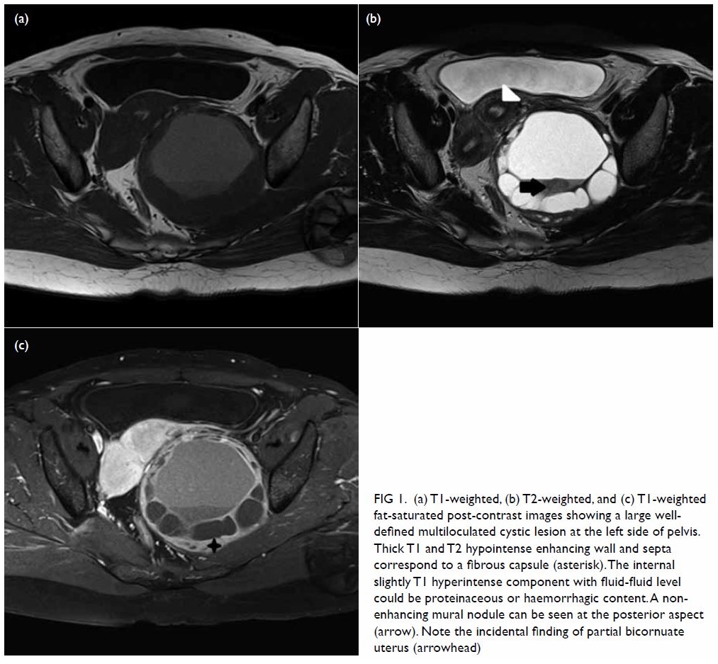
Figure 1. (a) T1-weighted, (b) T2-weighted, and (c) T1-weighted fat-saturated post-contrast images showing a large welldefined multiloculated cystic lesion at the left side of pelvis. Thick T1 and T2 hypointense enhancing wall and septa correspond to a fibrous capsule (asterisk). The internal slightly T1 hyperintense component with fluid-fluid level could be proteinaceous or haemorrhagic content. A nonenhancing mural nodule can be seen at the posterior aspect (arrow). Note the incidental finding of partial bicornuate uterus (arrowhead)
 

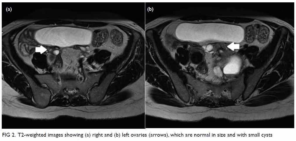
Figure 2. T2-weighted images showing (a) right and (b) left ovaries (arrows), which are normal in size and with small cysts
 
The majority of cystic pelvic masses originate from the ovary. Mimics of ovarian cystic masses have a wide variety of diagnoses. It is important to understand the relationship of a mass with its anatomic location, identify normal ovaries at imaging, and correlate imaging findings with the patient’s clinical history to avoid misdiagnosis.
 
Retroperitoneal schwannoma is a rare tumour and is difficult to diagnose, accounting for only 6% of retroperitoneal neoplasms. A retroperitoneal schwannoma is usually located in the paravertebral space or pre-sacral pelvic retroperitoneum.1 It usually occurs in young to middle-aged adults, and women are affected twice as often as men.1 The patient is usually asymptomatic, or complains of a wide variety of non-specific symptoms when the tumour is large in size.2 Malignant transformation is rare.1 On magnetic resonance images, a schwannoma appears as a well-defined mass with hypo- or iso-intensity on T1-weighted images and with hyperintensity on T2-weighted images. The nerve of origin is often difficult to identify. It is not unusual for a schwannoma to display cystic changes. However, prominent cystic changes are uncommon and point to ancient schwannoma, a rare variant of schwannoma that is characterised by degeneration and decreased cellularity.3 On magnetic resonance images, ancient schwannoma appears as a well-defined, complex cystic mass with a variable enhancement pattern. Thick T1 and T2 hypointense enhancing wall and septa correspond to a fibrous capsule, consisting of epineurium and residual nerve fibres.4
 
Identification of the nerve adjacent to or along the tumour is useful for differentiating ancient schwannomas from other complex cystic lesions, such as serous or mucinous cystadenocarcinoma, abscess, necrotic soft-tissue sarcoma, or necrotic metastatic lymphadenopathy.5
 
In the present case, the patient did not complain of any neurological symptoms at presentation. The presence of normal ovaries and fibrous capsule indicated a preoperative diagnosis of ancient schwannoma. This case illustrates the importance of considering this uncommon diagnosis when a pelvic complex cystic lesion is detected in imaging, and seeking specific imaging features (such as fibrous capsule and close relationship to the nerve) to confirm or exclude this diagnosis. This would facilitate surgical planning and minimise the risk of complications such as major neurological deficit.
 
Author contributions
All authors contributed to the concept or design, acquisition of data, analysis or interpretation of data, drafting of the manuscript, and critical revision for important intellectual content. All authors had full access to the data, contributed to the study, approved the final version for publication, and take responsibility for its accuracy and integrity.
 
Conflicts of interest
All authors have disclosed no conflicts of interest.
 
Funding/support
This research received no specific grant from any funding agency in the public, commercial, or not-for-profit sectors.
 
References
1. Rha SE, Byun JY, Jung SE, Chun HJ, Lee HG, Lee JM. Neurogenic tumors in the abdomen: tumor types and imaging characteristics. Radiographics 2003;23:29-43. Crossref
2. Kim SH, Choi BI, Han MC, Kim YI. Retroperitoneal neurilemoma: CT and MR findings. AJR Am J Roentgenol 1992;159:1023-6. Crossref
3. Dahl I. Ancient neurilemmoma (schwannoma). Acta Pathol Microbiol Scand A 1977;85:812-8. Crossref
4. Takeuchi M, Matsuzaki K, Nishitani H, Uehara H. Ancient schwannoma of the female pelvis. Abdom Imaging 2008;33:247-52. Crossref
5. Isobe K, Shimizu T, Akahane T, Kato H. Imaging of ancient schwannoma. AJR Am J Roentgenol 2004;183:331-6. Crossref

Catheterised hutch diverticulum masquerading as intraperitoneal bladder perforation

© Hong Kong Academy of Medicine. CC BY-NC-ND 4.0
 
PICTORIAL MEDICINE
Catheterised hutch diverticulum masquerading as intraperitoneal bladder perforation
Victor SH Chan, MB, BS, FRCR (UK)1; WM Kwok, MB, BS2; Stephen CW Cheung, MRCP, FHKAM (Radiology)1
1 Department of Radiology, Queen Mary Hospital, Pokfulam, Hong Kong
2 Department of Accident and Emergency, Queen Mary Hospital, Pokfulam, Hong Kong
 
Corresponding author: Dr Victor SH Chan (victorchansh@gmail.com)
 
 Full paper in PDF
 
A computed tomography (CT) scan was performed in a 64-year-old man with a history of end-stage renal failure to evaluate recent acute-on-chronic graft failure. To conserve remnant renal function, no intravenous contrast was administered. There was no hydronephrosis but the balloon of the indwelling Foley catheter was seen adjacent to but exterior to the bladder (Fig 1). No evidence of pneumoperitoneum was seen. Free fluid was noted in the pelvis, compatible with the history of peritoneal dialysis. The suspicion of perforated urinary bladder was conveyed to the referring physicians, and the patient was admitted for further evaluation. No abdominal distension, tenderness or guarding was elicited on physical examination. Urine output via the indwelling catheter was within normal limits. The patient was haemodynamically stable with no leukocytosis.
 

Figure 1. Coronal non-contrast maximum-intensity projection computed tomography scan of the pelvis demonstrates an apparently externally sited urinary bladder catheter and balloon; suspicion of bladder perforation is highlighted
 
Computed tomography cystography was performed: 20 mL of water-soluble contrast medium (Omnipaque 300) was diluted in 500 mL of normal saline. Transurethral perfusion of 250 mL of the prepared contrast medium was performed by free gravitational flow. Pre-instillation and post-instillation CT cystography (5 minutes and delayed 10 minutes) was performed. No intravenous contrast was administered.
 
Cross-sectional imaging revealed two large urinary bladder diverticula sited posteriorly, close to the native vesicoureteric junctions. The right diverticulum measured 3.8 × 4.6 cm with a 0.9-cm neck; the left diverticulum measured 4.5 × 5.8 cm with a 1.1-cm neck. On coronal images, a “Mickey-Mouse” appearance was noted, compatible with bilateral hutch diverticula. The inflated balloon of the urinary catheter was sited within the left diverticulum (Figs 2 3 4). The bladder contour was smooth. No evidence of intraperitoneal rupture was demonstrated. On pre-cystography images, the appearance had mimicked an externally sited catheter balloon due to the collapsed state of the bladder.
 

Figure 2. Coronal post–cystography maximum-intensity projection image of the pelvis demonstrates bilateral large hutch diverticula; the urinary bladder catheter tip and balloon are seen within the left diverticulum
 

Figure 3. Axial post–computed tomography cystography maximum-intensity projection image of the pelvis, which confirms the position of the balloon of the catheter within the left diverticulum
 

Figure 4. Sagittal post–computed tomography cystography maximum-intensity projection image of the pelvis is obtained, mirroring the findings seen in the axial and coronal planes. The tip of the Foley catheter is seen abutting against the superior aspect of the diverticulum
 
Spontaneous bladder perforation is rare but potentially fatal. Most such cases present with features of peritonitis. Some possible aetiologies include gonorrhoeal infection, radiation therapy, diabetes mellitus, neurogenic bladder, bladder diverticula, and indwelling urinary catheter.1 2 As our patient was largely pain-free with minimal abdominal symptoms, the overall clinical evidence did not favour bladder rupture even though the CT images were alarming.
 
Saline instillation and bedside ultrasound for rapid disposition of polytrauma patients and early diagnosis of bladder rupture has been described in the literature, with sensitivity reaching 90%.3 In cases with low clinical risk for perforation and when the patient is unfit for immediate CT, instillation of sterile saline to distend the bladder followed by ultrasound assessment provides a possible alternative to exclude bladder perforation. However, ultrasound results are highly operator-dependent and this procedure should be performed only in carefully selected patients. In experienced hands, following retrograde instillation of approximately 300 mL of sterile saline via the catheter, this imaging modality can be used to determine presence of ascites, evaluate the distension and configuration of the urinary bladder, look for bladder diverticula, and determine the position of the catheter balloon. This can be performed at the bedside for critically ill patients, involves no radiation and removes the risk of intravenous contrast-related anaphylaxis.
 
Author contributions
All authors contributed to the concept, image acquisition, image and data interpretation, manuscript drafting and critical revision for important intellectual content. All authors had full access to the data, contributed to the study, approved the final version for publication, and take responsibility for its accuracy and integrity.
 
Conflicts of interest
The authors have no conflicts of interest to disclose.
 
Funding/support
This research received no specific grant from any funding agency in the public, commercial, or not-for-profit sectors.
 
Ethics approval
Relevant patient consent was obtained for the purpose of this case study.
 
References
1. Sawalmeh H, Al-Ozaibi L, Hussein A, Al-Badri F. Spontaneous rupture of the urinary bladder (SRUB); A case report and review of literature. Int J Surg Case Rep 2015;16:116-8. Crossref
2. Ogawa S, Date T, Muraki O. Intraperitoneal urinary bladder perforation observed in a patient with an indwelling urethral catheter. Case Rep Urol 2013;2013:765704. Crossref
3. Karim T, Topno M. Bedside sonography to diagnose bladder trauma in the emergency department. J Emerg Trauma Shock 2010;3;305. Crossref

Dynamic dual-source computed tomography imaging for myocardial perfusion

© Hong Kong Academy of Medicine. CC BY-NC-ND 4.0
 
PICTORIAL MEDICINE
Dynamic dual-source computed tomography imaging for myocardial perfusion
Allen Li, MB, ChB, FHKAM (Radiology)1; YH Chan, MB, BS, FHKAM (Medicine)2; BE Wu, MB, BS, FHKAM (Medicine)3; CS Lam, MB, BS, FHKAM (Medicine)2
1 Department of Radiology and Nuclear Medicine, Tuen Mun Hospital, Tuen Mun, Hong Kong
2 Department of Medicine and Geriatrics, Pok Oi Hospital, Yuen Long, Hong Kong
3 Department of Medicine and Therapeutics, Prince of Wales Hospital, Shatin, Hong Kong
 
Corresponding author: Dr Allen Li (liallen@yahoo.com)
 
 Full paper in PDF
 
A 54-year-old man who was an ex-smoker was admitted to Pok Oi Hospital in August 2015 with acute chest pain that was subsequently confirmed to be a non-ST elevation myocardial infarction. Echocardiogram revealed anterior wall hypokinesia. Computed tomography (CT) coronary angiography demonstrated chronic total occlusion of the right coronary artery and a 90% stenotic lesion in the proximal to mid left anterior descending artery that was deemed to be the culprit lesion. Percutaneous coronary intervention was subsequently performed with a drug-eluting stent deployed across the proximal to mid left anterior descending artery stenosis. Angiographic results were excellent.
 
At clinical follow-up, the patient complained of persistent chest discomfort. Repeated echocardiogram was unremarkable showing normal left ventricular function without any regional wall motion abnormality. A CT stress myocardial perfusion and viability study was requested to guide subsequent management. The study protocol included quantitative evaluation of myocardial perfusion with pharmacological stress using a dynamic approach, followed by a delayed scan for the presence or absence of late myocardial enhancement.
 
Adenosine stress myocardial perfusion study with colour-coded maps demonstrated perfusion defects in the apicoseptal segment, the mid-inferoseptal segment, and to a lesser extent the basal inferoseptal segment (Fig 1). For quantitative evaluation, the normal areas had a myocardial blood flow of approximately 128 mL/100 mL/min, whereas areas with ischaemia had a flow of around 40 mL/100 mL/min. No delayed enhancement of the corresponding segments was evident to suggest scarring due to prior myocardial infarction (Fig 2).
 

Figure 1. Colour-coded maps of myocardial blood flow derived from stress dynamic computed tomography myocardial perfusion imaging (radiation dose about 8.5 mSv) showed significant perfusion hypoenhancement that involved (a) the apicoseptal segment, (b) the mid-inferoseptal segment, and to a lesser extent (c) the inferior aspect of the mid-basal anteroseptal segment (blue areas as indicated by the black arrows). Of note is the presence of artefacts at the subepicardial region of the mid-basal inferior segments (white arrowheads) and basal anteroseptal segment near the insertion point (blue arrow). Hibernating myocardium with reduced myocardial blood flow in the anterior segments from basal to apical levels
 

Figure 2. (a) Dual-energy computed tomography delayed enhancement showing no suspicious areas of late enhancement (radiation dose about 0.88 mSv). (b) Computed tomography myocardial perfusion study at the corresponding level demonstrates perfusion hypoenhancement (as indicated by the blue areas) of the inferoseptal segments
 
Eventually the patient underwent a percutaneous coronary intervention to the chronic total occlusion of the right coronary artery in 2016 via a combined radial and femoral arterial approach with successful stent deployment across the occluded segment. Final angiography showed excellent results with mild residual stenosis in patent ductus arteriosus ostium (Fig 3). To date, the patient remains symptom-free with improvements in both his exercise tolerance and mood, and psychiatric reports revealing reduced dosage of antidepressants.
 

Figure 3. Coronary angiography demonstrating (a) chronic total occlusion of the right coronary artery with retrograde septal collaterals supplied from the left coronary system and (b) successful percutaneous coronary intervention with minimal residual stenosis of the patent ductus arteriosus ostium
 
Discussion
Various imaging modalities are available for stress myocardial perfusion assessment.1 The present case demonstrates how a state-of-the-art dynamic and quantitative assessment of myocardial perfusion using a dual-source CT scanner enables detection of ischaemia along with viability assessment in a rapid and non-invasive fashion within an acceptable radiation dose.2
 
Conventional “static” CT myocardial imaging allows visual qualitative assessment of a single snapshot of myocardial iodine contrast attenuation that requires precise timing of the arrival of contrast to preserve diagnostic integrity. With a dual-source CT, quantitative assessment of myocardial perfusion in multiple cardiac phases with precise anatomic localisation of the ischaemic area becomes possible. To date, many studies have assessed the reliability of dual-source multiple-detector CT in the dynamic and quantitative evaluation of myocardial perfusion.2 3 4 5
 
Stress CT myocardial perfusion is emerging as a potentially promising non-invasive technique to detect myocardial ischaemia both qualitatively and quantitatively. With new-generation multiple-detector CT scanners, a one-stop non-invasive comprehensive evaluation of the heart including the coronary artery, ventricular function, myocardial perfusion, and viability is possible. Stress CT myocardial perfusion provides incremental benefit to standard coronary CT angiography, particularly for intermediate coronary lesions.2
 
Author contributions
All authors had full access to the data, contributed to the study, approved the final version for publication, and take responsibility for its accuracy and integrity.
 
Concept or design: All authors.
Acquisition of data: A Li, YH Chan.
Analysis of data: A Li, YH Chan.
Drafting of manuscript: A Li, YH Chan.
Critical revision for important intellectual content: All authors.
 
Conflicts of interest
All authors have disclosed no conflicts of interest.
 
Funding/support
This research received no specific grant from any funding agency in the public, commercial, or not-for-profit sectors.
 
References
1. Ko SM, Hwang HK, Kim SM, Cho IH. Multi-modality imaging for the assessment of myocardial perfusion with emphasis on stress perfusion CT and MR imaging. Int J Cardiovasc Imaging 2015;31(Suppl 1):1-21. Crossref
2. Rossi A, Dharampal A, Wragg A, et al. Diagnostic performance of hyperaemic myocardial blood flow index obtained by dynamic computed tomography: does it predict functionally significant coronary lesions? Eur Heart J Cardiovasc Imaging 2014;15:85-94. Crossref
3. Kim SM, Choi JH, Chang SA, Choe YH. Additional value of adenosine-stress dynamic CT myocardial perfusion imaging in the reclassification of severity of coronary artery stenosis at coronary CT angiography. Clin Radiol 2013;68:659-68. Crossref
4. Bamberg F, Becker A, Schwarz F, et al. Detection of hemodynamically significant coronary artery stenosis: incremental diagnostic value of dynamic CT-based myocardial perfusion imaging. Radiology 2011;260:689-98. Crossref
5. Varga-Szemes A, Meinel FG, De Cecco CN, Fuller SR, Bayer RR 2nd, Schoepf UJ. CT myocardial perfusion imaging. AJR Am J Roentgenol 2015;204:487-97. Crossref

Caesarean scar ectopic pregnancy: imaging findings of this rare but potentially life-threatening condition

DOI: 10.12809/hkmj176953
© Hong Kong Academy of Medicine. CC BY-NC-ND 4.0
 
PICTORIAL MEDICINE
Caesarean scar ectopic pregnancy: imaging findings of this rare but potentially life-threatening condition
Joseph Andrew WK Tang, MB, ChB, FRCR1; Esther MF Wong, MB, BS, FHKAM (Radiology)1; Wendy Shu, MB, BCh, FHKCOG2
1 Department of Radiology, Pamela Youde Nethersole Eastern Hospital, Chai Wan, Hong Kong
2 Department of Obstetrics and Gynaecology, Pamela Youde Nethersole Eastern Hospital, Chai Wan, Hong Kong
 
Corresponding author: Dr Joseph Andrew WK Tang (tangwingkin2000@gmail.com)
 
 Full paper in PDF
 
Caesarean scar ectopic pregnancy is a rare pregnancy complication with an estimated incidence of 1/1800 to 1/2500 pregnancies.1 2 Complications include uterine rupture, massive haemorrhage, placenta accrete, and pregnancy loss.3 Ultrasound examination is usually the first-line investigation. Magnetic resonance imaging (MRI) serves as a powerful confirmation tool. With its inherent superior tissue contrast and mulitplanar capability, MRI depicts anatomical details with robust reproducibility.4 Caesarean scar ectopic pregnancy is associated with a high risk of uterine rupture and uncontrollable haemorrhage. Expectant management is possible but not advocated. Surgical treatment leads to quicker postoperative recovery but may be associated with major haemorrhage.5 Other treatment includes systemic methotrexate and uterine artery embolisation.3
 
A high index of suspicion is required to diagnose this condition so that timely treatment can be initiated and life-threatening complications from a ruptured ectopic pregnancy prevented.
 
Case
A 34-year-old woman with a history of Caesarean section presented to the emergency department with per vaginal bleeding. Her pregnancy was 7 weeks of gestation by date.
 
On vaginal examination, the cervical os was closed and mildly blood-stained. She was haemodynamically stable with a normal haemoglobin of 12.9 g/dL and beta–human chorionic gonadotropin 15 877 mIU/mL. Transvaginal ultrasound revealed a single intrauterine gestational sac in the lower segment of the uterus, closely related to the myometrium (Fig 1). The fetal pole with positive fetal heart beat was identified. Crown to rump length was 11 mm, corresponding with 7 weeks and 1 day of gestation. The anterior uterine wall adjacent to the gestational sac was very thin with a thickness of only 4 mm (Fig 2). However, it was uncertain whether the placenta was directly implanted onto the Caesarean scar. A provisional diagnosis was made of pending abortion or Caesarean scar ectopic pregnancy. Magnetic resonance imaging of the pelvis was performed to determine whether the thin layer of soft tissue at the anterior uterine wall represented myometrium in the Caesarean scar with placental implantation elsewhere or if the placental tissue was implanted directly onto the scar.
 

Figure 1. Transvaginal ultrasound image showing the gestational sac with fetal pole within the uterus
 

Figure 2. Transvaginal ultrasound image showing only minimal thickness of anterior myometrium adjacent to the gestational sac
 
Magnetic resonance imaging showed a 1.7-cm defect at the anterior lower segment of the myometrium, corresponding to the Caesarean section scar. It was distended by a gestational sac. A singleton pregnancy was identified with crown-rump length consistent with gestational age. Trophoblastic tissue was seen implanted onto the serosa of the uterus (Fig 3). Overall MRI findings were compatible with Caesarean scar ectopic pregnancy. There was no direct extension of trophoblastic tissue into adjacent organs such as the urinary bladder or sign of uterine rupture (Fig 4).
 

Figure 3. (a) Sagittal T2-weighted magnetic resonance imaging (MRI) showing a T2 hyperintense structure at the inferior uterus, compatible with the gestational sac (white arrow) with fetal pole partially seen within. A defect at the anterior myometrium is seen at the lower anterior uterine wall, corresponding to Caesarean scar (white arrowheads) and is infiltrated by T2 hyperintense trophoblastic tissue. (b) Sagittal T2-weighted MRI showing a thin T2 hypointense layer that represents serosa (empty white arrow) covering the anterior aspect of the trophoblastic tissue. No evidence of uterine perforation is noted
 

Figure 4. (a) Axial T1-weighted magnetic resonance imaging (MRI) of the pelvis showing no abnormal T1 hyperintense fluid in the pouch of Douglas to suggest haemoperitoneum. (b) Axial T2-weighted fat-suppressed MRI of the pelvis showing no abnormal T2 hyperintense signal in the pouch of Douglas to suggest intraperitoneal free fluid. A T2 hyperintense structure is seen at the inferior aspect of the uterus, corresponding to the gestational sac (white arrow). The cervix (white arrowhead) is seen more posteriorly
 
The superior contrast resolution in MRI for different soft tissues is advantageous in the differentiation of uterine serosa, myometrium, endometrium, and trophoblastic tissue. This helped confirm the diagnosis of Caesarean scar ectopic pregnancy in our patient and would have been difficult if only ultrasound findings were available.
 
The patient received intramuscular methotrexate therapy. Serial beta–human chorionic gonadotropin levels showed a decreasing trend. Subsequent definitive treatment with suction evacuation was performed. The patient made an uneventful recovery.
 
Author contributions
Concept and design of the study: All authors.
Acquisition of data: EMF Wong, W Shu.
Analysis and interpretation of data: EMF Wong, W Shu.
Drafting of the manuscript: JAWK Tang.
Critical revision for important intellectual content: JAWK Tang, EMF Wong.
 
Declaration
All authors have disclosed no conflicts of interest. All authors had full access to the data, contributed to the study, approved the final version for publication, and take responsibility for its accuracy and integrity.
 
References
1. Seow KM, Huang LW, Lin YH, Lin MY, Tsai YL, Hwang JL. Caesarean scar pregnancy: issues in management. Ultrasound Obstet Gynecol 2004;23:247-53. Crossref
2. Jurkovic D, Hillaby K, Woelfer B, Lawrence A, Salim R, Elson CJ. First-trimester diagnosis and management of pregnancies implanted into the lower uterine segment caesarean section scar. Ultrasound Obstet Gynecol 2003;21:220-7. Crossref
3. Michaels AY, Washburn EE, Pocius KD, Benson CB, Doubilet PM, Carusi DA. Outcome of cesarean scar pregnancies diagnosed sonographically in the first trimester. J Ultrasound Med 2015;34:595-9. Crossref
4. Kao LY, Scheinfeld MH, Chernyak V, Rozenblit AM, Oh S, Dym RJ. Beyond ultrasound: CT and MRI of ectopic pregnancy. AJR Am J Roentgenol 2014;202:904-11. Crossref
5. Alalade AO, Smith FJ, Kendall CE, Odejinmi F. Evidencebased management of non-tubal ectopic pregnancies. J Obstet Gynaecol 2017;37:982-91. Crossref

Magnetic resonance imaging monitoring of post-treatment changes to Crohn’s disease–related anal fistula in patients prescribed infliximab

DOI: 10.12809/hkmj176964
© Hong Kong Academy of Medicine. CC BY-NC-ND 4.0
 
PICTORIAL MEDICINE
Magnetic resonance imaging monitoring of post-treatment changes to Crohn’s disease–related anal fistula in patients prescribed infliximab
KY Man, MB, ChB, FRCR1; Esther MF Wong, MB, BS, FHKCR1; Francis KY Cho, MB, BS, FHKCR1; CM Leung, FHKAM (Medicine)2
1 Department of Radiology, Pamela Youde Nethersole Eastern Hospital, Chai Wan, Hong Kong
2 Department of Medicine, Pamela Youde Nethersole Eastern Hospital, Chai Wan, Hong Kong
 
Corresponding author: Dr KY Man (dsgundam@hotmail.com)
 
 Full paper in PDF
 
The incidence of inflammatory bowel disease, particularly Crohn’s disease, is rising in Hong Kong.1 The age-adjusted incidence of Crohn’s disease increased from 0.01 per 100 000 population in 1985 to 1.46 per 100 000 population in 2014.1 Crohn’s disease is a multisystem disorder with specific radiological features such as transmural inflammation, fistulation, and skip lesions. Perianal fistulas often complicate Crohn’s disease, affecting up to 36% of patients.2
 
Infliximab, a monoclonal antibody against tumour necrosis factor-α, has revolutionised the treatment of Crohn’s disease–related anal fistula. Current evidence shows encouraging results for closure of perianal fistulas. According to a local consensus statement, biologics are advocated in patients with active fistulising Crohn’s disease, particularly those with complex perianal fistulising disease.3
 
Response to monoclonal antibody therapy needs to be monitored. This pictorial review illustrates the post-treatment changes on magnetic resonance imaging (MRI) of anal fistula in patients prescribed infliximab.
 
Patients with a known history of Crohn’s disease complicated by perianal fistula and prescribed infliximab between 2012 and 2016 were reviewed. The treatment regimen at our centre comprises an intravenous loading dose of infliximab 5 mg/kg, followed by the same dose at week 2 and week 6. Thereafter a maintenance dose of 5 mg/kg is given every 8 weeks.
 
Magnetic resonance images were acquired with the 1.5T Siemens Magnetom Avanto system (Erlangen, Germany). The pelvic MRI protocol for perianal fistula evaluation consists of T1-weighted and high-spatial-resolution T2-weighted imaging sequences without fat saturation to delineate the muscle groups, fat planes, and fistula tract. T2-weighted imaging with fat suppression is used to assess oedema and fluid-containing tracts and cavities, whereas fat-suppressed T1-weighted unenhanced and contrast-enhanced sequences are used to assess the presence and degree of inflammation (Table). Diffusion-weighted imaging is not routinely performed in view of the need for an extended examination time. Information about the presence of fluid, oedema, cavities, and inflammation can be obtained through these sequences. Anal fistulae are classified according to the Parks’ classification system (Fig 1).4
 

Table. Pelvic MRI protocol for evaluation of anal fistula
 

Figure 1. Schematic diagram demonstrating different types of perianal fistula according to Parks’ classification. Internal sphincter (thin arrow); external sphincter (thick arrow); puborectalis (asterisk); and levator ani (curved arrow). Intersphincteric fistula tracks between internal and external sphincter (1); trans-sphincteric fistula pierces through the external sphincter (2); suprasphincteric fistula tracks superior to the puborectalis (3); and extrasphincteric fistula penetrates the levator ani (4)
 
Three patients (two men, one woman) were reviewed and all received infliximab. At least one pre-treatment and one post-treatment MRI were performed.
 
Patient A was a 34-year-old woman with a history of systemic lupus erythematosus, retinitis, and neuropsychiatric lupus. She had had recurrent ischiorectal abscess and perianal fistula since 2002. Rectal biopsy confirmed Crohn’s disease. Despite treatment with azathioprine, the perianal fistula failed to close. She was scheduled to initially receive three doses of infliximab. Close monitoring was essential in view of the potential to develop lupus-like disease. Progress MRI after the third dose of infliximab showed slight interval improvement in her perianal fistula. Biologics were continued in view of the residual disease. After the seventh dose of infliximab, progress MRI revealed a largely quiescent perianal fistula (Fig 2). In view of the radiologically healed fistula, clinical improvement and potential risk of lupus-like disease, the decision was taken to stop the infliximab infusion but continue close clinical and radiological monitoring.
 

Figure 2. Patient A. (a and b) T2-weighted axial and T1-weighted post-contrast fat suppression axial magnetic resonance images showing an active trans-sphincteric tract at the 8 o’clock position with T2-weighted hyperintense signal and contrast enhancement (arrows). (c and d) One-year post-treatment. Loss of T2-weighted hyperintense signal and contrast enhancement suggested quiescent disease (arrows)
 
Patient B was a 24-year-old man with a history of perianal fistula since 2015 and an episode of perianal abscess that required incision and drainage. Crohn’s disease was confirmed on rectal biopsy. He had previously developed azathioprine-induced pancytopenia. Subsequent infusion of infliximab infusion resulted in responsive disease, evident on MRI (Fig 3). Clinical and radiological monitoring (progress MRI) at 6-month intervals was carried out to determine progress of the perianal fistula. Infliximab would be stopped when there was evidence of healed tract and clinical improvement.
 

Figure 3. Patient B. (a and b) T2-weighted axial and T1-weighted post-contrast fat suppression coronal magnetic resonance images showing an active intersphincteric tract at the right-sided natal cleft with T2-weighted hyperintense signal and contrast enhancement (arrows). (c and d) Six months post-treatment. Loss of T2-weighted hyperintense signal and contrast enhancement suggested quiescent disease (arrows)
 
Patient C, a 42-year-old man had a history of ileocolic Crohn’s disease since 2008, with episodes of perianal abscess and fistula refractory to steroid and azathioprine treatment. Magnetic resonance imaging showed progressive perianal fistula (Fig 4) after the second maintenance dose of infliximab. Previous infliximab dose/frequency was continued and progress MRI planned for the purpose of reassessment and consideration of alternative treatment if there was persistent progression.
 

Figure 4. Patient C. (a and b) T2-weighted axial and T1-weighted post-contrast with fat suppression axial magnetic resonance images showing a T2-weighted hyperintense-enhancing active trans-sphincteric tract (arrows) passing through the external sphincter at the 11 o’clock position. (c and d) Five months post-treatment. Interval progression of the 11 o’clock transsphincteric tract (arrows) and a new T2-weighted hyperintense-enhancing active intersphincteric tract over the 7-8 o’clock position (thick arrows)
 
Crohn’s disease–related anal fistulae are frequently encountered in radiologic practice due to their complexity and propensity for incomplete treatment response and relapse.
 
Magnetic resonance imaging is a well-established diagnostic tool for anal fistula. Its inherent high spatial and contrast resolution allows precise anatomical delineation.5 Magnetic resonance imaging plays a critical role in helping determine the appropriate treatment that should be individualised according to the type of perianal fistula and the degree of involvement of surrounding pelvic structures. Clinical examination can often be difficult because of induration and inflammation in patients with anal sepsis. At MRI, identification and localisation of the entire fistula, including the external opening, the primary track, secondary tracks, abscesses, and the internal opening, are essential for fistula classification and treatment. Inadequate assessment may result in progression of a simple fistula to a complex fistula, and failure to recognise secondary extensions can result in recurrent sepsis. Anti–tumour necrosis factor antibodies (infliximab) have been introduced with good clinical results. Magnetic resonance imaging also plays an important role in evaluation of the response to medical therapy. Magnetic resonance imaging does not have field of view limitations and offers excellent views of the supralevator, retrorectal and anteroanal spaces, where occult sepsis may be missed clinically due to extensive scarring or a remote location.6
 
This pictorial review demonstrates the ability of MRI to monitor the response to therapy of anal fistula in Crohn’s disease patients receiving infliximab.
 
Author contributions
KY Man is responsible for the design, acquisition and interpretation of data, and drafting of the article. EMF Wong, FKY Cho, and CM Leung are responsible for critical revision for important intellectual content.
 
Declaration
All authors have disclosed no conflicts of interest. All authors had full access to the data, contributed to the study, approved the final version for publication, and take responsibility for its accuracy and integrity.
 
References
1. Ng SC, Leung WK, Shi HY, et al. Epidemiology of inflammatory bowel disease from 1981 to 2014: results from a territory-wide population-based registry in Hong Kong. Inflamm Bowel Dis 2016;22:1954-60. Crossref
2. Leong RW, Lau JY, Sung JJ. The epidemiology and phenotype of Crohn’s disease in the Chinese population. Inflamm Bowel Dis 2004;10:646-51. Crossref
3. Leung WK, Ng SC, Chow DK, et al. Use of biologics for inflammatory bowel disease in Hong Kong: consensus statement. Hong Kong Med J 2013;19:61-8.
4. Parks AG, Gordon PH, Hardcastle JD. A classification of fistula-in-ano. Br J Surg 1976;63:1-12. Crossref
5. Buchanan G, Halligan S, Williams A, et al. Effect of MRI on clinical outcome of recurrent fistula-in-ano. Lancet 2002;360:1661-2. Crossref
6. Bell SJ, Halligan S, Windsor AC, Williams AB, Wiesel P, Kamm MA. Response of fistulating Crohn’s disease to infliximab treatment assessed by magnetic resonance imaging. Aliment Pharmacol Ther 2003;17:387-93. Crossref

Inadvertent arterial insertion of a central venous catheter

DOI: 10.12809/hkmj176866
© Hong Kong Academy of Medicine. CC BY-NC-ND 4.0
 
PICTORIAL MEDICINE
Inadvertent arterial insertion of a central venous catheter
Victor WT Chan, MB, BS, FRCR; KW Shek, MB, BS, FRCR
Department of Radiology and Imaging, Queen Elizabeth Hospital, Jordan, Hong Kong
 
Corresponding author: Dr Victor WT Chan (chanwaitat@gmail.com)
 
 Full paper in PDF
 
Central venous catheterisation is a common procedure that allows venous access for delivering medications, infusing fluids or blood products, and monitoring volume status. Traditionally, anatomical landmarks of the sternocleidomastoid muscle provide a pathway to catheterise the internal jugular vein (IJV). However, inadvertent arterial puncture is a risk. Currently, ultrasound guidance by experienced operators is recommended for reducing the risk of mechanical complications during central venous catheter (CVC) insertion.1
 
For venous access via the neck, common carotid and subclavian artery injuries have been reported.2 The risk of artery injury is about 0.5%, and practice should be reviewed if the risk exceeds 1%. Other known mechanical complications include haematoma formation, haemothorax, and pneumothorax.
 
A 55-year-old woman with end-stage renal failure on continuous ambulatory peritoneal dialysis was admitted to our hospital for fever and abdominal pain. The clinical diagnosis of continuous ambulatory peritoneal dialysis peritonitis was made, and the peritoneal dialysis catheter was removed. Bedside insertion of a CVC was selected for temporary haemodialysis. The CVC insertion was initially attempted via the right IJV, unsuccessfully. The CVC was subsequently inserted via the left IJV. The procedure was performed under ultrasound guidance using the Seldinger technique; however, inadvertent arterial puncture was not recognised, and the procedure was continued.
 
After CVC insertion, a chest radiograph was taken, showing an abnormal vertical course of the catheter with suspected malposition (Fig 1). Urgent contrast computed tomographic angiogram (Figs 2 and 3) revealed that the catheter had been inserted via the left IJV, subsequently exiting posteromedially, entering the left vertebral artery, and harbouring at the origin of the left subclavian artery. Computed tomographic angiogram also showed abnormal contrast pooling over the right neck suggestive of a pseudoaneurysm formation from the right subclavian artery.
 

Figure 1. Chest radiograph after central venous catheter insertion showing an abnormal vertical course of the catheter with suspected malposition. The catheter tip is seen close to the aortic arch (arrow)
 

Figure 2. Contrast computed tomographic angiogram with maximum intensity projection showing the central venous catheter (thick white arrow) with a vertical course and punctured the left vertebral artery. The catheter tip is seen at the origin of the left subclavian artery (thin white arrow). Over the right neck, a contrast pooling pseudoaneurysm (black arrow) connected to the right subclavian artery was identified
 

Figure 3. Contrast computed tomographic angiogram showing the central venous catheter puncturing through the left internal jugular vein, exiting medially (black arrow). The catheter enters the left vertebral artery (white arrow) located posteromedially
 
The opinion of a vascular surgeon was sought and the catheter was removed under general anaesthesia with repair of the vertebral artery. Oozing was noted from the left IJV exit site. Haemostasis was controlled by direct pressure onto the IJV. Urgent right subclavian angiogram (Fig 4) was performed by interventional radiologists, confirming the presence of a pseudoaneurysm, which was successfully embolised with coils.
 

Figure 4. Right subclavian angiogram showing a pseudoaneurysm (black arrow) arising from the right thyrocervical trunk, which was successfully embolised with coils (white arrows)
 
Postoperatively, the patient progressed well, with her peritonitis controlled by intravenous antibiotics. A new CVC for temporary haemodialysis was inserted via her right IJV by interventional radiologists under fluoroscopic and real-time ultrasound guidance.
 
This case concurs with a previous report that the incidence of mechanical complications after multiple attempts is higher than after one attempt.2 Real-time ultrasound guided venepuncture of the IJV has a higher first insertion attempt success rate, and decreased rate of arterial puncture as compared with the anatomic landmark approach; this technique is currently recommended by the Association of Anaesthetists of Great Britain and Ireland for all CVC insertions.3 Credentialing of ultrasound-guided CVC insertion should be advocated in Hong Kong, with adequate training provided by accredited trainers.
 
Author contributions
All authors have made substantial contributions to the concept of this pictorial medicine; acquisition and interpretation of data, drafting of the article, and critical revision for important intellectual content.
 
Funding/support
This research received no specific grant from any funding agency in the public, commercial, or not-for-profit sectors.
 
Declaration
All authors have disclosed no conflicts of interest. All authors had full access to the data, contributed to the study, approved the final version for publication, and take responsibility for its accuracy and integrity.
 
References
1. McGee DC, Gould MK. Preventing complications of central venous catheterization. N Engl J Med 2003;348:1123-33. Crossref
2. Schummer W, Schummer C, Rose N, Niesen WD, Sakka SG. Mechanical complications and malpositions of central venous cannulations by experienced operators. A prospective study of 1794 catheterizations in critically ill patients. Intensive Care Med 2007;33:1055-9. Crossref
3. Bodenham A, Babu S, Bennett J, et al. Association of Anaesthetists of Great Britain and Ireland: Safe Vascular Access 2016. Anaesthesia 2016;71:573-85. Crossref

Emphysematous epididymo-orchitis: an uncommon but life-threatening cause of scrotal pain

DOI: 10.12809/hkmj176876
© Hong Kong Academy of Medicine. CC BY-NC-ND 4.0
 
PICTORIAL MEDICINE
Emphysematous epididymo-orchitis: an uncommon but life-threatening cause of scrotal pain
HW Lau, MB, ChB, FRCR1; CH Yu, MB, ChB1; SM Yu, FRCR, FHKCR2; LF Lee, MB, ChB3
1 Department of Radiology, Tuen Mun Hospital, Tuen Mun, Hong Kong
2 Department of Radiology, United Christian Hospital, Kwun Tong, Hong Kong
3 Division of Urology, Department of Surgery, United Christian Hospital, Kwun Tong, Hong Kong
 
Corresponding author: Dr HW Lau (jackylauhw@gmail.com)
 
 Full paper in PDF
 
An 80-year-old man presented to the emergency department in August 2016 with a 2-day history of painful scrotal swelling and fever. There was no history of trauma. He had a history of hypertension, diabetes mellitus and bullous pemphigoid, and was taking long-term steroid therapy. Upon presentation, the patient had a fever of 38.8°C and fast atrial fibrillation with a heart rate of 170 beats per minute. Blood pressure was 149/104 mm Hg. Physical examination of the patient revealed scrotal skin erythema and a firm left scrotal swelling that was tender on palpation. No cough impulse could be elicited. His white cell count was 21.8 × 10⁹ /L and blood glucose level was 21.6 mmol/L. Ultrasound of the left hemiscrotum showed a fluid collection with hyperechoic interfaces and ring-down artefact suggestive of the presence of gas, obscuring the underlying structures (Fig 1). Subsequent non-contrast computed tomographic scan of the pelvis confirmed that the mottled gas and fluid density were confined to the left hemiscrotum (Fig 2a). The left testis and epididymis were poorly delineated. There was no gas density in the subcutaneous layer of the scrotum or perineum or in the intraperitoneal cavity. There was also no evidence of indirect inguinal hernia (Fig 2b). A diagnosis of emphysematous epididymo-orchitis was made. Emergency surgery was arranged a few hours after imaging. Intra-operatively, gas and pus were seen within the left tunica vaginalis. The left epididymis was necrotic and almost destroyed but the left testis was relatively spared with friable tissue and patches of necrosis (Fig 3). Drainage of pus and left orchidectomy were performed. Pus culture revealed Escherichia coli. The patient subsequently developed septic shock and died 3 days after the operation.
 

Figure 1. Ultrasound image of the left hemiscrotum showing a fluid collection with hyperechoic interfaces (white arrows) and ring-down artefact (black arrow) suggestive of the presence of gas. Underlying structures including the testis and epididymis were obscured
 

Figure 2. Axial computed tomography of the pelvis showing (a) mottled gas densities confined to the left hemiscrotum (white arrow) with no evidence of gas in the subcutaneous layer of the scrotum or perineum, virtually excluding the possibility of Fournier’s gangrene; and (b) absence of bowel loop in the left inguinal canal (white arrow) excluded the possibility of incarcerated indirect inguinal hernia
 

Figure 3. Intra-operative photo showing a grossly inflamed and slightly necrotic epididymis (white arrow). The testis (black arrow) was relatively spared with friable tissue and patches of necrosis
 
Emphysematous cholecystitis, pyelonephritis, and cystitis are not uncommonly seen in patients with poorly controlled diabetes mellitus. It is nonetheless rare to see gas-forming infection of the epididymis and testis although this is also reported to be associated with diabetes mellitus.1 2 3 Long-term use of steroid in our patient may have been an additional risk factor due to immunosuppression. Based on the clinical presentation and ultrasound findings of our patient, the main differential diagnoses included Fournier’s gangrene and incarcerated indirect inguinal hernia. Computed tomographic scan was very helpful in delineating the definitive diagnosis. Gas pockets confined to the scrotal sac without involvement of the subcutaneous layer of the perineum virtually excluded the possibility of Fournier’s gangrene. Incarcerated indirect inguinal hernia was excluded due to the absence of bowel loops in the scrotal sac.
 
When a diabetic patient presents with acute scrotal pain and features of infection, careful examination during the initial ultrasound scan for the presence of gas within the scrotum is essential as this will alter the subsequent management plan as it implies a much poorer prognosis than non–gas-forming epididymo-orchitis.
 
In conclusion, emphysematous epididymo-orchitis is an uncommon but life-threatening disease. Ultrasound and computed tomographic scan are essential to identify this entity for early treatment.
 
Author contributions
All authors have made substantial contributions to the concept or design of this study; acquisition of data; analysis or interpretation of data; drafting of the article; and critical revision for important intellectual content.
 
Funding/support
This research received no specific grant from any funding agency in the public, commercial, or not-for-profit sectors.
 
Declaration
All authors have disclosed no conflicts of interest. All authors had full access to the data, contributed to the study, approved the final version for publication, and take responsibility for its accuracy and integrity.
 
References
1. Mathur A, Manish A, Maletha M, Luthra NB. Emphysematous epididymo-orchitis: a rare entity. Indian J Urol 2011;27:399-400. Crossref
2. Mandava A, Rao RP, Kumar DA, Naga Prasad IS. Imaging in emphysematous epididymo-orchitis: a rare cause of acute scrotum. Indian J Radiol Imaging 2014;24:306-9. Crossref
3. Hegde RG, Balani A, Merchant SA, Joshi AR. Synchronous infection of the aorta and the testis: emphysematous epididymo-orchitis, abdominal aortic mycotic aneurysm, and testicular artery pseudoaneurysm diagnosed by use of MDCT. Jpn J Radiol 2014;32:425-30. Crossref

Pancreatic pseudocyst rupture into the portal vein diagnosed by magnetic resonance imaging

DOI: 10.12809/hkmj164980
© Hong Kong Academy of Medicine. CC BY-NC-ND 4.0
 
PICTORIAL MEDICINE
Pancreatic pseudocyst rupture into the portal vein diagnosed by magnetic resonance imaging
HC Lee, FRCR, FHKCR1; KH Tse, FRCR, FHKCR2
1 Department of Radiology, United Christian Hospital, Kwun Tong, Hong Kong
2 Department of Radiology, Princess Margaret Hospital, Laichikok, Hong Kong
 
Corresponding author: Dr HC Lee (lhc874@ha.org.hk)
 
 Full paper in PDF
 
A 38-year-old man presented to the United Christian Hospital, Hong Kong, with acute epigastric pain in October 2014. He was a chronic drinker and had experienced intermittent abdominal pain for 6 months. His serum amylase level was elevated (454 IU/L), and a diagnosis of acute-on-chronic pancreatitis was made. The patient was treated conservatively.
 
Magnetic resonance cholangiopancreatography performed 3 months after hospital discharge showed a 5.8-cm-diameter unilocular cystic mass over the pancreatic head (Fig 1). The main and right portal veins showed a signal intensity identical to that of the cystic pancreatic lesion on all phases, without any contrast enhancement (Fig 2). There was communication between the main portal vein and the cystic mass (Fig 3). The presence of multiple collateral veins in the hepatic hilum was consistent with cavernous transformation (Fig 2c). Features were suggestive of a pancreatic head pseudocyst that had ruptured into the main portal vein.
 

Figure 1. Coronal T2-weighted magnetic resonance image showing a cystic lesion at the pancreatic head (arrow)
 

Figure 2. Axial magnetic resonance images: (a) T1-weighted pre-contrast image and (b) T1-weighted post-contrast arterial phase image showing low signal in the main and right portal veins without contrast enhancement (arrows); (c) T1-weighted post-contrast portal venous phase image showing low signal in the main and right portal veins without contrast enhancement (arrow), and with collateral veins at the hepatic hilum (arrowhead) suggesting cavernous transformation; and (d) T2-weighted image showing high signal intensity within the right and main portal veins (arrow)
 

Figure 3. Two-dimensional slab magnetic resonance cholangiopancreatogram showing a pancreatic head cystic lesion with communication (arrow) with the main portal vein, both as high signal intensity (a normal portal vein with flowing blood should be of low signal intensity); the non-dilated biliary tree (arrowheads) and the gallbladder are also visible
 
The patient presented again 1 month later with recurrent pancreatitis. Contrast computed tomography (CT) showed that the pancreatic pseudocyst had enlarged, to 7.6 cm in diameter (Fig 4). Pancreatic cystojejunostomy and cholecystectomy were performed. Intra-operatively, a 10-cm cystic lesion at the pancreatic head was found, and 200 mL of clear fluid was aspirated. Intra-operative ultrasonography showed the lack of flow in the main portal vein.
 

Figure 4. Reformatted coronal computed tomographic image in portal venous phase showing fluid attenuation in the main and right portal veins (PV), absence of contrast enhancement, and communication between the portal veins and the pancreatic cystic lesion (arrow)
 
The patient had a few more episodes of recurrent pancreatitis thereafter. The last CT examination, performed 2 years after surgery, showed a reduction in the size of the pseudocyst, to 2 cm. The patient remains on regular follow-up.
 
Discussion
Rupture of a pancreatic pseudocyst into the portal vein is an uncommon complication; only a handful of cases have been reported in the literature.1 It has been postulated that portal vein thrombosis occurs first, followed by erosion of the portal vein by pancreatic enzymes present in the pseudocyst, and subsequent lysis of the thrombus and filling of the portal vein with fluid.1 2 It has also been reported that rupture of the pseudocyst into the portal vein may be the initial event, followed by the development of portal vein thrombosis.3 4
 
Previously reported cases have used various diagnostic techniques. Invasive methods include endoscopic retrograde cholangiopancreatography and portography with surgery. Non-invasive methods include ultrasonography, CT, and magnetic resonance imaging (MRI). In all reported cases in which MRI was performed, the signal intensity of fluid in the portal vein matched that of the pancreatic pseudocyst.1 2 3 Direct communication between the portal vein and the pancreatic pseudocyst was clearly seen in most cases. The presence of residual thrombus or concomitant existence of complete thrombosis of the portal vein has also been reported.1
 
There is no well-established treatment protocol. Options include conservative management, endoscopic or percutaneous procedures, or surgery. The patient’s clinical condition and symptoms, patency of the portal vein, communication between the pseudocyst and pancreatic duct, size of pseudocyst, and any other complicating factors should be considered in treatment planning.3
 
In summary, rupture of a pancreatic pseudocyst into the portal vein is an uncommon complication. On MRI, demonstration of fluid signal in the portal vein that matches the signal intensity of a pancreatic pseudocyst allows the diagnosis to be confidently made, obviating the need for more invasive investigations.
 
Declaration
The authors have no conflicts of interest to disclose.
 
References
1. Dayal M, Sharma R, Madhusudhan KS, et al. MRI diagnosis of rupture of pancreatic pseudocyst into portal vein: case report and review of literature. Ann Gastroenterol 2014;27:173-6.
2. Riddell A, Jhaveri K, Haider M. Pseudocyst rupture into the portal vein diagnosed with MRI. Br J Radiol 2005;78:265-8. Crossref
3. Ng TS, Rochefort H, Czaplicki C, et al. Massive pancreatic pseudocyst with portal vein fistula: case report and proposed treatment algorithm. Pancreatology 2015;15:88-93. Crossref
4. Dawson BC, Kasa D, Mazer MA. Pancreatic pseudocyst rupture into the portal Vein. South Med J 2009;102:728-32. Crossref

Characteristic imaging features of clonorchiasis

DOI: 10.12809/hkmj166198
© Hong Kong Academy of Medicine. CC BY-NC-ND 4.0
 
PICTORIAL MEDICINE
Characteristic imaging features of clonorchiasis
WK Lo, MB, BS, FRCR1; SM Yu, FRCR, FHKAM (Radiology)2; James CS Chan, FRCR, FHKAM (Radiology)2
1 Department of Diagnostic and Interventional Radiology, Kwong Wah Hospital, Yaumatei, Hong Kong
2 Department of Radiology and Organ Imaging, United Christian Hospital, Kwun Tong, Hong Kong
 
Corresponding author: Dr WK Lo (waikglo@gmail.com)
 
 Full paper in PDF
 
A 43-year-old Chinese man presented to United Christian Hospital, Hong Kong, in January 2016, with a 2-year history of vague right upper quadrant pain. Blood test results were unremarkable except for an episode of transient eosinophilia. Ultrasonography showed mildly dilated intrahepatic ducts. Magnetic resonance cholangiopancreatography showed diffuse and uniform mild dilatation of peripheral intrahepatic bile ducts (Fig 1) consistent with the ‘too many intrahepatic ducts’ sign (Fig 2). Notably, there was no central extrahepatic bile duct dilatation. Subsequently requested stool microscopy was positive for Clonorchis sinensis ova. The patient was given a course of praziquantel.


Figure 1. Axial T2-weighted magnetic resonance image showing dilatation of the peripheral intrahepatic ducts (arrows)


Figure 2. Reformatted magnetic resonance cholangiographic images showing the ‘too many intrahepatic ducts’ sign in a patient with clonorchiasis (a), compared with a healthy individual (b)
 
Clonorchiasis is a foodborne zoonosis caused by consumption of raw or undercooked freshwater fish infested by the liver fluke C sinensis. C sinensis larvae penetrate the scales of freshwater fish and encyst in subcutaneous tissues. Cysts, when consumed by a definitive host, hatch in the intestine and migrate to the biliary tree, where the adult flukes establish residence. Clonorchiasis is endemic in many parts of Asia, including China, Korea, and Vietnam.1 Clonorchiasis and its complications are not commonly seen in affluent populations, such as those in Hong Kong. However, the increasing mobility of people around the region means that clinicians should be vigilant of the condition and alert to its symptoms. Knowledge of the characteristic imaging features of clonorchiasis would prompt stool microscopy and allow early diagnosis.
 
Within the biliary tree, adult C sinensis flukes reside in small- or medium-sized peripheral intrahepatic ducts. The organisms cause dilatation of the intrahepatic ducts. In heavy infestations, the extrahepatic ducts, the gallbladder, or even the pancreatic duct can also be involved. The dilated peripheral ducts can be visualised by direct cholangiography, or to a better extent by magnetic resonance cholangiopancreatography (owing to the independence of rate and volume of the contrast injection), as the ‘too many intrahepatic ducts’ sign.2 Occasionally, linear or elliptical filling defects representing the flukes can be seen.3
 
Light infestations with C sinensis can be asymptomatic. Heavier infestations can result in fever, jaundice, right upper quadrant pain, and eosinophilia. Untreated, chronic clonorchiasis induces chronic inflammation of the bile ducts. Recurrent pyogenic cholangitis, cholelithiasis, pancreatitis, and cholangiocarcinoma are the main complications.4
 
Infestations are diagnosed by observation of C sinensis ova during stool microscopy. Immunological and molecular techniques of diagnosis are still at the experimental stage.1 Praziquantel is the only drug treatment for clonorchiasis that is recommended by the World Health Organization.1
 
In summary, clinicians in Asia should be vigilant of clonorchiasis. In modern clinical practice, magnetic resonance cholangiopancreatography is often requested for non-invasive evaluation of the biliary tree. Knowledge and recognition of the characteristic imaging features of clonorchiasis would prompt timely investigation by stool microscopy. Early diagnosis and treatment of clonorchiasis can prevent complications such as recurrent pyogenic cholangitis and cholangiocarcinoma.
 
Declaration
The authors have no conflicts of interest to disclose.
 
References
1. World Health Organization. Foodborne trematode infections—clonorchiasis. Available from: http://www.who.int/foodborne_trematode_infections/clonorchiasis/en/. Accessed on 27 Nov 2016.
2. Choi D, Hong ST. Imaging diagnosis of clonorchiasis. Korean J Parasitol 2007;45:77. Crossref
3. Lim JH. Radiologic findings of clonorchiasis. AJR Am J Roentgenol 1990;155:1001-8. Crossref
4. Choi BI, Han JK, Hong ST, Lee KH. Clonorchiasis and cholangiocarcinoma: etiologic relationship and imaging diagnosis. Clin Microbiol Rev 2004;17:540-52. Crossref

Cutaneous manifestation mimicking Stevens-Johnson syndrome in a critically ill patient: looks similar but totally different

DOI: 10.12809/hkmj165051
© Hong Kong Academy of Medicine. CC BY-NC-ND 4.0
 
PICTORIAL MEDICINE
Cutaneous manifestation mimicking Stevens-Johnson syndrome in a critically ill patient: looks similar but totally different
Jo A Lim, MD, MRCP, D Derm1; SE Chong, MD, MMed2,3; Huda Zainal Abidin, MD, MMed3; Mohd H Hassan, MBBS, MMed3
1 Department of Internal Medicine, Hospital Sultan Abdul Halim, 08000 Sungai Petani, Kedah, Malaysia
2 Regenerative Medicine Cluster, Advanced Medical and Dental Institute, Universiti Sains Malaysia, Bertam, 13200 Kepala Batas, Penang, Malaysia
3 Department of Anesthesiology and Intensive Care, School of Medical Sciences, Universiti Sains Malaysia, 16150 Kota Bharu, Kelantan, Malaysia
 
Corresponding author: Dr SE Chong (sechong@usm.my)
 
 Full paper in PDF
 
Case
A 60-year-old gentleman was intubated due to severe leptospirosis and multi-organ failure: acute kidney injury and liver failure with disseminated intravascular coagulation (DIC). He was ventilated in the intensive care unit for 7 days. He was treated with ceftriaxone, pantoprazole, fentanyl, midazolam, and required frequent fluid challenge and inotropic support.
 
On the day of extubation, the patient was noted to have ulcers over the angles of the mouth with crusted blood and seropurulent discharge (Fig 1). He also had diffuse erythema and desquamation over the tips of the fingers (Fig 2), and a large purpuric patch over the lateral aspect of both thighs (Fig 3) with generalised scaly dry skin over the body.
 

Figure 1. Localised oral and lip ulcers confined to previous endotracheal tube placement site, as opposed to the diffuse oral and lip ulcers seen in Stevens-Johnson syndrome
 

Figure 2. Erythematous fingers with skin desquamation over the fingertips
 

Figure 3. Purpuric patch with scaly skin rash over lateral thigh
 
He was treated as Stevens-Johnson syndrome (SJS). Antibiotic therapy was stopped and intravenous hydrocortisone was started but his ulcers continued to worsen. A dermatological opinion was arranged and revealed that the oral and tongue mucosa erosions were confined to the site of previous endotracheal tube placement rather than being the diffuse oral and lips erosions of SJS. Nikolsky sign was negative. The conjunctiva was clear, and there was involvement of the nasal, urethral or anal mucosa.
 
In view of the confined area of mucosa involvement, he was diagnosed with medical device–related pressure ulcers. The purpura and ecchymosis were due to the underlying coagulopathy secondary to septic shock with DIC. Potential infective causes, eg vegetating herpes simplex, staphylococcal scalded skin syndrome, were excluded by negative wound culture. There were also no features of SJS on skin biopsy.
 
The steroid was stopped immediately and antibiotics resumed. Albumin level was optimised. After 2 weeks of oral care, the patient’s skin condition improved (Fig 4) and he finally attained full recovery.
 

Figure 4. Ulcers improved 4 days after stopping hydrocortisone and restarting antibiotics
 
Discussion
Tracheal intubation is one of the best methods of airway protection1 but serious complications may occur, especially in critically ill patients.2
 
Stevens-Johnson syndrome is part of a spectrum of severe cutaneous adverse reactions that affect skin and mucous membranes. It is commonly caused by certain medications or infections. Skin lesions may be purpuric macule spots, erythema, or sometimes violaceous target-like lesions. Mucous membrane erosions and ulcers almost always appear in the eyes, mouth and lips, but may also occur in the upper airway, genitourinary and gastrointestinal tract. Assessment is often difficult when mucous membrane involvement is not yet apparent.3
 
Hypotensive episodes during septic shock may lead to reduced perfusion pressure to the skin and mucosa. This may cause pressure point areas to develop pressure sores.4 Hypoalbuminaemia and DIC may cause further deterioration of the ulcers. Pressure sores are one of the most common complications among patients undergoing mechanical ventilation in a poorly managed setting.5
 
Physicians should always be extra cautious when diagnosing SJS. The consensus definition should always be consulted with referral to a dermatologist or performance of a skin biopsy if there is any doubt.3 An incorrect diagnosis of SJS may lead to a totally inappropriate spectrum of treatment such as cessation of appropriate life-saving antibiotics and the commencement of steroid therapy that will increase the risk of infection and impair wound healing.
 
References
1. International Liaison Committee on Resuscitation. 2005 International Consensus on Cardiopulmonary Resuscitation and Emergency Cardiovascular Care Science with Treatment Recommendations. Part 4: Advanced life support. Resuscitation 2005;67:213-47.
2. von Goedecke A, Herff H, Paal P, Dörges V, Wenzel V. Field airway management disasters. Anesth Analg 2007;104:481-3. Crossref
3. Mockenhaupt M. Severe drug-induced skin reactions: clinical pattern, diagnostics and therapy. J Dtsch Dermatol Ges 2009;7:142-60. Crossref
4. Black JM, Edsberg LE, Baharestani MM, et al. Pressure ulcers: avoidable or unavoidable? Results of the National Pressure Ulcer Advisory Panel Consensus Conference. Ostomy Wound Manage 2011;57:24-37.
5. Tang WM, Tong CK, Yu WC, Tong KL, Buckley TA. Outcome of adult critically ill patients mechanically ventilated on general medical wards. Hong Kong Med J 2012;18:284-90.

Pages