Lingual thyroid in a young woman

ABSTRACT

Hong Kong Med J 1997;3:111 | Number 1, March 1997
PICTORIAL MEDICINE
Lingual thyroid in a young woman
PMJ Scott, G Soo, CA van Hasselt, J Kew
Department of Surgery, The Chinese University of Hong Kong, Prince of Wales Hospital, Shatin, Hong Kong
 
 
No abstract available.
 
View this abstract indexed in MEDLINE:
 

Otitic barotrauma in a nurse attending a patient receiving hyperbaric treatment

ABSTRACT

Hong Kong Med J 1996;2:340 | Number 3, September 1996
PICTORIAL MEDICINE
Otitic barotrauma in a nurse attending a patient receiving hyperbaric treatment
PMJ Scott, JKS Woo, CA van Hasselt
Department of Surgery, Division of ENT, The Chinese University of Hong Kong, Shatin, Hong Kong
 
 
No abstract available.
 
View this abstract indexed in MEDLINE:
 

A rare cause of haematochezia: pyogenic granuloma in colon

Hong Kong Med J 2014;20:168.e1–2 | Number 2, April 2014
DOI: 10.12809/hkmj133844
© Hong Kong Academy of Medicine. CC BY-NC-ND 4.0
 
PICTORIAL MEDICINE
A rare cause of haematochezia: pyogenic granuloma in colon
KL Lui, MRCP(UK), FHKCP1; KS Ng, MB, ChB2; Michael KK Li, FRCP, FHKAM (Medicine)1
1 Division of Gastroenterology and Hepatology, Department of Medicine and Geriatrics, Tuen Mun Hospital, Tuen Mun, Hong Kong
2 Department of Pathology, Tuen Mun Hospital, Tuen Mun, Hong Kong
 
Corresponding author: Dr MKK Li (klluitc@yahoo.com.hk)
 
 
A 74-year-old man presented with haematochezia (passage of fresh blood per rectum) for 1 day, and a history of diabetic nephropathy and fatty liver going back 10 years. The haemoglobin level dropped from 140 g/L to 90 g/L over 6 months. Colonoscopy revealed a ‘malignant looking’ ascending colon polyp with a whitish coating and easy contact bleeding (Fig 1). Polypectomy was performed but complicated with profuse bleeding which was controlled with a haemoclip. The histology confirmed the lesion to be pyogenic granuloma (PG) with a lobular arrangement of capillaries in an oedematous stroma (Fig 2) and endothelial cells staining positive for CD31, CD34 (Fig 3), and tissue factor VIII.
 

Figure 1. The endoscopic appearance of pyogenic granuloma with a whitish coating on its surface (red arrow)
 

Figure 2. High-power view of the tumour
 

Figure 3. Immunohistochemical staining showing (a) CD31-positive endothelial cells and (b) CD34-positive endothelial cells
 
This form of granuloma is a very rare cause of haematochezia. Usually, PG occurs on the skin after repeated trauma; only a few colonic cases have been reported.1 2 Macroscopically, it is usually described as a red, polypoid mass of apparent granulation tissue and surface ulceration or with a whitish coating that bleeds easily upon contact. Microscopically it is best described as a capillary haemangioma arranged in a lobular pattern, with clusters of small capillaries lined by a single layer of bland endothelial cells. The stroma is often oedematous and filled with a dense neutrophilic infiltrate. The most useful markers are the presence of elements that stain for tissue factors VIII and CD31, and CD34, on endothelial cells lining capillary loops. The exact cause of PG remains unknown, but trauma, post-irritation, post-surgery, viral causes (eg human herpesvirus–8) have all been postulated but never proven.1 2 Colonic PGs usually present with haematochezia with or without anaemia and sometimes the bleeding can be massive. The lesion is usually completely excised by endoscopic polypectomy. However, since it bleeds extremely easily, post-polypectomy haemostasis is usually necessary and sometimes angiographic embolisation is performed.3 Therefore, early recognition of the endoscopic appearance of PGs is essential.
 
References
1. Iwasaka C, Yazu T, Suehiro A, et al. A case of pyogenic granuloma in the sigmoid colon [in Japanese]. Nippon Shokakibyo Gakkai Zasshi 1995;92:885-8.
2. Nakaya T, Tokunaga T, Aono S, et al. Pyogenic granuloma of the descending colon. Endoscopy 2007;39(Suppl 1):E259-60. CrossRef
3. Kusakabe A, Kato H, Hayashi K, et al. Pyogenic granuloma of the stomach successfully treated by endoscopic resection after transarterial embolization of the feeding artery. J Gastroenterol 2005;40:530-5. CrossRef

Biliary papillomatosis

Hong Kong Med J 2014;20:168.e3 | Number 2, April 2014
DOI: 10.12809/hkmj133976
© Hong Kong Academy of Medicine. CC BY-NC-ND 4.0
 
PICTORIAL MEDICINE
Biliary papillomatosis
YQ Hsu, FRCP, FHKAM (Medicine)
Department of Medicine, St. Teresa’s Hospital, 327 Prince Edward Road West, Kowloon, Hong Kong
 
Corresponding author: Dr YQ Hsu (hsuyauque@yahoo.com)
 
 
A 65-year-old man presented with cholangitis which was treated effectively with antibiotics in October 2012. Abdominal computed tomography revealed gallstones inside the gallbladder; the intrahepatic duct (IHD) and common bile duct (CBD) were dilated but no stones were detected therein. However, a mural lesion was suspected inside the upper CBD. Subsequently, endoscopic retrograde cholangiopancreatography (ERCP) was performed and revealed dilated IHD and CBD, and multiple filling defects throughout the entire dilated CBD (Fig a). The right and left hepatic ducts were dilated but no filling defects were seen. After biliary sphincterotomy, and sweeping of the CBD with a balloon catheter, abundant gelatinous material mixed with tissue and three small pigmented stones were extracted through the papilla. Repeated sweeping of the CBD and imaging showed an extensive frondy mass attached to the CBD wall floating inside the lumen (Fig b). Some tissues were extracted for histological examination which revealed dysplastic cells. The ERCP findings were highly suggestive of the diagnosis of intraductal papillary mucinous neoplasm. Because of its malignant potential, resection of extrahepatic bile duct was performed. Pathological examination showed the CBD was extensively involved by high-grade dysplastic glands forming papillomatosis; no invasive malignancy was seen. The patient recovered uneventfully after the operation.
 

Figure. Endoscopic retrograde cholangiopancreatogram view showing (a) multiple irregular filling defects inside the common bile duct (CBD), and (b) an extensive frondy mass (arrow) attached to the CBD wall floating inside the lumen
 
Biliary papillomatosis1 is a rare disorder characterised by multiple papillary adenomas in the biliary tree. It affects mainly middle-aged, or elderly persons and commonly presents with obstructive jaundice and cholangitis. The papillomatosis varies in extent and distribution within the intrahepatic and/or extrahepatic biliary tree. The papillomas can be classified into mucin or non-mucin secreting, and are premalignant with definite malignant potential. The pathogenesis of this condition is unknown, although it has been suggested that the malignant transformation follows the pathway of adenoma to carcinoma sequence, similar to colonic polyps adenoma. The definitive treatment is surgical resection.
 
Reference
1. Lee SS, Kim MH, Lee SK, et al. Clinicopathologic review of 58 patients with biliary papillomatosis. Cancer 2004;100:783-93. CrossRef

Transient myeloproliferative disorder and non-immune hydrops fetalis in a neonate with trisomy 21

Hong Kong Med J 2014;20:78.e3–4 | Number 1, February 2014
DOI: 10.12809/hkmj133750
© Hong Kong Academy of Medicine. CC BY-NC-ND 4.0
 
PICTORIAL MEDICINE
Transient myeloproliferative disorder and non-immune hydrops fetalis in a neonate with trisomy 21
KL Hon, MD, FCCM1; TY Leung, FHKCOG, FHKAM (Obstetrics and Gynaecology)2
1 Department of Paediatrics, The Chinese University of Hong Kong, Prince of Wales Hospital, Shatin, Hong Kong
2 Department of Obstetrics and Gynaecology, The Chinese University of Hong Kong, Prince of Wales Hospital, Shatin, Hong Kong
 
Corresponding author: Dr KL Hon (ehon@cuhk.edu.hk)
 
A 39-year-old Rhesus-positive mother had been well. She had been screened low risk (1:2496) for Down syndrome (DS) at the first-trimester combined screening in late 2012. The fetal morphology scan at 20 weeks of gestation was normal. Nevertheless, an ultrasound scan at 32 weeks of gestation showed bilateral pleural effusions. Amnioreduction and left pleural tap yielded 35 mL of pleural chyle. There was no evidence of a viral infection. A girl was delivered at 33-weeks-6-days of gestation by emergency caesarean section because of recurrent fetal pleural effusions. The neonate was mildly oedematous with a distended abdomen and hepatomegaly (Fig 1). The baby was intubated and transferred to the neonatal intensive care unit (ICU) for further management. Pleural effusions (Fig 2) were drained by pleural tapping and chest drains. Echocardiography showed that her cardiac structure and function appeared normal, but a high pulmonary pressure and a patent ductus arteriosus were evident. Plasma total protein was 35 g/L (reference range, 65-82 g/L) and albumin 22 g/L (reference range, 35-52 g/L). The highest blood white cell count was 84.2 x 10⁹ /L (50% blasts). What is the underlying diagnosis for this infant’s chylothoraces, hypoproteinaemia, and leukocytosis?
1. Immune hydrops
2. Trisomy syndrome
3. Congenital infection
4. Congenital lymphoma
5. Inborn error of protein metabolism
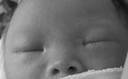
 

Figure 1. Neonate immediately intubated following delivery
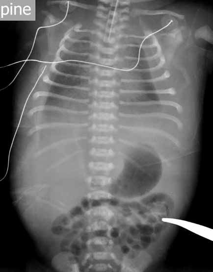
 

Figure 2. Bilateral pleural effusions
 
In the neonatal ICU, she improved with full intensive support (mechanical ventilation, thoracostomy drainage of pleural fluids, and treatment with intravenous octreotide). Trisomy 21 (47,XY,+21) was subsequently confirmed following chromosomal evaluation.
 
Hydrops fetalis (fetal hydrops) is a serious fetal condition defined as an abnormal accumulation of fluid in two or more fetal compartments, and includes ascites, pleural effusion, pericardial effusion, and skin oedema.1 It may be due to immune or nonimmune aetiologies.1 2 Rhesus isoimmunisation is the commonest immune aetiology, and alpha-thalassaemia is a non-immune cause.1 Immediate diagnosis of other aetiologies is often not possible without extensive investigations. A prompt spot diagnosis of DS was made in this neonate with typical facial features, which obviated the need for an extensive search for an underlying aetiology and enabled target therapies to be instituted (Fig 1). Trisomy 21 is a known association with hydrops fetalis and myeloproliferative disorder.1 2
 
Transient myeloproliferative disorder (TMD) is a self-limiting disorder characterised by leukocytosis and the presence of megakaryoblasts in the peripheral blood and bone marrow, anaemia, thrombocytopenia, and organomegaly. It occurs in approximately 10% of newborn infants with DS.2 Hepatic fibrosis is encountered in the severe form of TMD with DS, and is characterised by diffuse intralobular sinusoidal fibrosis and extramedullary haematopoiesis.3 Although TMD in most patients resolves spontaneously within the first 3 months of life, in a few severe cases there can be hepatic fibrosis or cardiopulmonary failure. Acute megakaryocytic leukaemia (AML-M7) is noted in 20 to 30% of babies with DS and TMD within the first 4 years of life.4 Cytokines produced by megakaryocytes (including transforming growth factor-beta, platelet-derived growth factor, and platelet factor 4) could be responsible for the pathogenesis of TMD.3 The imbalance between intravascular or capillary hydrostatic pressure and transcapillary filtration may be responsible from hydrops fetalis.2 3 4 5
 
Prompt recognition of the facial features of DS is important to facilitate immediate diagnosis and management of this neonate with hydrops fetalis.
 
References
1. Bellini C, Hennekam RC, Fulcheri E, et al. Etiology of nonimmune hydrops fetalis: a systematic review. Am J Med Genet A 2009;149A:844-51. Crossref
2. Oetama BK, Tucay RF, Morgan DL. Pathologic quiz case: nonimmune hydrops in a newborn. Down syndrome with acute (transient) leukemia. Arch Patho Lab Med 2001;125:1609-10.
3. Hongeng S, Pakakasama S, Hathirat P, Phuapradid P, Worapongpaiboon S. Diffuse hepatic fibrosis with transient myeloproliferative disorders in Down syndrome. J Pediatr Hematol Oncol 2000;22:543-4. Crossref
4. Al-Kasim F, Doyle JJ, Massey GV, Weinstein HJ, Zipursky A, Pediatric Oncology Group. Incidence and treatment of potentially lethal diseases in transient leukemia of Down syndrome: Pediatric Oncology Group Study. J Pediatr Hematol Oncol 2002;24:9-13. Crossref
5. De Groot CJ, Oepkes D, Egberts J, Kanhai HH. Evidence of endothelium involvement in the pathophysiology of hydrops fetalis? Early Hum Dev 2000;57:205-9. Crossref

Ketamine-induced cholangiopathy

Hong Kong Med J 2014;20:78.e1–2 | Number 1, February 2014
DOI: 10.12809/hkmj133796
© Hong Kong Academy of Medicine. CC BY-NC-ND 4.0
 
PICTORIAL MEDICINE
Ketamine-induced cholangiopathy
KL Lui, MMedSc, FHKCP1; WK Lee, FRCPath, FHKAM (Pathology)2; Michael KK Li, FRCP, FHKAM (Medicine)
1 Division of Gastroenterology and Hepatology, Department of Medicine and Geriatrics, Tuen Mun Hospital, Tuen Mun, Hong Kong
2 Department of Pathology, Tuen Mun Hospital, Tuen Mun, Hong Kong
 
Corresponding author: Dr KL Lui (klluitc@yahoo.com.hk)
 
A 28-year-old woman presented to us in November 2010 because of deranged liver function test results; predominantly she had raised ductal enzyme levels (gamma-glutamyl transferase, 1088; reference range, 12-57 IU/mL); alkaline phosphatase (ALP) 579 (reference range, 46-127) IU/mL, alanine transaminase (ALT) 183 (reference range, 10-57) IU/mL with normal bilirubin levels. Upon further questioning, she had been a ketamine abuser for 5 years and was followed up by psychiatrists. She was completely asymptomatic and physical examination yielded nil abnormal. Her ALP level was excessive (154 IU/mL) and her ALT level was 48 IU/mL. Ultrasound of hepatobiliary system (HBS) showed a dilated common bile duct (CBD) of 1.1 cm in diameter with tapering over lower end. A gallstone was present in the gallbladder. Therefore, the endoscopic retrograde cholangiopancreatography (ERCP) was performed in November 2011, and showed a 5-cm stricture at the lower end of the CBD together with small bilateral segmental strictures in the intrahepatic ducts (Fig 1). Brush cytology of the stricture of CBD revealed no malignant cells. A plastic stent bypassing the CBD was inserted for drainage. Liver function test findings did not improve after stenting but repeated ultrasonography of the HBS showed that with the 5.7-mm stent in situ, the CBD was not dilated. A liver biopsy was therefore performed, and showed mild-to-moderate portal fibrosis with ductular proliferation (Fig 2) and periportal copper deposits were noted (Fig 3). These findings were consistent with chronic cholestasis at both the extrahepatic and intrahepatic level. There were no features suggestive of primary biliary cirrhosis, or primary sclerosing cholangitis. The colonoscopy was normal and showed no evidence of inflammatory bowel disease. The patient’s liver function improved after she ceased the recreational use of ketamine. However, her stricture remained unchanged in the follow-up ERCP and repeated biopsies over the CBD stricture only showed reactive changes.
 

Figure 1. A 5-cm stricture at the lower end of the common bile duct with irregularity over intrahepatic ducts on both sides (arrows)
 

Figure 2. Intermediate power view showing portal fibrosis and ductal proliferation (arrows) [Masson’s trichrome stain, original magnification x 200]
 

Figure 3. Small periportal copper deposits are evident (arrows) [orange-red granules, rhodanine stain; original magnification x 400]
 
The first report on the association of liver injury with ketamine dates back to 1980.1 The exact cause of the ketamine-induced stricture is not known, but chronic use is associated with hepatocyte damage and fibrosis to the liver.2 Ketamine intake also stimulates the N-methyl-D-aspartic acid receptor in the smooth muscle cells of the bile duct and chronic stimulation may induce inflammation and fibrosis finally resulting in strictures.3 4 Affected patients are usually asymptomatic initially, and only manifest abnormal ductal enzyme level after 1 to 2 years of recreational ketamine use, indicating that chronicity and repeated use seem to be involved. Both intrahepatic and extrahepatic stricture might also develop and complicated with cholangitis, especially in the presence of gallstones. Definitive management entails cessation of ketamine intake, whereupon liver function improves, though the stricture may be permanent and warrant stenting to relieve any obstruction.4 5 This case report points that ketamine abuse also causes liver and biliary damage, quite apart from urinary and neurological sequelae.
 
References
1. Dundee JW, Fee JP, Moore J, Mcllroy PD, Wilson DB. Changes in serum enzyme levels following ketamine infusions. Anaesthesia 1980;35:12-6. Crossref
2. Wai MS, Chan WM, Zhang AQ, Wu Y, Yew DT. Longterm ketamine and ketamine plus alcohol treatments produced damages in liver and kidney. Hum Exp Toxicol 2012;31:877-86. Crossref
3. Jankovic SM, Jankovic SV, Stojadinovic D, Jakovljevic M, Milovanovic D. Effect of exogenous glutamate and N-Methyl-Daspartic acid on spontaneous activity of isolated human ureter. Int J Urol 2007;14:833-7. Crossref
4. Lo RS, Krishnamoorthy R, Freeman JG, Austin AS. Cholestasis and biliary dilatation associated with chronic ketamine abuse: a case series. Singapore Med J;52:e52-5.
5. Seto WK, Ng M, Chan P, et al. Ketamine-induced cholangiopathy: a case report. Am J Gastroenterol;106:1004-5. Crossref

Pages