An uncommon complication of infective endocarditis

DOI: 10.12809/hkmj144248
© Hong Kong Academy of Medicine. CC BY-NC-ND 4.0
 
PICTORIAL MEDICINE
An uncommon complication of infective endocarditis
KW Lam, MB, BS, FHKAM (Medicine); KW Au Yeung, MB, BS, FHKAM (Anaesthesiology); KY Lai, MB, BS, FHKAM (Medicine)
Intensive Care Unit, Queen Elizabeth Hospital, Jordan, Hong Kong
Corresponding author: Dr KW Lam (lamkw1@ha.org.hk)
 Full paper in PDF
 
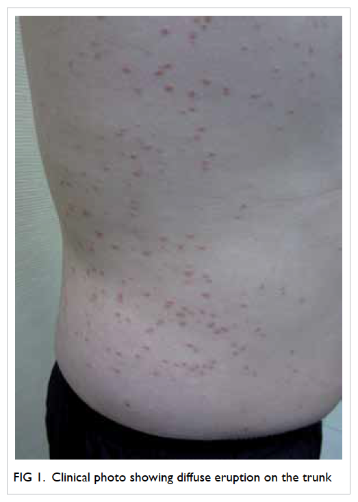
A 20-year-old man presented to us with confusion and generalised skin rash in April 2010. On examination, he was febrile and in shock. He was detected with a mitral regurgitation murmur and mild neck stiffness. His white cell count was elevated and platelet count was low. His liver function was mildly impaired. Chest X-ray showed acute pulmonary oedema. 
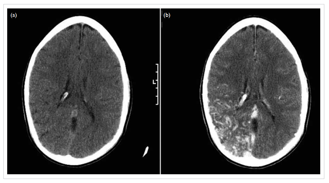
Computed tomography (CT) scan of brain showed multiple haematomas in his left occipital lobe and right parietal lobe, and subarachnoid haemorrhage (Fig 1a). Blood culture showed methicillin-sensitive Staphylococcus aureus (MSSA).
 

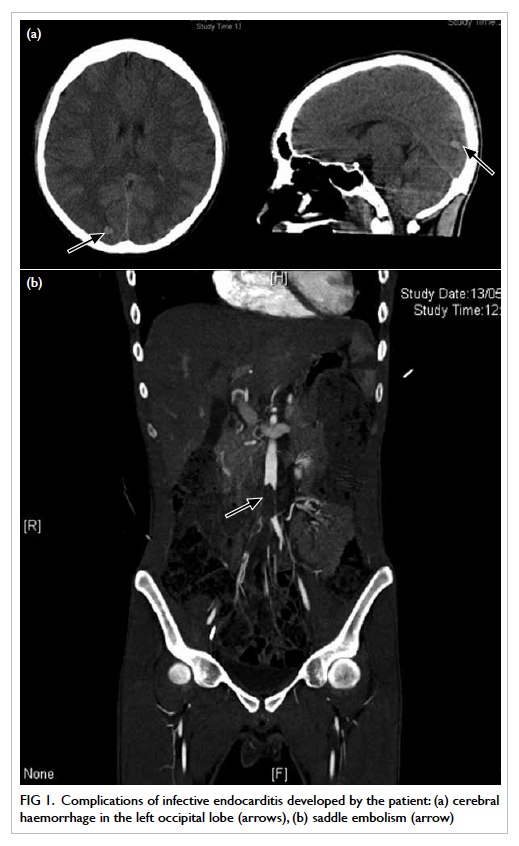
Figure 1. Complications of infective endocarditis developed by the patient: (a) cerebral haemorrhage in the left occipital lobe (arrows), (b) saddle embolism (arrow)
 
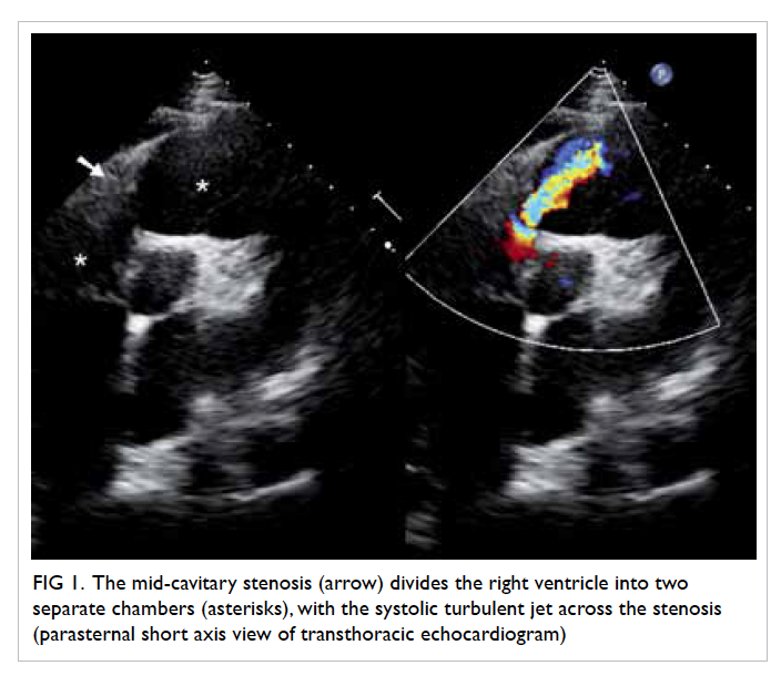
Transthoracic echocardiogram showed a huge vegetation measuring 4 cm x 2.22 cm attached to the base of anterior leaflet of mitral valve, resulting in perforation of the base of leaflet with severe mitral regurgitation (Fig 2a). The diagnosis was infective endocarditis due to MSSA, complicated by ruptured chordae tendineae with severe acute mitral regurgitation and multiple, septic cerebral emboli. The source of infection was suspected to be the skin. He was treated with high-dose intravenous cloxacillin and gentamicin. His heart failure was treated with frusemide.
 

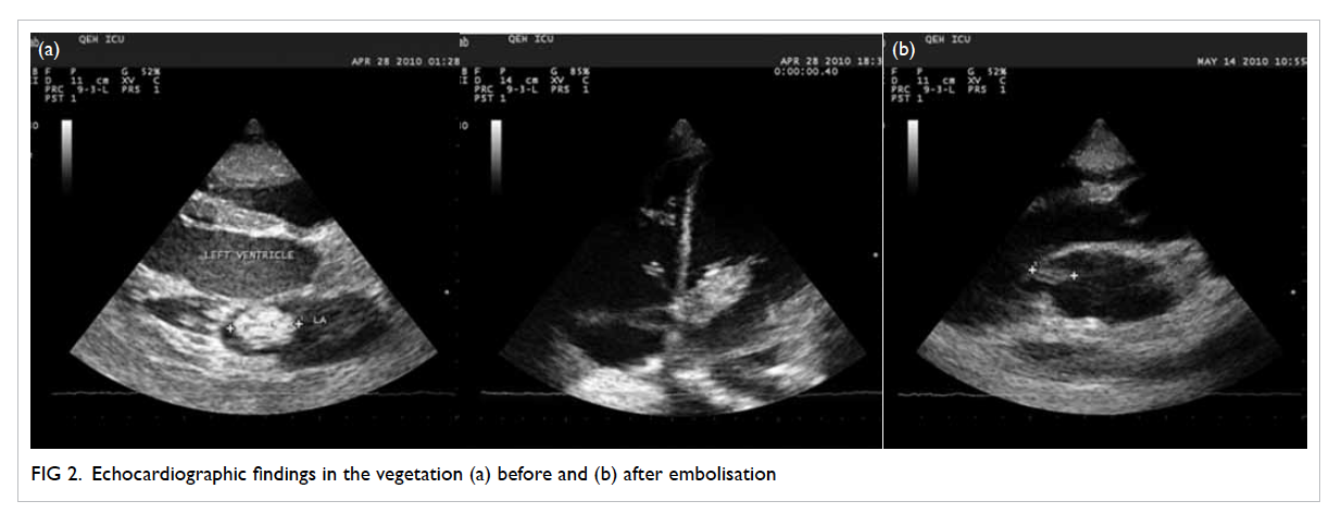
Figure 2. Echocardiographic findings in the vegetation (a) before and (b) after embolisation
 
Three weeks later, he complained of sudden onset of right lower limb pain and numbness. His CT angiogram showed a large saddle embolus in the lower abdominal aorta (Fig 1b). Emergent right-sided lower femoral and bilateral iliac artery embolectomy was performed.
 
On postoperative echocardiogram, the size of the vegetation was found to be decreased to 1.5 cm in diameter (Fig 2b). He then underwent mitral valvular replacement about 1 week later.
 
Discussion
The incidence of community-acquired native-valve endocarditis in western countries ranges from 1.7 to 6.2 cases per 100 000 person-years, with a male-to-female ratio of 1.7:1.1 Recently, the incidence of S aureus infective endocarditis is on the rise. Infective endocarditis due to S aureus is more common among young adults, especially the intravenous injection drug users.1 Usually, the tricuspid valve is involved.2
 
Extra cardiac complications of infective endocarditis usually include embolic events. The rate of embolic events has a relationship with the initiation of antibiotic therapy. In one study, after the commencement of appropriate antimicrobial treatment, the rate of embolism fell from 13 per 1000 patient-days during the first week of treatment to fewer than 1.2 per 1000 patient-days 2 weeks after treatment.3 A review involving 281 patients with suspected infective endocarditis demonstrated that the incidence of embolic events was greater with mitral than aortic valve vegetations (25% vs 10%).4 For mitral valve vegetations, the rate of embolism was higher if these were attached to the anterior leaflet rather than posterior leaflet. Some studies showed that the rate of embolism correlated with the size of vegetation, with the risk being higher if the diameter of the vegetation was greater than 1 cm.1
 
Up to 65% of embolic events of infective endocarditis are associated with neurological involvement. Such neurological complications account for 20% to 40% of all patients with infective endocarditis.5 Saddle embolus is a large embolus that straddles the arterial bifurcation and, thus, blocks both branches of the aorta. Saddle embolisation at the aortic bifurcation is an uncommon but serious complication. From the literature search, only eight cases were reported and all were caused by fungal endocarditis.6
 
References
1. Mylonakis E, Calderwood SB. Infective endocarditis in adults. N Engl J Med 2001;345:1318-30. Crossref
2. Hecht SR, Berger M. Right-sided endocarditis in intravenous drug users. Prognostic features in 102 episodes. Ann Intern Med 1992;117:560-6. Crossref
3. Heiro M, Nikoskelainen J, Engblom E, Kotilainen E, Marttila R, Kotilainen P. Neurologic manifestations of infective endocarditis: a 17-year experience in a teaching hospital in Finland. Arch Inern Med 2000;160:2781-7. Crossref
4. Rohmann S, Erbel R, Görge G, et al. Clinical relevance of vegetation localization by transoesophageal echocardiography in infective endocarditis. Eur Heart J 1992;13:446-52.
5. Røder BL, Wandall DA, Espersen F, Frimodt-Møller N, Skinhøj P, Rosdahl VT. Neurologic manifestations in Staphylococcus aureus endocarditis: a review of 260 bacteremic cases in nondrug addicts. Am J Med 1997;102:379-86. Crossref
6. Kawamoto T, Nakano S, Matsuda H, Hirose H, Kawashima Y. Candida endocarditis with saddle embolism: a successful surgical intervention. Ann Thorac Surg 1989;48:723-4. Crossref

Inflammatory myoglandular polyps of the rectum

DOI: 10.12809/hkmj134189
© Hong Kong Academy of Medicine. CC BY-NC-ND 4.0
 
PICTORIAL MEDICINE
Inflammatory myoglandular polyps of the rectum
Akira Hokama, MD#; Chiharu Kobashigawa, MD#; Jiro Fujita, MD
Department of Infectious, Respiratory, and Digestive Medicine, University of the Ryukyus, 207 Uehara, Nishihara, Okinawa 903-0215, Japan
# Currently at Department of Endoscopy, University of the Ryukyus, 207 Uehara, Nishihara, Okinawa 903-0215, Japan
 
Corresponding author: Dr Akira Hokama (hokama-a@med.u-ryukyu.ac.jp)
 
 Full paper in PDF
Case report
An 84-year-old woman with advanced pharyngeal cancer underwent colonoscopy for intermittent rectal bleeding in October 2012. Colonoscopy disclosed two red sessile polyps in the rectum (Fig 1). The larger one was spherical with a smooth surface, measuring approximately 15 mm in diameter (Fig 2). Biopsy showed hyperplastic glands and marked proliferation of smooth muscle cells in the lamina propria, consistent with a diagnosis of inflammatory myoglandular polyp (IMGP) [Fig 3]. The patient denied colonoscopic treatment and stays in a hospice.
 

Figure 1. Colonoscopy disclosing two red sessile polyps in the rectum
 

Figure 2. The larger polyp is spherical with a smooth surface, measuring approximately 15 mm in diameter
 

Figure 3. Biopsy shows hyperplastic glands and marked proliferation of smooth muscle cells in the lamina propria, consistent with a diagnosis of inflammatory myoglandular polyp (H&E; original magnification, x 100)
 
Inflammatory myoglandular polyp is a rare, non-neoplastic polyp of the colorectum with histological features of inflammatory granulation tissue in the lamina propria, proliferation of smooth muscle cells, and hyperplastic glands with variable cystic changes.1 Since Nakamura et al1 first documented IMGP in 1992, only 60 cases of IMGPs have been reported worldwide.2 As most IMGPs are located in the rectum and the sigmoid colon, a common symptom of the condition is haematochezia. Although the causes of IMGP are obscure, chronic trauma from the faecal stream and peristalsis may contribute to its pathogenesis.1 With prolonged irritation, small, sessile IMGPs can enlarge and become pedunculated. Characteristic features include hyperaemic surface with patchy mucous exudation and erosion. Inflammatory myoglandular polyp differs from other non-neoplastic polyps including inflammatory cap polyps, inflammatory cloacogenic polyps, juvenile polyps, inflammatory fibroid polyps, polyps secondary to mucosal prolapse syndrome, polypoid prolapsing mucosal folds of diverticular disease in terms of its clinical and histopathological features.2 Most IMGPs can be treated by endoscopic resection. Because IMGP follows a benign course, endoscopic resection might be unnecessary when biopsy confirms the histopathological diagnosis.3
 
References
1. Nakamura S, Kino I, Akagi T. Inflammatory myoglandular polyps of the colon and rectum. A clinicopathological study of 32 pedunculated polyps, distinct from other types of polyps. Am J Surg Pathol 1992;16:772-9. CrossRef
2. Meniconi RL, Caronna R, Benedetti M, et al. Inflammatory myoglandular polyp of the cecum: case report and review of literature. BMC Gastroenterol 2010;10:10. CrossRef
3. Hirasaki S, Okuda M, Kudo K, Suzuki S, Shirakawa A. Inflammatory myoglandular polyp causing hematochezia. World J Gastroenterol 2008;14:5353-5. CrossRef
 
Find HKMJ in MEDLINE:
 

Double-chambered right ventricle: a commonly overlooked diagnosis

DOI: 10.12809/hkmj134187
© Hong Kong Academy of Medicine. CC BY-NC-ND 4.0
 
PICTORIAL MEDICINE
Double-chambered right ventricle: a commonly overlooked diagnosis
Joe KT Lee, MRCP (UK), FHKAM (Medicine); KL Tsui, FRCP (Edin, Glasg), FHKAM (Medicine)
Department of Medicine, Pamela Youde Nethersole Eastern Hospital, Chai Wan, Hong Kong
 
Corresponding author: Dr Joe KT Lee (jktlee@gmail.com)
 
 Full paper in PDF
A 72-year-old woman presented with decreased exercise tolerance since 2007. Based on a transthoracic echocardiogram (TTE) in another hospital, the patient was diagnosed to have perimembranous ventricular septal defect (VSD) with pulmonary hypertension. Upon referral to our unit in 2013, a more meticulous TTE examination revealed a mid-cavitary stenosis in the right ventricle (RV) which was best appreciated in the parasternal short axis and the subcostal short axis view (Figs 1 and 2). The systolic pressure gradient measured by continuous-wave Doppler between the two RV chambers was markedly elevated to 80 mm Hg. There was also severe tricuspid regurgitation with a dilated RV. The findings were confirmed on a transoesophageal echocardiogram (TEE) and the diagnosis was revised as double-chambered right ventricle (DCRV) with perimembranous VSD (Fig 3). A right heart catheterization study showed a stenotic band over the right ventricular outflow tract (RVOT) [Fig 4]. The systolic pressure at the proximal RV chamber was markedly elevated to 75 mm Hg. However, we failed to manipulate the catheter across the stenotic band to measure the pressure gradient across the two chambers. The coronary angiographic results were normal. The patient was referred to cardiothoracic surgeons and open heart surgery was undertaken. After right ventriculotomy, the anomalous infundibular muscle bundle and a small perimembranous VSD were identified. The obstructive muscle bundle was resected and VSD was repaired. The tricuspid valve was repaired by means of annuloplasty. In postoperative TTE, the previously noted high pressure gradient across the RVOT was no longer present. The right ventricular systolic pressure normalised to 15 mm Hg.
 

Figure 1. The mid-cavitary stenosis (arrow) divides the right ventricle into two separate chambers (asterisks), with the systolic turbulent jet across the stenosis (parasternal short axis view of transthoracic echocardiogram)
 

Figure 2. On subcostal short axis view of transthoracic echocardiogram, a more parallel alignment of the turbulent jet to the transducer can be obtained, which allows more accurate pressure gradient assessment by Doppler between the two right ventricular chambers (asterisks) across the stenotic band (arrow)
 

Figure 3. The systolic turbulent jet across the mid-cavitary stenosis (arrow) in between the two right ventricle chambers (asterisks) [75° on transoesophageal echocardiogram]
 

Figure 4. The right-heart ventriculogram on postero-anterior projection reveals a stenotic band in the right ventricular outflow tract (arrows) and the two right ventricle chambers (asterisks)
 
Discussion
Double-chambered RV is characterised by the presence of an anomalous muscle bundle (AMB), which divides the RV into two separate chambers, namely, the proximal high-pressure and the distal low-pressure chambers. The AMB is considered a hypertrophied moderator band or the accentuated septoparietal trabeculation.1 2 These congenital anatomical substrates and other acquired haemodynamic factors lead to the development of DCRV.
 
Double-chambered RV is an uncommon condition and it is only seen in 0.5% to 2% of all cases of congenital heart disease.3 Most cases are diagnosed in childhood or adolescence before the age of 20 years. The occurrence in adults is rare and has only been described in case reports and small case series. About 80% to 90% of DCRV cases are associated with VSD, or sometimes with other congenital cardiac anomalies. Patients with DCRV usually present with shortness of breath and decreased exercise tolerance, but they may also have atypical symptoms such as chest pain, dizziness, and syncope.
 
The irregular shape and retrosternal position of RV, and its close proximity to the precordium impose diagnostic difficulty by TTE, especially in adults. Quite often, the mid-cavitary turbulent jet on TTE is mistaken as an intracardiac shunt, and the high RV systolic pressure is falsely interpreted as pulmonary hypertension. In a case series, only 15.6% of patients with DCRV could be correctly diagnosed by TTE.3 A high clinical suspicion and awareness of this clinical entity are required for precise diagnosis. The subcostal view of TTE may sometimes provide better visualisation of the RV and RVOT. It also allows better alignment of the turbulent jet for pressure gradient measurement. When TTE is not confirmative, TEE and cardiac catheterization serve as complementary tools. The use of cardiac magnetic resonance imaging as non-invasive assessment is now emerging as an alternative diagnostic modality for DCRV.
 
Surgical repair of RV by resection of the AMB is indicated in symptomatic patients, and it yields excellent long-term haemodynamic and functional results.4 Surgical treatment is also suggested in asymptomatic patients who have significantly elevated midventricular pressure gradient, that is, >40 mm Hg as the obstruction can progress rapidly over just a few years.5
 
References
1. Wong PC, Sanders SP, Jonas RA, et al. Pulmonary valve-moderator band distance and association with development of double-chambered right ventricle. Am J Cardiol 1991;68:1681-6. CrossRef
2. Alva C, Ho SY, Lincoln CR, et al. The nature of the obstructive muscular bundles in double-chambered right ventricle. J Thorac Cardiovasc Surg 1999;117:1180-9. CrossRef
3. Hoffman P, Wojcik AW, Rozanski J, et al. The role of echocardiography in diagnosing double chambered right ventricle in adults. Heart 2004;90:789-93. CrossRef
4. Telagh R, Alexi-Meskishvili V, Hetzer R, et al. Initial clinical manifestations and mid- and long-term results after surgical repair of double-chambered right ventricle in children and adults. Cardiol Young 2008;18:268-74. CrossRef
5. Oliver JM, Garrido A, Gonzalez A, et al. Rapid progression of midventricular obstruction in adults with double-chambered right ventricle. J Thorac Cardiovasc Surg 2003;126:711-7. CrossRef
 
Find HKMJ in MEDLINE:
 

Walker-Warburg syndrome: rare congenital muscular dystrophy associated with brain and eye abnormalities

DOI: 10.12809/hkmj134137
© Hong Kong Academy of Medicine. CC BY-NC-ND 4.0
 
PICTORIAL MEDICINE
Walker-Warburg syndrome: rare congenital muscular dystrophy associated with brain and eye abnormalities
CY Lee, FRCR, MB, ChB
Department of Radiology, Tuen Mun Hospital, Tuen Mun, Hong Kong
 
Corresponding author: Dr CY Lee (prodigycat@gmail.com)
 
 Full paper in PDF
Case report
A 7-month-old boy was found to have developmental delay, abnormal muscle tone, and abnormal eye movement in December 2012. Physical examination of the eyes revealed wandering gaze with convergent squint. Ophthalmology was consulted and bilateral retrolental masses were suspected. Blood tests revealed elevated serum creatine kinase level.
 
Computed tomography of orbits showed bilateral hyperdense retrolental tubular opacities with small retinal haemorrhage on the right (Fig 1). Computed tomography of brain also showed communicating hydrocephalus. Magnetic resonance imaging of orbits showed deformed bilateral globes, abnormal T1-weighted and T2-weighted hypo–to–iso-intense contrast-enhancing triangular bands with base near the optic disc and apex at the posterior surface of lens, compatible with bilateral persistent hyperplastic primary vitreous. T1- and T2-weighted hyperintensity at right vitreous body was compatible with previous haemorrhage.
 

Figure 1. Computed tomography of orbits shows bilateral, hyperdense retrolental tubular opacities (arrows), later confirmed to be bilateral, persistent hyperplastic primary vitreous on magnetic resonance imaging
 
Magnetic resonance imaging of the brain showed pachygyria, hydrocephalus, absent septum pellucidum, and hypoplasia of corpus callosum forming type II lissencephaly (Fig 2). Mega cisterna magna, hypoplastic pons, and cerebellar vermis were compatible with posterior cranial fossa malformation (Fig 3). Band-like structures in the bilateral periventricular regions with signal changes similar to grey matter were suggestive of band heterotopic grey matter.
 

Figure 2. T1-weighted axial magnetic resonance imaging of brain shows pachygyria, hydrocephalus, absent septum pellucidum, and hypoplasia of corpus callosum forming type II lissencephaly (arrows)
 

Figure 3. (a) T1-weighted sagittal and (b) axial magnetic resonance imaging of brain show mega cisterna magna, hypoplastic pons, and cerebellar vermis compatible with posterior cranial fossa malformation (arrows)
 
Radiological findings of type II lissencephaly, posterior fossa malformation and retinal anomaly, together with clinical findings of developmental delay, abnormal muscle tone, and elevated serum creatine kinase level were compatible with diagnosis of Walker-Warburg syndrome.
 
Discussion
Congenital muscular dystrophy (CMD) comprises a heterogeneous group of disorders. Walker-Warburg syndrome is one phenotype of CMD known to occur due to dystroglycanopathy,1 which is an autosomal recessive condition. The overall incidence is unknown but a survey in Northeastern Italy has reported an incidence rate of 1.2 per 100 000 live births.2
 
Walker-Warburg syndrome affects the brain, eye, and muscles with characteristic malformation. Diagnostic criteria for Walker-Warburg syndrome include type II lissencephaly, cerebellar malformation, retinal malformation, and CMD.3 Common associated anomalies include anterior chamber malformation of the eye and hydrocephalus. The less commonly observed anomalies include Dandy-Walker malformation, cleft lip and palate, congenital macrocephaly or microcephaly, posterior encephalocoele, ocular colobomas, congenital cataracts, and genital abnormalities. Neuroimaging findings other than lissencephaly include band heterotopia, cerebellar vermian hypoplasia, dysgenesis of corpus callosum, abnormal white matter changes, hypoplastic cerebral peduncles, intraventricular haemorrhage, cerebellar polymicrogyria, collicular fusion, and fusion of occipital poles.4 Laboratory investigations usually show elevated serum creatine kinase level, myopathic/dystrophic muscle pathology, and altered alpha-dystroglycan.2
 
Differentiation of Walker-Warburg syndrome from other dystroglycanopathies, for example, muscle-eye-brain disease or Fukuyama CMD, depends on the severity of clinical presentation including motor function and intellectual disability, and involvement of the central nervous system and eye.1 Walker-Warburg syndrome is believed to be the most severe form of dystroglycanopathy with most children dying before the age of 3 years.2 No specific treatment is available for this syndrome. Management is mainly supportive and preventive.
 
References
1. Sparks S, Quijano-Roy S, Harper A, et al. Congenital Muscular Dystrophy Overview. 2001 Jan 22 [Updated 2012 Aug 23]. In: Pagon RA, Adam MP, Ardinger HH, et al, editors. GeneReviews® [Internet]. Seattle (WA): University of Washington, Seattle; 1993-2014. Available from: http://www.ncbi.nlm.nih.gov/books/NBK1291/. Accessed Sep 2013.
2. Vajsar J, Schachter H. Walker-Warburg syndrome. Orphanet J Rare Dis 2006;1:29. CrossRef
3. Dobyns WB, Pagon RA, Armstrong D, et al. Diagnostic criteria for Walker-Warburg syndrome. Am J Med Genet 1989;32:195-210. CrossRef
4. Zaleski CG, Abdenour GE. Pediatric case of the day. Walker-Warburg syndrome (cerebro-ocular dysplasia-muscular dystrophy). Radiographics 1997;17:1319-23. CrossRef

Pneumonitis and extreme failure to thrive

DOI: 10.12809/hkmj134106
© Hong Kong Academy of Medicine. CC BY-NC-ND 4.0
 
PICTORIAL MEDICINE
Pneumonitis and extreme failure to thrive
KL Hon, MD, FCCM1; TF Leung, FRCPCH, FHKAM (Paediatrics)1; YS Yau, MRCP (UK), FHKAM (Paediatrics)2
1 Department of Paediatrics, The Chinese University of Hong Kong, Prince of Wales Hospital, Shatin, Hong Kong
2 Department of Paediatrics, Queen Elizabeth Hospital, Jordan, Hong Kong
 
Corresponding author: Prof KL Hon (ehon@cuhk.edu.hk)
 
 Full paper in PDF
Failure to thrive is an uncommon, challenging, but important basket of differential diagnoses to manage in the city of Hong Kong. Diagnosis may be non-organic or functional. During outbreaks of avian influenza in Mainland China and MERS/SARI (Middle East respiratory syndrome/severe acute respiratory illness) coronavirus infection in the Middle East in early 2013,1 a 27-month-old girl was brought by her parents to Hong Kong following a long period of hospitalisation and investigations in Mainland China for recurrent pneumonia, chronic diarrhoea, lymphadenopathy, oral candidiasis, and failure to thrive. Reportedly, no bacterial or viral pathogens had been found. Antenatal anti–human immunodeficiency virus (HIV) antibody testing was negative in the mother. There had been no adverse reaction to Bacille Calmette-Guérin and routine immunisations. The child had been exclusively breastfed and her growth and development were normal until 9 months of age. The child was admitted to the paediatric intensive care unit (ICU) of a Hong Kong hospital for management. She was noted to have profound failure to thrive with a body weight of only 5 kg (Fig 1). Chest X-ray showed diffuse pneumonitis (Fig 2). She required oxygen supplementation but mechanical ventilation was not needed.
 

Figure 1. Cachexic girl with pneumonitis
 

Figure 2. Chest X-ray showing fulminant pneumonitis
 
Which of the following investigations will most likely give the underlying diagnosis?
1. White cell counts and differentials for congenital neutrophil abnormality
2. Complement C3 and C4 levels for congenital complement deficits
3. Immunoglobulin A for immunoglobinopathy
4. H7N9, coronavirus, mycoplasma, and chlamydia serology for atypical pneumonia
5. HIV testing
 
After performing various investigations (Table), the child tested positive for HIV infection, confirming that she had acquired immunodeficiency syndrome (AIDS). Computed tomographic scan of thorax showed pneumonitis (Fig 3). Treatment for Pneumocystis jiroveci pneumonia and cytomegalovirus infection was commenced. Following stabilisation, the child was referred to a paediatric infectious disease specialist for continuation of care. Highly active antiretroviral therapy was started when opportunistic infections were under control. The child was last seen in June 2014; she was asymptomatic and had been thriving well.
 

Table. Some relevant investigations performed in the patient
 

Figure 3. Computed tomography thorax with contrast showing pneumonitis with diffuse ground-glass opacities and interseptal thickening, but no lung consolidation or hilar lymphadenopathy
 
The parents refused HIV testing but reported that the child had received blood products after onset of illness while in the Mainland hospital. Both were subsequently confirmed HIV positive. Nevertheless, HIV was most likely to be vertically (mother to child) transmitted.2 3 4 5 In many areas of the world, HIV/AIDS has become a chronic rather than an acutely fatal disease.3 Half of the infants born with HIV die before 2 years of age without treatment.2 3 4 5
 
Pneumonitis is usually caused by viruses or atypical pathogens. Since the atypical pneumonitis epidemic of coronavirus in 2003, Hong Kong and the rest of the world have heightened surveillance for outbreaks of atypical pneumonitis with novel pathogens such as avian or swine influenza, or coronavirus.1 In the cosmopolitan city of Hong Kong, paediatric HIV remains a relatively rare diagnosis. Nevertheless, due to the busy trafficking between Hong Kong and Mainland China, paediatricians in Hong Kong must be vigilant of such possibility in Hong Kong children of Mainland parents. Despite the misleading history by the parents about negative screening for HIV, paediatricians at the paediatric ICU were prompt to arrive at the definitive diagnosis by requesting HIV testing and considering HIV infection as a possibility to explain the combination of pneumonitis and extreme failure to thrive in this child.
 
This case is interesting and highlights the importance of excluding HIV infection in a child with dual symptoms of recurrent infections and failure to thrive, even when the mother had tested negative for HIV antibodies initially; the test may have been performed in a window period during pregnancy.
 
Differential diagnoses for failure to thrive include child abuse and neglect, cystic fibrosis (rare in Hong Kong), gastroesophageal reflux, growth failure, growth hormone deficiency, and HIV infection.6 7 The history and physical examination should guide any laboratory or ancillary testing. Most infants and children with growth failure related to environmental factors need very limited laboratory screenings. This child presented with recurrent infections and candidiasis. Approach to recurrent infections resulting in failure to thrive may include HIV testing, sweat test for cystic fibrosis (if history is relevant), metabolic and endocrinology screening, tuberculosis testing, and stool studies.
 
Basing on disease onset, this is most likely a case of vertical transmission of HIV. In infants, the onset of AIDS symptoms can take a few months; in contrast, it can be many years before adults develop symptoms of HIV. Thus, repeated HIV testing is very important to initiate timely treatment in the parents.
 
References
1. Hon KL. Severe respiratory syndromes: travel history matters. Travel Med Infect Dis 2013;11:285-7. CrossRef
2. Sepkowitz KA. AIDS—the first 20 years. N Engl J Med 2001;344:1764-72. CrossRef
3. Knoll B, Lassmann B, Temesgen Z. Current status of HIV infection: a review for non–HIV-treating physicians. Int J Dermatol 2007;46:1219-28. CrossRef
4. Coutsoudis A, Kwaan L, Thomson M. Prevention of vertical transmission of HIV-1 in resource-limited settings. Expert Rev Anti Infect Ther 2010;8:1163-75. CrossRef
5. Thorne C, Newell ML. HIV. Semin Fetal Neonatal Med 2007;12:174-81. CrossRef
6. Nangia S, Tiwari S. Failure to thrive. Indian J Pediatr 2013;80:585-9. CrossRef
7. Hendaus M, Al-Hammadi A. Failure to thrive in infants (review). Georgian Med News 2013;(214):48-54.

Trachyonychia in a patient with chronic myeloid leukaemia after imatinib mesylate

DOI: 10.12809/hkmj134084
© Hong Kong Academy of Medicine. CC BY-NC-ND 4.0
 
PICTORIAL MEDICINE
Trachyonychia in a patient with chronic myeloid leukaemia after imatinib mesylate
YM Lau, FHKCP, FHKAM (Medicine); YK Lam, FHKCP, FHKAM (Medicine); KH Leung, MRCP; SY Lin, FHKCP, FHKAM (Medicine)
Department of Medicine and Geriatrics, United Christian Hospital, Kwun Tong, Hong Kong
 
Corresponding author: Dr YM Lau (lym570@ha.org.hk)
 
 Full paper in PDF
An 86-year-old man presented with leukocytosis in December 2009. Bone marrow biopsy showed chronic myeloid leukaemia in chronic phase and cytogenetic studies showed t(9;22)(q34;q11.2) translocation. He was initially put on imatinib 300 mg daily; subsequently, this was increased to 400 mg daily. He developed pruritic skin rash within 3 months of initiating imatinib. Initially, the skin condition improved with topical steroid. However, there was progressive development of white streaks and scaling of skin over the face, scalp, trunk, limbs, and trachyonychia with onycholysis of fingers and toes. There were no mucosal lesions. Skin biopsy findings were consistent with lichenoid drug reaction. Imatinib was stopped and changed to nilotinib. The skin and nail conditions progressively improved while the patient was on nilotinib.
 
Imatinib mesylate has been the standard treatment for chronic myeloid leukaemia for 10 years.1 Imatinib mesylate inhibits tyrosine kinases of bcr/abl, c-kit, and platelet-derived growth factor receptors, and cutaneous reactions are the commonest side-effects in patients receiving this drug.2 3 Trachyonychia results from disruption of the nail matrix cells, and can be induced by chemotherapeutic agents.4 Although paronychial inflammation is commonly induced by kinase inhibitors, trachyonychia is rarely reported. Cross-reactivity between different tyrosine kinases has rarely been reported.5 The absence of cross-reactivity between imatinib and nilotinib in this patient suggests that the mechanism of drug reaction is not related to the inhibition of tyrosine kinase.
 

Figure. Severe trachyonychia with onycholysis
 
References
1. Peggs K, Mackinnon S. Imatinib mesylate—the new gold standard for treatment of chronic myeloid leukemia. N Engl J Med 2003;348:1048-50. CrossRef
2. Gardembas M, Rousselot P, Tulliez M, et al. Results of a prospective phase 2 study combining imatinib mesylate and cytarabine for the treatment of Philadelphia-positive patients with chronic myelogenous leukemia in chronic phase. Blood 2003;102:4298-305. CrossRef
3. Wahiduzzaman M, Pubalan M. Oral and cutaneous lichenoid reaction with nail changes secondary to imatinib: report of a case and literature review. Dermatol Online J 2008;14:14.
4. Chen W, Yu YS, Liu YH, Sheen JM, Hsiao CC. Nail changes associated with chemotherapy in children. J Eur Acad Dermatol Venereol 2007;21:186-90. CrossRef
5. Novitzky-Basso I, Craddock C. Cross-intolerance to imatinib, dasatinib and nilotinib therapy in a patient with chronic myeloid leukaemia. Eur J Haematol 2011;86:548-9. CrossRef

To scan or not to scan, to enhance or not to enhance? That is the question

DOI: 10.12809/hkmj134120
© Hong Kong Academy of Medicine. CC BY-NC-ND 4.0
 
PICTORIAL MEDICINE
To scan or not to scan, to enhance or not to enhance? That is the question
Lily Li, MRCS (Eng), MB/BChir1; Jonathan Costello, MRCPI, FCEM2
1 Department of Trauma and Orthopaedics, Lister Hospital, Stevenage, Hertfordshire SG1 4AB, United Kingdom
2 Emergency Department, Royal Free Hospital, London NW3 2QG, United Kingdom
 
Corresponding author: Dr Lily Li (xl228@doctors.org.uk)
 
 Full paper in PDF
A 12-year-old boy presented to the Emergency Department (ED) with reduced level of consciousness in February 2011. Collateral history established a pre-hospital witnessed seizure (requiring benzodiazepine administration) preceded by auditory hallucinations. Apart from uncomplicated malaria at the age of 5 years, there was no other medical history of relevance. Initial review was consistent with post-ictal presentation. An additional generalised seizure was witnessed in the ED within 30 minutes of admission requiring termination with additional intravenous benzodiazepine. In view of recurrent presentation, he was electively intubated and commenced on parenteral phenytoin. In addition, empirical acyclovir and ceftriaxone were administered. An unenhanced computed tomographic (CT) scan was normal (a). However, a subsequent enhanced scan revealed diffuse right parieto-occipital arteriovenous malformation (b). This case challenges the prevalent practice of non-performance of CT in new-onset seizure disorders and, if performed, the practice of performing solely non-enhanced CT scans in such presentations.
 

Should we perform polypectomy or not?

DOI: 10.12809/hkmj134090
© Hong Kong Academy of Medicine. CC BY-NC-ND 4.0
 
PICTORIAL MEDICINE
Should we perform polypectomy or not?
WY Mak, MB, BS, MRCP (UK); YT Hui, MRCP (UK), FHKAM (Medicine); Jodis TW Lam, FHKAM (Medicine), FRCP (Edin)
Department of Medicine, Queen Elizabeth Hospital, Jordan, Hong Kong
 
Corresponding author: Dr WY Mak (mwy612@ha.org.hk)
 
 Full paper in PDF
A 78-year-old woman, with a known history of rheumatoid arthritis, complained of dizziness and was found to have iron-deficiency anaemia with a haemoglobin level of 105 g/L in January 2013. She was on treatment with methotrexate, sulphasalazine, and non-steroidal anti-inflammatory medications. Oesophagogastroduodenoscopy performing for anaemia showed a linear ulcer over the anterior wall of the stomach. Subsequent colonoscopic examination was done to look for the cause of iron-deficiency anaemia and revealed a 1 cm–long everted umbilicated polypoid lesion in the ascending colon (Fig a). On close examination of the lesion using narrow band imaging (NBI), the mucosal pattern was normal with no endoscopic features of adenomatous polyp. Upon further air insufflation, the everted lesion invaginated and turned into a diverticulum (Fig b). Thus, a diagnosis of an inverted colonic diverticulum (ICD) was made.
 

Figure. (a) An inverted colonic diverticulum (white arrow) was found next to two small diverticula (black arrows) by narrow band imaging. (b) The inverted colonic diverticulum (white arrow) which was located next to the two small diverticula (black arrows) invaginated and became a diverticulum
 
Inverted colonic diverticulum is a rare condition. A retrospective analysis of colonic examinations showed that the prevalence was only 0.7%.1 The majority (approximately 75%) of ICDs were found in the sigmoid colon.1 Right-sided colonic ICD, as illustrated in our case, was not commonly seen. An ICD is typically described as a broad-based lesion with normal overlying mucosa lying within a bed of colonic diverticula. It can resemble an adenomatous polyp of variable size. It is essential to correctly diagnose this condition as inadvertent ‘polypectomy’ may potentially lead to bowel perforation. Currently, there are several endoscopic strategies that can help in distinguishing ICD from an adenomatous polyp. Firstly, gentle air insufflation may cause evertion of inverted diverticula. In some cases, a jet of water may be used to flatten the lesion.2 Secondly, probing the lesion gently with a biopsy forceps will show a soft lesion with easy indentation. Interestingly, it was recently suggested that the presence of Aurora rings can support the diagnosis of an ICD.3 Aurora rings are described as concentric rings surrounding the base of an ICD which can be demonstrated with the use of NBI or chromoendoscopy. If doubt exists, double-contrast barium enema or computed tomography colonography may help to distinguish between the two entities. Endoscopic ultrasound (EUS) has also been used to characterise such lesions. In a report, the diagnosis of sigmoid ICD was made by the EUS features of a thickened but normal-looking colonic mucosa in a polyp-like lesion.4 Most importantly, endoscopic removal and biopsy of ICD should be avoided as potentially fatal bowel perforation may occur.5
 
In conclusion, ICD is an uncommon but important clinical finding. Endoscopists should always be aware of the possibility of ICD during colonoscopic examination as inadvertent biopsy or resection of these lesions can lead to potentially serious complications.
 
References
1. Merino R, Kinney T, Santander R, et al. Inverted colonic diverticulum: an infrequent and dangerous endoscopic finding [abstract]. Gastrointest Endosc 2005;61:AB257. CrossRef
2. Cappell MS. The water jet deformation sign: a novel provocative colonoscopic maneuver to help diagnose an inverted colonic diverticulum. South Med J 2009;102:295-8. CrossRef
3. Share MD, Avila A, Dry SM, Share EJ. Aurora rings: a novel endoscopic finding to distinguish inverted colonic diverticula from colon polyps. Gastointest Endosc 2013;77:308-12. CrossRef
4. Yoshida M, Kawabata K, Kutsumi H, et al. Polypoid prolapsing mucosal folds associated with diverticular disease in the sigmoid colon: usefulness of colonoscopy and endoscopic ultrasonography for diagnosis. Gastrointest Endosc 1996;44:489-91. CrossRef
5. D’Ovidio V, Di Camillo M, Pimpo MT, Meo D, Vernia P, Caprilli R. An unusual complicated polypectomy and inverted colonic diverticula. Colorectal Dis 2010;12:491-2. CrossRef

Urinary bladder inguinal hernia: an uncommon cause of scrotal swelling

DOI: 10.12809/hkmj134057
© Hong Kong Academy of Medicine. CC BY-NC-ND 4.0
 
PICTORIAL MEDICINE
Urinary bladder inguinal hernia: an uncommon cause of scrotal swelling
HL She, MB, BS; KC Lam, MB, BS; KK Wong, MB, BS; Wendy WM Lam, MB, BS
Department of Radiology, Queen Mary Hospital, Pokfulam, Hong Kong
 
Corresponding author: Dr HL She (helenshe1025@gmail.com)
 
 Full paper in PDF
A 77-year-old man with benign prostate hypertrophy (BPH) presented to our hospital in October 2011 with a history of right groin swelling for several months. He was otherwise asymptomatic. Physical examination revealed a reducible right inguinal hernia. Ultrasound (USG) examination of the groins showed a fluid-filled lesion within the right scrotum. It had a beaked appearance at its cranial portion, which could be traced entering the right inguinal canal (Fig 1a). A tiny calcified focus was noted within this fluid-filled structure (Fig 1b). The normal right testis was displaced inferiorly (Fig 1c). Findings were suggestive of urinary bladder inguinal hernia with a bladder stone within. It was confirmed with non-contrast computed tomography (CT) of the abdomen and pelvis, which showed herniation of the urinary bladder along the inguinal canal and into the right scrotum, with a small bladder stone within (Fig 2).
 

Figure 1. (a) Ultrasonography demonstrates a fluid-filled structure with a beaked appearance within the right scrotum. (b) A tiny calcified focus within the fluid-filled structure in the right scrotum is seen (white arrow). (c) A normal right testis is displaced inferiorly by the fluid-filled structure (black arrow)
 

Figure 2. (a) Non-contrast computed tomography (CT) of the abdomen and pelvis in a reformatted image demonstrates herniation of the urinary bladder into the right scrotum (arrow). (b) Non-contrast CT axial view shows herniation of the urinary bladder into right scrotum (arrow) with a tiny bladder stone within (arrowhead)
 
Urinary bladder herniation is an uncommon condition, encountered in 1% to 4% of inguinal hernias. However, over the age of 50 years, the frequency increases to about 10%.1 Most patients are asymptomatic and usually found incidentally on imaging for workup of inguinal hernias, or even at the time of herniorrhaphy. Occasionally, patients may complain of urinary symptoms especially at an advanced stage, and may entail double-phase urination, that is, manually compressing the scrotum for complete bladder emptying. Predisposing factors include obesity, bladder outlet obstruction (eg due to BPH), and weakened abdominal musculature.2 Imaging modalities including USG, intravenous urogram, CT, and magnetic resonance imaging usually facilitate the diagnosis. Standard treatment entails surgical repair. It is important to be aware of this diagnosis, as apart from complications like urinary tract obstruction, urinary traction infection and urinary bladder infarction, unknowing herniorrhaphy may lead to bladder injury.1 3
 
References
1. Bisharat M, O’Donnell ME, Thompson T, et al. Complications of inguinoscrotal bladder hernias: a case series. Hernia 2009;13:81-4. CrossRef
2. Vindlacheruvu RR, Zayyan K, Burgess NA, Wharton SB, Dunn DC. Extensive bladder infarction in a strangulated inguinal hernia. Br J Urol 1996;77:926-7. CrossRef
3. Oruç MT, Akbulut Z, Ozozan O, Coşkun F. Urological findings in inguinal hernias: a case report and review of the literature. Hernia 2004;8:76-9. CrossRef

Diffuse xanthomatous eruption

DOI: 10.12809/hkmj134046
© Hong Kong Academy of Medicine. CC BY-NC-ND 4.0
 
PICTORIAL MEDICINE
Diffuse xanthomatous eruption
HF Cheng, MB, BS, MRCP (UK); William YM Tang, FRCP (Edin), FHKAM (Medicine); KC Lee, FRACPath, FHKAM (Pathology)
DERM 1 Skin Specialists Centre, Room 1102, Champion Building, 301-309 Nathan Road, Kowloon, Hong Kong
 
Corresponding author: Dr HF Cheng (chf@doctor.com)
 
 Full paper in PDF
A 37-year-old non-smoker with no history of drug allergy and history of childhood asthma presented with itchy rash over his back for 1 month, which progressed to involve his limbs and both axillae, in January 2013. The patient was not taking any medication apart from health supplements. He did not have any complaints of joint pain or fever. He was seen by a general practitioner who managed the rash as viral infection. Family history of hyperlipidaemia was negative. On examination, the patient was an obese man with body mass index of 32 kg/m2, blood pressure of 152/89 mm Hg, and pulse rate of 100 beats/min. There were widespread, reddish-yellow papular eruptions over both sides of the trunk and limbs, sparing the face, scalp, oral cavity, and ears (Fig 1). There were no scales, vesicles, pus formation, or erosions. The size of the lesions ranged from to 0.1 cm to 0.4 cm (Fig 2). There was no corneal arcus, regional lymphadenopathy, abdominal organomegaly or arthropathy. A skin biopsy of the lesion showed features of eruptive xanthoma (Figs 3 and 4). Fasting blood examination showed markedly elevated levels of total cholesterol (12.1 mmol/L), serum triglycerides (40.36 mmol/L), and plasma glucose (14.7 mmol/L). Thus, he was urgently referred to an endocrinologist.
 

Figure 1. Clinical photo showing diffuse eruption on the trunk
 

Figure 2. Close-up view demonstrating the colour, surface contour, and configuration of the lesion. Note also the absence of epidermal change
 

Figure 3. A wedge-shaped dermal lesion consists of histiocytes and lymphocytic infiltration (H&E; original magnification, x 40)
 

Figure 4. Characteristic lace-like granular material between collagen in the dermis is shown. Note the presence of foamy histiocytes (arrows) with admixed lymphocytes (arrowhead). Neutrophils are not conspicuous in this case (H&E; original magnification, x 200)
 
Discussion
Eruptive xanthoma is a benign lesion and patients usually consult because of itchiness or for cosmetic reasons. Morbidity arises from metabolic complications such as acute pancreatitis or myocardial infarction. The macroscopic lesions arise from phagocytosis in the dermis of plasma lipoproteins that leak from capillaries.1 Laboratory workup is mandatory to exclude diabetes, nephrotic syndrome, or hypothyroidism. Screening of family members is essential as genetic factors may contribute in the development of the condition.2 Eruptive xanthoma can occur in individuals with normal lipid levels.3 Under these circumstances, it is prudent to exclude occult malignancy (eg lymphoproliferative disorders and monoclonal paraproteinaemia) or infections (eg human immunodeficiency virus infection).4 Solitary lesions necessitate enquiry about previous local trauma, dermatoses, or surgical operation for Köbner phenomenon might have happened.
 
Differential diagnoses include eruptive xanthogranuloma, xanthoma disseminatum, and Langerhans cell histiocytosis (LCH). Xanthogranuloma usually arises in the head-and-neck regions of children. It is mostly a solitary, small-sized papule or nodule. Ophthalmologic evaluation is indicated if ocular involvement is suspected. Histopathology shows collection of lipidised histiocytes, inflammatory infiltrates, and Touton giant cells in the dermis. Known as non-LCH, xanthoma disseminatum is a non-familial histiocytic disorder. Mucocutaneous as well as systemic involvement has been reported. It shares similar histopathological features with eruptive xanthogranuloma but eosinophils may be absent, and Touton giant cells may be inconspicuous. Treatment, by far, is unsatisfactory. Langerhans cell histiocytosis comprises a spectrum of disorders with varied clinical manifestations including cutaneous involvement. Confirmation of diagnosis rests on histopathology. Within the lesion is dense infiltration by abnormal Langerhans cells which are characterised by their folded nuclei. Presence of a mixed inflammatory infiltrate with eosinophils in the background forms the classical picture of LCH. The Langerhans cells in LCH differ from the typical Langerhans cells by the lack of dendritic cell processes, and this feature is best demonstrated by CD1a immunostaining. All these differential diagnoses lack the characteristic deposits of lace-like material between collagen seen in eruptive xanthoma.
 
Gradual resolution of the cutaneous lesions is usually expected upon normalisation of lipid level in patients with eruptive xanthoma. En-bloc surgical excision or carbon dioxide laser vaporisation is equally practical, depending on the extent of the disease. The use of carbon dioxide laser has been reported in a skin phototype VI patient with xanthoma disseminatum with cosmetically acceptable post-inflammatory hyperpigmentation.5 Finally, referral to a physician is needed in patients with concomitant metabolic syndrome.
 
References
1. Massengale WT, Nesbitt LT Jr. Xanthomas. In: Bolognia JL, Jorizzo JL, Rapini RP, editors. Dermatology. Vol 2. 2nd ed. London, England: Mosby Elsevier; 2008: 1411-9.
2. Pickens S, Farber G, Mosadegh M. Eruptive xanthoma: a case report. Cutis 2012;89:141-4.
3. Williford PM, White WL, Jorizzo JL, Greer K. The spectrum of normolipemic plane xanthoma. Am J Dermatopathol 1993;15:572-5. CrossRef
4. Ramsay HM, Garraido MC, Smith AG. Normolipaemic xanthomas in association with human immunodeficiency virus infection. Br J Dermatol 2000;142:571-3. CrossRef
5. Carpo BG, Grevelink SV, Brady S, Gellis S, Grevelink JM. Treatment of cutaneous lesions of xanthoma disseminatum with a CO2 laser. Dermatol Surg 1999;25:751-4. CrossRef

Pages