Dermatomyositis following COVID-19 vaccination

Hong Kong Med J 2025 Feb;31(1):74.e1–2 | Epub 10 Feb 2025
© Hong Kong Academy of Medicine. CC BY-NC-ND 4.0
 
PICTORIAL MEDICINE
Dermatomyositis following COVID-19 vaccination
TK Kong, FRCP, FHKAM (Medicine)
Department of Medicine and Therapeutics, Prince of Wales Hospital, The Chinese University of Hong Kong, Hong Kong SAR, China
 
Corresponding author: Dr TK Kong (tkkong@cuhk.edu.hk)
 
 Full paper in PDF
 
 
A 59-year-old Chinese woman, a workman with hypertension and non-smoking history, sought medical advice in April 2024 for a 16-month history of progressive weight loss, reduced appetite, proximal myalgia, arthralgia of the hands, abdominal discomfort and constipation since February 2023; and a 10-month history of dry cough and exertional dyspnoea. She had received four Comirnaty messenger ribonucleic acid coronavirus disease 2019 (COVID-19) vaccinations (Pfizer-BioNTech; Pfizer Inc, Philadelphia [PA], United States) between 8 July 2021 and 12 January 2023 and had two COVID-19 infections, one each in March 2022 and June 2023. She carried the beta thalassaemia trait, as did her father. Both her parents had lung cancer.
 
Investigations in January 2024 revealed raised carcinoembryonic antigen level (11.4 ug/L; reference range, <5.0); negative stool for occult blood; negative sputum for acid-fast bacilli smear/culture and positive anti–nuclear antigen antibodies (1:160). Whole-body positron emission tomography–computed tomography in January 2024 revealed patchy ground-glass opacities and fibrosis with mild 18F-fluorodeoxyglucose uptake in the lower lobes of the lungs, but no hypermetabolic lesion to suggest malignancy. Twelve days prior to consultation she had been hospitalised for dizziness and found to have a low haemoglobin level (6.7 g/dL). She was transfused to 8.5 g/dL and discharged the next day.
 
When seen at the clinic in April 2024, the patient’s body weight was 47.9 kg, compared with 68.2 kg 16 months previously. She expressed difficulty with working and in getting up from bed because of weakness. Physical examination 
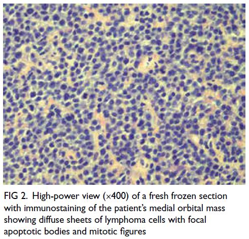
revealed pallor, a rash on her hands suggestive of dermatomyositis (Fig), puffy eyelids without heliotrope rash, and proximal muscle wasting and weakness. Fine end-inspiratory crepitations were heard at the base of the lungs. There was no cervical lymphadenopathy nor palpable abdominal masses. Review showed a serial drop in haemoglobin level (from 10.2 g/dL in February 2023 to 6.7 g/dL in April 2024), increasing microcytosis (decrease in mean corpuscular volume from 63.1 fL in February 2023 to 59.0 fL in April 2024), and iron deficiency in the setting of chronic inflammation. The clinical diagnosis was dermatomyositis with myopathy, interstitial lung disease, and probable colon cancer with iron deficiency anaemia superimposed on beta thalassaemia trait. She was referred to hospital for further management. Muscle enzyme tests revealed normal creatine kinase level (113 U/L) but elevated lactate dehydrogenase (499 U/L) and alanine aminotransferase levels (65 U/L). Myositis antibody screening confirmed the diagnosis of anti–melanoma differentiation–associated protein 5 (anti-MDA5) antibody–positive dermatomyositis.
 

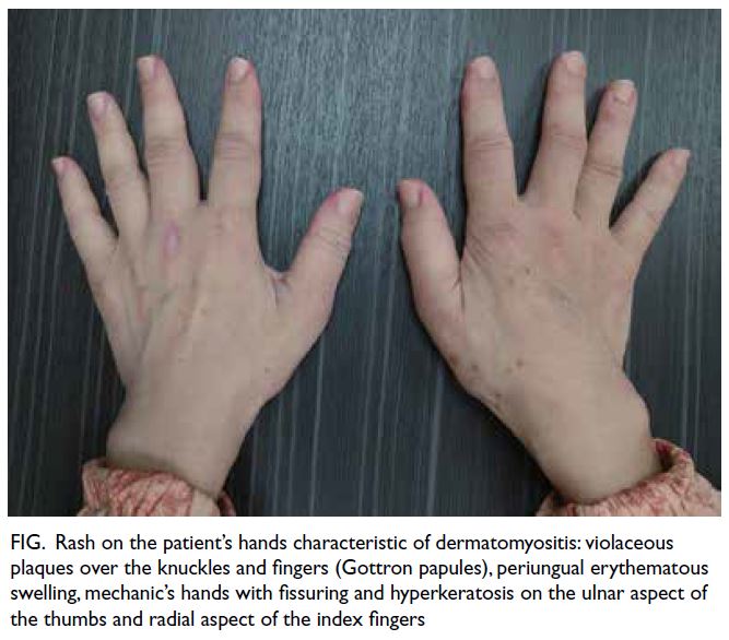
Figure. Rash on the patient’s hands characteristic of dermatomyositis: violaceous plaques over the knuckles and fingers (Gottron papules), periungual erythematous swelling, mechanic’s hands with fissuring and hyperkeratosis on the ulnar aspect of the thumbs and radial aspect of the index fingers
 
The patient’s illness onset in February 2023 following COVID-19 vaccination the month before suggested a trigger by vaccination, further aggravated by her COVID-19 infection in June 2023. Dermatomyositis, inflammatory myopathy, and rheumatic immune-mediated inflammatory diseases have been reported following COVID-19 vaccination and infection.1 2 3 In a systematic review up to May 2023,3 24 cases of post–COVID-19 vaccination dermatomyositis were reported worldwide, the majority following vaccination with Pfizer-BioNTech vaccine, and some following that with Moderna and Oxford–AstraZeneca vaccines. Only two such cases were reported among Chinese, one from mainland China (after Sinopharm’s inactivated Vero cell),4 one from Taiwan (after Oxford–AstraZeneca),5 but none from Hong Kong. The close temporal sequence and surge against a background of the reported case series suggest an association between severe acute respiratory syndrome coronavirus 2 infection/vaccination and the development of dermatomyositis, although proof of causality requires further research because of the limited number of cases reported.1 3 A recent bioinformatic study and transcriptome-derived insights point to a potential causal link between the surge in the Yorkshire region in the United Kingdom between 2020 and 2022 in anti-MDA5–positive dermatomyositis, autoimmune interstitial lung disease and COVID-19.6 The COVID-19 vaccination and infection may trigger a proinflammatory immune response involving type I interferon and stimulate production of dermatomyositis-specific autoantibodies such as MDA5 that are closely related to viral defence or viral RNA interaction supporting the concept of infection and vaccination-associated dermatomyositis.1 2 3 6
 
This case demonstrates that dermatomyositis can be induced by COVID-19 vaccination, ignorance of which and of the diagnostic clinical signs would lead to delayed diagnosis and management. Coronavirus disease 2019 vaccines are widely used. When patients present with constitutional symptoms with persistent muscle aches and weakness following COVID-19 vaccination, clinicians should consider dermatomyositis as a differential diagnosis and examine the skin for pathognomonic signs.
 
Author contributions
The author is solely responsible for the concept or design, acquisition of data, analysis or interpretation of data, drafting of the manuscript, and critical revision of the manuscript for important intellectual content. The author had full access to the data, contributed to the study, approved the final version for publication, and takes responsibility for its accuracy and integrity.
 
Conflicts of interest
The author has no conflicts of interest to disclose.
 
Funding/support
This study received no specific grant from any funding agency in the public, commercial, or not-for-profit sectors.
 
Ethics approval
The patient was treated in accordance with the Declaration of Helsinki. Patient consent was obtained for clinical photo of her hands and for publication of the article.
 
References
1. Holzer MT, Krusche M, Ruffer N, et al. New-onset dermatomyositis following SARS-CoV-2 infection and vaccination: a case-based review. Rheumatol Int 2022;42:2267-76. Crossref
2. Ding Y, Ge Y. Inflammatory myopathy following coronavirus disease 2019 vaccination: a systematic review. Front Public Health 2022;10:1007637. Crossref
3. Nune A, Durkowski V, Pillay SS, et al. New-onset rheumatic immune-mediated inflammatory diseases following SARS-CoV-2 vaccinations until May 2023: a systematic review. Vaccines (Basel) 2023;11:1571. Crossref
4. Yang L, Ye T, Liu H, Huang C, Tian W, Cai Y. A case of anti-MDA5–positive dermatomyositis after inactivated COVID-19 vaccine. J Eur Acad Dermatol Venereol 2023;37:e127-9. Crossref
5. Huang ST, Lee TJ, Chen KH, et al. Fatal myositis, rhabdomyolysis and compartment syndrome after ChAdOx1 nCoV-19 vaccination. J Microbiol Immunol Infect 2022;55:1131-3. Crossref
6. David P, Sinha S, Iqbal K, et al. MDA5-autoimmunity and interstitial pneumonitis contemporaneous with the COVID-19 pandemic (MIP-C). EBioMedicine 2024:104:105136. Crossref

Duplication of the portal vein and the implications for procedural planning

Hong Kong Med J 2025 Feb;31(1):72–3.e1–3 | Epub 6 Feb 2025
© Hong Kong Academy of Medicine. CC BY-NC-ND 4.0
 
PICTORIAL MEDICINE
Duplication of the portal vein and the implications for procedural planning
OL Chan, MB, BS, FRCR1; YS Lee, FRCR, FHKAM (Radiology)1; CH Ho, FRCR, FHKAM (Radiology)1; CC Lee, FRCS, FHKAM (Surgery)2; CC Cheung, FRCS, FHKAM (Surgery)2
1 Department of Radiology and Nuclear Medicine, Tuen Mun Hospital, Hong Kong SAR, China
2 Department of Surgery, Tuen Mun Hospital, Hong Kong SAR, China
 
Corresponding author: Dr OL Chan (col950@ha.org.hk)
 
 Full paper in PDF
 
 
A 72-year-old man with recurrent hepatitis B virus–related hepatocellular carcinoma was referred for right portal vein embolisation (PVE) prior to right hepatectomy. He had Child-Pugh class A cirrhosis, with calculated indocyanine green–R15 of 8%. Portal embolisation was indicated due to the presence of multiple co-morbidities and marginal future liver remnant volume of 35%.
 
Preprocedural computed tomography revealed duplication of the portal vein (DPV) [Fig 1]. The anatomy and feasibility of the procedure was discussed with hepatic surgeons. Right PVE was successfully performed with n-butyl cyanoacrylate glue. Left hepatic lobe hypertrophy from 430 cm3 to 560 cm3 was achieved. The patient subsequently underwent an uneventful right hepatectomy.
 

Figure 1. Axial image (a), coronal maximum intensity projection image (b) and three-dimensional reconstruction image (c) from contrast computed tomography of the abdomen showing duplication of the portal vein. The anatomical portal vein (arrowheads) arises from the confluence of the superior mesenteric vein and the splenic vein with a retroduodenal and retropancreatic course. The anomalous portal vein (arrows) arises from the superior mesenteric vein with a retroduodenal prepancreatic course. There is both intrahepatic and extrahepatic communication of the duplicated portal vein
 
Portal vein embolisation is a commonly adopted strategy to induce future liver remnant hypertrophy prior to hepatectomy. Knowledge of the portal venous anatomy and its variants is vital for treatment planning. Duplication of the portal vein is a rare congenital anomaly that has been described only in case reports. It is related to the spectrum of vitelline vein regression anomaly with pathogenesis believed to be failed regression of the left cranial part of the vitelline vein (Fig 2a-e).1 A variation of DPV has been reported; some authors describe two portal veins arising separately without extrahepatic communications,2 while some describe an additional portal vein arising anomalously from either the superior mesenteric vein or the splenic vein (Fig 2f-i).3 The latter was evident in our patient (Fig 1).
 

Figure 2. Schematic diagram illustrating the embryology of portal vein (PV) development and postulated pathogenesis of duplication of the portal vein (DPV), normal PV anatomy and various types of DPV described in the literature.1,3 (a) The paired vitelline veins (VVs), ie, the right vitelline vein (R VV) and the left vitelline vein (L VV), anastomose with each other around the primitive gut. (b, c) The caudal part of the R VV and the cranial part of the L VV degenerate to become the PV. (d) Duplication of the portal vein (the anatomical PV [PV1] and the anomalous PV [PV2]) due to failure of the cranial part of L VV to degenerate. (e) Duplication of the portal vein due to abnormal degeneration of the VV anastomoses. (f) The normal anatomy of a PV formed by the confluence of the superior mesenteric vein and splenic vein. (g) One variant of DPV, with PV1 arising from the superior mesenteric vein and PV2 arising from the splenic vein, without extrahepatic communication. (h) Another variant of DPV with an additional PV2 arising from the superior mesenteric vein. (i) Another variant of DPV with an additional PV2 arising from the splenic vein
 
Another anomaly with double channel portal vein is portal vein fenestration in which there is a small fenestration at the mid portion of the main portal vein.4 The exact pathogenesis and its relationship with portal vein duplication remains unknown.
 
In the presence of DPV, there was altered flow dynamic with preferential opacification of the right or left portal vein branches depending on different catheter tip positions (Fig 3). There was preferential flow towards the left portal branches at the intrahepatic communication at the hepatic hilum, giving a narrow safety margin for embolisation to prevent non-target embolisation of the left portal vein that could jeopardise the future liver remnant.
 

Figure 3. Right portal vein embolisation via the ipsilateral percutaneous approach. (a) An angiographic catheter was passed through the intrahepatic communication to the anatomical portal vein (arrowhead). Portography shows opacification of duplicated portal vein and left portal branches. (b) The catheter was passed through the extrahepatic communication to the anomalous portal vein (arrow). Portography shows preferential opacification of the right portal branches, the intrahepatic communication and some of the left portal branches. (c) Schematic diagram illustrating the catheter position during glue embolisation. The catheter is directed towards the right portal vein branches without bypassing the intrahepatic or extrahepatic communications of duplication of the portal vein (purple kinked line). (d) Post–portal vein embolisation. The radio-opaque branching pattern of glue cast at the right-side portal veins
 
Our patient successfully underwent PVE without complication. The degree of hypertrophy was similar to that reported in local cohorts.5 Surgeons discussed whether the anomalous portal vein could be embolised to improve the efficacy of PVE but there was also a risk of jeopardising venous return from small branches of the superior mesenteric vein that may worsen liver function.
 
During hepatectomy, DPV was confirmed (Fig 4). It did not affect surgical planning and the patient underwent right hepatectomy uneventfully.
 

Figure 4. Intra-operative photo at the hepatic hilum demonstrating duplication of the portal vein. The anatomical portal vein (PV1) is located posterior to the common bile duct (yellow), whereas the anomalous portal vein (PV2) is located anterior to the common bile duct
 
Duplication of the portal vein is a rare congenital anomaly. Because of the possible altered flow dynamics, it is important to identify this anomaly on preprocedural imaging and arrange multidisciplinary team discussion to plan PVE and ensure a safe and effective procedure.
 
Author contributions
Concept or design: All authors.
Acquisition of data: All authors.
Analysis or interpretation of data: OL Chan, YS Lee.
Drafting of the manuscript: OL Chan, YS Lee.
Critical revision of the manuscript for important intellectual content: All authors.
 
All authors had full access to the data, contributed to the study, approved the final version for publication, and take responsibility for its accuracy and integrity.
 
Conflicts of interest
All authors have disclosed no conflicts of interest.
 
Funding/support
This study received no specific grant from any funding agency in the public, commercial, or not-for-profit sectors.
 
Ethics approval
This study was approved by the Central Institutional Review Board of Hospital Authority, Hong Kong (Ref No.: CIRB-2023-064-1). Written informed consent was obtained from the patient for publication of this article.
 
References
1. Qin Y, Wen H, Liang M, et al. A new classification of congenital abnormalities of UPVS: sonographic appearances, screening strategy and clinical significance. Insights Imaging 2021;12:125. Crossref
2. Dighe M, Vaidya S. Case report. Duplication of the portal vein: a rare congenital anomaly. Br J Radiol 2009;82:e32-4. Crossref
3. Kitagawa S. Anomalous duplication of the portal vein with prepancreatic postduodenal portal vein. J Rural Med 2022;17:259-61. Crossref
4. Balradja I, Har B, Rastogi R, Agarwal S, Gupta S. Portal vein fenestration: a case report of an unusual portal vein developmental anomaly. Korean J Transplant 2022;36:298-301. Crossref
5. Yu KC, Wong SS, Wong YC, et al. Procedure time, efficacy, and safety of portal vein embolisation using a sheathless needle-only technique compared with traditional technique. Hong Kong J Radiol 2022;25:35-44. Crossref

Atypical imaging manifestations in non-alcoholic Wernicke’s encephalopathy: a potentially reversible neurological condition not to be missed

Hong Kong Med J 2024 Dec;30(6):509.e1-3 | Epub 18 Dec 2024
© Hong Kong Academy of Medicine. CC BY-NC-ND 4.0
 
PICTORIAL MEDICINE
Atypical imaging manifestations in non-alcoholic Wernicke’s encephalopathy: a potentially reversible neurological condition not to be missed
Cherry CY Chan, MB, ChB, FRCR (Radiology)1; Kevin KF Fung, FHKCR, FHKAM (Radiology)1,2; Elaine YL Kan, FHKCR, FHKAM (Radiology)2
1 Department of Diagnostic and Interventional Radiology, Kwong Wah Hospital, Hong Kong SAR, China
2 Department of Radiology, Hong Kong Children’s Hospital, Hong Kong SAR, China
 
Corresponding author: Dr Cherry CY Chan (chancherrycy@gmail.com)
 
 Full paper in PDF
 
 
An 18-year-old female with good past health was diagnosed with right tibial osteosarcoma in February 2019. She underwent wide excision of the right proximal tibia and distal femur with total knee replacement. Postoperatively, her adjuvant chemotherapy was complicated by multiple episodes of opportunistic infection, acute renal impairment due to drug toxicity and electrolyte disturbance. She was hospitalised for >6 months with suboptimal oral intake.
 
Over the course of a week, the patient had two episodes of seizure. Her general consciousness deteriorated acutely to a Glasgow Coma Scale score of 8/15 (E4V1M3). Physical examination revealed decorticate posture, generalised flaccidity and areflexia. Serum sodium level and urea were markedly elevated (154 mmol/L and 17.0 mmol/L, respectively), in keeping with hypernatraemic dehydration. Electroencephalogram showed diffuse slow-wave encephalopathy. Her Glasgow Coma Scale score did not improve following correction of hypernatraemia.
 
Magnetic resonance imaging of the brain revealed an abnormal high T2-weighted signal and restricted diffusion in bilateral frontal lobe cortices, dorsomedial thalami, periaqueductal grey, tectal plate of the midbrain and mammillary bodies (Fig 1a-d). Based on these findings, the patient was diagnosed with Wernicke’s encephalopathy and high-dose intravenous thiamine (vitamin B1) was initiated. Although her level of consciousness improved rapidly, there was poor recovery of limb power. Follow-up magnetic resonance imaging of the brain demonstrated cortical laminar necrosis and haemorrhage at bilateral frontal cortices (Figs 1e and 2). After 2 years of intensive rehabilitation, she regained most of her upper limb power, but lower limb power remained impaired.
 

Figure 1. Magnetic resonance imaging (MRI) of the brain demonstrated atypical imaging features in addition to the classic findings in Wernicke’s encephalopathy. Axial T2-weighted MRI showing abnormal symmetrical T2-weighted signals in bilateral dorsomedial thalami (a) and periaqueductal grey, tectal plate of the midbrain and mammillary bodies (b), which are typically seen in Wernicke’s encephalopathy (black arrows). (c) Axial fluid-attenuated inversion recovery MRI showing abnormal high signal involving cortices of bilateral frontal lobes. (d) Diffusion-weighted imaging (b value=1000 s/mm2) showing restricted diffusion in bilateral frontal lobe cortices. (e) Susceptibility-weighted imaging showed blooming artifacts in bilateral frontal lobe cortices, indicating microhaemorrhage (white arrows). The involvement of frontal lobe cortices with haemorrhage is an atypical imaging feature and more commonly seen in non-alcoholic Wernicke’s encephalopathy
 

Figure 2. (a, b) Susceptibility-weighted sequences in follow-up magnetic resonance imaging of the brain revealed curvilinear blooming artifacts at cortices of bilateral frontal lobes, indicating development of cortical laminar necrosis (white arrows). This is associated with poor neurological prognosis due to irreversible damage
 
Wernicke’s encephalopathy is an acute neurological syndrome caused by depletion of intracellular thiamine in neurons that is essential for production of neurotransmitters. The bodily reserve of thiamine in a healthy individual is exhausted within 4 to 6 weeks in the absence of dietary thiamine.1 Wernicke’s encephalopathy is most commonly associated with chronic alcoholism but can result from any condition that causes malnutrition or malabsorption.1 The classic clinical triad consists of confusion, ataxia and ophthalmoplegia, although only a small proportion of patients exhibits all three.2 Left untreated, Wernicke’s encephalopathy carries significant neurological morbidity and death. The condition is potentially reversible if recognised and treated early with intravenous thiamine replacement.
 
Classic imaging features of alcohol-associated Wernicke’s encephalopathy include abnormal signal involving deep periventricular and periaqueductal grey matter in basal ganglia and brainstem, most notably in the mamillary bodies.3 Atypical findings are more frequently seen in non-alcoholic Wernicke’s encephalopathy. These include abnormal signal in other locations such as the cerebral cortex, splenium, caudate nuclei, red nuclei, cranial nerve nuclei, cerebellum and vermis.4 Further progression to cortical laminar necrosis and haemorrhage, as seen in our case, is rare and associated with a poor prognosis due to irreversible neurological damage.5
 
In patients with a poor nutritional state who present with reduced consciousness, a high index of clinical suspicion and prompt imaging are important to establish the diagnosis of Wernicke’s encephalopathy. Atypical imaging manifestations are more commonly seen in non-alcoholic Wernicke’s encephalopathy. Timely diagnosis is crucial since the neurological impairment is potentially reversible with intravenous thiamine replacement therapy.
 
Author contributions
Concept or design: CCY Chan, KKF Fung.
Acquisition of data: CCY Chan, KKF Fung.
Analysis or interpretation of data: All authors.
Drafting of the manuscript: CCY Chan, KKF Fung.
Critical revision of the manuscript for important intellectual content: All authors.
 
All authors had full access to the data, contributed to the study, approved the final version for publication, and take responsibility for its accuracy and integrity.
 
Conflicts of interest
All authors have disclosed no conflicts of interest.
 
Funding/support
This study received no specific grant from any funding agency in the public, commercial, or not-for-profit sectors.
 
Ethics approval
The patient was treated in accordance with the Declaration of Helsinki. Informed consent was obtained from the patient for all treatments and procedures, and consent for publication.
 
References
1. Chandrakumar A, Bhardwaj A, ’t Jong GW. Review of thiamine deficiency disorders: Wernicke encephalopathy and Korsakoff psychosis. J Basic Clin Physiol Pharmacol 2018;30:153-62. Crossref
2. Harpe CG, Giles M, Finlay-Jones R. Clinical signs in the Wernicke–Korsakoff complex: a retrospective analysis of 131 cases diagnosed at necropsy. J Neurol Neurosurg Psychiatry 1986;49:341-5. Crossref
3. Zuccoli. G, Pipitone N. Neuroimaging findings in acute Wernicke’s encephalopathy: review of the literature. AJR Am J Roentgenol 2009;192:501-8. Crossref
4. Bae SJ, Lee HK, Lee JH, Choi CG, Suh DC. Wernicke’s encephalopathy: atypical manifestation at MR imaging. AJNR Am J Neuroradiol 2001;22:1480-2.
5. Pereira DB, Pereira ML, Gasparetto EL. Nonalcoholic Wernicke encephalopathy with extensive cortical involvement: cortical laminar necrosis and hemorrhage demonstrated with susceptibility-weighted MR phase images. AJNR Am J Neuroradiol 2011;32:E37-8. Crossref

Marchiafava–Bignami disease

Hong Kong Med J 2024 Oct;30(5):417.e1-2 | Epub 15 Oct 2024
© Hong Kong Academy of Medicine. CC BY-NC-ND 4.0
 
PICTORIAL MEDICINE
Marchiafava–Bignami disease
F Ren, MD1; Q Wang, MD2
1 Department of Radiology, Hospital of Chengdu University of Traditional Chinese Medicine, Chengdu, China
2 Department of Ultrasound, Hospital of Chengdu University of Traditional Chinese Medicine, Chengdu, China
 
Corresponding author: Dr Q Wang (444028177@qq.com)
 
 Full paper in PDF
 
 
A 67-year-old female was admitted to the neurology department in October 2020 with abnormal behaviour and cognitive impairment. Her memory and numeracy had declined, and symptoms progressed over the preceding week. She had a 30-year history of chronic alcohol abuse with an average daily intake of 500-mL liquor.
 
Routine biochemistry including electrolytes, liver function, and vitamin B12 were within normal limits. Magnetic resonance imaging of the brain showed areas of hyperintensity of the corpus callosum (splenium, body, and genu), bilateral middle cerebellar peduncles, periventricular white matter, and subcortical white matter of the frontal lobe on T2-weighted and fluid-attenuated inversion recovery images. Diffusion-weighted imaging revealed prominent high-intensity signal lesions involving the splenium, and these corresponding lesions were hypointense on the apparent diffusion coefficient map (Fig). Based on her history, physical examination and magnetic resonance imaging features, the patient was diagnosed with Marchiafava–Bignami disease (MBD). Despite a normal level of serum vitamins, the patient was prescribed vitamin B and neurotrophic treatment. Symptoms improved and she made a good recovery over the next 30 days.
 

Figure. Magnetic resonance imaging of the brain showing (a) the entire corpus callosum with hyperintensity on T2-weighted imaging, (b) the entire corpus callosum with hyperintensity on fluid-attenuated inversion recovery imaging, (c) bilateral middle cerebellar peduncles with hyperintensity on fluid-attenuated inversion recovery imaging, and (d) the entire corpus callosum with hypointensity on T1-weighted imaging. The splenium was strongly hyperintense on diffusion-weighted imaging (e) and hypointense on apparent diffusion coefficient map (f). The genu was moderately hyperintense on diffusion-weighted imaging (e) and hypointense on apparent diffusion coefficient map (f)
 
Marchiafava–Bignami disease is a rare neurological syndrome characterised by primary degeneration and necrosis of the corpus callosum associated with chronic alcoholism and malnutrition. The clinical manifestations of MBD are severe and nonspecific and include an altered mental state, impaired walking, deficient memory, and dysarthria. Symptoms and imaging findings may improve following thiamine treatment.1 2 The role of magnetic resonance imaging is essential to confirm the diagnosis. Chronic alcohol abuse plays an important role in its development although MBD has been occasionally diagnosed in patients with no history of alcohol abuse, in particular individuals with poorly controlled diabetes mellitus or following surgery.3 4 The aetiology and pathophysiology of MBD remain unclear. Possible mechanisms include cytotoxic oedema, blood-brain barrier breakdown, demyelination, and necrosis. Early pathological manifestations are mainly intramyelinic or cytotoxic oedema. In the later stages, demyelination and necrosis of the corpus callosum (especially in the genu and the body) may follow5 with necrosis leading to atrophy and cavitation in chronic stages, and a decreased number of oligodendrocytes. Occasionally, similar lesions can involve extracallosal regions, such as the anterior and posterior commissures, subcortical white matter, middle cerebellar peduncle, optic chiasm, putamen, internal capsules, hippocampus, and frontal cortex.2
 
The recent advances in neuroradiology techniques help understand the pathophysiological processes of MBD. Studies with diffusion-weighted imaging have shown a low apparent diffusion coefficient, which has been interpreted as irreversible cytotoxic oedema, and may precede the development of demyelination and necrosis and predict a poor or partial recovery.2 5 Nonetheless the high apparent diffusion coefficient showing reversible signal changes favoured an underlying vasogenic oedema-related process. In magnetic resonance spectroscopy studies, an increased choline/creatine ratio indicates demyelination during the acute phase of MBD, while a reduced N-acetylaspartate/creatine ratio suggests secondary axonal injury. In addition, decreased cerebral blood flow and cerebral blood volume in magnetic resonance perfusion suggest hypoperfusion. Recognition of the neuroradiological features is crucial to establish a diagnosis.
 
Author contributions
Concept or design: Both authors.
Acquisition of data: Q Wang.
Analysis or interpretation of data: F Ren.
Drafting of the manuscript: F Ren.
Critical revision of the manuscript for important intellectual content: Q Wang.
 
Both authors had full access to the data, contributed to the study, approved the final version for publication, and take responsibility for its accuracy and integrity.
 
Conflicts of interest
Both authors have disclosed no conflicts of interest.
 
Funding/support
This study received no specific grant from any funding agency in the public, commercial, or not-for-profit sectors.
 
Ethics approval
This study was approved by the Hospital of Chengdu University of Traditional Chinese Medicine Research Ethics Committee, China. Informed consent for all treatments and procedures, and consent for publication were obtained from the patient.
 
References
1. Tsai CY, Huang PK, Huang P. Simultaneous acute Marchiafava–Bignami disease and central pontine myelinolysis: a case report of a challenging diagnosis. Medicine (Baltimore) 2018;97:e9878. Crossref
2. Hillbom M, Saloheimo P, Fujioka S, Wszolek ZK, Juvela S, Leone MA. Diagnosis and management of Marchiafava–Bignami disease: a review of CT/MRI confirmed cases. J Neurol Neurosurg Psychiatry 2014;85:168-73. Crossref
3. Pérez Álvarez AI, Ramón Carbajo C, Morís de la Tassa G, Pascual Gómez J. Marchiafava–Bignami disease triggered by poorly controlled diabetes mellitus [in English, Spanish]. Neurologia 2016;31:498-500. Crossref
4. Bachar M, Fatakhov E, Banerjee C, Todnem N. Rapidly resolving nonalcoholic Marchiafava–Bignami disease in the setting of malnourishment after gastric bypass surgery. J Investig Med High Impact Case Rep 2018;6:2324709618784318. Crossref
5. Ménégon P, Sibon I, Pachai C, Orgogozo JM, Dousset V. Marchiafava–Bignami disease: diffusion-weighted MRI in corpus callosum and cortical lesions. Neurology 2005;65:475-7. Crossref

Pseudo fat-saturation and orbital lipolysis in cancer cachexia: a diagnostic trap

Hong Kong Med J 2024 Aug;30(4):331.e1-3 | Epub 11 Jul 2024
© Hong Kong Academy of Medicine. CC BY-NC-ND 4.0
 
PICTORIAL MEDICINE
Pseudo fat-saturation and orbital lipolysis in cancer cachexia: a diagnostic trap
SM Yu, MB, BS, FRCR; William KM Kwong, BAS, MHlthSc (MRS); Yan YY Law, BSc, MSc; Ann D King, MD, FRCR
Department of Imaging and Interventional Radiology, Prince of Wales Hospital, Hong Kong SAR, China
 
Corresponding author: Dr SM Yu (fayeyupwr@gmail.com)
 
 Full paper in PDF
 
 
A 59-year-old woman was diagnosed in June 2022 with locally advanced nasopharyngeal carcinoma. She declined standard chemoradiotherapy and opted to pursue traditional Chinese Medicine. In May 2023, she presented with bilateral sixth nerve palsy, poor oral intake, and progressive weight loss from 32 kg to 20 kg over 6 months.
 
The restaging positron emission tomography–computed tomography showed that she was extremely emaciated and had locoregionally advanced nasopharyngeal carcinoma without distant metastases (Fig 1). Magnetic resonance imaging (MRI) of the head and neck revealed diffuse loss of T1 hyperintense signal in the fat of the subcutaneous and deep soft tissues and in the bone marrow of the cervical spine and skull vault giving the images a pseudo fat-saturated appearance (Fig 2). The scanning parameters were verified to ensure the correct repetition time and echo time (568 ms and 7 ms, respectively) had been selected. Compared with the earlier MRI performed in November 2022, there was complete loss of normal T1-weighted hyperintense signals in the retrobulbar fat with development of diffuse oedema and enhancement in the post-septal orbits (Fig 3); similar changes with bilateral enophthalmos and diffuse symmetric enhancement of post-septal orbits were seen on computed tomography (Fig 4). Overall, this picture was that of pseudo fat-saturation and orbital lipolysis in a patient with cancer cachexia. Following assessment, the patient agreed to undergo palliative radiotherapy.
 

Figure 1. Maximum intensity projection coronal image of positron emission tomography of the patient. It shows a very emaciated condition with a huge nasopharyngeal carcinoma (black arrow) and bilateral bulky cervical node metastases (open arrows). There was no evidence of distant metastatic deposits
 

Figure 2. Magnetic resonance imaging (MRI) of the head and neck of the patient. (a) Axial T1-weighted MRI showing complete loss of fat signal in the cheeks (open arrows) and infratemporal fossae (curve arrows). (b) Sagittal T1-weighted MRI showing a locally advanced nasopharyngeal carcinoma with full-length clival invasion and dural invasion at the retroclival region (solid arrows), diffuse loss of normal fat signals within the bone marrow of the skull vault (arrowheads) and cervical spine (open arrows), compatible with pseudo fat-saturated appearance
 

Figure 3. Axial magnetic resonance imaging (MRI) showing the development of orbital lipolysis. (a) T1-weighted MRI (gradient echo) in November 2022 showing expected T1-weighted hyperintensity in bilateral retrobulbar fat. (b-d) Subsequent MRI in June 2023. (b) T1-weighted MRI (spin echo) showing complete loss of fat signal in bilateral retrobulbar fat. The imaging appearance mimics a fat-saturated T1-weighted image. (c) T2-weighted MRI with fat saturation showing oedema in bilateral retrobulbar fat. (d) T1-weighted subtraction post-gadolinium MRI showing diffuse, symmetrical contrast enhancement in the retrobulbar fat
 

Figure 4. (a) Plain and (b) contrast-enhanced computed tomography of the head showing marked volume loss of retrobulbar fat content resulting in bilateral enophthalmos (arrows in [a]) and diffuse symmetric enhancement of retrobulbar fat (arrows in [b])
 
Long-term cachexia, a wasting syndrome common in cancer patients, is marked by extreme weight loss and malnutrition and can lead to severe metabolic disturbances that cause excessive lipolysis and lipid peroxidation. Characteristic imaging features are often found in severe cases.1 2 3 4 Pseudo fat-saturated appearance is seen on T1-weighted images due to complete loss of subcutaneous adipose tissue, similar to the fat-saturated T1-weighted image.1 Diffuse loss of normal T1-weighted hyperintense bone marrow signal was a result of bone marrow fat atrophy and deposition of extracellular gelatinous substance, a process known as ‘gelatinous transformation of bone marrow’.2 This loss of fat signal gives the images an appearance similar to that of a fat-saturated T1-weighted image. Orbital fat is typically preserved until the late stages of severe cachexia during which a condition called orbital lipolysis may develop.3 4 This condition is related to endothelial injury and increased permeability of vessel walls resulting in diffuse oedema and contrast enhancement in the post-septal orbits.
 
Cachexia is common in patients with longstanding cancer and malnutrition. Doctors should recognise this phenomenon to prevent attributing these imaging findings to incorrect scanning parameters or alternative diagnoses. The diffuse hypointense T1-weighted bone marrow signal might be misdiagnosed as widespread metastatic disease or other bone marrow–infiltrating diseases such as myelofibrosis or haematological malignancies, while diffuse orbital oedema and enhancement may be misdiagnosed as orbital inflammatory conditions such as idiopathic orbital inflammation.
 
Understanding the characteristic imaging features of long-term cachexia is crucial for doctors to avoid diagnostic pitfalls and unnecessary additional investigations or invasive procedures.
 
Author contributions
All authors contributed to the concept or design, acquisition of data, analysis or interpretation of data, drafting of the manuscript, and critical revision of the manuscript for important intellectual content. All authors had full access to the data, contributed to the study, approved the final version for publication, and take responsibility for its accuracy and integrity.
 
Conflicts of interest
All authors have disclosed no conflicts of interest.
 
Funding/support
This study received no specific grant from any funding agency in the public, commercial, or not-for-profit sectors.
 
Ethics approval
This study was conducted in accordance with the Declaration of Helsinki. The patient provided written informed verbal consent for the publication of this case report.
 
References
1. Jegatheeswaran V, Chan M, Kucharczyk W, Chen YA. Pseudo fat-saturated appearance of magnetic resonance head and neck images in 2 cachectic patients. Radiol Case Rep 2020;15:2693-7. Crossref
2. Böhm J. Gelatinous transformation of the bone marrow: the spectrum of underlying diseases. Am J Surg Pathol 2000;24:56-65. Crossref
3. Li J, Rajput A, Kosoy D, et al. Rapid orbital lipolysis associated with critical illness and colectomy. Radiol Case Rep 2021;16:2347-50. Crossref
4. Demaerel P, Dekimpe P, Muls E, Wilms G. MRI demonstration of orbital lipolysis in anorexia nervosa. Eur Radiol 2002;12 Suppl 3:S4-6. Crossref

Two-in-one: concomitant diffuse large B-cell lymphoma and cavernous haemangioma within the same orbit

Hong Kong Med J 2024 Jun;30(3):249 | Epub 4 Jun 2024
© Hong Kong Academy of Medicine. CC BY-NC-ND 4.0
 
PICTORIAL MEDICINE
Two-in-one: concomitant diffuse large B-cell lymphoma and cavernous haemangioma within the same orbit
Kenneth KH Lai, MB, ChB, AFCOphth1; Tiffany Ong, MB, ChB1; Tiffany HT Chan, MB, ChB2; Edwin Chan, FCOphth1; Andrew KT Kuk, FCOphth1
1 Department of Ophthalmology, Tung Wah Eastern Hospital, Hong Kong SAR, China
2 Department of Anatomical and Cellular Pathology, Pamela Youde Nethersole Eastern Hospital, Hong Kong SAR, China
 
Corresponding author: Dr Andrew KT Kuk (aktkuk@hku.hk)
 
 Full paper in PDF
 
 
Orbital tumours encompass a wide range of benign and malignant space occupying lesions that may arise primarily from the orbit or have spread from other sites in the body. They are rare with an incidence of 1 in every 100 000 and may lead to devastating complications of which mechanical compression causing optic neuropathy is the most important.1 Multiple orbital tumours of the same orbit are even rarer with most reported cases being benign homologous tumours such as cavernous haemangiomas or myxofibrosarcomas.2 3 A 58-year-old ethnic Han Chinese male presented in December 2021 with a 1-month history of right eye proptosis. He had no clinical sign of optic neuropathy. Computed tomography of the orbit revealed right axial proptosis and two separate lesions in the right orbit. One lesion appeared infiltrative and measured 2.2 × 1.5 × 1.7 cm3 (anteroposterior × transverse × longitudinal) at the extraconal space with retrobulbar extension between the lamina papyracea and the medial rectus. The other was an encapsulated mass with regular border located in the superolateral intraconal region measuring 1.9 × 1.7 × 2.0 cm3 and abutting the optic nerve. Both lesions enhanced mildly with intravenous contrast (Fig 1). Blood results revealed normal thyroid function and immunoglobulin G4 and white blood cell levels. Based on the distinguishing radiological features of each lesion, we performed a two-stage surgery for theranostic reasons. An incisional biopsy of the medial infiltrative lesion was performed first through an anterior orbitotomy via an upper lid skin crease approach. Frozen section of the medial yellow jelly-like mass revealed atypical lymphoid cells with enlarged vesicular nuclei and amphophilic cytoplasm, highly suspicious of lymphoproliferative malignancy. We then performed a complete excision of the vascular encapsulated intraconal lesion using cryotherapy via a lateral orbitotomy. Formal histopathology reports revealed the first medial infiltrative lesion to be consistent with diffuse large B-cell lymphoma with positive immunostaining for CD20, BCL2, BCL6, MUM1, and CMYC1 (Fig 2). The second intraconal lesion was consistent with cavernous haemangioma (Fig 3).
 

Figure 1. Computed tomography scan with intravenous contrast of the patient showing a cavernous haemangioma (red arrow) and diffuse large B-cell lymphoma (blue arrow)
 

Figure 2. High-power view (×400) of a fresh frozen section with immunostaining of the patient’s medial orbital mass showing diffuse sheets of lymphoma cells with focal apoptotic bodies and mitotic figures
 

Figure 3. High-power view (×400) of a section from the patient’s lateral orbit mass, stained with haemotoxylin and eosin showing dilated congested venous type–looking vessels lined by bland-looking endothelial cells, consistent with cavernous haemangioma
 
At 4 months post-surgery there was no clinical sign of optic nerve damage and best-corrected visual acuity was 0.8 in the right eye. Positron emission tomography–computed tomography showed a residual hypermetabolic lesion over the medial aspect of the right orbit with no extra orbital lesion. The patient is receiving chemotherapy under the care of our haematology team.
 
Benign multiple homogenous lesions in the same orbit have been reported. Although multiple solitary fibrous tumours of the same orbit without malignant degeneration have been reported,4 multiple heterogeneous tumours in the same orbit are extremely rare. Ma et al5 reported a case of concurrent schwannoma and cavernous haemangioma in the same orbit of a 54-year-old female. To the best of our knowledge, concurrent benign and malignant lesions of the same orbit have not been reported in the English literature.
 
Author contributions
All authors contributed to the concept and design of this study, acquisition of data, analysis of data, drafting the manuscript, and critical revision for important intellectual content. All authors had full access to the data, contributed to the study, approved the final version for publication, and take responsibility for its accuracy and integrity.
 
Conflicts of interest
The authors have no conflicts of interest to disclose.
 
Funding/support
This study received no specific grant from any funding agency in the public, commercial, or not-for-profit sectors.
 
Ethics approval
The patient was treated in accordance with the Declaration of Helsinki and provided informed consent for the treatment and consent for publication.
 
References
1. Demirci H, Shields CL, Shields JA, Honavar SG, Mercado GJ, Tovilla JC. Orbital tumors in the older adult population. Ophthalmology 2002;109:243-8. Crossref
2. Deng C, Hu W. Multiple cavernous hemangiomas in the orbit: a case report and review of the literature. Medicine (Baltimore) 2020;99:e20670. Crossref
3. Du B, He X, Wang Y, He W. Multiple recurrent myxofibrosarcoma of the orbit: case report and review of the literature. BMC Ophthalmol 2020;20:264. Crossref
4. Griepentrog GJ, Harris GJ, Zambrano EV. Multiply recurrent solitary fibrous tumor of the orbit without malignant degeneration: a 45-year clinicopathologic case study. JAMA Ophthalmol 2013;131:265-7. Crossref
5. Ma M, Su F, Yang X. Multiple heterogeneous tumors in orbit: a case report. Int J Clin Exp Pathol 2019;12:4137-41.

Doxycycline-induced gastric injury

Hong Kong Med J 2024 Jun;30(3):248 | Epub 15 Apr 2024
© Hong Kong Academy of Medicine. CC BY-NC-ND 4.0
 
PICTORIAL MEDICINE
Doxycycline-induced gastric injury
Henry HW Liu, FHKCP, FHKAM (Medicine)1; TW Ho, MB, BS2; Nelson SK Tsang, MB, BS1; Jodis TW Lam, FRCP (Edin), FHKAM (Medicine)1; YT Hui, FRCP (Edin), FHKAM (Medicine)1
1 Department of Medicine, Queen Elizabeth Hospital, Hong Kong SAR, China
2 Department of Pathology, Queen Elizabeth Hospital, Hong Kong SAR, China
 
Corresponding author: Dr Henry HW Liu (lhw738@ha.org.hk)
 
 Full paper in PDF
 
 
A 28-year-old man with good past health presented with fever, dyspnoea, and pleuritic chest pain in April 2022. Blood tests showed hypochromic microcytic anaemia with haemoglobin level of 7 g/dL but no bleeding symptoms. Chest X-ray revealed left-sided pleural effusion. The working diagnosis was chest infection with parapneumonic effusion. He was treated with empirical co-amoxiclav and doxycycline.
 
Thoracocentesis yielded blood-stained fluid with an exudative biochemistry. Pleural fluid adenosine deaminase level was 58 U/L and cytology was negative. Findings were suspicious of tuberculous pleural effusion. Gastroscopy was arranged for his anaemia and revealed severe gastritis over the body and fundus with overlying yellowish exudates (Fig 1a). Biopsies excluded Helicobacter pylori infection but revealed superficial mucosal necrosis with acute neutrophilic infiltrates (Fig 2). The superficial capillaries displayed eosinophilic degeneration (Fig 3) with microthrombi formation (Fig 4). These histological findings are characteristic of doxycycline-induced gastric mucosal injury.1 2 3
 

Figure 1. (a) Severe fundal and body gastritis with overlying yellowish exudates. (b) Complete resolution of gastritis on follow-up oesophagogastroduodenoscopy
 

Figure 2. Superficial mucosal necrosis with acute neutrophilic infiltrates (haematoxylin and eosin staining, ×20)
 

Figure 3. Eosinophilic degeneration of capillaries with fibrinoid material (arrowheads) [haematoxylin and eosin staining, ×40]
 

Figure 4. Capillary with microthrombi formation (haematoxylin and eosin staining, ×60)
 
Doxycycline was withdrawn and the patient was started on antituberculosis treatment. Pleural fluid culture later confirmed infection with Mycobacterium tuberculosis. Follow-up upper endoscopy at 3 months showed complete resolution of gastric mucosal injury (Fig 1b). Haemoglobin analysis revealed findings compatible with Haemoglobin H disease.
 
Doxycycline is a tetracycline-class antibiotic commonly prescribed to cover atypical organisms in community-acquired pneumonia. It is a well-reported cause of oesophagitis and even ulcerations.4 On the contrary, doxycycline-induced gastric mucosal injury is rare and has a distinctive histological pattern. The underlying mechanism of injury is poorly understood. Duration of doxycycline treatment ranges from 5 days to 3 years.1 2 3 These patients often present with dysphagia, chest and epigastric pain or anaemia. Symptoms typically resolve following drug withdrawal and healing of the mucosa.1 2 3
 
Awareness of an association with doxycycline use, coupled with endoscopic and unique histological findings, facilitate prompt diagnosis of this condition.
 
Author contributions
All authors contributed to the concept or design of the study, acquisition of the data, analysis or interpretation of the data, drafting of the manuscript, and critical revision of the manuscript for important intellectual content. All authors had full access to the data, contributed to the study, approved the final version for publication, and take responsibility for its accuracy and integrity.
 
Conflicts of interest
All authors have disclosed no conflicts of interest.
 
Funding/support
This study received no specific grant from any funding agency in the public, commercial, or not-for-profit sectors.
 
Ethics approval
The patient was treated in accordance with the Declaration of Helsinki and provided informed consent for all procedures.
 
References
1. Affolter K, Samowitz W, Boynton K, Kelly ED. Doxycycline-induced gastrointestinal injury. Hum Pathol 2017;66:212-5. Crossref
2. Shih AR, Lauwers GY, Mattia A, Schaefer EA, Misdraji J. Vascular injury characterizes doxycycline-induced upper gastrointestinal tract mucosal injury. Am J Surg Pathol 2017;41:374-81. Crossref
3. Xiao SY, Zhao L, Hart J, Semrad CE. Gastric mucosal necrosis with vascular degeneration induced by doxycycline. Am J Surg Pathol 2013;37:259-63. Crossref
4. Gencosmanoglu R, Kurtkaya-Yapicier O, Tiftikci A, Avsar E, Tozun N, Oran ES. Mid-esophageal ulceration and candidiasis-associated distal esophagitis as two distinct clinical patterns of tetracycline or doxycycline-induced esophageal injury. J Clin Gastroenterol 2004;38:484-9. Crossref

Non-ketotic hyperglycaemic hemichorea: a rare complication of uncontrolled diabetes mellitus

Hong Kong Med J 2023 Dec;29(6):556 | Epub 21 Nov 2023
© Hong Kong Academy of Medicine. CC BY-NC-ND 4.0
 
PICTORIAL MEDICINE
Non-ketotic hyperglycaemic hemichorea: a rare complication of uncontrolled diabetes mellitus
PL Lam, MB, BS; PP Iu, FRCR, FHKAM (Radiology); Danny HY Cho, FRCR, FHKAM (Radiology)
Department of Diagnostic and Interventional Radiology, Kwong Wah Hospital, Hong Kong SAR, China
 
Corresponding author: Dr PL Lam (lpl404@ha.org.hk)
 
 Full paper in PDF
 
 
Case 1
A 65-year-old man with a 1-month history of left upper and lower limb chorea was admitted to the medical ward of our institution in February 2021. Blood tests revealed new-onset diabetes mellitus with markedly elevated fasting glucose level of 28.5 mmol/L. Urinalysis for ketones was negative. Urgent non-contrast computed tomography (CT) of the brain showed subtle hyperdensity at the right putamen without mass effect or surrounding oedema (Fig 1). The patient was started on subcutaneous insulin, and upon normalisation of blood glucose level, his hemichorea subsided without additional antichorea medications. Follow-up non-contrast magnetic resonance imaging of the brain performed 2 months later revealed T1-weighted hyperintensity in the right putamen, without restricted diffusion (Fig 2). Imaging findings were in keeping with non-ketotic hyperglycaemic hemichorea (NHH). The patient was prescribed a biphasic insulin regimen upon discharge.
 

Figure 1. Case 1. Non-contrast computed tomography of the brain shows subtle hyperdensity in right putamen (arrow) without mass effect or surrounding oedema, in keeping with non-ketotic hyperglycaemic hemichorea
 

Figure 2. Case 1. Non-contrast magnetic resonance imaging of the brain with non-ketotic hyperglycaemic hemichorea. (a) T1-weighted sequence showing hyperintense signal in right putamen (white arrow). (b) Susceptibility weighted imaging showing rim of susceptibility signal in right putamen (black arrow). (c) T2-weighted sequence and (d) T2-weighted fluid-attenuated inversion recovery sequence showing symmetrical signals in bilateral basal ganglia. (e) Diffusion-weighted imaging and (f) apparent diffusion coefficient map showing no restricted diffusion
 
Case 2
An 87-year-old man with a 2-day history of left upper limb chorea was hospitalised in April 2022. He had known diabetes mellitus but was noncompliant with oral hypoglycaemic therapy. Blood tests revealed markedly elevated random glucose level of 30.8 mmol/L. Urgent non-contrast CT of the brain showed asymmetric subtle hyperdensity in bilateral putamen and caudate nuclei, more extensive on the right, but without mass effect or surrounding oedema (Fig 3). Findings were compatible with NHH. The patient resumed metformin, with gliclazide and subcutaneous insulin injection added for optimal control. Upon normalisation of blood glucose level, his hemichorea resolved without antichorea medications.
 

Figure 3. Case 2. Non-contrast computed tomography of the brain showing asymmetric subtle hyperdensity in bilateral putamen (a) [arrows] and bilateral caudate nuclei (b) [arrowheads], more extensive on the right, without mass effect or surrounding oedema, in keeping with non-ketotic hyperglycaemic hemichorea
 
Discussion
Unilateral or asymmetric basal ganglia hyperdensity in brain CT of patients with focal neurological symptoms can be alarming, and intracerebral haemorrhage may be suspected. Nonetheless NHH, a rare complication of uncontrolled diabetes, should not be overlooked.
 
Case reports of NHH have been documented as early as 1960.1 Previously reported cases were frequent in Asian elderly women with uncontrolled diabetes. In most patients, unilateral chorea was observed, although bilateral involvement could be present.2 3 4 Of note, the aetiology of hemichorea is diverse, and other causes include infarct, haemorrhage and neoplasm. Imaging of the brain is therefore crucial.
 
Non-contrast CT of the brain of NHH typically shows subtle hyperdensity in contralateral putamen and/or caudate nucleus, although bilateral involvement is also seen. There will be no mass effect or perilesional oedema, and this differentiates NHH from haemorrhage or tumour.2 3 4
 
Similarly, magnetic resonance imaging of the brain typically shows corresponding signal changes in striatal regions contralateral to the symptomatic side. There will be T1-weighted hyperintense signal. Differentials of increased basal ganglia T1-weighted signal are diverse. They include toxin-related causes such as methanol poisoning or hepatic-related causes such as acquired hepatocerebral degeneration, but they commonly show bilateral and symmetrical involvement.5 T2-weighted or fluid-attenuated inversion recovery signals can be variable. Of note, restricted diffusion is not expected in NHH,2 3 4 and this differentiates it from acute ischaemic stroke.
 
The pathophysiology of NHH is not fully understood. Proposed mechanisms include depleted gamma-aminobutyric acid and disrupted blood-brain barrier at the corpus striatum.2 3 4 Recognising this rare complication of uncontrolled diabetes mellitus enables prompt medical intervention. Neurological symptoms of NHH usually show substantial improvement after normalisation of blood glucose level without additional intervention.2 3 4
 
Author contributions
All authors contributed to the concept and design of the study, acquisition of data, analysis and interpretation of data, drafting of the manuscript, and critical revision of the manuscript for important intellectual content. All authors had full access to the data, contributed to the study, approved the final version for publication, and take responsibility for its accuracy and integrity.
 
Conflicts of interest
All authors have disclosed no conflicts of interest.
 
Funding/support
This study received no specific grant from any funding agency in the public, commercial, or not-for-profit sectors.
 
Ethics approval
The patients were treated in accordance with the Declaration of Helsinki. They provided informed consent for all treatments and procedures, and consent for publication.
 
References
1. Bedwell SF. Some observations on hemiballismus. Neurology 1960;10:619-22. Crossref
2. Zheng W, Chen L, Chen JH, et al. Hemichorea associated with non-ketotic hyperglycemia: a case report and literature review. Front Neurol 2020;11:96. Crossref
3. Narayanan S. Hyperglycemia-induced hemiballismus hemichorea: a case report and brief review of the literature. J Emerg Med 2012;43:442-4. Crossref
4. Cherian A, Thomas B, Baheti NN, Chemmanam T, Kesavadas C. Concepts and controversies in nonketotic hyperglycemia-induced hemichorea: further evidence from susceptibility-weighted MR imaging. J Magn Reson Imaging 2009;29:699-703. Crossref
5. Hegde AN, Mohan S, Lath N, Lim CC. Differential diagnosis for bilateral abnormalities of the basal ganglia and thalamus. Radiographics 2011;31:5-30. Crossref

A curious case of early-onset dementia

Hong Kong Med J 2023 Aug;29(4):359.e1-3 | Epub 14 Jun 2023
© Hong Kong Academy of Medicine. CC BY-NC-ND 4.0
 
PICTORIAL MEDICINE
A curious case of early-onset dementia
Whitney CT Ip, MRCP (UK)1; YF Shea, FHKAM (Medicine)1; TK Ling, MHKCPath2; CY Law, FHKAM (Pathology)2; CW Lam, FHKAM (Pathology)2,3; Patrick KC Chiu, FHKAM (Medicine)1
1 Department of Medicine, Queen Mary Hospital, The University of Hong Kong, Hong Kong SAR, China
2 Division of Chemical Pathology, Department of Pathology, Queen Mary Hospital, Hong Kong SAR, China
3 Department of Pathology, The University of Hong Kong, Hong Kong SAR, China
 
Corresponding author: Dr YF Shea (syf321@ha.org.hk)
 
 Full paper in PDF
 
 
A 63-year-old woman was referred to the memory clinic of Queen Mary Hospital, Hong Kong in September 2021 for early-onset dementia (defined as onset before age of 65 years). Three months previously, she had developed stepwise deterioration in cognitive function and self-care ability following a right occipital lobe infarction. After a course of rehabilitation, her Montreal Cognitive Assessment–Hong Kong version and the Barthel Index scores were 5/30 and 9/20, respectively. The Montreal Cognitive Assessment–Hong Kong version score would have remained below the 2nd percentile even if the visual components, affected due to her potential visual deficit from stroke, had been excluded from the total score. A detailed review of her medical history revealed that she had had progressive anterograde amnesia since the age of 56 years. She worked previously as a professional accountant but had retired since the onset of cognitive impairment. Within 1 year, she developed executive dysfunction and personality change with aggressiveness. She reportedly had disorientation, prosopagnosia, and apraxia prior to her stroke in 2021. 18F-fluorodeoxyglucose positron emission tomography–computed tomography and Pittsburgh Compound B positron emission tomography at the age of 61 years, before the episode of stroke, showed bilateral temporoparietal hypometabolism (Fig 1) and diffuse amyloid load, especially over bilateral frontal lobes, parietal lobes, and posterior cingulate gyrus (Fig 2). The imaging findings were compatible with Alzheimer’s disease (AD). Further examination of her family history revealed multiple first-degree relatives with early-onset dementia with an autosomal dominant inheritance pattern (Fig 3).
 

Figure 1. 18F-fluorodeoxyglucose positron emission tomography–computed tomography showing bilateral temporoparietal hypometabolism
 

Figure 2. Pittsburgh Compound B positron emission tomography showing diffuse amyloid load over the brain as depicted by the reddish-yellowish areas, especially over the bilateral frontal lobes, parietal lobes, and posterior cingulate gyrus
 

Figure 3. Pedigree of the patient’s family showing an autosomal dominant inheritance pattern
 
Given the strong family history of early-onset dementia, familial AD gene panel, which included amyloid protein precursor (APP), presenilin-1 (PSEN1), and presenilin-2 (PSEN2), by next-generation sequencing was performed. Presenilin-1 was positive for a heterozygous mutation with a missense variant (c.786G>C, p.Leu262Phe), confirming the diagnosis of familial AD. No known pathogenic variants were detected in APP or PSEN2 genes. The family was referred for genetic counselling.
 
Familial AD accounts for less than 0.5% of early-onset AD cases.1 It is caused by mutations in the PSEN1, PSEN2 or APP gene, resulting in early deposition of amyloid plaques due to overproduction and deposition of Aβ42 leading to early neurodegeneration (the amyloid hypothesis).1 Nonetheless a newer presenilin hypothesis suggests alternative mechanisms, eg, loss-of-function of PSEN1 with suppressed γ-secretase activity and increased Aβ42/Aβ40 ratios, resulting in neurodegeneration.2 Presenilin-1 mutations account for up to 71.5% of Asian cases of familial AD.1 These patients may have an atypical presentation such as parkinsonism or spastic paraparesis.1 With a few exceptions, familial AD mutations are considered fully penetrant with the development of dementia at a predictable age. Families should be referred for genetic counselling since carriers may have half the chance of transmitting the mutation to a child. Carriers may be referred to a tertiary centre for potential pre-implantation genetic testing. There have been three reported families in Hong Kong with familial AD and different mutations.3 4 Patients with PSEN1 p.Leu262Phe tend to have a decreased word-finding ability.5
 
In summary, familial AD should be considered when a patient presents with early-onset cognitive impairment and a strong family history of early-onset dementia. Referral to chemical pathologists for genetic testing is important for family planning and advance care planning.
 
Author contributions
All authors contributed to the concept or design of the study, acquisition of the data, analysis or interpretation of the data, drafting of the manuscript, and critical revision of the manuscript for important intellectual content. All authors had full access to the data, contributed to the study, approved the final version for publication, and take responsibility for its accuracy and integrity.
 
Conflicts of interest
The authors have no conflicts of interest to disclose.
 
Funding/support
This study received no specific grant from any funding agency in the public, commercial, or not-for-profit sectors.
 
Ethics approval
The patient was treated in accordance with the Declaration of Helsinki. Patient consent was obtained for all investigations including treatment, procedures, and publication.
 
References
1. Shea YF, Chu LW, Chan AO, Ha J, Li Y, Song YQ. A systematic review of familial Alzheimer’s disease: differences in presentation of clinical features among three mutated genes and potential ethnic differences. J Formos Med Assoc 2016;115:67-75. Crossref
2. Kelleher RJ 3rd, Shen J. Presenilin-1 mutations and Alzheimer’s disease. Proc Natl Acad Sci U S A 2017;114:629-31. Crossref
3. Shea YF, Chan AO, Chu LW, et al. Novel presenilin 1 mutation (p.F386I) in a Chinese family with early-onset Alzheimer’s disease. Neurobiol Aging 2017;50:168.e9-11. Crossref
4. Shea YF, Chu LW, Chan AO, Kwan JS. Delayed diagnosis of an old Chinese woman with familial Alzheimer’s disease. J Formos Med Assoc 2015;114:1020-1. Crossref
5. Forsell C, Froelich S, Axelman K, et al. A novel pathogenic mutation (Leu262Phe) found in the presenilin 1 gene in early-onset Alzheimer’s disease. Neurosci Lett 1997;234:3-6. Crossref

Chronic prostatitis with recurrent extended-spectrum beta-lactamase–producing Escherichia coli bacteraemia treated with prolonged fosfomycin

Hong Kong Med J 2023 Aug;29(4):358.e1-2 | Epub 13 Jul 2023
© Hong Kong Academy of Medicine. CC BY-NC-ND 4.0
 
PCITORIAL MEDICINE
Chronic prostatitis with recurrent extended spectrum beta-lactamase–producing Escherichia coli bacteraemia treated with prolonged fosfomycin
Michael Tang, MB, BS; Whitney CT Ip, MRCP (UK); Jacqueline KY Yuen, MD; YF Shea, FHKAM (Medicine)
Geriatrics Medical Unit, Grantham Hospital, Hong Kong SAR, China
 
Corresponding author: Dr YF Shea (syf321@ha.org.hk)
 
 Full paper in PDF
 
 
An 84-year-old man was admitted with his third episode over 4 months of extended-spectrum beta-lactamase (ESBL)–producing Escherichia coli bloodstream infection (BSI). He had hypertension, diabetes mellitus, benign prostatic hyperplasia, Parkinson’s disease, ischaemic heart disease, and severe aortic stenosis treated with transcutaneous aortic valvular implantation. His most recent glycohaemoglobin level was 6.4% and he was on a controlled diet. He was not on long-term steroids. His two initial infections (presenting only with fever) had each been managed with a 2-week course of intravenous carbapenem and consequent normalisation of inflammatory markers, white blood cell (WBC) count and bacterial clearance on blood culture. Repeated urine culture and liver biochemistry were unremarkable. Transthoracic echocardiogram showed no evidence of vegetation.
 
The patient was readmitted with a 1-day history of fever with blood culture showing ESBL-producing E coli (refer to the Table for antibiogram) characterised by leukocytosis (WBC count=20×109/L). Systemic review revealed no localising signs or symptoms. We administered 14 days of intravenous meropenem with rapid defervescence and normalisation of WBC. As a deep-seated infection was suspected, whole-body gallium scan was performed and showed intense uptake over the prostate (Fig a). We decided to treat his chronic prostatitis with a prolonged regimen of oral fosfomycin (3 g daily for 1 week, then 3 g every 48 hours for 6 weeks). The patient tolerated fosfomycin without adverse effects (eg, diarrhoea) and remained free of reinfection 3 months after discharge; interval gallium scan showed almost complete resolution of uptake (Fig b).
 

Table. Antibiogram of blood culture with Escherichia coli of the patient
 

Figure. (a) Gallium scan of our patient showed focal intense tracer uptake within the prostate and together with the clinical history of recurrent extended-spectrum beta-lactamase producing–Escherichia coli bacteraemia is suggestive of chronic prostatitis. (b) Interval gallium scan 3 months later showed almost complete resolution of tracer uptake
 
The primary site of ESBL-producing E coli BSI is predominantly the urinary tract, but may include intra-abdominal infections (eg, pyogenic liver abscess and psoas abscess).1 Typical prostatitis is diagnosed by a 10-time higher bacterial load in expressed prostatic fluid or urine sample collected after prostatic massage than that in a urine sample without prostatic massage.1 Our patient was asymptomatic with a negative urine culture; diagnosis was incidental on gallium scan, confirming its elusiveness. Once diagnosed, chronic prostatitis requires prolonged treatment for 4 to 6 weeks with an appropriate antibiotic.1
 
Extended-spectrum beta-lactamase–producing E coli arising from prostatitis has significant treatment implications. Few oral antibiotics can adequately penetrate the prostate to be clinically effective. They include fluoroquinolones, trimethoprim/sulfamethoxazole, and fosfomycin.2 3 Fosfomycin has a high clinical success rate and avoids the cardiac and musculoskeletal toxicities traditionally associated with fluoroquinolones.2 In our centre, it has a striking sensitivity of 97%.4
 
This report highlights three key points. First, chronic bacterial prostatitis should be considered in occult recurrent ESBL-producing Enterobacteriaceae BSI. Second, oral fosfomycin is an excellent choice for ESBL-producing E coli. Third, early stepdown from intravenous to oral antibiotics is effective in real life and validates historical retrospective studies.2 3 Early outpatient management is a pragmatic approach that is especially important within the current context of the coronavirus disease 2019 pandemic where healthcare facilities have been often overwhelmed. Gallium scan or positron emission tomography should be considered for patients with occult infection to determine its origin.5
 
Author contributions
All authors contributed to the concept or design, acquisition of data, analysis or interpretation of data, drafting of the manuscript, and critical revision of the manuscript for important intellectual content.
 
All authors had full access to the data, contributed to the study, approved the final version for publication, and take responsibility for its accuracy and integrity.
 
Conflicts of interest
All authors declared no conflicts of interest.
 
Funding/support
This study received no specific grant from any funding agency in the public, commercial, or not-for-profit sectors.
 
Ethics approval
The patient was treated in accordance with the Declaration of Helsinki and provided informed consent for the treatment/ procedures, and consent for publication.
 
References
1. Zhanel GG, Zhanel MA, Karlowsky JA. Oral fosfomycin for the treatment of acute and chronic bacterial prostatitis caused by multidrug-resistant Escherichia coli. Can J Infect Dis Med Microbiol 2018;2018:1404813. Crossref
2. Tamma PD, Conley AT, Cosgrove SE, et al. Association of 30-day mortality with oral step-down vs continued intravenous therapy in patients hospitalized with Enterobacteriaceae bacteremia. JAMA Intern Med 2019;179:316-23. Crossref
3. Kwan AC, Beahm NP. Fosfomycin for bacterial prostatitis: a review. Int J Antimicrob Agents 2020;56:106106. Crossref
4. Ho PL, Chan J, Lo WU, et al. Prevalence and molecular epidemiology of plasmid-mediated fosfomycin resistance genes among blood and urinary Escherichia coli isolates. J Med Microbiol 2013;62:1707-13. Crossref
5. Lin KH, Chen YS, Hu G, Tsay DG, Peng NJ. Chronic bacterial prostatitis detected by FDG PET/CT in a patient presented with fever of unknown origin. Clin Nucl Med 2010;35:894-5. Crossref

Pages