Bow hunter’s syndrome: a sinister cause of vertigo and syncope not to be missed

© Hong Kong Academy of Medicine. CC BY-NC-ND 4.0
 
PICTORIAL MEDICINE
Bow hunter’s syndrome: a sinister cause of vertigo and syncope not to be missed
SC Wong, MB, BS; TS Chan, FHKCR, FHKAM (Radiology); CH Chan, FHKCR, FHKAM (Radiology); Johnny KF Ma, FRCR (UK), FHKAM (Radiology)
Department of Radiology, Princess Margaret Hospital, Laichikok, Hong Kong
 
Corresponding author: Dr SC Wong (bennychun1021@gmail.com)
 
 Full paper in PDF
 
Case
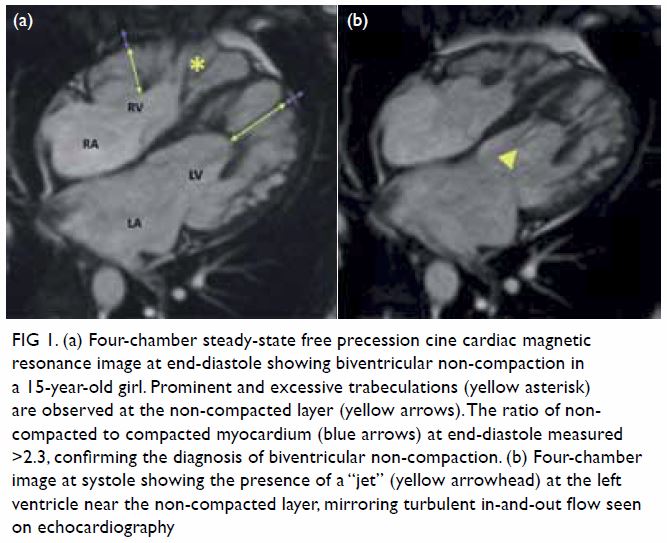
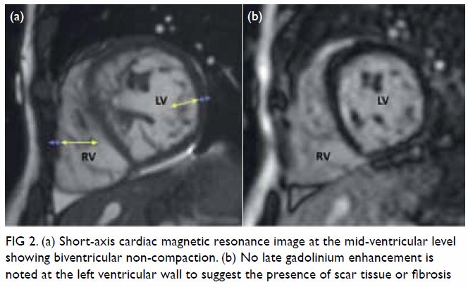
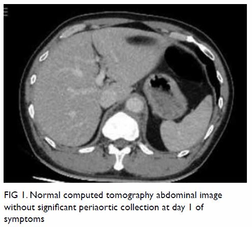
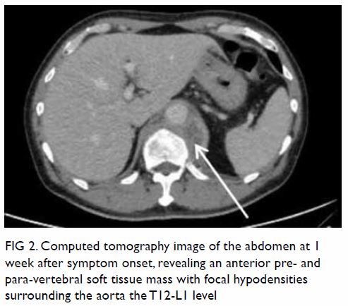
In May 2015, a 59-year-old woman presented to Princess Margaret Hospital, Hong Kong, with chronic intermittent vertigo and syncope, aggravated by head rotation to the right. Physical, otoscopy, and nasal endoscopy examination results were unremarkable. Cervical spine plain radiographs demonstrated cervical spondylosis with marginal osteophytosis (Fig 1). In May 2015, computed tomography angiography of the head and neck revealed focal moderate (50%) stenosis at bilateral vertebral arteries at C5/6 levels due to extrinsic compression from hypertrophied uncovertebral joints (Fig 2). In January 2016, magnetic resonance imaging demonstrated disco-osteophytic protrusion at C5/6 level without evidence of cord compression. In March 2016, digital subtraction angiography, performed with the patient’s head in neutral and bilateral rotated positions, demonstrated dynamic deterioration of focal stenosis of right vertebral artery at C5/6 level to up to 80% stenosis during head rotation to the right (Figs 3 and Fig 4).
 

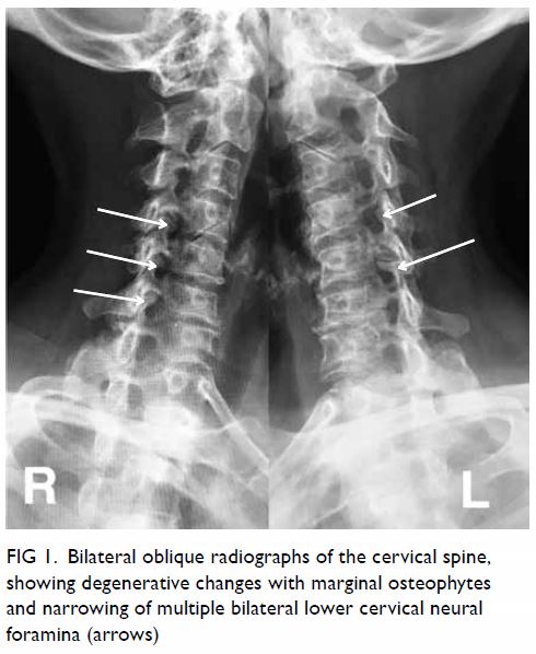
Figure 1. Bilateral oblique radiographs of the cervical spine, showing degenerative changes with marginal osteophytes and narrowing of multiple bilateral lower cervical neural foramina (arrows)
 

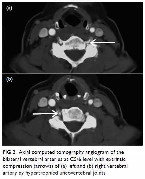
Figure 2. Axial computed tomography angiogram of the bilateral vertebral arteries at C5/6 level with extrinsic compression (arrows) of (a) left and (b) right vertebral artery by hypertrophied uncovertebral joints
 

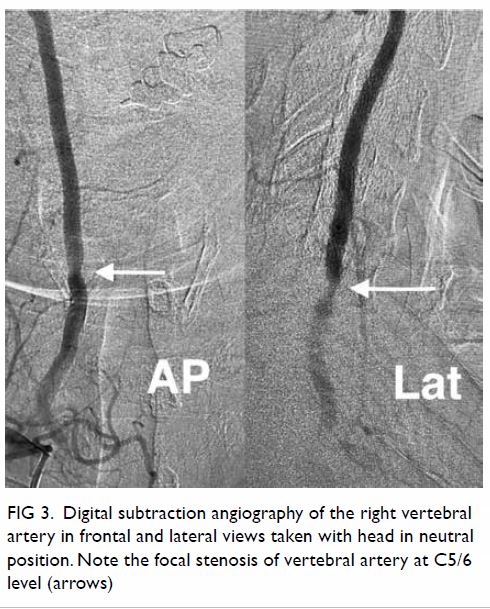
Figure 3. Digital subtraction angiography of the right vertebral artery in frontal (AP) and lateral (Lat) views taken with head in neutral position. Note the focal stenosis of vertebral artery at C5/6 level (arrows)
 

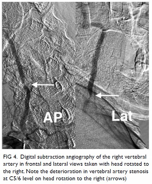
Figure 4. Digital subtraction angiography of the right vertebral artery in frontal (AP) and lateral (Lat) views taken with head rotated to the right. Note the deterioration in vertebral artery stenosis at C5/6 level on head rotation to the right (arrows)
 
A static focal moderate (50%) stenosis of the left vertebral artery at C5/6 was also present. Overall findings were compatible with bow hunter’s syndrome (BHS) with dynamic deterioration of right vertebral artery stenosis on head rotation, related to extrinsic compression by hypertrophied facet joint and disc protrusion. In April 2016, the patient underwent anterior C5/6 cervical discectomy and anterior spinal fusion with smooth recovery and symptomaticresolution.
 
Discussion
Bow hunter’s syndrome was first reported in 1978 when a patient developed lateral medullary syndrome during archery practice with lateral head rotation.1 It refers to symptomatic vertebrobasilar insufficiency secondary to mechanical occlusion or stenosis of vertebral arteries upon head and neck rotation.
 
The pathogenesis of BHS is related to the tortuous anatomical course of the vertebral artery along the cervical spine, which renders the artery susceptible to extrinsic compression, repetitive shear stress resulting in haemodynamic events in at-risk patients during head and neck rotation.2 Osteophytes, disc herniation, ligamentous or neck muscle hypertrophy are risk factors for BHS.3
 
Though rare, BHS is a not to be missed cause of vertigo, owing to its specific relationship with head and neck rotation and its potential risk of posterior circulation ischaemic stroke.1 2 Bow hunter’s syndrome is more common among males and those aged between 50 and 70 years old.3 Common clinical manifestations include vertigo and syncope. Other symptoms include nystagmus, emesis, Horner’s syndrome, and rarely motor and sensory deficits.2
 
Imaging is crucial in establishing the diagnosis of BHS, delineating the cause and site of extrinsic compression and evaluating complications such as infarction. Dynamic digital subtraction angiography remains the preferred modality for prompt and accurate localisation of stenotic segment and establishing causal relationship with head rotation.4 5 Non-invasive computed tomography or magnetic resonance angiography in both neutral and rotated head positions are also used. Computed tomography can delineate the relationship with surrounding compressive skeletal structures and magnetic resonance would be sensitive in documenting early ischaemic event.3
 
In this case, the patient’s complaint of vertigo exacerbation with specific direction of head rotation should raise the suspicion of BHS. Surgical treatment was offered in view of failed conservative approach, repeated fall related to syncope, and underlying uncovertebral joint hypertrophy and disc protrusion as the aetiological factors of vertebral artery compression.
 
Author contributions
All authors had full access to the data, contributed to the study, approved the final version for publication, and take responsibility for its accuracy and integrity.
 
Concept or design: All authors.
Acquisition of data: TS Chan, JKF Ma.
Analysis or interpretation of data: All authors.
Drafting of the manuscript: SC Wong. TS Chan.
Critical revision of the manuscript for important intellectual content: All authors.
 
Conflicts of interest
All authors have disclosed no conflicts of interest.
 
Funding/support
This pictorial medicine paper received no specific grant from any funding agency in the public, commercial, or not-for-profit sectors.
 
Ethics approval
All patients were treated in accordance with the Declaration of Helsinki and provided consent for all investigations and procedures.
 
References
1. Sorensen BF. Bow hunter’s stroke. Neurosurgery 1978;2:259-61. Crossref
2. Duan G, Xu J, Shi J, Cao Y. Advances in the pathogenesis, diagnosis and treatment of bow hunter’s syndrome: a comprehensive review of the literature. Interv Neurol 2016;5:29-38. Crossref
3. Rastogi V, Rawls A, Moore O, et al. Rare etiology of bow hunter’s syndrome and systematic review of literature. J Vasc Interv Neurol 2015;8:7-16.
4. Taylor WB 3rd, Vandergriff CL, Opatowsky MJ, Layton KF. Bowhunter’s syndrome diagnosed with provocative digital subtraction cerebral angiography. Proc (Bayl Univ Med Cent) 2012;25:26-7. Crossref
5. Go G, Hwang SH, Park IS, Park H. Rotational vertebral artery compression: bow hunter’s syndrome. J Korean Neurosurg Soc 2013;54:243-5. Crossref

Recognising eye implants on radiological imaging: pear or fishball shapes

© Hong Kong Academy of Medicine. CC BY-NC-ND 4.0
 
PICTORIAL MEDICINE
Recognising eye implants on radiological imaging: pear or fishball shapes
Sunny CL Au, MB, ChB, AFCOphthHK; Simon TC Ko, FHKAM (Ophthalmology), FCOphth HK
Department of Ophthalmology, Tung Wah Eastern Hospital (Hong Kong East Cluster Ophthalmic Service), Causeway Bay, Hong Kong
 
Corresponding author: Dr Sunny CL Au (kilihcua@gmail.com)
 
 Full paper in PDF
 
Radiological imaging is now readily available in Hong Kong, in both the public and private sector. Imaging helps diagnose or screen for diseases, but can create noise or false alarms.1 Ophthalmology consultations are common in both in-patient and out-patient settings. Floaters, visual field defects, and abnormal incidental findings on computed tomography or magnetic resonance imaging (MRI) are all common reasons for consultations.2
 
In 2018, a 60-year-old man underwent an MRI scan for acoustic neuroma screening, which revealed an abnormal eyeball shape (Fig 1). Diligent consultation with an ophthalmologist was reassuring for both the physician and the patient. In December 2017, the patient had been referred to our eye department for visual disturbance and was diagnosed with right eye macula-on retinal detachment. He had no history of laser or other eye surgery, nor trauma to the eyes. The patient reported right eye floaters and loss of the inferior visual field. Fundus examinations found a large retinal U-shaped tear at the superior retina, with bullous retinal detachment over the right eye. There were also multiple small flat holes with lattice degeneration over the retina in the temporal and nasal aspect. Retinal detachment repair surgery with encircling band as scleral buckling, and intravitreal gas injection was done for the presence of multiple retinal holes.3 Given the patient’s older age, the presence of cataract, and perceived cataract progression with postoperative intravitreal gas,4 cataract extraction with intraocular lens insertion was also done in the same operation. These are all evidenced on the MRI scan contrasting the presence of a natural thickness lens over the left eye.
 

Figure 1. T2-weighted magnetic resonance imaging scan transverse cut of the brain showing two T2 hyperintense structures highlighting the position of the eyeballs: the pear-shaped right eyeball, in contrast to the normal fishball- shaped left eyeball
 
Retinal detachment surgery can be of external or internal approach to relieve the vitreous traction and flatten the retina. External approach can be localised or 360-degree encircling depending on the retinal status.
 
The formation of the pear-shaped eyeball was due to the buckling effect of the encircling band (Fig 2). With basic knowledge of normal organ shapes, all practitioners would be concerned by such a deformed organ on plain radiographs. Typically, intraocular tumours or eyeball ruptures are not regular nor symmetrical, except large ring melanoma of the ciliary body. The key to differentiating a genuine pathology from a congenital/postoperative variant lies heavily on clinical history.1 Operative implants usually appear regular, or even symmetrical on imaging.
 

Figure 2. Schematic diagram of the eyeball cross-sectional plane showing the effect of the encircling band on the shape of the right eyeball. Solid arrows indicate the direction of force exerted by the encircling band on indenting the eyeball
 
Author contributions
All authors had full access to the data, contributed to the study, approved the final version for publication, and take responsibility for its accuracy and integrity.
 
Concept or design: SCL Au.
Acquisition of data: SCL Au.
Analysis or interpretation of data: SCL Au.
Drafting of the manuscript: SCL Au.
Critical revision of the manuscript for important intellectual content: All authors.
 
Conflicts of interest
All authors have disclosed no conflicts of interest.
 
Funding/support
This pictorial medicine paper received no specific grant from any funding agency in the public, commercial, or not-for-profit sectors.
 
Ethics approval
This study was conducted in accordance with the principles outlined in the Declaration of Helsinki. Relevant patient consent was obtained for the purpose of this case study.
 
References
1. Leslie A, Jones AJ, Goddard PR. The influence of clinical information on the reporting of CT by radiologists. Br J Radiol 2000;73:1052-5. Crossref
2. Carter K, Miller KM. Ophthalmology inpatient consultation. Ophthalmology 2001;108:1505-11. Crossref
3. Shanmugam PM, Ramanjulu R, Mishra KC, Sagar P. Novel techniques in scleral buckling. Indian J Ophthalmol 2018;66:909-15. Crossref
4. Thompson JT. The role of patient age and intraocular gases in cataract progression following vitrectomy for macular holes and epiretinal membranes. Trans Am Ophthalmol Soc 2003;101:485-98.

Giant perivascular spaces: an uncommon cause of obstructive hydrocephalus

© Hong Kong Academy of Medicine. CC BY-NC-ND 4.0
 
PICTORIAL MEDICINE
Giant perivascular spaces: an uncommon cause of obstructive hydrocephalus
MH So, MB, BS FRCR; WK Lo, FRCR, FHKAM (Radiology)
Department of Diagnostic and Interventional Radiology, Kwong Wah Hospital, Yaumatei, Hong Kong
 
Corresponding author: Dr MH So (manhon.so@gmail.com)
 
 Full paper in PDF
 
A 55-year-old man with good past health presented to the emergency department with unsteady gait for 6 months with recent mild left-sided weakness. Urgent computed tomography (CT) scan of the brain showed a multiseptated cystic lesion in the right mesencephalothalamic region with pressure effect on the third ventricle causing obstructive hydrocephalus (Fig 1). Urgent magnetic resonance imaging (MRI) scan (Fig 2) of the lesion showed no post-gadolinium enhancement, no restricted diffusion, complete suppression of the cystic areas on T2 fluid attenuation inversion recovery (FLAIR) sequence and no abnormal parenchymal signal intensities compared with normal brain parenchyma. These imaging findings are consistent with tumefactive perivascular space. Invasive biopsy and surgical excision were avoided, and the patient underwent surgery for ventricular drain insertion.
 

Figure 1. Plain computed tomography scan of the brain showing a multicystic lesion (arrow) in right mesencephalothalamic region with dilated frontal horns of the lateral ventricles and periventricular white matter hypodensities suggestive of transependymal oedema
 

Figure 2. Magnetic resonance images showing tumefactive perivascular space (arrows): (a) T2-weighted sequence; (b) T2 fluid attenuation inversion sequence with gadolinium showing complete suppression of fluid signal in the multicystic lesion; (c) T1-weighted sequence with gadolinium showing no enhancement; and (d) diffusionweighted imaging showing no restricted diffusion
 
Dilated perivascular spaces (PVSs) in the brain are interstitial fluid-filled structures lined by pia-mater that have accompanying patent penetrating arteries within, most commonly seen along the lenticulostriate arteries.1 They can be unilocular or multilocular and may have a radial pattern along the course of the penetrating arteries. The PVSs occur across all age-groups and are more frequent and larger with advancing age. The cause of dilated PVS remains unknown though numerous theories have been postulated including increased permeability of arterial wall and obstruction/disturbance of interstitial fluid drainage/flow. Dilated PVSs may be associated with microvascular diseases, trauma, non-vascular dementia, multiple sclerosis, and the mucopolysaccharidoses.
 
Rarely PVSs are markedly expanded and are termed tumefactive or giant PVSs. Some authors define tumefactive PVSs as those >1.5 cm. Tumefactive PVSs are most commonly located in mesencephalothalamic region.2 They are also seen in cerebral white matter and in the cerebellar dentate nuclei. They can exhibit mass effect and cause obstructive hydrocephalus when occurring in the mesencephalothalamic region as in our case. The MRI signal intensities of typical PVSs should follow cerebrospinal fluid in all sequences including FLAIR imaging with no post-gadolinium enhancement. There is no restricted diffusion as the compartments are communicating. Tumefactive PVSs in cerebral white matter may have perilesional abnormal T2 and FLAIR hyperintensities in up to 50% of cases. The mass effect of the tumefactive PVS may cause chronic ischaemic change in adjacent white matter.3 Histopathological results typically show a pial-lined cyst with no evidence of neoplasm or infection.
 
Differential diagnoses include cystic infarction, tumour, and infection.4 Cystic infarctions assume a slit-like or ovoid shape whereas PVSs are more rounded or linear. The cystic content of tumours is usually not isointense to cerebrospinal fluid on MRI. Solid components are often present, which may enhance after contrast and are surrounded by oedema. Parasitic infections have a range of appearances on CT or MRI scans with contrast enhancement and oedema during the active phase and calcifications in the quiescent phase.
 
Asymptomatic tumefactive PVSs can be managed by follow-up imaging for stability in size. Spontaneous regression of tumefactive PVSs without surgical intervention is rare. Tumefactive PVSs with mass effect and obstructive hydrocephalus can be treated surgically with ventriculostomy, cyst fenestration, ventriculoperitoneal shunting, or cystoperitoneal shunting. When the appearance is typical, surgical biopsy or excision should be avoided.
 
Author contributions
All authors contributed to the concept, acquisition and interpretation of data, drafting of the manuscript, and revision for important intellectual content. All authors had full access to the data, contributed to the study, approved the final version for publication, and take responsibility for its accuracy and integrity.
 
Conflicts of interest
All authors have disclosed no conflicts of interest.
 
Funding/support
This research received no specific grant from any funding agency in the public, commercial, or not-for-profit sectors.
 
Ethics approval
The patient was treated in accordance with the Declaration of Helsinki. The patient provided informed consent for all procedures.
 
References
1. AI Abdulsalam H, Alatar AA, Elwatidy S. Giant tumefactive perivascular spaces: a case report and literature review. World Neurosurg 2018;112:201-4. Crossref
2. Choh NA, Shaheen F, Robbani I, Singh M, Gojwari T. Tumefactive Virchow–Robin spaces: a rare cause of obstructive hydrocephalus. Ann Indian Acad Neurol 2014;17:345-6. Crossref
3. Salzman KL, Osborn AG, House P, et al. Giant tumefactive perivascular spaces. AJNR Am J Neuroradiol 2005;26:298- 305.
4. Kwee RM, Kwee TC. Virchow-Robin spaces at MR imaging. Radiographics 2007;27:1071-86. Crossref

Ruptured ovarian teratoma with granulomatous peritonitis

© Hong Kong Academy of Medicine. CC BY-NC-ND 4.0
 
PICTORIAL MEDICINE
Ruptured ovarian teratoma with granulomatous peritonitis
WL Wong, MB, BS, FRCR1; Anthony WT Chin, MB, ChB, FRCR1; WM Yu, MB, ChB1; FH Ng, FRCR, FHKAM (Radiology)2
1 Department of Radiology, United Christian Hospital, Kwun Tong, Hong Kong
2 Department of Radiology, Caritas Medical Centre, Shamshuipo, Hong Kong
 
Corresponding author: Dr WL Wong (jesswong723@gmail.com)
 
 Full paper in PDF
 
Case
In January 2017, a 35-year-old woman was admitted to our hospital with insidious onset of upper abdominal pain. A computed tomography (CT) scan showed bilateral ovarian cysts with fat fluid level, calcifications, and Rokitansky protuberance, compatible with ovarian teratoma (Fig 1). Anti-dependent fatty pockets with soft tissue rim were found at the bilateral subphrenic space, likely representing reactive changes to spilt cyst content, which also explained the patient’s upper abdominal pain. The patient had stable vital signs and was therefore treated conservatively. Follow-up ultrasonography scan showed globular fatty locules on the liver surface, compatible with escaped fatty cyst content (Fig 2). Subsequently, the patient underwent bilateral ovarian cystectomy. Histology confirmed bilateral ovarian mature cystic teratoma. Intra-operatively, widespread flimsy adhesions and multiple sebum-like implants were seen in the peritoneal cavity, consistent with changes related to teratoma rupture. The peritoneal cavity was irrigated and her symptoms gradually subsided; however, follow-up CT showed mild interval enlargement of the fat-attenuated lesions (Fig 3).
 

Figure 1. (a) Ultrasonograph showing a partially echogenic mass with posterior acoustic attenuation at left adnexal region and (b) computed tomography image showing a partly fat-attenuated mass with tooth like calcification, characteristic of ovarian teratoma
 

Figure 2. (a) Ultrasonograph showing a hypoechoic lesion and (b) computed tomography image showing a fat-attenuated nodule over the liver surface suggestive of liver capsular implant from ruptured ovarian teratoma
 

Figure 3. (a) Axial and (b) coronal computed tomography images showing progression of the bilateral subphrenic fatty implants (arrows) with increased adjacent stranding suggestive of chronic granulomatous peritonitis caused by the content of the mature cystic teratoma
 
Discussion
Mature cystic teratomas (also known as dermoid cysts) are common ovarian germ cell neoplasms accounting up to 10% to 25% of all ovarian neoplasms.1 They are cystic tumours composed of well-differentiated derivations from at least two of the three germ cell layers. Tumours are bilateral in about 10% of cases. On ultrasonography, cystic teratoma commonly manifests as a cystic lesion with a densely echogenic tubercle projecting into the cystic lumen; or a diffusely or partially echogenic mass with posterior attenuation by sebaceous material and hair. Multiple thin echogenic bands caused by hair in cyst cavity can also been seen. Pure sebum within the cyst can be hypoechoic or anechoic, fluid-fluid level can result from sebum floating on aqueous fluid which appears more echogenic than the sebum layer. On CT, the diagnosis of mature cystic teratoma is rather straightforward; fat attenuation within a cyst is diagnostic of mature cystic teratoma. Teeth or other calcifications can be seen in 56% of cases.2
 
Spontaneous rupture is an uncommon complication of dermoid cysts owing to the presence of a thick capsule, and is only seen in 1% to 4% of cases.1 Acute peritonitis can result from sudden rupture of tumour contents as seen in the present case. Chronic granulomatous peritonitis is caused by chronically leaking teratoma and is characterised by multiple small white peritoneal implants and dense adhesions with variable ascites. Visualisation of fatty implants within the peritoneal cavity is diagnostic.3
 
The reported CT findings of granulomatous peritonitis or intraperitoneal rupture of teratoma are inconsistent.1 3 In one case, capsular fatty implants were seen in the dome of the liver, similar to the CT appearance in the present case. In other patients there can be a significant amount of intraperitoneal free fluid and an omental cake appearance mimicking peritoneal carcinomatosis.
 
Chronic granulomatous peritonitis is a potentially serious complication of ruptured dermoid cyst and can lead to bowel obstruction resulting from adhesion. Removal of cystic content and copious peritoneal lavage should be performed to prevent new adhesion and peritoneal granuloma, and can be a successful method for treating chemical peritonitis caused by ruptured ovarian teratoma.4 In the present case, progression of the right subphrenic fatty implant, possibly related to incomplete removal of the cystic content, resulted in chronic granulomatous peritonitis. The patient may have benefitted from further peritoneal lavage.
 
Author contributions
WL Wong contributed to acquisition of data. All authors contributed to concept or design, analysis or interpretation of data, drafting of the article, and critical revision for important intellectual content. All authors had full access to the data, contributed to the study, approved the final version for publication, and take responsibility for its accuracy and integrity.
 
Conflicts of interest
All authors have disclosed no conflicts of interest.
 
Funding/support
This research received no specific grant from any funding agency in the public, commercial, or not-for-profit sectors.
 
Ethics approval
This study was approved by the Kowloon Central/Kowloon East Research Ethics Committee (Ref KC/KE-19-0158/ER-4). Informed consent was obtained from the patient.
 
References
1. Erbay G. Ruptured ovarian dermoid cyst mimicking peritoneal carcinomatosis: CT and MRI. J Clin Anal Med 2016;6:701-3.
2. Outwater EK, Siegelman ES, Hunt JL. Ovarian teratomas: tumor types and imaging characteristics. Radiographics 2001;21:475-90. Crossref
3. Fibus TF. Intraperitoneal rupture of a benign cystic ovarian teratoma: findings at CT and MR imaging. AJR Am J Roentgenol 2000;174:261-2. Crossref
4. Shamshirsaz AA, Shamshirsaz AA, Vibhaka JL, Broadwell C, Van Voorhis BJ. Laparoscopic management of chemical peritonitis caused by dermoid cyst spillage. JSLS 2011;15:403-5. Crossref

Sudden cardiac arrest with pericardial contrast during computed tomography aortogram in a type A aortic dissection patient

© Hong Kong Academy of Medicine. CC BY-NC-ND 4.0
 
PICTORIAL MEDICINE
Sudden cardiac arrest with pericardial contrast during computed tomography aortogram in a type A aortic dissection patient
WM Yu, MB, ChB, FRCR1; WL Wong, MB, ChB, FRCR1; FH Ng, MB ChB, FRCR2
1 Department of Radiology, United Christian Hospital, Kwun Tong, Hong Kong
2 Department of Radiology, Caritas Medical Centre, Shamshuipo, Hong Kong
 
Corresponding author: Dr WM Yu (wenmingyu@hotmail.com)
 
 Full paper in PDF
 
A 69-year-old man with long-standing hypertension and history of pulmonary embolism on warfarin presented to Accident and Emergency Department with sudden upper back pain and transient loss of consciousness. On admission, the patient was in haemodynamic shock with blood pressure 83/57 mm Hg and pulse 42 beats per minute. Cardiovascular examination revealed radial-radial and radial-femoral delay. Electrocardiogram showed complete heart block and chest radiograph demonstrated a widened mediastinum. An immediate bedside echocardiogram found an intimal flap in the aortic root with evidence of aortic regurgitation and a thin rim of pericardial effusion.
 
Fluid resuscitation and inotropic support was given and urgent computed tomography (CT) aortogram was arranged to further delineate the extent of involvement of aortic dissection. However, the patient developed sudden cardiac arrest immediately after being given 80 mL intravenous Omnipaq at a rate of 3 mL/s via injector during CT aortogram. A review of CT images showed Stanford type A aortic dissection with an intimal flap extending into the pericardial sac (Fig 1). No obvious pericardial effusion was visible on the pre-contrast CT images (Fig 2a). However, significant contrast was seen in the pericardial sac on post-contrast CT images (Fig 2b), indicating a sudden rupture with haemopericardium had occurred during the CT scan. Despite prompt bedside pericardiocentesis and cardiopulmonary resuscitation, the patient died.
 

Figure 1. Axial (a) and coronal (b) computed tomography aortogram images showing the extension of the dissection flap into pericardial sac. The rupture was seen at the right lateral wall of the ascending aorta (white arrow)
 

Figure 2. (a) Pre-contrast computed tomography image showing normal pericardium. (b) Computed tomography aortogram image showing significant contrast in the pericardium. A dissection flap can be seen within the descending thoracic aorta. The false lumen was attenuated due to delayed opacification. The right atrium was obliterated, in line with cardiac tamponade
 
Aortic dissection is the most common acute emergency condition of the aorta. The mortality rate is high, and rupture is the cause of death in approximately one-third of affected patients.1 The pathology is due to a tear in the intimal layer allowing blood to propagate into the media and create a false lumen. In the ascending thoracic aorta, the primary tear is most often within 3 cm of the aortic cusps.2 The false lumen of aorta may rupture due to loss of elastic recoil and increased wall stress with dilatation. Often, the rupture site is close to the initial intimal-medial tear over the right lateral wall where it receives the ejected blood from the left ventricle.2 This ends up into the pericardial sac causing haemopericardium and subsequent fatal cardiac tamponade.2
 
Computed tomography aortogram is the first-line modality in the diagnosis of aortic dissection, delineation of its extent of involvement and end-organ ischaemia. In our case, rupture of the aorta into the pericardial sac was evidenced by significant contrast-enhanced haemopericardium on CT aortogram images which was absent in pre-contrast CT images. This could be related to the rapid injection of a large volume bolus of intravenous contrast by power-injector, resulting in a sudden elevation of left ventricular pressure, supported by previous study in human subjects demonstrating a significant increase in blood pressure after bolus injection of low osmolarity, non-ionic contrast agent.3
 
The treatment of type A aortic dissection with rupture is immediate surgical repair.4 5 Although it was previously considered controversial to perform pericardiocentesis as there is a risk of worsening the leak, recent evidence suggests that controlled pericardiocentesis may reduce haemodynamic instability in critical cardiac tamponade to allow sufficient time for urgent operative repair.5
 
Author contributions
WM Yu and FH Ng are responsible for the concept of study, acquisition and analysis of data, and drafting of the article. All authors are responsible for critical revision for important intellectual content. All authors had full access to the data, contributed to the study, approved the final version for publication, and take responsibility for its accuracy and integrity.
 
Conflicts of interest
All authors have disclosed no conflicts of interest.
 
Funding/support
This research received no specific grant from any funding agency in the public, commercial, or not-for-profit sectors.
 
Ethics approval
The present study was reviewed and approved by the Kowloon Central Cluster/Kowloon East Cluster Research Ethics Committee (KCC/KEC-2019-0179). Because the concerned patient was deceased, the requirement for consent was waived by the ethics board.
 
References
1. Mehta RH, Suzuki T, Hagan PG, et al. Predicting death in patients with acute type a aortic dissection. Circulation 2002;105:200-6. Crossref
2. Patel YD. Rupture of an aortic dissection into the pericardium. Cardiovascular Intervent Radiol 1986;9:222-4. Crossref
3. John AM, Yadar S. Evaluation of blood pressure variations during the administration of intravascular contrast media in CECT Abdomen. Asian J Pharm Clin Res 2018;11:309-11. Crossref
4. Adler Y, Charron P, Imazio M, et al. 2015 ESC Guidelines for the diagnosis and management of pericardial diseases: The Task Force for the Diagnosis and Management of Pericardial Diseases of the European Society of Cardiology (ESC). Endorsed by: The European Association for Cardio-Thoracic Surgery (EACTS). Eur Heart J 2015;36:2921-64. Crossref
5. Hayashi T, Tsukube T, Yamashita T, et al. Impact of controlled pericardial drainage on critical cardiac tamponade with acute type A aortic dissection. Circulation. 2012;126(11 Suppl 1):S97-S101. Crossref

Diagnosis of Wunderlich syndrome in a patient with flank pain

© Hong Kong Academy of Medicine. CC BY-NC-ND 4.0
 
PICTORIAL MEDICINE
Diagnosis of Wunderlich syndrome in a patient with flank pain
YY Lin, MD; CW Hsu, PhD; HM Li, MD; HY Su, MD
Department of Emergency Medicine, E-Da Hospital, I-Shou University, Kaohsiung, Taiwan
 
Corresponding author: Dr HY Su (hys927@hotmail.com)
 
 Full paper in PDF
 
In September 2018, a 62-year-old man without underlying disease presented to the emergency department of E-Da Hospital, Kaohsiung, Taiwan, with right flank pain for 1 day. The patient reported sharp and persistent pain radiating to the right upper abdomen. On arrival at the emergency department, the patient had heart rate 120 beats per minute and blood pressure 85/54 mm Hg. Physical examination revealed right flank knocking tenderness. Laboratory test results, including blood test and urinary analysis, were unremarkable. Abdominal plain film radiograph revealed a large right renal mass displacing surrounding structures (Fig 1). Point-of-care ultrasound demonstrated a right renal mass with hyperechogenicity, which was surrounded by hypoechoic haematoma in the perinephric space (Fig 2). Subsequent abdominal computed tomography (CT) revealed rupture of right renal angiomyolipoma with pericapsular haematoma (Fig 3). Wunderlich syndrome complicated by hypovolaemic shock was diagnosed, and proper fluid resuscitation and blood transfusion were performed in the emergency department. The patient received partial nephrectomy of right kidney on the next day, and was discharged uneventfully from the hospital 2 weeks after admission.
 

Figure 1. Plain abdominal radiograph showing a large right renal mass displacing surrounding structure (arrows). The low density of the mass is suggestive of a lesion with a lipomatous component
 

Figure 2. Point-of-care ultrasound showing hypoechoic haematoma in the perinephric space (arrows). Hyperechogenicity can indicate a lipomatous component such as angiomyolipoma in the kidney (star)
 

Figure 3. (a) Coronal and (b) axial contrast-enhanced computed tomography images showing rupture of a right renal angiomyolipoma (stars) with pericapsular haematoma (arrows)
 
Wunderlich syndrome, a rare but life-threatening entity, is defined as spontaneous nontraumatic renal haemorrhage confined to the subcapsular and perirenal space.1 Lenk’s triad, which consists of acute flank pain, palpable flank mass, and hypovolemic shock, is the classical clinical feature of Wunderlich syndrome.2 The aetiologies of Wunderlich syndrome are classified into neoplastic and non-neoplastic origins. Up to 60% of patients with Wunderlich syndrome are caused by neoplasm, including benign tumours such as angiomyolipoma and malignancies such as renal cell carcinoma.3 A variety of diseases account for non-neoplastic origins of Wunderlich syndrome, including vasculitis, renal artery aneurysm, arteriovenous malformation, renal vein thrombosis, nephritis, cystic renal disease, and coagulopathy.3 Angiomyolipoma, a benign neoplasm composed of smooth muscle, adipose tissue, and thick-walled blood vessels, is the most common cause of Wunderlich syndrome.3 The risk of tumour rupture leading to fatal internal haemorrhage increases when angiomyolipoma grows >40 mm in diameter.4 Aneurism formation due to poor elastic vascular structure might be the reason for angiomyolipoma rupture, especially during tumour growth.
 
For diagnosis of Wunderlich syndrome, contrast-enhanced CT scan is a standard medical imaging modality with 100% sensitivity in identifying perirenal haemorrhage.4 Computed tomography scan can present renal vascular structure, origins of tumours and pathological change in adjacent tissues. Furthermore, CT scan can also provide detailed vascular anatomy to provide a roadmap for superselective renal embolisation in management of perirenal haemorrhage. In contrast with CT scan, point-of-care ultrasound might be considered as a prompt tool to diagnose patients with Wunderlich syndrome. Point-of-care ultrasound can be used to screen the renal structure, quickly identify internal bleeding, and evaluate the hemodynamic condition by measuring the diameter of the inferior vena cava and assessing the cardiac preload and contractility. Ultrasound can also facilitate the initial differential diagnosis of patients with flank pain, such as renal colic, renal abscess or acute pyelonephritis. Initial treatments for Wunderlich syndrome include selective arterial embolisation and surgical intervention. However, clinical guidelines for management of Wunderlich syndrome are not yet well established.5 Selective arterial embolisation has the advantage of minimal invasiveness, renal preservation, and efficiency in treating acute renal haemorrhage. However, surgical intervention can provide a delicate strategy for tumour resection, especially if suspicious for malignancy, and prevent recurrent tumour bleeding.5 Since Wunderlich syndrome is a life-threatening condition, clinicians should be aware while approaching patients presenting with flank pain and in shock to facilitate timely emergency surgery or embolisation if needed.
 
Author contributions
All authors had full access to the data, contributed to the study, approved the final version for publication, and take responsibility for its accuracy and integrity.
 
Concept and design of the study: HY Su.
Acquisition of data: YY Lin.
Analysis or interpretation of data: YY Lin.
Drafting of the article: HY Su.
Critical revision for important intellectual content: HM Li, CW Hsu.
 
Conflicts of interest
All authors have disclosed no conflicts of interest.
 
Funding/support
This study received no specific grant from any funding agency in the public, commercial, or not-for-profit sectors.
 
Ethics approval
This study was conducted in accordance with the principles outlined in the Declaration of Helsinki.
 
References
1. Medda M, Picozzi SC, Bozzini G, Carmignani L. Wunderlich’s syndrome and hemorrhagic shock. J Emerg Trauma Shock 2009;2:203-5. Crossref
2. Simkins A, Maiti A, Cherian SV. Wunderlich syndrome. Am J Med 2017;130:e217-8. Crossref
3. Katabathina VS, Katre R, Prasad SR, Surabhi VR, Shanbhogue AK, Sunnapwar A. Wunderlich syndrome: cross-sectional imaging review. J Comput Assist Tomogr 2011;35:425-33. Crossref
4. Albi G, del Campo L, Tagarro D. Wünderlich’s syndrome: causes, diagnosis and radiological management. Clin Radiol 2002;57:840-5. Crossref
5. Flum AS, Hamoui N, Said MA, et al. Update on the diagnosis and management of renal angiomyolipoma. J Urol 2016;195(4 Pt 1):834-46. Crossref

Cardiac magnetic resonance imaging in the diagnosis of biventricular non-compaction in a young but failing heart

© Hong Kong Academy of Medicine. CC BY-NC-ND 4.0
 
PICTORIAL MEDICINE
Cardiac magnetic resonance imaging in the diagnosis of biventricular non-compaction in a young but failing heart
Victor SH Chan, MB, BS, FRCR1; Carmen WS Chan, MB, BS, FRCP (Lond)2; Stephen CW Cheung, MRCP, FHKAM (Radiology)1
1 Department of Radiology, Queen Mary Hospital, Pokfulam, Hong Kong
2 Department of Medicine, Queen Mary Hospital, Pokfulam, Hong Kong
 
Corresponding author: Dr Victor SH Chan (victorchansh@gmail.com)
 
 Full paper in PDF
 
A 15-year-old Chinese girl with a history of scoliosis presented to Queen Mary Hospital, Hong Kong in April 2016 with an incidental finding of an ejection systolic murmur at the left lower sternal border. No history of chest pain, syncope, reduced effort tolerance, or significant family history of congenital cardiac disease was present. Echocardiography revealed heavy trabeculations over the left ventricular (LV) apical region. Colour Doppler revealed abnormal in-and-out flow at the deep crypts. Overall features were suspicious of LV non-compaction (NC). The patient subsequently underwent cardiac magnetic resonance imaging for further assessment, using a 1.5-T magnetic resonance imaging scanner (Magnetom Aera; Siemens Healthcare, Forchheim, Germany). Cardiac magnetic resonance confirmed the diagnosis of biventricular NC with diffuse involvement (Fig 1). The ratio of non-compacted to compacted diastolic myocardium was 2.95 (>2.3). The LV ejection fraction (EF) was 32.6%, and the right ventricular (RV) EF was 32.5%. Moderate global hypokinesia of both ventricles was observed. Mild mitral regurgitation was present. No significant left to right cardiac shunt or late gadolinium enhancement was seen at the LV wall to suggest presence of scar or fibrosis (Fig 2). No abnormal thinning of the wall, focal/regional RV wall motion abnormality, or aneurysmal change was noted. No thrombus was present within the cardiac chambers. A normal configuration of a left-sided aortic arch was observed with absence of coarctation. Subsequent genetic testing for pathogenic mutations for NC was negative in this patient.
 

Figure 1. (a) Four-chamber steady-state free precession cine cardiac magnetic resonance image at end-diastole showing biventricular non-compaction in a 15-year-old girl. Prominent and excessive trabeculations (yellow asterisk) are observed at the non-compacted layer (yellow arrows). The ratio of noncompacted to compacted myocardium (blue arrows) at end-diastole measured >2.3, confirming the diagnosis of biventricular non-compaction. (b) Four-chamber image at systole showing the presence of a “jet” (yellow arrowhead) at the left ventricle near the non-compacted layer, mirroring turbulent in-and-out flow seen on echocardiography
 

Figure 2. (a) Short-axis cardiac magnetic resonance image at the mid-ventricular level showing biventricular non-compaction. (b) No late gadolinium enhancement is noted at the left ventricular wall to suggest the presence of scar tissue or fibrosis
 
Ventricular NC of the myocardium, also known as spongiform cardiomyopathy, is a rare cardiomyopathy arising from arrested endomyocardial development during embryogenesis,1 with an incidence of approximately 0.05%.2 Non-compaction is a group of genetically heterogeneous disorders and can be inherited in autosomal dominant, autosomal recessive and X-linked recessive pattern.3 However, the majority of NC have idiopathic pathogenesis, and the diagnostic yield of gene panel testing in LVNC is low (~9%). Patients with isolated NC are less likely to have a positive genetic test result.3 Morphologically, NC is characterised by an altered myocardial wall with resultant prominent trabeculae and deep intertrabecular recesses,4 leading to an abnormal thickened bilayer of compacted and non-compacted myocardium. The LV is more frequently involved and biventricular involvement is less commonly encountered. The absence of wall thinning, RV wall motion abnormality or aneurysmal change, although not diagnostic, suggests an alternative diagnosis to that of arrhythmogenic RV dysplasia. Principal clinical manifestations of NC include: heart failure, arrhythmia, cardioembolic events, syncope, and sudden cardiac death.4 Even though our patient had remained asymptomatic prior to diagnosis, there were notable reductions in LVEF and RVEF, suggesting heart failure.
 
Cardiac magnetic resonance in establishing suspected NC cases would be crucial in: (1) confirming the diagnosis, (2) establishing residual cardiac function, and (3) determining presence of other associated cardiac malformations, such as LV outflow tract abnormalities (eg, bicuspid aortic valve), Ebstein anomaly, tetralogy of Fallot (more commonly diagnosed at a younger age-group) or coarctation of the aorta. Cardiac magnetic resonance is also superior to echocardiography in delineating RV anatomy and function, evaluating RV involvement of NC, determining presence of intracardiac thrombus and myocardial scarring. After the above diagnostic considerations have been addressed, management of biventricular NC may include anticoagulation, treatment of heart failure, and the placement of implantable cardioverter defibrillator or pacemaker where clinically appropriate. However, cardiac transplantation remains as the only definitive treatment of biventricular NC.
 
Author contributions
All authors had full access to the data, contributed to the study, approved the final version for publication, and take responsibility for its accuracy and integrity. All authors contributed to the concept, image acquisition, image and data interpretation, drafting of the article, and critical revision for important intellectual content.
 
Conflicts of interest
The authors have no conflicts of interest to disclose.
 
Funding/support
This research received no specific grant from any funding agency in the public, commercial, or not-for-profit sectors.
 
Ethics approval
Patient consent was obtained for the purpose of this case report.
 
References
1. Maron BJ, Towbin JA, Thiene G, et al. Contemporary definitions and classification of the cardiomyopathies: an American Heart Association scientific statement from the Council on Clinical Cardiology, Heart Failure and Transplantation Committee; Quality of Care and Outcomes Research and Functional Genomics and Translational Biology Interdisciplinary Working Groups; and Council on Epidemiology and Prevention. Circulation 2006;113:1807-16. Crossref
2. Richardson P, McKenna RW, Bristow M, et al. Report of the 1995 World Health Organization/International Society and Federation of Cardiology task force on the definition and classification of cardiomyopathies. Circulation 1996;93:841-2. Crossref
3. Miller EM, Hinton RB, Czosek R, et al. Genetic testing in pediatric left ventricular noncompaction. Circ Cardiovasc Genet 2017;10. pii:e001735. Crossref
4. Odiete O, Nagendra R, Lawson MA, Okafor H. Biventricular noncompaction cardiomyopathy in a patient presenting with new onset seizure: case report. Case Rep Cardiol article 2012;2012:924865. Crossref

Radiological progression of penicillin-sensitive Staphylococcus aureus aortitis

© Hong Kong Academy of Medicine. CC BY-NC-ND 4.0
 
PICTORIAL MEDICINE
Radiological progression of penicillin-sensitive Staphylococcus aureus aortitis
S Zheng, MB, BS, MRCP
Department of General Medicine, Sengkang General Hospital, Singapore
 
Corresponding author: Dr S Zheng ( zheng.shuwei@singhealth.com.sg)
 
 Full paper in PDF
 
In June 2017, a 58-year-old man, with no known cardiovascular risk factors, was admitted to a hospital in Singapore, presenting with a 1-week history of fever and progressively worsening epigastric pain. On the first day of his symptoms, he had visited the emergency department at another hospital where a computed tomography (CT) scan of the abdomen performed had not revealed significant pathology (Fig 1). He was treated symptomatically as for a viral infection, but he represented to our hospital a week later without significant symptomatic improvement. The patient had a known history of reaction to penicillin and a history of traumatic spinal injury more than 20 years ago requiring spinal instrumentation at the L4/5 level. On examination, the patient was febrile but haemodynamically stable. Abdominal examination revealed epigastric tenderness on deep palpation. Cardiorespiratory examination was unremarkable. The patient had leucocytosis of 15.37 × 10³/uL, raised C-reactive protein level of 109.2 mg/L, and erythrocyte sedimentation rate of 46 mm/h. Renal and liver function tests, and serum amylase and lipase levels were unremarkable. He was started empirically on intravenous ceftriaxone following blood cultures.
 

Figure 1. Normal computed tomography abdominal image without significant periaortic collection at day 1 of symptoms
 
A new CT scan of the abdomen was performed, revealing an anterior pre- and para-vertebral soft tissue mass with focal hypodensities surrounding the aorta at the T12-L1 level (Fig 2). Blood culture results (which were received 3 days later) were positive for penicillin-sensitive Staphylococcus aureus. Magnetic resonance imaging of the thoracolumbar spine illustrated paravertebral soft tissue thickening at level of T12-L1 that appears multiloculated with rim enhancement, suggestive of an underlying paravertebral abscess without epidural extension. A transthoracic echocardiogram did not reveal any valvular lesions. The presence of an inflammatory collection around the aorta prompted concerns for an infectious aortitis.
 

Figure 2. Computed tomography image of the abdomen at 1 week after symptom onset, revealing an anterior pre- and para-vertebral soft tissue mass with focal hypodensities surrounding the aorta the T12-L1 level
 
Antimicrobial therapy was switched to intravenous cefazolin, which was continued for 6 weeks with symptomatic improvement. Repeated blood cultures did not show evidence of persistent S aureus bacteraemia. Monitoring of C-reactive protein level and erythrocyte sedimentation rate showed gradual improvement. A CT aortogram, after 6 weeks of parenteral antibiotics, showed interval improvement of the paravertebral collections and soft tissue thickening around the aorta, suggesting improving aortitis (Fig 3). Antibiotics were switched to oral trimethoprim and sulfamethoxazole. Another CT aortogram 8 months later showed complete resolution of aortitis and paravertebral abscess (Fig 4).
 

Figure 3. Computed tomography aortogram, after 6 weeks of parental antibiotics, showing interval improvement but residual paravertebral collections and soft tissue thickening around the aorta
 

Figure 4. Complete resolution of aortitis on computed tomography aortogram 8 months later
 
In the antibiotic era, infectious aortitis is a rare clinical entity. Gram-positive micro-organisms are most commonly implicated, in up to 60% of cases, with S aureus being the most frequently encountered micro-organism. Other micro-organisms commonly implicated include Enterococcus species, Streptococcus pneumoniae, Salmonella species, Mycobacterium tuberculosis, and in the more distant past, syphilis.1 Rare case reports have featured the radiological evolution of infectious aortitis while on conservative treatment, and these often illustrate peri-aortic soft tissue masses progressing to aneurysmal formation from S aureus or Salmonella species infection.1 2 3 4 5 Prompt antimicrobial therapy is crucial along with endovascular or surgical intervention.5 Our patient demonstrated radiological normality to pathology within a week of symptom onset and subsequent improvement while on conservative therapy alone, followed by full radiological resolution. We believe that his successful recovery is in part due to early appropriate and prolonged antimicrobial therapy.
 
Author contributions
The author designed the study, contributed to acquisition and analysis of data, drafted the article, and contributed to the critical revision for important intellectual content. The author had full access to the data, contributed to the study, approved the final version for publication, and takes responsibility for its accuracy and integrity.
 
Conflicts of interest
The author has disclosed no conflicts of interest.
 
Funding/support
This research received no specific grant from any funding agency in the public, commercial, or not-for-profit sectors.
 
Ethics approval
This study was conducted in accordance with the principles outlined in the Declaration of Helsinki.
 
References
1. Cevasco M, Menard MT, Bafford R, McNamee CJ. Acute infectious pseudoaneurysm of the descending thoracic aorta and review of infectious aortitis. Vasc Endovascular Surg 2010;44:697-700. Crossref
2. Carreras M, Larena JA, Tabernero G, Langara E, Pena JM. Evolution of salmonella aortitis towards the formation of abdominal aneurysm. Eur Radiol 1997;7:54-6. Crossref
3. Rozenblit A, Bennett J, Suggs W. Evolution of the infected abdominal aortic aneurysm: CT observation of early aortitis. Abdom Imaging 1996;21:512-4. Crossref
4. Wein M, Bartel T, Kabatnik M, Sadony V, Dirsch Olaf, Erbel R. Rapid progression of bacterial aortitis to an ascending aortic mycotic aneurysm documented by transesophageal echocardiography. J Am Soc Echocardiogr 2001;14:646-9. Crossref
5. Kan CD, Lee HL, Yang YJ. Outcome after endovascular stent graft treatment for mycotic aortic aneurysm: a systematic review. J Vasc Surg 2007;46:906-12. Crossref

Native T1 mapping for the diagnosis of Anderson-Fabry disease with myocardial hypertrophy

© Hong Kong Academy of Medicine. CC BY-NC-ND 4.0
 
PICTORIAL MEDICINE
Native T1 mapping for the diagnosis of Anderson-Fabry disease with myocardial hypertrophy
Victor SH Chan, MB, BS, FRCR1; W Zhou, MB, BS2; Stephen CW Cheung, MRCP, FHKAM (Radiology)1; MY Ng, BMBS, FRCR2
1 Department of Radiology, Queen Mary Hospital, Pokfulam, Hong Kong
2 Department of Diagnostic Radiology, Li Ka Shing Faculty of Medicine, The University of Hong Kong, Pokfulam, Hong Kong
 
Corresponding author: Dr MY Ng (myng2@hku.hk)
 
 Full paper in PDF
 
A 66-year-old woman previously treated for hypertrophic cardiomyopathy (HCM) underwent cardiac magnetic resonance imaging for further assessment. The modified Look-Locker sequence 5s(3s)3s was performed on a Philips Achieva 3T magnetic resonance scanner (Philips, Amsterdam, Netherlands). Cardiac magnetic resonance had revealed a combined apical and asymmetrical hypertrophy of the left ventricle (Fig 1). There was a diffuse, non-ischaemic pattern of late gadolinium enhancement in the region of hypertrophy in keeping with HCM. The septal:lateral wall ratio on the 4-chamber view was 2.2:1. The mid-inferoseptal:anterolateral ratio on the short-axis view was 1.32:1. However, native T1 mapping was abnormally low (1009 ms; normal range on our scanner, 1226-1256 ms) [Fig 2]. T2 mapping values were in the normal range indicating no underlying iron deposition to account for the low T1 values. Low native T1 mapping values are atypical of HCM that would normally be associated with a slight increase in native T1. This suggested underlying Anderson-Fabry disease (AFD), subsequently proven by genetic testing in this patient.
 

Figure 1. Cardiac magnetic resonance imaging of the patient demonstrating the following: (a) 4-chamber late gadolinium enhancement (LGE) image showing diffuse mid-wall LGE at the mid-ventricular and apical left ventricular walls (arrows); (b) 4-chamber cine image showing hypertrophy of the lateral and septal walls with a septal:lateral wall ratio of 2.2:1; and (c) 2-chamber LGE image demonstrating mid-wall LGE particularly in the inferior apical wall (arrow)
 

Figure 2. Native T1 mapping images comparing an Anderson-Fabry’s disease (AFD) patient with cardiac involvement (left-sided images) and a normal volunteer (right-sided images). Abnormally reduced T1 values are seen in the AFD patient despite a diffuse, non-ischaemic pattern of late gadolinium enhancement (LGE) that would usually result in elevation of T1 values (native T1 mapping of the AFD patient region of interest (ROI) = 1009 ± 33 ms compared with the normal volunteer [ROI = 1222 ± 47 ms]). The ROI was measured at the basal septal wall. The native T1 values at the mid (1161 ms) and apical short axis (1135 ms) views were also reduced
 
Anderson-Fabry disease is an uncommon X-linked sphingolipid storage disorder resulting from deficiency of the lysosomal enzyme α-galactosidase. The principal driver of mortality in AFD is cardiac disease.1 Disease manifestations include left ventricle hypertrophy that can mimic HCM.2 Other complications of AFD include valve thickening, myocardial scarring, cardiac failure and arrhythmic death.3 Enzyme replacement therapy for AFD should be started early to prevent progression of cardiac disease. Cardiac magnetic resonance myocardial native T1 is decreased in AFD and is a non-invasive method that may raise suspicion of AFD in the context of left ventricle hypertrophy or suspected HCM. Native T1 mapping is an established reproducible technique.1 Raising suspicion and later confirming AFD is crucial to managing these patients appropriately as well as initiating screening in asymptomatic family members.
 
Author contributions
All authors have made substantial contributions to the concept or design of this study; acquisition of data; analysis or interpretation of data; drafting of the manuscript; and critical revision for important intellectual content. All authors had full access to the data, contributed to the study, approved the final version for publication, and take responsibility for its accuracy and integrity.
 
Conflicts of interest
The authors have no conflicts of interest to disclose.
 
Funding/support
This research received no specific grant from any funding agency in the public, commercial, or not-for-profit sectors.
 
Ethics approval
Relevant patient consent was obtained for the purpose of this case study.
 
References
1. Pica S, Sado DM, Maestrini V, et al. Reproducibility of native myocardial T1 mapping in the assessment of Fabry disease and its role in early detection of cardiac involvement by cardiovascular magnetic resonance. J Cardiovasc Magn Reson 2014;16:99. Crossref
2. Deva DP, Hanneman K, Li Q, et al. Cardiovascular magnetic resonance demonstration of the spectrum of morphological phenotypes and patterns of myocardial scarring in Anderson-Fabry disease. J Cardiovasc Magn Reson 2016,18:14. Crossref
3. O’Mahony C, Elliott P. Anderson-Fabry disease and the heart. Prog Cardiovasc Dis 2010;52:326-35. Crossref

Diabetic mastopathy: a breast carcinoma mimic

© Hong Kong Academy of Medicine. CC BY-NC-ND 4.0
 
PICTORIAL MEDICINE
Diabetic mastopathy: a breast carcinoma mimic
WK Ng, MB, BS, FRCR1; SK Chan, MB, ChB, FHKCPath2; KM Kwok, FRCR, FHKAM (Radiology)3; PY Fung, FRCR, FHKAM (Radiology)3
1 Department of Radiology, Tuen Mun Hospital, Hong Kong
2 Department of Pathology, Kwong Wah Hospital, Hong Kong
3 Department of Diagnostic and Interventional Radiology, Kwong Wah Hospital, Hong Kong
 
Corresponding author: Dr WK Ng (wingki.ng712@gmail.com)
 
 Full paper in PDF
 
A 62-year-old woman with a long-standing history of type 1 diabetes mellitus presented to the breast clinic with a palpable breast lump. She had incidentally discovered a painless lump in her left breast that had increased in size over the last 3 months. She denied nipple discharge or overlying skin changes but reported a family history of one maternal aunt who had breast cancer diagnosed in her sixties.
 
Clinical examination revealed an irregular hard mass at the upper outer quadrant of the left breast with no evidence of axillary lymphadenopathy. The right breast was unremarkable.
 
Mammography showed heterogeneously dense breasts with asymmetrical density at the left upper breast, but no discrete mass or spiculations (Fig 1). There were also no suspicious microcalcifications or architectural distortion. Ultrasonography revealed an approximately 4-cm irregular hypoechoic lesion with strong posterior acoustic shadowing at the upper outer quadrant of the left breast and no increase in vascularity (Fig 2). Overall features were suspicious of malignancy.
 

Figure 1. Bilateral mammogram (mediolateral oblique views) showing heterogeneously dense breasts with asymmetrical density at the left upper breast. No suspicious microcalcifications or architectural distortions are visible. Prominent axillary lymph nodes with fatty hila are visible bilaterally
 

Figure 2. Ultrasonogram of the left breast (transverse and longitudinal images) showing that the palpable lump corresponded to an approximately 4-cm irregular hypoechoic lesion with strong posterior acoustic shadowing at the upper outer quadrant of left breast
 
Ultrasound-guided core biopsy was performed. Histological examination showed lymphocytic lobular mastitis associated with stromal fibrosis of the breast, findings compatible with diabetic mastopathy (Fig 3).
 

Figure 3. Micrograph showing atrophic breast tissue with well-delineated dense perilobular lymphocytic infiltrates. The stroma is fibrosclerotic with scattered plump to stellate shaped fibroblasts. Findings are characteristic of diabetic mastopathy. No malignancy is evident (haematoxylin and eosin; ×100)
 
Diabetic mastopathy is a rare fibro-inflammatory disease of the breast. It is usually seen in association with type 1 diabetes mellitus,1 although rarely can also been with long-standing type 2 diabetes mellitus. It is typically found in premenopausal women. Many such patients are known to have other complications of diabetes mellitus such as retinopathy, nephropathy, and neuropathy.1 Its exact pathogenesis is not well understood but likely multifactorial, probably related to an inflammatory or immunological reaction.
 
Clinically, diabetic mastopathy often presents as a hard, painless, irregular breast mass that can also be multiple and bilateral (60% of the cases). The clinical findings are often suspicious of breast carcinoma and patients are thus referred for imaging.
 
On mammogram, diabetic mastopathy may appear as an ill-defined mass or asymmetric density, without associated calcifications or spiculations, corresponding to the site of presenting palpable abnormality, but very often obscured by dense breast tissue.2 On ultrasonogram, diabetic mastopathy appears as an irregular poorly defined hypoechoic mass of between 2 and 6 cm in size, with moderate to marked posterior shadowing and absence of vascularity on colour Doppler imaging.3
 
Clinical examination and imaging studies cannot differentiate diabetic mastopathy from breast carcinoma, and ultimately the diagnosis can only be made on histology from core or excisional biopsy.
 
Diabetic mastopathy is a benign entity without malignant potential4 5 and should therefore be treated conservatively. Surgery should be avoided as the recurrence rate following surgical excision has been reported to be rather high at around 32%, and usually within 5 years.6 Recurrences can be single or multiple, and can occur at the ipsilateral, contralateral, or bilateral breasts. Clinicians should be aware of this entity if a diabetic patient presents with a palpable breast lump, after eliminating the possibility of breast carcinoma. Once this benign condition is diagnosed, the patient should be advised to perform routine breast self-examination and have regular clinical breast examinations. If any changes are detected, they should be referred for imaging and core biopsy performed if necessary.
 
In summary, diabetic mastopathy is an uncommon but important benign entity that can mimic breast carcinoma clinically and radiologically. Ultrasound-guided core needle biopsy of the lesion is required to establish the diagnosis. Increasing awareness of this condition and careful correlation of radiological and pathological findings are essential to avoid unnecessary surgical intervention, reduce patient anxiety, and ensure optimal patient care.
 
Author contributions
All authors have made substantial contributions to the concept or design of the study, acquisition of data, analysis or interpretation of data, drafting of the manuscript, and critical revision for important intellectual content. All authors had full access to the data, contributed to the study, approved the final version for publication, and take responsibility for its accuracy and integrity.
 
Conflicts of interest
The authors have no conflicts of interest to disclose.
 
Funding/support
This research received no specific grant from any funding agency in the public, commercial, or not-for-profit sectors.
 
Ethics approval
This study was conducted in accordance with the principles outlined in the Declaration of Helsinki. The patient provided verbal informed consent.
 
References
1. Kudva YC, Reynolds C, O’Brien T, Powell C, Oberg AL, Crotty TB. “Diabetic mastopathy,” or sclerosing lymphocytic lobulitis, is strongly associated with type 1 diabetes. Diabetes Care 2002;25:121-6. Crossref
2. Wong KT, Tse GM, Yang WT. Ultrasound and MR imaging of diabetic mastopathy. Clin Radiol 2002;57:730-5. Crossref
3. Baratelli GM, Riva C. Diabetic fibrous mastopathy: sonographic-pathologic correlation. J Clin Ultrasound 2005;33:34-7. Crossref
4. Camuto PM, Zetrenne E, Ponn T. Diabetic mastopathy: a report of 5 cases and a review of the literature. Arch Surg 2000;135:1190-3. Crossref
5. Thorncroft K, Forsyth L, Desmond S, Audisio RA. The diagnosis and management of diabetic mastopathy. Breast J 2007;13:607-13. Crossref
6. Ely KA, Tse G, Simpson JF, Clarfeld R, Page DL. Diabetic mastopathy. A clinicopathologic review. Am J Clin Pathol 2000;113:541-5. Crossref

Pages