Emergency thrombectomy for acute ischaemic stroke: current evidence, international guidelines, and local clinical practice

DOI: 10.12809/hkmj176296
© Hong Kong Academy of Medicine. CC BY-NC-ND 4.0
 
MEDICAL PRACTICE  CME
Emergency thrombectomy for acute ischaemic stroke: current evidence, international guidelines, and local clinical practice
Anderson CO Tsang, MB, BS, MRCSEd1; Ryo WL Yeung2; Mona MY Tse, MB, BS, FHKAM (Medicine)3; Raymand Lee, MB, BS, FHKAM (Radiology)4; WM Lui, MB, BS, FCSHK1
1 Division of Neurosurgery, Department of Surgery, Queen Mary Hospital, The University of Hong Kong, Pokfulam, Hong Kong
2 Li Ka Shing Faculty of Medicine, The University of Hong Kong, Pokfulam, Hong Kong
3 Division of Neurology, Department of Medicine, Queen Mary Hospital, Pokfulam, Hong Kong
4 Department of Radiology, Queen Mary Hospital, Pokfulam, Hong Kong
 
Corresponding author: Dr Anderson CO Tsang (acotsang@hku.hk)
 
 Full paper in PDF
 
Abstract
Acute ischaemic stroke due to large vessel occlusion leads to grave neurological morbidity and mortality. Conventional intravenous thrombolysis is ineffective in achieving timely reperfusion in this group of patients. The publication of five positive randomised controlled trials of emergency thrombectomy for acute ischaemic stroke in 2015 provided strong evidence to support endovascular reperfusion therapy and represented a paradigm shift in acute stroke management. In this article, we review the current evidence and international guidelines, and report on the findings of a survey study of the clinical practice and opinions of local neurologists, neurosurgeons, and interventional radiologists in emergency thrombectomy. We also discuss the controversies around thrombectomy treatment, local experience, and suggestions to incorporate thrombectomy in acute stroke treatment.


Introduction
Before 2015, the standard of care for emergency ischaemic stroke treatment was intravenous thrombolysis with tissue plasminogen activator (IV-tPA). This stemmed from the National Institute of Neurological Disorders and Stroke trial in 1995, which showed that compared with placebo, patients who were given IV-tPA within 3 hours from symptom onset were 30% more likely to have minimal or no disability at 3 months.1 The therapeutic window was further extended to 4.5 hours from symptom onset by the European Cooperative Acute Stroke Study III, which demonstrated similar benefits when IV-tPA was administered between 3 and 4.5 hours to selected patients.2
 
Nonetheless, in patients with ischaemia due to occlusion of a major cerebral artery, such as the intracranial internal carotid artery or the first and second segment of middle cerebral artery (M1, M2), the efficacy of intravenous thrombolysis was limited, with a recanalisation rate of only 4% to 30%.3 This group of patients frequently had a grave neurological prognosis and high mortality owing to the large infarct territory, and could develop malignant cerebral oedema that required decompressive craniectomy.4
 
To improve the recanalisation rate, endovascular mechanical thrombectomy to remove the occluding clot was proposed. Early studies of this technique showed conflicting results and were attributed to poor patient selection and suboptimal endovascular devices that resulted in a low recanalisation rate, thus failing to show the expected benefits of endovascular thrombectomy.5 6 7
 
With modern improved thrombectomy devices, the publication of five positive randomised trials of acute mechanical thrombectomy for ischaemic stroke resulting from anterior circulation large-vessel occlusion in the New England Journal of Medicine in 2015 marked a paradigm shift in stroke treatment.8 9 10 11 12 Since then, emergency endovascular thrombectomy has been internationally regarded as the new standard of care for acute ischaemic stroke caused by major vessel occlusion, and is recommended by all major stroke guidelines.
 
Summary of current evidence
The five independent randomised controlled trials that provided strong evidence to support endovascular thrombectomy were MR CLEAN,8 REVASCAT,9 ESCAPE,10 EXTEND-IA,11 and SWIFT PRIME,12 conducted in the US, Europe, and Australia from 2010 to 2015. Although there were differences in terms of inclusion and exclusion criteria, all five studies recruited only acute stroke patients with angiogram-proven major vessel occlusion in the anterior circulation, and used newer-generation thrombectomy devices (mostly stent retrievers) that achieved higher recanalisation rates than earlier devices used in previous trials.
 
All patients in the five trials were given IV-tPA as standard treatment when eligible, and were then randomised to receive endovascular thrombectomy or standard care alone. All these trials unanimously showed significant benefit in the thrombectomy group in terms of improved functional outcome at 3 months, as measured by the modified Rankin scale (mRS) [Table 18 9 10 11 12 13].
 

Table 1. Summary of functional outcome and mortality at 90 days of five thrombectomy for ischaemic stroke trials8 9 10 11 12 13
 
Functional outcome benefit
A meta-analysis of the above five trials with a total of 1287 eligible patients showed that 46% of patients treated by endovascular thrombectomy achieved functional independence (mRS score, 0-2) at 90 days, compared with 26.5% of patients in the control group13 (Table 1). The number needed-to-treat was 2.6 for one patient to improve functionally by at least 1 point on mRS. In terms of safety, there was no significant difference between the intervention group and the control group in symptomatic intracranial haemorrhage (4.4% vs 4.3%; P=0.81) or mortality rate at 90 days (15.3% vs 18.9%; P=0.15).13
 
Moreover, the clinical benefit was maintained whether the patient was eligible to receive IV-tPA in the first place, and across all age-groups including the patients older than 80 years. Patients improved after thrombectomy regardless of the initial severity of stroke, as documented by the National Institutes of Health Stroke Scale (NIHSS) and the initial Alberta Stroke Program Early CT Score.
 
Therapeutic time frame
Regarding the treatment time frame, thrombectomy within 6 hours from symptom onset was consistently beneficial across all five thrombectomy trials. In the REVASCAT trial that included patients within 8 hours of symptom onset, the median onset-to-reperfusion time was still within 6 hours in the thrombectomy group, although no separate data were provided for those who presented between 6 and 8 hours.9 Similarly, in the ESCAPE trial, which included patients up to 12 hours from stroke onset, the median time to reperfusion was 4 hours from symptom onset and very few patients beyond 6 hours were recruited.10 At present, two clinical trials are underway to investigate the benefits of endovascular thrombectomy for anterior circulation stroke beyond 6 hours, and both require computed tomography (CT) perfusion or magnetic resonance imaging (MRI) to assess infarct core and perfusion mismatch to determine eligibility (NCT02586415, NCT02142283). Therefore, the efficacy and risk of anterior circulation thrombectomy beyond 6 hours without advanced imaging selection criteria are uncertain, and should be performed with discretion or in a clinical research setting.
 
Posterior circulation and the paediatric population
At present, there is no evidence from randomised trials regarding thrombectomy for posterior circulation large-vessel occlusion or paediatric patients.
 
Contemporary thrombectomy devices
One major improvement of these five trials8 9 10 11 12 compared with previous negative thrombectomy trials5 6 7 was the exclusive use of newer stent retriever devices and a consequent higher recanalisation rate. Near-complete/complete (TICI 2b/3) recanalisation was achieved in 59% to 88% of patients, compared with only 25% to 41% in early studies that used intra-arterial tPA and first-generation devices.14
 
Another contemporary thrombectomy device was the direct aspiration catheter (ADAPT technique),15 which applied suction to remove the clot via a large-bore endovascular catheter (Fig 1). This technique was supported by multiple single-centre series that showed comparable and sometimes superior results over stent retrievers, although none of these were head-to-head comparative trials.16 17 18 The 2016 THERAPY trial was the only randomised study to compare aspiration thrombectomy versus IV-tPA alone.19 It was terminated prematurely with a limited sample size after publication of the positive stent retriever trials, and as such failed to demonstrate a statistically significant benefit for aspiration thrombectomy over intravenous thrombolysis alone.
 

Figure 1. (a) Angiogram showing acute occlusion of the right middle cerebral artery (arrow). (b) Post-thrombectomy angiogram showing revascularisation of the right middle cerebral artery territory. (c) Thrombus removed by endovascular thrombectomy
 
Cost-effectiveness of thrombectomy
Cost-utility analyses have been performed in several health care systems around the world and all have shown the cost-effectiveness of endovascular thrombectomy. The cost per quality-adjusted life year (QALY) gained for thrombectomy over standard thrombolysis treatment varies between US$7988 (Sweden), US$9386 (US), US$11 651 (UK), and US$11 990 (Canada).18 20 21 22 Using internationally accepted willingness-to-pay per QALY threshold of the gross domestic product per capita, all of the above health care systems found endovascular thrombectomy to be cost-effective.
 
International guidelines
The overwhelming clinical evidence to support application of thrombectomy in anterior circulation stroke prompted the European Stroke Organisation (ESO) and subsequently the American Heart Association/American Stroke Association (AHA/ASA) to release a focused update of early stroke treatment guidelines in 2015, recommending endovascular thrombectomy with stent retriever device if onset of symptoms was within 6 hours and was due to a major vessel occlusion of the anterior circulation, and when suitable criteria were met (Class I, Level of Evidence A).23 24 The Box lists the latest AHA/ASA and ESO recommendations regarding endovascular thrombectomy.23 24
 

Box. New international guidelines for endovascular thrombectomy23 24
 
Emergency thrombectomy in Hong Kong
Endovascular thrombectomy for ischaemic stroke is a relatively new procedure in Hong Kong and is not routinely available. Although it was practised in certain centres prior to 2015 for selected cases,25 there was no consensus on treatment indications, patient selection criteria, or operative techniques. With acute mechanical thrombectomy now becoming the international standard of care for acute anterior circulation ischaemic stroke, we undertook a survey to identify the availability, clinical practice, and potential obstacles of an acute stroke thrombectomy service in Hong Kong.
 
Design of the survey
We designed a survey which addressed the availability of expertise, the indications, patient selection for stroke treatment, interventional techniques, and perceived obstacles to timely stroke treatment. This was a web-based survey consisting of 43 questions and was administered from May to August 2016. Respondents were drawn from the Hong Kong Stroke Society, Hong Kong Neurosurgical Society, and Hong Kong Society of Interventional and Therapeutic Neuroradiology. Invitations to participate were sent by email. Participants did not receive any incentive to complete this survey. We received 44 responses from the three societies. Background characteristics of the respondents are shown in Figure 2.
 

Figure 2. Background characteristics of respondents (n=44)
 
Service availability
Overall, 24 (54.5%) respondents from six public hospitals and five private practices were regularly involved in the decision and provision of mechanical thrombectomy for patients with acute ischaemic stroke. Most were neurosurgeons (75.0%), and the remainder were neurologists and interventional radiologists. In most centres, the interventionist capable of performing thrombectomy was a neurosurgeon or interventional radiologist, whereas in half of the responding centres there were also neurologists who were able to perform this procedure. For the majority of interventionists (62.5%), mechanical thrombectomy was offered only on an ‘ad hoc’ basis with no official service hours. The mean number of interventionists currently available was 4.3 per centre. The questions and responses of the survey are shown in Table 2.
 

Table 2. Questions and responses on endovascular thrombectomy for acute ischaemic stroke (n=24)
 
Clinical practice
Direct aspiration technique (ADAPT15) was the most popular first-line thrombectomy technique, adopted by 83.3% of respondents. A stent retriever was chosen as a second-line thrombectomy device by 91.7% of interventionists. The majority of local practitioners used 6 hours as the time limit for anterior circulation thrombectomy in accordance with current guidelines, but a significant number of respondents (37.5%) adopted a more liberal limit of 8 hours or beyond. Most practitioners would prescribe intravenous thrombolysis using the standard dosage in patients who fulfilled thrombolysis indications, regardless of thrombectomy decision, and in line with current guidelines.
 
For patient selection, most local interventionists used CT angiography as the predominant imaging modality. This finding is unsurprising considering CT perfusion and MRI scanners were not routinely available in an emergency setting in many public hospitals where most acute stroke patients were treated. Apart from angiographic evidence of major vessel occlusion (intracranial internal carotid artery, M1/M2 segment of middle cerebral artery and basilar artery), stroke symptom severity of NIHSS score of >5 and pre-stroke functional status (mRS score ≤2) were regarded as important selection criteria by over 70% of respondents.
 
Up to 87.5% of respondents performed thrombectomy without general anaesthesia. This practice has been recommended after a recent meta-analysis that confirmed better outcome in patients treated under conscious sedation than in those under general anaesthesia.26 A more recent randomised trial, however, showed no difference in outcome whether general anaesthesia or conscious sedation was used.27
 
Incorporating emergency thrombectomy in acute stroke care in Hong Kong
Locally, an acute ischaemic stroke service is heavily dependent on a public health system that handles over 80% of emergency hospital admissions in Hong Kong.28 Our neurologists and stroke physicians in public hospitals have made major contributions over the past two decades in implementing intravenous thrombolysis and spearheading 24-hour acute stroke care. As a result of much effort, currently seven of 17 emergency hospitals in the public hospital system provide 24-hour IV-tPA thrombolysis service.29 Future enhancement of an acute stroke service should aim to provide timely and universal access for patients with acute ischaemic stroke, and divert potentially eligible thrombectomy patients to centres that can provide such service (Fig 3).
 

Figure 3. Treatment algorithm for patients with acute ischaemic stroke
 
Our survey identified the most common reported obstacles in implementing prompt stroke interventions as poor inter- and in-hospital logistics of patient transfer and triage, delayed presentation, and insufficient interventionists, as well as difficulty in obtaining emergency CT angiogram in an urgent setting.
 
The two-tier primary and comprehensive stroke centre model that aims to give priority access to patients with suspected acute stroke may be seen as a framework for acute stroke service.30 31 32 The first-tier stroke centre should be capable of providing 24-hour urgent CT and CT angiogram with round-the-clock IV-tPA service. All patients with suspicious stroke symptoms, such as acute hemiplegia or dysphasia, should be directly transferred to one of these first-tier stroke centres, bypassing other non-stroke centres in emergency hospitals to avoid unnecessary delay in diagnosis. When major vessel occlusion is suspected, immediate CT angiography should be performed in the same setting to determine thrombectomy eligibility. Second-tier stroke centres should additionally be capable of 24-hour endovascular thrombectomy, and be equipped with full-time neurosurgery, neurocritical care, and advanced radiological imaging support.31 Among the local stroke specialists, a median number of four second-tier comprehensive stroke centres is believed to be appropriate in Hong Kong. Support from the health administration, structured training for endovascular techniques, and efficient use of existing resources are required to effectively incorporate emergency thrombectomy into routine clinical service locally.
 
Conclusion
There is a strong body of clinical evidence and international guidelines to support emergency endovascular thrombectomy for acute ischaemic stroke due to anterior circulation major vessel occlusion. As the local stroke community embraces this new treatment modality, efforts should be directed towards providing universal and timely access to emergency ischaemic stroke therapy for the Hong Kong population.
 
Declaration
All authors have disclosed no conflicts of interest.
 
Acknowledgements
This paper is supported by the Hong Kong Stroke Society Research Scholarship 2015 and the Health and Medical Research Fund (01150027). We thank Dr GKK Leung for his valuable advice in the course of this study. We also thank the Hong Kong Stroke Society, Hong Kong Neurosurgical Society, and Hong Kong Society of Interventional and Therapeutic Neuroradiology for their support in the survey study.
 
References
1. National Institute of Neurological Disorders and Stroke rt-PA Stroke Study Group. Tissue plasminogen activator for acute ischemic stroke. N Engl J Med 1995;333:1581-7. Crossref
2. Hacke W, Kaste M, Bluhmki E, et al. Thrombolysis with alteplase 3 to 4.5 hours after acute ischemic stroke. N Engl J Med 2008;359:1317-29. Crossref
3. Bhatia R, Hill MD, Shobha N, et al. Low rates of acute recanalization with intravenous recombinant tissue plasminogen activator in ischemic stroke: real-world experience and a call for action. Stroke 2010;41:2254-8. Crossref
4. Jüttler E, Schwab S, Schmiedek P, et al. Decompressive surgery for the treatment of malignant infarction of the middle cerebral artery (DESTINY): a randomized, controlled trial. Stroke 2007;38:2518-25. Crossref
5. Broderick JP, Palesch YY, Demchuk AM, et al. Endovascular therapy after intravenous t-PA versus t-PA alone for stroke. N Engl J Med 2013;368:893-903. Crossref
6. Ciccone A, Valvassori L, Nichelatti M, et al. Endovascular treatment for acute ischemic stroke. N Engl J Med 2013;368:904-13. Crossref
7. Kidwell CS, Jahan R, Gornbein J, et al. A trial of imaging selection and endovascular treatment for ischemic stroke. N Engl J Med 2013;368:914-23. Crossref
8. Berkhemer OA, Fransen PS, Beumer D, et al. A randomized trial of intraarterial treatment for acute ischemic stroke. N Engl J Med 2015;372:11-20. Crossref
9. Jovin TG, Chamorro A, Cobo E, et al. Thrombectomy within 8 hours after symptom onset in ischemic stroke. N Engl J Med 2015;372:2296-306. Crossref
10. Goyal M, Demchuk AM, Menon BK, et al. Randomized assessment of rapid endovascular treatment of ischemic stroke. N Engl J Med 2015;372:1019-30. Crossref
11. Campbell BC, Mitchell PJ, Kleinig TJ, et al. Endovascular therapy for ischemic stroke with perfusion-imaging selection. N Engl J Med 2015;372:1009-18. Crossref
12. Saver JL, Goyal M, Bonafe A, et al. Stent-retriever thrombectomy after intravenous t-PA vs. t-PA alone in stroke. N Engl J Med 2015;372:2285-95. Crossref
13. Goyal M, Menon BK, van Zwam WH, et al. Endovascular thrombectomy after large-vessel ischaemic stroke: a meta-analysis of individual patient data from five randomised trials. Lancet 2016;387:1723-31. Crossref
14. Higashida RT, Furlan AJ, Roberts H, et al. Trial design and reporting standards for intra-arterial cerebral thrombolysis for acute ischemic stroke. Stroke 2003;34:e109-37. Crossref
15. Turk AS, Frei D, Fiorella D, et al. ADAPT FAST study: a direct aspiration first pass technique for acute stroke thrombectomy. J Neurointerv Surg 2014;6:260-4. Crossref
16. Hentschel KA, Daou B, Chalouhi N, et al. Comparison of non-stent retriever and stent retriever mechanical thrombectomy devices for the endovascular treatment of acute ischemic stroke. J Neurosurg 2017;126:1123-30. Crossref
17. Lapergue B, Blanc R, Guedin P, et al. A direct aspiration, first pass technique (ADAPT) versus stent retrievers for acute stroke therapy: an observational comparative study. AJNR Am J Neuroradiol 2016;37:1860-5. Crossref
18. Turk AS, Turner R, Spiotta A, et al. Comparison of endovascular treatment approaches for acute ischemic stroke: cost effectiveness, technical success, and clinical outcomes. J Neurointerv Surg 2015;7:666-70. Crossref
19. Mocco J, Zaidat OO, von Kummer R, et al. Aspiration thrombectomy after intravenous alteplase versus intravenous alteplase alone. Stroke 2016;47:2331-8. Crossref
20. Aronsson M, Persson J, Blomstrand C, Wester P, Levin LÅ. Cost-effectiveness of endovascular thrombectomy in patients with acute ischemic stroke. Neurology 2016;86:1053-9. Crossref
21. Nguyen-Huynh MN, Johnston SC. Is mechanical clot removal or disruption a cost-effective treatment for acute stroke? AJNR Am J Neuroradiol 2011;32:244-9. Crossref
22. Ganesalingam J, Pizzo E, Morris S, Sunderland T, Ames D, Lobotesis K. Cost-utility analysis of mechanical thrombectomy using stent retrievers in acute ischemic stroke. Stroke 2015;46:2591-8. Crossref
23. Wahlgren N, Moreira T, Michel P, et al. Mechanical thrombectomy in acute ischemic stroke: Consensus statement by ESO-Karolinska Stroke Update 2014/2015, supported by ESO, ESMINT, ESNR and EAN. Int J Stroke 2016;11:134-47. Crossref
24. Powers WJ, Derdeyn CP, Biller J, et al. 2015 American Heart Association/American Stroke Association focused update of the 2013 guidelines for the early management of patients with acute ischemic stroke regarding endovascular treatment: a guideline for healthcare professionals from the American Heart Association/American Stroke Association. Stroke 2015;46:3020-35. Crossref
25. Wong EH, Yu SC, Lau AY, et al. Intra-arterial revascularisation therapy for acute ischaemic stroke: initial experience in a Hong Kong hospital. Hong Kong Med J 2013;19:135-41.
26. Brinjikji W, Murad MH, Rabinstein AA, Cloft HJ, Lanzino G, Kallmes DF. Conscious sedation versus general anesthesia during endovascular acute ischemic stroke treatment: a systematic review and meta-analysis. AJNR Am J Neuroradiol 2015;36:525-9. Crossref
27. Schönenberger S, Uhlmann L, Hacke W, et al. Effect of conscious sedation vs general anesthesia on early neurological improvement among patients with ischemic stroke undergoing endovascular thrombectomy: a randomized clinical trial. JAMA 2016;316:1986-96. Crossref
28. Public health. Hong Kong: the facts. Information Services Department, Hong Kong SAR Government. February 2016.
29. Hospital Authority Annual plan 2015-2016. Hong Kong: The Hospital Authority; 2015.
30. Alberts MJ, Latchaw RE, Jagoda A, et al. Revised and updated recommendations for the establishment of primary stroke centers: a summary statement from the brain attack coalition. Stroke 2011;42:2651-65. Crossref
31. Alberts MJ, Latchaw RE, Selman WR, et al. Recommendations for comprehensive stroke centers: a consensus statement from the Brain Attack Coalition. Stroke 2005;36:1597-616. Crossref
32. Gorelick PB. Primary and comprehensive stroke centers: history, value and certification criteria. J Stroke 2013;15:78-89. Crossref

The current treatment landscape of irritable bowel syndrome in adults in Hong Kong: consensus statements

DOI: 10.12809/hkmj177060
© Hong Kong Academy of Medicine. CC BY-NC-ND 4.0
 
MEDICAL PRACTICE
The current treatment landscape of irritable bowel syndrome in adults in Hong Kong: consensus statements
Justin CY Wu, MB, ChB, MD1; Annie OO Chan, MB, ChB, PhD2; Yawen Chan, MSocSci3; Gordon CL Cheung, MPhil, RD (UK)4; TK Cheung, MB, BS, PhD5; Ambrose CP Kwan, MB, BS5; Vincent KS Leung, MB, BS6; Arthur DP Mak, MB, BS, MRCPsych7; WC Sze, MB, BS, GradDFM5; Raymond Wong, MD, PhD5
 
1 Department of Medicine and Therapeutics, The Chinese University of Hong Kong, Shatin, Hong Kong
2 Department of Gastroenterology and Hepatology, Hong Kong Sanatorium & Hospital, Happy Valley, Hong Kong
3 Hong Kong Institute of Integrative Medicine, The Chinese University of Hong Kong, Shatin, Hong Kong
4 Hong Kong Nutrition Association, Hong Kong
5 Private specialist in Gastroenterology and Hepatology, Hong Kong
6 Department of Gastroenterology and Hepatology, Hong Kong Baptist Hospital, Kowloon Tong, Hong Kong
7 Department of Psychiatry, The Chinese University of Hong Kong, Shatin, Hong Kong
 
Corresponding author: Prof Justin CY Wu (justinwu@cuhk.edu.hk)
 
 Full paper in PDF
 
Abstract
Objective: The estimated prevalence of irritable bowel syndrome in Hong Kong is 6.6%. With the increasing availability of pharmacological and non-pharmacological treatments, the Hong Kong Advisory Council on Irritable Bowel Syndrome has developed a set of consensus statements intended to serve as local recommendations for clinicians about diagnosis and management of irritable bowel syndrome.
 
Participants: A multidisciplinary group of clinicians constituting the Hong Kong Advisory Council on Irritable Bowel Syndrome—seven gastroenterologists, one clinical psychologist, one psychiatrist, and one nutritionist—convened on 20 April 2017 in Hong Kong.
 
Evidence: Published primary research articles, meta-analyses, and guidelines and consensus statements issued by different regional and international societies on the diagnosis and management of irritable bowel syndrome were reviewed.
 
Consensus Process: An outline of consensus statements was drafted prior to the meeting. All consensus statements were finalised by the participants during the meeting, with 100% consensus.
 
Conclusions: Twenty-four consensus statements were generated at the meeting. The statements were divided into four parts covering: (1) patient assessment; (2) patient’s psychological distress; (3) dietary and alternative approaches to managing irritable bowel syndrome; and (4) evidence to support pharmacological management of irritable bowel syndrome. It is recommended that primary care physicians assume the role of principal care provider for patients with irritable bowel syndrome. The current statements are intended to guide primary care physicians in diagnosing and managing patients with irritable bowel syndrome in Hong Kong.


Introduction
Irritable bowel syndrome (IBS) is a common condition encountered by primary care physicians, with an estimated local prevalence of 6.6%,1 yet it remains poorly understood. Irritable bowel syndrome is believed to be a multifactorial disease involving motility dysfunction, visceral hypersensitivity, psychiatric co-morbidity, neuroendocrine dysfunction, genetics and epigenetics, dysbiosis, diet, and immune activation.2
 
First-line pharmacological treatment for IBS may include smooth muscle relaxants, antidiarrhoeal drugs, or laxatives. Nonetheless significant recent advances have been made in the understanding of IBS and new treatment modalities have emerged, such as dietary modifications and the use of probiotics, as well as pharmacological therapies, including antidepressants, non-systemic antibiotics, serotonin-receptor modulators, chloride channel activators, guanylate cyclase C receptor agonists, mixed µ- and κ-opioid receptor agonist and δ antagonists, and alpha 2 δ ligands. Traditional Chinese medicine, including herbal medicine and acupuncture, which is valued by many Hong Kong Chinese people as an important form of complementary medical care, has also been investigated as a treatment for IBS.
 
The current standard of diagnosis and management of IBS is based principally on data from western studies; nonetheless cultural differences must be considered in local practice, including the differences in perception of symptoms, dietary trends, and treatment goals within the Hong Kong Chinese population.
 
For this reason, the Hong Kong Advisory Council on IBS developed a set of consensus statements offering guidance on the diagnosis and management of IBS in Hong Kong.
 
Methods
A multidisciplinary group of clinicians constituting the Hong Kong Advisory Council on IBS—seven gastroenterologists, one clinical psychologist, one psychiatrist, and one nutritionist—convened on 20 April 2017 in Hong Kong. An outline of consensus statements was created prior to the meeting; this was divided into four parts covering: (1) patient assessment; (2) psychological distress; (3) dietary and alternative approaches to managing IBS; and (4) evidence for pharmacological management of IBS. Published primary research articles, meta-analyses, and guidelines and consensus statements issued by different regional and international societies on the diagnosis and management of IBS were reviewed during the meeting. All consensus statements were finalised by the participants during the meeting, with 100% unanimity.
 
Results
Patient assessment—from primary care to diagnosis
Statement 1: The Rome IV criteria allow for an objective diagnosis of IBS. The long-term duration of symptoms required by the criteria to make a diagnosis, however, is too restrictive. Patients with a shorter duration of symptoms should also be treated for IBS.
 
The revised Rome IV criteria specify abdominal pain as a requirement for diagnosis of IBS,3 while the former Rome III criteria specified abdominal pain or discomfort (Table).4 Due to cultural differences and connotations of the word ‘pain’ in Chinese languages, Chinese patients are more likely to complain of ‘bloating’ and ‘discomfort’ than ‘pain’ when they were describing their symptoms.5 Patients with abdominal discomfort or bloating without pain as the dominant symptom should also be considered for diagnosis of IBS in real-life clinical practice.
 

Table. Rome III and Rome IV criteria for irritable bowel syndrome3 4
 
Statement 2: Physicians should make a positive symptom-based clinical diagnosis; there are no confirmatory diagnostic tests for IBS.3
 
Physicians should exercise clinical judgement in determining appropriate investigations (eg blood tests, stool tests, diagnostic imaging), considering age, family history, and the presence of alarming symptoms.6 Specific investigations such as colonoscopy or abdominal imaging are not routinely recommended in patients younger than 50 years without specific risk factors.7
 
Statement 3: Inflammatory bowel disease (Crohn’s disease and ulcerative colitis) and colorectal cancer are the most important differential diagnoses of IBS and should be actively excluded in patients who present with IBS-related symptoms.6 8
 
Additional differential diagnoses include enteric infections, medications, gynaecological pathologies in female patients (eg endometriosis, uterine fibroids, pelvic inflammatory disease), pancreatic disorders, metabolic diseases (eg hypercalcaemia), and ischaemic bowel disease in elderly patients.8 9
 
Statement 4: Primary care providers should be the principal physicians to diagnose and manage IBS.
 
Compared with specialists, primary care providers have the advantage of being more familiar with a patient and are able to provide medical care with a holistic approach, which is important when managing a multifaceted disease such as IBS.6 Psychological well-being and lifestyle factors (eg exercise, diet, stress-coping strategies, sleep) should be addressed.6 Primary care providers should also educate patients about the disease, provide reassurance about prognosis, and manage expectations of treatment.6 10 Primary care providers should recognise alarming symptoms or indicators suggestive of organic pathology of the gastrointestinal tract (eg anaemia, weight loss, bleeding) or significant health problems; referral to other disciplines should be made where appropriate.6 11 It is also important to recognise the risk of (unwarranted) frequent consultations and specialist referrals that will cause unnecessary stress for the patient and prolonged anxiety regarding their health.10
 
Statement 5: The main treatment objectives in IBS are: symptomatic relief, improved quality of life, reduced functional impairment, education, and empowerment.6
 
It is important for physicians to verbally acknowledge to patients that they have a clinical condition with bothersome symptoms, while reassuring them about their fears of severe underlying conditions or deterioration of health; or to reassure patients about their prognosis in the case of post-infectious IBS.10
 
Understanding the patient’s psychological distress
Statement 6: Anxiety and depressive disorders are mental morbidities that are commonly observed in patients with IBS and should be actively screened for and managed. Sleep disturbances may be a symptom of more severe mental distress.
 
In a community-based survey in Hong Kong, the prevalence of generalised anxiety disorder was 16.5% in patients with IBS, compared with 3.3% in the general population (odds ratio [OR]=5.8).12 A Taiwanese cohort of 4689 patients also found increased risks of depressive disorder (hazard ratio [HR]=2.89; 95% confidence interval [CI], 2.30-3.19), anxiety disorder (HR=2.89; 95% CI, 2.42-3.46), and sleep disorder (HR=2.47; 95% CI, 2.02-3.02) in patients with IBS.13 In a cross-sectional study of 201 subjects with IBS, 67.2% were poor sleepers.14 The correlation between sleep score and IBS severity was independent of anxiety and depression; nonetheless the prevalence of sleep disturbances was higher in patients with co-morbid anxiety and depression.14
 
Statement 7: Mental health morbidities in patients with IBS should be screened for in the primary care setting. Patients with mental health morbidities should be encouraged to consult mental health professionals. Referral to a psychiatrist is indicated for psychosis, suicidal ideation, violent behaviour, or other life-threatening conditions.6
 
Hints of mental morbidities include14 15;
• persistently low mood and/or reduced enjoyment of pleasurable activities;
• multiple and extra-intestinal somatic symptoms;
• stress-related gastrointestinal symptoms;
• family history of mental illness;
• suicidal ideation or a history of such attempts;
• sleep disturbance;
• significant functional impairment; and
• health anxiety10:
 o repeated investigations
 o relentless search for health information
 
Standardised instruments (eg Patient Health Questionnaire [PHQ]) can easily be administered to facilitate clinical assessments. The PHQ is available online in Cantonese and is appropriate for use in primary care clinics.16 Mild-to-moderate anxiety and depression can be managed in the primary care setting. Physicians should routinely counsel patients on the importance of mental health in the management of IBS.
 
Counselling and face-to-face psychological interventions have been found to be efficacious in the management of IBS. A study of 149 patients with moderate or severe IBS resistant to the antispasmodic agent mebeverine found that the addition of cognitive behavioural therapy, delivered by primary care nurses, had a considerable initial benefit on symptom severity compared with mebeverine alone, with the benefit persisting after 3 and 5 months.17 Cognitive behavioural therapy also showed a significant benefit on the work and social adjustment scale that persisted 12 months after therapy (mean reduction of 2.8 points).17 A meta-analysis also demonstrated similar positive benefits of cognitive behavioural therapy (HR=0.60; 95% CI, 0.44-0.83), dynamic psychotherapy (HR=0.60; 95% CI, 0.39-0.93), hypnotherapy (HR=0.74; 95% CI, 0.63-0.87), and multi-component psychotherapy (HR=0.72; 95% CI, 0.62-0.83) compared with control treatment.18
 
Dietary and alternative approaches to irritable bowel syndrome
Statement 8: A short trial of low-FODMAP diet has been shown to improve symptoms of IBS.19 Involvement of dieticians may improve accuracy and adherence to the low-FODMAP diet or other specific diets.20
 
In a randomised, controlled, single-blind, crossover trial, a low-FODMAP (fermentable oligosaccharides, disaccharides, monosaccharides, and polyols) diet for 21 days led to a significant improvement in symptoms of IBS (including abdominal pain, bloating, passage of gas and dissatisfaction with stool consistency) and quality of life compared with a standard Australian diet.19 Possible mechanisms include a decrease in osmotic diarrhoea, fermentation and altered gut microbiota, immune activation and visceral sensitivity.21 22
 
Statement 9: Other dietary concerns that may affect IBS include lactose intolerance, high-fat diet, high-fibre diet, chilli, and gluten.
 
These can all be potential aggravators of IBS, but do not apply to all patients.23 Coeliac disease and non-coeliac gluten sensitivity are rare in Chinese populations and therefore trial of a gluten-free diet is not warranted in Chinese patients.24
 
Statement 10: Health care practitioners should exercise caution in recommending excessively restrictive diets that could lead to malnutrition, quality of life impairment, or psychological distress (as a result of the difficulty of adherence).
 
For selected patients on long-term restrictive diets or with multiple food intolerances, referral to a dietician may help to minimise the risk of nutrient deficiency.20
 
Statement 11: The role of food allergy in the pathophysiology of IBS in unclear. Routine food allergy testing is not recommended.25
 
Statement 12: Herbal medicine has been shown to be effective only if an individualised approach is taken. This requires assessment by a Chinese medicine practitioner.
 
The benefit of individualised herbal medicine was shown in a 1998 study in which 116 patients with IBS were randomised to receive placebo (n=35), individualised (n=38), or a standard (n=43) Chinese herbal medicine for 16 weeks.26 Only the individualised treatment group maintained improvement at 14 weeks after completion of treatment.26 This was confirmed by a 2006 Hong Kong study by Leung et al27 in 199 diarrhoea-predominant IBS patients randomised to receive placebo (n=59) or standard (n=60) Chinese herbal formula for 16 weeks. No differences in global or individual IBS symptoms or quality of life were observed at any follow-up visits.
 
Statement 13: The current evidence does not support acupuncture as an effective treatment for IBS.
 
A meta-analysis that evaluated evidence from 17 randomised controlled trials reported that acupuncture is not more effective than sham treatment for improving symptom severity (P=0.36) or quality of life (P=0.83).28
 
Pharmacological management of irritable bowel syndrome
Statement 14: The currently approved drug classes for treatment of IBS are antispasmodics, laxatives, and antidiarrhoeal drugs.
 
Statement 15: There are good efficacy and safety data to support antispasmodics as first-line therapy for IBS.
 
A 2008 meta-analysis evaluated data from 22 randomised controlled trials comparing antispasmodics (including otilonium bromide, cimetropium, hyoscine, pinaverium, trimebutine, rociverine, alverine, dicycloverine, mebeverine, pirenzepine, prifinium, and propinox) with placebo. Of 905 patients assigned to antispasmodics, 350 (39%) had persistent symptoms after treatment compared with 485 (56%) of 873 allocated to placebo (relative risk [RR]=0.68; 95% CI, 0.57-0.81; P<0.001).29 Otilonium bromide (RR=0.55; 95% CI, 0.31-0.97) and hyoscine (RR=0.63; 95% CI, 0.51-0.78) were the only antispasmodics to show consistent evidence of efficacy. The most frequent adverse events were dry mouth, dizziness and blurred vision, but none of the trials reported any serious adverse events.29
 
It is important to recognise that not all antispasmodics share the same efficacy and safety profile. Moreover, there are additional safety concerns (eg blurred vision, mental confusion, aggravation of prostatism, tachycardia) with antispasmodics of the anticholinergic subclass; additional monitoring is required with such agents.30
 
Statement 16: There is a lack of head-to-head studies comparing the efficacy and safety of different antispasmodics. Moreover, antispasmodics have varying mechanisms of action.
 
Hyoscine is an antispasmodic that blocks the action of muscarinic and nicotinic acetylcholine receptors in smooth muscle and secretory glands causing decreased motility of the gastrointestinal tract.31
 
Otilonium bromide is an antispasmodic with several modes of action that are not shared by other antispasmodics. It works by blocking L-type calcium channels on smooth muscle cells thereby restoring physiological motility. It also exhibits an antisecretory effect and reduces spasm through inhibition of muscarinic M3 receptor–coupled calcium signals. Finally, otilonium bromide antagonises tachykinin receptors on the intestinal smooth muscle cells and afferent nervous terminations, thus modulating the development of intestinal hyperalgesia and reducing visceral hypersensitivity by enhancing sensory thresholds to rectosigmoid distension.32
 
Statement 17: Otilonium bromide can be prescribed by primary care physicians as first-line therapy for IBS.
 
Data from a total of 883 patients with IBS from three randomised controlled trials were included in a pooled analysis. A significant therapeutic effect of otilonium bromide was observed after 10 and 15 weeks of treatment compared with placebo, with reference to intensity and frequency of abdominal pain, severity of bloating, and rate of responders as evaluated by patients and physicians.33 The most common treatment-emergent adverse events associated with otilonium bromide were gastrointestinal events (abdominal pain, flatulence, worsening IBS) and infections. Nearly all were mild to moderate (99% in the otilonium bromide group and 98% in the placebo group) and were considered unrelated to the study treatment (92% in the otilonium bromide group and 94% in the placebo group).34
 
Statement 18: Further study is warranted to establish an optimal treatment period for otilonium bromide.
 
Many patients use otilonium bromide on an as-needed basis or as prophylaxis prior to known triggering events (eg travel, large meals). Others use otilonium bromide on a long-term basis (eg those with frequent daily symptoms). A randomised, double-blind clinical trial demonstrated a lower rate of symptom relapse (P=0.009) and higher relapse-free probability (P=0.038) in patients treated with otilonium bromide for 15 weeks compared with patients treated with placebo.34 A 2-year study demonstrated a significant improvement in abdominal pain, abdominal distension, and bowel movements in patients treated with otilonium bromide, compared with a high-roughage diet.35
 
Statement 19: Antispasmodics are also commonly prescribed in combination with antidiarrhoeal drugs or laxatives. No clinical data, however, are available on combination therapy.
 
Statement 20: Selective serotonin reuptake inhibitors (SSRIs) are used in patients with co-morbid anxiety or depressive disorder or as off-label treatment for patients who do not respond to first-line treatment for IBS. Treatment with SSRIs requires close monitoring for efficacy and safety.
 
Selective serotonin reuptake inhibitors have proven efficacy for IBS, anxiety and depressive disorders, and should be considered when organ-based treatment and psychological treatment are not accessible or effective. A meta-analysis of five randomised controlled trials found that SSRIs were more effective and better tolerated than placebo as treatment for IBS (RR=0.62; 95% CI, 0.45-0.87).36 Nonetheless, SSRIs should be prescribed by physicians or mental health professionals with experience and training in antidepressant drug treatment. Potential adverse events, including suicidal ideation in non-suicidal patients, warrants careful attention to patients taking antidepressants.37
 
Statement 21: Probiotics have demonstrated positive results in the treatment of IBS.
 
A 2013 meta-analysis found that probiotics consisting of Lactobacillus, Bifidobacterium, Escherichia, Streptococcus or combination probiotics had beneficial effects on the persistence of IBS symptoms (RR=0.79; 95% CI, 0.70-0.89), global IBS, abdominal pain, bloating and flatulence scores, and led to an increase in the number of stools per week.38 The exact mechanism of action, optimal regimen and delivery mode, and durability of efficacy remains to be determined. Moreover, although adverse events with probiotics are rare, there are little long-term safety data available.38 Finally, the efficacy of different probiotic strains is variable and limits their use as a first-line treatment.38
 
Statement 22: Short-term rifaximin has been found to be effective in relieving bloating symptoms.
 
Rifaximin is a poorly absorbed, luminally active antibiotic. A meta-analysis of five studies found that short-term use of rifaximin was effective in relieving bloating symptoms (OR=1.55; 95% CI, 1.23-1.96) and led to global IBS symptom improvement (OR=1.57; 95% CI, 1.22-2.01).39 The role of rifaximin has not been fully acknowledged in the management algorithm of IBS owing to the concern of antibiotic resistance, risk of Clostridium difficile infection, and long-term effectiveness.
 
Statement 23: Other novel therapies that are Food and Drug Administration–approved based on positive results in patients with IBS, but are not yet available in the primary care setting in Hong Kong include: serotonin receptor modulators, secretagogues, and peripherally acting opioid receptor modulators.
 
Statement 24: There are insufficient efficacy and safety data to justify the clinical use of faecal microbiota transplantation in the management of IBS.
 
Conclusions
Irritable bowel syndrome is a common disorder encountered in general practice, yet effective treatment remains a challenge for primary care physicians and gastroenterologists. This is the first consensus statement on the appropriate approach to diagnosis and management of IBS in Hong Kong. This paper summarises important considerations in managing patients with IBS, along with clinical efficacy and safety data on pharmacological treatments. These consensus statements aimed to provide local general practitioners with information to counsel and manage patients with IBS in Hong Kong.
 
The treatment of IBS depends on patient symptoms.6 After actively excluding relevant and serious pathologies, psychological and dietary aspects of IBS should first be addressed.6
 
Food allergy testing is not recommended in patients with IBS25; nonetheless important dietary considerations include FODMAPs, fibre, chilli, lactose, and gluten.23 Coeliac disease is rare in the Chinese population; data suggest that wheat is not completely absorbed in the small bowel and may produce gastrointestinal symptoms.23 Although the primary carbohydrate in the Chinese diet is rice, there is a strong influence of western cuisine in Hong Kong and wheat is found in many traditional Hong Kong–style foods.
 
Anxiety and depression are common in patients with IBS. Psychological interventions such as counselling, cognitive behavioural therapy, and hypnotherapy are effective treatments for patients with mental morbidities and IBS.17 18 Physicians should routinely counsel patients on the importance of mental health in the management of IBS.
 
Motivated patients may consider traditional Chinese medicine but an individualised approach must be taken.26 At this time, there is insufficient evidence to recommend acupuncture for patients with IBS.28
 
The currently approved drug classes for treatment of IBS are antispasmodics, laxatives, and antidiarrhoeal drugs. Antispasmodics are a heterogeneous drug class with varying mechanisms of action. Otilonium bromide and hyoscine are the only antispasmodics to show consistent evidence of efficacy but the anticholinergic side-effect of hyoscine has limited its frequent use in IBS.29 31
 
Acknowledgements
English language editing and writing support, funded by an unrestricted educational grant from A. Menarini Hong Kong Limited, was provided by Cassandra Thomson of MIMS (Hong Kong) Limited.
 
Declaration
All authors have disclosed no conflicts of interest.
 
References
1. Kwan AC, Hu WH, Chan YK, Yeung YW, Lai TS, Yuen H. Prevalence of irritable bowel syndrome in Hong Kong. J Gastroenterol Hepatol 2002;17:1180-6. Crossref
2. Camilleri M. Peripheral mechanisms in irritable bowel syndrome. N Engl J Med 2012;367:1626-35. Crossref
3. Lacy BE, Mearin F, Chang L, et al. Bowel disorders. Gastroenterology 2016 Feb 18. Epub ahead of print. Crossref
4. Shih DQ, Kwan LY. All roads lead to Rome: update on Rome III criteria and new treatment options. Gastroenterol Rep 2007;1:56-65.
5. Gwee KA, Lu CL, Ghoshal UC. Epidemiology of irritable bowel syndrome in Asia: something old, something new, something borrowed. J Gastroenterol Hepatol 2009;24:1601-7. Crossref
6. Khanbhai A, Singh Sura D. Irritable bowel syndrome for primary care physicians. Br J Med Pract 2013;6:a608.
7. Hong Kong Department of Health. About colorectal cancer 2016. Available from: http://www.colonscreen.gov. hk/en/public/about_crc/who_should_be_screened_and_who_need_not_be_screened.html. Accessed 1 Jun 2017.
8. Zammit E. The irritable bowel syndrome. Malta Med J 2009;21:34-40.
9. Cartwright SL, Knudson MP. Evaluation of acute abdominal pain in adults. Am Fam Physician 2008;77:971-8.
10. O’Sullivan MA, Mahmud N, Kelleher DP, Lovett E, O’Morain CA. Patient knowledge and educational needs in irritable bowel syndrome. Eur J Gastroenterol Hepatol 2000;12:39-43. Crossref
11. Hookway C, Buckner S, Crosland P, Longson D. Irritable bowel syndrome in adults in primary care: summary of updated NICE guidance. BMJ 2015;350:h701. Crossref
12. Lee S, Wu J, Ma YL, Tsang A, Guo WJ, Sung J. Irritable bowel syndrome is strongly associated with generalized anxiety disorder: a community study. Aliment Pharmacol Ther 2009;30:643-51. Crossref
13. Lee YT, Hu LY, Shen CC, et al. Risk of psychiatric disorders following irritable bowel syndrome: a nationwide population-based cohort study. PLoS One 2015;10:e0133283. Crossref
14. Moradian-Shahrbabaki M, Vahedi H, Sadeghniiat-Haghighi K, Shamsipour M. Tracing the relationships between sleep disturbances and symptoms of irritable bowel syndrome. J Sleep Sci 2016;1:101-8.
15. Fadgyas-Stanculete M, Buga AM, Popa-Wagner A, Dumitrascu DL. The relationship between irritable bowel syndrome and psychiatric disorders: from molecular changes to clinical manifestations. J Mol Psychiatry 2014;2:4. Crossref
16. Yu X, Stewart SM, Wong PT, Lam TH. Screening for depression with the Patient Health Questionnaire–2 (PHQ-2) among the general population in Hong Kong. J Affect Disord 2011;134:444-7. Crossref
17. Kennedy T, Jones R, Darnley S, Seed P, Wessely S, Chalder T. Cognitive behaviour therapy in addition to antispasmodic treatment for irritable bowel syndrome in primary care: randomised controlled trial. BMJ 2005;331:435. Crossref
18. Ford AC, Quigley EM, Lacy BE, et al. Effect of antidepressants and psychological therapies, including hypnotherapy, in irritable bowel syndrome: systematic review and meta-analysis. Am J Gastroenterol 2014;109:1350-65. Crossref
19. Halmos EP, Power VA, Shepherd SJ, Gibson PR, Muir JG. A diet low in FODMAPs reduces symptoms of irritable bowel syndrome. Gastroenterology 2014;146:67-75.e5. Crossref
20. Nanayakkara WS, Skidmore PM, O’Brien L, Wilkinson TJ, Gearry RB. Efficacy of the low FODMAP diet for treating irritable bowel syndrome: the evidence to date. Clin Exp Gastroenterol 2016;9:131-42.
21. Hayes PA, Fraher MH, Quigley EM. Irritable bowel syndrome: the role of food in pathogenesis and management. Gastroenterol Hepatol (N Y) 2014;10:164-74.
22. De Palma G, Reed DE, Pigrau M, et al. Diet-microbiota interactions underlie symptoms’ generation in IBS. Gastroenterology 2017;152(5 Suppl 1):S160. Crossref
23. Gonlachanvit S. Are rice and spicy diet good for functional gastrointestinal disorders? J Neurogastroenterol Motil 2010;16:131-8. CrossRef
24. Jiang LL, Zhang BL, Liu YS. Is adult celiac disease really uncommon in Chinese? J Zhejiang Univ Sci B 2009;10:168-71. Crossref
25. Kennedy DA, Lewis E, Cooley K, Fritz H. An exploratory comparative investigation of Food Allergy/Sensitivity Testing in IBS (the FAST study): a comparison between various laboratory methods and an elimination diet. Adv Integr Med 2014;1:124-30. Crossref
26. Bensoussan A, Talley NJ, Hing M, Menzies R, Guo A, Ngu M. Treatment of irritable bowel syndrome with Chinese herbal medicine: a randomized controlled trial. JAMA 1998;280:1585-9. Crossref
27. Leung WK, Wu JC, Liang SM, et al. Treatment of diarrhea-predominant irritable bowel syndrome with traditional Chinese herbal medicine: a randomized placebo-controlled trial. Am J Gastroenterol 2006;101:1574-80. Crossref
28. Manheimer E, Wieland LS, Cheng K, et al. Acupuncture for irritable bowel syndrome: systematic review and meta-analysis. Am J Gastroenterol 2012;107:835-47. Crossref
29. Ford AC, Talley NJ, Spiegel BM, et al. Effect of fibre, antispasmodics, and peppermint oil in the treatment of irritable bowel syndrome: systematic review and meta-analysis. BMJ 2008;337:a2313. Crossref
30. Hesch K. Agents for treatment of overactive bladder: a therapeutic class review. Proc (Bayl Univ Med Cent) 2007;20:307-14.
31. Samuels LA. Pharmacotherapy update: hyoscine butylbromide in the treatment of abdominal spasms. Clin Med Ther 2009;1:647-55. Crossref
32. Triantafillidis JK, Malgarinos G. Long-term efficacy and safety of otilonium bromide in the management of irritable bowel syndrome: a literature review. Clin Exp Gastroenterol 2014;7:75-82. Crossref
33. Clavé P, Tack J. Efficacy of otilonium bromide in irritable bowel syndrome: a pooled analysis. Therap Adv Gastroenterol 2017;10:311-22. Crossref
34. Clavé P, Acalovschi M, Triantafillidis JK, et al. Randomised clinical trial: otilonium bromide improves frequency of abdominal pain, severity of distention and time to relapse in patients with irritable bowel syndrome. Aliment Pharmacol Ther 2011;34:432-42. Crossref
35. Villagrasa M, Boix J, Humbert P, Quer JC. Aleatory clinical study comparing otilonium bromide with a fiber-rich diet in the treatment of irritable bowel syndrome. Ital J Gastroenterol 1991;23(8 Suppl 1):67-70.
36. Ford AC, Talley NJ, Schoenfeld PS, Quigley EM, Moayyedi P. Efficacy of antidepressants and psychological therapies in irritable bowel syndrome: systematic review and meta-analysis. Gut 2009;58:367-78. Crossref
37. Bielefeldt AØ, Danborg PB, Gøtzsche PC. Precursors to suicidality and violence on antidepressants: systematic review of trials in adult healthy volunteers. J R Soc Med 2016;109:381-92. Crossref
38. Ford AC, Quigley EM, Lacy BE, et al. Efficacy of prebiotics, probiotics, and synbiotics in irritable bowel syndrome and chronic idiopathic constipation: systematic review and meta-analysis. Am J Gastroenterol 2014;109:1547-61. Crossref
39. Menees SB, Maneerattannaporn M, Kim HM, Chey WD. The efficacy and safety of rifaximin for the irritable bowel syndrome: a systematic review and meta-analysis. Am J Gastroenterol 2012;107:28-35. Crossref

Public access defibrillation in Hong Kong in 2017

DOI: 10.12809/hkmj176810
© Hong Kong Academy of Medicine. CC BY-NC-ND 4.0
 
MEDICAL PRACTICE
Public access defibrillation in Hong Kong in 2017
KL Fan, FHKAM (Emergency Medicine)1; CT Lui, FHKAM (Emergency Medicine)2; LP Leung, FRCSEd, FHKAM (Emergency Medicine)3
1 Accident and Emergency Department, The University of Hong Kong–Shenzhen Hospital, Shenzhen, China
2 Accident and Emergency Department, Tuen Mun Hospital, Tuen Mun, Hong Kong
3 Emergency Medicine Unit, Li Ka Shing Faculty of Medicine, The University of Hong Kong, Pokfulam, Hong Kong
 
Corresponding author: Dr LP Leung (leunglp@hku.hk)
 
 Full paper in PDF
 
Abstract
The concept of public access defibrillation was proposed more than 20 years ago. Since then, various programmes have been implemented in many major cities although not all have been successful. Fourteen years ago, the question of whether Hong Kong needed public access defibrillation was raised. This article aimed to answer this question based on the best available evidence. Over the years, the clinical effectiveness of public access defibrillation in out-of-hospital cardiac arrest has been proven. Nonetheless various studies have indicated that among others, cost-effectiveness, knowledge and attitudes of the public, and incidence of ventricular fibrillation are important factors that will affect the likelihood of success of such programmes. In Hong Kong, because of the long interval between recognition of arrest and first defibrillation, public access defibrillation is probably needed. To ensure the success of such a programme, careful planning in addition to the installation of more automated external defibrillators are essential.


Introduction
The prognosis of out-of-hospital cardiac arrest (OHCA) in Hong Kong is poor. Fewer than one in 44 persons with OHCA survive to hospital discharge. The number of survivors with good neurological outcome is even smaller (one in 67 persons with OHCA).1 Public access defibrillation (PAD) has been proposed as a strategy to improve survival. The concept was first put forward by the American Heart Association Task Force on Automatic External Defibrillation in 1995.2 There is no strict definition of PAD but it is considered to include defibrillation by persons not medically trained. There were initially four levels of responders, ie persons performing the defibrillation before the arrival of the emergency medical services (EMS). Level 1 referred to the traditional first responders, eg police and firefighters. Level 2 were persons who had a duty of care, eg life guards and security personnel. Laypersons who had been trained in cardiopulmonary resuscitation (CPR) or use of an automated external defibrillator (AED) constituted the third level. Level 4 referred to minimally trained or untrained laypersons who may respond to an emergency.
 
Advances in technology have ensured the current AED is compact in size and easy to operate. Visual and auditory instructions allow a person without prior training to perform defibrillation.3 Fourteen years ago, a group of emergency physicians questioned whether PAD was needed in Hong Kong.4 In response to the culminating scientific evidence to support the use of PAD in OHCA, and the change in the environment and population demographics of Hong Kong over the past 14 years, this paper aimed to answer the same question again. A literature search was conducted using the electronic database of MEDLINE, Embase, and Scopus for primary clinical studies, as well as Cochrane Reviews and Health Technology Assessment and Database for secondary reviews, published from 1995 to the present. Keywords of cardiac arrest, survival, defibrillator, defibrillation, effectiveness, and AED were used singly or in combination. Additionally a manual search was performed of bibliographies listed in articles used for review. An article was included for review if all investigators agreed that it could provide data to answer the research question. It is hoped that the findings of this review will inform the health authorities and the government about community-based PAD programmes.
 
Clinical effectiveness of automated external defibrillator
Ventricular fibrillation (VF) is one of the causes of sudden OHCA. Defibrillation is the most effective treatment for patients with VF. The probability of successful defibrillation is time-dependent, dropping by 7% to 10% with each passing minute.5 Defibrillation by a bystander with an AED prior to the arrival of EMS may shorten the time between arrest and defibrillation and thus increase the chance of survival. Early studies that compared CPR versus CPR plus AED by level 1 responders, ie police and firefighters, did not reveal any survival advantage of using an AED.6 7 In 2004, the PAD trial investigators published their report on PAD and survival after OHCA and presented the most convincing evidence of the clinical effectiveness of an AED.8 They randomised over 19 000 volunteer responders from nearly 1000 communities in North America to an intervention group that used CPR and AED or a control group that used CPR alone. The intervention resulted in a 2-fold increase in the chance of survival to hospital discharge.8 The failure to demonstrate any benefits of AED in the early studies may be related to the exclusion of the lay public in using AED. A meta-analysis that studied nearly 1600 cases of OHCA also demonstrated an increased probability of survival to hospital discharge if an AED was used for OHCA before EMS arrival (relative risk of 1.39 of surviving to hospital discharge for people treated with CPR + AED compared with CPR-only).9 In conclusion, there is concrete evidence that AED is clinically effective in OHCA.
 
Cost-effectiveness of automated external defibrillator
Before a PAD programme is implemented, policymakers need to consider many factors. Clinical effectiveness alone is not sufficient to justify the implementation of a PAD programme when there are competing demands for resources. An important question is how cost-effective of an AED is. Clinical effectiveness cannot be directly translated into cost-effectiveness. In fact, opinions about the cost-effectiveness of public placement of AEDs are divided.10 11
 
Factors that affect cost-effectiveness
In general, important factors that affect the cost-effectiveness of public placement of AEDs include the incidence of OHCA at the placement site, the existing survival rate of OHCA in the community, and the number of AEDs required to provide adequate coverage. Different recommendations exist regarding the incidence of OHCA. The American Heart Association (AHA) recommends an AED be placed in sites where an OHCA can be expected every 5 years while the European Resuscitation Council recommends placing an AED in sites where an OHCA is expected every 2 years.12 13 If cardiac arrest at the placement site is rare, the AED is unlikely to be cost-effective. The survival to hospital discharge rate of OHCA in most regions is lower than 10%.14 The higher the survival rate, the lower the incremental benefit of adding an AED. It should also be noted that the survival rate is influenced by multiple factors and is not easily modifiable. A way to evaluate adequacy of coverage by an AED is to check whether a layperson can get an AED to the patient in 1 to 1.5 minutes.15 Based on the average speed of brisk walking, this can be translated into an AED being placed within 100 metres of a cardiac arrest. Nonetheless it is difficult to estimate the number of AEDs required even with perfect matching with the sites of possible arrest. This is because historical data used for matching cannot predict the future risk of an arrest at the same site. Therefore, the incidence of cardiac arrest at the placement site is probably a more significant factor to consider when the cost-effectiveness of an AED is analysed. A number of studies have been conducted in the last decade on the cost-effectiveness of AED in public sites in terms of cost per quality-adjusted life year (QALY). Depending on the analytic model, maintenance and system support cost and the chance that the AED was used, in turn related to the incidence of cardiac arrests, the QALY ranged from US$31 000 to $198 000.16 17 18 Overall these studies concluded that sites where the incidence of cardiac arrests is more than once every 5 to 7 years will assume a more sustainable cost-benefit for AED placement. Examples include casinos, airports, and fitness centres.19
 
It is more than clinical effectiveness and cost-effectiveness
Presence of an AED does not mean that it will or can be used. The modern AED is easy to operate and skill retention by a lay rescuer is good. Data from the PAD trial indicate that it took 7 minutes only for re-training in AED operation more than a year after the initial training.20 Nonetheless completion of AED training does not guarantee that lay responders will use it when it is needed. In a Finnish study, the AED was available but not used in 65% of OHCAs.21 A similar figure was found in the PAD trial.8 There are likely multiple reasons for this high underutilisation rate. It may be that the responder is simply unaware of the presence of an AED or does not know its location. In addition, the public’s willingness to use the AED may play a part. Fear of legal liability because a lack of Good Samaritan legislation is sometimes quoted as a reason for not providing help to the needy.22 Factors related to the AED itself may also be a concern. A survey on the functional status of public AEDs by Haskell et al23 identified problems such as battery expiry, inaccessibility, and invisibility of the AED. In other words, mere installation of AEDs is not enough to ensure success in a PAD programme. Proper organisation and maintenance of the AEDs must accompany any PAD programme.
 
Another important factor is the characteristics of the population at risk, eg age of patients with sudden cardiac death. The incidence of VF is lower in those older than 70 years than those below.24 The overall incidence of VF in OHCA has also fallen over the last two decades.25 This is believed to be a result of improved primary and secondary prevention of coronary heart disease as evidenced by the drop in the associated mortality in many parts of the world.26 Since defibrillation is only effective for OHCA with an initial rhythm of VF or pulseless ventricular tachycardia (VT), the need to implement PAD in regions, where the risk of VF is low, is less compelling.
 
Whether public access defibrillation is needed in Hong Kong
On the basis of the previous discussion, this question is to be addressed from two perspectives: the likelihood of PAD to improve survival of OHCA in Hong Kong, and factors that will affect the likelihood of successful implementation of a PAD programme.
 
Likelihood of public access defibrillation to improve survival of out-of-hospital cardiac arrest
The chain of survival is a widely accepted framework to improve OHCA survival. The chain is composed of early recognition with a call for EMS, early CPR, early defibrillation, effective advanced life support, and integrated post-arrest care. Almost all early local studies of OHCA highlighted the need to strengthen particularly the first three links in the chain.27 28 29 30 Of note, OHCA patients with VF in Hong Kong have a better prognosis than those with a non-shockable rhythm.31 32 33 The rate of survival to hospital discharge of patients with VF initially is 6 times that of patients with a non-VF rhythm.33 It is therefore not surprising to find that the time to first defibrillation is an independent predictor of OHCA survival in Hong Kong.1
 
Over the past 20 years, there has been a remarkable improvement in the time from arrest recognition to first defibrillation. The time interval was shortened by nearly 11 minutes from 23 minutes to 12 minutes.1 34 A possible important contribution to this improvement is the reduced recognition-to-activation interval from over 7 minutes to almost instantly. The reason for this reduction is unknown although wider use of mobile phones is a possibility. Whatever the reason, a time gap of at least 12 minutes between collapse and defibrillation is still far from desirable. The chance of successful defibrillation remains low. It is vital that means to reduce this time gap be found in order to increase the chance of successful defibrillation. Fourteen years ago, public education about OHCA, graded dispatch by ambulances, and a first responder (AED) programme by police or firefighters were discussed.4 No local studies have specifically addressed the issue of public education. On the contrary, there is evidence that the percentage of the public who have received CPR training has increased over the last 10 years.35 36 Whether this increase in number of CPR-trained citizens can be translated to an increase in OHCA awareness is questionable. This is because even in the survey published in 2014, only 21% of the 1013 respondents had been trained; and overall, their CPR knowledge was poor.35 A graded dispatch system refers to one that prioritising the EMS response time is based on the urgency of calls. Similar to 14 years ago, there remains concern about affecting the overall service commitment by the Fire Services Department. It is unlikely that this practice will be adopted in the foreseeable future. With regard to the last suggestion to recruit police or firefighters as first responders, overseas experience has already shown that it is unlikely to improve OHCA survival.6 7 Based on the calculation of the investigators 14 years ago, employing this means could reduce the collapse to defibrillation interval by 4 minutes. Even with this optimistic assumption, the interval of 8 minutes remains long when applying the latest data. As a result, PAD by level 2 to 4 responders is probably needed to shorten the time to first defibrillation to 5 minutes or shorter, and hopefully improve the probability of OHCA survival.
 
Factors affecting the likelihood of success of a public access defibrillation programme
Public access defibrillation primarily involves public placement of AEDs to be used by non–medically trained members of the public in OHCA patients who present with a shockable rhythm. Factors that may affect the success of a PAD programme are multiple and those specific to Hong Kong are discussed below.
 
Public placement of AEDs has to be pre-planned. Evidence suggests that unguided AED placement is not effective in improving survival in OHCA.37 In general, appropriate sites include locations where one can expect an OHCA every 5 years (AHA recommendation), EMS response time beyond 5 minutes, or facilities serving high-risk people.38 How the sites of AED placement are related to the cost-effectiveness of a PAD programme has also been discussed. In Hong Kong, because there is no compulsory AED registry, the location of all AEDs is unknown to the public. To make the best use of these AEDs, first of all, their location should be known by the public. The Government is in the best position to lead the development of an AED registry. Efforts to facilitate public knowledge of AED locations are also underway. For instance, a mobile phone application with an AED locating function is now available.39 Besides, the AED should be accessible by the public. A study of the accessibility and availability of 207 AEDs in 670 facilities in New Territories (NT) West published in 2014 provided some insight into this issue.40 The investigators found that many of the AEDs (37.7%) were placed in schools in NT West. Whether these AEDs were truly accessible by the public was questionable as schools have limited opening hours. Nonetheless in Hong Kong, most OHCAs occur with the patient at home. Only 13.5% of cases occurred in public places or streets.1 Detailed planning of AED placement is thus essential to ensure that they are accessible and can be used by the public in an emergency. Regarding accessibility, Hong Kong may take reference from Japan and Singapore. In Japan, AEDs can be found in many vending machines on the streets and internet-based maps for AED location have been created in several cities.41 In 2015, Singapore launched a pilot programme that installed an AED in taxis with taxi drivers trained to use it.42
 
Since it is expected that an AED will be used by a layperson, public knowledge of and attitude to AED will influence the success of a PAD programme. According to a recently published survey, only 18% of respondents would use an AED in an OHCA and approximately 77% had no knowledge of the location of an AED near their home or workplace.22 These findings probably explain the very low rate of bystander defibrillation (1.4%) in OHCA in Hong Kong.1 Simply increasing the number of AEDs installed without more extensive engagement of the public is bound to fail. Education, including recurrent training in AED use, by government or non-governmental organisations is indispensable in this aspect.
 
As defibrillation is only indicated for VF or pulseless VT, the incidence of these shockable rhythms in OHCA is an important consideration in a PAD programme. In Hong Kong, a fall in the incidence of VF in OHCA has been observed alongside a similar downward trend elsewhere.1 28 The latest study revealed an incidence rate of 8.7%.1 Whether the advanced age of the local OHCA patients is a contributing factor is unknown. It seems reasonable to postulate that like elsewhere, improved medical care of patients with coronary artery disease may be contributory. Nonetheless caution is needed in interpreting this incidence rate when PAD is considered. It is well known that patients with VF will soon become asystolic in the absence of any intervention. With a call to patient’s side with an interval of 9 minutes by the EMS, it is possible that a proportion of VF cases will have already degenerated into asystole when the EMS connect them to the cardiac monitor. It has been estimated that about 53% of patients may be in VT or VF within 4 minutes of collapse from OHCA.43 If an effective PAD programme is in place, more VF cases will be identified by the AED machine.
 
Conclusion
Automated external defibrillator is clinically effective in improving the survival outcome of OHCA. Cost-effectiveness is nonetheless dependent on multiple factors. In Hong Kong, there is a need to implement a PAD programme in order to shorten the time to first defibrillation, with itself being a predictor of survival. Based on the best available evidence for Hong Kong, strategic planning, eg matching the incidence of OHCA with AED placement, ensuring accessibility, and establishing an AED registry with an infrastructure of AED maintenance are recommended. Unguided placement of AEDs is discouraged because it is likely a waste of resources. In parallel, public engagement is essential. Both knowledge and attitude should be enhanced through education. Early defibrillation is just one of the links in the chain of survival. From the community perspective, basic life support by a bystander, eg CPR, deserves continued encouragement despite the increased bystander CPR rate to nearly 30% over the past 14 years. This is because high-quality bystander CPR may help prevent degeneration to asystole.
 
Declaration
All authors have disclosed no conflicts of interest.
 
References
1. Fan KL, Leung LP, Siu YC. Out-of-hospital cardiac arrest in Hong Kong: a territory-wide study. Hong Kong Med J 2017;23:48-53.
2. Weisfeldt ML, Kerber RE, McGoldrick RP, et al. Public access defibrillation. A statement for healthcare professionals from the American Heart Association Task Force on Automatic External Defibrillation. Circulation 1995;92:2763. Crossref
3. Eames P, Larsen PD, Galletly DC. Comparison of ease of use of three automated external defibrillators by untrained lay people. Resuscitation 2003;58:25-30. Crossref
4. Lo CB, Wong TW, Lai KK. Is public access defibrillation needed in Hong Kong? Hong Kong Med J 2003;9:113-8.
5. Valenzuela TD, Roe DJ, Cretin S, Spaite DW, Larsen MP. Estimating effectiveness of cardiac arrest interventions: a logistic regression survival model. Circulation 1997;96:3308-13. Crossref
6. van Alem AP, Vrenken RH, de Vos R, Tijssen JG, Koster RW. Use of automated external defibrillator by first responders in out of hospital cardiac arrest: prospective controlled trial. BMJ 2003;327:1312. Crossref
7. Sayre MR, Evans J, White LJ, Brennan TD. Providing automated external defibrillators to urban police officers in addition to a fire department rapid defibrillation program is not effective. Resuscitation 2005;66:189-96. Crossref
8. Hallstrom AP, Ornato JP, Weisfeldt M, et al. Public-access defibrillation and survival after out-of-hospital cardiac arrest. N Engl J Med 2004;351:637-46. Crossref
9. Sanna T, La Torre G, de Waure C, et al. Cardiopulmonary resuscitation alone vs. cardiopulmonary resuscitation plus automated external defibrillator use by nonhealthcare professionals: a meta-analysis on 1583 cases of out-of-hospital cardiac arrest. Resuscitation 2008;76:226-32. Crossref
10. Gold LS, Eisenberg M. Cost-effectiveness of automated external defibrillators in public places: pro. Curr Opin Cardiol 2007;22:1-4. Crossref
11. Pell J, Walker A, Cobbe SM. Cost-effectiveness of automated external defibrillators in public places: con. Curr Opin Cardiol 2007;22:5-10. Crossref
12. Hazinski MF, Idris AH, Kerber RE, et al. Lay rescuer automated external defibrillator (“public access defibrillation”) programs: lessons learned from an international multicenter trial: advisory statement from the American Heart Association Emergency Cardiovascular Committee; the Council on Cardiopulmonary, Perioperative, and Critical Care; and the Council on Clinical Cardiology. Circulation 2005;111:3336-40. Crossref
13. Handley AJ, Koster R, Monsieurs K, et al. European Resuscitation Council guidelines for resuscitation 2005. Section 2. Adult basic life support and use of automated external defibrillators. Resuscitation 2005;67 Suppl 1:S7-23. Crossref
14. Berdowski J, Berg RA, Tijssen JG, Koster RW. Global incidences of out-of-hospital cardiac arrest and survival rates: Systematic review of 67 prospective studies. Resuscitation 2010;81:1479-87. Crossref
15. Aufderheide T, Hazinski MF, Nichol G, et al. Community lay rescuer automated external defibrillation programs: key state legislative components and implementation strategies: a summary of a decade of experience for healthcare providers, policymakers, legislators, employers, and community leaders from the American Heart Association Emergency Cardiovascular Care Committee, Council on Clinical Cardiology, and Office of State Advocacy. Circulation 2006;113:1260-70. Crossref
16. Cram P, Vijan S, Fendrick AM. Cost-effectiveness of automated external defibrillator deployment in selected public locations. J Gen Intern Med 2003;18:745-54. Crossref
17. Nichol G, Huszti E, Birnbaum A, et al. Cost-effectiveness of lay responder defibrillation for out-of-hospital cardiac arrest. Ann Emerg Med 2009;54:226-35.e1-2.
18. Groeneveld PW, Owens DK. Cost-effectiveness of training unselected laypersons in cardiopulmonary resuscitation and defibrillation. Am J Med 2005;118:58-67. Crossref
19. Reed DB, Birnbaum A, Brown LH, et al. Location of cardiac arrests in the public access defibrillation trial. Prehosp Emerg Care 2006;10:61-76. Crossref
20. Riegel B, Birnbaum A, Aufderheide TP, et al. Predictors of cardiopulmonary resuscitation and automated external defibrillator skill retention. Am Heart J 2005;150:927-32. Crossref
21. Kuisma M, Castren M, Nurminen K. Public access defibrillation in Helsinki—costs and potential benefits from a community-based pilot study. Resuscitation 2003;56:149-52. Crossref
22. Fan KL, Leung LP, Poon HT, Chiu HY, Liu HL, Tang WY. Public knowledge of how to use an automatic external defibrillator in out-of-hospital cardiac arrest in Hong Kong. Hong Kong Med J 2016;22:582-8.
23. Haskell SE, Post M, Cram P, Atkins DL. Community public access sites: compliance with American Heart Association recommendations. Resuscitation 2009;80:854-8. Crossref
24. Tresch DD, Thakur R, Hoffmann RG, Brooks HL. Comparison of outcome of resuscitation of out-of-hospital cardiac arrest in persons younger and older than 70 years of age. Am J Cardiol 1988;61:1120-2. Crossref
25. Bunch TJ, White RD. Trends in treated ventricular fibrillation in out-of-hospital cardiac arrest: ischemic compared to non-ischemic heart disease. Resuscitation 2005;67:51-4. CrossRef
26. World Health Organization (WHO). Health for all database. Available from: http://www.euro.who.int/en/data-and-evidence/databases. Accessed 1 Mar 2017.
27. Wong TW, Yeung KC. Out-of-hospital cardiac arrest: two and a half years experience of an accident and emergency department in Hong Kong. J Accid Emerg Med 1995;12:34-9. Crossref
28. Leung LP, Wong TW, Tong HK, Lo CB, Kan PG. Out-of-hospital cardiac arrest in Hong Kong. Prehosp Emerg Care 2001;5:308-11. Crossref
29. Wai AK, Cameron P, Cheung CK, Mak P, Rainer TH. Out-of-hospital cardiac arrest in a teaching hospital in Hong Kong: descriptive study using the Utstein style. Hong Kong J Emerg Med 2005;12:148-55.
30. Lau CL, Lai JC, Hung CY, Kam CW. Outcome of out-of-hospital cardiac arrest in a regional hospital in Hong Kong. Hong Kong J Emerg Med 2005;12:224-7.
31. Leung KL, Lui CT, Cheung KH, Tsui KL, Tang YH. Outcome and prognostic factors of patients in out-of-hospital cardiac arrests presenting with non-shockable rhythm in Hong Kong. Hong Kong J Emerg Med 2012;19:6-12.
32. Chan TH, Lui CT, Cheung KH, Tang YH, Tsui KL. Outcome predictors of patients in out-of-hospital cardiac arrests with pre-hospital defibrillation in Hong Kong. Hong Kong J Emerg Med 2013;20:131-7.
33. Fan KL, Leung LP. Prognosis of patients with ventricular fibrillation in out-of-hospital cardiac arrest in Hong Kong: prospective study. Hong Kong Med J 2002;8:318-21.
34. Lui JC. Evaluation of the use of automatic external defibrillation in out-of-hospital cardiac arrest in Hong Kong. Resuscitation 1999;41:113-9. Crossref
35. Chair SY, Hung MS, Lui JC, Lee DT, Shiu IY, Choi KC. Public knowledge and attitudes towards cardiopulmonary resuscitation in Hong Kong: telephone survey. Hong Kong Med J 2014;20:126-33.
36. Cheung BM, Ho C, Kou KO, et al. Knowledge of cardiopulmonary resuscitation among the public in Hong Kong: telephone questionnaire survey. Hong Kong Med J 2003;9:323-8.
37. Ringh M, Herlitz J, Hollenberg J, Rosenqvist M, Svensson L. Out of hospital cardiac arrest outside home in Sweden, change in characteristics, outcome and availability for public access defibrillation. Scand J Trauma Resusc Emerg Med 2009;17:18. Crossref
38. Atkins DL. Public access defibrillation: where does it work? Circulation 2009;120:461-3. Crossref
39. The University of Hong Kong. Technology Enhanced Learning Initiatives (TELI). Available from: http://teli.hku.hk/portfolio/aed-locator-app/. Accessed 1 Mar 2017.
40. Ho CL, Lui CT, Tsui KL, Kam CW. Investigation of availability and accessibility of community automated external defibrillators in a territory in Hong Kong. Hong Kong Med J 2014;20:371-8.
41. Mitamura H. Public access defibrillation: advances from Japan. Nat Clin Pract Cardiovasc Med 2008;5:690-2. Crossref
42. Singapore Civil Service College. SCDF’s myResponder App helps saves lives. Available from: https://www.cscollege.gov.sg/Knowledge/Pages/SCDF-myResponder-App-Helps-Saves-Lives.aspx. Accessed 5 Jul 2017.
43. Herlitz J, Ekström L, Wennerblom B, Axelsson A, Bång A, Holmberg S. Type of arrhythmia at EMS arrival on scene in out-of-hospital cardiac arrest in relation to interval from collapse and whether a bystander initiated CPR. Am J Emerg Med 1996;14:119-23. Crossref

Spinal cord stimulation for chronic non-cancer pain: a review of current evidence and practice

DOI: 10.12809/hkmj176288
© Hong Kong Academy of Medicine. CC BY-NC-ND 4.0
 
MEDICAL PRACTICE
Spinal cord stimulation for chronic non-cancer pain: a review of current evidence and practice
Stanley SC Wong, MB, BS, FHKAM (Anaesthesiology)1; CW Chan, MB, BS, FHKAM (Anaesthesiology)2; CW Cheung, MD, FHKAM (Anaesthesiology)1
1 Laboratory and Clinical Research Institute for Pain, Department of Anaesthesiology, The University of Hong Kong, Pokfulam, Hong Kong
2 Department of Anaesthesiology, Queen Mary Hospital, Pokfulam, Hong Kong
 
Corresponding author: CW Cheung (cheucw@hku.hk)
 
 Full paper in PDF
 
Abstract
Spinal cord stimulation provides analgesia through electrical stimulation of the dorsal column of the spinal cord via electrode leads placed into the epidural space. In traditional tonic stimulation, a painful sensation is replaced with paraesthesia. Spinal cord stimulation is effective in reducing neuropathic pain, enhancing function, and improving quality of life in different chronic pain conditions. Currently, there is most evidence to support its use for failed back surgery syndrome when multidisciplinary conventional management is unsuccessful. Temporary trial leads are inserted in carefully selected patients to test their responsiveness prior to permanent implantation. Newer neuromodulation modalities are now available. These include burst stimulation, high-frequency stimulation, and dorsal root ganglion stimulation. Results are encouraging to date, and they may provide superior analgesia and cover for deficiencies of traditional tonic stimulation. Although complications are not uncommon, they are rarely life threatening or permanently disabling. Nonetheless, device removal is occasionally needed.


Introduction
Neuromodulation involves the use of an advanced medical device to alter the activity of the nervous system. Spinal cord stimulation (SCS) is a neuromodulation technique that reduces pain by electrical stimulation of the dorsal column of the spinal cord. Electrical leads are placed into the epidural space either percutaneously or by laminotomy. The electrical leads are then connected to a power source, either an implantable pulse generator (IPG) or a radiofrequency unit. The IPG can be surgically implanted under the skin.1
 
A recent study showed that 28.7% of people in Hong Kong have chronic pain.2 This can be a major reason for reduced psychosocial function, impaired quality of life, and increased health care costs.2 Spinal cord stimulation mainly targets neuropathic pain and has limited efficacy for nociceptive pain.1 Neuropathic pain is common in Hong Kong, affecting 9.03% of the total population and 14.7% of chronic pain sufferers.2 For some patients, severe pain persists despite multidisciplinary management. Strong opioids are often prescribed, despite their side-effects and lack of good longterm efficacy.3 For some of these patients, SCS offers effective pain relief and consequent improved function. The Neuromodulation Appropriateness Consensus Committee (NACC) recommends use of neuromodulation techniques before long-term opioids for neuropathic pain.4 Spinal cord stimulation is currently approved by the Food and Drug Administration (FDA) for chronic pain of the trunk and limbs, low back pain, leg pain, and failed back surgery syndrome (FBSS). European guidelines also approve the use of SCS for refractory angina pectoris and peripheral limb ischaemia.
 
More recently, newer neurostimulation modalities have been introduced. These include high-frequency spinal cord stimulation (HF-SCS), burst stimulation, and dorsal root ganglion (DRG) stimulation. These techniques may improve efficacy and compensate for deficiencies of traditional tonic SCS.
 
Patient selection
Careful patient assessment is required to confirm the indication, assess suitability, and exclude contra-indications. The NACC recommends that SCS be considered after conventional multidisciplinary management (usually 3-6 months) has failed in patients with neuropathic or mixed pain.4 5 Patients should have a well-defined, non-cancer, physiological cause of pain.4 Contra-indications should be excluded. These include systemic or local infection, coagulopathy, need for anticoagulant or antiplatelet therapy that cannot be temporarily stopped, and uncontrolled psychiatric/psychological problems.5 Depression, anxiety, somatisation, and poor coping are associated with poorer outcomes following SCS implantation so psychological evaluation is advised to ensure that there are no uncontrolled psychiatric/psychological issues.5 6 7 Unresolved social issues, in particular those related to litigation and secondary gain, should also be excluded. The patient should have a reasonable cognitive ability, reasonable expectations, and be motivated to comply with the post-implantation rehabilitation programme.
 
Spinal cord stimulation: technical aspects
The SCS device comprises electrode leads, an extension cable, a pulse generator, and a programmer. Most percutaneous leads have four to 20 electrode contacts, and these are introduced into the epidural space via an epidural needle. In the percutaneous approach, entry of the epidural needle into the epidural space is achieved using the loss of resistance (LOR) technique with a LOR syringe under X-ray guidance. After entry of the needle into the epidural space, the electrode leads are advanced to the target level under live X-ray screening to stimulate the dorsal column (Fig). The electrodes are then placed around the midline of the epidural space to avoid stimulating the dorsal nerve roots that can result in uncomfortable motor responses and dysaesthesia.1 Usually two electrode leads are placed. The target level depends on the area that needs to be stimulated (Table1). The final position of the electrode lead is adjusted based on patient feedback during the procedure to ensure that the area of stimulation matches the area of pain. This is called paraesthesia mapping.
 

Figure. Implantation techniques of spinal cord stimulation (courtesy of the Division of Pain Medicine, Department of Anaesthesiology, Queen Mary Hospital) (a) Anterior-posterior fluoroscopic image of percutaneous leads placed in the thoracic epidural space. (b) Lateral fluoroscopic image of percutaneous leads placed in the thoracic epidural space. (c) The implantable pulse generator and retrograde percutaneous leads for management of coccydynia. (d) Anterior-posterior fluoroscopic image of percutaneous paddle lead placed in the thoracic epidural space
 

Table. A guide for level of initial lead placement for different anatomical areas1
 
Three parameters are adjusted to provide neurostimulation: frequency, amplitude, and pulse width. The frequency determines the quality of paraesthesia: 50 Hz is most commonly used.1 The pulse width affects the size of the area of paraesthesia and amplitude affects stimulation intensity.1 A trial period where temporary electrode leads are inserted for approximately 5 to 7 days is needed to determine analgesic efficacy.1 Pain relief of 50% or greater is considered a positive trial.5 This should be accompanied by a stable level of daily activity and use of analgesic drugs. After a successful trial, a permanent SCS implant can be placed several weeks to 1 month later. In permanent SCS implantation, the electrode leads are tunnelled and connected to the IPG that is implanted under the skin in the gluteal region or lower abdominal area.
 
Specific pain conditions
Failed back surgery syndrome
Failed back surgery syndrome is present when persistent pain (axial back pain and/or radicular leg pain) continues despite back surgery. It is the most common indication for SCS with level I-II evidence supporting the use of traditional tonic SCS for managing FBSS.8 In the PROCESS (Prospective Randomised Controlled Multicentre trial of the Effectiveness of Spinal Cord Stimulation) trial, a multicentre randomised controlled trial, SCS together with conservative medical management was superior to conservative medical management alone in reducing leg pain, improving quality of life, and enhancing functional capacity in patients with FBSS.9 At 12 months, 48% of patients with SCS plus medical management obtained 50% or greater leg pain reduction versus 18% in those with medical management only.9
 
A systematic review of cost-effectiveness showed that SCS has a higher initial cost, but is more cost-effective in the long term compared with conventional medical management.10 Such stimulation technique is also more cost-effective than reoperation.11
 
Complex regional pain syndrome
Complex regional pain syndrome (CRPS) can cause disabling pain and dysfunction of the limbs. In a randomised controlled trial to compare SCS plus physical therapy with physical therapy alone in the treatment of CRPS, patients with SCS had better pain control and health-related quality of life.12 In those with an implanted SCS, 39% experienced ‘much improved’ global perceived effect versus only 6% of control patients.12 At 2 years of follow-up, the visual analogue pain scale decreased by 2.1 cm in the SCS–plus–physical therapy group, but did not change in the physical therapy–only group.13 By 3 years after implantation, there was no longer any significant difference between the groups but 95% of patients with an implant would choose to repeat the treatment.14 A cost-effective analysis for CRPS over a 15-year period indicated that SCS was cost-effective. 15
 
Refractory angina pectoris and peripheral ischaemic limb pain
Spinal cord stimulation can cause vasodilation with consequent improved blood flow. This is an option in the management of patients with severe coronary artery disease and angina and for whom revascularisation is unsuitable. It is associated with reduced angina attacks, reduced nitrate use, and increased exercise duration compared with conventional medical management.16 When compared with coronary artery bypass grafting, patients with SCS achieved similar symptom relief, but required more nitrate and had lower exercise capacity.17 It has been shown that SCS is cost-effective for refractory angina.18
 
Spinal cord stimulation is also a therapeutic option for patients with critical limb ischaemia where surgical treatment is not possible. A meta-analysis of randomised controlled trials showed improved analgesia and limb salvage rates.19
 
Other conditions
Other chronic pain conditions where SCS may be useful include painful diabetic peripheral neuropathy, post-herpetic neuralgia, abdominal/pelvic pain, post-amputation pain, and chest wall pain syndromes. Results from a small randomised controlled trial indicate better pain relief with SCS compared with medical treatment for painful diabetic neuropathy.20 Evidence for the other conditions is limited.
 
Newer neuromodulation modalities
Newer neuromodulation modalities have been introduced in recent years, and they may further improve patient outcomes. These include HF-SCS, burst stimulation, and DRG stimulation. The HF-SCS and burst stimulation differ in their programming to traditional tonic SCS. Traditional tonic SCS delivers a consistent set of pulses at a certain amplitude, frequency, and pulse width. For tonic SCS, pulse width is usually 300-500 µs, amplitude is 2-5 mA, and frequency is 30-100 Hz.5 21 Axial back pain, and groin and foot pain are areas that are more difficult to target with tonic SCS.22 Tonic SCS produces paraesthesia to surround and replace the area of pain. Some patients, however, find this tingling sensation unpleasant. This can be especially problematic with change in body position (especially from sitting to standing).4 Newer neuromodulation modalities may help tackle some of these problems.
 
Burst stimulation
Burst stimulation provides high-density stimulation where groups of high-frequency impulses or ‘bursts’ are delivered intermittently at 40 Hz. Within each ‘burst’, five pulses with a 1-ms pulse width and 1-ms spike interval are delivered at a high frequency of 500 Hz.21 Amplitude is reduced with the aim of providing paraesthesia-free stimulation.
 
Current evidence regarding burst SCS is limited. A small randomised controlled trial showed that burst stimulation reduced back and general pain more than that of tonic SCS.23 A systematic review, however, concluded that there was insufficient evidence to support or discourage use of burst SCS for chronic back and limb pain.21 Full results of the SUNBURST (Success Using Neuromodulation with BURST) study, a multicentre randomised controlled trial comparing burst SCS with traditional tonic SCS in 121 patients, are awaited. The preliminary results involving 85 patients followed up at 24 weeks show statistically better analgesia with burst stimulation (mean difference of 6 mm visual analogue scale points), although the clinical significance appears to be small.24 Burst SCS was preferred to tonic by 69% of patients.24 Burst SCS may provide improved pain control (particularly the back) without paraesthesia.
 
High-frequency stimulation
The HF-SCS provides electrical stimulation at a high frequency of 1 kHz to 10 kHz; 10 kHz is most frequently used.25 Such therapy ensures paraesthesia-free stimulation but is not available in Hong Kong. Unlike other forms of SCS, lead implantation for HF-SCS is based only on anatomical landmarks so paraesthesia mapping that requires a patient to be woken up from sedation for assessment is avoided. Implantation without paraesthesia mapping makes the procedure simpler, and duration of surgery is more predictable.
 
Clinical studies support HF-SCS for back and leg pain.26 27 This therapy has been shown to reduce back and leg pain, decrease opioid use, and improve sleep and functional status.26 A large randomised controlled trial, SENZA, compared high-frequency 10-kHz stimulation with traditional tonic SCS for back and leg pain in 198 patients.27 Patients with HF-SCS reported significantly better back and leg pain relief than those who received tonic SCS.27 At 24-month follow-up, 76.5% and 72.9% of patients with HF-SCS had at least a 50% reduction in back pain and leg pain, respectively.28 This reduction of pain was significantly higher than that for patients using tonic stimulation, where only 49.3% had at least a 50% reduction in back pain and leg pain.28 Of note, HF-SCS also resulted in better outcomes in terms of disability and patient satisfaction, and one third of the patients had reduced opioid consumption.27 A retrospective study showed that 68% of patients who did not receive satisfactory pain relief with tonic SCS had a positive HF-SCS trial, suggesting that HF-SCS may salvage patients who are not responsive to tonic SCS.29 The FDA has labelled HF-SCS at 10 kHz as superior to traditional tonic SCS.22
 
Dorsal root ganglion stimulation
Stimulation of DRG involves insertion of the electrode lead into the epidural space and then positioning of it into the neural foramen laterally to stimulate the DRG.1 While tonic, burst, and HF-SCS act on second-order neurons in the spinal cord, DRG exerts its effect on the primary afferent level. The DRG is especially useful for targeting discrete focal areas of pain, such as the groin and foot that are difficult to target using other SCS modalities. Another advantage of DRG stimulation is a lack of change in paraesthesia intensity with change in position, possibly due to the stable position of the DRG.30
 
Clinical studies have shown encouraging results for DRG stimulation. A non-comparative study of patients with FBSS, CRPS, and chronic post-surgical pain reported overall pain reduction, improved mood, and better quality of life with DRG stimulation.31 The ACCURATE trial was the largest randomised controlled trial to compare DRG stimulation against traditional tonic SCS.32 In 152 patients who had CRPS and/or peripheral causalgia of the lower limbs for over 6 months, DRG stimulation resulted in better pain control at 3 and 12 months, greater improvement in quality of life, better functional status, and better psychological well-being.32 With DRG stimulation, 74.2% of patients had over 50% pain reduction at 12 months, compared with only 53% of those with traditional tonic stimulation.32 The FDA has approved the use of DRG stimulation for the treatment of lower-limb CRPS.
 
Complications
The overall safety profile of SCS is good and most complications can be reversed by removal of the implant. Incidences of complications range from 30% to 40%, but life-threatening complications are rare.33 34
 
Hardware problems
Notable hardware problems include electrode lead migration, lead fracture and malfunction, and battery failure. Lead migration is the most common complication occurring in 2.1% to 27% of cases, with a mean of 15.49%.33 One study showed that it was the most common reason for surgical revision apart from battery change.35 Lead migration usually presents as a change in area of paraesthesia and loss of analgesia. Diagnosis can be confirmed with X-ray that should show an unintended relocation of the lead. Minor lead migrations can be managed by reprogramming the stimulator. If this is unsuccessful, surgical repositioning is required.
 
The incidence of electrode lead fracture ranges from 0% to 10.2%, with a mean of 6.37%.33 Lead fracture presents as loss of pain relief. X-ray may sometimes show the site of fracture. An impedance check needs to be performed to diagnose lead fracture, and this usually exceeds 4000 ohms.34 The fractured lead has to be removed and a new one placed.
 
Other less common hardware complications include battery failure and extension wire failures. Battery failure occurs when the battery inside the IPG becomes exhausted and requires replacement before the expected date; its incidence is around 1.9%.36 Rarely, extension cable breakage or disconnection can occur, and most will require replacement.34
 
Biological complications
Biological complications include infection, pain, or discomfort over device components, dural puncture, skin erosion, and neurological injury.
 
Infection is a major complication of SCS implantation, and a common reason for removal of the device. Infection rate ranges from 2.5% to 14%, with a mean incidence of around 5%.34 Severe infection such as epidural abscess is rare. Infection involving the subcutaneous IPG pocket is more common than infection involving the spinal canal. In a review of over 100 cases of infection, 48% were caused by staphylococcus and 3% by pseudomonas.37 Some risk factors included diabetes, debility, malnutrition, obesity, a very thin body, autoimmune disorders, use of steroids, pre-existing infection, poor hygiene, urinary or faecal incontinence, malabsorption syndrome, and decubitus ulcers.37
 
Clinical symptoms and signs of infection include fever, local pain, erythema, swelling, wound secretion, and dehiscence. Maintaining a high index of clinical suspicion is important for early diagnosis, and antibiotic treatment should be started without waiting for culture results.37 38 A positive staining and/or culture of micro-organisms from the surgical wound or implant site confirms the diagnosis. Superficial infections may be successfully managed with antibiotics alone. Deep infections close to the device usually necessitate device removal. Overall, treatment of infection without device removal is associated with lower success rates, and it is the reason most infections ultimately result in device removal.37 After infection is controlled, the same device can be replaced in an anatomical location removed from the site of the infection.34 37 Some strategies to reduce risk of infection are listed in the Box.34
 

Box. Strategies to reduce the risk of infection after spinal cord stimulation implantation34
 
Neurological injury is very rare but can occur as a result of direct spinal cord injury from needle puncture or lead placement. Epidural haematoma may rarely develop and lead to delayed neurological damage. The incidence of epidural haematoma and paralysis has been reported to be 0.3% and 0.03%, respectively.36 Early surgical consultation for exploration and decompression is required if epidural haematoma is diagnosed.
 
Patients sometimes experience pain around the SCS device such as the IPG site. The mean incidence is around 6.15%.33 Pain is usually temporary and diminishes after 7 to 14 days. Inadvertent dural puncture can occur uncommonly during Tuohy needle insertion or electrode lead manipulation. The rate of dural puncture has been estimated to be 0% to 0.3%, and this can result in post-dural puncture headache.36 Subcutaneous haematoma or seroma may develop, and they most commonly occur in the IPG pocket. The IPG pocket may subsequently become infected. Aspiration or surgical evacuation is occasionally indicated. Skin erosions by leads or hardware are rare, with an incidence of only 0.2%.36
 
Implanter training and mentorship
Appropriate training in SCS implantation is essential to ensure optimal outcomes. The NACC recommends fellowship training for at least 6 months, with at least 12 hours of continuing medical education directly related to neuromodulation each year.5 Those without formal fellowship training should perform implantation only after appropriate hands-on training with active mentorship.5 During formal training, the trainee should perform 10 cases under supervision as the primary implanter.5
 
Practice and challenges in Hong Kong
Spinal cord stimulation is considered for management of significant chronic pain that is refractory to conventional management. It can be performed by pain physicians, orthopaedic surgeons, or neurosurgeons. A percutaneous approach by pain physicians and an open approach via laminotomy by surgeons have been performed. Patients typically undergo multidisciplinary assessment by the SCS surgeon, pain nurses, clinical psychologist, and physiotherapists. Suitable patients will undergo a trial of SCS and proceed to implant if successful.
 
Although SCS has been available for many years in other developed countries such as the United States, it has only started to attract more interest in Hong Kong over the last few years. Few SCS implants have been performed and therefore there are little local data about its use. Despite the presence of local expertise in SCS implantation, there is a lack of awareness and familiarity on the part of both medical professionals and the general public. This means that potentially suitable patients are rarely referred for an SCS trial. The cost of an SCS implant is around HK$150 000, making it unaffordable for many patients. While SCS may be covered by medical insurance in some other developed countries, this is not the case in Hong Kong. There is also a lack of government funding. Education of the general public and medical professionals about chronic pain management and SCS, as well as financial support from the government is imperative in order to successfully implement SCS as an effective treatment option in Hong Kong. Competent SCS implanters who can produce good results are crucial to generate support from the government, other medical professionals, and the general public.
 
Conclusion
Spinal cord stimulation provides an effective treatment of various chronic pain conditions such as FBSS and CRPS. It reduces pain, improves function, increases patient satisfaction, improves quality of life, and is also cost-effective in the long term. The option of SCS should be considered after conservative management has failed. Careful patient selection and assessment including placement of trial leads are required before permanent SCS implantation. Newer neuromodulation modalities such as burst stimulation, HF-SCS, and DRG stimulation are producing promising results. Life-threatening or debilitating complications are rare. Most complications can be reversed with device removal.
 
Declaration
All authors have disclosed no conflicts of interest.
 
References
1. Song JJ, Popescu A, Bell RL. Present and potential use of spinal cord stimulation to control chronic pain. Pain Physician 2014;17:235-46.
2. Cheung CW, Choi SW, Wong SS, Lee Y, Irwin MG. Changes in prevalence, outcomes, and help-seeking behavior of chronic pain in an aging population over the last decade. Pain Pract 2017;17:643-54. Crossref
3. Cheung CW, Chan TC, Chen PP, et al. Opioid therapy for chronic non-cancer pain: guidelines for Hong Kong. Hong Kong Med J 2016;22:496-505. Crossref
4. Deer TR, Mekhail N, Provenzano D, et al. The appropriate use of neurostimulation: avoidance and treatment of complications of neurostimulation therapies for the treatment of chronic pain. Neuromodulation Appropriateness Consensus Committee. Neuromodulation 2014;17:571-97; discussion 597-8. Crossref
5. Deer TR, Mekhail N, Provenzano D, et al. The appropriate use of neurostimulation of the spinal cord and peripheral nervous system for the treatment of chronic pain and ischemic diseases: the Neuromodulation Appropriateness Consensus Committee. Neuromodulation 2014;17:515-50; discussion 550. Crossref
6. Celestin J, Edwards RR, Jamison RN. Pretreatment psychosocial variables as predictors of outcomes following lumbar surgery and spinal cord stimulation: a systematic review and literature synthesis. Pain Med 2009;10:639-53. Crossref
7. Nagel SJ, Lempka SF, Machado AG. Percutaneous spinal cord stimulation for chronic pain: indications and patient selection. Neurosurg Clin N Am 2014;25:723-33. Crossref
8. Grider JS, Manchikanti L, Carayannopoulos A, et al. Effectiveness of spinal cord stimulation in chronic spinal pain: a systematic review. Pain Physician 2016;19:E33-54.
9. Kumar K, Taylor RS, Jacques L, et al. Spinal cord stimulation versus conventional medical management for neuropathic pain: a multicentre randomised controlled trial in patients with failed back surgery syndrome. Pain 2007;132:179-88. Crossref
10. Bala MM, Riemsma RP, Nixon J, Kleijnen J. Systematic review of the cost-effectiveness of spinal cord stimulation for people with failed back surgery syndrome. Clin J Pain 2008;24:741-56. Crossref
11. North RB, Kidd D, Shipley J, Taylor RS. Spinal cord stimulation versus reoperation for failed back surgery syndrome: a cost effectiveness and cost utility analysis based on a randomized, controlled trial. Neurosurgery 2007;61:361-8; discussion 368-9. Crossref
12. Kemler MA, Barendse GA, van Kleef M, et al. Spinal cord stimulation in patients with chronic reflex sympathetic dystrophy. N Engl J Med 2000;343:618-24. Crossref
13. Kemler MA, De Vet HC, Barendse GA, Van Den Wildenberg FA, Van Kleef M. The effect of spinal cord stimulation in patients with chronic reflex sympathetic dystrophy: two years' follow-up of the randomized controlled trial. Ann Neurol 2004;55:13-8. Crossref
14. Kemler MA, de Vet HC, Barendse GA, van den Wildenberg FA, van Kleef M. Effect of spinal cord stimulation for chronic complex regional pain syndrome Type I: five-year final follow-up of patients in a randomized controlled trial. J Neurosurg 2008;108:292-8. Crossref
15. Kemler MA, Raphael JH, Bentley A, Taylor RS. The cost-effectiveness of spinal cord stimulation for complex regional pain syndrome. Value Health 2010;13:735-42. Crossref
16. Hautvast RW, DeJongste MJ, Staal MJ, van Gilst WH, Lie KI. Spinal cord stimulation in chronic intractable angina pectoris: a randomized, controlled efficacy study. Am Heart J 1998;136:1114-20. Crossref
17. Mannheimer C, Eliasson T, Augustinsson LE, et al. Electrical stimulation versus coronary artery bypass surgery in severe angina pectoris: the ESBY study. Circulation 1998;97:1157-63. Crossref
18. Kumar K, Rizvi S. Cost-effectiveness of spinal cord stimulation therapy in management of chronic pain. Pain Med 2013;14:1631-49. Crossref
19. Ubbink DT, Vermeulen H. Spinal cord stimulation for non-reconstructable chronic critical leg ischaemia. Cochrane Database Syst Rev 2013;(2):CD004001.
20. Slangen R, Schaper NC, Faber CG, et al. Spinal cord stimulation and pain relief in painful diabetic peripheral neuropathy: a prospective two-center randomized controlled trial. Diabetes Care 2014;37:3016-24. Crossref
21. Hou S, Kemp K, Grabois M. A systematic evaluation of burst spinal cord stimulation for chronic back and limb pain. Neuromodulation 2016;19:398-405. Crossref
22. Verrills P, Sinclair C, Barnard A. A review of spinal cord stimulation systems for chronic pain. J Pain Res 2016;9:481-92. Crossref
23. De Ridder D, Plazier M, Kamerling N, Menovsky T, Vanneste S. Burst spinal cord stimulation for limb and back pain. World Neurosurg 2013;80:642-9.e1. Crossref
24. Deer TR. SUNBURST Trial Results. Proceedings of the 19th North American Neuromodulation Society Annual Meeting; 2015 Dec 10-13; Las Vegas, USA.
25. Russo M, Van Buyten JP. 10-kHz high-frequency SCS therapy: a clinical summary. Pain Med 2015;16:934-42. Crossref
26. AI-Kaisy A, Van Buyten JP, Smet I, Palmisani S, Pang D, Smith T. Sustained effectiveness of 10 kHz high-frequency spinal cord stimulation for patients with chronic, low back pain: 24-month results of a prospective multicenter study. Pain Med 2014;15:347-54. Crossref
27. Kapural L, Yu C, Doust MW, et al. Novel 10-kHz high-frequency therapy (HF10 therapy) is superior to traditional low-frequency spinal cord stimulation for the treatment of chronic back and leg pain: The SENZA-RCT randomized controlled trial. Anesthesiology 2015;123:851-60. Crossref
28. Kapural L, Yu C, Doust MW, et al. Comparison of 10-kHz high-frequency and traditional low-frequency spinal cord stimulation for the treatment of chronic back and leg pain: 24-month results from a multicenter, randomized, controlled pivotal trial. Neurosurgery 2016;79:667-77. Crossref
29. Russo M, Verrills P, Mitchell B, Salmon J, Barnard A, Santarelli D. High frequency spinal cord stimulation at 10 kHz for the treatment of chronic pain: 6-month Australian clinical experience. Pain Physician 2016;19:267-80.
30. Kramer J, Liem L, Russo M, Smet I, Van Buyten JP, Huygen F. Lack of body positional effects on paresthesias when stimulating the dorsal root ganglion (DRG) in the treatment of chronic pain. Neuromodulation 2015;18:50-7; discussion 57. Crossref
31. Liem L, Russo M, Huygen FJ, et al. One-year outcomes of spinal cord stimulation of the dorsal root ganglion in the treatment of chronic neuropathic pain. Neuromodulation 2015;18:41-8; discussion 48-9. Crossref
32. Deer TR, Levy RM, Kramer J, et al. Dorsal root ganglion stimulation yielded higher treatment success rate for complex regional pain syndrome and causalgia at 3 and 12 months: a randomized comparative trial. Pain 2017;158:669-81. Crossref
33. Eldabe S, Buchser E, Duarte RV. Complications of spinal cord stimulation and peripheral nerve stimulation techniques: a review of the literature. Pain Med 2016;17:325-36.
34. Bendersky D, Yampolsky C. Is spinal cord stimulation safe? A review of its complications. World Neurosurg 2014;82:1359-68. Crossref
35. Turner JA, Loeser JD, Deyo RA, Sanders SB. Spinal cord stimulation for patients with failed back surgery syndrome or complex regional pain syndrome: a systematic review of effectiveness and complications. Pain 2004;108:137-47. Crossref
36. Cameron T. Safety and efficacy of spinal cord stimulation for the treatment of chronic pain: a 20-year literature review. J Neurosurg 2004;100(3 Suppl Spine):254-67.
37. Follett KA, Boortz-Marx RL, Drake JM, et al. Prevention and management of intrathecal drug delivery and spinal cord stimulation system infections. Anesthesiology 2004;100:1582-94. Crossref
38. Rudiger J, Thomson S. Infection rate of spinal cord stimulators after a screening trial period. A 53-month third party follow-up. Neuromodulation 2011;14:136-41; discussion 141. Crossref

Diabetes in older people: position statement of The Hong Kong Geriatrics Society and the Hong Kong Society of Endocrinology, Metabolism and Reproduction

DOI: 10.12809/hkmj166140
© Hong Kong Academy of Medicine. CC BY-NC-ND 4.0
 
MEDICAL PRACTICE
Diabetes in older people: position statement of The Hong Kong Geriatrics Society and the Hong Kong Society of Endocrinology, Metabolism and Reproduction
CW Wong, FHKAM (Medicine), FHKCP1; Jenny SW Lee, FHKAM (Medicine), MD (CUHK)2; KF Tam, FHKAM (Medicine), FRCP (Edin, Glasg)3; HF Hung, FHKAM (Medicine), FHKCP4; WY So, FHKAM (Medicine), MD (CUHK)5; CK Shum, FHKAM (Medicine), FHKCP6; CY Lam, FHKAM (Medicine), FHKCP7; JN Cheng, FHKAM (Medicine), FHKCP1; SP Man, FHKAM (Medicine), FHKCP8; TW Auyeung, FHKAM (Medicine), FHKCP8
1 Department of Medicine and Geriatrics, Caritas Medical Centre, Sham Shui Po, Hong Kong
2 Department of Medicine and Geriatrics, Tai Po Hospital, Tai Po, Hong Kong
3 Department of Medicine, Hong Kong Buddhist Hospital, Lok Fu, Hong Kong
4 Department of Medicine and Geriatrics, Princess Margaret Hospital, Lai Chi Kok, Hong Kong
5 Department of Medicine and Therapeutics, Prince of Wales Hospital, Shatin, Hong Kong
6 Department of Medicine and Geriatrics, Tuen Mun Hospital, Tuen Mun, Hong Kong
7 Department of Medicine, Queen Elizabeth Hospital, Jordan, Hong Kong
8 Department of Medicine and Geriatrics, Pok Oi Hospital, Yuen Long, Hong Kong
 
Corresponding author: Dr CW Wong (chitwaiwong@hotmail.com)
 
 Full paper in PDF
 
Abstract
Following a survey on the clinical practice of geriatricians in the management of older people with diabetes and a study of hypoglycaemia in diabetic patients, a round-table discussion with geriatricians and endocrinologists was held in January 2015. Consensus was reached for six domains specifically related to older diabetic people: (1) the considerations when setting an individualised diabetic management; (2) inclusion of geriatric syndrome screening in assessment; (3) glycaemic and blood pressure targets; (4) pharmacotherapy; (5) restrictive diabetic diet; and (6) management goals for nursing home residents.


Introduction
The prevalence of diabetes increases with age such that among the older population (age ≥65 years), it was 6 times that of the younger population (age 18-64 years), reaching 21.4% in Hong Kong in 2004 to 2005.1 Although the prevalence among older people is quite constant over time, with an ageing population the number of older people with diabetes is expected to continuously increase in the future.
 
In addition to the increasing population of older diabetic people, heterogeneity among older people with varying levels of cognitive and functional ability, life expectancy, and social support present a challenge in clinical practice because there is no single treatment goal or management plan that can address all of the issues in this patient group. Recently, individualised and tailored care approaches to cater for the individual characteristics of older people have been promoted.2 3 4 5 6 7 Increasing attention to avoid treatment-related hypoglycaemia has also been emphasised.2 3 4 5 6 7 However, there is a gap in knowledge of the optimal management due to the paucity of clinical trials among older diabetic people, in particular those with frailty. This has led to a lack of consensus and variation of management in clinical practice.
 
A special interest group on diabetes mellitus, under the auspices of The Hong Kong Geriatrics Society (HKGS), has been established to raise the awareness of diabetes among older people; to address the special issues of older people associated with their varied physical, cognitive, and social needs; and to enhance their care. A survey on the opinions of local geriatricians about diabetes management in older people and data analysis of hypoglycaemia among diabetic patients in the local public sector were performed. A round-table discussion with geriatricians from the HKGS and endocrinologists from the Hong Kong Society of Endocrinology, Metabolism and Reproduction was then held on 24 January 2015. Discussion was based on evidence-based review of the current literature, scientific presentations by experts in the field, opinions from both geriatricians and endocrinologists, and the analysis of local data on hypoglycaemia of the diabetic patients. After the meeting, statements were drafted and circulated among the council members of HKGS and the Hong Kong Society of Endocrinology, Metabolism and Reproduction for comments. The final version was approved by all participants. The purpose of the round-table discussion was to arrive at a consensus on the management approach for older people with diabetes. This position statement was developed to serve as a reference for local clinicians.
 
Survey on the clinical practice of geriatricians in the management of older people with diabetes
An online survey was conducted to collect the opinions of geriatricians about the management of type 2 diabetes in older people between December 2014 and January 2015 (Appendix 1). The questionnaires were distributed to all 113 members of the HKGS. Approximately half (46.3%) of the HKGS members returned the questionnaires. The following summarises the results of the questionnaire; details are shown in Appendix 2.
 
Individualised diabetes management
Most (60%) of the respondents ranked risk of hypoglycaemia as the most important domain in setting an individualised diabetes management plan. Physical and mental functions, and co-morbidities and associated vascular diseases were also considered to be important.
 
Geriatric syndromes
Almost all of the respondents agreed that physical frailty (98%) and cognitive impairment (96%) should be assessed when managing older people with diabetes. More than 80% of respondents agreed that polypharmacy and nutritional problems should also be assessed.
 
Target glycaemic and blood pressure control
Respondents agreed that an individual’s co-morbidities, cognitive and functional status, and life expectancy must be considered when determining a glycaemic goal, such that more stringent control (target glycated haemoglobin [HbA1c] 6.5%-7%) should be considered for robust elderly people (59% of respondents), less stringent control (target HbA1c 7%-9%) for those who are physically frail or cognitively impaired (52%-57% of respondents), and relaxed control (target HbA1c ≥9%) for those receiving end-of-life care (50% of respondents). Blood pressure targets set for older diabetic people were quite varied among the respondents; these ranged from ≤130/80 mm Hg to ≤150/90 mm Hg for robust elderly people, and ≤160/100 mm Hg and avoidance of diastolic blood pressure of <60 mm Hg for those at the end of life.
 
Pharmacotherapy
The risk of hypoglycaemia was the most important concern for almost all the respondents (98%) when prescribing glucose-lowering therapy. Dosing frequency was also a major concern (76% of respondents). A vast majority (93% of respondents) would prescribe metformin as the first-line glucose-lowering therapy for robust elderly people, while a dipeptidyl peptidase-IV (DPP-IV) inhibitor (45%) or sulphonylurea (45%) was considered suitable for older people with organ failure or estimated glomerular filtration rate of <30 mL/min, and metformin (52%) and a DPP-IV inhibitor (34%) was considered suitable for those at high risk of hypoglycaemia.
 
Non-pharmacotherapy
A majority (>80%) of respondents considered that a restrictive diabetic diet should only be allocated for robust elderly people, but not for octogenarians, physically frail or cognitively impaired patients, and nursing home residents.
 
Goals for nursing home residents
Preventing hypoglycaemia was the goal of almost all the respondents (98%). This was followed by preventing hospitalisation and avoiding acute metabolic complications (approximately 80% of respondents).
 
A study of hypoglycaemia in diabetic patients in Hong Kong
A study of hypoglycaemia in the older diabetic population was performed. The study involved collection of data from the Clinical Data Analysis and Reporting System. Clinical data on diabetic people attending the accident and emergency department (AED) with the diagnosis of hypoglycaemia were collected. The study involved two parts: (1) analysis of all the AED attendance data on diabetic patients diagnosed with hypoglycaemia during the period between 1 July 2013 and 30 June 2014; and (2) subanalysis of data of diabetic patients attending the AED for hypoglycaemia from five hospitals (Alice Ho Miu Ling Nethersole Hospital, Caritas Medical Centre, Pok Oi Hospital, Queen Elizabeth Hospital, and Tuen Mun Hospital) during the period between 1 January 2014 and 31 January 2014.
 
Accident and emergency department visits for hypoglycaemia among diabetic patients in Hong Kong during a 1-year period between 1 July 2013 and 30 June 2014
A total of 2416 diabetic patients had attended all AEDs under the Hospital Authority in Hong Kong for hypoglycaemia between 1 July 2013 and 30 June 2014. The majority (78.2%) of them were aged 65 years or older; 14.4% were from old-age homes, and the hospital admission rate was 81.1% (Table 1). Older patients (≥65 years) had a significantly higher rate of hospital admission and 12-month mortality than patients younger than 65 years.
 

Table 1. Accident and emergency department attendance for hypoglycaemia among diabetic patients in Hong Kong from 1 July 2013 to 30 June 2014
 
Subanalysis of accident and emergency department visits for hypoglycaemia from five hospitals between 1 January 2014 and 31 January 2014
Of the 133 diabetic patients included in the subanalysis, 105 (78.9%) were older patients (≥65 years) [Table 2]. Tight glycaemic control with HbA1c of ≤7% was associated with a significantly higher 12-month mortality in older diabetic patients than those with less stringent control (27.8% vs 11.8%; P=0.04). In addition, older patients with very tight glycaemic control (HbA1c ≤6%) had a longer duration of stay in hospital than those with less stringent control (6.8 days vs 3.8 days; P=0.001). On the other hand, loose glycaemic control with HbA1c of ≤8% and patients aged younger than 65 years were not associated with increased short-term (28-day readmission) and long-term (12-month mortality) adverse outcomes. Multivariate analysis showed that male sex, higher Charlson Comorbidity Index score, dementia, and lower HbA1c level were independent predictors for 12-month mortality among the older diabetic patients (Table 3).
 

Table 2. Comparison of adverse outcomes of older diabetic patients (≥65 years) who presented to five accident and emergency departments with hypoglycaemia according to different glycated haemoglobin values between 1 January 2014 and 31 January 2014
 

Table 3. Multivariate analysis of 12-month mortality among the older diabetic patients (≥65 years) following the index AED attendance (n=105)
 
Factors to be considered in the management of older people with type 2 diabetes mellitus
Hypoglycaemic risk
Advanced age is an independent risk factor for hypoglycaemia.8 Older people are intrinsically prone to hypoglycaemia. With increasing age, hypoglycaemic warning symptoms become less intense and hypoglycaemic unawareness becomes more common even with intact physiological glucose counter-regulatory response (ie decreased insulin secretion, and increased glucagon and epinephrine secretion).9 10 11 12 Furthermore, the physiologically higher blood glucose level for the initiation of neurogenic warning symptoms (eg palpitation, tremor, and sweating—the result of automonic activation) than that for onset of neuroglycopenic symptoms (eg confusion, seizure, and loss of consciousness—the result of brain glucose deprivation), which allows time to take measures to avoid neuroglycopenia and severe hypoglycaemia, tends to be lost in the older people.12 The impaired perception of the warning symptoms of hypoglycaemia and the narrower or even absence of a glycaemic threshold gap between the development of neurogenic and neuroglycopenic symptoms put elderly people at a high risk for severe hypoglycaemia.
 
Furthermore, with a longer duration of type 2 diabetes and subsequent progression to endogenous insulin deficiency, counter-regulatory responses to hypoglycaemia are compromised. Additionally, the neurogenic warning symptoms become attenuated and lead to hypoglycaemic unawareness. Compromised glucose counter-regulation and hypoglycaemia unawareness increase the risk of severe iatrogenic hypoglycaemia by 25-fold and 6-fold, respectively.13 14 Multiple co-morbidities, polypharmacy, and cognitive dysfunction associated with advancing age are also risk factors for hypoglycaemia (see below).
 
Geriatric syndromes
Diabetes predisposes older people to the development of geriatric syndromes as it is associated with risk for dementia, depression, polypharmacy, fall and fracture, urinary incontinence, visual impairment, and chronic pain. The presence of geriatric syndromes is linked with functional decline and increasing frailty that would limit a patient’s functional independence and complicate medical management. Early recognition and including geriatric syndromes in the management plan are recommended.
 
Cognitive dysfunction
Patients with diabetes are at increased risk for dementia. They have been shown to have a 1.2- to 1.5-fold higher rate of decline in cognitive function than those without diabetes,15 and were at a higher risk for developing Alzheimer’s disease and vascular dementia by approximately 1.5-fold and 2.5-fold, respectively.16 Additionally, adverse effect of treatment-related hypoglycaemia, especially if it is severe, has been shown to be associated with subsequent dementia in older diabetic patients.17 18 19 There was also a graded increase in dementia risk with the number of severe hypoglycaemic episodes experienced, such that the risk was almost double for three or more episodes when compared with only one episode.17 Furthermore, there is a bidirectional association of hypoglycaemia with dementia in which hypoglycaemia damages the brain and that, in turn, decreases one’s ability to manage diabetes or recognise hypoglycaemic symptoms leading to the subsequent risk of hypoglycaemia, that further impairs cognitive function in a vicious cycle.20
 
Depression
A systematic review and meta-analysis found people with type 2 diabetes had a 24% increased risk of developing depression.21 On the other hand, depression was associated with a 60% increased risk of type 2 diabetes in another systematic review.22 Despite the fact that depression in diabetic patients is common, it is often undiagnosed and untreated.23 Like cognitive dysfunction, depression may impede functionality and diabetic self-management causing erratic timing of medication intake, irregular eating, inability to self-monitor blood glucose, and failure to recognise hypoglycaemic symptoms to enable prompt management. These may worsen glycaemic control and increase the risk of diabetic and treatment-related complications. Early detection of depression, especially in those with unexplained decline in clinical status, is warranted.24
 
Polypharmacy
Medications prescribed for co-morbidities predispose patients to the impact of polypharmacy. Because of the age-related changes in pharmacokinetics and pharmacodynamics, the adverse effects of drugs and drug-drug interactions are further exacerbated in older patients. Older diabetic patients using four or more concomitant medications have been found to be at increased risk for developing serious hypoglycaemia.8 25 Polypharmacy can also precipitate geriatric syndromes such as fall, cognitive impairment, urinary incontinence, and malnutrition.26 27
 
Fall and fracture
Diabetic complications (such as autonomic dysfunction with orthostatic hypotension, peripheral neuropathy with gait disorder, and diabetic retinopathy with poor vision)28 and treatment complications (such as metformin-associated vitamin B12 deficiency with resultant neuropathy)29 increase the susceptibility of diabetic patients to fall. Besides, diabetes has been shown to be an independent risk factor for fracture.30 Patients who have longer diabetes duration, suboptimal glucose control, diabetic retinopathy, insulin use or thiazolidinedione use in women, and increased risk for fall are particularly at high risk for fracture.30 31 32
 
Chronic pain and urinary incontinence
Neuropathic pain affects up to one third of patients with diabetes and is more prevalent in women.33 Its occurrence may not relate to the severity of neuropathy and may even occur in patients without clinical neuropathy. Besides, pain from other sources such as bone, joint, and back is common in older people. Urinary incontinence is also common in diabetic patients, especially women. Up to one third of female patients with diabetes had reported incontinence at least weekly in a survey in which urge incontinence was associated with advancing age.34 Managing treatable causes such as urinary tract infection, faecal impaction, use of offensive medications, and poor glycaemic control with polyuria may alleviate incontinence.24 Both pain and urinary incontinence are often neglected in clinical practice and may lead to adverse outcomes such as anxiety, depression, decreased socialisation, fall and fracture if left untreated.35 36 37
 
Co-morbidities and other cardiovascular risk factors
As many as 40% of older people with diabetes have four or more chronic conditions.38 Multiple co-morbidities may have profound effects on patients’ ability to self-care. Additionally, the level of co-morbidities affects treatment outcomes. Diabetic patients with low-to-moderate co-morbidity have been found to have a lower incidence of cardiovascular events than those with high co-morbidity, even with comparable HbA1c levels.39 Furthermore, co-morbidity, especially renal impairment, hepatic disease and cognitive dysfunction increase the risk of severe hypoglycaemia,25 which is associated with twice the risk of cardiovascular disease (myocardial infarction, congestive heart failure, stroke, and cardiovascular death) in diabetic patients.40 Diabetic patients with concomitant coronary artery disease who experience hypoglycaemia are particularly prone to ischaemic heart attack.41 These have clinical implications—in patients with multiple co-morbidities, intensive glucose treatment may not be beneficial, but might make patients prone to treatment-related hypoglycaemia which, in turn, may exacerbate the cardiovascular event, especially in those at risk for cardiovascular disease.
 
Management of other cardiovascular factors to lower the cardiovascular risk is also important. Systolic blood pressure (SBP) of ≥140 mm Hg increases the risk of cardiovascular events, whereas lowering blood pressure from a high level reduces both cardiovascular and microvascular complications in older diabetic patients.42 43 44 There is no further benefit to lowering SBP to <130 mm Hg however, as compared to SBP of 130-140 mm Hg, but may increase mortality.45 46 Furthermore, a low diastolic blood pressure of <70 mm Hg that may result from SBP reduction is associated with higher cardiovascular disease risk.42 Thus, the recommended target blood pressure for older diabetic patients is <140/90 mm Hg, if tolerated.2
 
Lipid lowering by statins has been shown to reduce the incidents of major vascular events by approximately 20% per mmol/L low-density lipoprotein cholesterol reduction in diabetic patients and in patients age ≥65 years.47 This benefit emerges quite rapidly, within 1 to 2 years of treatment, suggesting that most older people could benefit from statins except for those with very limited life expectancy. There is limited evidence with drugs other than statins for reduction of cardiovascular risk.
 
Is stringent glycaemic control beneficial?
The UKPDS (UK Prospective Diabetes Study) that recruited patients with newly diagnosed type 2 diabetes (mean age, 53 years) showed a 25% risk reduction in microvascular complications in the intensive-therapy group (HbA1c achieved, 7%) after a median follow-up of 11 years.48 Macrovascular benefit, in terms of a 15% risk reduction for myocardial infarction, emerged only during 10 years of extended post-trial follow-up.49 In three more recent large-scale trials—ACCORD (Action to Control Cardiovascular Risk in Diabetes), ADVANCE (Action in Diabetes and Vascular Disease: Preterax and Diamicron Modified Release Controlled Evaluation), and VADT (Veterans Affairs Diabetes Trial)—which recruited older people (mean age, 60-66 years) with type 2 diabetes duration of 8 to 11.5 years and of whom 32% to 40% had a history of cardiovascular events, the intensive-therapy group (HbA1c achieved, 6.4%-6.9%) showed no benefit in the reduction of overall major cardiovascular events and death over 5 years of follow-up but only a lower rate of non-fatal myocardial infarction in the ACCORD trial.50 51 52 53 Instead, there was higher mortality in the intensive-therapy group of the ACCORD trial that led to premature discontinuation of intensive therapy after 3.5 years of follow-up. For the microvascular outcomes, the intensive-therapy group had lowered macroalbuminuria by 30%, decreased progression of retinopathy by 33%, and a modest risk reduction in the development of peripheral neuropathy.51 54 55 All of these trials revealed that intensive therapy was associated with a higher rate of hypoglycaemic episodes, with up to 2- to 3-fold increase in severe hypoglycaemia. The findings imply that good glycaemic control is most beneficial if it commences earlier, before the establishment of long-term complications. Furthermore, it takes time for intensive glycaemic control to reap microvascular benefit (over 5 years) and even longer for macrovascular benefit (over 10-20 years).48 49 51 52 54 55 For patients with limited life expectancy and multiple co-morbidities, the adverse effects are likely to outweigh the benefits.
 
Individualised approach
The primary aim of diabetes management is to optimise glycaemic control to avoid acute hyperglycaemia complications and prevent long-term diabetic complications, both microvascular and macrovascular, and to minimise the adverse effect of treatment-related hypoglycaemia. Given the heterogeneous health status of older people, diabetes intervention strategies designed for long-term benefits may not be appropriate for all older people. A patient-centred approach for diabetes management that takes account of the potential benefits and risks of treatment, health and functional state, and social background for an individual patient has been increasingly emphasised. Accordingly, various frameworks or guidelines have been developed to assist in determining glycaemic treatment goals in older diabetic patients.2 3 4 5 6 In general, for older people who are relatively young, healthy and active, the same glycaemic target as for young people may be worthwhile to prevent long-term complications. For frail older people with multiple co-morbidities and limited life expectancy, the aim of glycaemic control is to prevent acute hyperglycaemic complications (polyuria, dehydration, hyperglycaemic hyperosmolar syndrome, infection, and poor wound healing) while avoiding treatment adverse effects, rather than to gain long-term benefit. The suggested target HbA1c varies from 7.0% to 7.5% for healthy older people to 8% to 9% for those who are in very poor health.2 3 4 5 6 The choice of anti-glycaemic agents should focus on safety, with low risk for hypoglycaemia, and metformin is generally considered to be the first-line therapy for older people.2 3 4 5 6 Avoidance of drugs with potential adverse effects that may exacerbate underlying conditions such as heart failure, osteoporosis with risk of fracture, and renal dysfunction is also advised.
 
Restrictive diabetic diet
Because of altered taste and smell, anorexia of ageing, difficulty in swallowing, and decreased functional state, food intake tends to decline with advancing age. A restrictive diet for glycaemic control that is designed for young and middle-aged diabetic patients may not be suitable for all older diabetic patients.56 Instead, a restrictive diet may limit the variety and flavour of food offered, which may exacerbate poor food intake leading to unintentional weight loss and undernutrition. Those who are frail, institutionalised, or underweight are particularly at risk, with increased morbidity and mortality.57 Thus, a less restrictive diet or even a liberal diet with modification of medications to control blood glucose may be advisable for susceptible patients.58 Nutritional assessment taking account of a patient’s circumstances to guide individual nutritional intervention is advocated.
 
Consensus statement for the management of older people with diabetes
After the round-table discussion, consensus was reached on the following six domains to address the management of older diabetic people:
(1) When setting an individualised glycaemic goal, the important considerations should include:
   (a) risk of hypoglycaemia;
   (b) physical and mental function;
   (c) co-morbidities and associated vascular disease; and
   (d) family support and community resources.
(2) In view of the high risk of the associated co-morbidities with functional and cognitive impairment, use of an extended diabetic complication screening tool to include the geriatric syndromes is recommended. Other important reasons for screening include the close association of geriatric syndromes with diabetes, implications for choosing therapeutic interventions, and the considerable impact on quality of life. Common syndromes that could be included are:
   (a) frailty;
   (b) cognitive dysfunction;
   (c) polypharmacy;
   (d) nutrition;
   (e) falls;
   (f) hearing, visual impairment;
   (g) depression;
   (h) pain; and
   (i) urinary incontinence.
(3) Because of the heterogeneous health status of older people, glucose and blood pressure targets should be individualised. An important consideration would be whether the time frame of potential benefits from treatment in long-term clinical trials is within the life expectancy of an individual patient:
   (a) glycaemic target:
      (i) HbA1c goal similar to that of general adults, but without excessive hypoglycaemia, should be considered for robust elderly people;
      (ii) higher HbA1c up to 8.5% can be considered for those who are physically and cognitively frail or in nursing homes; and
      (iii) liberal HbA1c without setting a target, aiming at symptomatic control, for those at the end of life.
      (NB: HbA1c level would be potentially influenced by co-morbidities such as anaemia, which is more prevalent in older adults.)
   (b) blood pressure target:
      (i) similar to general adults (≤140/90 mm Hg) for robust elderly people;
      (ii) ≤150/90 mm Hg for physically or cognitively frail elderly people, with avoidance of hypotension; and
      (iii) liberal without setting a target for those at the end of life.
(4) In view of the risk of polypharmacy and the age-related changes in pharmacokinetics and pharmacodynamics, the following points need to be noted:
   (a) when prescribing glucose-lowering agent(s), the major considerations should include:
      (i) risk of hypoglycaemia;
      (ii) dosing frequency and complexity of drug regimen;
      (iii) tolerability and adverse effects such as gastrointestinal intolerance, change in fluid status, heart failure, fracture risk, weight change, and risk of urogenital infection;
      (iv) glucose-lowering effect; and
      (v) overall health status and quality of life of the patient.
   (b) Choice of drugs:
      (i) Metformin is generally chosen as a first-line agent because of robust clinical efficacy and low risk of hypoglycaemia. Its use is mainly limited by gastrointestinal tolerability, renal insufficiency, risk of lactic acidosis, and subclinical vitamin B12 deficiency.
      (ii) Sulphonylureas are of low cost with high anti-glycaemic efficacy. However, they are associated with higher risk of hypoglycaemia and should be used with caution in older people. A long-acting sulphonylurea such as glibenclamide should be avoided because of the high incidence of prolonged hypoglycaemia and possibly increased mortality.
      (iii) DPP-IV inhibitors have a low hypoglycaemia risk, modest clinical efficacy, good tolerability, and convenient dosing. The disadvantage is their higher cost.
      (iv) Thiazolidinediones have low hypoglycaemia risk, and good efficacy and durability. The adverse effects include fluid retention, weight gain, and increased fractures. Lower doses are generally better tolerated.
      (v) Sodium-glucose cotransporter 2 inhibitors have low hypoglycaemia risk, with decreased body weight, and modest clinical efficacy with some favourable cardiovascular safety data. Adverse effects include urogenital infection, urinary frequency, and dehydration. There may also be reduction of blood pressure so dose adjustment of any anti-hypertensive agent may be needed. They are of a higher cost with limited efficacy in patients with impaired renal function.
      (vi) Alpha-glucosidase inhibitors have modest anti-glycaemic efficacy and low hypoglycaemic risk. Adverse effects include bloating, flatulence, and diarrhoea.
      (vii) Glucagon-like peptide 1 agonists have low hypoglycaemia risk and good antiglycaemic efficacy with associated weight loss. The disadvantages are the high cost; the need for injections; and adverse effects of nausea, vomiting, and anorexia.
      (viii) Insulins are highly effective in lowering glucose with various regimens. They are associated with significant hypoglycaemic risk and weight gain. The requirement for a high level of self-management education may be difficult for older people with physical or mental disabilities.
(5) A restrictive (therapeutic) diabetic diet may not be beneficial for some elderly diabetic people and may lead to decreased intake, unintentional weight loss, and undernutrition. Individualised nutritional approaches addressing personal food preferences and goals with a wider variety of food choices should be adopted. This is particularly applicable to those who are:
   (a) >80 years old;
   (b) physically frail;
   (c) cognitively frail;
   (d) underweight; and
   (e) nursing home residents.
(6) Nursing home residents are distinct from community-dwelling older people because they are generally more frail with co-morbidities requiring high levels of care. The staff at long-term care facilities should be offered appropriate education and training in diabetes. The management of elderly nursing home residents with diabetes should aim to:
   (a) prevent hypoglycaemia;
   (b) prevent hospitalisation;
   (c) avoid acute metabolic complications; and
   (d) provide timely end-of-life care and advance care planning.
 
Conclusion
With the increasing population of older people with diabetes and the complexity and heterogeneity of older people, it is time to change our clinical practice in managing diabetes in older people—management should not be solely based on the clinical guidance for younger people with diabetes. We need to consider the course of the disease in the context of individual characteristics (co-morbidities, frailty, cognitive impairment, life expectancy, risk of treatment-induced hypoglycaemia, patients’ attitudes, social support, etc) to tailor a treatment goal and management plan. This approach has recently been advocated by several international organisations such as the American Diabetes Association and the American Geriatrics Society. Our consensus statement takes the initiative in promoting better diabetes care for older people in our locality. The guidance takes into consideration of local experience to address issues specifically related to older diabetic people, such as the inclusion of a comprehensive geriatric assessment to screen for geriatric syndromes and psychosocial needs, which is often missed in a busy clinic, and the glycaemic targets for broadly classified groups of patients, which could guide clinicians in daily practice. Nonetheless, the consensus statement is far from complete in addressing all the issues—the details on how to implement the geriatric assessment for optimal therapy, the appropriate treatment goals for all the multifaceted scenarios of older people, the optimal level of blood pressure control, and the allocation of social support for care in the community and much more, remain to be determined.
 
Appendices
Additional material related to this article can be found on the HKMJ website. Please go to http://www.hkmj.org, and search for the article.
 
Declaration
All authors have disclosed no conflicts of interest.
 
References
1. McGhee SM, Cheung WL, Woo J, et al. Trends of disease burden consequent to diabetes in older persons in Hong Kong: implications of population ageing. Hong Kong SAR: Hong Kong Jockey Club; 2009.
2. American Geriatrics Society Expert Panel on Care of Older Adults with Diabetes Mellitus, Moreno G, Mangione CM, Kimbro L, Vaisberg E. Guidelines abstracted from the American Geriatrics Society Guidelines for Improving the Care of Older Adults with Diabetes Mellitus: 2013 update. J Am Geriatr Soc 2013;61:2020-6. Crossref
3. Ismail-Beigi F, Moghissi E, Tiktin M, Hirsch IB, Inzucchi SE, Genuth S. Individualizing glycemic targets in type 2 diabetes mellitus: implications of recent clinical trials. Ann Intern Med 2011;154:554-9. Crossref
4. Sinclair A, Morley JE, Rodriguez-Mañas L, et al. Diabetes mellitus in older people: position statement on behalf of the International Association of Gerontology and Geriatrics (IAGG), the European Diabetes Working Party for Older People (EDWPOP), and the International Task Force of Experts in Diabetes. J Am Med Dir Assoc 2012;13:497-502. Crossref
5. Inzucchi SE, Bergenstal RM, Buse JB, et al. Management of hyperglycemia in type 2 diabetes: a patient-centered approach: position statement of the American Diabetes Association (ADA) and the European Association for the Study of Diabetes (EASD). Diabetes Care 2012;35:1364-79. Crossref
6. Kirkman MS, Briscoe VJ, Clark N, et al. Diabetes in older adults: a consensus report. J Am Geriatr Soc 2012;60:2342-56. Crossref
7. Inzucchi SE, Bergenstal RM, Buse JB, et al. Management of hyperglycemia in type 2 diabetes, 2015: A patient-centered approach: update to a position statement of American Diabetes Association and the European Association for the Study of Diabetes. Diabetes Care 2015;38:140-9. Crossref
8. Shorr RI, Ray WA, Daugherty JR, Griffin MR. Incidence and risk factors for serious hypoglycemia in older persons using insulin or sulfonylureas. Arch Intern Med 1997;157:1681-6. Crossref
9. Bremer JP, Jauch-Chara K, Hallschmid M, Schmid S, Schultes B. Hypoglycemia unawareness in older compared with middle-aged patients with type 2 diabetes. Diabetes Care 2009;32:1513-7. Crossref
10. Brierley EJ, Broughton DL, James OF, Alberti KG. Reduced awareness of hypoglycaemia in the elderly despite an intact counter-regulatory response. QJM 1995;88:439-45.
11. Meneilly GS, Cheung E, Tuokko H. Altered responses to hypoglycemia of healthy elderly people. J Clin Endocrinol Metab 1994;78:1341-8. Crossref
12. Matyka K, Evans M, Lomas J, Cranston I, Macdonald I, Amiel SA. Altered hierarchy of protective responses against severe hypoglycemia in normal aging in healthy men. Diabetes Care 1997;20:135-41. Crossref
13. White NH, Skor DA, Cryer PE, Levandoski LA, Bier DM, Santiago JV. Identification of type I diabetic patients at increased risk for hypoglycemia during intensive therapy. N Engl J Med 1983;308:485-91. Crossref
14. Gold AE, Macleod KM, Frier BM, Frequency of severe hypoglycemia in patients with type I diabetes with impaired awareness of hypoglycemia. Diabetes Care 1994;17:697-703. Crossref
15. Cukierman T, Gerstein HC, Williamson JD. Cognitive decline and dementia in diabetes—systematic overview of prospective observational studies. Diabetologia 2005;48:2460-9. Crossref
16. Lu FP, Lin KP, Kuo HK. Diabetes and the risk of multi-system aging phenotypes: a systematic review and meta-analysis. PLoS One 2009;4:e4144. Crossref
17. Whitmer RA, Karter AJ, Yaffe K, Quesenberry CP Jr, Selby JV. Hypoglycemic episodes and risk of dementia in older patients with type 2 diabetes mellitus. JAMA 2009;301:1565-72. Crossref
18. Lin CH, Sheu WH. Hypoglycaemic episodes and risk of dementia in diabetes mellitus: 7-year follow-up study. J Intern Med 2013;273:102-10. Crossref
19. Aung PP, Strachan MW, Frier BM, et al. Severe hypoglycaemia and late-life cognitive ability in older people with type 2 diabetes: the Edinburgh Type 2 Diabetes Study. Diabet Med 2012;29:328-36. Crossref
20. Yaffe K, Falvey CM, Hamilton N, et al. Association between hypoglycemia and dementia in a biracial cohort of older adults with diabetes mellitus. JAMA Intern Med 2013;173:1300-6. Crossref
21. Nouwen A, Winkley K, Twisk J, et al. Type 2 diabetes mellitus as a risk factor for the onset of depression: a systematic review and meta-analysis. Diabetologia 2010;53:2480-6. Crossref
22. Mezuk B, Eaton WW, Albrecht S, Golden SH. Depression and type 2 diabetes over the lifespan: a meta-analysis. Diabetes Care 2008;31:2383-90. Crossref
23. Li C, Ford ES, Zhao G, Ahluwalia IB, Pearson WS, Mokdad AH. Prevalence and correlates of undiagnosed depression among U.S. adults with diabetes: the Behavioral Risk Factor Surveillance System, 2006. Diabetes Res Clin Pract 2009;83:268-79. Crossref
24. Brown AF, Mangione CM, Saliba D, Sarkisian CA; California Healthcare Foundation/American Geriatrics Society Panel on Improving Care for Elders with Diabetes. Guidelines for improving the care of the older person with diabetes mellitus. J Am Geriatr Soc 2003;51(5 Suppl Guidelines):S265-80.
25. Holstein A, Plaschke A, Egberts EH. Clinical characterisation of severe hypoglycaemia—a prospective population-based study. Exp Clin Endocrinol Diabetes 2003;111:364-9. Crossref
26. Huang ES, Karter AJ, Danielson KK, Warton EM, Ahmed AT. The association between the number of prescription medications and incident falls in a multi-ethnic population of adult type-2 diabetes patients: the diabetes and aging study. J Gen Intern Med 2010;25:141-6. Crossref
27. Maher RL, Hanlon J, Hajjar ER. Clinical consequences of polypharmacy in elderly. Expert Opin Drug Saf 2014;13:57-65. Crossref
28. Mayne D, Stout NR, Aspray TJ. Diabetes, falls and fractures. Age Ageing 2010;39:522-5. Crossref
29. Bauman WA, Shaw S, Jayatilleke E, Spungen AM, Herbert V. Increase intake of calcium reverses vitamin B12 malabsorption induced by metformin. Diabetes Care 2000;23:1227-31. Crossref
30. Schneider AL, Williams EK, Brancati FL, Blecker S, Coresh J, Selvin E. Diabetes and risk of fracture-related hospitalization: the Atherosclerosis Risk in Communities Study. Diabetes Care 2013;36:1153-8. Crossref
31. Loke YK, Singh S, Furberg CD. Long-term use of thiazolidinediones and fractures in type 2 diabetes: a meta-analysis. CMAJ 2009;180:32-9. Crossref
32. Ivers RQ, Cumming RG, Mitchell P, Peduto AJ; Blue Mountains Eye Study. Diabetes and risk of fracture: The Blue Mountains Eye Study. Diabetes Care 2001;24:1198-203. Crossref
33. Abbott CA, Malik RA, van Ross ER, Kulkarni J, Boulton AJ. Prevalence and characteristics of painful diabetic neuropathy in a large community-based diabetic population in the U.K. Daibetes Care 2011;34:2220-4. Crossref
34. Brown JS, Vittinghoff E, Lin F, Nyberg LM, Kusek JW, Kanaya AM. Prevalence and risk factors for urinary incontinence in women with type 2 diabetes and impaired fasting glucose: findings from the National Health and Nutrition Examination Survey (NHANES) 2001-2002. Diabetes Care 2006;29:1307-12. Crossref
35. AGS Panel on Persistent Pain in Older Persons. The management of persistent pain in older persons. J Am Geriatr Soc 2002;50(6 Suppl):S205-24.
36. Brown JS, Vittinghoff E, Wyman JF, et al. Urinary incontinence: does it increase risk for falls and fracture? J Am Geriatr Soc 2000;48:721-5. Crossref
37. Dugan E, Cohen SJ, Bland DR, et al. The association of depressive symptoms and urinary incontinence among older adults. J Am Geriatr Soc 2000:48:413-6. Crossref
38. Wolff JL, Starfield B, Anderson G. Prevalence, expenditures, and complications of multiple chronic conditions in the elderly. Arch Intern Med 2002;162:2269-76. Crossref
39. Greenfield S, Billmek J, Pellegrini F, et al. Comorbidity affects the relationship between glycemic control and cardiovascular outcomes in diabetes: a cohort study. Ann Intern Med 2009;151:854-60. Crossref
40. Goto A, Arah OA, Goto M, Terauchi Y, Noda M. Severe hypoglycaemia and cardiovascular disease: systematic review and meta-analysis with bias analysis. BMJ 2013;347:f4533. Crossref
41. Desouza C, Salazar H, Cheong B, Murgo J, Fonseca V. Association of hypoglycemia and cardiac ischemia: a study based on continuous monitoring. Diabetes Care 2003;26:1485-9. Crossref
42. Anderson RJ, Bahn GD, Moritz TE, et al. Blood pressure and cardiovascular disease risk in the Veterans Affairs Diabetes Trial. Diabetes Care 2011;34:34-8. Crossref
43. Curb JD, Pressel SL, Cutler JA, et al. Effect of diuretic-based antihypertensive treatment on cardiovascular disease risk in older diabetic patients with isolated systolic hypertension. Systolic Hypertension in the Elderly Program Cooperative Research Group. JAMA 1996;276:1886-92. Crossref
44. Tight blood pressure control and risk of macrovascular and microvascular complications in type 2 diabetes: UKPDS 38. UK Prospective Diabetes Study Group. BMJ 1998:317:703-13. Crossref
45. Cooper-DeHoff RM, Gong Y, Handberg EM, et al. Tight blood pressure control and cardiovascular outcomes among hypertensive patients with diabetes and coronary artery disease. JAMA 2010;304:61-8. Crossref
46. Sleight P, Redon J, Verdecchia P, et al. Prognostic value of blood pressure in patients with high vascular risk in the Ongoing Telmisartan Alone and in combination with Ramipril Global Endpoint Trial study. J Hypertens 2009;27:1360-9. Crossref
47. Baigent C, Keech A, Kearney PM, et al. Efficacy and safety of cholesterol-lowering treatment: prospective metaanalysis of data from 90,056 participants in 14 randomised trials of statins. Lancet 2005;366:1267-78. Crossref
48. Intensive blood-glucose control with sulphonylureas or insulin compared with conventional treatment and risk of complications in patients with type 2 diabetes (UKPDS 33). UK Prospective Diabetes Study (UKPDS) Group. Lancet 1998;352:837-53. Crossref
49. Holman RR, Paul SK, Bethel MA, Matthews DR, Neil HA. 10-year follow-up of intensive glucose control in type 2 diabetes. N Engl J Med 2008;359:1577-89. Crossref
50. Action to Control Cardiovascular Risk in Diabetes Study Group, Gerstein HC, Miller ME, Byington RP, et al. Effects of intensive glucose lowering in type 2 diabetes. N Engl J Med 2008;358:2545-59. Crossref
51. ADVANCE Collaborative Group, Patel A, MacMahon S, Chalmers J, et al. Intensive blood glucose control and vascular outcomes in patients with type 2 diabetes. N Engl J Med 2008;358:2560-72. Crossref
52. Duckworth W, Abraira C, Moritz T, et al. Glucose control and vascular complications in veterans with type 2 diabetes. N Engl J Med 2009;360:129-39. Crossref
53. ACCORD Study Group, Gerstein HC, Miller ME, Genuth S, et al. Long-term effects of intensive glucose lowering on cardiovascular outcomes. N Engl J Med 2011;364:818-28. Crossref
54. Ismail-Beigi F, Craven T, Banerji MA, et al. Effect of intensive treatment of hyperglycaemia on microvascular outcomes in type 2 diabetes: an analysis of the ACCORD randomised trial. Lancet 2010;376:419-30. Crossref
55. ACCORD Study Group; ACCORD Eye Study Group, Chew EY, Ambrosius WT, Davis MD, et al. Effects of medical therapies on retinopathy progression in type 2 diabetes. N Engl J Med 2010;363:233-44. Crossref
56. Dorner B, Friedrich EK, Posthauer ME; American Dietetic Association. Position of the American Dietetic Association: individualized nutrition approaches for older adults in health care communities. J Am Diet Assoc 2010;110:1549-53. Crossref
57. Schafer RG, Bohannon B, Franz MJ, et al. Diabetes nutrition recommendations for health care institutions. Diabetes Care 2004;27 Suppl 1:S55-7. Crossref
58. American Diabetes Association, Bantle JP, Wylie-Rosett J, Albright AL, et al. Nutrition recommendations and interventions for diabetes: a position statement of the American Diabetes Association. Diabetes Care 2008;31 Suppl 1:S61-78.

In-situ medical simulation for pre-implementation testing of clinical service in a regional hospital in Hong Kong

DOI: 10.12809/hkmj166090
© Hong Kong Academy of Medicine. CC BY-NC-ND 4.0
 
MEDICAL PRACTICE
In-situ medical simulation for pre-implementation testing of clinical service in a regional hospital in Hong Kong
PP Chen, FHKAM (Anaesthesiology)1; Nick TK Tsui, FHKAM (Anaesthesiology)1; Arthur SW Fung, MB, BS1; Alick HF Chiu, BScN1; Wendy CW Wong, FHKAM (Anaesthesiology)1; HT Leong, FHKAM (Surgery)2; Paul SF Lee, FHKAM (Radiology)3; James YW Lau, FHKAM (Surgery)4
1 Department of Anaesthesiology and Operating Services, North District Hospital, Sheung Shui, Hong Kong
2 Department of Surgery, North District Hospital, Sheung Shui, Hong Kong
3 Department of Radiology, North District Hospital, Sheung Shui, Hong Kong
4 Department of Surgery, Prince of Wales Hospital, The Chinese University of Hong Kong, Shatin, Hong Kong
 
Corresponding author: Dr PP Chen (chenpp@ha.org.hk)
 
 Full paper in PDF
 
Abstract
The implementation of a new clinical service is associated with anxiety and challenges that may prevent smooth and safe execution of the service. Unexpected issues may not be apparent until the actual clinical service commences. We present a novel approach to test the new clinical setting before actual implementation of our endovascular aortic repair service. In-situ simulation at the new clinical location would enable identification of potential process and system issues prior to implementation of the service. After preliminary planning, a simulation test utilising a case scenario with actual simulation of the entire care process was carried out to identify any logistic, equipment, settings or clinical workflow issues, and to trial a contingency plan for a surgical complication. All patient care including anaesthetic, surgical, and nursing procedures and processes were simulated and tested. Overall, 17 vital process and system issues were identified during the simulation as potential clinical concerns. They included difficult patient positioning, draping pattern, unsatisfactory equipment setup, inadequate critical surgical instruments, blood products logistics, and inadequate nursing support during crisis. In-situ simulation provides an innovative method to identify critical deficiencies and unexpected issues before implementation of a new clinical service. Life-threatening and serious practical issues can be identified and corrected before formal service commences. This article describes our experience with the use of simulation in pre-implementation testing of a clinical process or service. We found the method useful and would recommend it to others.
 
 
Introduction
The implementation of clinical service in a new hospital facility or location is associated with challenges that may prevent a smooth and safe execution of the service.1 New equipment, unfamiliar surroundings and setup, untested emergency care support, alarm systems and logistics, and new interdisciplinary staff mix may contribute to unexpected negative outcomes. Anticipated and actual practices may differ significantly and need to be modified to adapt to the new environment. In addition, some unexpected issues may not be apparent until the actual clinical service commences. Such concerns may lead to significant anxiety and stress in both administrative and clinical staff involved in the planning of the new service.
 
Clinical simulation is commonly employed in education and patient safety training of frontline health care professionals.2 It is now becoming increasingly common that simulation is also used in assessment, credentialing, and even in health care system integration and feasibility testing of clinical process and equipment.3 4 Advanced simulation methods have previously been adopted to assess and optimise workflow and setup at new facility prior to clinical operation.5 6 Usability testing of new equipment with simulation techniques may enhance medical safety before clinical application. In-situ simulation at a new clinical location offers a unique opportunity to identify potential process and system issues prior to implementation of a new service. This type of simulation method is valuable to assess, troubleshoot, or develop new system processes.7
 
Our hospital is a local district hospital with about 480 beds. A new endovascular aortic repair (EVAR) service was planned in the angiography suite located in the Department of Radiology on the hospital ground floor. No elective surgical procedure under anaesthesia had previously been performed at this location. This location is remote from the operating theatre suite and intensive care unit that are located on the second floor of the same building. We conducted an in-situ simulation of a clinical case 2 weeks before the first procedure in a real patient to assess the readiness of our preparation for the clinical service.
 
Definitions
Simulation is defined as a technique that creates a situation or environment that allows a person to experience a representation of a real event for the purpose of practice, learning, evaluation and testing, or to gain an understanding of systems or human actions.7 In-situ simulation refers to simulation exercises that take place in the actual patient care setting/environment in an effort to achieve a high level of fidelity and realism.7
 
Planning a new service
Effective and safe planning of a new service is not only challenging but often a tedious and fretful procedure. In our case, key stakeholders including the operating theatre nursing team, anaesthesiologists, surgeons, and angiography suite staff met on four occasions over 2 months to discuss the logistics of patient flow, clinical service needs, and setup. Patient selection criteria, preparation, transfer logistics, location setup, equipment, drugs and consumables stocks, and postoperative care were discussed. Workflow logistics; clinical care plans; detailed emergency and contingency plans; and lists of essential surgical, anaesthetic, and nursing consumables and equipment were worked out and agreed by all parties. Several visits to the angiography suites were held to assess the site and familiarise staff. The position of patient and health care staff, equipment, and overall setup were tried out.
 
At the last planning session, a simulated case was proposed to be performed at the new clinical location to test our preparation before the first real patient case was conducted.
 
Research approval and informed consent
Clinical research ethics committee approval was not required as this was a simulated site-testing exercise with no study participant or data involved. All parties involved consented to participate voluntarily, and the project was supported and approved by the Hospital’s Chief Executive and the Hospital’s Working Group on EVAR service.
 
Clinical scenario
In order to facilitate the simulation test, a clinical case scenario was developed by two co-authors (TTK and WCW) who are simulation education–trained instructors and were the main coordinators of the exercise. The scenario was of a routine case scheduled for EVAR procedure under general anaesthesia in the angiography suite. The case background involved a 78-year-old man with multiple stable medical conditions including hypertension, history of congestive cardiac failure, chronic renal impairment, and a 7-cm infrarenal abdominal aortic aneurysm scheduled for endovascular aortic surgery. He was on aspirin, metoprolol, lisinopril, amlodipine, and isosorbide dinitrate. Echocardiography assessment had reported a left ventricular ejection fraction of 35%. Blood type and screen, and a postoperative bed in intensive care unit were available before the operation. Premedication with oral N-acetylcysteine, midazolam, and his usual medications was prescribed and administered in the ward prior to transfer to the angiography suite.
 
The scenario began with patient transferring to the angiography suite by ward staff and on arrival, checking by the angiography suite nurse. Timeout was then performed with the anaesthesiologists, surgeon, and nurse in the angiography suite before the anaesthetic procedure commenced. Invasive arterial pressure monitoring was set up, and the patient was anaesthetised on the transfer trolley before being moved onto the angiography table. An anaesthetic workstation (Primus; Drägerwerk AG & Co. KGaA, Germany) with Infinity C700 physiological monitor (Drägerwerk AG & Co. KGaA, Germany) was used for the general anaesthesia. The anaesthesiologist simulated the induction of general anaesthesia on a training simulator with full virtual physiological monitoring. The anaesthetic procedure tested the setup at the head of the ‘operating table’ and the logistics of delivering anaesthesia to the patient in the small and compact room.
 
Following general anaesthesia, the surgeon cleaned and draped the surgical field, and commenced the surgical procedure according to his usual practice at another hospital. One nurse scrubbed in and another two circulating nurses assisted the procedure. All required surgical instruments and equipment were laid out on sterile trolleys. The surgical team performed each step of the procedure as in a real clinical case including imitating the femoral arterial puncture and insertion of the guidewire and aortic stent to test the surgical setup, equipment locations, and the positions of staff and patient. No EVAR simulator or manikin was used for the surgical procedure part as we did not intend to assess the technical aspects of the surgical procedure.
 
When the surgical part of the exercise was completed, the scenario proceeded to evaluate the contingency plan in case of a surgical complication resulting in haemorrhagic shock. The patient would need to be transferred to the operating theatre for open surgery to stop the bleeding, instead of directly to the intensive care unit from the angiography suite after completion of the procedure as per preoperative plan. The surgeon was able to test the availability of surgical instruments for haemostasis while the anaesthesiology team was able to test the logistics, clinical support, and treatment protocols for fluid and blood resuscitation, and urgent arrangements for transferring to the main operating theatre. The site of surgical complication was not specified and the surgeon went through a mental exercise with the surgical team of different surgical management requirements during the crisis.
 
Simulation exercise
Two co-authors were responsible for coordinating, directing, and running the exercise. Frontline representatives from the anaesthetic, vascular surgery, operating theatre nursing, and angiography suite teams who will be involved in the new future clinical service were nominated by their heads of department to participate in the simulation exercise. An actor who was a ward nurse played the role of the patient during transfer from the ward to the angiography suite as it facilitated the checking procedure. The patient checking part of the scenario also involved a participant nurse from the surgical ward who accompanied the patient from the ward. The participants involved are shown in Table 1.
 

Table 1. Resources involved in the simulation exercise
 
A high-fidelity simulator (SimMan 3G; Laerdal, Stavanger, Norway) was used during the perioperative phase from timeout procedure onwards. The SimMan 3G can be controlled by a wireless laptop, and is able to generate virtual vital signs on a monitor. This simulator has peripheral pulses, chest expansion, pupillary responses, and can simulate normal and abnormal breath sounds, heart sounds, pupil size, and normal and difficult airway, among other functions. The entire induction of anaesthesia including tracheal intubation was performed on the simulator. The technical part of the simulation was supported by two staff from the Simulation and Training Centre.
 
The simulation exercise took place in the afternoon of a normal working day. The area of simulation exercise was an isolated part of the Department of Radiology with minimal patient traffic. A half-hour briefing of involved parties was conducted prior to the commencement of the exercise. The objective of the exercise to test the workflow logistics, setup, and care plans of the new service was explained. All participants were instructed to play their professional role during the simulated exercise, as they would in the real clinical situation (Fig 1). The exercise was carried out in four phases: a pre-anaesthetic phase, anaesthesia induction phase, operative phase, and postoperative phase.
 

Figure 1. Surgeon ready to start
 
Data collection and evaluation
Three observers who were nurse managers from the operating theatre and surgical wards evaluated the scenario and made comments independently. An evaluation checklist was designed by the exercise coordinators together with members of the planning team for observers to identify any deficiency and safety issues associated with the event at each phase (Table 2). The checklist followed the anticipated flow of events during the case. The evaluation began with pre-medication and sending of the patient to the angiography suite. Positioning, surgical draping, range of X-ray table, and blood ordering logistics were tested. After the scenario, all participants were debriefed, and observers were invited to share their observations. Areas of improvement were noted and required alterations made by relevant team members before scheduling first real patient for the EVAR procedure in the angiography suite.
 

Table 2. Observer evaluation checklist
 
Issues identified
The in-situ simulation took place in the angiography suite over four phases in 3 hours. During the simulation, 17 vital process and system issues were identified, including anaesthetic and surgical issues such as poor patient positioning, draping pattern, unsatisfactory equipment setup, inadequate critical surgical instruments, blood products requesting logistics, and inadequate nursing support during the crisis (Table 3). Some of these deficiencies were corrected immediately during the simulation exercise. During the post-exercise, observer checklist deficiencies and participant experience were reviewed and discussed, and solutions devised.
 

Table 3. Issues identified (critical issues in italics)
 
 
Benefits of simulation testing
This is the first reported use of simulation for the purpose of testing a new service in Hong Kong. Previous publications have reported the effectiveness of simulation-based methodology to identify process gaps before major institutional change.5 6 Kobayashi et al5 found the utility of simulated patient encounters in testing a new emergency department functions and operational capabilities useful. The trial run also helped in the design of an optimal clinical care environment. In their experience, most issues that arose were not apparent with traditional preparation efforts. Staff also felt more comfortable and confident attending to critical patients in the new facility after participating in simulated scenarios. Bender et al6 evaluated the function of a new neonatal intensive care unit using simulation-based technology and methodology. The exercise involved multidisciplinary stakeholders in two simulated clinical scenarios. They identified 164 latent safety hazards in communication, facilities, supplies, staffing, and training with over 90% of them resolved at transition to the new facility. They concluded that it provided valuable benefits for system refinement and patient safety.6
 
Issues such as inadequate electrical sockets and equipment electrical cables, breathing circuit too short, and various positioning difficulties were encountered in our simulation exercise despite earlier planning and preparation (Fig 2). One may attribute such deficiencies to poor planning in some situations, but simulation testing is in fact a most suitable method to identify and iron out these unanticipated problems, regardless of whether or not they were the result of poor planning. Some issues that may appear trivial and simple to correct were identified as potential patient safety concerns in real life. It was critical that these life-threatening problems and other serious practical issues were identified during the in-situ simulation as this enabled us to make improvements to facilitate a more efficient and safe service. Another example was related to the management of a surgical complication during the procedure, resulting in some urgency for the surgeon to control the bleeding, a sudden and urgent blood request for resuscitation by the anaesthesiologists, and immediate need for emergency surgery in the main theatre suite, all happening simultaneously. An essential surgical vascular clip was found lacking while there was confusion about the logistics of getting blood products requested urgently. Other concerns and difficulties were identified during the crisis and were eventually rectified.
 

Figure 2. Patient head-end
 
We did not repeat the simulation testing after correcting the deficiencies identified at the initial exercise because of lack of time. We did not feel that it was necessary to repeat the simulation although a second test may still have produced some unexpected results. The first real case therefore became an assessment of the final preparation. The case was conducted 3 weeks after the simulation testing. The procedures at each stage went as anticipated without any problem. The first patient did not require any blood transfusion and we were not able to test the transfusion logistics in real life after simulation testing. In a further subsequent patient (real case) blood transfusion was required, and the logistics of ordering and obtaining blood products urgently were carried out smoothly and uneventfully, indicating successful correction of the initial problems with the blood collection procedure.
 
Challenges
A significant amount of time was spent in planning the exercise so that it resembled closely the actual workflow and procedures. As the clinical activity involved several different specialties, it was challenging to organise a multispecialty group to plan and participate in the exercise. The number of participants depends on the usual staffing need of the clinical activity. The two coordinators who were experienced medical simulation trainers were the key persons who devised the scenario and coordinated and executed the simulation exercise. Most participants were required to attend the briefing and simulation exercise only on the day. Some discussions were conducted by email. Although different specialty teams could have conducted their own individual exercise separately, the cross-specialty interactions and benefits may not have been apparent.
 
The leadership of the exercise and the hospital’s support were vital for the smooth conduct of the simulation testing. All the heads of department involved in the new service, and our Hospital Chief Executive supported the exercise making it easy to organise the participants and observers.
 
It is important to test out the actual clinical location. The setting up of the venue required some time prior to the simulation exercise. In our case, the availability of the angiography suite was a challenge as we needed to find an unused session when all stakeholders were also available. This will be especially difficult in a busy hospital where the clinical area may not be available during office hours. In such cases, running the simulation exercise after hours may be considered.
 
Although in-situ simulation remains an effective method to test out a new location and service, the organiser should ensure that the exercise is not confused with a real clinical case. Special attention should be given to the use of training equipment, drugs, and consumables in the clinical area, so that they are not inadvertently used in a real patient. Staff and other patients in the clinical area should also be aware that the simulation exercise is in progress. It was helpful in our case that the angiography suite was located in an area where patient traffic flow was easily controlled in the afternoon.
 
In this exercise, we borrowed a high-fidelity human patient simulator from our Simulation and Training Centre. The manikin came with physiological features and computer-controlled virtual vital signs. It was a bonus to have access to this simulator although a successful simulation test such as ours may also be performed with a lower-fidelity manikin such as that used for cardiopulmonary resuscitation training. The advantage for us with this exercise was that the high-fidelity simulator provided real-time temporal changes in vital signs of the patient during the surgical complication, thereby creating a sense of urgency and crisis to the situation. This facilitated the testing of contingency plans during the emergency. The most important aspect of the exercise, however, was not the simulator but the actual role play of the scenario script and evaluation of the settings, workflows logistics, and clinical processes. We were fortunate that technical support from the Centre was also available. In a hospital where such expertise and resources may not be available, this should not be a barrier to simulation-based testing as there is the option to consult a simulation training centre for assistance.
 
The procedure and results of simulation testing should be clearly documented. One limitation of this study was that we did not document the exercise with video recordings. Photographs were taken but their quality was modest. These would have been helpful in the discussion and sharing process.
 
Conclusion
It is advantageous to identify critical deficiencies and unexpected issues that may not be apparent prior to implementation of a new service. The testing of a new clinical service, facility, or environment with simulation technology requires considerable time, effort, and expense. Very often traditional checks and testing will only evaluate individual aspects of a new facility in isolation and separately. Exposing the new environment and critical services to a simulated clinical event allows actual experience of the settings, workflow, and clinical management to reveal issues not apparent with the usual manual checks.
 
Acknowledgements
The authors would like to express their appreciation to staff at the Department of Radiology, Department of Anaesthesiology and Operating Services, Department of Surgery, and New Territories East Cluster Simulation and Training Centre for their support and contributions.
 
References
1. Berwick DM. A primer on leading the improvement of systems. BMJ 1996;312:619-22. Crossref
2. Okuda Y, Bryson EO, DeMaria S Jr, et al. The utility of simulation in medical education: what is the evidence? Mt Sinai J Med 2009;76:330-43. Crossref
3. Holmboe E, Rizzolo MA, Sachdeva AK, Rosenberg M, Ziv A. Simulation-based assessment and the regulation of healthcare professionals. Simul Healthc 2011;6 Suppl:S58-62. Crossref
4. Landman AB, Redden L, Neri P, et al. Using a medical simulation center as an electronic health record usability laboratory. J Am Med Inform Assoc 2014;21:558-63. Crossref
5. Kobayashi L, Shapiro MJ, Sucov A, et al. Portable advanced medical simulation for new emergency department testing and orientation. Acad Emerg Med 2006;13:691-5. Crossref
6. Bender J, Shields R, Kennally K. Transportable enhanced simulation technologies for pre-implementation limited operations testing: neonatal intensive care unit. Simul Healthc 2011;6:204-12. Crossref
7. Healthcare simulation dictionary. Society for Simulation in Healthcare. June 2016. Available from: http://www.ssih.org/dictionary. Accessed Dec 2016.

The feeding paradox in advanced dementia: a local perspective

DOI: 10.12809/hkmj166110
© Hong Kong Academy of Medicine. CC BY-NC-ND 4.0
 
MEDICAL PRACTICE
The feeding paradox in advanced dementia: a local perspective
James KH Luk, FHKCP, FHKAM (Medicine)1; Felix HW Chan, FHKCP, FHKAM (Medicine)1; Elsie Hui, FHKCP, FHKAM (Medicine)2; CY Tse, FHKCCM, FHKAM (Medicine)3
1 Department of Medicine and Geriatrics, Fung Yiu King Hospital, Pokfulam, Hong Kong
2 Department of Medicine and Geriatrics, Shatin Hospital, Shatin, Hong Kong
3 Hospital Authority Clinical Ethics Committee, Hospital Authority, Hong Kong
 
Corresponding author: Dr James KH Luk (lukkh@ha.org.hk)
 
 Full paper in PDF
 
 
Abstract
Feeding problems are common in older people with advanced dementia. When eating difficulties arise tube feeding is often initiated, unless there is a valid advance directive that refuses enteral feeding. Tube feeding has many pitfalls and complications. To date, no benefits in terms of survival, nutrition, or prevention of aspiration pneumonia have been demonstrated. Careful hand feeding is an alternative to tube feeding with advanced dementia. In Hong Kong, the Hospital Authority has established clear ethical guidelines for careful hand feeding. Notwithstanding, there are many practical issues locally if tube feeding is not used in older patients with advanced dementia. Training of doctors, nurses, and other members of the health care team is vital to the promulgation of careful hand feeding. Support from the government and Hospital Authority policy, health care staff training, public education, and promotion of advance care planning and advance directive are essential to reduce the reliance on tube feeding in advanced dementia.
 
 
Introduction
Hong Kong is facing an unparalleled challenge of rapid population ageing.1 This demographic change results in an impending need for end-of-life care among older people with advanced dementia.2 One of the natural stages of the dementia disease process is eating problems with poor appetite and swallowing difficulty, leading to malnutrition, weight loss, and aspiration pneumonia (AP).3 4 Unless there is a valid advance directive (AD) refusing enteral feeding, family members and the health care team often feel compelled to initiate tube feeding. This leads to a very high prevalence of tube feeding in elderly with advanced dementia, especially those living in residential care homes for the elderly (RCHEs).5 6
 
Pitfalls of tube feeding
There are many reasons for placing a feeding tube in patients with advanced dementia. Medical, social, cultural, economic, ethical, psychological, and medicolegal factors all play a part in the decision.7 Many older patients are commenced on tube feeding when they are dysphagic or are feeding inadequately. Probably due to inadequate information about the pitfalls of tube feeding, risk of AP and survival are the most frequently cited reasons by health care teams to insert a feeding tube.8 To date, however, evidence has proven that tube feeding does not prevent AP.9 On the contrary, AP might be increased by the use of enteral feeding.10 Placement of a nasogastric tube weakens the lower oesophageal sphincter and reduces the efficiency of the valve that prevents gastric reflux into the upper digestive tract.11 The use of tube feeding without oral feeding also leads to neglect of oral hygiene, resulting in bacterial colonisation and an increased risk of AP. Enteral feeding is unable to improve serum albumin, body weight, or lean muscle mass.12 The use of a feeding tube causes patient discomfort, increased use of restraints, and consequent greater likelihood of pressure sore development.13 14 Studies showed that RCHE residents with feeding tubes are frequently transferred to an emergency department for tube complications such as blockage and dislodgement.15 To date, studies have not shown survival benefits in older people with tube feeding.16 In a local study of 312 advanced cognitively impaired RCHE residents, 164 (53%) were being tube fed.6 The 1-year mortality rate was 34% and enteral feeding was cited as an important risk factor for 1-year mortality (odds ratio=2.0; 95% confidence interval, 2.0-3.4; P=0.008).6
 
Careful hand feeding as an alternative
Careful hand feeding (CHF) has been advocated as an alternative for older people with advanced dementia and eating problems.17 In CHF, the carer makes use of feeding techniques such as frequent reminders to swallow, multiple swallows, encouraging gentle coughs after each swallow, limiting bolus size to less than one teaspoon, and judicious use of thickeners. The carer observes the patient for choking and pocketing of food in the mouth. The carer focuses on the older person during the entire feeding process and avoids distraction. The older person is placed in an upright position during the meal. Moistening foods with water or sauces, or alternating food with appropriate liquid consistency may help swallowing, for example, in patients with a dry mouth.
 
In the 2014 position statement on feeding tubes in advanced dementia published by the American Geriatrics Society, feeding tubes are not recommended.18 It emphasises that CHF should be offered as it is at least as good as tube feeding for the outcomes of death, AP, functional status, and comfort.19 20 Older patients with dementia can still form a relationship with their carer. Actions by the carer can influence food intake of an older person with dementia and include touching, kissing, hugging, and responding to non-verbal cues.21 Caregivers can provide patients frequent reminders to swallow, perform multiple swallows, make gentle small coughs between feeds, and assume an appropriate posture to reduce the risk of AP. A pleasant quiet environment with less distraction is desirable during the whole feeding process.
 
Reasons for a high prevalence of tube feeding in advanced dementia in Hong Kong
Family factors
Tube feeding is prevalent in Hong Kong among older patients with advanced dementia for multiple reasons. Family members may think that they cannot allow the demented relative to starve. This may be affected by the Chinese culture that emphasises eating and avoidance of hunger at all costs. To achieve this, there seems to be little other choice. Physicians may be too optimistic and inform family members that the tube can be removed if the patient regains the ability to eat normally.22 The chance of stopping tube feeding, however, is lower than 20% in all indications for tube feeding.23 Family members may insist on aggressive measures at all costs, despite the futility.
 
Health care team factors
The current medical culture in Hong Kong is predominantly biomedical, with life preservation the overwhelming principle.24 Physicians may recommend tube feeding in older patients with advanced dementia because they believe clinical outcomes can be thereby improved.25 Many physicians are under pressure from family members when discussing tube feeding.26 The health care team may be unfamiliar with the current literature about the pitfalls of tube feeding and may not realise that there is also an option of CHF. The health care team may also fear legal consequences if patients with advanced dementia are not fed with a feeding tube.
 
Lack of an advance directive and advance care planning
Advance care planning (ACP) is a process of communication among patients, their family, and important others about the care they wish to receive if they are unable to make decisions.27 Often one of the discussions relates to the decision to start tube feeding in the presence of severe eating problems. One outcome of ACP is an expressed wish that is not legally binding. Another option is for the patient to sign an AD, a formal tool that respects the autonomy of patients and in which any decision must be adhered to by the health care team.28 In Hong Kong, life-sustaining treatment, including tube feeding, can be withheld if there is a valid AD when the patient is in an irreversible coma, persistent vegetative state, terminal illnesses, or other end-stage irreversible life-limiting condition.29 Nonetheless until recently ACP and AD have been seldomly discussed in Hong Kong.30 When a patient without an AD is unconscious due to an advanced irreversible illness, the decision to withhold or withdraw tube feeding is made by consensus of the health care team and family members according to the best interests of the patient, taking into account any prior wish or treatment preference. Without knowing the exact wishes of the patient, many health care teams and family members are compelled to start tube feeding.
 
Practical issues in not using tube feeding
In Hong Kong, there are practical issues associated with not using a feeding tube. Hand feeding is time-consuming. In the hospital environment, because of staff shortages, it is difficult to provide quality CHF to all patients with advanced dementia having eating problems. If an older patient is feeding poorly, it is difficult to discharge them from hospital, especially if they are returning to a RCHE. The environment can also affect feeding.31 Medical wards in Hong Kong public hospitals are often elderly unfriendly, crowded, noisy, and without privacy. In addition, nurses may be reluctant to hand feed the advanced dementia patient with dysphagia after assessment by a speech therapist. Without strong hospital policy support, nurses understandably are concerned about medicolegal consequences should the dysphagic elderly patient aspirate following CHF. Hence, not uncommonly, they will ask relatives who have ‘refused’ tube feeding of an elderly dysphagic older to feed them. Family members who are unable to come to the hospital 2 or 3 times a day will have little choice but to alter their decision and agree to tube feeding. In RCHEs, manpower issues and the crowded environment are barriers to quality feeding of those with dementia. Older RCHE residents who are offered CHF but are feeding poorly will soon become dehydrated, especially in summer. Staff in RCHEs will soon bring their older residents back to the emergency ward/department if they cannot eat or are eating poorly, leading to a ‘revolving door’ phenomenon. Alternative ways of hydration, including hypodermoclysis (subcutaneous fluid infusion), are not practised in RCHEs in Hong Kong.32 Not many family doctors are equipped with the knowledge or have the time to take care of advanced dementia cases with feeding problems in RCHEs. Many medications need to be taken orally and administration via an enteral tube may appear to be the only alternative in dysphagic patients.
 
Hospital Authority guidelines on life-sustaining treatment in the terminally ill
Artificial nutrition and hydration (ANH) refers specifically to those techniques for providing nutrition or hydration which are used to bypass the swallowing process. They include the use of nasogastric tubes, percutaneous endoscopic gastrostomy, intravenous or subcutaneous fluid, and parenteral nutrition. In September 2015, the Hospital Authority guidelines on life-sustaining treatment in the terminally ill was updated. Among other key end-of-life care issues, the guidelines provide a clear picture of CHF and ANH from the ethical perspective.33 It states that when death is imminent (death is expected within a few hours or days) and inevitable in a mentally incompetent patient without a valid AD, it is acceptable to withhold or withdraw ANH. This follows the same principles that apply to other life-sustaining treatments. Notwithstanding, if a patient is in or near the end stage of a disease or condition and is mentally incompetent, and death is not imminent, the balance of benefits and burdens of ANH may become unclear. The guideline states that if the patient does not have a valid AD refusing ANH, the consideration of withholding or withdrawing ANH requires additional safeguards. There must be consensus within the health care team and with the family (if any) that a decision to withhold or withdraw ANH is in the best interests of the patient, taking into account their prior wishes and values. The health care team must include at least two doctors, one of whom must be a specialist in a relevant field, eg geriatrician or palliative care specialist. In addition, if the patient is unable to swallow, the health care team should seek advice from the ‘cluster clinical ethics committee’, before making a decision to withhold or withdraw ANH, unless before losing capacity, the patient has clearly expressed a wish to refuse ANH (as reported clearly by family members or documented in medical notes when the patient was still competent) or the patient actively and repeatedly resists ANH such as repeatedly pulling out a nasogastric tube. Based on the principles stipulated in the Hospital Authority guidelines,33 Figures 1 and 2 were drawn showing the flowcharts when death is imminent/inevitable and when death is not imminent, respectively.
 

Figure 1. Careful hand feeding workflow for patients facing imminent death
 

Figure 2. Careful hand feeding workflow for patients not facing imminent death
 
The way forward for feeding patients with advanced dementia in Hong Kong
There is no definitive solution for feeding problems in older patients with advanced dementia. In the absence of a valid AD, patient management should be individualised, and the decision for tube feeding or CHF should be shared between the health care team and family members, based on the patient’s previously expressed wishes and best interests. The health care team should accept and respect the family’s choice of CHF instead of tube feeding. Experienced nurses and doctors should be responsible for discussing the pros and cons of tube feeding with the family to achieve a consensus. Clear hospital guidelines and protocols should facilitate CHF and effect a cultural change.34 Staff sentiments and medicolegal concerns should be addressed. Clear Hospital Authority or hospital policy to support CHF will help alleviate the concerns of nursing staff. Training of doctors, nurses, and other members of the health care team is vital to the promulgation of CHF. There is an urgent need to enhance the environment of public hospital wards so that they are more elderly friendly. Training of RCHE staff and the staff ratio are important factors that will determine the success of CHF in the community of Hong Kong. Without a well-prepared staff, patients on CHF will soon be put on enteral feeding. The Social Welfare Department can ensure it is part of the licensing requirement to have end-of-life care that includes CHF in most, if not all, RCHEs. More palliative care training should be given to primary doctors who look after older people with advanced dementia.35 Recently, all medical students at the University of Hong Kong have been seconded to RCHEs to learn about community geriatrics as part of their undergraduate training. They have first-hand experience, under the guidance of geriatricians, of how the elderly with advanced dementia are cared for in RCHEs. More education about feeding issues in dementia should be offered to the public. Furthermore, ACP and AD should be promoted in Hong Kong so that patients can elect a particular mode of feeding while they are mentally capable.36 At the time of writing this article, the Hong Kong SAR Government is exploring the realisation of enduring power of attorney for health care decision, allowing mentally incapacitated older people to express their wishes through a chosen advocate.37 It is hoped that the decision to accept enteral feeding or not can be included in the scope of the power of attorney.
 
References
1. Hong Kong population projections 2012-2041. Census and Statistics Department, Hong Kong SAR Government; 2012.
2. Luk JK, Liu A, Ng WC, Lui B, Beh P, Chan FH. End-of-life care: towards a more dignified dying process in residential care homes for the elderly. Hong Kong Med J 2010;16:235-6.
3. Mitchell SL, Teno JM, Keily DK, et al. The clinical course of advanced dementia. N Engl J Med 2009;361:1529-38. Crossref
4. Hoffer LJ. Tube feeding in advanced dementia: the metabolic perspective. BMJ 2006;333:1214-5. Crossref
5. Luk JK, Chan FH, Pau MM, Yu C. Outreach geriatrics service to private old age homes in Hong Kong West Cluster. J Hong Kong Geriatr Soc 2002;11:5-11.
6. Luk JK, Chan WK, Ng WC, et al. Mortality and health services utilization among older people with advanced cognitive impairment living in residential care homes. Hong Kong Med J 2013;19:518-24.
7. Luk JK, Chan DK. Preventing aspiration pneumonia in older people: do we have the “know-how”? Hong Kong Med J 2014;20:421-7.
8. Li I. Feeding tubes in patients with severe dementia. Am Fam Physician 2002;65:1605-10, 1515.
9. Finucane TE, Christmas C, Travis K. Tube feeding in patients with advanced dementia: a review of the evidence. JAMA 1999;282:1365-70. Crossref
10. Vergis EN, Brennen C, Wagener M, Muder RR. Pneumonia in long-term care: a prospective case-control study of risk factors and impact on survival. Arch Intern Med 2001;161:2378-81. Crossref
11. Gomes GF, Pisani JC, Macedo ED, Campos AC. The nasogastric feeding tube as a risk factor for aspiration and aspiration pneumonia. Curr Opin Clin Nutr Metab Care 2003;6:327-33. Crossref
12. Ciocon JO, Silverstone FA, Graver LM, Foley CJ. Tube feedings in elderly patients. Indications, benefits, and complications. Arch Intern Med 1988;148:429-33. Crossref
13. Kuo S, Rhodes RL, Mitchell SL, Mor V, Teno JM. Natural history of feeding-tube use in nursing home residents with advanced dementia. J Am Dir Assoc 2009;10:264-70. Crossref
14. Teno JM, Mitchell SL, Kuo SK, et al. Decision-making and outcome of feeding tube insertion: a five-state study. J Am Geriatr Soc 2011;59:881-6. Crossref
15. Odom SR, Barone JE, Docimo S, Bull SM, Jorgensson D. Emergency departments visits by demented patients with malfunctioning feeding tubes. Surg Endosc 2003;17:651-3. Crossref
16. Mitchell SL, Tetroe JM. Survival after percutaneous endoscopic gastrostomy placement in older persons. J Gerontol A Biol Sci Med Sci 2000;55:M735-9. Crossref
17. DiBartolo MC. Careful hand feeding: a reasonable alternative to PEG tube placement in individuals with dementia. J Gerontol Nurs 2006;32:25-33.
18. American Geriatrics Society Ethics Committee and Clinical Practice and Models of Care Committee. American Geriatrics Society feeding tubes in advanced dementia position statement. J Am Geriatr Soc 2014;62:1590-3. Crossref
19. Hanson LC, Ersek M, Gilliam R, Carey TS. Oral feeding options for people with dementia: a systematic review. J Am Geriatr Soc 2011;59:463-72. Crossref
20. Hanson LC. Tube feeding versus assisted oral feeding for persons with dementia: using evidence to support decision-making. Ann Longterm Care 2013;21:36-9.
21. Lange-Alberts ME, Shott S. Nutritional intake. Use of touch and verbal cuing. J Gerontol Nurs 1994;20:36-40. Crossref
22. Carey TS, Hanson L, Garrett JM, et al. Expectations and outcomes of gastric feeding tubes. Am J Med 2006;119:527.e11-6.
23. Wolfsen HC, Kozarek RA, Ball TJ, Patterson DJ, Botoman VA, Ryan JA. Long-term survival in patients undergoing percutaneous endoscopic gastrostomy and jejunostomy. Am J Gastroenterol 1990;85:1120-2.
24. Pang MC, Volicer L, Chung PM, Chung YM, Leung WK, White P. Comparing the ethical challenges of forgoing tube feeding in American and Hong Kong patients with advanced dementia. J Nutr Health Aging 2007;11:495-501.
25. Shega JW, Hougham GW, Stocking CB, Cox-Hayley D, Sachs GA. Barriers to limiting the practice of feeding tube placement in advanced dementia. J Palliat Med 2003;6:885-93. Crossref
26. Solomon MZ, O’Donnell L, Jennings B, et al. Decisions near the end of life: professional views on life-sustaining treatments. Am J Public Health 1993;83:14-23. Crossref
27. Teno JM, Nelson HL, Lynn J. Advance care planning. Priorities for ethical and empirical research. Hasting Cent Rep 1994;24:S32-6. Crossref
28. Chu LW, Luk JK, Hui E, et al. Advance directive and end-of-life care preferences among Chinese nursing home residents in Hong Kong. J Am Med Dir Assoc 2011;12:143-52. Crossref
29. Guideline for HA clinicians on advance directives in adults (2014). Hong Kong: Hospital Authority; 2014.
30. Tse CY. Reflections on the development of advance directives in Hong Kong. Asian Bioethics Rev 2016;8:211-23. Crossref
31. Amella EJ. Factors influencing the proportion of food consumed by nursing home residents with dementia. J Am Geriatr Soc 1999;47:879-85. Crossref
32. Luk KH, Chan HW, Chu LW. Is hypodermoclysis suitable for frail Chinese elderly? Asian J Gerontol Geriatr 2008;3:49-50.
33. HA guidelines on life-sustaining treatment in the terminally ill 2015. Hong Kong: Hospital Authority; 2015.
34. Palecek EJ, Teno JM, Casarett DJ, Hanson LC, Rhodes RL, Mitchell SL. Comfort feeding only: a proposal to bring clarity to decision-making regarding difficulty with eating for persons with advanced dementia. J Am Geriatr Soc 2010;58:580-4. Crossref
35. Hong TC, Lam TP, Chao VK. Barriers for primary care physicians in providing palliative care service in Hong Kong—qualitative study. Hong Kong Pract 2010;32:3-9.
36. Luk JK, Liu A, Ng WC, Beh P, Chan FH. End of life care in Hong Kong. Asian J Gerontol Geriatr 2011;6:103-6.
37. The Law Reform Commission of Hong Kong Report. Enduring powers of attorney: personal care. July 2011. Available from: http://www.hkreform.gov.hk/en/docs/repa2_e.pdf. Accessed 29 Apr 2017.

Clinical management of sepsis

DOI: 10.12809/hkmj165057
© Hong Kong Academy of Medicine. CC BY-NC-ND 4.0
 
MEDICAL PRACTICE
Clinical management of sepsis
SM Lam, MB, BS, FHKAM (Medicine)1; Arthur CW Lau, MB, BS, FHKAM (Medicine)1; Rex PK Lam, MB, BS, FHKAM (Emergency Medicine)2; WW Yan, MB, BS, FHKAM (Medicine)1
1 Department of Intensive Care, Pamela Youde Nethersole Eastern Hospital, Chai Wan, Hong Kong
2 Emergency Medicine Unit, Li Ka Shing Faculty of Medicine, The University of Hong Kong, Pokfulam, Hong Kong
 
Corresponding author: Dr SM Lam (lamsm2@ha.org.hk)
 
 Full paper in PDF
 
 
Abstract
Sepsis is a common cause of hospital admission worldwide and contributes significantly to morbidity and mortality. The definition of sepsis has evolved from the 1991 American College of Chest Physicians/Society of Critical Care Medicine definition based on the criteria of systemic inflammatory response syndrome, to the 2016 Sepsis-3 definition that incorporates the Sequential Organ Failure Assessment score. The landmark trial on protocolised early goal-directed therapy was published in 2001, but three subsequent multicentre randomised controlled trials (ProCESS, ARISE, and ProMISe) in 2014-2015 did not confirm a survival benefit with protocolised care. Over the years, there has been considerable improvement in sepsis outcome and management that hinges on early detection; timely source control; prompt, appropriate, and correctly dosed antibiotics; aggressive fluid resuscitation; and shock reversal. These are all directed by repeated bedside assessment. This article summarises recent developments and landmark trials that should guide current sepsis management.
 
 
Introduction
Sepsis is a common cause of hospital admission worldwide. The annual incidence of sepsis has been reported to be approximately 300 to 1031 per 100 000 population in the US, and is increasing.1 In-hospital mortality, however, decreased from 35% in 2000 to <20% in 2013.2 Numerous studies have been performed or are ongoing in this field. The following discussion provides an update on the change to sepsis definition, three recent trials on protocolised early goal-directed therapy (EGDT), and individual components of sepsis management.
 
Defining and recognising sepsis: from systemic inflammatory response syndrome to Sequential Organ Failure Assessment score and the role of biomarkers
In 1991, sepsis was defined as fulfilling two or more than two systemic inflammatory response syndrome (SIRS) criteria in the presence of infection (Table 1).3 Many seriously infected patients (eg old or immunocompromised), however, are unable to mount a SIRS. Using SIRS criteria to define severe sepsis will miss one in eight otherwise similar patients with substantial mortality.2 In addition, the mortality risk has been shown to increase linearly with each additional SIRS criterion and there is no transition point noted at a threshold of two SIRS criteria.
 

Table 1. Old and new definitions of sepsis3
 
To acknowledge the above shortcomings, the Sepsis-3 (Third International Consensus Definitions for Sepsis and Septic Shock) in 2016 redefined sepsis as “life-threatening organ dysfunction caused by a dysregulated host response to infection”.4 Organ dysfunction is identified as “an acute change in the total Sequential Organ Failure Assessment (SOFA) score of ≥2 points” (Table 1). In addition, a quick SOFA (qSOFA) score was introduced for bedside screening. Meeting two or more than two qSOFA criteria (respiratory rate ≥22/min, altered mentation with a Glasgow Coma Scale score of <15, systolic blood pressure ≤100 mm Hg) should prompt consideration of possible infection in undiagnosed patients, investigation for organ dysfunction that defines sepsis in infected patients, and initiation of sepsis management where appropriate.4 Despite these recent changes in definition, clinicians should maintain a high index of suspicion and always consider sepsis as a possible explanation/diagnosis when faced with new-onset organ dysfunction in a patient.
 
Apart from clinical assessment, various serum biomarkers have been studied for their potential role in early diagnosis of sepsis. C-reactive protein is commonly used but it has a low specificity for sepsis. Procalcitonin (PCT) is a prohormone of calcitonin that is released into the circulation in response to severe systemic inflammation due to bacterial infection. A recent meta-analysis showed its clinical value in diagnosing sepsis in critically ill patients (an area under the receiver operating characteristic curve of 0.85).5 The meta-analysis, however, was limited by substantial heterogeneity across different studies, a wide range of cut-offs used, absence of a true reference diagnostic standard, and potential publication bias. It is noteworthy that PCT can be falsely elevated in inflammation due to other causes, such as trauma, rhabdomyolysis, surgery, severe pancreatitis, autoimmune disorders, cardiogenic shock, and following prolonged resuscitation. It cannot be solely relied on to discriminate sepsis from other causes of inflammation, but a plasma PCT level of ≥0.5 ng/mL is a helpful adjunct when interpreted along with additional clinical information and serial monitoring might have a role in guiding subsequent antibiotic treatment (see below). In case of doubt, it is advisable to initiate treatment for sepsis early, and adjust subsequent management and antibiotics according to the patient’s clinical progress, results of investigations, and possibly serial PCT monitoring.
 
Update on protocolised management: from early goal-directed therapy to ProCESS-ARISE-ProMISe
In 2001, Rivers et al6 randomised 263 patients with severe sepsis or septic shock in an emergency department (ED) to EGDT or usual care. The sequential goals of EGDT were central venous pressure (CVP) achieved by fluid resuscitation, mean arterial pressure (MAP) with vasopressors, and central venous oxygen saturation (ScvO2) with red cell transfusion and dobutamine. The result was an absolute reduction in in-hospital mortality of 16%.6 The protocol was incorporated into the surviving sepsis campaign (SSC) 2004, 2008, and the 2012 Guidelines.7
 
Over the years, concerns have remained about the external validity of the original trial, haemodynamic goals, use of CVP and ScvO2 monitoring, blood transfusion, dobutamine, and resultant higher costs of its implementation. Subsequently, three large-scale multicentre randomised controlled trials were published in 2014-2015: the ProCESS (Protocolized Care for Early Septic Shock) trial,8 the ARISE (Australasian Resuscitation in Sepsis Evaluation) trial,9 and the ProMISe (Protocolised Management in Sepsis) trial.10 All three studies were negative; there was no survival benefit using protocolised care compared with usual care (Table 2).6 8 9 10
 

Table 2. Comparison of the original EGDT trial, with the ProCESS, ARISE and ProMISe trials. (a) Study characteristics and (b) interventions and outcomes6 8 9 10
 
A lack of survival benefit of EGDT in the latest three trials may be the result of improved sepsis management since the original trial: nearly all patients received antibiotics within 6 hours of presentation, and a significant amount of fluid was already administered before randomisation (Table 2). Treatment in the usual care groups was guided by clinical assessment of volume and perfusion status, and achieved similar mean and systolic blood pressures at the end of the intervention period. These trials demonstrated that CVP and ScvO2 goals confer no additional benefit for sepsis survival.
 
In response to the new evidence, the SSC Guidelines updated its 6-hour bundle in April 2015, and recommended reassessment in the event of persistent arterial hypotension with either physical examination or “two of the following (measure CVP, measure ScvO2, bedside cardiovascular ultrasound, dynamic assessment of fluid responsiveness with passive leg raise or fluid challenge)”.11
 
Clinical management: initial resuscitation and treatment
Source control
Source control includes drainage of any infected fluid collection, debridement of infected solid tissue, and removal of infected foreign bodies or devices. It should best be achieved within 12 hours of identification by imaging and/or diagnostic sampling of the infection foci. Minimally invasive intervention including percutaneous and endoscopic treatments should be considered, but surgery is indicated if control remains inadequate or if there is diagnostic uncertainty. Damage-control surgery for life-threatening peritonitis is associated with improved outcomes.12 It involves an abbreviated initial laparotomy for haemorrhage and contamination control, followed by resuscitation before the final definitive repair and abdominal closure.
 
Antibiotics
Delay in antimicrobial treatment is associated with increased mortality, adverse clinical outcome, and longer intensive care unit (ICU) and hospital stay.13 Effective intravenous antimicrobials should be initiated as soon as possible after recognition and within 1 hour for both sepsis and septic shock.14 One study showed that each hour delay reduces survival by 7.6% in the first 6 hours following the onset of septic shock.15 Although a recent meta-analysis pooling data from 11 observational studies showed no survival benefit of early antibiotic therapy,16 it failed to analyse all eligible studies and lacked microbiological considerations. Early therapy remains logical, especially in patients with severe infections, although the optimum time frame for administration remains unknown.17 Delay in administration can occur anywhere, from ED triage, making a diagnosis, antibiotic order, drug dispensary to the actual administration, and should be addressed with clinical, administrative, and logistics measures to improve timeliness of treatment.
 
The choice of initial empirical antimicrobials should be broad enough to cover the likely pathogens, while also taking into account recent culture results, host factors, and susceptibility patterns. The EPIC II (Extended Prevalence of Infection in Intensive Care) study showed that in Asia, the common infective sources are the respiratory system, abdomen, blood stream, and renal/urinary tract, while the commonest organisms are Streptococcus pneumoniae, vancomycin-sensitive Enterococcus, Klebsiella spp, Pseudomonas spp, and Acinetobacter spp.18 Local microbiologists can regularly provide antibiotic sensitivity patterns for reference.
 
Combination therapy, defined as administration of two or more different classes of antimicrobials with different mechanisms of action, has the advantages of broadening the spectrum of coverage, and possible additive or synergistic effects on pathogens. Meta-analyses showed that combination antibiotic therapy improves survival in the most severely ill patients with septic shock, but may be detrimental to low-risk patients and increases nephrotoxicity.19 20 Once the causative pathogens and their susceptibility patterns are known, de-escalation of antimicrobial therapy should follow to prevent development of resistance, as well as reduce drug toxicity and cost. Discontinuing antibiotics can be considered when PCT is ≤0.5 ng/mL or serial monitoring shows a decline of ≥80% of its peak value.21
 
Appropriate antibiotic dosing to achieve effective bacterial killing while preventing toxicity and emergence of resistance is also important. This is particularly relevant in critically ill patients with substantial pharmacokinetic variability (Table 3), who are infected by pathogens with higher minimum inhibitory concentrations (MIC). Individualised dosing adjustment requires knowledge of pharmacokinetic targets and MIC for the organism. Readers can refer to published reviews for specific recommendations.22 23 In general, changes to the volume of distribution will affect initial dosing, while changes in drug clearance will affect the maintenance dose. Killing by time-dependent antibiotics (eg β-lactams) correlates with the time fraction when serum drug concentration exceeds MIC. This can be achieved by frequent dosing and use of continuous infusion. Conversely, concentration-dependent killing (eg aminoglycosides) correlates with the ratio of peak drug concentration to MIC. A higher dosage with extended dosing intervals will maximise killing while minimising toxicity. Due to the complexity and variability of various factors at play, therapeutic drug monitoring has been advocated in the critically ill patients but remains to be available universally.22 23 24 Therapeutic drug monitoring involves direct measurement of serum antibiotic concentrations and timely comparison with a therapeutic target to facilitate adjustments by the clinician or any dosing software.
 

Table 3. Factors affecting antibiotic pharmacokinetics in critically ill patients
 
Fluid
Type of fluid
Choice of non-blood product can be broadly divided into crystalloids and colloids. Crystalloids include normal saline or balanced solutions (eg lactated Ringer’s solution [B Braun, US], Hartmann’s solution [Fresenius Kabi, Australia], Plasma-Lyte 148 [Baxter, US]). Colloids include natural human albumin and semi-synthetic solutions (gelatin-like Gelofusine [B Braun, US] or Haemaccel [Sanofi, France], dextran, and hydroxyethyl starch). Normal saline has a high chloride content and may produce hyperchloraemic acidosis and renal vasoconstriction. Balanced solutions minimise these side-effects by using lactate or acetate as buffers. Weak evidence suggests that balanced solutions compared with normal saline reduce acute kidney injury (AKI), the need for renal replacement therapy (RRT), and mortality in sepsis.25 26 Nonetheless, the recent SPLIT trial did not find a difference in AKI among a heterogeneous group of ICU patients who received a balanced crystalloid or saline, although the study recruited predominantly postoperative patients at low risk who received small doses (median, 2 L) of fluid.27
 
Colloids theoretically maintain a higher oncotic pressure and hence intravascular volume but the CRISTAL (Colloids Versus Crystalloids for the Resuscitation of the Critically Ill) trial found no mortality difference among ICU patients with hypovolaemic shock who were resuscitated with either colloids or crystalloids.28 Dextran has ceased to be a resuscitation fluid due to its high anaphylactoid potential, impact on platelet aggregation with resultant bleeding complications, and interference with erythrocyte cross-matching. Gelatins have the highest risk among the colloids for anaphylactoid reaction and the lowest intravascular persistence due to their rapid urinary excretion. Hydroxyethyl starch is not advisable for acute volume resuscitation because it deposits in the kidneys, liver, skin and other tissues, and is associated with increased mortality, AKI, new-onset hepatic failure, and higher incidences of pruritus and rash.29 30 31 32 Concerning albumin, the SAFE (Saline versus Albumin Fluid Evaluation) trial33 demonstrated no survival benefit among a general ICU population when either 4% albumin or normal saline was used for fluid resuscitation, but predefined subgroup analysis suggested a trend towards improved survival in patients with severe sepsis who received albumin solution. A decade later, however, the ALBIOS (Albumin Italian Outcome Sepsis) study of patients with severe sepsis could not confirm a survival benefit when albumin was used in addition to crystalloids compared with crystalloids alone to maintain a serum albumin level of ≥30 g/L, although there was a small haemodynamic advantage and post-hoc subgroup analysis showed a significantly lower 90-day mortality in patients with septic shock who received albumin.34
 
In summary, crystalloids (possibly balanced solutions) remain the initial fluid of choice in the resuscitation of sepsis. Routine use of albumin is not warranted given its higher cost, but it may be considered in patients with septic shock who do not respond to crystalloid. There is no evidence that gelatins are more beneficial, and dextran and hydroxyethyl starch should be avoided.
 
Assessment of fluid responsiveness
Both under- and over-hydration can be harmful.35 It is therefore recommended that 30 mL/kg crystalloid be given with reassessment of fluid responsiveness (defined as >10%-15% increase in stroke volume in response to volume administration) or tissue perfusion afterwards. Static indices like CVP and pulmonary capillary wedge pressure are not good indicators of fluid responsiveness, and are not recommended for use to guide fluid therapy. Dynamic indices obtained by inducing a change in the preload and monitoring the corresponding change in cardiac output (CO) or its derivatives should be used instead (Table 4).
 

Table 4. Assessment of fluid responsiveness
 
An arterial waveform pulse pressure variation (PPV) of >13%, induced by heart-lung interactions during mechanical ventilation, predicts fluid responsiveness with a high degree of accuracy in controlled settings.36 37 Its accuracy, however, is lowered by arrhythmia, spontaneous breathing activity, low-tidal-volume ventilation (<8 mL/kg ideal body weight), low heart-rate-to-respiratory-rate ratio (<3.6), and right ventricular dysfunction (peak systolic velocity of tricuspid annulus <0.15 m/s). Raised intra-abdominal pressure (IAP) exaggerates PPV, and one study found that PPV/IAP of <1.41 could identify false-positive patients.38 These confounders limit the application of PPV in routine clinical practice, in particular during protective ventilation for acute respiratory distress syndrome (ARDS).
 
A virtual fluid challenge test with passive leg raising can avoid the above caveats as it does not rely on mechanical ventilation to induce changes in preload and, unlike other methods, has been validated in patients with breathing efforts and arrhythmia. When coupled with CO monitoring, passive leg raising has an excellent predictive accuracy.39 Accuracy drops when arterial pulse pressure instead of CO is used, as well as in patients with raised IAP of ≥16 mm Hg.40
 
A mini-fluid challenge (100 mL infused rapidly over 1 minute) is an alternative method and limits cumulative positive fluid balance in non-responders. Unlike PPV, it remains accurate at times of low-tidal-volume ventilation.41
 
Respiratory variation of the inferior vena cava diameter is another accurate marker of fluid responsiveness in patients who are mechanically ventilated,42 but its use in spontaneously breathing patients is more controversial.
 
Vasopressors
Guidelines of SSC recommend maintaining MAP at a minimal of 65 mm Hg.14 The SEPSISPAM (Sepsis and Mean Arterial Pressure) Investigators studied 776 septic shock patients, and found that targeting a MAP of 80-85 mm Hg rather than 65-70 mm Hg did not result in any difference in 28- or 90-day mortality.43 A vasopressor should be considered when MAP of ≥65 mm Hg cannot be maintained despite adequate fluid resuscitation.
 
For the choice of vasopressors, studies that compared norepinephrine (with or without additional dobutamine in patients with low CO) with epinephrine found no difference in all-cause 28-day mortality,44 or the time to achievement of a clinician-prescribed MAP goal.45 Epinephrine use was associated with significant tachycardia and lactic acidosis that did not affect haemodynamic stabilisation or survival. The hyperlactataemia represents exaggerated aerobic glycolysis instead of ongoing tissue hypoxia, but potentially interferes with interpretation of serial serum lactate measurements. The 2008 VASST (Vasopressin and Septic Shock Trial)46 randomised 779 septic shock patients to receive either norepinephrine alone or norepinephrine plus low-dose vasopressin (0.03 U/min), and found no difference in all-cause 28-day mortality. The SOAP II Trial randomised 1679 patients with shock to receive either norepinephrine or dopamine, and found no difference in all-cause 28-day mortality but a significantly higher rate of arrhythmias in the dopamine group (the number needed to harm was 9). In their subgroup analysis, dopamine was associated with higher mortality in cardiogenic shock, but not septic and hypovolaemic shock.47
 
Norepinephrine is therefore recommended as the first-line vasopressor for septic shock. In refractory hypotension, epinephrine or low-dose vasopressin (0.03 units/min) may be added. Dopamine should be avoided except in highly selected patients who are bradycardic and at low risk of tachyarrhythmias.
 
Resuscitation endpoints
The optimal goal for sepsis resuscitation remains unknown. While under resuscitation is detrimental, achieving supranormal targets has also been shown to cause harm.48 The MAP (perfusion pressure) of ≥65 mm Hg and urine output of ≥0.5 mL/kg/h are the recommended targets. The EMShockNet Trial showed that there was no difference in hospital mortality when using lactate clearance (>10%) or ScvO2 (>70%) as goals of early sepsis resuscitation.49 Hyperlactataemia in sepsis, however, can result from increased production driven by endogenous or exogenous epinephrine-stimulated aerobic glycolysis, endotoxin inhibition of pyruvate dehydrogenase, and decreased lactate metabolism due to liver and renal dysfunction. Thus, persistent hyperlactataemia does not necessarily indicate anaerobic metabolism and tissue hypoxia, and should not be solely relied on to guide therapy that aims to boost oxygen delivery in patients who are otherwise clinically improving. Conversely, normalisation of serum lactate is reassuring as it is associated with reduced hospital mortality in critically ill patients.50
 
Adjunctive therapy
Blood transfusion
In the original EGDT protocols, once ScvO2 drops below 70%, blood transfusion to achieve a haematocrit level of ≥30% was recommended to boost oxygen delivery. The 1999 TRICC (Transfusion Requirements in Critical Care) trial demonstrated lower rates of in-hospital mortality with a restrictive rather than liberal transfusion strategy. This trial, however, excluded septic shock patients. The 2014 TRISS (Transfusion Requirements in Septic Shock) trial randomised 998 septic shock patients to either a liberal blood transfusion strategy with a transfusion threshold of haemoglobin of ≤90 g/L or a restrictive strategy with a threshold of ≤70 g/L.51 Mortality at 90 days, rate of ischaemic events, and use of life support were similar. A transfusion threshold of 70 g/L is therefore recommended. For patients with ongoing acute coronary syndrome or chronic cardiovascular disease, targeting a higher haemoglobin level of 100 g/L might be beneficial but remains to be proven.52
 
Glucocorticoids
Glucocorticoids have anti-inflammatory and immunosuppressive effects. Despite the positive Annane Trial in 2002, the subsequently larger multicentre CORTICUS (Corticosteroid Therapy of Septic Shock) Trial53 was negative. It randomised 499 patients with septic shock to receive 6-hourly 50-mg hydrocortisone or placebo, with the dose tapered over 11 days. Hydrocortisone did not improve 28-day survival in patients with septic shock, and should not be routinely used for septic shock before adequate fluid resuscitation and vasopressor therapy.14 If used in refractory shock, early administration within 9 hours of commencement of vasopressor is advised.54
 
Glucose control
Tight glycaemic control (blood glucose, 4.4-6.1 mmol/L) was once commonly practised after the 2001 Leuven Surgical Trial. In 2009, the NICE-SUGAR (Normoglycemia in Intensive Care Evaluation—Surviving Using Glucose Algorithm Regulation) study randomised 6104 ICU patients, and showed that intensive glucose control (4.5-6.0 mmol/L) increased mortality compared with a target of <10 mmol/L.55 Post-hoc analysis further demonstrated an association between hypoglycaemia and an increased risk of death in a dose-response relationship. This association was strongest for death from distributive, including septic shock.56 Guidelines of SSC recommend targeting an upper blood glucose level of <10 mmol/L to reduce the risk of hypoglycaemia.14
 
Organ support
Kidney
The optimal timing of RRT in the absence of overt life-threatening complications (severe metabolic acidosis, hyperkalaemia, and/or fluid overload) is uncertain. Prior studies as well as the recent ELAIN (Early vs Late Initiation of Renal Replacement Therapy in Critically Ill Patients With Acute Kidney Injury57) and AKIKI (Artificial Kidney Initiation in Kidney Injury58) trials have yielded contradictory results, partly because of the heterogeneous definitions of ‘early’ and ‘late’ initiation of RRT. It is hoped that the upcoming IDEAL-ICU (Initiation of Dialysis Early versus Delayed in Intensive Care Unit59) and STARRT-AKI (Standard versus accelerated initiation of renal replacement therapy in acute kidney injury60) trials will provide more evidence on the subject. Regarding the intensity of renal support in critically ill patients with AKI, an effluent rate of 25 mL/kg/h is considered adequate and high-volume haemofiltration is not superior.61 62 Survival benefit of blood purification strategies has yet to be proven.
 
Lungs
Of note, ARDS is a frequent complication of sepsis. Optimal ventilatory support prevents further lung injury and the resultant biotrauma from cytokine release. A lung protective strategy with low tidal volume (6 mL/kg ideal body weight) remains the cornerstone of treatment.63 A higher positive end-expiratory pressure should be reserved for patients with moderate-to-severe ARDS as defined by the latest Berlin definition.64 Early (intubated for <36 hours) and sustained (≥16 consecutive hours per day) prone positioning in moderate-to-severe ARDS has proven survival advantage when practised in conjunction with lung protective ventilation.65
 
Conclusion
Optimal sepsis management involves both refinement of clinical interventions and administrative logistics for the timeliness of their delivery. Early recognition of sepsis, timely source control, prompt and effective antibiotic administration at the right dose, immediate fluid resuscitation as guided by bedside reassessment, and dynamic indices of fluid responsiveness remain the mainstay of sepsis management.
 
Declaration
All authors have disclosed no conflicts of interest.
 
References
1. Gaieski DF, Edwards JM, Kallan MJ, Carr BG. Benchmarking the incidence and mortality of severe sepsis in the United States. Crit Care Med 2013;41:1167-74. Crossref
2. Kaukonen KM, Bailey M, Pilcher D, Cooper DJ, Bellomo R. Systemic inflammatory response syndrome criteria in defining severe sepsis. N Engl J Med 2015;372:1629-38. Crossref
3. Bone RC, Balk RA, Cerra FB, et al. Definitions for sepsis and organ failure and guidelines for the use of innovative therapies in sepsis. The ACCP/SCCM Consensus Conference Committee. American College of Chest Physicians/Society of Critical Care Medicine. Chest 1992;101:1644-55. Crossref
4. Singer M, Deutschman CS, Seymour CW, et al. The Third International Consensus Definitions for Sepsis and Septic Shock (Sepsis-3). JAMA 2016;315:801-10. Crossref
5. Wacker C, Prkno A, Brunkhorst FM, Schlattmann P. Procalcitonin as a diagnostic marker for sepsis: a systematic review and meta-analysis. Lancet Infect Dis 2013;13:426-35. Crossref
6. Rivers E, Nguyen B, Havstad S, et al. Early goal-directed therapy in the treatment of severe sepsis and septic shock. N Engl J Med 2001;345:1368-77. Crossref
7. Dellinger RP, Levy MM, Rhodes A, et al. Surviving sepsis campaign: international guidelines for management of severe sepsis and septic shock: 2012. Crit Care Med 2013;41:580-637. Crossref
8. ProCESS Investigators, Yealy DM, Kellum JA, et al. A randomized trial of protocol-based care for early septic shock. N Engl J Med 2014;370:1683-93. Crossref
9. ARISE Investigators; ANZICS Clinical Trial Group, Peake SL, Delany A, et al. Goal-directed resuscitation for patients with early septic shock. N Engl J Med 2014;371:1496-506. Crossref
10. Mouncey PA, Osborn TM, Power GS, et al. Trial of early, goal-directed resuscitation for septic shock. N Engl J Med 2015;372:1301-11. Crossref
11. Surviving Sepsis Campaign. Updated bundles in response to new evidence. Available from: http://www.survivingsepsis.org/SiteCollectionDocuments/SSC_Bundle.pdf. Accessed 5 Jun 2016.
12. Waibel BH, Rotondo MF. Damage control for intra-abdominal sepsis. Surg Clin North Am 2012;92:243-57. Crossref
13. Ferrer R, Martin-Loeches I, Phillips G, et al. Empirical antibiotic treatment reduces mortality in severe sepsis and septic shock from the first hour: results from a guideline-based performance improvement program. Crit Care Med 2014;42:1749-55. Crossref
14. Rhodes A, Evans LE, Alhazzani W, et al. Surviving Sepsis Campaign: International Guidelines for Management of Sepsis and Septic Shock: 2016. Crit Care Med 2017;45:486-552. Crossref
15. Kumar A, Roberts D, Wood KE, et al. Duration of hypotension before initiation of effective antimicrobial therapy is the critical determinant of survival in human septic shock. Crit Care Med 2006;34:1589-96. Crossref
16. Sterling SA, Miller WR, Pryor J, Puskarich MA, Jones AE. The impact of timing of antibiotics on outcomes in severe sepsis and septic shock: a systematic review and meta-analysis. Crit Care Med 2015;43:1907-15. Crossref
17. Almeida M, Ribeiro O, Aragão I, et al. Differences in compliance with Surviving Sepsis Campaign recommendations according to hospital entrance time: day versus night. Crit Care 2013;17:R79. Crossref
18. Vincent JL, Rello J, Marshall J, et al. International study of the prevalence and outcomes of infection in intensive care units. JAMA 2009;302:2323-9. Crossref
19. Kumar A, Safdar N, Kethireddy S, Chateau D. A survival benefit of combination antibiotic therapy for serious infections associated with sepsis and septic shock is contingent only on the risk of death: a meta-analytic/meta-regression study. Crit Care Med 2010;38:1651-64. Crossref
20. Paul M, Benuri-Silbiger I, Soares-Weiser K, Leibovici L. Beta lactam monotherapy versus beta lactam-aminoglycoside combination therapy for sepsis in immunocompetent patients: systematic review and meta-analysis of randomised trials. BMJ 2004;328:668. Crossref
21. de Jong E, van Oers JA, Beishuizen A, et al. Efficacy and safety of procalcitonin guidance in reducing the duration of antibiotic treatment in critically ill patients: a randomised, controlled, open-label trial. Lancet Infect Dis 2016;16:819-27. Crossref
22. Jamal JA, Economou CJ, Lipman J, Roberts JA. Improving antibiotic dosing in special situations in the ICU: burns, renal replacement therapy and extracorporeal membrane oxygenation. Curr Opin Crit Care 2012;18:460-71. Crossref
23. Choi G, Gomersall CD, Tian Q, Joynt GM, Li AM, Lipman J. Principles of antibacterial dosing in continuous renal replacement therapy. Blood Purif 2010;30:195-212. Crossref
24. Roberts JA, Abdul-Aziz MH, Lipman J, et al. Individualised antibiotic dosing for patients who are critically ill: challenges and potential solutions. Lancet Infect Dis 2014;14:498-509. Crossref
25. Yunos NM, Bellomo R, Hegarty C, Story D, Ho L, Bailey M. Association between a chloride-liberal vs chloride-restrictive intravenous fluid administration strategy and kidney injury in critically ill adults. JAMA 2012;308:1566-72. Crossref
26. Rochwerg B, Alhazzani W, Sindi A, et al. Fluid resuscitation in sepsis: a systematic review and network meta-analysis. Ann Intern Med 2014;161:347-55. Crossref
27. Young P, Bailey M, Beasley R, et al. Effect of a buffered crystalloid solution vs saline on acute kidney injury among patients in the intensive care unit: The SPLIT Randomized Clinical Trial. JAMA 2015;314:1701-10. Crossref
28. Annane D, Siami S, Jaber S, et al. Effects of fluid resuscitation with colloids vs crystalloids on mortality in critically ill patients presenting with hypovolemic shock: the CRISTAL randomized trial. JAMA 2013;310:1809-17. Crossref
29. Brunkhorst FM, Engel C, Bloos F, et al. Intensive insulin therapy and pentastarch resuscitation in severe sepsis. N Engl J Med 2008;358:125-39. Crossref
30. Perner A, Haase N, Guttormsen AB, et al. Hydroxyethyl starch 130/0.42 versus Ringer’s acetate in severe sepsis. N Engl J Med 2012;367:124-34. Crossref
31. Myburgh JA, Finfer S, Bellomo R, et al. Hydroxyethyl starch or saline for fluid resuscitation in intensive care. N Engl J Med 2012;367:1901-11. Crossref
32. Guidet B, Martinet O, Boulain T, et al. Assessment of hemodynamic efficacy and safety of 6% hydroxyethylstarch 130/0.4 vs. 0.9% NaCl fluid replacement in patients with severe sepsis: the CRYSTMAS study. Crit Care 2012;16:R94. Crossref
33. Finfer S, Bellomo R, Boyce N, et al. A comparison of albumin and saline for fluid resuscitation in the intensive care unit. N Engl J Med 2004;350:2247-56. Crossref
34. Caironi P, Tognoni G, Masson S, et al. Albumin replacement in patients with severe sepsis or septic shock. N Engl J Med 2014;370:1412-21. Crossref
35. Boyd JH, Forbes J, Nakada TA, Walley KR, Russell JA. Fluid resuscitation in septic shock: a positive fluid balance and elevated central venous pressure are associated with increased mortality. Crit Care Med 2011;39:259-65. Crossref
36. Marik PE, Cavallazzi R, Vasu T, Hirani A. Dynamic changes in arterial waveform derived variables and fluid responsiveness in mechanically ventilated patients: a systematic review of the literature. Crit Care Med 2009;37:2642-7. Crossref
37. Michard F, Boussat S, Chemla D, et al. Relation between respiratory changes in arterial pulse pressure and fluid responsiveness in septic patients with acute circulatory failure. Am J Respir Crit Care Med 2000;162:134-8. Crossref
38. Royer P, Bendjelid K, Valentino R, Résière D, Chabartier C, Mehdaoui H. Influence of intra-abdominal pressure on the specificity of pulse pressure variations to predict fluid responsiveness. J Trauma Acute Care Surg 2015;78:994-9. Crossref
39. Monnet X, Marik P, Teboul JL. Passive leg raising for predicting fluid responsiveness: a systematic review and meta-analysis. Intensive Care Med 2016;42:1935-47. Crossref
40. Mahjoub Y, Touzeau J, Airapetian N, et al. The passive leg-raising maneuver cannot accurately predict fluid responsiveness in patients with intra-abdominal hypertension. Crit Care Med 2010;38:1824-9. Crossref
41. Mallat J, Meddour M, Durville E, et al. Decrease in pulse pressure and stroke volume variations after mini-fluid challenge accurately predicts fluid responsiveness. Br J Anaesth 2015;115:449-56. Crossref
42. Barbier C, Loubières Y, Schmit C, et al. Respiratory changes in inferior vena cava diameter are helpful in predicting fluid responsiveness in ventilated septic patients. Intensive Care Med 2004;30:1740-6. Crossref
43. Asfar P, Meziani F, Hamel JF, et al. High versus low blood-pressure target in patients with septic shock. N Engl J Med 2014;370:1583-93. Crossref
44. Annane D, Vignon P, Renault A, et al. Norepinephrine plus dobutamine versus epinephrine alone for management of septic shock: A randomised trial. Lancet 2007;370:676-84. Crossref
45. Myburgh JA, Higgins A, Jovanovska A, et al. A comparison of epinephrine and norepinephrine in critically ill patients. Intensive Care Med 2008;34:2226-34. Crossref
46. Russell JA, Walley KR, Singer J, et al. Vasopressin versus norepinephrine infusion in patients with septic shock. N Engl J Med 2008;358:877-87. Crossref
47. De Backer D, Biston P, Devriendt J, et al. Comparison of dopamine and norepinephrine in the treatment of shock. N Engl J Med 2010;362:779-89. Crossref
48. Hayes MA, Timmins AC, Yau EH, Palazzo M, Hinds CJ, Watson D. Elevation of systemic oxygen delivery in the treatment of critically ill patients. N Engl J Med 1994;330:1717-22. Crossref
49. Jones AE, Shapiro NI, Trzeciak S, et al. Lactate clearance vs central venous oxygen saturation as goals of early sepsis therapy: a randomized clinical trial. JAMA 2010;303:739-46. Crossref
50. Nichol A, Bailey M, Egi M, et al. Dynamic lactate indices as predictors of outcome in critically ill patients. Crit Care 2011;15:R242. Crossref
51. Holst LB, Haase N, Wetterslev J, et al. Lower versus higher hemoglobin threshold for transfusion in septic shock. N Engl J Med 2014;371:1381-91. Crossref
52. Docherty AB, O’Donnell R, Brunskill S, et al. Effect of restrictive versus liberal transfusion strategies on outcomes in patients with cardiovascular disease in a non-cardiac surgery setting: systematic review and meta-analysis. BMJ 2016;352:i1351. Crossref
53. Sprung CL, Annane D, Keh D, et al. Hydrocortisone therapy for patients with septic shock. N Engl J Med 2008;358:111-24. Crossref
54. Katsenos CS, Antonopoulou AN, Apostolidou EN, et al. Early administration of hydrocortisone replacement after the advent of septic shock: impact on survival and immune response. Crit Care Med 2014;42:1651-7. Crossref
55. NICE-SUGAR Study Investigators, Finfer S, Chittock DR, et al. Intensive versus conventional glucose control in critically ill patients. N Engl J Med 2009;360:1283-97. Crossref
56. NICE-SUGAR Study Investigators, Finfer S, Liu B, et al. Hypoglycemia and risk of death in critically ill patients. N Engl J Med 2012;367:1108-18. Crossref
57. Zarbock A, Kellum JA, Schmidt C, et al. Effect of early vs delayed initiation of renal replacement therapy on mortality in critically ill patients with acute kidney injury: the ELAIN randomized clinical trial. JAMA 2016;315:2190-9. Crossref
58. Gaudry S, Hajage D, Schortgen F, et al. Initiation strategies for renal-replacement therapy in the Intensive Care Unit. N Engl J Med 2016;375:122-33. Crossref
59. Barbar SD, Binquet C, Monchi M, Bruyère R, Quenot JP. Impact on mortality of the timing of renal replacement therapy in patients with severe acute kidney injury in septic shock: the IDEAL-ICU study (initiation of dialysis early versus delayed in the intensive care unit): study protocol for a randomized controlled trial. Trials 2014;15:270. Crossref
60. Wald R, Adhikari NK, Smith OM, et al. Comparison of standard and accelerated initiation of renal replacement therapy in acute kidney injury. Kidney Int 2015;88:897-904. Crossref
61. RENAL Replacement Therapy Study Investigators, Bellomo R, Cass A, et al. Intensity of continuous renal-replacement therapy in critically ill patients. N Engl J Med 2009;361:1627-38. Crossref
62. Joannes-Boyau O, Honoré PM, Perez P, et al. High-volume versus standard-volume haemofiltration for septic shock patients with acute kidney injury (IVOIRE study): a multicentre randomized controlled trial. Intensive Care Med 2013;39:1535-46. Crossref
63. Ventilation with lower tidal volumes as compared with traditional tidal volumes for acute lung injury and the acute respiratory distress syndrome. The Acute Respiratory Distress Syndrome Network. N Engl J Med 2000;342:1301-8. Crossref
64. Briel M, Meade M, Mercat A, et al. Higher vs lower positive end-expiratory pressure in patients with acute lung injury and acute respiratory distress syndrome: systematic review and meta-analysis. JAMA 2010;303:865-73. Crossref
65. Guérin C, Reignier J, Richard JC, et al. Prone positioning in severe acute respiratory distress syndrome. N Engl J Med 2013;368:2159-68. Crossref

2016 Consensus statement on prevention of atherosclerotic cardiovascular disease in the Hong Kong population

DOI: 10.12809/hkmj165045
© Hong Kong Academy of Medicine. CC BY-NC-ND 4.0
 
MEDICAL PRACTICE  CME
2016 Consensus statement on prevention of atherosclerotic cardiovascular disease in the Hong Kong population
Bernard MY Cheung, MB BChir (Cantab), PhD (Cantab)1; CH Cheng, MB, BS2; CP Lau, MB, BS, MD3; Chris KY Wong, MB, ChB (Glasg)4; Ronald CW Ma, MB BChir (Cantab)5; Daniel WS Chu, MB, BS (NSW)2; Duncan HK Ho, MB, BS4; Kathy LF Lee, MB, BS2; HF Tse, MD, PhD1; Alexander SP Wong, MB, BS2; Bryan PY Yan, MB, BS5; Victor WT Yan, MB, BS2
1 Department of Medicine, The University of Hong Kong, Queen Mary Hospital, Pokfulam, Hong Kong
2 Private practice, Hong Kong
3 Institute of Cardiovascular Science and Medicine, The University of Hong Kong, Pokfulam, Hong Kong
4 Hong Kong Sanatorium & Hospital, Happy Valley, Hong Kong
5 Department of Medicine and Therapeutics, The Chinese University of Hong Kong, Prince of Wales Hospital, Shatin, Hong Kong
 
Corresponding author: Prof Bernard MY Cheung (mycheung@hku.hk)
 
 Full paper in PDF
 
Abstract
Introduction: In Hong Kong, the prevalence of atherosclerotic cardiovascular disease has increased markedly over the past few decades, and further increases are expected. In 2008, the Hong Kong Cardiovascular Task Force released a consensus statement on preventing cardiovascular disease in the Hong Kong population. The present article provides an update on these recommendations.
 
Participants: A multidisciplinary group of clinicians comprising the Hong Kong Cardiovascular Task Force—10 cardiologists, an endocrinologist, and a family physician—met in September 2014 and June 2015 in Hong Kong.
 
Evidence: Guidelines from the American College of Cardiology/American Heart Association, the European Society of Hypertension/European Society of Cardiology, and the Eighth Joint National Committee for the Management of High Blood Pressure were reviewed.
 
Consensus Process: Group members reviewed the 2008 Consensus Statement and relevant international guidelines. At the meetings, each topical recommendation of the 2008 Statement was assessed against the pooled recommendations on that topic from the international guidelines. A final recommendation on each topic was generated by consensus after discussion.
 
Conclusions: It is recommended that a formal risk scoring system should be used for risk assessment of all adults aged 40 years or older who have at least one cardiovascular risk factor. Individuals can be classified as having a low, moderate, or high risk of developing atherosclerotic cardiovascular disease, and appropriate interventions selected accordingly. Recommended lifestyle modifications include adopting a healthy eating pattern; maintaining a low body mass index; quitting smoking; and undertaking regular, moderate-intensity physical activity. Pharmacological interventions should be selected as appropriate after lifestyle modification.


Introduction
Atherosclerotic cardiovascular disease (ASCVD), which includes coronary heart disease (CHD), peripheral vascular disease and stroke, is currently one of the most common causes of morbidity and mortality worldwide.1 Unfortunately the prevalence of ASCVD is expected to increase further over the next few decades due to a number of factors including an ageing population and increasing industrialisation. The latter is associated with increased exposure to known ASCVD risk factors such as smoking, low levels of physical activity, and poor dietary habits such as reduced consumption of fruit and vegetables and increased fat and salt intake.2
 
In Hong Kong, the prevalence of ASCVD risk factors has increased markedly over the past few decades. For example, the 2005-2008 Hong Kong Cardiovascular Risk Factor Prevalence Study-3 (CRISPS-3) reported an 8.6% increase in the prevalence of abdominal obesity (waist circumference ≥90 cm in men and ≥80 cm in women) and a 21.5% increase in the prevalence of hypertension among a cohort of 1803 subjects recruited from CRISPS-1, the first such survey conducted between 1995 and 1996.3 Of the 551 participants of the Hong Kong Cardiovascular Task Force Risk Management Programme, 65.4% had hypertension, 63.7% dyslipidaemia, and 33.3% diabetes at baseline (BMY Cheung, unpublished data).
 
Global efforts are underway to promote ASCVD prevention and reduce the risk of major ASCVD events. These efforts have yielded benefits—between 1990 and 2013, a substantial reduction in cardiovascular mortality was seen in central Europe (5.2%) and western Europe (12.8%), attributed primarily to birth cohorts’ decreased exposure to tobacco smoking, improvements in diet, improved treatment of cardiometabolic risk factors, and improved treatment of CVD.4
 
Methods
A multidisciplinary group of clinicians comprising the Hong Kong Cardiovascular Task Force—10 cardiologists, an endocrinologist, and a family physician—met in September 2014 and June 2015 in Hong Kong with the aim of updating the first Consensus Statement on Preventing Cardiovascular Disease in the Hong Kong Population published in 2008.5 Prior to the consensus meetings, group members reviewed the 2008 Consensus Statement and relevant guidelines from the American College of Cardiology/American Heart Association, the European Society of Hypertension/European Society of Cardiology, and the Eighth Joint National Committee for the Management of High Blood Pressure, among others.5 6 7 8 9 At the meetings, each topical recommendation of the 2008 Statement was assessed against the pooled recommendations on that topic from the international guidelines reviewed. A final recommendation on each topic was generated by consensus after discussion.
 
The recommendations included in this consensus statement constitute the consensus opinion of the members of the Hong Kong Cardiovascular Task Force regarding the most appropriate interventions for the Hong Kong population.
 
Recommendations
Risk assessment
Total cardiovascular risk
Total ASCVD risk is based on the complex interactions of a number of different risk factors that together have a multiplicative effect. That is, the risk of ASCVD is amplified to a greater extent by the interaction of multiple risk factors than would be expected due to the cumulative effect of each risk factor alone.7 9 The present standard of practice for the primary prevention of ASCVD is to determine a patient’s total ASCVD risk using a formal risk scoring algorithm.1 7 9
 
Who to assess?
In Hong Kong, it is recommended that ASCVD prevention efforts should be focused on adults aged 40 years or older who have at least one ASCVD risk factor.1 9 The total ASCVD risk should be formally calculated for such individuals, and they should receive ASCVD prevention advice and/or treatment according to their determined level of risk (high, moderate, or low).1 9 High-risk patients will benefit most from treatment and include:
  • patients with overt ASCVD (CHD, previous myocardial infarction, previous stroke, or peripheral vascular disease) or those who are symptomatic (eg have experience with angina)
  • patients with diabetes mellitus
  • patients with one major ASCVD risk factor (eg moderate-to-severe hypertension, severely elevated lipid levels)9
 
These patients automatically meet the threshold for intensive risk factor treatment and need not undergo formal risk scoring.1 9
 
How to assess?
It is important to note that the current recommendations do not espouse a preference for any particular method of risk projection, but recommend that formal ASCVD risk scoring should be performed for all potentially at-risk patients.
 
A patient’s 10-year (total) risk for ASCVD may be calculated using a variety of methods. The most recently published algorithm uses the American Pooled Cohort Risk Assessment Equations that are sex- and race-specific estimates for African-American and White men and women aged 40 to 79 years. They utilise age, total and high-density lipoprotein (HDL) cholesterol, systolic blood pressure (BP), diabetes, and current smoking status to calculate the total ASCVD risk.1 The European Systematic Coronary Risk Evaluation (SCORE) system is another well-validated system that uses sex, age, systolic BP, total cholesterol, and current smoking status to predict the risk of fatal cardiovascular events.7 Another risk assessment system, QRISK2, includes the risk factor of ‘self-assigned ethnicity’ (including Chinese) in the computation10 11; however, the model was validated for Chinese immigrants to the United Kingdom and, thus, its applicability to the local Chinese population is unknown.
 
The calculated risk score is used to stratify patients into low-, moderate-, and high-risk categories (Fig). When interpreting these scores, the clinician should bear in mind that they were developed and validated for western populations. In addition, it must be remembered that any calculated ASCVD score is simply an indicator of total cardiovascular risk.9 Although it can guide patient treatment, it cannot be a substitute for individualised patient evaluation and management. The clinician is advised to take all factors into account and to treat the patient individually rather than treat the risk score.
 

Figure. Algorithm for stratifying patients into low-, moderate-, and high-risk categories
 
Risk interventions
There is robust scientific evidence that the development of ASCVD in at-risk patients may be slowed and/or prevented by lifestyle modification, reduction of metabolic risk factors, and pharmacological treatment.7 9 10 11 12 13 14 15 16 17 18 19 Listed below are the major modifiable risk factors for ASCVD. The treatment goal is stated for each risk factor, along with general recommendations on how this goal may be achieved. Existing hypertension,7 8 dyslipidaemia,1 6 and diabetes20 21 22 treatment guidelines incorporate the latest evidence on how to treat these conditions and to what appropriate targets. The clinician is referred to these guidelines for further guidance.
 
Diet
Treatment goal: an overall healthy eating pattern
All patients at increased risk of ASCVD should be given advice and specific recommendations for eating a healthy diet. Advice should include9:
  • matching energy intake with energy needs;
  • eating a variety of fruits, vegetables, grains, low- or non-fat dairy products, legumes, fish, poultry, and lean meats;
  • reducing saturated and trans fats to <10% of total daily caloric intake, through replacement with polyunsaturated fats (vegetables, nuts, seeds, and seafood);
  • reducing cholesterol intake;
  • reducing salt intake; and
  • limiting alcohol intake to no more than two drinks per day for men and one drink per day for women.
 
Physical activity
Treatment goal: a minimum of 30 minutes of moderate-intensity physical activity at least 5 times a week, or a minimum of 15 minutes of vigorous-intensity physical activity at least 5 times a week9
  • ‘Moderate intensity’ is defined as exercising at 64% to 76% of maximum heart rate (ie 220 minus age); activities include brisk walking, slow cycling, vacuuming, gardening, golf, tennis (doubles), ballroom dancing, and water aerobics.9 ‘Vigorous intensity’ is defined as exercising at 77% to 93% of maximum heart rate; activities include race walking, jogging or running, bicycling, heavy gardening, swimming laps, and tennis (singles).9
  • The practice of the popular Chinese soft martial art tai chi may also be beneficial for individuals at risk of ASCVD. A systematic review has shown that tai chi has physiological and psychosocial benefits, and it also appears to be safe and effective in promoting flexibility, balance control, and cardiovascular fitness in older patients with chronic conditions.23
  • All patients should consult their doctor prior to initiating graded exercise programmes.
 
Overweight/obesity
Treatment goal: maintenance of normal body mass index and waist circumference
  • Normal body mass index is 18.5-22.9 kg/m2 for Asians,24 and normal waist circumference is <90 cm (35.4 inches) for men and <80 cm (31.5 inches) for women.25
  • Patients who are overweight or obese should strive to achieve normal body weight by restricting caloric intake and increasing physical activity.
  • Drug therapy or surgical interventions may be a helpful adjunct for the treatment of severe obesity in some patients.
 
Smoking
Treatment goal: complete smoking cessation
  • Assess the patient’s tobacco use and strongly urge the patient to stop smoking.
  • Determine the patient’s degree of nicotine addiction and his/her readiness to quit smoking. For patients identified as willing to quit, a plan should be developed that may involve pharmacotherapy, counselling, cessation support mechanisms (eg follow-up calls and visits), and referral to specialised programmes, if available.20 26
 
Hypertension
Risk factor reduction goal: blood pressure of <140/90 mm Hg for the general population aged <60 years, including patients with previous stroke or transient ischaemic attack; patients with coronary heart disease; and patients with chronic kidney disease. For patients with diabetes, a target blood pressure of <140/85 mm Hg is recommended. For the general population aged ≥60 years, a target blood pressure of <150/90 mm Hg is recommended7 8 9
  • Patients with a systolic BP of ≥130 mm Hg or diastolic BP of ≥80 mm Hg should be given advice and specific recommendations on reducing lifestyle risk factors.
  • Patients who do not meet their primary goals as defined above should be given drug therapy tailored to their circumstances.
  • The choice of first-line therapy is the prerogative of the attending physician. Suitable antihypertensive drugs include calcium channel blockers, angiotensin-converting enzyme (ACE) inhibitors, and angiotensin receptor blockers (ARBs), either alone or in combination (Tables 1 and 2).7 9 Diuretics (chlorthalidone and indapamide) and β-blockers may also be used, but their long-term use is associated with increased risk of new-onset diabetes30 31; of note, there is no evidence that hydrochlorothiazide—one of the most commonly prescribed antihypertensives—in its usual dose of 12.5 to 25 mg daily reduces myocardial infarction, stroke, or death.32
  • When target BP cannot be achieved with monotherapy or with a two-drug combination, doses can be increased; if target BP cannot be achieved by a two-drug combination at full doses, switching to another two-drug combination, or adding a third drug, may be considered.7 8 9 In patients with uncontrolled BP despite treatment with maximally tolerated doses of three antihypertensive medications, addition of the aldosterone antagonist spironolactone has achieved larger reductions in systolic BP than addition of the β-blocker bisoprolol, the alpha-adrenergic blocker doxazosin, or placebo.33
 

Table 1. Summary of blood pressure management guidelines7 8 27 28 29
 

Table 2. Classes of antihypertensive drugs
 
Dyslipidaemia
Risk factor reduction goal: low-density lipoprotein cholesterol level of <3 mmol/L. For patients with overt atherosclerotic cardiovascular disease, the target level should be <1.8 mmol/L9
  • Low-density lipoprotein cholesterol (LDL-C) reduction decreases cardiovascular events.9
  • Recommended target LDL-C level for patients stratified by ASCVD risk is as follows:
  • Very high ASCVD risk: LDL-C <1.8 mmol/L, or a ≥50% reduction if the baseline is between 1.8 and 3.5 mmol/L (Table 3)
  • High ASCVD risk: LDL-C <2.6 mmol/L, or a ≥50% reduction if the baseline is between 2.6 and 5.1 mmol/L
  • Low-to-moderate ASCVD risk: LDL-C <3.0 mmol/L9
  • Patients at low and moderate risk should be given advice and specific recommendations on lowering LDL-C through dietary adjustments, increased physical activity, and weight reduction. If the target is not met after 6 months, they should be given a lipid-lowering agent (Tables 4 and 5).35
  • Patients at high risk should immediately be started on lipid-lowering therapy with a high-intensity statin (Tables 4 and 5).35 Importantly, however, pharmacokinetic studies have shown that Chinese patients achieve a higher plasma concentration of statin compared with Caucasians, and this may be associated with an increased risk of adverse effects.36 Consequently, the maximum approved doses of the statins available in Asia are around half the maximum approved doses in the United States.37 The clinician should, therefore, exercise caution when prescribing high-intensity statin therapy.
  • Inhibitors of proprotein convertase subtilisin/kexin type 9 have recently been approved for use in the United Kingdom and the United States as an adjunct to diet and maximally tolerated statin therapy for the treatment of individuals with primary hypercholesterolaemia or mixed dyslipidaemia, or those with clinical ASCVD who require additional lowering of LDL-C.38 39 Clinical trials have demonstrated decreases in LDL-C by up to 60% in subjects receiving these agents38; definitive evidence of reduced cardiovascular event rates associated with their use may be provided by ongoing trials.
 

Table 3. Summary of lipid management goals6 9 34
 

Table 4. Agents recommended for achieving lipid-lowering goals6 9 34
 

Table 5. Classes of lipid-lowering agents
 
Diabetes
Risk factor treatment goal: glycated haemoglobin <7%20 21 22
  • All diabetic patients are considered high risk for the development of ASCVD9 21 and should receive appropriate management upon diagnosis. This includes guidance on diet modification and increased physical activity in conjunction with pharmacotherapy.20 21 22 Early initiation of medication is recommended to avoid any delay in treatment. Insulin is administered if treatment goals are not achieved with oral therapy.20 21 22 Treatment goal for glycaemia should be tailored according to the patient profile in order to avoid hypoglycaemia in those with co-morbidities or in elderly patients.22 40
  • In diabetic patients, treat other ASCVD risk factors more aggressively,22 including hypertension. Nevertheless, present evidence suggests that a BP target of <140/85 mm Hg is appropriate in patients with diabetes, with a lower BP (systolic BP of <130 mm Hg) as an option in patients with hypertension and nephropathy. It should be noted that lower BP may be associated with increased risk of adverse events, especially in older patients or those with a long duration of diabetes, and the risk and benefit of intensive BP lowering needs to be considered individually according to the patient profile.22
 
Discussion
The gap between evidence and practice
Although clear, evidence-based guidelines and recommendations for ASCVD prevention have been available for a number of years and are regularly updated, there is evidence that they are not routinely implemented in clinical practice.9 41 42 For example, Yusuf et al43 reported worldwide poor use of medications for the secondary prevention of ASCVD. Their study included 153 996 adults aged 35 to 70 years from rural and urban communities in high-, upper-middle–, lower-middle–, and low-income countries, 5650 of whom had had a self-reported CHD event and 2292 a stroke. Few individuals with ASCVD took antiplatelet drugs (25.3%), β-blockers (17.4%), ACE inhibitors or ARBs (19.5%), or statins (14.6%). As expected, drug use was higher in high-income countries, with 11.2% of patients in these countries not receiving any drugs compared with 45.1% of patients in upper-middle–income countries, 69.3% in lower-middle–income countries, and 80.2% in low-income countries. Notably, despite the relative accessibility of drugs for secondary prevention of ASCVD in high- and upper-middle–income countries, many patients remained untreated.
 
Of the patients who do receive treatment for ASCVD risk factors, only a few attain their treatment goals. Findings from the Hong Kong Cardiovascular Task Force Risk Management Programme indicate that 84% of enrolled hypertensive patients were treated with one or two antihypertensive drugs, most commonly ARBs (63.5%) and calcium channel blockers (47.2%; BMY Cheung, unpublished data). Similarly 64% of the diabetic patients were treated with metformin (68.8%) and/or gliptins (36%), while 78.1% of patients with dyslipidaemia were treated with a statin. Notably, however, treatment goals for hypertension (<130/80 mm Hg for diabetic patients, <140/90 mm Hg for non-diabetics) and diabetes (glycated haemoglobin <7%) were met by just over 50% of hypertensive patients and approximately 60% of diabetics.
 
Ensuring physician compliance with evidence-based guidelines and improving clinician understanding of factors affecting patient compliance with treatment may be the key to decreasing ASCVD risk in the Hong Kong population.
 
Differences between the 2008 and 2016 consensus statements
The present update of the 2008 Consensus Statement introduces the use of the new American Pooled Cohort Risk Assessment Equations that have superseded the Framingham Risk Evaluation; ASCVD risk can be assessed using either these equations or the European SCORE system to stratify patients into low-, moderate-, or high-risk categories to aid targeting of therapies as well as the establishment of suitable treatment goals. The risk factor reduction goals for hypertension and dyslipidaemia have been updated to reflect the most current recommendations from the Eighth Joint National Committee, the European guidelines on the management of arterial hypertension, and the European guidelines on cardiovascular disease prevention in clinical practice. The HDL-C target included in the 2008 Consensus Statement has been omitted from the current update as increased HDL-C has not been proven to reduce ASCVD risk.
 
Conclusions
The development of ASCVD in at-risk patients may be slowed and/or prevented by lifestyle modification, reduction of metabolic risk factors, and pharmacological treatment. The clinician plays a central role in ASCVD prevention—identifying at-risk patients, calculating the total ASVCD risk score, encouraging lifestyle changes, and providing targeted interventions to achieve specific treatment goals. Nonetheless, it is vital that the clinician is not overly focused on the treatment of isolated ASCVD risk factors but should instead adopt a ‘whole-person’ approach to diagnosis and therapy. Many patients present with multiple risk factors and, therefore, individualised, nuanced patient evaluation and management is essential to achieve optimum outcomes. Finally, none of these interventions will result in ASVCD prevention without the cooperation of the patient. Clinicians are encouraged to build strong partnerships with their patients, with the aim of establishing individual ownership of their treatment plans and, thus, improved treatment compliance.
 
Acknowledgements
The authors would like to acknowledge Ms Lianne Cowie and Dr Jose Miguel (Awi) Curameng of MIMS (Hong Kong) Limited for providing editorial and writing support, which was funded by Pfizer Corporation Hong Kong Limited. The meetings during which these consensus points were formulated and discussed were supported by an unrestricted educational grant from Pfizer Corporation Hong Kong Limited.
 
Declaration
Source of support: Editorial and writing services, and the meetings during which these consensus points were formulated and discussed, were supported by an unrestricted educational grant from Pfizer Corporation Hong Kong Limited.
 
References
1. Goff DC Jr, Lloyd-Jones DM, Bennett G, et al. 2013 ACC/AHA guideline on the assessment of cardiovascular risk: a report of the American College of Cardiology/American Heart Association Task Force on Practice Guidelines. Circulation 2014;129(25 Suppl 2):S49-73. Crossref
2. Yusuf S, Reddy S, Ounpuu S, Anand S. Global burden of cardiovascular diseases. Part I: general considerations, the epidemiologic transition, risk factors, and impact of urbanization. Circulation 2001;104:2746-53. Crossref
3. Cheung MY, Ong KL, Tso WK, Lam TH, Lam SL. Increasing prevalence of hypertension in Hong Kong Cardiovascular Risk Factor Prevalence Study: role of general and central obesity. Proceedings of the 16th Medical Research Conference; 2011 Jan 22; Department of Medicine, The University of Hong Kong, Hong Kong. Hong Kong Med J 2011;17(Suppl 1):16S.
4. Roth GA, Huffman MD, Moran AE, et al. Global and regional patterns in cardiovascular mortality from 1990 to 2013. Circulation 2015;132:1667-78. Crossref
5. Cheung MY, Chow CC, Chu DW, et al. The Hong Kong Cardiovascular Task Force. Towards cardiovascular disease protection. Consensus statement on preventing cardiovascular disease in the Hong Kong population. Med Prog 2008;35:473-9.
6. Stone NJ, Robinson JG, Lichtenstein AH, et al. 2013 ACC/AHA guideline on the treatment of blood cholesterol to reduce atherosclerotic cardiovascular risk in adults: a report of the American College of Cardiology/American Heart Association Task Force on Practice Guidelines. J Am Coll Cardiol 2014;63(25 Pt B):2889-934. Crossref
7. Mancia G, Fagard R, Narkiewicz K, et al. 2013 ESH/ESC guidelines for the management of arterial hypertension: the Task Force for the Management of Arterial Hypertension of the European Society of Hypertension (ESH) and of the European Society of Cardiology (ESC). Eur Heart J 2013;34:2159-219. Crossref
8. James PA, Oparil S, Carter BL, et al. 2014 Evidence-based guideline for the management of high blood pressure in adults: report from the panel members appointed to the Eighth Joint National Committee (JNC 8). JAMA 2014;311:507-20. Crossref
9. Piepoli MF, Hoes AW, Agewall S, et al. 2016 European guidelines on cardiovascular disease prevention in clinical practice: The Sixth Joint Task Force of the European Society of Cardiology and Other Societies on Cardiovascular Disease Prevention in Clinical Practice (constituted by representatives of 10 societies and by invited experts) developed with the special contribution of the European Association for Cardiovascular Prevention & Rehabilitation (EACPR). Eur Heart J 2016;37:2315-81. Crossref
10. Collins GS, Altman DG. Predicting the 10 year risk of cardiovascular disease in the United Kingdom: independent and external validation of an updated version of QRISK2. BMJ 2012;344:e4181. Crossref
11. QRISK2. Available from: https://www.qrisk.org/2016/. Accessed 9 Jan 2017.
12. Scandinavian Simvastatin Survival Group. Randomized trial of cholesterol lowering in 4,444 patients with coronary heart disease: the Scandinavian Simvastatin Survival Study (4S). Lancet 1994;344:1383-9.
13. Shepherd J, Cobbe SM, Ford I, et al. Prevention of coronary heart disease with pravastatin in men with hypercholesterolemia. West of Scotland Coronary Prevention Study Group. N Engl J Med 1995;333:1301-7. Crossref
14. Intensive blood-glucose control with sulphonylureas or insulin compared with conventional treatment and risk of complications in patients with type 2 diabetes (UKPDS 33). UK Prospective Diabetes Study (UKPDS) Group. Lancet 1998;352:837-53. Crossref
15. Retinopathy and nephropathy in patients with type 1 diabetes four years after a trial of intensive therapy. The Diabetes Control and Complications Trial/Epidemiology of Diabetes Interventions and Complications Research Group. N Engl J Med 2000;342:381-9. Crossref
16. Antithrombotic Trialists’ Collaboration. Collaborative meta-analysis of randomised trials of antiplatelet therapy for prevention of death, myocardial infarction, and stroke in high risk patients. BMJ 2002;324:71-86. Crossref
17. US Department of Health and Human Services. The health benefits of smoking cessation. Washington DC: US Department of Health and Human Services; 1990.
18. Taylor R, Brown A, Ebrahim S, et al. Exercise-based rehabilitation for patients with coronary heart disease: systematic review and meta-analysis of randomized controlled trials. Am J Med 2004;116:682-92. Crossref
19. Prevention of cardiovascular disease. Guidelines for assessment and management of cardiovascular risk. Geneva, World Health Organization; 2007.
20. American Diabetes Association. Standards of medical care in diabetes—2014. Diabetes Care 2014;37(Suppl 1):S14-80. Crossref
21. The Hong Kong reference framework for diabetes care for adults in primary care settings. Available from: http://www.pco.gov.hk/english/resource/files/RF_DM_full.pdf. Accessed 1 Dec 2014.
22. Rydén L, Grant PJ, Anker SD, et al. ESC guidelines on diabetes, pre-diabetes, and cardiovascular diseases developed in collaboration with the EASD: the Task Force on diabetes, pre-diabetes, and cardiovascular diseases of the European Society of Cardiology (ESC) and developed in collaboration with the European Association for the Study of Diabetes (EASD). Eur Heart J 2013;34:3035-87. Crossref
23. Wang C, Collet JP, Lau J. The effect of Tai Chi on health outcomes in patients with chronic conditions: a systematic review. Arch Intern Med 2004;164:493-501. Crossref
24. WHO Expert Consultation. Appropriate body-mass index for Asian populations and its implications for policy and intervention strategies. Lancet 2004;363:157-63. Crossref
25. International Diabetes Federation. The IDF consensus worldwide definition of the metabolic syndrome. Available from: http://www.idf.org/webdata/docs/MetSyndrome_FINAL.pdf. Accessed 10 Jul 2015.
26. West R, McNeill A, Raw M. Smoking cessation guidelines for health professionals: an update. Health Education Authority. Thorax 2000;55:987-99. Crossref
27. National Institute for Health and Clinical Excellence. Hypertension in adults: diagnosis and management. NICE guideline (CG127). August 2011. Available from: https://www.nice.org.uk/guidance/cg127. Accessed 10 Jul 2015.
28. National Institute for Health and Clinical Excellence. Type 2 diabetes: The management of type 2 diabetes. NICE guideline (CG87). May 2009. Accessed 10 Jul 2015.
29. National Institute for Health and Care Excellence. Chronic kidney disease in adults: assessment and management. NICE guideline (CG182). July 2014. Accessed 10 Jul 2015.
30. Shen L, Shah BR, Reyes EM, et al. Role of diuretics, β blockers, and statins in increasing the risk of diabetes in patients with impaired glucose tolerance: reanalysis of data from the NAVIGATOR study. BMJ 2013;347:f6745. Crossref
31. Bangalore S, Parkar S, Grossman E, Messerli FH. A meta-analysis of 94,492 patients with hypertension treated with beta blockers to determine the risk of new-onset diabetes mellitus. Am J Cardiol 2007;100:1254-62. Crossref
32. Messerli FH, Bangalore S. Half a century of hydrochlorothiazide: facts, fads, fiction, and follies. Am J Med 2011;124:896-9. Crossref
33. Williams B, MacDonald TM, Morant S, et al. Spironolactone versus placebo, bisoprolol, and doxazosin to determine the optimal treatment for drug-resistant hypertension (PATHWAY-2): a randomised, double-blind, crossover trial. Lancet 2015;386:2059-68. Crossref
34. National Institute for Health and Care Excellence. Cardiovascular disease: risk assessment and reduction, including lipid modification. NICE guideline (CG181). July 2014. Accessed 10 Jul 2015.
35. Baigent C, Keech A, Kearney PM, et al. Efficacy and safety of cholesterol-lowering treatment: prospective meta-analysis of data from 90 056 participants in 14 randomised trials of statins. Lancet 2005;366:1267-78. Crossref
36. Tomlinson B, Hu M, Zhang Y, Liu ZM, Chan P. Response to the letter “No evidence to support high-intensity statin in Chinese patients with coronary heart disease”. Int J Cardiol 2016;209:192-3. Crossref
37. Liao JK. Safety and efficacy of statins in Asians. Am J Cardiol 2007;99:410-4. Crossref
38. Everett BM, Smith RJ, Hiatt WR. Reducing LDL with PCSK9 inhibitors—the clinical benefit of lipid drugs. N Engl J Med 2015;373:1588-91. Crossref
39. Mayor S. NICE recommends PCSK9 inhibitors for patients not responding to statins. BMJ 2016;353:i2609. Crossref
40. Inzucchi SE, Bergenstal RM, Buse JB, et al. Management of hyperglycemia in type 2 diabetes, 2015: a patient-centered approach: update to a position statement of the American Diabetes Association and the European Association for the Study of Diabetes. Diabetes Care 2015;38:140-9. Crossref
41. Ma J, Sehgal NL, Ayanian JZ, Stafford RS. National trends in statin use by coronary heart disease risk category. PLoS Med 2005;2:e123. Crossref
42. Pearson TA. The prevention of cardiovascular disease: have we really made progress? Health Aff (Millwood) 2007;26:49-60. Crossref
43. Yusuf S, Islam S, Chow CK, et al. Use of secondary prevention drugs for cardiovascular disease in the community in high-income, middle-income, and low-income countries (the PURE Study): a prospective epidemiological survey. Lancet 2011;378:1231-43. Crossref

An update of the Hong Kong Epilepsy Guideline: consensus statement on the use of antiepileptic drugs in Hong Kong

DOI: 10.12809/hkmj166027
© Hong Kong Academy of Medicine. CC BY-NC-ND 4.0
 
MEDICAL PRACTICE
An update of the Hong Kong Epilepsy Guideline: consensus statement on the use of antiepileptic drugs in Hong Kong
Jason KY Fong, FHKCP, FHKAM (Medicine)1; Eric LY Chan, FHKCP, FHKAM (Medicine)2; Howan Leung, FHKCP, FHKAM (Medicine)3; Iris Chan, PhD4; Richard SK Chang, FHKCP, FHKAM (Medicine)5; Gardian CY Fong, FHKCP, FHKAM (Medicine)1; Eva LW Fung, FHKCP, FHKAM (Paediatrics)6; Colin HT Lui, FHKCP, FHKAM (Medicine)7; Ben BH Fung, FHKCP, FHKAM (Medicine)8; TL Poon, FCSHK, FHKAM (Surgery)9; Deyond Siu, FHKCR, FHKAM (Radiology)10; HT Wong, FCSHK, FHKAM (Surgery)11; Eric Yeung, FHKCP, FHKAM (Medicine)12; Ada WY Yung, FHKCP, FHKAM (Paediatrics)13; Cannon XL Zhu, FRCS, FHKAM (Surgery)14; Subcommittee on the Consensus Statement of The Hong Kong Epilepsy Society
1 Private practice, Hong Kong
2 Department of Medicine and Geriatrics, Tuen Mun Hospital, Tuen Mun, Hong Kong
3 Department of Medicine and Therapeutics, Prince of Wales Hospital, Shatin, Hong Kong
4 Department of Medicine, Queen Elizabeth Hospital, Jordan, Hong Kong
5 Department of Medicine, Queen Mary Hospital, Pokfulam, Hong Kong
6 Department of Paediatrics, Prince of Wales Hospital, Shatin, Hong Kong
7 Department of Medicine, Tseung Kwan O Hospital, Tseung Kwan O, Hong Kong
8 Department of Medicine, United Christian Hospital, Kwun Tong, Hong Kong
9 Department of Neurosurgery, Queen Elizabeth Hospital, Jordan, Hong Kong
10 Department of Radiology, Kwong Wah Hospital, Yaumatei, Hong Kong
11 Department of Neurosurgery, Kwong Wah Hospital, Yaumatei, Hong Kong
12 Department of Medicine, Pamela Youde Nethersole Eastern Hospital, Chai Wan, Hong Kong
13 Department of Paediatrics, Queen Mary Hospital, Pokfulam, Hong Kong
14 Department of Surgery, Prince of Wales Hospital, Shatin, Hong Kong
 
Corresponding authors: Dr Howan Leung (howanleung@cuhk.edu.hk)
 
 Full paper in PDF
 
Abstract
Objective: New information about antiepileptic drugs has arisen since the publication of the Hong Kong Epilepsy Guideline in 2009. This article set out to fill the knowledge gap between 2007 and 2016 on the use of antiepileptic drugs in Hong Kong.
 
Participants: Between May 2014 and April 2016, four consensus meetings were held in Hong Kong, where a group comprising 15 professionals (neurologists, paediatricians, neurosurgeons, radiologists, and clinical psychologists) from both public and private sectors aimed to review the best available evidence and update all practising physicians on a range of clinical issues including drug-related matters. All participants were council members of The Hong Kong Epilepsy Society.
 
Evidence: A literature review of the clinical use of antiepileptic drugs as monotherapy suggested Level A evidence for levetiracetam and Level B evidence for lacosamide. No change in the level of evidence was found for oxcarbazepine (Level A evidence) or pregabalin (undesignated), and no evidence was found for perampanel. A literature review on the clinical use of antiepileptic drugs as adjunctive therapy suggested Level A evidence for both lacosamide and perampanel. No change to the level of evidence was found for levetiracetam (Level A evidence), oxcarbazepine (Level A evidence), or pregabalin (Level A evidence). A literature search on the use of generic antiepileptic drugs suggested Level A evidence for the use of lamotrigine in generic substitution.
 
Consensus process: Three lead authors of the Subcommittee drafted the manuscript that consisted of two parts—part A: evidence on new antiepileptic drugs, and part B: generic drugs. The recommendations on monotherapy/adjunctive therapy were presented during the meetings. The pros and cons for our health care system of generic substitution were discussed. The recommendations represent the ‘general consensus’ of the participants in keeping with the evidence found in the literature.
 
Conclusions: Recommendations for the use of levetiracetam, lacosamide, oxcarbazepine, pregabalin, and perampanel were made. The consensus statements may provide a reference to physicians in their daily practice. Controversy exists over the use of generic products among patients who are currently taking brand medications. In this regard, approvals from prescriber and patient are pivotal. Good communication between doctors and patients is essential, as well as enlisting the assistance of doctors, nurses, and pharmacists, therapeutic blood monitoring if available, and the option of brand antiepileptic drug as a self-financed item. The physical appearance of generic drugs should be considered as it may hamper drug compliance. Support from medical services is recommended. In the longer term, the benefit of flexibility and the options to have a balance between the generic and brand drug market may need to be addressed by institutions and regulatory bodies.


Introduction
Epilepsy is a chronic neurological condition that places a high economic burden on patients from childhood to senescence. In Hong Kong alone, more than 70 000 patients have seizures as a chronic condition and many more have developed seizures as a result of an acute symptomatic medical condition; both of which may require the use of antiepileptic drugs (AEDs). There are currently 155 registered pharmaceutical products in Hong Kong classed as AEDs and approved by the Department of Health, excluding drugs that are prescribed off-label. The general guiding principles for physicians in the selection of AEDs are derived from evidence-based medicine and the last version of The Hong Kong Epilepsy Guideline already provides ample advice.1 As the number of published papers and meta-analysis is fast-growing, The Hong Kong Epilepsy Society (HKES) considers it important to review the best available evidence and to update all practising physicians with regard to their position on a range of clinical issues including drug-related matters. As such, HKES prepared a series of consensus statements to supplement The Hong Kong Epilepsy Guideline of 2009.
 
Four consensus meetings were convened between May 2014 and April 2016 during which time a group of 15 professionals consisting of neurologists, paediatricians, neurosurgeons, radiologists, and clinical psychologists participated in structured discussions in four major areas: AEDs, status epilepticus, refractory epilepsy, and women and epilepsy. The participants represented both the public and private sectors. They were all council members of HKES. The current paper addresses the topic of AEDs.
 
In part A of this consensus statement, we have compiled all the papers and studies published in 2007 or later, using the citation index from PubMed, Ovid and Google Scholar, that are concerned with the clinical use of AEDs as either monotherapy or adjunctive therapy. The research papers must be written in English with seizure outcome as their primary endpoint. Only AEDs licensed in Hong Kong after 2001 are included in this review. Studies pertaining to benzodiazepine and intravenous preparations only of any AED were not reviewed, nor were those that focused exclusively on subgroups of patients in which prognosis may be affected by parameters other than drug treatment (eg neurosurgical cohorts).
 
The research papers were rated as randomised controlled trial, cohort study (including retrospective study), meta-analysis or review, and where possible, graded as class I, II, or III level of evidence, in line with the previous version of The Hong Kong Epilepsy Guideline.1 Level A evidence is defined as the availability of one Class I study or more, or meta-analysis suggesting a similar rating. Level B evidence is defined as the availability of one Class II study or more, or meta-analysis suggesting a similar rating. Level C evidence is defined as the presence of more than two Class III studies.
 
In part B of this consensus statement, we have compiled all the studies published in 2007 or later, using the citation index from PubMed, Ovid and Google Scholar, that are related to human studies of generic preparations of AEDs. The same classification of evidence is employed. The analyses in both parts A and B are of particular importance to local health care providers, because Hong Kong has a special health-financing situation in which the majority of patients are treated under the public hospital system. As a result, hospital-based practice is likely to influence the standard of care delivered to the majority of chronic epilepsy patients and the health care costs of medical treatment.
 
Part A: evidence on new antiepileptic drugs
A total of 95 eligible papers were submitted for the purpose of writing this consensus statement. Articles that focused on zonisamide, eslicarbazepine, and brivaracetam were not reviewed because these agents were not registered with Department of Health at the time of writing. Papers pertaining to topiramate were not reviewed as the drug was registered in Hong Kong before 2001. Papers on retigabine were not reviewed as this drug has currently limited usage in Hong Kong following an alert from the Food and Drug Administration (FDA) of the United States. The remaining drugs of interest were collated based on their indications.
 
Monotherapy
Levetiracetam
Two Class I studies, 10 Class II studies, and 16 Class III studies were found under this indication for levetiracetam (LEV). One Class I study that randomised patients to LEV or carbamazepine found non-inferiority of LEV.2 Another Class I study randomised paediatric patients with juvenile absence epilepsy to LEV or placebo and reported a non-significant superiority in terms of seizure response.3 One Class II study compared LEV with lamotrigine (LTG) and another Class II study compared LEV with carbamazepine or sodium valproate. Both studies demonstrated that LEV was as efficacious as the other standard regimens.4 5
 
The evidence in the paediatric population was generally positive.3 4 6 7 At the opposite end of the spectrum, geriatric patients were also shown in a Class II study to benefit from LEV monotherapy.8 One Class II study detailed the conversion of treatment in patients with existing partial-onset epilepsy to extended-release LEV monotherapy.9 In the Chinese population, one Class III study demonstrated the usefulness of LEV monotherapy.10 The overall level of conclusion is supported by an expedited review from the International League Against Epilepsy (ILAE).11
 
Statement 1: The level of evidence for LEV monotherapy reaches Level A.
 
Oxcarbazepine
Four Class III studies and one meta-analysis were found under this indication for oxcarbazepine (OXC). Another three Class III studies recruited patients with mixed indications (Table 112 13 14 15 16 17 18 19). The evidence in the paediatric subgroup suggested that OXC may be useful in children across a range of conditions, from idiopathic to symptomatic and cryptogenic epilepsy.12 Of interest, one study that recruited Chinese patients for the purpose of both mono- and adjunctive therapy showed that OXC was as effective as LTG or topiramate.13 Oxcarbazepine is already indicated as monotherapy in partial epilepsy. The recommendation for the use of OXC remains unchanged.
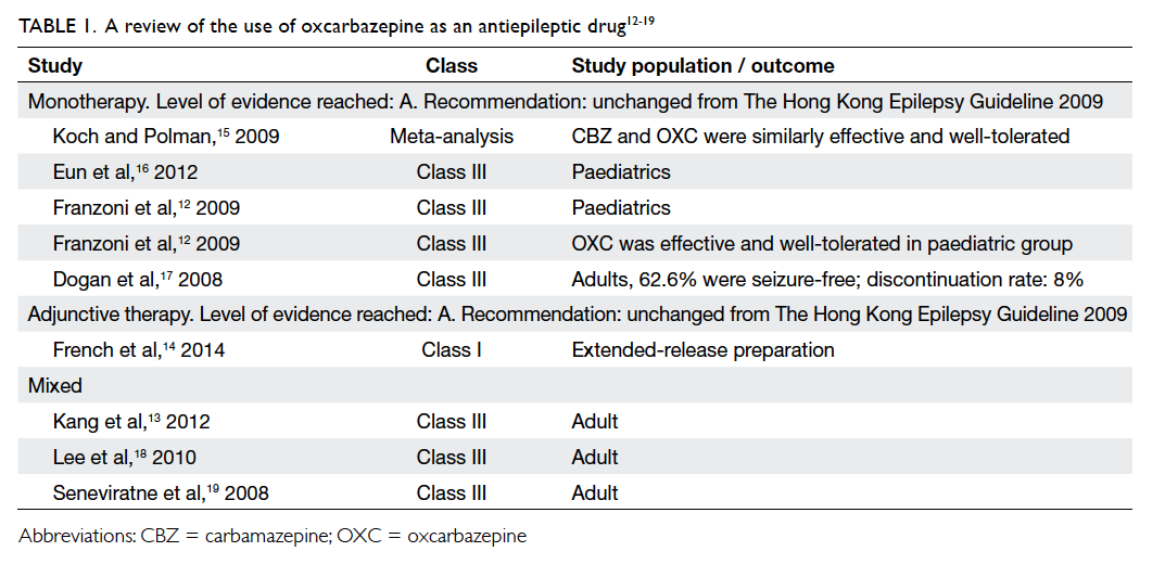
 

Table 1. A review of the use of oxcarbazepine as an antiepileptic drug12 13 14 15 16 17 18 19
 
Statement 2: The level of evidence for OXC monotherapy remains unchanged (Level A).
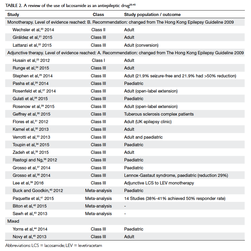
 
Lacosamide
Lacosamide (LCS) produces slow inactivation of neuronal sodium channels. We found one Class II study and two Class III studies on the use of LCS monotherapy and two Class III studies with mixed indications (Table 220 21 22 23 24 25 26 27 28 29 30 31 32 33 34 35 36 37 38 39 40 41 42 43 44 45). One conversion study showed that 425 patients completed LCS maintenance with a favourable safety profile at a nominal dose of 400 mg per day.20 In another study, the seizure-free rate was 72.3% at 1 year and the withdrawal rate was 15%.21 In the study by Lattanzi et al,22 58 patients were converted from a background single AED to LCS with just over half (55.2%) becoming seizure-free. Only 20.8% of patients reported mild-to-moderate adverse events. The FDA has approved use of LCS as monotherapy in epilepsy since September 2014 and there was a plan to seek its approval for use with the same indication in Europe in 2016.
 

Table 2. A review of the use of lacosamide as an antiepileptic drug20 21 22 23 24 25 26 27 28 29 30 31 32 33 34 35 36 37 38 39 40 41 42 43 44 45
 
Statement 3: The level of evidence for LCS monotherapy reaches Level B.
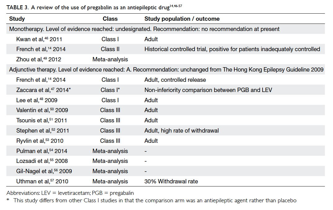
 
Pregabalin
Pregabalin (PGB) has binding properties to the alpha-2-delta units of calcium channels. We found one Class I study, one Class II study, and one meta-analysis for PGB under this indication (Table 314 46 47 48 49 50 51 52 53 54 55 56 57). Pregabalin was compared with LTG in a study of 330 patients using a double-blind, non-inferiority design with the primary efficacy endpoint being the proportion of patients to achieve seizure freedom for 6 months. In the study, however, PGB was inferior to LTG on both intention-to-treat and per-protocol analyses.46 In the study by French et al,14 conversion from a first or second AED to PGB was undertaken in 125 patients and the results showed that PGB monotherapy was safe and efficacious in partial epilepsy. No recommendation may be given at this stage regarding the use of PGB monotherapy in epilepsy.
 

Table 3. A review of the use of pregabalin as an antiepileptic drug14 46 47 48 49 50 51 52 53 54 55 56 57
 
Statement 4: The level of evidence for PGB monotherapy remains unchanged (not designated).
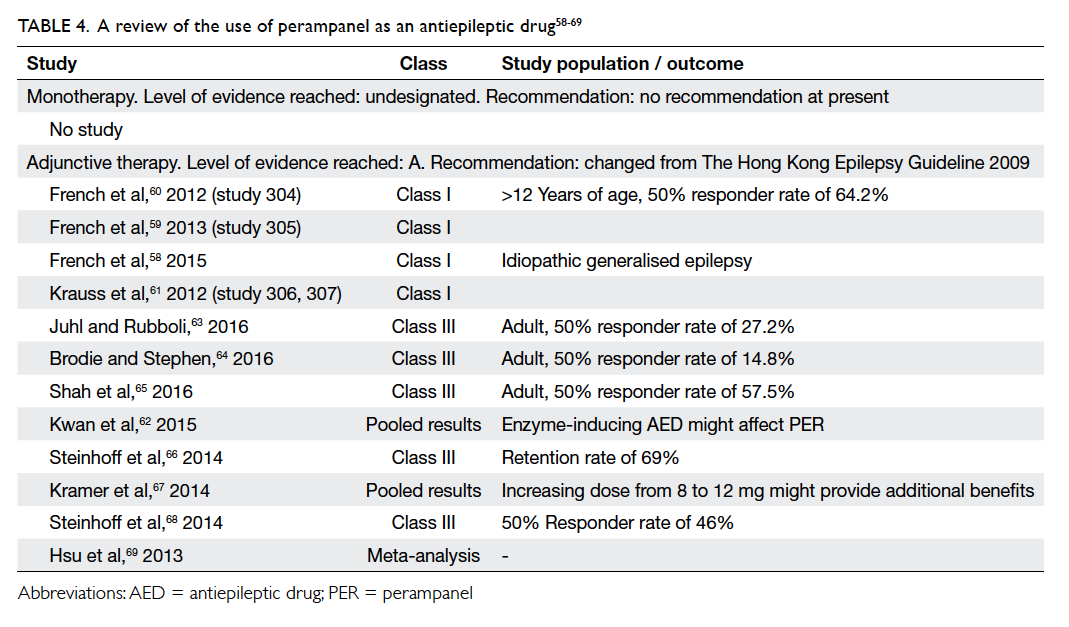
 
Perampanel
No study on the use of perampanel (PER) monotherapy could be found using the current search criteria. Other information pertaining to PER is shown in Table 4.58 59 60 61 62 63 64 65 66 67 68 69
 

Table 4. A review of the use of perampanel as an antiepileptic drug58 59 60 61 62 63 64 65 66 67 68 69
 
Statement 5: The level of evidence for PER monotherapy remains unchanged (no recommendation).
 
Adjunctive therapy
Levetiracetam
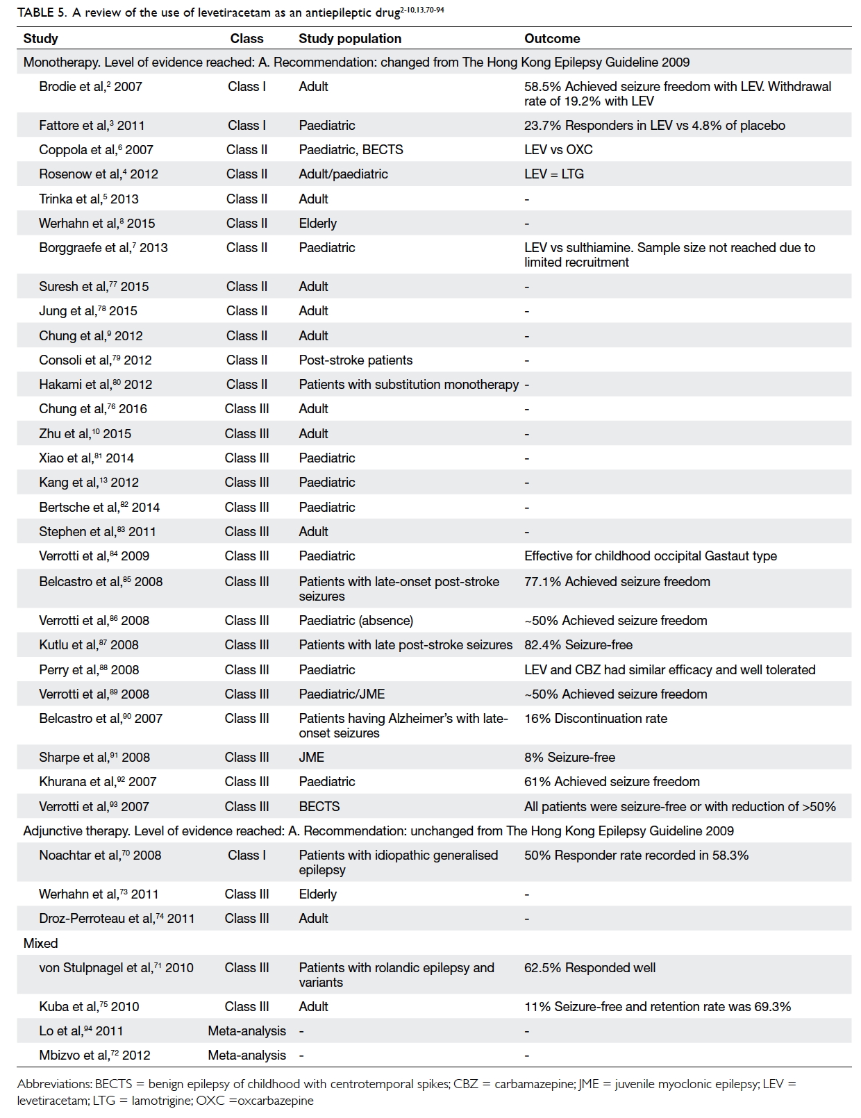
One Class I and two Class III studies were identified using the search criteria. In addition, two Class III studies reported mixed indications and two meta-analyses were published (Table 52 3 4 5 6 7 8 9 10 13 70 71 72 73 74 75 76 77 78 79 80 81 82 83 84 85 86 87 88 89 90 91 92 93 94). In the only Class I study available for this indication, patients with idiopathic generalised epilepsy were randomised to receive LEV 3000 mg per day or placebo. The results suggested that a reduction by ≥50% of myoclonic seizures may be achieved in 58.3% of patients.70 One Class III study reported the use of LEV among patients with rolandic epilepsy or variants: a >50% reduction in seizure frequency was achieved by 62.5% of patients.71 There is no new recommended level of evidence for LEV under this indication.
 

Table 5. A review of the use of levetiracetam as an antiepileptic drug2 3 4 5 6 7 8 9 10 13 70 71 72 73 74 75 76 77 78 79 80 81 82 83 84 85 86 87 88 89 90 91 92 93 94
 
A review of the behavioural side-effects of LEV revealed possible variation among paediatric and adult subjects. Nervousness, aggression, and hostile behaviour have been reported as putative behavioural adverse events. In paediatric cohorts, the proportion of such adverse events was 20% to 30%.70 71 72 94 By comparison, the behavioural side-effects in adults were less prominent.72 73 74 75 94
 
Statement 6: The level of evidence for LEV adjunctive therapy remains unchanged (Level A).
 
Oxcarbazepine
One Class I study and three Class III studies (with mixed indications) were identified (Table 112 13 14 15 16 17 18 19). In the study by the PROSPER Investigators Study Group, adjunctive OXC reduced seizure magnitude by 38.2% to 42.9%. Adverse event rates and safety profiles suggested improved tolerability.95 Oxcarbazepine is currently licensed for adjunctive therapy in epilepsy and no change to the current recommended level of evidence was made.
 
Statement 7: The level of evidence for OXC adjunctive therapy remains unchanged (Level A).
 
Lacosamide
Three pivotal clinical studies outlined the clinical usefulness of LCS in patients with refractory epilepsy: one Phase II and two Phase III studies.76 96 97 These 12-week, randomised, double-blind, placebo-controlled, multicentre trials enrolled subjects with partial-onset seizures with or without secondary generalisation who were not adequately controlled with one to three concomitant AEDs. Study 1 compared doses of LCS 200, 400, and 600 mg/day with placebo.96 Study 2 compared doses of LCS 400 and 600 mg/day with placebo.76 Study 3 compared doses of LCS 200 and 400 mg/day with placebo.97 Following an 8-week phase to establish baseline seizure frequency, subjects were titrated to the randomised dose. During the titration phase in all three trials, treatment was initiated at 100 mg/day (50 mg given twice daily) and increased by weekly increments of 100 mg/day to the target dose. The titration phase lasted 6 weeks in Study 1 and Study 2 and 4 weeks in Study 3. In all three trials, the titration phase was followed by a maintenance phase for 12 weeks. The primary endpoint was reduction in 28-day seizure frequency (baseline to maintenance phase) compared with the placebo group. A statistically significant effect was observed with LCS treatment at doses of 200 mg/day (Study 3), 400 mg/day (Study 1, 2, and 3), and 600 mg/day (Study 1 and 2).
 
An observational phase IV open-label study to assess the efficacy, safety, tolerability, and additional outcomes of LCS in Hong Kong patients aged ≥18 years showed that LCS had efficacy and adverse effects similar to those described in the literature from other parts of the world. In a cohort of 105 patients, the proportion who achieved a 50% reduction in seizure frequency was 54.5 with a mean titration time of 6.75 weeks and a mean maintenance dose of 158.6 mg/day. The efficacy profile was satisfactory whether or not LCS was combined with concomitant sodium channel blockers (45.8% vs 46.5%). The side-effect profile included apprehension and aggression, drowsiness and tiredness, headache, memory problems, dizziness, numbness, and gait disturbance (local data).
 
Statement 8: The level of evidence for LCS as adjunctive therapy reaches Level A.
 
Pregabalin
Three Class I studies, four Class III studies, and four meta-analyses were found pertaining to PGB under this indication (Table 314 46 47 48 49 50 51 52 53 54 55 56 57). One study evaluated the efficacy and tolerability of adjunctive PGB as a controlled-release formulation. The 50% responder rate (ie percentage of patients achieving 50% reduction in seizure frequency) was 45.9% for a daily dose of 330 mg.98 Another randomised study tested PGB versus LEV in a head-to-head comparison in 409 patients. The drug PGB was non-inferior to LEV with a similar tolerability to LEV as adjunctive therapy.47 In a multicentre, randomised study of PGB versus placebo, PGB was effective and tolerable as adjunctive therapy in the Asian population.48 This drug is currently licensed for adjunctive therapy in epilepsy and there is no change to the level of evidence regarding its recommended use.
 
Statement 9: The level of evidence for PGB as adjunctive therapy remains unchanged (Level A).
 
Perampanel
A total of four Class I clinical studies demonstrated the efficacy of PER among patients with refractory epilepsy.58 59 60 61 These were all double-blind studies and all evaluated the 50% responder rate as a seizure outcome. The corresponding risk ratio for 50% responder rate for 4 mg, 8 mg, and 12 mg were 1.54, 1.8, and 1.72. The most common treatment-emergent adverse effects were dizziness, drowsiness, headache, fatigue, and nasopharyngitis. The pooled results suggested that a higher dose was more efficacious if the side-effects could be tolerated.62 There was one ongoing study on the use of PER among patients with secondary generalised seizures.
 
Statement 10: The level of evidence for PER as adjunctive therapy reaches Level A.
 
Part B: Generic drugs
The last version of The Hong Kong Epilepsy Guideline gave advice on the use of generic drugs, details of which can be revisited in the original guideline of 2009.1 There might be a perceived difference between pharmaceutical equivalence, which is the requirement of the exact product, and bioequivalence, which is the concept of assigning no difference among products in terms of drug absorption. There have been positional statements that outline the possible risks involved when switching antiepileptic agents from a brand to a generic preparation.99 Clinicians are understandably perturbed by the prospect of inadvertent seizures and loss of quality of life for their patients. The criteria applied by authorities to license generic products give rise to various issues. For instance, the concept of bioequivalence does not require the generic product to demonstrate clinical efficacy among patients. Most bioequivalence studies are performed among healthy subjects rather than individual patients. Antiepileptic drugs are placed in the same category as immunosuppressants and psychotropic drugs, in which generic substitution is necessarily given consideration before implementation. The benefit of generic AEDs is clear in countries where health care financing is either state-run or public-funded, but may still be important in terms of patient choice in countries where private health care or an insurance-based system is practised because patients may want to lower their premium by using generic products. It may be argued that the use of generic products will increase the potential availability of drugs to a broader population of patients including those who are underprivileged or resident in communities where the drug budget is restricted.
 
There is a growing need for review and update of recommended guidelines on issues related to generic products as the evidence for newer drugs has become more eminent. The prescription of and expenditure on newer agents has risen sharply over the last 5 to 10 years. Clinicians now have a far greater number of AEDs at their disposal compared with a decade ago. There is divided opinion in the professional community about the use of generic products and when it will be considered optimal and safe for epilepsy patients. In general, communities that rely on a state-financed or government-funded health care system are under greater pressure to consider generic product prescription, compared with private-funded or out-of-pocket payment health care financing systems.
 
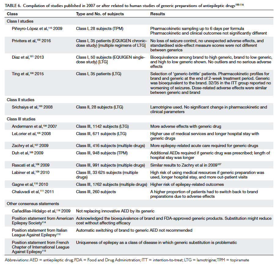
Our literature search identified 13 studies published in or after 2007 that fulfilled the initial inclusion criteria. Four studies were of the Class I category, one of the Class II category, and eight of the Class III category (Table 6100 101 102 103 104 105 106 107 108 109 110 111 112 113 114 115 116). Six studies had LTG as the study AED.100 101 102 103 104 105 Two studies had topiramate as the study focus106 115 and the remaining studies adopted multiple drug regimens.107 108 109 110 111 A good level of evidence came from a randomised controlled trial of ‘generic-brittle’ patients in a double-blind, multiple-dose, steady-state, fully replicated crossover bioequivalence study of LTG. The study demonstrated that the generic product was bioequivalent to the brand medication. Such observations were supported by the secondary outcomes of seizure control and tolerability—32 of 35 patients reported no deterioration of seizures, and dose-related adverse events were experienced by 14 patients while on the generic product and 15 patients while on the brand product. The study highlighted the use of the therapeutic level as a guide over a period of time while the patient is switched from brand to generic or vice versa.100 Two Class I studies with preliminary results disseminated during the annual meeting of the American Epilepsy Society in 2015 showed no deviation from FDA’s bioequivalence standards in Cmax and area under the curve when comparing two most disparate generic products in a single dose and chronic disease model respectively (methodology given in Diaz et al in 2013101). One well-designed study of 35 patients randomised patients from six epilepsy centres to receive LTG as one of two treatment sequences that comprised four study periods of 14 days each, during which time balanced doses of an oral generic LTG product were given every 12 hours. Disparate generic LTG in patients with epilepsy demonstrated bioequivalence with no detectable difference in clinical effects.102 A similar result was found from the only Class II study from our literature search.103 The best level of evidence in epilepsy patients supported the switch of LTG (sodium channel blocker) from brand to generic preparation. It remains controversial whether these findings can be extrapolated to other AEDs because LTG is by far one of the most widely used first-line AEDs.
 

Table 6. Compilation of studies published in 2007 or after related to human studies of generic preparations of antiepileptic drugs100 101 102 103 104 105 106 107 108 109 110 111 112 113 114 115 116
 
Most Class III studies indicated an opposite result compared with the Class I and II studies. These studies showed that generic substitution may result in increased acute seizure–related events and higher use of medical services. The switch-back rates for AEDs from generic to brand were higher in these studies. Of note, these studies had larger sample sizes but all the studies were retrospective in nature. These studies might also have involved a wide range of prescribing practices and some patient factors might not have been taken into account.
 
Overall, most studies suggested bioequivalence of brand and generic AEDs. This result was also in keeping with a meta-analysis which concluded that if only the highest level of evidence is considered, there is no significant difference in terms of seizure control, whether or not the patient is taking brand or generic products.117 A UK pharmacovigilance body, the Medicine and Healthcare products Regulatory Agency, issued guidelines regarding the use of generic products in 2013 and specifically divided AEDs into three categories, each of which had specific recommendations regarding the switching of brand to generic products (Appendix).118 Category 1 relates to products among which a specific manufacturer’s product should be ensured (eg phenytoin, carbamazepine, phenobarbital, and primidone). Category 2 relates to products for which generic switching is considered neutral, but clinical judgement should be exercised in so doing (eg sodium valproate, LTG, OXC, topiramate). Category 3 relates to products for which generic substitution is considered safe (eg LEV, gabapentin) [Table 6100 101 102 103 104 105 106 107 108 109 110 111 112 113 114 115 116]. The UK National Institute for Health and Care Excellence guideline119 recommended that a consistent supply should be made available to the epilepsy patient unless the prescriber, in consultation with the patient, considers that this is not a concern.
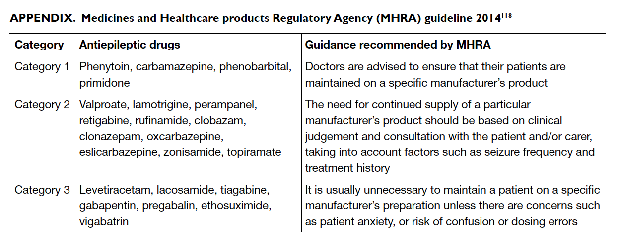
 

Appendix. Medicines and Healthcare products Regulatory Agency (MHRA) guideline 2014118
 
We acknowledge the controversy about switching from a brand to a generic product. There appears to be a divide in the positional statements and guidelines between countries with public-funded health care and those with private health care. Many associations, including the Italian League Against Epilepsy,112 American Academy of Neurology,114 and the French Chapter of ILAE113 have expressed concerns about generic substitution of AEDs, emphasising the uniqueness of epilepsy as a class of disease in which generic substitution is problematic when carried out for this indication. The latest position statement from the American Epilepsy Society acknowledges the bioequivalence of brand and FDA-approved generic products and the fact that substitution may reduce cost without compromising efficacy. The Society advises the importance of using either immediate-release or extended-release preparations uniformly throughout the switching process. They acknowledge that tablet or capsule colour or shape may impact drug compliance. They also state that the counselling of switching should include an indication of bioequivalence and not inferiority when the information is conveyed to the patient(s) and their family members.114
 
A pilot study pioneered by the Hospital Authority Head Office on the switching of phenytoin from a generic back to a brand product due to supplier issues suggested that proper counselling and follow-up logistics in conjunction with a pre- and post-drug level at 2 weeks may be adequate for the exercise. In 40 patients recruited from the Prince of Wales Hospital and Queen Mary Hospital, no patients developed a toxic level of plasma phenytoin during the switching process (four patients had a toxic-level pre-switching that remained post-switching). Plasma phenytoin concentration increased in 23 patients and decreased in 17. The conclusion was that there was no consistent trend in the change of plasma drug level (personal communication). Apart from isolated cases of reported dizziness, no serious adverse event occurred. The rate of hospitalisation as a result of the switch in that study was not available to us at the time of writing this review.
 
Statement 11: There is Level A evidence for generic substitution of LTG (a sodium channel blocker), taking into account the drug’s pharmacodynamics and pharmacokinetics.
 
The HKES upholds the safety of patients above all else. Following a review of the current evidence, the HKES has made the following revisions for the reference of physicians. Doctors can initiate treatment in patients with epilepsy with either a brand or generic product. Switching from a brand to a generic product or between generic products requires great care by clinicians and health care administrators. Automatic substitution at a pharmacy level is not recommended. If switching takes place as a result of cost considerations, prescriber and patient approval must be sought, in liaison with the pharmacist. Prescriber approval is not equivalent to a medical decision. The course of treatment, including choice of drug and dosage, is determined by the doctor and forms part of a medical decision. When the use of generic drugs is based on cost-effective analyses, prescriber approval is a logistic and economic decision. Depending on the type of health care setting, a request for generic substitution may begin with the patient or the health administrator, in liaison with the attending doctor/pharmacist. Patient approval may not be equivalent to medical consent. This can be a requirement of the health care system to which the patient belongs or a self-initiated step from the patient who has subscribed to insurance plans with affordable premiums. The physician should discuss any switch with the patient from both a medical and layman’s perspective. Good communication is considered fundamental to the provision of care.120 Therefore, in a private health care system, the choice for generic drugs may begin with a patient’s request, followed by prescriber approval. In a public health care system, the choice for generic drugs may begin with prescriber’s request, followed by patient approval. Follow-up and monitoring logistics should be mutually agreed to ensure patient safety. A change in the physical appearance of medications may hinder compliance. This facet of the switch must be taken into account by all parties. In the special situation where switching from a brand to a generic product takes place among patients who have achieved remission while on antiepileptic therapy, clinicians must take into account the drug’s pharmacokinetics and the support of medical services. Assistance from nursing staff, enlisting therapeutic blood monitoring, and the option to use the AED as a self-financed item (both public and private setting) should be made available.
 
Statement 12: Controversy exists over the use of generic products among patients who are currently taking brand medications. Prescriber and patient approval is pivotal. There should be good communication between doctors and patients; enlisting assistance from doctors, nurses, and pharmacists; therapeutic blood monitoring if available; and the option of brand AED as a self-financed item. The physical appearance of generic drugs may hamper drug compliance. Support from medical services is recommended. In the longer term, the benefit of flexibility and the option to have balanced use of generic and brand drugs may need to be addressed by institutions and regulatory bodies.
 
Conclusions
New evidence on AEDs has arisen since the publication of the Hong Kong Epilepsy Guideline in 2009. There is Level A evidence for LEV monotherapy and Level B evidence for LCS monotherapy. There is Level A evidence for LCS and PER adjunctive therapy. No change to the level of evidence is evident for LEV, OXC, and PGB. The use of generic preparations of AEDs should be considered following prescriber and patient approval, with support from medical services (doctors, nurses, pharmacists). It is important to emphasise that a generic preparation is not inferior, that shape and colour of tablets may be different, there may be therapeutic blood monitoring (if available), and patients may have the option of self-financing items.
 
Appendix
Additional material related to this article can be found on the HKMJ website. Please go to <http://www.hkmj.org>, and search for the article.
 
Acknowledgement
This project was supported in part by an unrestricted grant from the Hong Kong Epilepsy Society.
 
Disclaimer
This consensus statement is designed to assist clinicians by providing an analytical framework for the drug treatment of epilepsy. It is not intended to establish a community standard of care, replace a clinician’s medical judgement, or establish a protocol for all patients.
 
References
1. Guideline Development Group, Hong Kong Epilepsy Society. The Hong Kong Epilepsy Guideline 2009. Hong Kong Med J 2009;15 Suppl 5:6S-28S.
2. Brodie MJ, Perucca E, Ryvlin P, et al. Comparison of levetiracetam and controlled-release carbamazepine in newly diagnosed epilepsy. Neurology 2007;68:402-8. Crossref
3. Fattore C, Boniver C, Capovilla G, et al. A multicenter, randomized, placebo-controlled trial of levetiracetam in children and adolescents with newly diagnosed absence epilepsy. Epilepsia 2011;52:802-9. Crossref
4. Rosenow F, Schade-Brittinger C, Burchardi N, et al. The LaLiMo Trial: lamotrigine compared with levetiracetam in the initial 26 weeks of monotherapy for focal and generalised epilepsy—an open label, prospective, randomised controlled multicenter study. J Neurol Neurosurg Psychiatry 2012;83:1093-8. Crossref
5. Trinka E, Marson AG, Van Paesschen W, et al. KOMET: an unblinded, randomised, two parallel-group, stratified trial comparing the effectiveness of levetiracetam with controlled-release carbamazepine and extended-release sodium valproate as monotherapy in patients with newly diagnosed epilepsy. J Neurol Neurosurg Psychiatry 2013;84:1138-47. Crossref
6. Coppola G, Franzoni E, Verrotti A, et al. Levetiracetam or oxcarbazepine as monotherapy in newly diagnosed benign epilepsy of childhood with centrotemporal spikes (BECTS): An open-label, parallel group trial. Brain Dev 2007;29:281-4. Crossref
7. Borggraefe I, Bonfert M, Bast T, et al. Levetiracetam vs. sulthiame in benign epilepsy with centrotemporal spikes in childhood: A double-blinded, randomized, controlled trial (German HEAD study). Eur J Paediatr Neurol 2013;17:507-14. Crossref
8. Werhahn KJ, Trinka E, Dobesberger J, et al. A randomized, double-blind comparison of antiepileptic drug treatment in the elderly with new-onset focal epilepsy. Epilepsia 2015;56:450-9. Crossref
9. Chung S, Ceja H, Gawlowicz J, et al. Levetiracetam extended release conversion to monotherapy for the treatment of patients with partial-onset seizures: A double-blind, randomised, multicentre, historical control study. Epilepsy Res 2012;101:92-102. Crossref
10. Zhu F, Lang SY, Wang XQ, et al. Long-term effectiveness of antiepileptic drug monotherapy in partial epileptic patients: A 7-year study in an epilepsy center in China. Chin Med J (Engl) 2015;128:3015-22. Crossref
11. Glauser T, Ben-Menachem E, Bourgeois B, et al. Updated ILAE evidence review of antiepileptic drug efficacy and effectiveness as initial monotherapy for epileptic seizures and syndromes. Epilepsia 2013;54:551-63. Crossref
12. Franzoni E, Gentile V, Pellicciari A, et al. Prospective study on long-term treatment with oxcarbazepine in pediatric epilepsy. J Neurol 2009;256:1527-32. Crossref
13. Kang HC, Hu Q, Liu XY, et al. A follow-up study on newer anti-epileptic drugs as add-on and monotherapy for partial epilepsy in China. Chin Med J (Engl) 2012;125:646-51.
14. French J, Kwan P, Fakhoury T, et al. Pregabalin monotherapy in patients with partial-onset seizures: a historical-controlled trial. Neurology 2014;82:590-7. Crossref
15. Koch MW, Polman SK. Oxcarbazepine versus carbamazepine monotherapy for partial onset seizures. Cochrane Database Syst Rev 2009;(4):CD006453. Crossref
16. Eun SH, Kim HD, Chung HJ, et al. A multicenter trial of oxcarbazepine oral suspension monotherapy in children newly diagnosed with partial seizures: A clinical and cognitive evaluation. Seizure. 2012;21:679-84. Crossref
17. Dogan EA, Usta BE, Bilgen R, Senol Y, Aktekin B. Efficacy, tolerability, and side effects of oxcarbazepine monotherapy: A prospective study in adult and elderly patients with newly diagnosed partial epilepsy. Epilepsy Behav 2008;13:156-61. Crossref
18. Lee SA, Heo K, Kim WJ, et al. Clinical feasibility of immediate overnight switching from slow-release carbamazepine to oxcarbazepine in Korean patients with refractory partial epilepsy. Seizure 2010;19:356-8. Crossref
19. Seneviratne U, D’Souza W, Cook M. Long-term assessment of oxcarbazepine in a naturalistic setting: a retrospective study. Acta Neurol Scand 2008;117:367-9. Crossref
20. Wechsler RT, Li G, French J, et al. Conversion to lacosamide monotherapy in the treatment of focal epilepsy: Results from a historical-controlled, multicenter, double-blind study. Epilepsia 2014;55:1088-98. Crossref
21. Giráldez BG, Toledano R, García-Morales I, et al. Long-term efficacy and safety of lacosamide monotherapy in the treatment of partial-onset seizures: a multicenter evaluation. Seizure 2015;29:119-22. Crossref
22. Lattanzi S, Cagnetti C, Foschi N, Provinciali L, Silvestrini M. Lacosamide monotherapy for partial onset seizures. Seizure 2015;27:71-4. Crossref
23. Husain A, Chung S, Faught E, Isojarvi J, McShea C, Doty P. Long-term safety and efficacy in patients with uncontrolled partial-onset seizures treated with adjunctive lacosamide: results from a Phase III open-label extension trial. Epilepsia 2012;53:521-8. Crossref
24. Runge U, Arnold S, Brandt C, et al. A noninterventional study evaluating the effectiveness and safety of lacosamide added to monotherapy in patients with epilepsy with partial-onset seizures in daily clinical practice: The VITOBA study. Epilepsia 2015;56:1921-30. Crossref
25. Stephen LJ, Kelly K, Parker P, Brodie MJ. Adjunctive lacosamide—5 years’ clinical experience. Epilepsy Res 2014;108:1385-91. Crossref
26. Pasha I, Kamate M, Didagi SK. Efficacy and tolerability of lacosamide as an adjunctive therapy in children with refractory partial epilepsy. Pediatr Neurol 2014;51:509-14. Crossref
27. Rosenfeld W, Fountain NB, Kaubrys G, et al. Safety and efficacy of adjunctive lacosamide among patients with partial-onset seizures in a long-term open-label extension trial of up to 8 years. Epilepsy Behav 2014;41:164-70. Crossref
28. Gulati P, Cannell P, Ghia T, et al. Lacosamide as adjunctive therapy in treatment-resistant epilepsy in childhood. J Paediatr Child Health 2015;51:794-7. Crossref
29. Rosenow F, Kelemen A, Ben-Menachem E, et al. Long-term adjunctive lacosamide treatment in patients with partial-onset seizures. Acta Neurol Scand 2015 Jul 2. Epub ahead of print.
30. Geffrey AL, Belt OD, Paolini JL, Thiele EA. Lacosamide use in the treatment of refractory epilepsy in tuberous sclerosis complex. Epilepsy Res 2015;112:72-5. Crossref
31. Flores L, Kemp S, Colbeck K, et al. Clinical experience with oral lacosamide as adjunctive therapy in adult patients with uncontrolled epilepsy: a multicentre study in epilepsy clinics in the United Kingdom (UK). Seizure 2012;21:512-7. Crossref
32. Kamel JT, DeGruyter MA, D’Souza WJ, Cook MJ. Clinical experience with using lacosamide for the treatment of epilepsy in a tertiary centre. Acta Neurol Scand 2013;127:149-53. Crossref
33. Verrotti A, Loiacono G, Pizzolorusso A, et al. Lacosamide in pediatric and adult patients: comparison of efficacy and safety. Seizure 2013;22:210-6. Crossref
34. Toupin JF, Lortie A, Major P, et al. Efficacy and safety of lacosamide as an adjunctive therapy for refractory focal epilepsy in paediatric patients: a retrospective single-centre study. Epileptic Disord 2015;17:436-43.
35. Zadeh WW, Escartin A, Byrnes W, et al. Efficacy and safety of lacosamide as first add-on or later adjunctive treatment for uncontrolled partial-onset seizures: A multicentre open-label trial. Seizure 2015;31:72-9. Crossref
36. Rastogi RG, Ng YT. Lacosamide in refractory mixed pediatric epilepsy: a prospective add-on study. J Child Neurol 2012;27:492-5. Crossref
37. Grosso S, Parisi P, Spalice A, Verrotti A, Balestri P. Efficacy and safety of lacosamide in infants and young children with refractory focal epilepsy. Eur J Paediatr Neurol 2014;18:55-9. Crossref
38. Grosso S, Coppola G, Cusmai R, et al. Efficacy and tolerability of add-on lacosamide in children with Lennox-Gastaut syndrome. Acta Neurol Scand 2014;129:420-4. Crossref
39. Lee JW, Alam J, Llewellyn N, et al. Open label trial of add on lacosamide versus high dose levetiracetam monotherapy in patients with breakthrough seizures. Clin Neuropharmacol 2016;39:128-31. Crossref
40. Buck ML, Goodkin HP. Use of lacosamide in children with refractory epilepsy. J Pediatr Pharmacol Ther 2012;17:211-9. Crossref
41. Paquette V, Culley C, Greanya ED, Ensom MH. Lacosamide as adjunctive therapy in refractory epilepsy in adults: a systematic review. Seizure 2015;25:1-17. Crossref
42. Biton V, Gil-Nagel A, Isojarvi J, et al. Safety and tolerability of lacosamide as adjunctive therapy for adults with partial-onset seizures: Analysis of data pooled from three randomized, double-blind, placebo-controlled clinical trials. Epilepsy Behav 2015;52:119-27. Crossref
43. Sawh SC, Newman JJ, Deshpande S, Jones PM. Lacosamide adjunctive therapy for partial-onset seizures: a meta-analysis. PeerJ 2013;1:e114. Crossref
44. Yorns WR Jr, Khurana DS, Carvalho KS, Hardison HH, Legido A, Valencia I. Efficacy of lacosamide as adjunctive therapy in children with refractory epilepsy. J Child Neurol 2014;29:23-7. Crossref
45. Novy J, Bartolini E, Bell GS, Duncan JS, Sander JW. Long-term retention of lacosamide in a large cohort of people with medically refractory epilepsy: a single centre evaluation. Epilepsy Res 2013;106:250-6. Crossref
46. Kwan P, Brodie MJ, Kälviäinen R, Yurkewicz L, Weaver J, Knapp LE. Efficacy and safety of pregabalin versus lamotrigine in patients with newly diagnosed partial seizures: a phase 3, double-blind, randomised, parallel-group trial. Lancet Neurol 2011;10:881-90. Crossref
47. Zaccara G, Almas M, Pitman V, Knapp L, Posner H. Efficacy and safety of pregabalin versus levetiracetam as adjunctive therapy in patients with partial seizures: a randomized, double-blind, noninferiority trial. Epilepsia 2014;55:1048-57. Crossref
48. Lee BI, Yi S, Hong SB, et al. Pregabalin add-on therapy using a flexible, optimized dose schedule in refractory partial epilepsies: a double-blind, randomized, placebo-controlled, multicenter trial. Epilepsia 2009;50:464-74. Crossref
49. Zhou Q, Zheng J, Yu L, Jia X. Pregabalin monotherapy for epilepsy. Cochrane Database Syst Rev 2012;(10):CD009429. Crossref
50. Valentin A, Moran N, Hadden R, et al. Pregabalin as adjunctive therapy for partial epilepsy: an audit study in 96 patients from the South East of England. Seizure 2009;18:450-2. Crossref
51. Tsounis S, Kimiskidis VK, Kazis D, et al. An open-label, add-on study of pregabalin in patients with partial seizures: a multicenter trial in Greece. Seizure 2011;20:701-5. Crossref
52. Stephen LJ, Parker P, Kelly K, Wilson EA, Leach V, Brodie MJ. Adjunctive pregabalin for uncontrolled partial-onset seizures: findings from a prospective audit. Acta Neurol Scand 2011;124:142-5. Crossref
53. Ryvlin P, Kälviäinen R, Von Raison F, Giordano S, Emir B, Chatamra K. Pregabalin in partial seizures: a pragmatic 21-week, open-label study (PREPS). Eur J Neurol 2010;17:726-32. Crossref
54. Pulman J, Hemming K, Marson AG. Pregabalin add-on for drug-resistant partial epilepsy. Cochrane Database Syst Rev 2014;(3):CD005612. Crossref
55. Lozsadi D, Hemming K, Marson AG. Pregabalin add-on for drug-resistant partial epilepsy. Cochrane Database Syst Rev 2008;(1):CD005612.
56. Gil-Nagel A, Zaccara G, Baldinetti F, Leon T. Add-on treatment with pregabalin for partial seizures with or without generalisation: pooled data analysis of four randomised placebo-controlled trials. Seizure 2009;18:184-92. Crossref
57. Uthman BM, Bazil CW, Beydoun A, et al. Long-term add-on pregabalin treatment in patients with partial-onset epilepsy: pooled analysis of open-label clinical trials. Epilepsia 2010;51:968-78. Crossref
58. French JA, Krauss GL, Wechsler RT, et al. Perampanel for tonic-clonic seizures in idiopathic generalized epilepsy. A randomized trial. Neurology 2015;85:950-7. Crossref
59. French JA, Krauss GL, Steinhoff BJ, et al. Evaluation of adjunctive perampanel in patients with refractory partial-onset seizures: results of randomized global phase III study 305. Epilepsia 2013;54:117-25. Crossref
60. French JA, Krauss GL, Biton V, et al. Adjunctive perampanel for refractory partial-onset seizures. Randomized phase III study 304. Neurology 2012;79:589-96. Crossref
61. Krauss GL, Serratosa JM, Villanueva V, et al. Randomized phase III study 306. Adjunctive perampanel for refractory partial-onset seizures. Neurology 2012;78:1408-15. Crossref
62. Kwan P, Brodie MJ, Laurenza A, FitzGibbon H, Gidal BE. Analysis of pooled phase III trials of adjunctive perampanel for epilepsy: Impact of mechanism of action and pharmacokinetics on clinical outcomes. Epilepsy Res 2015;117:117-24. Crossref
63. Juhl S, Rubboli G. Perampanel as add-on treatment in refractory focal epilepsy. The Dianalund experience. Acta Neurol Scand 2016;134:374-7. Crossref
64. Brodie MJ, Stephen LJ. Prospective audit with adjunctive perampanel: Preliminary observations in focal epilepsy. Epilepsy Behav 2016;54:100-3. Crossref
65. Shah E, Reuber M, Goulding P, Flynn C, Delanty N, Kemp S. Clinical experience with adjunctive perampanel in adult patients with uncontrolled epilepsy: A UK and Ireland multicentre study. Seizure 2016;34:1-5. Crossref
66. Steinhoff BJ, Hamer H, Trinka E, et al. A multicenter survey of clinical experiences with perampanel in real life in Germany and Austria. Epilepsy Res 2014;108:986-8. Crossref
67. Kramer LD, Satlin A, Krauss GL, et al. Perampanel for adjunctive treatment of partial-onset seizures: A pooled dose-response analysis of phase III studies. Epilepsia 2014;55:423-31. Crossref
68. Steinhoff BJ, Bacher M, Bast T, et al. First clinical experiences with perampanel—The Kork experience in 74 patients. Epilepsia 2014;55 Suppl 1:16-8. Crossref
69. Hsu WW, Sing CW, He Y, Worsley AJ, Wong IC, Chan EW. Systematic review and meta-analysis of the efficacy and safety of perampanel in the treatment of partial-onset epilepsy. CNS Drugs 2013;27:817-27. Crossref
70. Noachtar S, Andermann E, Meyvisch P, et al. Levetiracetam for the treatment of idiopathic generalized epilepsy with myoclonic seizures. Neurology 2008;70:607-16. Crossref
71. von Stulpnagel C, Kluger G, Leiz S, Holthausen H. Levetiracetam as add-on therapy in different subgroups of “benign” idiopathic focal epilepsies in childhood. Epilepsy Behav 2010;17:193-8. Crossref
72. Mbizvo GK, Dixon P, Hutton JL, Marson AG. Levetiracetam add-on for drug-resistant focal epilepsy: an updated Cochrane Review. Cochrane Database Syst Rev 2012;(9):CD001901. Crossref
73. Werhahn KJ, Klimpe S, Balkaya S, Trinka E, Krämer G. The safety and efficacy of add-on levetiracetam in elderly patients with focal epilepsy: A one-year observational study. Seizure 2011;20:305-11. Crossref
74. Droz-Perroteau C, Dureau-Pournin C, Vespignani H, et al. The EULEV cohort study: rates of and factors associated with continuation of levetiracetam after 1 year. Br J Clin Pharmacol 2011;71:121-7. Crossref
75. Kuba R, Novotná I, Brázdil M, et al. Long-term levetiracetam treatment in patients with epilepsy: 3-year follow up. Acta Neurol Scand 2010;121:83-8. Crossref
76. Chung S, Ceja H, Gawłowicz J, McShea C, Schiemann J, Lu S. Levetiracetam extended release for the treatment of patients with partial-onset seizures: A long-term, open-label follow-up study. Epilepsy Res 2016;120:7-12. Crossref
77. Suresh SH, Chakraborty A, Virupakshaiah A, Kumar N. Efficacy and safety of levetiracetam and carbamazepine as monotherapy in partial seizures. Epilepsy Res Treat 2015;2015:415082. Crossref
78. Jung DE, Yu R, Yoon JR, et al. Neuropsychological effects of levetiracetam and carbamazepine in children with focal epilepsy. Neurology 2015;84:2312-9. Crossref
79. Consoli D, Bosco D, Postorino P, et al. Levetiracetam versus carbamazepine in patients with late poststroke seizures: a multicenter prospective randomized open-label study (EpIC Project). Cerebrovasc Dis 2012;34:282-9. Crossref
80. Hakami T, Todaro M, Petrovski S, et al. Substitution monotherapy with levetiracetam vs older antiepileptic drugs: A randomized comparative trial. Arch Neurol 2012;69:1563-71. Crossref
81. Xiao F, An D, Deng H, Chen S, Ren J, Zhou D. Evaluation of levetiracetam and valproic acid as low-dose monotherapies for children with typical benign childhood epilepsy with centrotemporal spikes (BECTS). Seizure 2014;23:756-61. Crossref
82. Bertsche A, Neininger MP, Dahse AJ, et al. Initial anticonvulsant monotherapy in routine care of children and adolescents: levetiracetam fails more frequently than valproate and oxcarbazepine due to a lack of effectiveness. Eur J Pediatr 2014;173:87-92. Crossref
83. Stephen LJ, Kelly K, Parker P, Brodie MJ. Levetiracetam monotherapy—outcomes from an epilepsy clinic. Seizure 2011;20:554-7. Crossref
84. Verrotti A, Parisi P, Loiacono G, et al. Levetiracetam monotherapy for childhood occipital epilepsy of gastaut. Acta Neurol Scand 2009;120:342-6. Crossref
85. Belcastro V, Costa C, Galletti F, et al. Levetiracetam in newly diagnosed late-onset post-stroke seizures: A prospective observational study. Epilepsy Res 2008;82:223-6. Crossref
86. Verrotti A, Cerminara C, Domizio S, et al. Levetiracetam in absence epilepsy. Dev Med Child Neurol 2008;50:850-3. Crossref
87. Kutlu G, Gomceli YB, Unal Y, Inan LE. Levetiracetam monotherapy for late poststroke seizures in the elderly. Epilepsy Behav 2008;13:542-4. Crossref
88. Perry S, Holt P, Benatar M. Levetiracetam versus carbamazepine monotherapy for partial epilepsy in children less than 16 years of age. J Child Neurol 2008;23:515-9. Crossref
89. Verrotti A, Cerminara C, Coppola G, et al. Levetiracetam in juvenile myoclonic epilepsy: long-term efficacy in newly diagnosed adolescents. Dev Med Child Neurol 2008;50:29-32. Crossref
90. Belcastro V, Costa C, Galletti F, et al. Levetiracetam monotherapy in Alzheimer patients with late-onset seizures: a prospective observational study. Eur J Neurol 2007;14:1176-8. Crossref
91. Sharpe DV, Patel AD, Abou-Khalil B, Fenichel GM. Levetiracetam monotherapy in juvenile myoclonic epilepsy. Seizure 2008;17:64-8. Crossref
92. Khurana DS, Kothare SV, Valencia I, Melvin JJ, Legido A. Levetiracetam monotherapy in children with epilepsy. Pediatr Neurol 2007;36:227-30. Crossref
93. Verrotti A, Coppola G, Manco R, et al. Levetiracetam monotherapy for children and adolescents with benign rolandic seizures. Seizure 2007;16:271-5. Crossref
94. Lo BW, Kyu HH, Jichici D, Upton AM, Akl EA, Meade MO. Meta-analysis of randomized trials on first line and adjunctive levetiracetam. Can J Neurol Sci 2011;38:475-86. Crossref
95. French JA, Baroldi P, Brittain ST, Johnson JK; PROSPER Investigators Study Group. Efficacy and safety of extended-release oxcarbazepine (Oxtellar XR) as adjunctive therapy in patients with refractory partial-onset seizures: a randomized controlled trial. Acta Neurol Scand 2014;129:143-53. Crossref
96. Ben-Menachem E, Biton V, Jatuzis D, Abou-Khalil B, Doty P, Rudd GD. Efficacy and safety of oral lacosamide as adjunctive therapy in adults with partial-onset seizures. Epilepsia 2007;48:1308-17. CrossRef
97. Halász P, Kälviäinen R, Mazurkiewicz-Beldzińska M, et al. Adjunctive lacosamide for partial-onset seizures: efficacy and safety results from a randomized controlled trial. Epilepsia 2009;50:443-53. Crossref
98. French J, Brandt C, Friedman D, et al. Adjunctive use of controlled-release pregabalin in adults with treatment-resistant partial seizures: a double-blind, randomized, placebo-controlled trial. Epilepsia 2014;55:1220-8. Crossref
99. Liow K, Barkley GL, Pollard JR, Harden CL, Bazil CW; American Academy of Neurology. Position statement on the coverage of anticonvulsant drugs for the treatment of epilepsy. Neurology 2007;68:1249-50. Crossref
100. Ting TY, Jiang W, Lionberger R, et al. Generic lamotrigine versus brand-name Lamictal bioequivalence in patients with epilepsy: A field test of the FDA bioequivalence standard. Epilepsia 2015;56:1415-24. Crossref
101. Diaz FJ, Berg MJ, Krebill R, et al. Random-effects linear modeling and sample size tables for two special crossover designs of average bioequivalence studies: the four-period, two-sequence, two-formulation and six-period, three-sequence, three-formulation designs. Clin Pharmacokinet 2013;52:1033-43. Crossref
102. Privitera MD, Welty TE, Gidal BE, et al. Generic-to-generic lamotrigine switches in people with epilepsy: the randomised controlled EQUIGEN trial. Lancet Neurol 2016;15:365-72. Crossref
103. Srichaiya A, Longchoopol C, Oo-Puthinan S, Sayasathid J, Sripalakit P, Viyoch J. Bioequivalence of generic lamotrigine 100-mg tablets in healthy Thai male volunteers: A randomized, single-dose, two-period, two-sequence crossover study. Clin Ther 2008;30:1844-51. Crossref
104. Andermann F, Duh MS, Gosselin A, Paradis PE. Compulsory generic switching of antiepileptic drugs: High switchback rates to branded compounds compared with other drug classes. Epilepsia 2007;48:464-9. Crossref
105. LeLorier J, Duh MS, Paradis PE, et al. Clinical consequences of generic substitution of lamotrigine for patients with epilepsy. Neurology 2008;70(22 Pt 2):2179-86. Crossref
106. Duh MS, Paradis PE, Latrémouille-Viau D, et al. The risks and costs of multiple-generic substitution of topiramate. Neurology 2009;72:2122-9. Crossref
107. Zachry WM 3rd, Doan QD, Clewell JD, Smith BJ. Case-control analysis of ambulance, emergency room, or inpatient hospital events for epilepsy and antiepileptic drug formulation changes. Epilepsia 2009;50:493-500. Crossref
108. Rascati KL, Richards KM, Johnsrud MT, Mann TA. Effects of antiepileptic drug substitutions on epileptic events requiring acute care. Pharmacotherapy 2009;29:769-74. Crossref
109. Labiner DM, Paradis PE, Manjunath R, et al. Generic antiepileptic drugs and associated medical resource utilization in the United States. Neurology 2010;74:1566-74. Crossref
110. Gagne JJ, Avorn J, Shrank WH, Schneeweiss S. Refilling and switching of antiepileptic drugs and seizure-related events. Clin Pharmacol Ther 2010;88:347-53. Crossref
111. Chaluvadi S, Chiang S, Tran L, Goldsmith CE, Friedman DE. Clinical experience with generic levetiracetam in people with epilepsy. Epilepsia 2011;52:810-5. Crossref
112. Perucca E, Albani F, Capovilla G, Bernardina BD, Michelucci R, Zaccara G. Recommendations of the Italian League Against Epilepsy working group on generic products of antiepileptic drugs. Epilepsia 2006;47 Suppl 5:16-20. Crossref
113. French Chapter of the International League Against Epilepsy (LFCE): Recommendations on the use of generics for the treatment of epilepsy. Available from: http://www.ilae.org/visitors/MeetingProceedings/documents/PRESSRELEASEONGENERICAEDsFRENCHCHAPTER OFTHEILAE_000.pdf. Accessed Mar 2016.
114. American Epilepsy Society. Substitution of different formulations of antiepileptic drugs for the treatment of epilepsy. Available from: https://www.aesnet.org/about_aes/generic-position-statement. Accessed Mar 2016.
115. Piñeyro-López A, Piñeyro-Garza E, Gómez-Silva M, et al. Bioequivalence of single 100-mg doses of two oral formulations of topiramate: An open-label, randomized-sequence, two-period crossover study in healthy adult male Mexican volunteers. Clin Ther 2009;31:411-7. Crossref
116. Cañadillas-Hidalgo FM, Sánchez-Alvarez JC, Serrano-Castro PJ, Mercadé-Cerdá JM; en representación de la Sociedad Andaluza de Epilepsia. Consensus clinical practice guidelines of the Andalusian Epilepsy Society on prescribing generic antiepileptic drugs [in Spanish]. Rev Neurol 2009;49:41-7.
117. Yamada M, Welty TE. Generic substitution of antiepileptic drugs: a systematic review of prospective and retrospective studies. Ann Pharmacother 2011;45:1406-15. Crossref
118. Medicines & Healthcare products Regulatory Agency. Available from: https://www.gov.uk/government/organisations/medicines-and-healthcare-products-regulatory-agency. Accessed Mar 2016.
119. NICE guidance CG137. Epilepsies: diagnosis and management. Available from: https://www.nice.org.uk/guidance/cg137. Accessed Mar 2016.
120. The Medical Council of Hong Kong. Available from: http://www.mchk.org.hk/code.htm. Accessed Mar 2016.

Pages