Hip fractures are preventable: a proposal for osteoporosis screening and fall prevention in older people

Hong Kong Academy of Medicine. CC BY-NC-ND 4.0
 
MEDICAL PRACTICE  CME
Hip fractures are preventable: a proposal for osteoporosis screening and fall prevention in older people
Timothy CY Kwok, MD, FHKAM (Medicine)1,2,3; SW Law, MB, ChB, FHKAM (Orthopaedic Surgery)3,4; Edward MF Leung, MB, BS, FRCP3,5; Dicky TK Choy, MB, ChB, DCH2,3; Patti MS Lam, BSc, MSc (Health Services Management)2; Jason CS Leung, MSc2; SH Wong, MB, BS, FHKAM (Orthopaedic Surgery)6; TP Ip, MB, BS, FHKAM (Medicine)6,7; CL Cheung, BSc, PhD6,8
1 Department of Medicine and Therapeutics, Prince of Wales Hospital, The Chinese University of Hong Kong, Hong Kong
2 Jockey Club Centre for Osteoporosis Care and Control, The Chinese University of Hong Kong, Hong Kong
3 Hong Kong Osteoporosis Foundation
4 Department of Orthopaedics and Traumatology, Alice Ho Miu Ling Nethersole Hospital, Hong Kong
5 Hong Kong Association of Gerontology
6 The Osteoporosis Society of Hong Kong
7 Department of Medicine, Tung Wah Hospital, Hong Kong
8 Department of Pharmacology and Pharmacy, Li Ka Shing Faculty of Medicine, The University of Hong Kong, Hong Kong
 
Corresponding author: Prof Timothy CY Kwok (tkwok@cuhk.edu.hk)
 
 Full paper in PDF
 
Abstract
Osteoporosis is highly prevalent but underdiagnosed and undertreated in Hong Kong. Fragility fractures associated with osteoporosis often result in loss of independence and increased mortality for home-dwelling patients, imposing a high socio-economic burden on society. This issue requires urgent attention given the rapid growth of the elderly population in Hong Kong by approximately 4.3% each year. To address this situation, a group of experts convened to discuss practical ways to reduce the burden of fractures and formulated three recommendations: first, all men (aged ≥70 years) and women (aged ≥65 years) should receive universal dual-energy X-ray absorptiometry assessment for osteoporosis. Second, all men (aged ≥70 years) and women (aged ≥65 years) with a fracture-risk assessment-derived 10-year risk (hip fracture with bone mineral density) ≥3% should receive ≥3 years of anti-osteoporotic treatment. Third, comprehensive structured assessment (including dual-energy X-ray absorptiometry) should be conducted in older patients with a history of falling. By implementing these recommendations, we estimate that we could prevent 5234 hip fractures in 10 years, an annual incidence reduction of approximately 7%, and save HK$425 million in direct medical costs plus substantial indirect savings. Ample clinical and cost-effectiveness data support these recommendations, and studies in Hong Kong and abroad could serve as models on how to implement them. We are confident that by applying these recommendations rigorously and systematically, a significant reduction in hip fractures in Hong Kong is achievable.


Introduction
The Hong Kong eldercare and healthcare system faces a high burden from osteoporosis and hip fragility fractures. The prevalence of osteoporosis in people aged ≥50 years in Hong Kong has been measured as high as 37% in some studies.1 A study of 4000 community-dwelling older people in Hong Kong found that 7% of men and 11% of women had ≥1 incident of major osteoporotic fracture over 9.9 and 8.8 years of follow-up, respectively.2 The number of patients admitted for hip fracture surgery increased from 3678 in 2000 to an estimated 6300 in 2020,3 a 71.3% increase over 20 years. Although the risk of geriatric hip fracture in Hong Kong is declining slightly, this is outweighed by the growing elderly population.3 The life expectancy in Hong Kong (81.9 years for men; 87.6 years for women) is among the highest in the world,4 5 and projections suggest that by 2036, 31.1% of the population will be aged ≥65 years.6 Without intervention, the ageing of the population is predicted to result in a considerable increase in the incidence of fragility fracture, with estimates for hip fractures rising to more than 14 500 in 2040.3
 
Direct costs per hip fracture to Hong Kong public hospitals have been estimated at HK$81 120 (2017 data by Su et al7). This equates to an annual estimated cost of HK$511 million for the territory’s Hospital Authority. Although no estimates of indirect costs are available for Hong Kong, Taiwanese data from 2016 suggest they are likely to be substantial. Estimated annual indirect costs in Taiwan are HK$13 728, HK$3744, or HK$9687 per patient if discharged to a nursing home, foreign-paid home care, or domestic-paid home care, respectively.8 A Hong Kong study found that 23% of patients with hip fractures who had previously been living at home were discharged to residential care homes for the elderly.9
 
Follow-up care after hip fracture in Hong Kong is suboptimal, with low usage of bone-enhancing medication with poor follow-up rates.9 In a territory-wide retrospective study, only 8.2% of patients were prescribed anti-osteoporotic medication during the year after hip fracture.10 Among patients with primary hip fractures, 6% had fracture complications, and 4% had secondary fracture(s) within 12 months.9 The post-discharge mortality rate was also high: 17.3% died within 1 year of hip surgery versus 1.6% of age-matched controls.9
 
Local data on other major types of osteoporotic fractures are scarce. In the Hong Kong Osteoporosis Study (HKOS),11 the incidence of vertebral fracture was 194 per 100 000 person-years in men and 508 per 100 000 person-years women aged ≥50 years.12 In 2013, in Hong Kong, the prevalence of vertebral fracture was 17% in men and 30% in women aged 70 to 79 years.13 Many vertebral fractures do not require immediate medical attention, making their impact difficult to study,13 but international data suggest they are associated with substantial morbidity, possibly contributing to increased mortality.14
 
European and North American studies have demonstrated that dual-energy X-ray absorptiometry (DXA) screening followed by osteoporosis treatment in older people can substantially reduce hip fracture rates by 25% to 50% in ≤5 years and is cost-effective.15 16 17 Therefore, many fractures and associated complications, including secondary fractures and mortality, could be prevented by routine osteoporosis screening in older people and timely treatment initiation in at-risk individuals.
 
The rising burden of fragility hip fractures demands a response. Based on existing studies from Hong Kong and abroad, we believe that reducing this burden is achievable. Here, we provide clear, practical, evidence-based recommendations on how to reduce hip fracture rates in Hong Kong with the goal of reducing incidence by 6.8% annually, thereby curbing rising incidence.
 
Methods
Roundtable meetings of experts reviewing hip fracture burden in Hong Kong were held in October and December 2018 and June 2019. Tactics to relieve the primary fracture burden were discussed, and a consensus on a rigorous approach to screening, prevention, and treatment was formed.
 
The calculated predictive performance and simulated effects of our proposed strategies used a similar population to that used by Su et al18 in the Mr OS and Ms OS Hong Kong Cohort Study, employing the same statistical model and simulation analysis methods. Simulated analysis of the cohort was based on DXA scans of the hip and spine performed on men aged ≥70 years and women aged ≥65 years, without pre-selection, followed by treatment if the Fracture Risk Assessment Tool (FRAX; including bone mineral density [BMD]) score was ≥3%.18
 
The number needed to DXA scan and number needed to treat (NNT) were calculated using data derived from the same simulated analysis as that performed by Su et al.18 The present epidemiological analysis assumes that only 50% of eligible patients agree to undergo the DXA scan and engage in follow-up steps.
 
Results
Better awareness of osteoporosis screening and treatment among healthcare providers is urgently needed, as is public financial support for evidence-based tools that can reduce this problem. Discussions resulted in the formulation of three evidence-based recommendations.
 
Recommendation 1: Universal dual-energy X-ray absorptiometry screening of the hip and spine in all men aged ≥70 years and women aged ≥65 years
Rationale for using age as screening criterion
We propose that the lower age limits for screening of women and men be set to 65 and 70 years, respectively, based on a 2017 study showing that the average age of fragility fracture in Hong Kong was 82.1 years.9 From ages 65 to 69 years to 70 to 74 years, the annual incidence of hip fracture rose exponentially from 156.0 to 364.4 per 100 000 person-years in women and 102.6 to 212.2 per 100 000 person-years in men.19 The incidence continued to double in every subsequent 5-year period.19 Our proposed age thresholds align with those specified in international guidelines and reflect the differing ages at which the prevalence of reduced bone mass increases in men and women.15 20
 
Rationale for universal screening
A prospective cohort study of almost 4000 men and women aged ≥65 years in Hong Kong estimated the effectiveness of hip fracture prevention strategies using DXA.18 Based on the calculation method used in this study, we recommend a strategy of universal DXA measurement and treating patients with FRAX (including BMD measurement) risk ≥3%. The FRAX threshold of 3% was selected because our statistical analysis suggests that this prevents the greatest number of hip fractures while maintaining acceptable NNT. Furthermore, this threshold aligns with current local and international treatment guidelines,13 and a United States study found that this threshold was cost-effective.21
 
With our strategy, we found that one hip fracture can be prevented for every 111 men (aged ≥70 years) DXA scanned, with 54 subsequently treated; or for every 111 women (aged ≥65 years) DXA scanned, with 73 subsequently treated (Table 1). Using the projected older population of 1.16 million,6 and assuming that only 50% of the target population would be scanned, we could prevent 5234 hip fractures over the next 10 years.
 

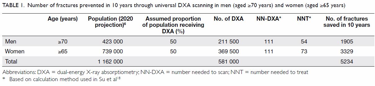
Table 1. Number of fractures prevented in 10 years through universal DXA scanning in men (aged ≥70 years) and women (aged ≥65 years)
 
To put the number of hip fractures prevented into context, we considered the projected number of hip fractures and the estimated growth rate. The estimated number of hip fractures in 2020 according to Man et al3 was 6300. If we apply the 4.3% estimated annual growth of the older population to this figure,6 by 2029, we would expect 9167 hip fractures in Hong Kong. Assuming the same 4.3% estimated annual increase also applies to the number of fractures prevented each year,6 we expect to prevent 431 hip fractures in 2020, and 627 in 2029. Based on these calculations, we estimate that by DXA screening and treating 581 000 men and women, the incidence of hip fracture could be reduced by approximately 6.8% (Table 1). As the estimated growth of the older population is 4.3% each year,6 an 6.8% annual hip fracture reduction rate would outweigh the annual increase in hip fracture cases among elderly patients due to population growth.
 
Rationale for using dual-energy X-ray absorptiometry
We selected DXA for screening because reduced hip BMD in the femoral neck measured by DXA is widely used in treatment guidelines to define osteoporosis and set drug-treatment thresholds.22 Hip BMD is strongly associated with hip fracture risk in both men and women23: its association with fragility fracture is stronger than that of spine BMD in Hong Kong Chinese people.24 25 Dual-energy X-ray absorptiometry is cheaper than magnetic resonance imaging (each DXA session cost approximately HK$500 in Hong Kong in 2018)7; it is non-invasive, and uses less irradiation than computed tomography.26 Local guidelines do not recommend the use of quantitative ultrasound for monitoring or treatment of osteoporosis, and currently consider the evidence for peripheral DXA to be insufficient.13
 
This recommendation is supported by health economics studies. Screening strategies based on DXA are more cost-effective in individuals aged ≥65 years than in younger people because hip fracture is rare in the latter.15 American and European studies have shown that screening strategies, including DXA screening, are cost-effective for prevention of hip fracture in elderly men and women.15 27 A recent Hong Kong study that modelled screening strategies found that DXA-based approaches, including universal DXA with or without pre-screening, were more cost-effective than no screening.7 In women aged ≥65 years and men aged ≥70 years, the incremental effectiveness gained by universal DXA screening versus no screening incurred no additional cost, which is well below the widely acceptable willingness to pay (US$50 000 per quality-adjusted life year).7 A modelling analysis using North American data found that universal densitometry and treatment with alendronate was highly cost-effective for women aged ≥65 years, and it was cost-saving for ambulatory women aged ≥85 years.28 The International Society for Clinical Densitometry recommends DXA assessment for all women and men aged ≥65 and ≥70 years, respectively.29 While the United States Preventative Services Task Force recommends screening in women aged ≥65 years and in younger postmenopausal women with elevated risk, it makes no recommendation for men because of insufficient evidence.22 In Hong Kong, however, the analysis of Su et al,18 which is based on extensive local data, suggested that strategies using DXA, including universal screening, can potentially be cost-effective for fracture prevention in both men and women at the age thresholds we propose. Other countries such as Australia, Malaysia, Japan, and Singapore also wholly or partially fund universal DXA screening for older people.30 31 32 33
 
Recommendation 2: ≥3 years of treatment with antiresorptive therapy for all men aged ≥70 years and women aged ≥65 years with Fracture Risk Assessment Tool score ≥3%
Rationale for using Fracture Risk Assessment Tool to guide treatment
Scanning alone would only be impactful if followed up by a treatment plan. We recommend using FRAX to guide treatment because it calculates the 10-year fracture risk from a patient’s age, body mass index, and risk-factor profile (fracture history, family history, tobacco or alcohol use, use of oral glucocorticoids, and presence of rheumatoid arthritis).34 It can be used with or without BMD data; it is the most widely adopted fracture risk assessment tool, and FRAX has been extensively validated and adapted to local populations, including that of Hong Kong.34 35
 
The FRAX risk thresholds are widely used to guide intervention in local and international osteoporosis treatment guidelines.13 36 The 2019 European guidelines recommend country-specific use of FRAX to assess risk in postmenopausal women, including BMD in intermediate-risk individuals.34 An example of a successful real-world fragility fracture-reducing intervention incorporating FRAX has been documented.37 A United Kingdom trial used a questionnaire incorporating FRAX to promote DXA screening in women aged 70 to 85 years, leading to a 28% hip fracture reduction over 5 years.37
 
Rationale for treatment duration of ≥3 years
We suggest 3 years as the duration of antiresorptive treatment based on local studies of alendronate, which showed increases in BMD of 5% to 6% at the lumbar spine and 3% to 4% at the hip after 1 year of treatment in postmenopausal women with osteoporosis.38 39 Local guidelines recommend 5 years of initial therapy with oral bisphosphonates but acknowledge that no consensus exists regarding the optimal treatment duration.13 We believe that 3 years of duration offers an appropriate balance of efficacy and cost. Additional financial support for longer-term therapy may become feasible when our recommendations begin demonstrating effectiveness in fragility fracture reduction, with improved compliance, during the first 3 years.
 
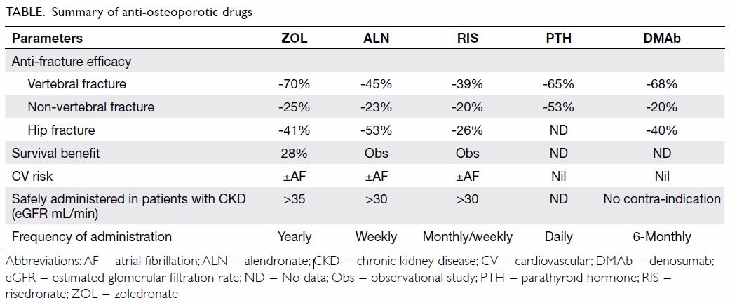
Rationale for treatment with antiresorptive drugs
Current osteoporosis treatments are appropriate for addressing the hip fracture burden in Hong Kong (Table 213). The efficacy and safety of locally approved antiresorptive therapies have been extensively validated in numerous clinical trials in men and postmenopausal women, including long-term studies.40 41 42 Cost-effectiveness analyses support their use for fracture prevention, with gains in quality-adjusted life years for patients and incremental cost-effectiveness ratios well within acceptability thresholds (vs no treatment).7 21 A meta-analysis of eight studies of four agents found that antiresorptive therapy for osteoporosis led to significant mortality-rate reductions.43 Recently, a study in Hong Kong showed that real-world use of bisphosphonates among hip fracture patients may protect against cardiovascular diseases.10 Adverse event (AE) imbalances suggesting cardioprotective effects were also observed in a randomised trial of zoledronate in 2000 women with osteopenia in New Zealand.44 Bisphosphonates are easily accessible in Hong Kong, and many patients with osteoporosis could feasibly be treated in primary care.13 Our analysis found that, with universal DXA scanning of men aged ≥70 years and women aged ≥65 years, a treatment threshold of FRAX (with BMD) risk ≥3% for hip fracture yields a NNT of 54 for men and 73 for women to prevent one fracture.
 

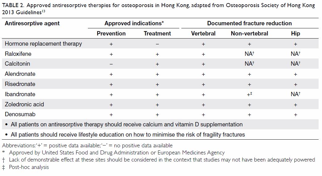
Table 2. Approved antiresorptive therapies for osteoporosis in Hong Kong, adapted from Osteoporosis Society of Hong Kong 2013 Guidelines13
 
In line with local treatment guidelines, we recommend lifestyle measures, including a balanced diet rich in calcium and vitamin D, regular exercise, avoidance of smoking or excessive alcohol intake, and adequate sunlight exposure, as a universal approach to prevention and non-pharmacological management of osteoporosis.13 Vitamin D supplementation should be given alongside antiresorptive medications, and calcium supplementation is recommended for those unable to achieve adequate levels through diet alone.13 This is particularly pertinent in Hong Kong, where dietary vitamin D intake is generally low and the prevalence of vitamin D insufficiency or deficiency has been observed to be high.45 A Cochrane systematic review found that exercise has a small but significant effect on BMD in postmenopausal women with osteoporosis.46 People with osteoporosis and high hip fracture risk should be treated with antiresorptive drugs combined with calcium or vitamin D and physical exercise.
 
Osteonecrosis of the jaw (ONJ) and atypical femoral fracture (AFF) are well-known AEs of antiresorptive therapies.13 47 However, the absolute risk of ONJ with the administration of antiresorptive therapy in patients with osteoporosis patients is low, and the risk-benefit balance is highly favourable.48 49 Risk-minimising precautions against ONJ, including close attention to good dental hygiene during treatment and endodontic (rather than surgical) corrective procedures, have already been incorporated into local guidelines.13
 
Poor adherence is a challenge for long-term use of oral bisphosphonates in osteoporosis.50 51 One study showed persistence declining to <50% over 2 years.51 Factors associated with poor adherence include advanced age, fear of AEs, and inadequate education.52 Improving patient education,53 using antiresorptive therapies with longer dosing intervals,54 and managing AEs appropriately may help to improve adherence.55
 
Cost of Recommendations 1 and 2
For screening and follow-up treatment programmes to be worthwhile, assessing the cost-benefit ratio is important. The estimated costs of Recommendation 1 (universal DXA screening in men aged ≥70 years and women aged ≥65 years) in 10 years are shown in Table 3.25
 

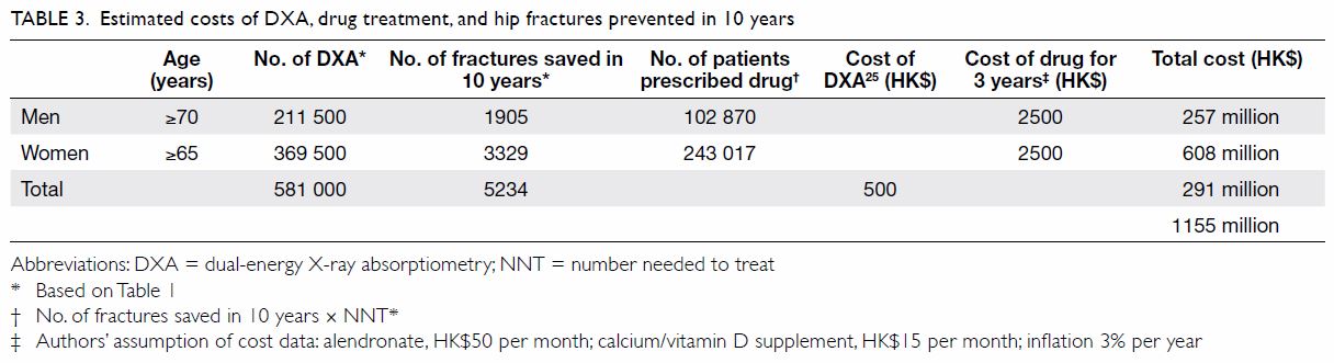
Table 3. Estimated costs of DXA, drug treatment, and hip fractures prevented in 10 years
 
We assumed that 3 years of generic alendronate plus calcium and vitamin D supplementation would be given for treatment, costing approximately HK$2500 (accounting for inflation; Table 325). We chose alendronate because it is currently the lowest-priced generic antiresorptive; patients may self-fund other options should they prefer. For instance, denosumab may be an option, particularly for patients with high fracture risk, poor adherence, contraindications, or intolerance to bisphosphonates.13 49 The incidence of ONJ in bisphosphonate-treated patients with osteoporosis in Hong Kong appears to be low (73.53 per 100 000 person-years of oral treatment), and the reported cases were in patients with readily modifiable risk factors.56 Estimates for the incidence of AFF in Hong Kong are not available, but data from abroad suggest that AFF is also very rare (13.6 per 100 000 patient-years at 2.0-3.9 years of bisphosphonate use).57 On this basis, we have not included the costs associated with ONJ and AFF in our calculations, as the associated cost is likely to be low.
 
The total cost of Recommendations 1 and 2 is HK$1155 million in 10 years (Table 325). In terms of benefits, we estimate that our proposals will prevent 5234 hip fractures over 10 years; if the estimated direct cost per hip fracture is HK$81 120 (2017),7 this would save the healthcare system HK$425 million in 10 years, with further savings from reduced outpatient and indirect costs. Additionally, elderly people and their families would avoid costs associated with institutional care. Based on 6300 hip fractures in Hong Kong in 2020,3 fracture reduction of 6.8%, the proportion of all patients with hip fractures discharged to institutional care (17%),9 and local institutional care costs (HK$120 000 annually for 5 years), we estimate that the savings from reduced institutional care needs would be HK$534 million in 10 years. Therefore, the total cost savings would be HK$959 million over 10 years; it should be noted, though, that this estimation is conservative and has not taken into account the year-on-year increase in hip fracture rate or inflation. Although the prevention of hip fractures alone may not reduce the institutional care cost for all patients, it would still likely be associated with substantial cost savings for the majority of patients. A full cost-effectiveness analysis of our proposal has not been performed, but the above cost-savings calculation has not included other fragility fractures, indirect healthcare cost savings (eg, transportation, costs associated with work productivity in family members), and inflation. Therefore, we believe that our recommendations are potentially cost-effective.
 
Recommendation 3: Comprehensive structured assessment (including dual-energy X-ray absorptiometry) of older people with a history of falling
Rationale for assessing patients with a history of falls
Because of ageing of muscles and declining balance control, older adults are more likely to experience falls than their younger counterparts.58 Fall history is a risk factor for subsequent falls59 60 61 and an independent predictor of fracture in older men and women.62 Recently, a hip fracture prediction score (HKOS score) was developed and validated for older adults aged ≥80 years in Hong Kong, and fall history is one of the risk factors included.63
 
Older people who have a history of falling should therefore be offered a comprehensive fall risk assessment. We also recommend DXA screening in these high-risk individuals. Although a history of falling is a good reason for fall risk and DXA assessments, these assessments should not be confined to those with a history of falling. Guidelines from the American Geriatrics Society/British Geriatrics Society recommend annual screening of all community-dwelling older people aged ≥65 years for falls and risk of falling.64
 
A meta-analysis of 159 trials in over 79 000 community-dwelling patients, including diverse fall-risk reduction strategies, concluded that home or group-based exercise programmes, including Tai Chi and home modifications, can reduce fall risk and fall-related fracture.65 In a study of elderly Hong Kong patients with a history of falling, a single risk assessment and home modification visit by an occupational therapist resulted in a reduced fall prevalence in the following 12 months compared with controls (13.7% vs 20.4%, P=0.03).66
 
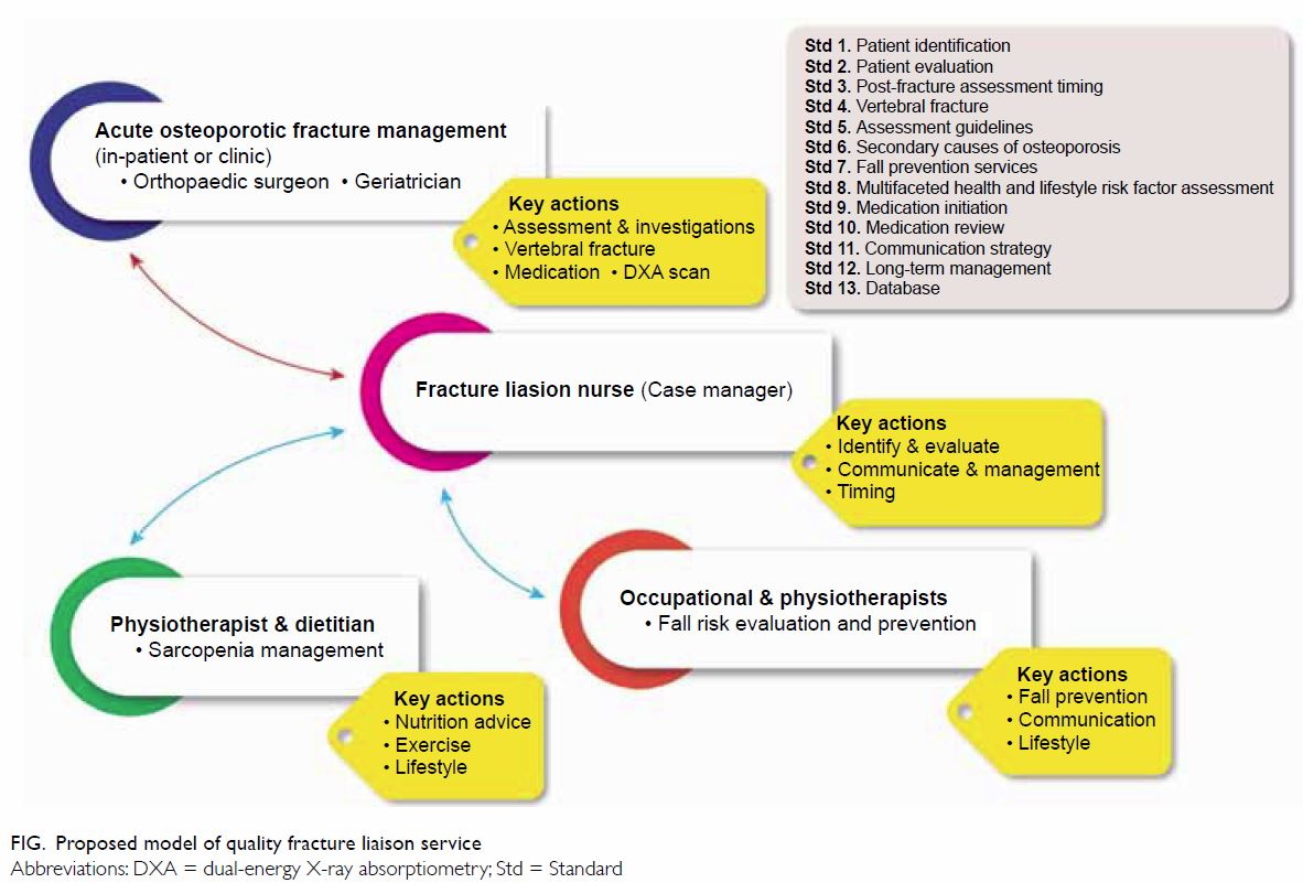
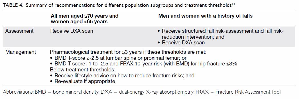
Suggestions for provision of care
Table 413 summarises the key elements of our recommendations, including treatment thresholds specified by local guidelines. Osteoporosis and fall risk assessment can be managed in the primary care setting, especially in Hong Kong, where access to DXA screening is excellent.
 

Table 4. Summary of recommendations for different population subgroups and treatment thresholds13
 
To promote DXA screening, Shepstone et al37 showed that a self-completed questionnaire capturing FRAX risk factors was effective at alerting older women to their fracture risk, resulting in a 28% reduction in hip fracture incidence in 5 years. On the basis of local prospective data, we demonstrated that combining a short questionnaire for sarcopenia (SARC-F) and the FRAX questionnaire could identify >75% of older people who suffered an incident hip fracture within 10 years.18 Furthermore, the well-calibrated HKOS score may accurately identify the oldest old (≥80 years) who are at high hip fracture risk.63
 
Conclusion and call to action
The high primary hip fracture burden in Hong Kong can be reduced with an evidence-based screening programme. High-risk patients identified by our proposed screening can be effectively managed using well-established and affordable therapies within the primary care field. Our proposals are supported by extensive evidence: similar programmes have shown efficacy, both in Hong Kong and abroad.
 
We propose that the Hong Kong government provide public funding support for a universal DXA screening programme for all men aged ≥70 years and women aged ≥65 years. Patients with a history of falling must receive systematic fracture-risk evaluations; patients with high fracture risk identified through these measures should be treated with antiresorptive agents and receive appropriate multidisciplinary follow-up.
 
We recommend a multidisciplinary effort including colleagues in general practice, orthopaedics, geriatric medicine, etc, to improve awareness of already available screening tools and effective treatment options for osteoporosis. If properly implemented and supported, we believe that our goal of a 7% annual reduction in hip fracture incidence in Hong Kong can be achieved, helping to offset the annual increase in the elderly population. Adoption of our proposals will shift government spending from post-fracture management to proactive osteoporosis management and fracture prevention. If our proposals are adopted, the estimated HK$1 billion that would be spent in 10 years on post-fracture management will instead be spent on improved osteoporosis management and fracture prevention for tens of thousands of patients. Furthermore, preventing fractures would also preserve the quality of life of many patients and their families. Achieving better control of hip fracture burden will be challenging, but it is well within our reach.
 
Author contributions
All authors contributed to the literature review and recommendations, critically revised the manuscript for important intellectual content, approved the final version for publication, and take responsibility for its accuracy and integrity.
 
Conflicts of interest
This publication is funded by a supportive grant from Amgen Asia Holding Limited to the Hong Kong Osteoporosis Foundation (HKOF) and the Osteoporosis Society of Hong Kong (OSHK) Consensus Group on Prevention of Fractures in Older People. All authors declare that they have no additional conflicts of interest.
 
Acknowledgement
Editorial support for this manuscript was provided by Magdalene Chu and Dr Alister Smith of MIMS Hong Kong, funded by the Hong Kong Osteoporosis Foundation.
 
Funding/support
This publication is funded by a supportive grant from Amgen Asia Holding Limited to the Hong Kong Osteoporosis Foundation (HKOF) and the Osteoporosis Society of Hong Kong (OSHK) Consensus Group on Prevention of Fractures in Older People.
 
References
1. Yu F, Xia W. The epidemiology of osteoporosis, associated fragility fractures, and management gap in China. Arch Osteoporos 2019;14:32. Crossref
2. Su Y, Leung J, Hans D, Aubry-Rozier B, Kwok T. Added clinical use of trabecular bone score to BMD for major osteoporotic fracture prediction in older Chinese people: the Mr. OS and Ms. OS cohort study in Hong Kong. Osteoporos Int 2017;28:151-60. Crossref
3. Man LP, Ho AW, Wong SH. Excess mortality for operated geriatric hip fracture in Hong Kong. Hong Kong Med J 2016;22:6-10. Crossref
4. The World Bank. World Development Indicators. Available from: https://datacatalog.worldbank.org/dataset/world-development-indicators. Accessed 18 Mar 2019.
5. Department of Health, Hong Kong SAR Government. Life expectancy. Available from: https://www.healthyhk.gov.hk/phisweb/en/healthy_facts/health_indicators/life_exp/. Accessed 3 Dec 2019.
6. Census and Statistics Department, Hong Kong SAR Government. Hong Kong Population Projections 2017-2066. Available from: https://www.statistics.gov.hk/pub/B1120015072017XXXXB0100.pdf. Accessed 1 Dec 2019.
7. Su Y, Lai FT, Yip BH, Leung JC, Kwok TC. Cost-effectiveness of osteoporosis screening strategies for hip fracture prevention in older Chinese people: a decision tree modeling study in the Mr. OS and Ms. OS cohort in Hong Kong. Osteoporos Int 2018;29:1793-805. Crossref
8. Chan DC, McCloskey EV, Chang CB, et al. Establishing and evaluating FRAX® probability thresholds in Taiwan. J Formos Med Assoc 2017;116:161-8. Crossref
9. Leung KS, Yuen WF, Ngai WK, et al. How well are we managing fragility hip fractures? A narrative report on the review with the attempt to setup a Fragility Fracture Registry in Hong Kong. Hong Kong Med J 2017;23:264-71. Crossref
10. Sing CW, Wong AY, Kiel DP, et al. Association of alendronate and risk of cardiovascular events in patients with hip fracture. J Bone Miner Res 2018;33:1422-34. Crossref
11. Cheung CL, Tan KC, Kung AW. Cohort Profile: The Hong Kong Osteoporosis Study and the follow-up study. Int J Epidemiol 2018;47:397-8f. Crossref
12. Bow CH, Cheung E, Cheung CL, et al. Ethnic difference of clinical vertebral fracture risk. Osteoporos Int 2012;23:879- 85. Crossref
13. OSHK Task Group for Formulation of 2013 OSHK Guideline for Clinical Management of Postmenopausal Osteoporosis in Hong Kong, Ip TP, Cheung SK, et al. The Osteoporosis Society of Hong Kong (OSHK): 2013 OSHK guideline for clinical management of postmenopausal osteoporosis in Hong Kong. Hong Kong Med J 2013;19 Suppl 2:1-40.
14. Hirsch JA, Beall DP, Chambers MR, et al. Management of vertebral fragility fractures: a clinical care pathway developed by a multispecialty panel using the RAND/UCLA Appropriateness Method. Spine J 2018;18:2152-61. Crossref
15. Dell R, Greene D. Is osteoporosis disease management cost effective? Curr Osteoporos Rep 2010;8:49-55. Crossref
16. Dell R, Greene D, Schelkun SR, Williams K. Osteoporosis disease management: the role of the orthopaedic surgeon. J Bone Joint Surg Am 2008;90 Suppl 4:188-94. Crossref
17. Newman ED, Ayoub WT, Starkey RH, Diehl JM, Wood GC. Osteoporosis disease management in a rural health care population: hip fracture reduction and reduced costs in postmenopausal women after 5 years. Osteoporos Int 2003;14:146-51. Crossref
18. Su Y, Woo JW, Kwok TC. The added value of SARC-F to prescreening using FRAX for hip fracture prevention in older community adults. J Am Med Dir Assoc 2019;20:83-9. Crossref
19. Tsang SW, Kung AW, Kanis JA, Johansson H, Oden A. Ten-year fracture probability in Hong Kong Southern Chinese according to age and BMD femoral neck T-scores. Osteoporos Int 2009;20:1939-45. Crossref
20. Cheung CL, Ang SB, Chadha M, et al. An updated hip fracture projection in Asia: the Asian Federation of Osteoporosis Societies study. Osteoporos Sarcopenia 2018;4:16-21. Crossref
21. Tosteson AN, Melton LJ 3rd, Dawson-Hughes B, et al. Cost-effective osteoporosis treatment thresholds: the United States perspective. Osteoporos Int 2008;19:437-47. Crossref
22. US Preventive Services Task Force, Curry SJ, Krist AH, et al. Screening for Osteoporosis to prevent fractures: US preventive services task force recommendation statement. JAMA 2018;319:2521-31. Crossref
23. Cummings SR, Cawthon PM, Ensrud KE, et al. BMD and risk of hip and nonvertebral fractures in older men: a prospective study and comparison with older women. J Bone Miner Res 2006;21:1550-6. Crossref
24. Kung AW, Lee KK, Ho AY, Tang G, Luk KD. Ten-year risk of osteoporotic fractures in postmenopausal Chinese women according to clinical risk factors and BMD T-scores: a prospective study. J Bone Miner Res 2007;22:1080-7. Crossref
25. Bow CH, Tsang SW, Loong CH, Soong CS, Yeung SC, Kung AW. Bone mineral density enhances use of clinical risk factors in predicting ten-year risk of osteoporotic fractures in Chinese men: the Hong Kong Osteoporosis Study. Osteoporos Int 2011;22:2799-807. Crossref
26. Choi YJ. Dual-energy X-ray absorptiometry: beyond bone mineral density determination. Endocrinol Metab (Seoul) 2016;31:25-30. Crossref
27. Nayak S, Greenspan SL. Cost-effectiveness of osteoporosis screening strategies for men. J Bone Miner Res 2016;31:1189-99. Crossref
28. Schousboe JT, Ensrud KE, Nyman JA, Melton LJ 3rd, Kane RL. Universal bone densitometry screening combined with alendronate therapy for those diagnosed with osteoporosis is highly cost-effective for elderly women. J Am Geriatr Soc 2005;53:1697-704. Crossref
29. International Society for Clinical Densitometry. 2019 ISCD Official Positions–Adult. International Society for Clinical Densitometry (ISCD). Available from: https://www.iscd. org/official-positions/2019-iscd-official-positions-adult/. Accessed 7 Aug 2019.
30. The Department of Health. Australian Government. Bone densiometry. Available from: https://www1.health.gov.au/ internet/main/publishing.nsf/Content/diagnosticimagingbd. htm. Accessed 7 Aug 2019.
31. International Osteoporosis Foundation. 2013 Asia- Pacific Audit: Malaysia. Available from: https://www.iofbonehealth.org/sites/default/files/media/PDFs/Regional%20Audits/2013-Asia_Pacific_Audit-Malaysia_0_0.pdf. Accessed 23 Oct 2019.
32. International Osteoporosis Foundation. The Asian Audit. Epidemiology, costs and burden of osteoporosis in Asia 2009. Available from: https://www.iofbonehealth.org/sites/default/files/PDFs/Audit%20Asia/Asian_regional_audit_2009.pdf. Accessed 23 Oct 2019.
33. International Osteoporosis Foundation. 2013 Asia- Pacific Audit: Singapore. Available from: https://www.iofbonehealth.org/sites/default/files/media/PDFs/Regional%20Audits/2013-Asia_Pacific_Audit-Singapore_0_0.pdf. Accessed 23 Oct 2019.
34. Kanis JA, Cooper C, Rizzoli R, et al. European guidance for the diagnosis and management of osteoporosis in postmenopausal women. Osteoporos Int 2019;30:3-44. Crossref
35. The University of Sheffield, UK. FRAX Fracture Risk Assessment Tool (Hong Kong). Available from: https://www.sheffield.ac.uk/FRAX/tool.aspx?country=20. Accessed 20 Mar 2019.
36. Kanis JA, Harvey NC, Cooper C, et al. A systematic review of intervention thresholds based on FRAX: a report prepared for the National Osteoporosis Guideline Group and the International Osteoporosis Foundation. Arch Osteoporos 2016;11:25. Crossref
37. Shepstone L, Lenaghan E, Cooper C, et al. Screening in the community to reduce fractures in older women (SCOOP): a randomised controlled trial. Lancet 2018;391:741-7. Crossref
38. Kung AW, Yeung SS, Chu LW. The efficacy and tolerability of alendronate in postmenopausal osteoporotic Chinese women: a randomized placebo-controlled study. Calcif Tissue Int 2000;67:286-90. Crossref
39. Lau EM, Woo J, Chan YH, Griffith J. Alendronate prevents bone loss in Chinese women with osteoporosis. Bone 2000;27:677-80. Crossref
40. Tu KN, Lie JD, Wan CK, et al. Osteoporosis: a review of treatment options. P T 2018;43:92-104.
41. Zhou J, Ma X, Wang T, Zhai S. Comparative efficacy of bisphosphonates in short-term fracture prevention for primary osteoporosis: a systematic review with network meta-analyses. Osteoporos Int 2016;27:3289-300. Crossref
42. Lyu H, Jundi B, Xu C, et al. Comparison of denosumab and bisphosphonates in osteoporosis patients: a meta-analysis of randomized controlled trials. J Clin Endocrinol Metab 2019;104:1753-65. Crossref
43. Bolland MJ, Grey AB, Gamble GD, Reid IR. Effect of osteoporosis treatment on mortality: a meta-analysis. J Clin Endocrinol Metab 2010;95:1174-81. Crossref
44. Reid IR, Horne AM, Mihov B, et al. Fracture prevention with zoledronate in older women with osteopenia. N Engl J Med 2018;379:2407-16. Crossref
45. Leung RY, Cheung BM, Nguyen US, Kung AW, Tan KC, Cheung CL. Optimal vitamin D status and its relationship with bone and mineral metabolism in Hong Kong Chinese. Bone 2017;97:293-8. Crossref
46. Howe TE, Shea B, Dawson LJ, et al. Exercise for preventing and treating osteoporosis in postmenopausal women. Cochrane Database Syst Rev 2011;(7):CD000333. Crossref
47. Amgen. Prolia (denosumab prescribing information). Highlights of prescribing information. Prolia (denosumab prescribing information). Available from: https://www. pi.amgen.com/~/media/amgen/repositorysites/pi-amgencom/ prolia/prolia_pi.pdf. Accessed 25 Apr 2019.
48. Khan AA, Morrison A, Hanley DA, et al. Diagnosis and management of osteonecrosis of the jaw: a systematic review and international consensus. J Bone Miner Res 2015;30:3-23. Crossref
49. Bone HG, Wagman RB, Brandi ML, et al. 10 Years of denosumab treatment in postmenopausal women with osteoporosis: results from the phase 3 randomised FREEDOM trial and open-label extension. Lancet Diabetes Endocrinol 2017;5:513-23. Crossref
50. Abrahamsen B. Are long-term bisphosphonate users a reality? Dose years for current bisphosphonate users assessed using the Danish National Prescription Database. Osteoporos Int 2013;24:369-72. Crossref
51. Burden AM, Paterson JM, Solomon DH, Mamdani M, Juurlink DN, Cadarette SM. Bisphosphonate prescribing, persistence and cumulative exposure in Ontario, Canada. Osteoporos Int 2012;23:1075-82. Crossref
52. Diab DL, Watts NB. Postmenopausal osteoporosis. Curr Opin Endocrinol Diabetes Obes 2013;20:501-9. Crossref
53. Sewerynek E, Horst-Sikorska H, Stępień-Kłos W, et al. The role of counselling and other factors in compliance of postmenopausal osteoporotic patients to alendronate 70 therapy. Arch Med Sci 2013;9:288-96. Crossref
54. Cooper A, Drake J, Brankin E, PERSIST Investigators. Treatment persistence with once-monthly ibandronate and patient support vs. once-weekly alendronate: results from the PERSIST study. Int J Clin Pract 2006;60:896-905. Crossref
55. Inderjeeth CA, Inderjeeth AJ, Raymond WD. Medication selection and patient compliance in the clinical management of osteoporosis. Aust Fam Physician 2016;45:814-7.
56. Kwok T, Choy TK, Kwok WL. Prevalence of bisphosphonate-related osteonecrosis of the jaw in Hong Kong. Hong Kong Med J 2016;22 Suppl 2:S46-7.
57. Dell RM, Adams AL, Greene DF, et al. Incidence of atypical nontraumatic diaphyseal fractures of the femur. J Bone Miner Res 2012;27:2544-50. Crossref
58. Guerado E, Sandalio RM, Caracuel Z, Caso E. Understanding the pathogenesis of hip fracture in the elderly, osteoporotic theory is not reflected in the outcome of prevention programmes. World J Orthop 2016;7:218-28. Crossref
59. Scott V, Votova K, Scanlan A, Close J. Multifactorial and functional mobility assessment tools for fall risk among older adults in community, home-support, long-term and acute care settings. Age Ageing 2007;36:130-9. Crossref
60. Wu TY, Chie WC, Yang RS, Kuo KL, Wong WK, Liaw CK. Risk factors for single and recurrent falls: a prospective study of falls in community dwelling seniors without cognitive impairment. Prev Med 2013;57:511-7. Crossref
61. Woo J, Leung J, Wong S, Kwok T, Lee J, Lynn H. Development of a simple scoring tool in the primary care setting for prediction of recurrent falls in men and women aged 65 years and over living in the community. J Clin Nurs 2009;18:1038-48. Crossref
62. Harvey NC, Odén A, Orwoll E, et al. Falls predict fractures independently of FRAX probability: a meta-analysis of the osteoporotic fractures in men (MrOS) study. J Bone Miner Res 2018;33:510-6. Crossref
63. Lam MT, Sing CW, Li GH, Kung AW, Tan KC, Cheung CL. Development and validation of a risk score to predict the first hip fracture in the oldest old: a retrospective cohort study. J Gerontol A Biol Sci Med Sci 2020;75:980-6. Crossref
64. Panel on Prevention of Falls in Older Persons, American Geriatrics Society, British Geriatrics Society. Summary of the Updated American Geriatrics Society/British Geriatrics Society clinical practice guideline for prevention of falls in older persons. J Am Geriatr Soc 2011;59:148-57. Crossref
65. Gillespie LD, Robertson MC, Gillespie WJ, et al. Interventions for preventing falls in older people living in the community. Cochrane Database Syst Rev 2012;(9):CD007146. Crossref
66. Chu MM, Fong KN, Lit AC, et al. An occupational therapy fall reduction home visit program for community-dwelling older adults in Hong Kong after an emergency department visit for a fall. J Am Geriatr Soc 2017;65:364-72. Crossref

Biological disease-modifying antirheumatic drugs in juvenile idiopathic arthritis of polyarticular course, enthesitis-related arthritis, and psoriatic arthritis: a consensus statement

© Hong Kong Academy of Medicine. CC BY-NC-ND 4.0
 
MEDICAL PRACTICE
Biological disease-modifying antirheumatic drugs in juvenile idiopathic arthritis of polyarticular course, enthesitis-related arthritis, and psoriatic arthritis: a consensus statement
Assunta CH Ho, MB, ChB, FHKAM (Paediatrics)1; SN Wong, MB, BS, FHKAM (Paediatrics)2; Lettie CK Leung, MB, BS (Syd), FHKAM (Paediatrics)3; Winnie KY Chan, MB, BS, FHKAM (Paediatrics)4; Patrick CY Chong, MB, BS, FHKAM (Paediatrics)5; Niko KC Tse, MB, BS, FHKAM (Paediatrics)6; Roanna HM Yeung, MB, BS, FHKAM (Paediatrics)4; SY Kong, MB, ChB, FHKAM (Paediatrics)3; KP Lee, MB, ChB, FHKAM (Paediatrics)7
1 Department of Paediatrics, Prince of Wales Hospital, The Chinese University of Hong Kong, Shatin, Hong Kong
2 Department of Paediatrics and Adolescent Medicine, Tuen Mun Hospital, Tuen Mun, Hong Kong
3 Department of Paediatrics, Kwong Wah Hospital, Yaumatei, Hong Kong
4 Department of Paediatrics, Queen Elizabeth Hospital, Jordan, Hong Kong
5 Department of Paediatrics and Adolescent Medicine, Queen Mary Hospital, The University of Hong Kong, Pokfulam, Hong Kong
6 Department of Paediatrics and Adolescent Medicine, Princess Margaret Hospital, Laichikok, Hong Kong
7 Department of Paediatrics and Adolescent Medicine, Pamela Youde Nethersole Eastern Hospital, Chai Wan, Hong Kong
 
Corresponding author: Dr Assunta CH Ho (assuntaho@cuhk.edu.hk)
 
 Full paper in PDF
 
Abstract
Objectives: Juvenile idiopathic arthritis (JIA) is the most common type of inflammatory arthritis in children. Treatment options have been expanded since the introduction of biologics, which are highly effective. The existing local JIA treatment guideline was published more than a decade ago, when use of biologics was not as common. In this article, we review the latest evidence on using biologics in three JIA subtypes: JIA of polyarticular course (pcJIA), enthesitis-related arthritis (ERA), and psoriatic arthritis (PsA). Based on the latest information, an update on eligibility, response assessment, termination, and safety information for using biologics in these patients was performed.
 
Consensus process: The JIA Work Group, which consisted of nine paediatricians experienced in managing JIA, was convened in 2016. Publications before July 2017 were screened. Eligible articles were clinical trials, extension studies, systemic reviews, and recommendations from international societies and regulatory agencies about the use of biologics in pcJIA, ERA, and PsA. Evidence extraction, appraisal, and drafting of propositions were performed by two reviewers. Extracted evidence and drafted propositions were presented and discussed at the first two meetings. Overwhelming consensus was obtained at the final meeting in May 2018. Seven practice consensus statements were formulated. Regular review should be performed to keep the practice evidence-based and up-to-date.


Introduction
 
Biological disease-modifying antirheumatic drugs (DMARDs) were introduced for the treatment of juvenile idiopathic arthritis (JIA) in year 2000. They are highly effective, especially for patients who are refractory to conventional DMARDs (cDMARDs). The current treatment algorithm for JIA in Hong Kong was published more than a decade ago, when the use of biologics was not as common.1 There has been a huge expansion in knowledge and approved medications since then. An update of practice based on the latest evidence is required, not only for specialists but also paediatricians and family physicians who may provide care to children with JIA.
 
The Hong Kong Society for Paediatric Rheumatology commissioned the JIA Work Group in 2016 to review this topic. The work group consists of nine paediatricians experienced in managing JIA. Our aims were: (1) to review the latest evidence on biological DMARDs in polyarticular course JIA (pcJIA, ie, those having arthritis affecting ≥5 joints irrespective of subtype at onset), enthesitis-related arthritis (ERA), and psoriatic arthritis (PsA); and (2) to propose an updated practice consensus for using biologics in these patients.
 
Methods
Articles about the use of biologics in pcJIA, ERA, and PsA published in English before July 2017 were identified by searching MEDLINE and PubMed. The keywords used for searching included JIA, pcJIA, ERA, PsA, eoJIA (extended oligoarticular arthritis), biologics, biological DMARDs, guidelines, recommendation, practice review, registries, adverse effects, malignancy, and infection. Publications considered relevant were clinical trials (randomised trials, reports on long-term extension phase of clinical trials), open-label studies, results from analysis of major registries, and recommendations from international regulatory organisations (United Kingdom: Clinical Commissioning Policy of National Health Service, National Institute for Health and Care Excellence; United States: American College of Rheumatology [ACR]; Australia: the Pharmaceutical Benefits Scheme, reimbursement programme for biologics use). Article screening, grading of the level of evidence, and propositions drafting were performed by two reviewers. The extracted evidence and draft propositions were presented to work group members at the first meeting held in July 2017. They were discussed and deliberated in a subsequent meeting held in March 2018. At the final meeting in May 2018, overwhelming consensus was reached on seven practice consensus statements.
 
Statement 1: Level of care
Care of children with JIA should preferably be shared jointly with paediatricians experienced in managing JIA.
 
With modern treatment strategies, a high level of disease control is possible. One study reported that more than 70% of patients (except rheumatoid factor positive polyarticular JIA) were able to reach a clinically inactive disease state within 2 years.2 To achieve the best outcome, care plans and treatment targets should be carefully formulated together with patients and paediatricians experienced in managing JIA.
 
Statement 2: Therapeutic ladder
For pcJIA, ERA and PsA, patients with persistently active arthritis despite adequate use of one or more cDMARDs, or if patients are intolerant of them, treatment escalation to biological DMARDs should be considered.
 
Conventional DMARDs like methotrexate, leflunomide, and sulphasalazine are effective in treating pcJIA.3 4 5 6 The choice of cDMARDs usually depends on JIA subtype and patient tolerance. Nevertheless, some patients may not respond to or may be intolerant of cDMARDs. Switching to biologics should be considered in these cases.
 
Biological disease-modifying antirheumatic drugs: evidence of effectiveness
Anti-tumour necrosis factor inhibitors
Etanercept
Etanercept is a chimeric fusion protein that acts as soluble TNF receptor. It is approved for use in cases of: (1) pcJIA in children aged ≥2 years (Food and Drug Administration [FDA] and European Medicines Agency [EMA]) and (2) ERA and PsA in children aged ≥12 years (EMA). The dose is 0.4 mg/kg (max 25 mg) twice weekly or 0.8 mg/kg (max 50 mg) once weekly subcutaneously.
 
Etanercept was the first anti-TNF inhibitor approved for pcJIA. Its efficacy was demonstrated by a randomised withdrawal trial7, in which 69 patients with pcJIA aged 4 to 17 years who were refractory to methotrexate were enrolled in the open-label lead-in phase. Fifty one (74%) patients achieved an ACR 30 response at week 12. They were then randomised to receive either etanercept or placebo for 16 weeks. Etanercept was more effective in preventing arthritis flares (28% vs 81%, P=0.003). The median time to flare was significantly shorter in the placebo group (28 days vs 116 days, P<0.001). Long-term data from the extension phase confirmed the persistence of efficacy.8
 
The use of etanercept in eoJIA, ERA, and PsA was studied by Horneff et al.9 In all, 127 patients (60 patients with eoJIA aged 2-17 years, 38 patients with ERA aged 12-17 years, 29 patients with PsA aged 12-17 years) were recruited in an open-label multicentre study. All had persistently active disease despite the use of nonsteroidal anti-inflammatory drugs (NSAIDs; for ERA) and DMARDs (for eoJIA, ERA, and PsA). At week 12, 88.6% (95% confidence interval [CI]=81.6%-93.6%) had attained ACR 30 response. The proportion of response was similar across all three subtypes.
 
The efficacy of etanercept in ERA was further evaluated. In a phase III double-blinded study, 41 patients aged 6 to 17 years with active disease despite taking at least one NSAID and one DMARD were recruited. They received open-label etanercept for 24 weeks. An ACR 30 response was achieved in 38 (93%). These patients were then randomised to receive etanercept or placebo in the subsequent 24-week withdrawal phase. The odds for having a flare was significantly higher in placebo group (odds ratio=6, 95% CI=1.1-37, P=0.02).10
 
The use of etanercept has now been extended to 2 years old.11 The efficacy of administrating at 0.8 mg/kg weekly has been shown to be comparable to twice weekly dosing.12
 
Etanercept can be used concomitantly with methotrexate or as monotherapy.13
 
Adalimumab
Adalimumab is a fully humanised monoclonal anti- TNF antibody. It is approved for use in cases of: (1) pcJIA in children aged ≥2 years (FDA and EMA) and (2) ERA in children aged ≥6 years (EMA). The dose is once every other week at 10 mg for 10 to <15 kg, 20 mg for 15 to <30 kg, and 40 mg for ≥30 kg.
 
Adalimumab was shown to be effective in treating pcJIA by Lovell et al.14 In total, 171 patients aged 4 to 17 years with active arthritis despite taking NSAIDs or methotrexate were enrolled in the 16-week open-label phase. In all, 94% of the adalimumab-methotrexate group and 74% of the adalimumab monotherapy group demonstrated an ACR 30 response. In the 32-week randomised withdrawal phase, the flare rate was significantly lower for those who remained on adalimumab. The results were more pronounced in patients receiving concomitant methotrexate (flare in placebo and methotrexate vs adalimumab and methotrexate: 65% vs 37%, P=0.02; flare in placebo vs adalimumab: 71% vs 43%, P=0.03). Efficacy was maintained in the long-term extension phase.
 
Burgos-Vargas et al15 studied the effectiveness of adalimumab in 46 patients with ERA. These patients, aged 6 to 17 years, had active arthritis and enthesitis. They were randomised to receive either adalimumab or placebo. At week 12, the adalimumab group had a significantly greater percentage decrease in the number of active joints (-62.2% vs -11.6%, P=0.039).
 
The safety and effectiveness of adalimumab in children aged 2 to 4 years weighing <15 kg were also demonstrated.16
 
Adalimumab can be used together with methotrexate or as monotherapy.
 
T cell co-stimulation inhibitor
Abatacept
Abatacept is a co-stimulation modulator that binds to the cluster of differentiation (CD) 80 or CD 86 ligands of the antigen-presenting cell and interferes with its interactions with T cells. Abatacept is indicated for pcJIA in children aged ≥6 years who are unresponsive to cDMARDs and at least one anti-TNF (EMA, FDA). The dose is 10 mg/kg (max 1g) infusion on day 1, 15, 29, and then every 28 days.
 
The efficacy of abatacept in pcJIA was assessed in a randomised withdrawal trial by Ruperto et al.17 The enrolled patients were aged 6 to 17 years with active disease despite DMARDs including anti-TNF inhibitors. Systemic JIA patients without systemic manifestations were also eligible. Patients with ERA or PsA were not included. Among the 190 recruited patients, 27% had previously been exposed to anti-TNF therapy. At the end of the 4-month open-label phase, 123 (65%) demonstrated an ACR 30 response. After one patient left the study, the remaining 122 patients were randomised to receive abatacept (60 patients) or placebo (62 patients) for 6 months. The flare rate of the abatacept group was significantly lower than that of the placebo group (20% vs 53%, P=0.0003). The hazard ratio for having a flare in the abatacept group was 0.31 (95% CI=0.16-0.95). The median time to flare was 6 months for placebo and not assessable for abatacept because of an inadequate number of events (P=0.0002). Among patients who did not achieve ACR 30 at the end of the 4-month open-label phase, half achieved ACR 30 after longer exposure. By day 589 of the long-term extension phase, the proportions of patients achieving ACR 30, 50, 70, 90, and 100 were 90%, 88%, 75%, 57%, and 39%, respectively.18
 
Abatacept can be used concomitantly with methotrexate or as monotherapy.
 
Anti-interleukin 6 inhibitor
Tocilizumab
Tocilizumab is a humanised anti–interleukin 6 monoclonal antibody. It is indicated for pcJIA in child aged ≥2 years (FDA, EMA). The dose is once every 4 weeks intravenous infusion, 10 mg/kg for <30 kg, 8 mg/kg for ≥30 kg.
 
Tocilizumab is another non-anti-TNF option for pcJIA. In a phase 3, three-part randomised controlled study (CHERISH), 188 patients aged 2 to 17 years with active disease received open-label tocilizumab for 16 weeks. In all, 166 (88%) achieved an ACR 30 response. A total of 163 were then randomised to receive tocilizumab (n=82) or placebo (n=81) for 24 weeks. Flares of JIA occurred significantly less often in the tocilizumab group (25.6% vs 48.1%, difference in means adjusted for stratification -0.21; 95% CI= -0.35 to -0.08; P=0.0024). Numerically more patients on concurrent methotrexate achieved ACR 70 and 90 responses.19
 
Tocilizumab can be used concomitantly with methotrexate or as monotherapy.
 
Systemic reviews of biologics
The short-term efficacies of biologics have been confirmed by systemic reviews.20 21 22 While there have been no head-to-head trials between individual biologics, analysis with indirect comparisons has not shown obvious differences in efficacy. Longer-term data (mainly for etanercept) and reports from major registries also confirm biologics’ effectiveness and safety.23 The results of clinical trials are summarised in Table 1.
 

Table 1. Summary of trails on individual biologics for pcJIA, ERA, and PsA
 
Statement 3: Eligibility criteria for using biological disease-modifying antirheumatic drugs
The work group achieved overwhelming consensus on the following eligibility criteria.
 
Eligible conditions, age, and biologics
Table 2 lists the JIA subtypes and the corresponding biological DMARDs indicated. Anti-TNF inhibitors are usually the first biologics to commence, except in systemic JIA. The decision of which biologic to start also depends on JIA subtype, the presence of extra-articular manifestations (eg, uveitis, inflammatory bowel disease), patient preferences, etc.24 25
 

Table 2. Eligibility: age, subtype, and biologics
 
Definition of adequate use of conventional disease-modifying antirheumatic drugs
  • Patients should have been treated with at least one cDMARD at adequate dose for at least 3 months unless toxicities are present.
  • Use of intra-articular steroid injections or a short course of bridging systemic corticosteroids are acceptable during trial use of cDMARDs.
  • In addition to cDMARDs, at least one anti-TNF inhibitor should have been tried before starting abatacept.
  •  
    It usually takes 6 to 8 weeks before the effects of cDMARDs can be seen. “Adequate cDMARDs exposure” is often defined as at least 3 months. However, if one develops significant intolerance or toxicities during trial of cDMARDs, use of biologics should be considered earlier.
     
    Definition of persistently active disease
  • pcJIA: Five or more joints with active arthritis
  • ERA and PsA: three or more joints with active arthritis
  •  
    “Active arthritis” is defined as swelling, with or without limitation in movement. Joints with limitation in movement due to pain or tenderness are also considered as “active”, especially those in which swelling is difficult to assess.
     
    When deciding the number of joints required to define active disease, the work group adopted the commonly used inclusion criteria of the clinical trials. In view of the fact that the disease characteristics (eg, number of joints involved, clinical course, medication indicated) are somewhat different for ERA and PsA, the work group considered it more appropriate to have seperate definitions for ERA and PsA.
     
    Statement 4: Assessment of response
  • Assessment of response to biological DMARDs should be performed after adequate exposure. For etanercept, adalimumab and tocilizumab, assessment should take place at 16 weeks. For abatacept, assessment at 24 weeks is acceptable if the expected response is not reached by 16 weeks.
  • A 50% reduction of the initial active joint count (≥2 joints) is considered as a positive response.
  •  
    Patients usually experience improvement within or around 3 months after commencement. The exception is abatacept, which may take a bit longer to achieve the desired therapeutic effect.20
     
    The definition of positive response is extrapolated from Pharmaceutical Benefits Scheme. It is easy to apply and does not require inflammatory markers like erythrocyte sedimentation rate and C-reactive protein, as they are not always elevated.
     
    Statement 5: Termination of treatment
    Termination of treatment should be considered in the following situations:
     
  • Toxicities or medical conditions that contraindicate further use of biologics, including but not limited to severe injection reaction, severe infection, malignancy, demyelination, heart failure, etc
  • Pregnancy (temporary withdrawal may be appropriate for anti-TNF inhibitors)
  • Lack of 50% reduction in the number of joints with active arthritis after the 16th week (or 6th month for abatacept)
  • Persistent inactive disease state, defined as Clinical Juvenile Arthritis Disease Activity Score ≤1, for at least 1 year
  •  
    The above termination criteria represent the common “end points” for stopping biologics, ie, the development of side-effects, conditions contra-indicated for use of biologics, or continuous remission of the index disease.
     
    Pregnancy is no longer an absolute contraindication in adult patients with rheumatoid arthritis or ankylosing spondylitis who are receiving anti-TNF inhibitors. Temporary withdrawal, however, is necessary for some agents. There have been no recent updates from international organisations on the use of biologics in pregnant patients with JIA. This issue should be revisited during the next update.
     
    Several definitions are used to describe treatment response and disease remission in JIA.26 The Clinical Juvenile Arthritis Disease Activity Score is a composite measurement that evaluates three variables: active joint count, patient’s/parents’ global well-being score, and physician’s global disease activity score (both on visual analogue scales). It has been shown to outperform other activity measurements in predicting the need of escalation to anti-TNF inhibitors.27 For both oligoarticular arthritis and pcJIA, a score of ≤1 signifies inactive disease.
     
    Statement 6: Malignancy and infection
  • The risk of developing new-onset malignancy due to exposure to biological DMARDs is low. Careful screening and close monitoring are recommended.
  • Patients on biological DMARDs should follow vaccination schedules according to local recommendations except for live attenuated vaccines. Infection screening should be performed prior to commencement. Screening should include (but not be limited to) tuberculosis, hepatitis B and C. Individual risk profiles should also be taken into consideration.
  •  
    In 2008, the FDA issued a black box warning about the development of malignancy, particularly lymphoproliferative types, in 48 children after commencement of anti-TNF inhibitors between 2001 and 2008.28 In recent years, researchers investigated the risk of malignancy by analysing data generated from major registries and medical reimbursement databases. These data suggested that TNF inhibition alone is probably not causing the apparent increase in incident malignancy. Instead, those children’s background risk might have already been elevated compared with that of the general population. Nevertheless, much longer-term data on larger patient samples are needed, as both cancer and JIA are rare in children.29 30 31
     
    Data on the associations between malignancy and other non-anti-TNF biologics are still lacking. Careful screening prior to commencement and ongoing surveillance are necessary.
     
    As for infection, biological DMARDs are generally safe. Most infections associated with the use of biologics are mild. Nevertheless, data from registries and medical insurance databases did show an increase in bacterial or serious infections associated with TNF inhibition.32 33 34 Tuberculosis is a genuine concern in our locality.35 36 Careful screening and follow-up according to individual risk profiles is strongly encouraged. The baseline infection screening should include, but not be limited to, the following:
     
  • Tuberculosis: Mantoux test, chest X-ray, interferon-gamma release assay
  • Hepatitis screening: hepatitis B surface antigen, antibody to hepatitis B core antigen, antibody to hepatitis C antigen
  •  
    While live attenuated vaccines should be avoided during use of biologics, patients should follow local guidelines for non-live vaccines.
     
    Statement 7: Special consideration
  • For patients with refractory disease but who do not fulfil the above criteria, biologics can be considered on a case-by-case basis.
  •  
    The work group is aware that some patients who do not fulfil the above criteria might still benefit from biologics. These patients include those with persistently active and disabling multiple enthesitis or sacroiliitis despite NSAIDs and DMARDs or oligoarticular JIA with severely active arthritis and has exhausted treatment modalities including intra-articular steroid injections and multiple cDMARDs, etc. The use of biologics can be considered after careful assessment on a case-bycase basis.
     
    The key recommendations and treatment algorithm are summarised in Table 3 and the Figure.
     

    Table 3. Summary of key consensus
     

    Figure. Treatment algorithm
     
    Looking ahead: new indications after the review period
    The indications for use of biological DMARDs have been expanding. For example, subcutaneous abatacept and tocilizumab are approved for pcJIA in patients aged ≥2 years in the United States.37 38 39 The ACR is also preparing a new update of the 2011 and 2013 guidelines. A review of the practice should be performed regularly.
     
    Conclusion
    The use of biological DMARDs has greatly improved the management of patients with JIA. Both patients and physicians could benefit from incorporating the latest and best available evidence into disease management. Periodic review should be performed to keep clinical practice evidence-based and up-to-date.
     
    Author contributions
    All the authors contributed substantially to conception and design, acquisition and analysis of data, drafting and revising the article, and approval of the version to be published.
     
    Conflicts of interest
    All authors have no competing interest in the products mentioned in this paper.
     
    Funding/support
    This research received no specific grant from any funding agency in the public, commercial, or not-for-profit sectors.
     
    References
    1. Lee TL, Lau YL, Chan W, et al. Juvenile idiopathic arthritis in Hong Kong and its current management. HK J Paediatr (new series) 2003;8:21-30. >
    2. Guzman J, Oen K, Tucker LB, et al. The outcomes of juvenile idiopathic arthritis in children managed with contemporary treatments: results from the ReACCh-Out cohort. Ann Rheum Dis 2015;74:1854-60. Crossref
    3. Giannini EH, Brewer EJ, Kuzmina N, et al. Methotrexate in resistant juvenile rheumatoid arthritis. Results of the U.S.-U.S.S.R. double-blind, placebo-controlled trial. The Pediatric Rheumatology Collaborative Study Group and The Cooperative Children's Study Group. N Engl J Med 1992;326:1043-9. Crossref
    4. Woo P, Southwood TR, Prieur AM, et al. Randomised placebo-controlled, crossover trial of low-dose oral methotrexate in children with extended oligoarticular or systemic arthritis. Arthritis Rheum 2000;43:1849-57. Crossref
    5. Silverman E, Mouy R, Spiegel L, et al. Leflunomide or methotrexate for juvenile rheumatoid arthritis. N Engl J Med 2005;352:1655-66. Crossref
    6. van Rossum MA, Fiselier TJ, Franssen MJ, et al. Sulfasalazine in the treatment of juvenile chronic arthritis: a randomised, double-blind, placebo-controlled, multicenter study. Dutch Juvenile Chronic Arthritis Study Group. Arthritis Rheum 1998;41:808-16. Crossref
    7. Lovell DJ, Giannini EH, Reiff A, et al. Etanercept in children with polyarticular juvenile rheumatoid arthritis. Pediatric Rheumatology Collaborative Study Group. N Engl J Med 2000;342:763-9. Crossref
    8. Lovell DJ, Reiff A, Ilowite NT, et al. Safety and efficacy of up to eight years of continuous etanercept therapy in patients with juvenile rheumatoid arthritis. Arthritis Rheum 2008;58:1496-504. Crossref
    9. Horneff G, Burgos-Vargas R, Constantin T, et al. Efficacy and safety of open-label etanercept on extended oligoarticular juvenile idiopathic arthritis, enthesitis- related arthritis and psoriatic arthritis: part 1 (week 12) of the CLIPPER study. Ann Rheum Dis 2014;73:1114-22. Crossref
    10. Horneff G, Foeldvari I, Minden K, et al. Efficacy and safety of etanercept in patients with the enthesitis-related arthritis category of juvenile idiopathic arthritis. Arthritis Rheumatol 2015;67:2240-9. Crossref
    11. Windschall D, Horneff G. Safety and efficacy of etanercept and adalimumab in children aged 2 to 4 years with juvenile idiopathic arthritis. Clin Rheumatol 2016;35:2925-31. Crossref
    12. Horneff G, Ebert A, Fitter S, et al. Safety and efficacy of once weekly etanercept 0.8 mg/kg in a multicenter 12 week trial in active polyarticular course juvenile idiopathic arthritis. Rheumatology (Oxford) 2009;48:916-9. Crossref
    13. Horneff G, De Bock F, Foeldvari I, et al. Safety and efficacy of combination of etanercept and methotrexate compared to treatment with etanercept only in patients with juvenile idiopathic arthritis (JIA): preliminary data from the German JIA Registry. Ann Rheum Dis 2009;68:519-25. Crossref
    14. Lovell DJ, Ruperto N, Goodman S, et al. Adalimumab with or without methotrexate in juvenile rheumatoid arthritis. N Engl J Med 2008;359:810-20. Crossref
    15. Burgos-Vargas R, Tse SM, Horneff G, et al. A randomized, double-blind, placebo-controlled multicenter study of adalimumab in pediatric patients with enthesitis-related arthritis. Arthritis Care Res (Hoboken) 2015;67:1503-12. Crossref
    16. Kingsbury DJ, Bader-Meunier B, Patel G, Arora V, Kalabic J, Kupper H. Safety, effectiveness, and pharmacokinetics of adalimumab in children with polyarticular juvenile idiopathic arthritis aged 2 to 4 years. Clin Rheumatol 2014;33:1433-41. Crossref
    17. Ruperto N, Lovell DJ, Quartier P, et al. Abatacept in children with juvenile idiopathic arthritis: a randomized, double-blind, placebo-controlled withdrawal trial. Lancet 2008;372:383-91. Crossref
    18. Ruperto N, Lovell DJ, Quartier P, et al. Long-term safety and efficacy of abatacept in children with juvenile idiopathic arthritis. Arthritis Rheum 2010;62:1792-802. Crossref
    19. Brunner HI, Ruperto N, Zuber Z, et al. Efficacy and safety of tocilizumab in patients with polyarticular-course juvenile idiopathic arthritis: results from a phase 3, randomized, double-blind withdrawal trial. Ann Rheum Dis 2015;74:1110-7. Crossref
    20. Shepherd J, Cooper K, Harris P, Picot J, Rose M. The clinical effectiveness and cost-effectiveness of abatacept, adalimumab, etanercept and tocilizumab for treating juvenile idiopathic arthritis: a Crossref
    21. Ungar WJ, Costa V, Burnett HF, Feldman BM, Laxer RM. The use of biologic response modifiers in polyarticular- course juvenile idiopathic arthritis: a systematic review. Semin Arthritis Rheum 2013;42:597-618. Crossref
    22. Otten MH, Anink J, Spronk S, van Suijlekom-Smit LW. Efficacy of biological agents in juvenile idiopathic arthritis: a systematic review using indirect comparisons. Ann Rheum Dis 2013;72:1806-12. Crossref
    23. Klotsche J, Niewerth M, Haas JP, et al. Long-term safety of etanercept and adalimumab compared to methotrexate in patients with juvenile idiopathic arthritis (JIA). Ann Rheum Dis 2016;75:855-61. Crossref
    24. Anink J, Otten MH, Gorter SL, et al. Treatment choices of paediatric rheumatologists for juvenile idiopathic arthritis: etanercept or adalimumab? Rheumatology (Oxford) 2013;52:1674-9. Crossref
    25. Kearsley-Fleet L, Davies R, Baildam E, et al. Factors associated with choice of biologics among children with juvenile idiopathic arthritis: results from two UK paediatric biologic registers. Rheumatology (Oxford) 2016;55:1556-65. Crossref
    26. Smith EM, Foster HE, Beresford MW. The development and assessment of biological treatments for children. Br J Clin Pharmacol 2015;79:379-94. Crossref
    27. Swart JF, van Dijkhuizen EH, Wulffraat NM, de Roock S. Clinical juvenile arthritis disease activity score proves to be a useful tool in treat-to-target therapy in juvenile idiopathic arthritis. Ann Rheum Dis 2018;77:336-42. Crossref
    28. Diak P, Siegel J, La Grenada L, Choi L, Lemery S, McMahon A. Tumor necrosis factor alpha blockers and malignancy in children: forty-eight cases reported to the Food and Drug Administration. Arthritis Rheum 2010;62:2517-24. Crossref
    29. Beukelman T, Haynes K, Curtis JR, et al. Rate of malignancy associated with juvenile idiopathic arthritis and its treatment. Arthritis Rheum 2012;64:1263-71. Crossref
    30. Ruperto N, Martini A. Juvenile idiopathic arthritis and malignancy. Rheumatology (Oxford) 2014;53:968-74. Crossref
    31. Mannion ML, Beukelman T. Risk of malignancy associated with biologic agents in pediatric rheumatic disease. Curr Opin Rheumatol 2014;26:538-42. Crossref
    32. Beukelman T, Xie F, Chen L, et al. Rates of hospitalized bacterial infection associated with juvenile idiopathic arthritis and its treatment. Arthritis Rheum 2012;64:2773- 80. Crossref
    33. Davies R, Southwood TR, Kearsley-Fleet L, Lunt M, Hyrich KL; British Society for Paediatric and Adolescent Rheumatology Etanercept Cohort Study. Medically significant infections are increased in patients with juvenile idiopathic arthritis treated with etanercept: results from the British Society for Paediatric and Adolescent Rheumatology Etanercept Cohort Study. Arthritis Rheumatol 2015;67:2487-94. Crossref
    34. Becker I, Horneff G. Risk of serious infection in juvenile idiopathic arthritis patients associated with tumor necrosis factor inhibitors and disease activity in the German biologic in pediatric rheumatology registry. Arthritis Care Res (Hoboken) 2017;69:552-60. Crossref
    35. Mok CC, Tam LS, Chan TH, Lee GK, Li EK; Hong Kong Society of Rheumatology. Management of rheumatoid arthritis: consensus recommendations from the Hong Kong Society of Rheumatology. Clin Rheumatol 2011;30:303-12. Crossref
    36. Wan R, Mok CC. The Hong Kong Society of Rheumatology Biologics Registry: Updated Report (May 2016). HK Bulletin Rheum Dis 2016;16:24-6. Crossref
    37. Brunner HI, Tzaribachev N, Vega-Cornejo G, et al. Subcutaneous abatacept in patient with polyarticular- course juvenile idiopathic arthritis: results from a phase III open-label study. Arthritis Rheumatol 2018;70:1144-54. Crossref
    38. Brunner H, Ruperto N, Martini A, et al. Identification of optimal subcutaneous doses of tocilizumab in children with polyarticular course juvenile idiopathic arthritis. Arthritis Rheumatol 2017;69(Suppl 4):Abstract 41.
    39. De Benedetti F, Ruperto N, Lovell D, et al. THU0503 identification of optimal subcutaneous (SC) doses of tocilizumab in children with polyarticular-course juvenile idiopathic arthritis (PCJIA). Ann Rheum Dis 2017;76(Suppl 2):396. Crossref

    High-fidelity simulation training programme for final-year medical students: implications from the perceived learning outcomes

    © Hong Kong Academy of Medicine. CC BY-NC-ND 4.0
     
    MEDICAL PRACTICE
    High-fidelity simulation training programme for final-year medical students: implications from the perceived learning outcomes
    YF Choi, FHKCEM, MSc(Clinical Education)(Edin)1,2,3; TW Wong, FHKCEM, FHKAM (Emergency Medicine)1
    1 Accident and Emergency Department, Pamela Youde Nethersole Eastern Hospital, Chai Wan, Hong Kong
    2 Programme Director, Nethersole Clinical Simulation Training Centre, Hong Kong
    3 Medical Director, Hong Kong Fire Services Department, Hong Kong
     
    Corresponding author: Dr YF Choi (choiyf@gmail.com)
     
     Full paper in PDF
     
    Abstract
    We designed a session of high-fidelity simulation training course for final-year medical students in their emergency medicine specialty clerkship. This was a new initiative with clearly defined learning outcomes. We aimed to evaluate the learning outcomes. Students completed an evaluation form at the end of the session focusing on their perceived learning outcomes. Thematic analysis was conducted for data processing. We collected responses from 149 students. In addition to the intended outcomes of the course, students gained unexpected learning outcomes from the training and some of them matched a few identified learning gaps between undergraduate medical education and their subsequent transition to early clinical practice that have been described in the literature. High-fidelity simulation training in medical school could be an effective tool to address some of the identified gaps in the transition between undergraduate medical education and postgraduate practice.


    Introduction
    Clinical simulation training has become more widely practised in local hospitals and academic healthcare institutions in the past decade, with a wide range of training modalities, from part-task simulator to full-body manikin and from single skill training to interprofessional team-based training.
     
    The Nethersole Clinical Simulation Training Centre is a hospital-based training centre which has advocated high-fidelity multidisciplinary team training for hospital staff since its establishment in 2012 in Pamela Youde Nethersole Eastern Hospital. From 2008, the emergency department of the hospital was one of the centres for teaching emergency medicine specialty clerkship medical students from one of the medical schools in Hong Kong. In 2017, the emergency department collaborated with the Nethersole Clinical Simulation Training Centre in an initiative to design a high-fidelity simulation training session for medical students.
     
    In the past, medical students in local medical school did not have much high-fidelity simulation training because medical schools did not have high-fidelity training facilities. Furthermore, it was conventionally believed that high-fidelity simulation training is more beneficial for expert-level learners and that low-fidelity training was more suitable for beginners, because the sophisticated context in the high-fidelity environment might potentially jeopardise learning objectives by creating excessive cognitive burden.1 2 3 Medical students, owing to their low clinical exposure, were regarded as beginners.
     
    However, a recent study suggested that once medical students have learnt some basic skills, high-fidelity simulation training might benefit learners through psychological immersion, despite the extra cognitive burden.4 Therefore, we decided to trial high-fidelity simulation training for final-year medical students, who have already completed most of their academic studies and have acquired some basic practical skills.
     
    The prototype of the simulation training course was started in 2017 and at that time we mainly evaluated the “reaction” phase of the learners which is the basic level of outcome evaluation according to Kirkpatrick’s model (Fig). The feedback from the students was very positive and they welcomed the course very much.5 Encouraged by this, we revised the course material in 2018 with written intended learning outcomes. In the present study, we evaluate “learning”, which is a level higher in the Kirkpatrick’s model.
     

    Figure. Kirkpatrick model of course evaluation (evaluation training programmes. 1975)
     
    The aim of the present study was to evaluate the learning outcomes of a high-fidelity simulation training course for final-year medical students.
     
    Methods
    Design
    This was a qualitative study by a survey in English. The contents were checked against SRQR reporting guideline (Standards for Reporting Qualitative Research 2016 version).
     
    Setting
    The training course was conducted in the Nethersole Clinical Simulation Training Centre that has a simulation training suite with isolated simulation rooms and a debriefing room. The simulation rooms are equipped with ceiling-mounted cameras for video-assisted debriefing. The simulation room used was prepared to resemble the environment of a resuscitation room in the emergency department with a full-body high-fidelity manikin.
     
    Course design
    The training course consisted of a brief introduction followed by four short case scenarios in a 3-hour session. In the introduction, students were briefed about course structure, learning objectives, and clinical simulation rules and underwent a short simulation laboratory familiarisation session. A group of seven to eight students further divided into two subgroups played the four scenarios in turn. While one subgroup was doing the scenario, the other subgroup observed from the debriefing room via the audio-visual system. The instructor wore a nurse uniform and acted as an experienced nurse in the scenarios, which included no real-time coaching. In the four scenarios all the patients presented with acute or even life-threatening conditions that need prompt treatment. Such a design simulated the high psychological fidelity of real life. Participants had to treat the patients on their own with no guidance from the instructor. A debriefing session was carried out immediately after each scenario.
     
    Data collection
    A cohort of student participants in 2018 were selected by convenience sampling and they were asked to complete an evaluation form immediately after the session. The evaluation form invited the students to offer free text only with no rating scale. They could write up to three perceived learning outcomes, up to three reflections after the course and any extra comments or suggestions about the course. There was no space to enter the name of the students, so the data collection was completely anonymous.
     
    Data analysis
    The findings in the evaluation form were processed through thematic analysis shortly after each course and compared with the intended learning outcomes of the course (Table 1). The intended learning outcomes were designed to include both clinical learning outcomes and teamwork learning outcomes such as briefing, debriefing, communication, situation awareness, and leadership. The clinical learning outcomes, owing to their diversity were further divided into three separate categories (general approach to critical patients, use of investigations and resuscitation skills) while all the teamwork-related learning outcomes are grouped under one category. The written learning outcomes were first coded under these intended learning outcomes. Items not fitted under the intended learning outcomes were considered new findings. These new findings were further processed newly identified themes. The results of thematic analysis were checked serially by the authors for quality assurance. Consensus on new themes after further literature review was made between authors on items that could not be categorised under the intended learning outcomes.
     

    Table 1. Intended learning outcomes of the course with sample quotations from respondents
     
    Results
    Data saturation was attained after 20 sessions with 149 evaluation forms collected (data saturation is a point when no new theme was found by thematic analysis after analysing the responses after several courses). The response rate was 100% and more than 99% (148 out of 149) of the returned forms were complete, despite the voluntary nature of data collection.
     
    The intended learning outcomes are listed in Table 1. From the contents of the evaluation form, all of the intended learning outcomes were well received by the respondents, as reflected by the responses shown in Table 1.
     
    In addition to the intended learning outcomes, we discovered a large number of learning points or reflections that cannot be categorised under our pre-planned learning objectives. These additional unexpected learning points were processed by thematic analysis under new headings as listed in Table 2.
     

    Table 2. Unexpected/additional learning outcomes from the course with sample quotations from respondents
     
    Respondents were also given the opportunity to make open comments or suggestions about the course. Some of the responses are listed in Table 3.
     

    Table 3. Selected additional open-ended comments or suggestions on the course from respondents
     
    Discussion
    The original intention of course evaluation questionnaire was to evaluate the learning objectives as perceived by students. The results suggest that our intended learning outcomes were well received by our students. A number of learning outcomes were revealed that did not fall into our intended learning outcomes by thematic analysis. Additional literature review revealed that these unexpected outcomes match some gaps identified in the transition between undergraduate medical education and postgraduate practice.
     
    Fidelity
    In our training course, high fidelity was thought to be an important element to achieve the learning outcomes, including the unexpected ones. Fidelity is commonly defined as “the level of realism present to the learners during a simulation training”.3 4 Conventionally, it is believed that higher fidelity leads to more efficient learning.6
     
    However, fidelity is not a single-dimensional concept and there are different components of fidelity described in the literature using different terminology. For simplicity, we consider the definition of fidelity described by Feinstein and Cannon, in which fidelity of simulation has physical and functional aspects.3 Physical fidelity includes the environmental, visual, and spatial components, such as the design of the simulation room, the performance of manikin, and the settings of various instruments. Functional fidelity is a dynamic interaction between the participants and their task, including information, stimuli, and responses of learners. Other simulation educators have used the terms “conceptual”, “experiential”, “emotional” or more commonly “psychological” fidelity to describe the same or similar concepts,2 7 but further discussion is beyond the scope of the present study.
     
    High-fidelity simulation training was not always regarded as superior to lower-fidelity training. Besides the issue of cost, early studies in the last century in various disciplines (such as civil and military aviation) failed to show better learning outcomes after high physical fidelity training.3 8 9 10 This might be because the high physical fidelity training over-stimulated novice learners and resulted in cognitive overload that jeopardised the intended learning objectives.11 Such findings supported the conventional belief that low fidelity is better for beginners and high fidelity is for expert learners. Medical students are regarded as novice learners and have little or no working experience. These conventional beliefs lead to the conclusion that it is unreasonable and not cost-effective to expose medical students to high physical fidelity simulation training. However, this overlooks the importance of functional or psychological fidelity. In recent years, studies have suggested that functional or psychological fidelity is important in enhancing learning.7 12 13 14 15 In addition to simulating real-world functioning, high psychological fidelity also creates a stressful environment, increasing student arousal level and facilitating learning and performance. Therefore, in our training course, in addition to the relatively high physical fidelity provided by the well-equipped simulation training room and sophisticated manikin, we further enhanced functional or psychological fidelity by the following interventions:
     
  • A short briefing session before the scenario emphasised behavioural authenticity (such as doing real chest compression and discharging full energy for defibrillation) and psychological immersion. This is equivalent to a fiction contract to suspend disbelief described in clinical simulation training literature.15 16
  • All participants and the instructor wore clinical attire, including white coats and nurse uniforms that simulated real staffing in an emergency department.
  • The instructor behaved as a helpful nurse and talked in a submissive manner while occasionally offered hints with limited scientific knowledge out of past experiences.
  • The instructor might immediately remind or even criticise participants who demonstrated non-immersive clinical behaviour such as laughing, suboptimal chest compression, or non-participative gesture.
  • Stress level was enhanced by the absence of real-time coaching in critical clinical conditions. Students were made to solve challenges and overcome difficulties or uncertainty by themselves with a deteriorating patient in front of them.
  •  
    All of these measures enhanced functional fidelity by providing a realistic working environment and imposed psychological immersion for the participants.
     
    A further remark on the term “fidelity” is that physical and psychological fidelity are not mutually exclusive nor competitive, but are actually complementary to each other.17 18 19 20 For example, a high-fidelity manikin who can talk or moan can enhance psychological stimuli for the participants.
     
    Some of the learning outcomes of the course are closely related to psychological fidelity, namely teamwork, working under stress, identifying one’s own weakness, acute patient management, and situation awareness. In the written responses under these headings, we could see interaction, flow of information, stress, and self-reflection.
     
    Gaps in undergraduate medical education
    Although not the original objective of this study, we found that the unexpected learning outcomes of this study echoed some of the identified gaps between transitions from undergraduate medical education to early postgraduate medical practice in the literature. In the medical education literature there have been calls worldwide for reform of undergraduate medical curricula, to address gaps identified in undergraduate medical education that adversely affect medical interns in their early practice.21 22 23 24 25 These gaps include working under stress, working on call, uncertainties, helplessness, workload, difficulties prescribing medication, and managing acutely ill patients, and similar gaps have been identified in medical educations programmes across the world.
     
    Newly graduated interns perceive their work to be stressful and have feelings of being underprepared.23 24 25 Educators have suggested that undergraduate curricula should prepare medical students to deal with expected stress, such as facing uncertainty, knowing one’s limitations, and asserting one’s right for support.21 26 27 These wordings or identified gaps were found in the perceived learning outcomes of our course. In the literature, interns have indicated that they could not predict their shortcomings in their undergraduate study until they were in clinical practice. Furthermore, it was also suggested that undergraduate medical education should include more on communication skills and emotional involvement.21 28 29 Again these aspects match the perceived learning outcomes of our course.
     
    More work-related training should be put in the final year of medical school, particularly for dealing with acutely ill patients and prescribing medication.30 31 32 Studies have shown that despite curriculum reform, management of acute problems has remained an unclosed gap.30 33 34 Work-related training with acutely ill patients is difficult to achieve because such patients are not always readily available even if medical students do a period of assistant internship. This is a likely reason that curriculum reform and workplace placements during their final year closed some gaps but not others.20 21 The opportunities for students to experience real acute care are limited, making it difficult to build expertise through repetitive practice.35 36 Furthermore, there is always an ethical consideration of whether to allow students to treat acutely ill patients.37
     
    The learning outcomes of our study suggest that high-fidelity simulation training can be a solution to the gaps identified. High-fidelity simulation training can create a lot of scenarios with acutely ill patients in a short period of time, to create an environment in which students can make mistakes and learn from these mistakes without harming real patients. This idea is supported by the literature, which demonstrates simulation training is better than other forms of instruction method such as didactic or problem-based learning and should serve as an adjunct to other instruction methods.2 38 39 This point is particularly important because interns or junior doctors are the first medical responders called to attend acutely deteriorating patient in a ward and their suboptimal management might put patients at risk or delay appropriate treatment.21 29 The General Medical Council of the United Kingdom also recommends the use of simulation technology in medical school.22
     
    The limitations of this study include that data were collected from only one medical school, the current curriculum was not discussed, the cohort included students at different times in their final year, and most (but not all) students were recruited before formal clinical placement.
     
    Summary
    There are gaps between undergraduate medical education and transition to postgraduate clinical practice which could be eliminated through reform of undergraduate medical school curricula. The application of psychologically immersive high-fidelity simulation training for medical students is likely to be a helpful strategy to enhance their preparedness. Such training should emphasise management of acutely ill patients.
     
    Author contributions
    All authors had full access to the data, contributed to the study, approved the final version for publication, and take responsibility for its accuracy and integrity. YF Choi wrote the article. All authors contributed to the concept of study, acquisition and analysis of data, and critical revision for important intellectual content.
     
    Conflicts of interest
    As an editor of the journal, TW Wong was not involved in the peer review process. The other author has disclosed no conflicts of interest.
     
    Funding/support
    This research received no specific grant from any funding agency in the public, commercial, or not-for-profit sectors.
     
    Ethics approval
    No real patients were involved in this study and no personal data was collected from the participants. Verbal consent was obtained from all participants in the introduction session of the course; participation was non-compulsory. The study was approved by the hospital ethics committee (Ref HKECREC-2019-013).
     
    References
    1. Alessi SM. Fidelity in the design of instructional simulations. J Comput Based Instr 1988;14:40-7.
    2. Maran NJ, Glavin RJ. Low- to high- fidelity simulation—a continuum of medical education? Med Educ 2003;37(Suppl 1):22-8. Crossref
    3. Feinstein AH, Cannon HM. Fidelity, verifiability, and validity of simulation: constructs for evaluation. Developments in Business Simulation and Experiential Learning. 2001; 28: 57-65.
    4. Mills BW, Carter OB, Rudd CJ, Claxton LA, Ross NP, Strobel NA. Effects of low- versus high-fidelity simulations on the cognitive burden and performance of entry-level paramedicine students: a mixed-methods comparison trial using eye-tracking, continuous heart rate, difficulty rating scales, video observation and interviews. Simul Healthc 2016;11:10-8. Crossref
    5. Choi YF, Wong TW. High fidelity simulation training for medical students in emergency medicine specialty clerkship. Proceedings of the 9th Asia Medical Education Symposium cum Frontiers in Medical and Healthcare Science Education 2017. OP2.
    6. Hays RT, Singer MJ. Simulation Fidelity in Training System Design: Bridging the Gap between Reality and Training. New York: Springer-Verlag; 1989. Crossref
    7. Diederich E, Mahnken JD, Rigler SK, Williamson TL, Tarver S, Sharpe MR. The effect of model fidelity on learning outcomes of a simulation-based education program for central venous catheter insertion. Simul Healthc 2015;10:360-7. Crossref
    8. Matsumoto ED, Hamstra SJ, Radomski SC, Cusimano MD. The effect of bench model fidelity on endourological skill: a randomized controlled trial. J Urol 2002;167:1243-7. Crossref
    9. Chandra DB, Savoldelli GL, Joo HS, Weiss ID, Nail VN. Fiberoptic oral intubation: the effect of model fidelity on training for transfer to patient care. Anesthesiology 2008;109:1007-13. Crossref
    10. Grober ED, Hamstra SJ, Wanzel KR, et al. The educational impact of bench model fidelity on the acquisition of technical skill: the use of clinically relevant outcome measures. Ann Surg 2004;240:374-81. Crossref
    11. Martin EL, Waag WL. Contributions of platform motion to simulator training effectiveness: Study I—basic contact. 39. Brooks Air Force Base, Texas Air Force Human Resources Laboratory; 1978. Crossref
    12. Bland AJ, Topping A, Tobbell J. Time to unravel the conceptual confusion of authenticity and fidelity and their contribution to learning within simulation-based nursing education. A discussion paper. Nurse Educ Today 2014;24:1112-8. Crossref
    13. Chetwood JD, Grag P, Burton K. High-fidelity realistic acute medical simulation and SBAR training at a tertiary hospital in Blantyre, Malawi. Simul Healthc 2018;13:139-45. Crossref
    14. Rudolph JW, Simon R, Reamer DB. Which reality matters? Questions on the path to high engagement in healthcare simulation. Simul Healthc 2007;2:161-3. Crossref
    15. Hamstra SJ, Brydges R, Hatala R, Zendejas B, Cook DA. Reconsidering fidelity in simulation-based training. Acad Med 2014;89:387-92. Crossref
    16. Rudolph JW, Raemer DB, Simon R. Establishing a safe container for learning in simulation: the role of the presimulation briefing. Simul Healthc 2014;9:339-49. Crossref
    17. Kozlowski SW, DeShon RP. A psychological fidelity approach to simulation-based training: theory, research and principles. In: Elliott LR, Coovert MD, Schiflett SG, editors. Scaled Worlds: Development, Validation and Applications. New York: Ashgate Publishing; 2017.
    18. Oser RL, Cannon-Bowers J, Sala E, Dwyer DJ. Enhancing human performance in technology-rich environments: Guidelines for scenario-based training. Hum Tach Interact Complex Sys 1999;9:175-202.
    19. Beaubien JM, Baker DP. The use of simulation for training teamwork skill in health care: how low can we go? Qual Saf Health Care 2004;13 Suppl 1:i51-6. Crossref
    20. Tun JK, Alinier G, Tang J, Kneebone RL. Redefining simulation fidelity in healthcare education. Simul Gaming 2015;46:159-74. Crossref
    21. Bernnan N, Corrigan O, Allard J, et al. The transition from medical student to junior doctor: today’s experiences of tomorrow’s doctors. Med Educ 2010;44:449-58. Crossref
    22. General Medical Council. Tomorrow’s Doctors. London: GMC; 2009.
    23. Teo A. The current state of medical education in Japan: a system under reform. Med Educ 2007;41:302-8. Crossref
    24. Segouin C, Jouquan J, Hodges B, et al. Country report: medical education in France. Med Educ 2007;41:295-301. Crossref
    25. Ochsmann EB, Zier U, Drexler H, Schmid K. Well prepared for work? Junior doctors’ self-assessment after medical education. BMC Med Educ 2011;11:99. Crossref
    26. Miles S, Kellett J, Leinster SJ. Medical graduates’ preparedness to practice: a comparison of undergraduate medical school training. BMC Med Educ 2017;17:33. Crossref
    27. Lempp H, Cochrane M, Seabrook M, Rees J. Impact of educational preparation on medical students in transition from final year to PRHO year: a qualitative evaluation of final-year training following the introduction of new year 5 curriculum in a London medical school. Med Teach 2004;26:276-8. Crossref
    28. Watmough S, Garden A, Taylor D. Pre-registration house officers’ views on studying under a reformed medical curriculum in the UK. Med Educ 2006;40:893-9. Crossref
    29. O’Neill PA, Jones A, Willis SC, McArdle PJ. Does a new undergraduate curriculum based on Tomorrow’s Doctors prepare house officers better for their first post? A qualitative study of the views of pre-registration house officers using critical incidents. Med Educ 2003;37:1100-8. Crossref
    30. Illing JC, Peile E, Morrison J, et al. How prepared are medical graduate to begin practice? A comparison of three diverse UK medical school. London: General Medical Council; 2008.
    31. Smith CM, Perkins GD, Bullock I, Bion JF. Undergraduate training in the care of the acutely ill patient: a literature review. Intensive Care Med 2007;33:901-7. Crossref
    32. Jensen ML, Hesselfeldt R, Rasmussen MB, et al. Newly graduated doctors’ competence in managing cardiopulmonary arrests assessed using a standardized Advanced Life Support (ALS) assessment. Resuscitation 2008;77:63-8. Crossref
    33. Bosch J, Maaz A, Hitzblech T, Holzhausen Y, Peters H. Medical students’ preparedness for professional activities in early clerkship. BMC Med Educ 2017;17:140. Crossref
    34. Illing JC, Morrow GM, Rothwell nee Kergon CR, et al. Perception of UK medical graduates’ preparedness for practice: a multi-centre qualitative study reflecting the importance of learning on the job. BMC Med Educ 2013;13:34. Crossref
    35. Phillips PS, Nolan JP. Training in basic and advanced life support in UK medical schools: questionnaire survey. BMJ 2001;323:22-3. Crossref
    36. Vozenilek J, Huff JS, Reznek M, Gordon JA. See one, do one, teach one: advanced technology in medical education. Acad Emerg Med 2004;11:1149-54. Crossref
    37. Ziv A, Wolpe PR, Small SD, Glick S. Simulation-based medical education: an ethical imperative. Acad Med 2003;78:783-8. Crossref
    38. Beal MD, Kinnear J, Anderson CR, Martin TD, Wamboldt R, Hooper L. The effectiveness of medical simulation in teaching medical students critical care medicine: A systemic review and meta-analysis. Simul Healthc 2017;12:104-16. Crossref
    39. McCoy CE, Menchine M, Anderson C, Kollen R, Langdorf MI, Lotfipour S. Prospective randomized crossover study of simulation vs didactics for teaching medical students the assessment and management of critically ill patients. J Emerg Med 2011;40:448-55. Crossref

    Clinical considerations when adding a sodium-glucose co-transporter-2 inhibitor to insulin therapy in patients with diabetes mellitus

    © Hong Kong Academy of Medicine. CC BY-NC-ND 4.0
     
    MEDICAL PRACTICE  CME
    Clinical considerations when adding a sodium-glucose co-transporter-2 inhibitor to insulin therapy in patients with diabetes mellitus
    Kathryn Tan, MBBCH, MD1; WS Chow, FRCP (Edin), FHKAM (Medicine)2; Jenny Leung, MB, BS, FRCP (Lond)3; Andrew Ho, FHKCP4; Risa Ozaki, MB, ChB, FHKCP5; Grace Kam, FHKCP, FRCP (Glasgow)6; June Li, MB, ChB, FHKCP7; CH Choi, MB, ChB, FRCP (Lond)8; MW Tsang, MB, BS, FRCP9; Norman Chan, MD, FRCP9; KK Lee, MB, BS, FHKAM (Medicine)1; KW Chan, FRCP (Edin), FHKAM (Medicine)10
    1 Department of Medicine, The University of Hong Kong, Pokfulam, Hong Kong
    2 Department of Medicine, Queen Mary Hospital, Pokfulam, Hong Kong
    3 Department of Integrated Medical Service, Ruttonjee and Tang Shiu Kin Hospitals, Hong Kong
    4 Department of Medicine and Geriatrics, Tuen Mun Hospital, Tuen Mun, Hong Kong
    5 Department of Medicine and Therapeutics, The Chinese University of Hong Kong, Shatin, Hong Kong
    6 Department of Medicine and Geriatrics, United Christian Hospital, Kwun Tong, Hong Kong
    7 Department of Medicine, Yan Chai Hospital, Tsuen Wan, Hong Kong
    8 Department of Medicine, Queen Elizabeth Hospital, Jordan, Hong Kong
    9 Specialist in Endocrinology, Private Practice
    10 Department of Medicine and Geriatrics, Princess Margaret Hospital, Laichikok, Hong Kong
     
    Corresponding author: Prof Kathryn Tan (kcbtan@hku.hk)
     
     Full paper in PDF
     
    Abstract
    A consensus meeting was held to discuss add-on therapy of sodium-glucose co-transporter-2 (SGLT2) inhibitors in patients with diabetes mellitus treated with insulin. The objectives were to affirm the efficacy and safety of SGLT2 inhibitors as an add-on to insulin, empower clinicians to minimise the risk of adverse events, and provide clinical guidance. Administration of SGLT2 inhibitors as an add-on therapy to insulin is associated with significant reductions compared with placebo in glycosylated haemoglobin A1c, fasting plasma glucose, insulin dose, and body weight without an increased risk of hypoglycaemia. Compared with traditional therapies, SGLT2 inhibitors have shown cardiovascular and renal benefits. Adding an SGLT2 inhibitor to insulin increases the risk of urinary tract and genital tract infections. The use of SGLT2 inhibitor is also associated with a slightly increased incidence of diabetic ketoacidosis. Patients who may benefit most from add-on therapy with SGLT2 inhibitors include those with established atherosclerotic cardiovascular disease, heart failure, chronic kidney disease, high insulin doses, obesity, and metabolic syndrome. Routine monitoring for diabetic ketoacidosis is controversial, and patient and clinician education is essential to minimise risk. The decision to adjust insulin dose when adding an SGLT2 inhibitor is dependent on patient factors, but the insulin dose should not be reduced beyond 20% prior to the first dose of SGLT2 inhibitor. Patients should temporarily discontinue SGLT2 inhibitors during fasting, acute illness, or low/reduced carbohydrate intake. If ketonuria is detected, SGLT2 inhibitors but not insulin should be immediately discontinued and medical advice sought.


    Introduction
    Sodium-glucose co-transporter-2 (SGLT2) inhibitors are an important therapeutic option in the management of type 2 diabetes mellitus (T2DM). These oral agents treat hyperglycaemia by blocking the reabsorption of glucose in renal tubules, which results in increased urinary glucose excretion.1 As monotherapy, SGLT2 inhibitors have been shown to significantly lower glycated haemoglobin A1c (HbA1c), fasting glucose, and postprandial glucose compared with placebo in subjects with T2DM that was inadequately controlled with diet and exercise.2 3 4 The significant and consistent reduction in HbA1c observed with SGLT2 inhibitors is similar to or better than that produced by metformin, sulfonylureas, and dipeptidyl peptidase-4 (DPP-4) inhibitors, with a minimal risk of hypoglycaemia. These SGLT2 inhibitors can also bring about reductions in body weight and blood pressure. Sodium-glucose co-transporter- 2 inhibitor therapy is associated with an elevated risk of genital tract infections (GTIs) and, to a lesser degree, urinary tract infections (UTIs).1 5 In addition, in rare cases, SGLT2 inhibitors have been associated with diabetic ketoacidosis (DKA) and euglycaemic ketoacidosis.
     
    Many patients with T2DM fail to achieve glycaemic goals despite receiving two or more antidiabetic drug classes that target different core defects of the disease.6 Whereas the majority of antidiabetic drugs have an insulin-dependent mode of action, SGLT2 inhibitors have an insulin-independent mode of action, suggesting that the use of these drugs could offer therapeutic synergy when used in combination.6 7 Randomised controlled trials (RCTs),8 9 10 11 as well as real-world studies12 have confirmed the efficacy and tolerability of SGLT2 inhibitors when used as monotherapy and as an add-on therapy to insulin.13 14 At the time of writing, SGLT2 inhibitors are not approved for use in type 1 diabetes mellitus (T1DM); however, there are RCTs providing evidence of a potential role for SGLT2 inhibitors in patients with T1DM.15 16
     
    A meeting of esteemed endocrinologists in Hong Kong was held in August 2018 to develop a consensus on the role of add-on therapy with SGLT2 inhibitors in insulin-treated patients with diabetes mellitus. The expert panel considered all evidence relating to T2DM and also considered T1DM where possible. All statements pertaining to T1DM should be interpreted carefully given the paucity of data available for this condition and not construed as recommendations for off-label use. Herein we present the consensus findings of the expert panel, with the major objectives of this review summarised as follows: (1) to summarise the clinical approach and rationale for intensifying insulin therapy with SGLT2 inhibitors (2) to affirm the efficacy and safety of SGLT2 inhibitors in insulin-treated patients; (3) to empower clinicians to minimise the risk of hypoglycaemia and DKA; (4) to provide practical clinical guidance on adding SGLT2 inhibitors in insulin-treated patients; and (5) to guide clinicians on patient selection.
     
    Clinical approach and rationale
    Statement 1.1: The decision to add an additional therapy or intensify insulin therapy is dependent on individual patient factors that contribute to inadequate control
    Because T2DM is a progressive disease, many patients will need intensification of therapy. If the patient has experienced an episode of severe hypoglycaemia while on insulin therapy, then the addition of another therapy with a low risk of hypoglycaemia may be a better option compared with intensifying insulin therapy. Patient preference is an important consideration. Intensifying insulin heightens the potential risks for weight gain and hypoglycaemia, especially at high insulin doses. Moreover, a large proportion of patients with T2DM have co-morbid obesity and/or metabolic syndrome, with the latter characterised by hypertension and dyslipidaemia in addition to poor glycaemic control. Intensifying insulin in these patients could contribute to a vicious cycle of increasing appetite, further weight gain, and increasing insulin resistance.17
     
    Statement 1.2: Sodium-glucose co-transporter-2 inhibitors offer weight loss and cardiovascular-renal benefits with a low risk of hypoglycaemia compared with other oral agents
    Sodium-glucose co-transporter-2 inhibitors offer the benefit of improving glycaemic control without increasing the risks of hypoglycaemia or weight gain in insulin-treated patients. Compared with most other therapies, SGLT2 inhibitors have shown cardiovascular benefits in patients with established atherosclerotic cardiovascular disease, which appears to be a class effect.18 Because of their insulin-independent mechanism, SGLT2 inhibitors are often effective in patients in whom other therapies have failed. Add-on therapy with an SGLT2 inhibitor may have particular benefits in obese patients, as insulin intensification or add-on therapy with sulfonylurea or thiazolidinedione may exacerbate weight gain. Treatment with SGLT2 inhibitors can improve metabolic syndrome through reductions in weight and blood pressure while counteracting insulin resistance and improving insulin sensitivity. Continuous glucose monitoring studies have shown improvement in glucose excursions after initiating SGLT2 inhibitor therapy. In patients with T1DM, the patients who mainly benefit from SGLT2 inhibitor therapy are those on high insulin doses and those in whom features of T2DM—including excessive weight, high blood pressure, and other indices of metabolic syndrome—have accrued.
     
    Efficacy and safety
    Statement 2.1: Administration of sodium-glucose co-transporter-2 inhibitors as add-on therapy to insulin in type 2 diabetes mellitus is associated with significant reductions in haemoglobin A1c, fasting plasma glucose, insulin dose and body weight
    The efficacy of SGLT2 inhibitors as add-on therapy to insulin in T2DM has been demonstrated in randomised, placebo-controlled trials, with significant reductions in HbA1c, fasting plasma glucose, total daily insulin dose, and body weight compared with placebo achieved over periods of up to 24 weeks (Table).19 So far, no head-to-head trials have compared SGLT2 inhibitors and DPP-4 inhibitors as an add-on therapy to insulin. Indirect comparison via network meta-analysis has suggested that SGLT2 inhibitors showed better glycaemic control and greater weight reduction than DPP-4 inhibitors in patients with T2DM that was inadequately controlled with insulin (Table).20 In addition, SGLT2 inhibitors have proven cardiovascular-renal benefits, while most DPP-4 inhibitors have only achieved cardiovascular safety.
     

    Table. Efficacy and safety of SGLT2 inhibitors as add-on therapy to insulin
     
    Statement 2.2: Adding sodium-glucose co-transporter- 2 inhibitors to insulin in type 2 diabetes mellitus patients with a mean baseline haemoglobin A1c of 8.4% (range, 8.2%-8.9%) does not increase the risk of hypoglycaemia but increases the risks of urinary and genital tract infections
    In patients with T2DM, the use of SGLT2 inhibitors as add-on therapy to insulin is associated with a similar incidence of hypoglycaemia to placebo (Table).19 However, SGLT2 inhibitors are associated with higher risks of UTIs and GTIs compared with placebo.19
     
    Statement 2.3: Administration of sodium-glucose co-transporter-2 inhibitors as add-on therapy to insulin in type 1 diabetes mellitus is associated with significant reductions in haemoglobin A1c, fasting plasma glucose, daily total insulin dose, body weight, and glycaemic excursions
    The use of SGLT2 inhibitors in T1DM is currently off-label. The addition of an SGLT2 inhibitor in patients with T1DM offers therapeutic value in patients who are obese or with problems with large glucose excursions. Careful patient selection and meticulous patient education of precautions are important to minimise the risk of DKA.21 Evidence from several clinical trials showed that in patients with T1DM, dapagliflozin, or empagliflozin as adjunctive therapy to insulin improved glycaemic control and weight.16
     
    Statement 2.4: Adding sodium-glucose co-transporter-2 inhibitors to insulin in type 1 diabetes mellitus does not appear to increase the risks of hypoglycaemia or urinary tract infections but increases the risk of genital tract infections
    In patients with T1DM, the use of SGLT2 inhibitor therapy is not associated with an increased risk of hypoglycaemia or UTIs compared with placebo but is associated with an increased risk of GTIs (Table).15 Evidence from the EASE trials showed that in patients with T1DM, empagliflozin as adjunctive therapy to insulin did not increase the risk of hypoglycaemia.16
     
    Adverse events of special interest
    Statement 3.1: The incidence of genital tract infections is higher in patients with sodium-glucose co-transporter-2 inhibitors added to insulin therapy versus other agents or placebo; however, most events are classified as mild or moderate in intensity and readily respond to therapy
    In patients with T1DM or T2DM, the addition of an SGLT2 inhibitor to insulin therapy is associated with a significantly increased risk of GTIs (Table).15 19 The estimated risk ratio ranges from 3 to 5 compared with placebo. However, treatment cessation is not necessary, and most events are mild or moderate in intensity and readily respond to therapy.
     
    Statement 3.2: Administration of sodium-glucose co-transporter-2 inhibitors is associated with an increased, albeit low, incidence of euglycaemic diabetic ketoacidosis, a risk that is strongly associated with use of insulin
    In RCTs, the incidence of DKA among patients with T1DM or T2DM receiving SGLT2 inhibitor therapy is estimated to be <1 case per 1000, whereas in cohort studies, the incidence has been reported to be 1.6 cases per 1000 person-years.22 In patients with T1DM, the DEPICT-1,18 DEPICT-2,23 inTandem1,24 and inTandem325 trials showed a DKA incidence ranging from 1.5% to 4.0% in patients treated with selective SGLT2 inhibitors or dual SGLT1 and SGLT2 inhibitors for up to 1 year.
     
    Statement 3.3: The risk of diabetic ketoacidosis is heightened in patients with high haemoglobin A1c levels, frail and elderly patients, those with inadequate food intake, and patients with poor disease awareness/adherence or frequent complications
    In patients with T2DM, those with HbA1c levels of ≥10% may have a higher risk of developing DKA than patients with lower HbA1c levels. Risk may also be elevated in latent autoimmune diabetes in adults, frail and elderly patients with poor disease awareness, those who are repeatedly admitted to hospital for complications, and patients with long-term diabetes with depleted ß-cell reserves.
     
    In patients with T1DM, poor glycaemic control is also indicative of heightened risk of DKA, along with insulin pump use and suboptimal adherence.26 27 28 Other risk groups include those taking weight loss medications, those without steady dietary control, postoperative patients, and frail or elderly patients, particularly those with cognitive impairment.
     
    Patient selection
    Statement 4.1: Sodium-glucose co-transporter-2 inhibitors have the greatest overall benefit/risk profile in patients with obesity, cardiovascular or renal diseases, high insulin requirement, or large glycaemic excursions
    Randomised controlled trials support the use of SGLT2 inhibitors as an add-on therapy to insulin therapy in T2DM patients with obesity, cardiovascular or renal diseases, high insulin requirement, or large glycaemic excursions in whom insulin intensification would otherwise be the next step in achieving glycaemic control.9 29 30 31 32 33 34 35
     
    In patients with T1DM, SGLT2 inhibitors should only be prescribed by an endocrinologist, as the use of these therapies in T1DM is currently off-label. The use of SGLT2 inhibitors may be a useful adjunct to insulin therapy in patients with T1DM and obesity or large glycaemic variability. Hypoglycaemia unawareness is not an absolute contra-indication provided that the patient is compliant and knowledgeable about the disease. Patients should be capable of detecting insulin pump failure, and the ability to monitor urine or serum ketone levels is mandatory. In RCTs of patients with T1DM, patient selection included those aged 18 to 75 years with baseline HbA1c levels of 7.0% to 11.0% and a body mass index ≥18.5 kg/m2.21 23 24 25 In the DEPICT trials, patients using insulin for ≥12 months with a total daily insulin dose ≥0.3 IU/kg for ≥3 months were selected, and patients were additionally required to have a creatinine clearance of >60 mL/min and C-peptide level <0.7 ng/mL.21 23 In the inTandem trials, patients were required to have treatment with insulin at a stable dose via continuous subcutaneous insulin infusion or multi-dose insulin treatment, with no change in insulin delivery within 3 months.24 25 Patients were additionally required to perform self-monitoring of blood glucose (SMBG) and have an estimated glomerular filtration rate of >45 mL/min/1.73 m2.
     
    Switching versus adding
    Statement 5.1: In patients with type 2 diabetes mellitus, the decision to switch a sodium-glucose co-transporter-2 inhibitor to another oral agent or add a sodium-glucose co-transporter-2 inhibitor to an existing treatment regimen is based on factors such as efficacy, tolerability of the existing treatment, and cost
    In T2DM patients on combination therapy of insulin and oral antidiabetic agents, replacing one of the oral agents with an SGLT2 inhibitor can be considered. When the effectiveness of the DPP-4 inhibitor is no longer sustained owing to its limited durability or when the patient develops fluid retention with a glitazone, patients would be expected to benefit from a switch to an SGLT2 inhibitor. Switching to an SGLT2 inhibitor might be more cost-effective compared with adding an SGLT2 inhibitor to the existing drug regimen.
     
    Implementation
    Statement 6.1: Adjustment of the insulin dose when adding a sodium-glucose co-transporter-2 inhibitor may be appropriate in some but not all patients
    In patients with T2DM, the decision to adjust insulin dose upon initiation of an SGLT2 inhibitor is dependent on patient factors. If the patient is obese and insulin resistant with high HbA1c, then maintaining the insulin dose is a reasonable approach. Conversely, if a patient’s HbA1c is close to target, a reduction in insulin dose will be appropriate. For patients with frequent large glycaemic excursions, the expert panel recommends reduction of the insulin dose by up to 10% before initiating an SGLT2 inhibitor. Initiation of the SGLT2 inhibitor at the lowest available dose is also recommended.
     
    In the DEPICT trials with T1DM, after the first dose of study drug, basal and bolus insulin were reduced symmetrically by up to 20%.21 23 In the inTandem trials, in which dual SGLT1 and SGLT2 inhibitor therapy was employed, bolus insulin was reduced by 30%, with insulin dosing subsequently adjusted according to SMBG data to meet targets.24 25 The consensus of the expert panel was that the insulin dose should not be reduced beyond 20% upon initiation of an SGLT2 inhibitor. When the patient is receiving both basal and bolus doses of insulin, the bolus dose may be reduced, with addition of the SGLT2 inhibitor as appropriate.
     
    Monitoring
    Statement 7.1: Early and regular monitoring for diabetic ketoacidosis is recommended following initiation of sodium-glucose co-transporter-2 inhibitor therapy
    Patients should be monitored and closely followed after initiating an SGLT2 inhibitor. Self-monitoring of blood glucose is important, and titration of insulin may be necessary. Early follow-up in the form of telecommunication or nurse clinic instead of clinician visits may be more feasible, and monitoring of renal function within 4 weeks is recommended. Daily ketone monitoring is not practical because of the high cost and short shelf life of ketone strips. However, monitoring of ketones is recommended during acute illness. Education of patients and clinicians is essential for improved awareness, and the importance of sick day management needs to be emphasised.
     
    The risk of DKA is higher in T1DM. Urine or blood ketone monitoring should be considered during initiation of SGLT2 inhibitors and mandatory during acute stress. Education regarding optimal nutrition, situations of nausea/vomiting, and temporary cessation of SGLT2 inhibitor therapy is appropriate. Instruction on measures to reverse ketosis and prevent progression to DKA (including carbohydrates and fluid intake as well as additional correction of insulin doses) should be given. In T1DM, the STICH protocol is an appropriate strategy for mitigating DKA risk in patients receiving SGLT2 inhibitors. When DKA is suspected, the patients should stop SGLT2 inhibitor therapy, inject bolus insulin, consume 30 g of carbohydrates, and hydrate with water.36
     
    Statement 7.2: Sodium-glucose co-transporter-2 inhibitor therapy should be stopped in the event of high ketone levels
    In the event that a patient detects high ketone levels, they should be instructed to stop their SGLT2 inhibitor, continue insulin, ensure carbohydrate intake, and seek medical advice.
     
    Other practical advice
    Statement 8.1: Sodium-glucose co-transporter-2 inhibitor therapy should be temporarily stopped to avoid diabetic ketoacidosis during fasting or reduced intake of food, acute illness, and hospitalisation
    Although there are currently no guidelines on when to discontinue SGLT2 inhibitor therapy, SGLT2 inhibitor therapy should be withheld temporarily in the following situations37:
  • Fasting, reduced carbohydrate intake (withhold at onset);
  • Acute illness (withhold at onset);
  • Hospitalisation (withhold at onset);
  • Surgery, endoscopic procedures, contrast studies requiring fasting (withhold prior to procedure, with duration depending on risk (at least 3 days prior to major surgery or prolonged fasting and/or anticipated high risk of hypovolaemia); and
  • Bariatric surgery (withhold 1 to 2 weeks before initiation of ketotic diet).
  •  
    Sodium-glucose co-transporter-2 inhibitors may be re-introduced when the patient is able to eat normally, with recovery of renal function following acute illness.
     
    Further evidence to explore
    Statement 9.1: Additional real-world and renal and cardiovascular outcome data in patients with type 1 diabetes mellitus are needed to further support the use of sodium-glucose co-transporter-2 inhibitors in diabetes management
    Real-world data are needed to better understand long-term medication adherence and persistence, cost-effectiveness of ketone monitoring, and the role of carbohydrate intake and SMBG in the prevention of DKA. Renal and CVD outcome data are needed regarding the use of SGLT2 inhibitors in T1DM.
     
    Concluding remarks
    Sodium-glucose co-transporter-2 inhibitors are an effective and well tolerated therapeutic option as add-on therapy to insulin in patients with T2DM. Addition of SGLT2 inhibitors in this setting is associated with significant reductions in HbA1c, fasting plasma glucose, total daily insulin dose, and body weight without increasing the risk of hypoglycaemia. Sodium-glucose co-transporter-2 inhibitor use is accompanied by a slightly increased risk of UTIs and GTIs. There is a low risk of DKA (about 1 per 1000 person-years). Appropriate patient selection, education, and monitoring are helpful in mitigating this risk. The use of SGLT2 inhibitors in patients with T1DM is currently off-label and should only be attempted under the supervision of an endocrinologist in appropriately selected patients. Further research will help to clarify the role of this important oral antidiabetic drug class in both T1DM and T2DM.
     
    Post-meeting note
    The results of the EASE-2 and EASE-3 trials were published after the meeting. Data from these two trials showed that adding empagliflozin as an adjunct to insulin therapy in T1DM improved glycaemic control and weight without increasing hypoglycaemia. The rate of ketoacidosis was lower when a smaller dose of empagliflozin was used.
     
    Author contributions
    All authors have made substantial contributions to the concept or design of this study; acquisition of data; analysis or interpretation of data; drafting of the manuscript; and critical revision for important intellectual content. All authors had full access to the data, contributed to the study, approved the final version for publication, and take responsibility for its accuracy and integrity.
     
    Conflicts of interest
    All authors have declared no conflicts of interest.
     
    Acknowledgement
    Language editing and writing support, funded by an unrestricted educational grant from AstraZeneca Hong Kong Limited, were provided by Ben Searle and Howard Christian of MIMS (Hong Kong) Limited.
     
    Funding/support
    Editing and writing support was funded by an unrestricted educational grant from AstraZeneca Hong Kong Limited.
     
    References
    1. Scheen AJ. SGLT2 inhibition: efficacy and safety in type 2 diabetes treatment. Expert Opin Drug Saf 2015;14:1879- 904. Crossref
    2. Stenlöf K, Cefalu WT, Kim KA, et al. Efficacy and safety of canagliflozin monotherapy in subjects with type 2 diabetes mellitus inadequately controlled with diet and exercise. Diabetes Obes Metab 2013;15:372-82. Crossref
    3. Ji L, Ma J, Li H, et al. Dapagliflozin as monotherapy in drug-naive Asian patients with type 2 diabetes mellitus: a randomized, blinded, prospective phase III study. Clin Ther 2014;36:84-100.e9. Crossref
    4. Ferrannini E, Seman L, Seewaldt-Becker E, Hantel S, Pinnetti S, Woerle HJ. A Phase IIb, randomized, placebo-controlled study of the SGLT2 inhibitor empagliflozin in patients with type 2 diabetes. Diabetes Obes Metab 2013;15:721-8. Crossref
    5. Levine MJ. Empagliflozin for type 2 diabetes mellitus: an overview of phase 3 clinical trials. Curr Diabetes Rev 2017;13:405-23. Crossref
    6. Fioretto P, Giaccari A, Sesti G. Efficacy and safety of dapagliflozin, a sodium glucose cotransporter 2 (SGLT2) inhibitor, in diabetes mellitus. Cardiovasc Diabetol 2015;14:142. Crossref
    7. Kilov G, Leow S, Thomas M. SGLT2 inhibition with dapagliflozin—a novel approach for the management of type 2 diabetes. Aust Fam Physician 2013;42:706-10.
    8. Ishihara H, Yamaguchi S, Nakao I, Okitsu A, Asahina S. Efficacy and safety of ipragliflozin as add-on therapy to insulin in Japanese patients with type 2 diabetes mellitus (IOLITE): a multi-centre, randomized, placebo-controlled, double-blind study. Diabetes Obes Metab 2016;18:1207-16. Crossref
    9. Rosenstock J, Jelaska A, Zeller C, et al. Impact of empagliflozin added on to basal insulin in type 2 diabetes inadequately controlled on basal insulin: a 78-week randomized, double-blind, placebo-controlled trial. Diabetes Obes Metab 2015;17:936-48. Crossref
    10. Neal B, Perkovic V, de Zeeuw D, et al. Efficacy and safety of canagliflozin, an inhibitor of sodium-glucose cotransporter 2, when used in conjunction with insulin therapy in patients with type 2 diabetes. Diabetes Care 2015;38:403-11. Crossref
    11. Wilding JP, Norwood P, T’Joen C, Bastien A, List JF, Fiedorek FT. A study of dapagliflozin in patients with type 2 diabetes receiving high doses of insulin plus insulin sensitizers: applicability of a novel insulin-independent treatment. Diabetes Care 2009;32:1656-62. Crossref
    12. Thewjitcharoen Y, Yenseung N, Malidaeng A, et al. Effectiveness of long-term treatment with SGLT2 inhibitors: real-world evidence from a specialized diabetes center. Diabetol Metab Syndr 2017;9:96. Crossref
    13. Fala L. Jardiance (Empagliflozin), an SGLT2 inhibitor, receives FDA approval for the treatment of patients with type 2 diabetes. Am Health Drug Benefits 2015;8(Spec Feature):92-5.
    14. Handelsman Y. Potential place of SGLT2 inhibitors in treatment paradigms for type 2 diabetes mellitus. Endocr Pract 2015;21:1054-65. Crossref
    15. Yamada T, Shojima N, Noma H, Yamauchi T, Kadowaki T. Sodium-glucose co-transporter-2 inhibitors as add-on therapy to insulin for type 1 diabetes mellitus: Systematic review and meta-analysis of randomized controlled trials. Diabetes Obes Metab 2018;20:1755-61. Crossref
    16. Rosenstock J, Marquard J, Laffel LM, et al. Empagliflozin as adjunctive to insulin therapy in type 1 diabetes: the EASE trials. Diabetes Care 2018;41:2560-9. Crossref
    17. Herman ME, O’Keefe JH, Bell DS, Schwartz SS. Insulin therapy increases cardiovascular risk in type 2 diabetes. Prog Cardiovasc Dis 2017;60:422-34. Crossref
    18. Wu JH, Foote C, Blomster J, et al. Effects of sodium-glucose cotransporter-2 inhibitors on cardiovascular events, death, and major safety outcomes in adults with type 2 diabetes: a systematic review and meta-analysis. Lancet Diabetes Endocrinol 2016;4:411-9. Crossref
    19. Tang H, Cui W, Li D, et al. Sodium-glucose co-transporter 2 inhibitors in addition to insulin therapy for management of type 2 diabetes mellitus: a meta-analysis of randomized controlled trials. Diabetes Obes Metab 2017;19:142-7. Crossref
    20. Min SH, Yoon JH, Hahn S, Cho YM. Comparison between SGLT2 inhibitors and DPP4 inhibitors added to insulin therapy in type 2 diabetes: a systematic review with indirect comparison meta-analysis. Diabetes Metab Res Rev 2017;33(1). Epub 2016 Jun 8. Crossref
    21. Dandona P, Mathieu C, Phillip M, et al. Efficacy and safety of dapagliflozin in patients with inadequately controlled type 1 diabetes (DEPICT-1): 24 week results from a multicentre, double-blind, phase 3, randomised controlled trial. Lancet Diabetes Endocrinol 2017;5:864-76. Crossref
    22. Bonora BM, Avogaro A, Fadini GP. Sodium-glucose co-transporter- 2 inhibitors and diabetic ketoacidosis: an updated review of the literature. Diabetes Obes Metab 2018;20:25-33. Crossref
    23. Mathieu C, Dandona P, Gillard P, et al. Efficacy and safety of dapagliflozin in patients with inadequately controlled type 1 diabetes (the DEPICT-2 study): 24-week results from a randomized controlled trial. Diabetes Care 2018;41:1938-46. Crossref
    24. Buse JB, Garg SK, Rosenstock J, et al. Sotagliflozin in combination with optimized insulin therapy in adults with type 1 diabetes: The North American inTandem1 Study. Diabetes Care 2018;41:1970-80. Crossref
    25. Garg SK, Henry RR, Banks P, et al. Effects of sotagliflozin added to insulin in patients with type 1 diabetes. N Engl J Med 2017;377:2337-48. Crossref
    26. Al-Hayek AA, Robert AA, Braham RB, Turki AS, Al-Sabaan FS. Frequency and associated risk factors of recurrent diabetic ketoacidosis among Saudi adolescents with type 1 diabetes mellitus. Saudi Med J 2015;36:216-20. Crossref
    27. Giessmann LC, Kann PH. Risk and relevance of insulin pump therapy in the aetiology of ketoacidosis in people with type 1 diabetes. Exp Clin Endocrinol Diabetes 2018 Jul 26. Epub ahead of print. Crossref
    28. Fortin K, Pries E, Kwon S. Missed medical appointments and disease control in children with type 1 diabetes. J Pediatr Health Care 2016;30:381-9. Crossref
    29. Terauchi Y, Tamura M, Senda M, Gunji R, Kaku K. Efficacy and safety of tofogliflozin in Japanese patients with type 2 diabetes mellitus with inadequate glycaemic control on insulin therapy (J-STEP/INS): results of a 16-week randomized, double-blind, placebo-controlled multicentre trial. Diabetes Obes Metab 2017;19:1397-407. Crossref
    30. Ott C, Jumar A, Striepe K, et al. A randomised study of the impact of the SGLT2 inhibitor dapagliflozin on microvascular and macrovascular circulation. Cardiovasc Diabetol 2017;16:26. Crossref
    31. Araki E, Onishi Y, Asano M, Kim H, Yajima T. Efficacy and safety of dapagliflozin over 1 year as add-on to insulin therapy in Japanese patients with type 2 diabetes: the DAISY (Dapagliflozin Added to patients under InSulin therapY) trial. Diabetes Obes Metab 2017;19:562-70. Crossref
    32. Weber MA, Mansfield TA, Cain VA, Iqbal N, Parikh S, Ptaszynska A. Blood pressure and glycaemic effects of dapagliflozin versus placebo in patients with type 2 diabetes on combination antihypertensive therapy: a randomised, double-blind, placebo-controlled, phase 3 study. Lancet Diabetes Endocrinol 2016;4:211-20. Crossref
    33. Leiter LA, Cefalu WT, de Bruin TW, Gause-Nilsson I, Sugg J, Parikh SJ. Dapagliflozin added to usual care in individuals with type 2 diabetes mellitus with preexisting cardiovascular disease: a 24-week, multicenter, randomized, double-blind, placebo-controlled study with a 28-week extension. J Am Geriatr Soc 2014;62:1252-62. Crossref
    34. Wilding JP, Woo V, Rohwedder K, Sugg J, Parikh S; Dapagliflozin 006 Study Group. Dapagliflozin in patients with type 2 diabetes receiving high doses of insulin: efficacy and safety over 2 years. Diabetes Obes Metab 2014;16:124-36. Crossref
    35. Devineni D, Morrow L, Hompesch M, et al. Canagliflozin improves glycaemic control over 28 days in subjects with type 2 diabetes not optimally controlled on insulin. Diabetes Obes Metab 2012;14:539-45. Crossref
    36. Garg SK, Peters AL, Buse JB, Danne T. Strategy for mitigating DKA risk in patients with type 1 diabetes on adjunctive treatment with SGLT inhibitors: a STICH protocol. Diabetes Technol Ther 2018;20:571-5. Crossref
    37. Goldenberg RM, Berard LD, Cheng AY, et al. SGLT2 inhibitor-associated diabetic ketoacidosis: clinical review and recommendations for prevention and diagnosis. Clin Ther 2016;38:2654-64. Crossref

    Secondary prevention of fragility fractures: instrumental role of a fracture liaison service to tackle the risk of imminent fracture

    © Hong Kong Academy of Medicine. CC BY-NC-ND 4.0
     
    MEDICAL PRACTICE
    Secondary prevention of fragility fractures: instrumental role of a fracture liaison service to tackle the risk of imminent fracture
    Ronald MY Wong, MB, ChB, PhD1; SW Law, MB, ChB, FHKAM (Orthopaedic Surgery)2; KB Lee, FRCSEd (Ortho), FHKAM (Orthopaedic Surgery)3; Simon KH Chow, PhD1; WH Cheung, PhD1
    1 Department of Orthopaedics and Traumatology, The Chinese University of Hong Kong, Shatin, Hong Kong
    2 Department of Orthopaedics and Traumatology, Prince of Wales Hospital, Shatin, Hong Kong
    3 Department of Orthopaedics and Traumatology, Queen Elizabeth Hospital, Jordan, Hong Kong
     
    Corresponding author: Prof Simon KH Chow (skhchow@ort.cuhk.edu.hk)
     
     Full paper in PDF
     
    Abstract
    The occurrence of fragility fractures is strongly associated with significant morbidity and mortality. Effective recommendations should be set to treat these patients punctually for secondary prevention of fractures and ultimately decrease healthcare costs. The key pitfalls in the current management for patients with fragility fractures are the lack of fracture liaison services, low prescription rates for osteoporosis, inadequate referral for rehabilitation, and low follow-up attendance leading to poor compliance with treatment. Most imminent fractures occur within the first 2 years, and it is therefore important to raise the awareness of fracture risk and provide fracture liaison services to improve management. Fracture liaison services are coordinated and have been shown to be cost-effective. These services allow prompt identification of patients with fragility fractures. This leads to appropriate investigations of their bone health and fall risk. Information about and interventions for each patient are provided for secondary prevention of fractures. Implementation of the fracture liaison services model would play a major role in improving patient outcomes in our community.


    Introduction
    Osteoporosis is a socio-economic threat, and with the ageing population, the disease has grown into a global epidemic. The lifetime fracture risk in patients with osteoporosis can reach 40%, and the most common fracture regions are the hip, distal radius, and spine.1 In Hong Kong, the number of fragility fractures is on the rise, and hospital budgets are increasing. Currently, around 6000 hip fractures occur annually in Hong Kong, and these numbers are projected to double by 2050.2 A recent study showed that the number of hip fractures in Asia will increase from 1 124 060 in 2018 to 2 563 488 in 2050, a 2.28-fold increase.3 It is also expected that 50% of hip fractures will occur in Asia, with the majority in China.4
     
    According to the Osteoporosis Society of Hong Kong, 95% of direct costs of osteoporosis are incurred for acute management and rehabilitation of the fracture.5 Annual hospital expenditures for hip fractures in Hong Kong amount to approximately US$52 million and rising.6
     
    The occurrence of fragility fractures is strongly associated with significant morbidity and mortality. Mortality after a hip fracture is around 5% to 10% after 1 month, and one-third of patients die by 1 year.7 At least 10% of patients have care issues, and most have residual disability and pain. Many studies have also shown that mortality after vertebral compression fractures is almost as high as that after hip fractures.8 More importantly, after the occurrence of the first fracture, prompt measures and initiatives should be taken for secondary prevention to decrease healthcare costs.
     
    The single most predictive factor of a fragility fracture is the presence of a previous fracture. The relative risk is approximately 2-fold higher to sustain a hip or vertebral fracture after a prior fragility fracture. The risk of vertebral fracture is 4-fold higher for patients with prior vertebral fractures than for those without.9 The increased relative risk is not constant with time or age, as imminent fractures occur shortly after the initial one.10 A previous large-scale prospective cohort study in Australia showed that absolute repeat fracture risk persists up to 10 years and that 40% to 60% of surviving patients experience a subsequent fracture. However, 41% of refractures in women and 52% of refractures in men occur within the first 2 years.11 Effective recommendations should be made to treat these patients punctually for secondary prevention of fractures and ultimately decrease healthcare costs in Hong Kong.
     
    This guideline serves to provide recommendations about identifying patients with risk of imminent fracture. Prompt management with the incorporation of fracture liaison services (FLS) based on a review of the current literature is provided.
     
    Pitfalls in Hong Kong’s current fragility fracture management
    The PubMed database (date last accessed: 28 October 2018) was searched. The keywords used for the search criteria were “fragility fracture” and “Hong Kong” and “manage*”. Seven studies were retrieved in the initial search. From these results, four studies related to the management of fragility fractures in Hong Kong were included.12 13 14 15 The remaining studies were unrelated and excluded. The key pitfalls in the current management of patients with fragility fractures in Hong Kong are the lack of FLS (10%-25% in public hospitals), low prescription rates for osteoporosis on discharge (23% of hip fracture cases), inadequate referral rates for rehabilitation (22% of hip fracture cases), and low follow-up attendance (35.1% of hip fracture cases at 1 year). It is therefore important to raise awareness about imminent fractures and FLS to further improve the current management situation.
     
    Currently, there is a large treatment gap between osteoporotic fractures and secondary prevention. According to the International Osteoporosis Foundation (IOF), only 10% to 25% of public hospitals in Hong Kong have FLS.6 Furthermore, a study of six hospitals in Hong Kong located in different clusters showed that only 23% of patients were prescribed anti-osteoporotic medications postoperatively for hip fractures.15 Another study showed that 33% of anti-osteoporotic medications that were prescribed were given 6 months after discharge.14 Routine preoperative orthogeriatric co-management for hip fractures was given in only 3.5% of cases.15 A previous study had already established certain outcomes, showing a shorter length of stay, shorter time to surgery, lower in-hospital mortality, and lower hospital cost of US$170 224 annually with implementation of an orthogeriatric intervention for hip fracture patients in Hong Kong.16 Currently, there is poor coordination among different subspecialties in delivery of post-fragility fracture care. There is also low follow-up attendance after discharge: 74.8% at 3 months and 35.1% at 1 year.15 Internal surveys showed only 22% of patients are referred for rehabilitation, with inadequate fall prevention programmes provided.
     
    As the number of patients with osteoporosis continues to grow, regular follow-up is crucial, as long-term monitoring for chronic disease is required. Currently, fewer than five public hospitals have dedicated osteoporosis clinics for care of these patients. More importantly, many patients are seen at various subspecialty clinics, including general medicine, orthopaedics, endocrinology, and geriatrics, causing the standard of care to be suboptimal.
     
    There are currently seven dual-energy X-ray absorptiometry (DXA) scanning facilities in the public setting in Hong Kong. The average waiting time for a DXA scan is 1 to 6 years, depending on location. The long waiting time places the patient at high risk of imminent fractures occurring within 2 years of the initial fracture.
     
    According to the Asian Federation of Osteoporosis Societies Call-To-Action Committee, osteoporosis should be made a national health priority.17 It is also important to raise public awareness, have educational programmes for health professionals, and ultimately prevent secondary fractures. The current evidence suggests that a structured service delivery model (ie, an FLS) is therefore essential to improve the care of our patients. There is certainly a pressing need for further resource allocation to the prevention of secondary fractures to decrease healthcare costs, patient morbidity, and mortality.
     
    Preventing imminent fractures
    Imminent fractures, or fractures occurring within 2 years of the initial fracture, should be identified promptly to receive anti-osteoporotic treatment and fall prevention programmes.10 18 Prompt multidisciplinary assessment should be employed, and patients should undergo thorough evaluation to prevent imminent fractures. It is well documented that the cause of imminent fractures may be the increase of frailty during hospital admission.18 Immobility due to pain and disability causes an increased loss of cortical and trabecular bone.
     
    The Reykjavik Study fracture registrar from Iceland showed that the risk of a major osteoporotic fracture after a previous one was 2.7-fold higher compared with the general population risk at 1 year, and this risk elevation decreases to 1.4-fold at 10 years.10 The risk of a second major osteoporotic fracture also increases by 4% for each year of age. As the absolute risk is 6.1% for subsequent fractures at 1 year, the implementation of global fracture prevention strategies to prevent imminent fractures is crucial.10 The concept of a recent fracture as a more predictive risk factor than fracture history is important for future health policies.10 19 Therefore, the window of opportunity to treat imminent fractures is best taken advantage of by FLS, as it provides a holistic approach and treats osteoporosis from a public health perspective.20
     
    Importance and cost-effectiveness of fracture liaison services for patients with fragility fractures
    Fracture liaison services are coordinated services that identify patients with fragility fractures, assess and treat their bone health, make referrals for rehabilitation, and aim to prevent secondary fractures.21
     
    Most patients do not receive appropriate bone health assessment and treatment. In fact, only 9% to 50% of patients in the US, the UK, and Canada proceed with these assessments after a fragility fracture.21 International FLS guidelines in the US including initiatives by specialty groups, such as the American Orthopedics Association “Own the Bone” campaign, have been established to target these patients during the imminent fracture time interval.22 In a US nationwide study of 273 330 patients with index fractures, imminent fractures were common in the 1 year following hip, shoulder or wrist fractures. Therefore, national strategies to minimise further impairment have been urged, as subsequent fractures cause significant morbidity and loss of quality of life. However, many hospitals worldwide still lack this model of care.23 24
     
    A recent meta-analysis of 74 controlled studies showed that FLS programmes improved outcomes, with significant increases in bone mineral density assessment (48.0% vs 23.5%), treatment initiation (38.0% vs 17.2%) and adherence (57.0% vs 34.1%), and reductions in re-fracture incidence (6.4% vs 13.4%) and mortality (10.4% vs 15.8%).25 In Taiwan, 22 FLS programmes have already been established, of which 11 are accredited by the IOF.26 Taiwan has some of the best FLS coverage in the Asia-Pacific region. Randomised controlled trials are being conducted to assess outcomes in Taiwan.26 Other countries that have adopted FLS programmes include Japan, where it has been proven to be cost-effective. A recent study in Japan showed an additional lifetime cost of US$3396 per person for an additional 0.118 quality-adjusted life year (QALY), resulting in an incremental cost-effectiveness ratio of US$28 880 per QALY gained.27 Furthermore, a systematic review has also shown that FLS per the IOF Best Practice Standards conducted in Canada, Australia, the US, the UK, Japan, and Sweden were all found to be cost-effective in comparison with usual or no treatment, regardless of programme intensity or country.24 The costs per QALY ranged from US$3023 to US$28 800 in Japan and from US$14 513 to US$112 877 in the US. Several studies have also shown that FLS was cost-saving, which further reinforces that these services should be widely adopted and introduced.24 Fracture liaison services could effectively bridge the gap between the patient and prevention of imminent fractures.
     
    Creating a model for fracture liaison services in Hong Kong
    There are several published models to create an effective model of FLS care. Many hospitals have adopted the recommendations of the IOF Capture the Fracture Campaign, which consist of 13 Best Practice Standards.28 The recent FLS consensus meeting in the Asia-Pacific Region endorsed by the IOF, the Asian Federation of Osteoporosis Societies, and the Asia Pacific Osteoporosis Foundation reinforced that there is still a wide gap in terms of fragility fractures and secondary prevention.12
     
    Therefore, it is essential to establish FLS in Hong Kong (Fig). One essential element is a dedicated coordinator, often a nurse,29 who provides proactive recruitment of patients aged ≥50 years with new fragility fractures or vertebral fractures. All patients should be evaluated for future fracture risk within 3 months. In addition to DXA scanning, the cause of osteoporosis should also be recognised, and blood tests including serum calcium, phosphate, creatinine, and 25-hydroxyvitamin D should be performed to look for secondary osteoporosis. All patients with osteoporosis should be treated promptly with anti-osteoporotic medications and reviewed regularly during follow-up. Fall risk and health and lifestyle risk factors should be evaluated accordingly. A dedicated database with long-term management should be established for these patients.
     

    Figure. Proposed model of quality fracture liaison service
     
    The implementation of an FLS model would play a major role in improving patient outcomes to prevent imminent fractures. It is important to have policymaker and stakeholder engagement to achieve successful and widespread uptake of FLS in our community.
     
    Anti-osteoporotic drug use and challenges in decreasing imminent fractures
    In Hong Kong, only 23% of hip patients discharged are prescribed with anti-osteoporotic medications, excluding calcium and vitamin D supplements.15 An FLS model would be important to coordinate and improve on osteoporosis medication initiation and adherence and improve follow-up.30 Bisphosphonates are most commonly prescribed and are currently considered first-line drugs for treatment of osteoporosis.5 The Agency for Healthcare Research and Quality published a systematic review showing alendronate, risedronate, zoledronic acid, denosumab and teriparatide to be effective at reducing fractures.31 This further shows the importance of early treatment to prevent imminent fractures. A meta-analysis of 10 studies of five anti-osteoporotic agents (risedronate, alendronate, strontium ranelate, zoledronic acid, and denosumab) also showed an 11% reduction in mortality with treatment for established fragility fractures. Mortality reduction was highest in patients who were frail and older.32 The Table summarises a selection of anti-osteoporotic drugs.
     

    Table. Summary of anti-osteoporotic drugs
     
    Currently, the prescription of combination treatment has a low quality of evidence, except for the addition of teriparatide to on-going denosumab, which produces a large increase in bone mineral density compared with monotherapy.33 The use of bisphosphonates following teriparatide has been shown to produce an additional bone mineral density increase in both the hip and spine.33 34 Sequential anabolic drugs followed by anti-remodelling agents may therefore become the standard to treat imminent fractures in the future.35
     
    However, poor compliance with bisphosphonates is a major issue worldwide.18 Additional measures to tackle this problem are essential to ensure successful patient care during the period of imminent fractures.
     
    Improving compliance with bisphosphonates
    A systematic review has shown that 50% of all patients prescribed oral bisphosphonates stop treatment within 1 year.18 36 Although patients receiving weekly instead of daily oral bisphosphonates had higher compliance at 1 year, the overall treatment rate was still below the required standard for optimal fracture prevention.37 A meta-analysis of 15 articles describing 171 063 patients revealed a 46% increase of fracture risk in non-compliant patients compared with compliant patients.38 Adherence to bisphosphonates has become a major problem leading to subsequent fractures, morbidity, and mortality.
     
    International guidelines to improve adherence have been recommended. A systematic review showed that periodic follow-up interaction between patients and health professionals improved adherence and persistence.39 A review of 20 studies showed the importance of simplification of the dosing regimen.40 The Denosumab Adherence Preference Satisfaction study, a 24-month randomised, crossover comparison with alendronate in postmenopausal women, showed less frequent non-adherence with denosumab, which was injected every 6 months.41 Of the 250 women who enrolled, at 1 year and 2 years, 88.1% and 92.5% adhered to denosumab, whereas only 76.6% and 63.5% adhered to alendronate, respectively. Furthermore, of the 198 subjects who expressed treatment preference, 92.4% favoured injections over oral therapy.41 A US study consisting of 10 863 patients with newly initiated osteoporosis treatment showed that at 12 months of treatment, persistence varied from 28.9% to 35.1% for oral bisphosphonate users, 59.1% for teriparatide, and 68.3% for denosumab.42 Although there has been no comparison between denosumab and zoledronic acid, recent reviews have shown that adherence to and patient preference for zoledronic acid were greater compared with that for oral bisphosphonates.43 This further reinforces that patients prefer less frequent dosing and that switching from oral to injection therapy may improve compliance.44
     
    Prescribing anti-osteoporotic drugs that have higher compliance is an important consideration for clinicians, especially during the first 2 years, when imminent fracture risk is high.
     
    Fall prevention programmes to prevent imminent fractures
    Numerous studies have concluded that among elderly people, fall prevention is as important as treating osteoporosis.45 It is estimated that fall prevention reduces the number of fractures by over 50%. Fracture liaison services models have recommended assessment of fall risk, which is essential to prevent imminent fractures. Early referral for physiotherapy and exercise-based intervention (including multi-component exercises with strength, endurance, and balance training) reduces the rate and risk of falling.46 Balance training is also an important component of fall prevention for patients with fragility fractures during rehabilitation. Tai chi has been shown to significantly reduce fall risk and rate.47
     
    A recent systematic review and meta-analysis showed that vibration therapy reduced fall rate and may prevent fractures by reducing falls.48 Vibration therapy provides a non-invasive, cyclic mechanical stimulation that has been shown to improve quadriceps muscle strength, balancing, and movement velocity.49 Incorporating the device into multidisciplinary rehabilitation programmes for elderly patients with hip fractures has also been shown to be effective.13 The FLS programme is able to integrate fall risk assessments with adequate information and treatment for patients to prevent further falls and fractures, especially during the imminent fracture period.
     
    Increasing awareness of sarcopenia and fragility fractures
    Sarcopenia is an age-related decline in muscle bulk and strength, which is strongly associated with frailty.50 According to the practical definition and consensus for age-related sarcopenia in 2010 by the European Working Group on Sarcopenia in Older People and in 2014 by the Asian Working Group for Sarcopenia, low muscle mass and low muscle function or low physical performance are the criteria for diagnosis.51 52
     
    Sarcopenia leads to falls, disability, and increased mortality. More importantly, a recent multi-centre cross-sectional study showed that 37% of subjects with hip fractures were diagnosed with sarcopenia.53 Several studies have shown that osteoporosis is closely related to sarcopenia.54 A study of 2400 Japanese women also showed sarcopenia was highly associated with osteopenia (present in 16.8% of cases) and osteoporosis (in 20.4%).55
     
    A local study showed that the prevalence of sarcopenia was 73.6% in men and 67.7% in women with geriatric hip fractures.56 This prevalence is much higher than that in community-dwelling elderly people, and therefore, the health status of muscle tissue should be investigated during hospitalisation.51 A global evaluation of nutritional status is required in addition to early mobilisation of patients. Resistance exercises and supplements including vitamin D should be recommended to strengthen muscle and hence reduce falls.57 58 Studies have also shown that nutrition is important for sarcopenia and that protein intake of 1.0 to 1.2 g/kg per day is recommended for older adults.59 Dietary protein increases insulin-like growth factor, which has anabolic effects on bone and muscle. Furthermore, calcium absorption is increased, having positive effects on bone health.59 Awareness and understanding of the condition are crucial for better care and quality of life for elderly patients.
     
    Recommendation to establish fracture liaison services in Hong Kong
    Once an official FLS programme is established in Hong Kong based on the 13 best practice standards, serial workshops should be hosted to promote FLS expansion by a panel of local experts.26 Experts should be invited as clinical instructors and coordinators to share experiences. New programmes can also share challenges and interim progress for discussion. Furthermore, osteoporosis treatment promotion events can be held at each participating hospital to allow close interactions between healthcare providers and patients. After successful implementation, accreditation by the IOF can be achieved based on assessment of the practice guidelines.60
     
    Conclusion
    Fracture liaison service models should be adopted in hospitals for secondary prevention of fractures, particularly imminent fractures. Fracture liaison services can improve patient outcomes and decrease healthcare costs. With the current lack of resources and pitfalls in fragility fracture management in Hong Kong, major changes and engagement with stakeholders are crucial to achieve successful and widespread uptake of FLS to tackle the undertreatment of osteoporosis.
     
    Author contributions
    All authors had full access to the data, contributed to the study, approved the final version for publication, and take responsibility for its accuracy and integrity.
     
    Concept and design of study: All authors.
    Acquisition of data: RMY Wong, SKH Chow, WH Cheung.
    Drafting of the manuscript: RMY Wong, SW Law, WH Cheung.
    Critical revision for important intellectual content: KB Lee, SKH Chow.
     
    Conflicts of interest
    All authors have disclosed no conflicts of interest.
     
    Funding/support
    This research received no specific grant from any funding agency in the public, commercial, or not-for-profit sectors.
     
    References
    1. Rachner TD, Khosla S, Hofbauer LC. Osteoporosis: now and the future. Lancet 2011;377:1276-87. Crossref
    2. Lau PY. To improve the quality of life in elderly people with fragility fractures. Hong Kong Med J 2016;22:4-5. Crossref
    3. Cheung CL, Ang SB, Chadha M, et al. An updated hip fracture projection in Asia: The Asian Federation of Osteoporosis Societies study. Osteoporos Sarcopenia 2018;4:16-21. Crossref
    4. Bow CH, Tsang SW, Loong CH, Soong CS, Yeung SC, Kung AW. Bone mineral density enhances use of clinical risk factors in predicting ten-year risk of osteoporotic fractures in Chinese men: the Hong Kong Osteoporosis Study. Osteoporosis Int 2011;22:2799-807. Crossref
    5. OSHK Task Group for Formulation of 2013 OSHK Guideline for Clinical Management of Postmenopausal Osteoporosis in Hong Kong; Ip TP, Cheung SK, Cheung TC, et al. The Osteoporosis Society of Hong Kong (OSHK): 2013 OSHK guideline for clinical management of postmenopausal osteoporosis in Hong Kong. Hong Kong Med J 2013;19 Suppl 2:1-40.
    6. International Osteoporosis Foundation. Asia-Pacific regional audit. Epidemiology, costs, and burden of osteoporosis in 2013—Hong Kong. Available from: https://www.iofbonehealth.org/sites/default/files/media/PDFs/Regional%20Audits/2013-Asia_Pacific_Audit-Hong_Kong_0_0.pdf. Accessed 1 Jul 2018.
    7. Parker M, Johansen A. Hip fracture. BMJ 2006;333:27-30. Crossref
    8. Johnell O, Kanis JA, Odén A, et al. Mortality after osteoporotic fractures. Osteoporos Int 2004;15:38-42. Crossref
    9. Klotzbuecher CM, Ross PD, Landsman PB, Abbott TA 3rd, Berger M. Patients with prior fractures have an increased risk of future fractures: a summary of the literature and statistical synthesis. J Bone Miner Res 2000;15:721-39. Crossref
    10. Johansson H, Siggeirsdóttir K, Harvey NC, et al. Imminent risk of fracture after fracture. Osteoporos Int 2017;28:775-80. Crossref
    11. Center JR, Bliuc D, Nguyen TV, Eisman JA. Risk of subsequent fracture after low-trauma fracture in men and women. JAMA 2007;297:387-94. Crossref
    12. Chan DD, Chang LY, Akesson KE, et al. Consensus on best practice standards for Fracture Liaison Service in the Asia-Pacific region. Arch Osteoporos 2018;13:59. Crossref
    13. Cheung WH, Shen WY, Dai DL, et al. Evaluation of a multidisciplinary rehabilitation programme for elderly patients with hip fracture: a prospective cohort study. J Rehabil Med 2018;50:285-91. Crossref
    14. Kung AW, Fan T, Xu L, et al. Factors influencing diagnosis and treatment of osteoporosis after a fragility fracture among postmenopausal women in Asian countries: a retrospective study. BMC Womens Health 2013;13:7. Crossref
    15. Leung KS, Yuen WF, Ngai WK, et al. How well are we managing fragility hip fractures? A narrative report on the review with the attempt to set up a Fragility Fracture Registry in Hong Kong. Hong Kong Med 2017;23:264-71. Crossref
    16. Ho WW, Dai DL, Liu KW, et al. To investigate the effect and cost-effectiveness of implementing an orthogeriatric intervention for elderly patients with acute hip fracture: the experience in Hong Kong. J Am Geriatr Soc 2009;57:2153-4. Crossref
    17. Yeap SS, Jaisamrarn U, Park YS, Takeuchi Y, Xia W, AFOS Call-To-Action Committee. The Asian Federation of Osteoporosis Societies’ call to action to improve the undertreatment of osteoporosis in Asia. Osteoporos Sarcopenia 2017;3:161-3. Crossref
    18. Roux C, Briot K. Imminent fracture risk. Osteoporos Int 2017;28:1765-9. Crossref
    19. O’Hanlon CE, Parthan A, Kruse M, et al. A model for assessing the clinical and economic benefits of bone-forming agents for reducing fractures in postmenopausal women at high, near-term risk of osteoporotic fracture. Clin Ther 2017;39:1276-90. Crossref
    20. Noordin S, Allana S, Masri BA. Establishing a hospital based fracture liaison service to prevent secondary insufficiency fractures. Int J Surg 2018;54(Pt B):328-32. Crossref
    21. Walters S, Khan T, Ong T, Sahota O. Fracture liaison services: improving outcomes for patients with osteoporosis. Clin Interv Aging 2017;12:117-27. Crossref
    22. Bynum JP, Bell JE, Cantu RV, et al. Second fractures among older adults in the year following hip, shoulder, or wrist fracture. Osteoporosis Int 2016;27:2207-15. Crossref
    23. Huntjens KM, van Geel TA, van den Bergh JP, et al. Fracture liaison service: impact on subsequent nonvertebral fracture incidence and mortality. J Bone Joint Surg Am 2014;96:e29. Crossref
    24. Wu CH, Kao IJ, Hung WC, et al. Economic impact and cost-effectiveness of fracture liaison services: a systematic review of the literature. Osteoporos Int 2018;29:1227-42. Crossref
    25. Wu CH, Tu ST, Chang YF, et al. Fracture liaison services improve outcomes of patients with osteoporosis-related fractures: a systematic literature review and meta-analysis. Bone 2018;111:92-100. Crossref
    26. Chang LY, Tsai KS, Peng JK, et al. The development of Taiwan Fracture Liaison Service network. Osteoporos Sarcopenia 2018;4:47-52. Crossref
    27. Moriwaki K, Noto S. Economic evaluation of osteoporosis liaison service for secondary fracture prevention in postmenopausal osteoporosis patients with previous hip fracture in Japan. Osteoporos Int 2017;28:621-32. Crossref
    28. Akesson K, Marsh D, Mitchell PJ, et al. Capture the Fracture: a Best Practice Framework and global campaign to break the fragility fracture cycle. Osteoporos Int 2013;24:2135-52. Crossref
    29. Ganda K, Puech M, Chen JS, et al. Models of care for the secondary prevention of osteoporotic fractures: a systematic review and meta-analysis. Osteoporos Int 2013;24:393-406. Crossref
    30. Yates CJ, Chauchard MA, Liew D, Bucknill A, Wark JD. Bridging the osteoporosis treatment gap: performance and cost-effectiveness of a fracture liaison service. J Clin Densitom 2015;18:150-6. Crossref
    31. Levis S, Theodore G. Summary of AHRQ’s comparative effectiveness review of treatment to prevent fractures in men and women with low bone density or osteoporosis: update of the 2007 report. J Manag Care Pharm 2012;18(4 Suppl B):S1-15. Crossref
    32. Bolland MJ, Grey AB, Gamble GD, Reid IR. Effect of osteoporosis treatment on mortality: a meta-analysis. J Clin Endocrinol Metab 2010;95:1174-81. Crossref
    33. McClung MR. Using osteoporosis therapies in combination. Curr Osteoporos Rep 2017;15:343-52. Crossref
    34. Black DM, Bilezikian JP, Ensrud KE, et al. One year of alendronate after one year of parathyroid hormone (1-84) for osteoporosis. N Engl J Med 2005;353:555-65. Crossref
    35. Cosman F, Nieves JW, Dempster DW. Treatment sequence matters: anabolic and antiresorptive therapy for osteoporosis. J Bone Miner Res 2017;32:198-202. Crossref
    36. Maraka S, Kennel KA. Bisphosphonates for the prevention and treatment of osteoporosis. BMJ 2015;351:h3783. Crossref
    37. Rabenda V, Mertens R, Fabri V, et al. Adherence to bisphosphonates therapy and hip fracture risk in osteoporotic women. Osteoporos Int 2008;19:811-8. Crossref
    38. Imaz I, Zegarra P, González-Enríquez J, Rubio B, Alcazar R, Amate JM. Poor bisphosphonate adherence for treatment of osteoporosis increases fracture risk: systematic review and meta-analysis. Osteoporos Int 2010;21:1943-51. Crossref
    39. Gleeson T, Iversen MD, Avorn J, et al. Interventions to improve adherence and persistence with osteoporosis medications: a systematic literature review. Osteoporos Int 2009;20:2127-34. Crossref
    40. Hiligsmann M, Salas M, Hughes DA, et al. Interventions to improve osteoporosis medication adherence and persistence: a systematic review and literature appraisal by the ISPOR Medication Adherence & Persistence Special Interest Group. Osteoporos Int 2013;24:2907-18. Crossref
    41. Freemantle N, Satram-Hoang S, Tang ET, et al. Final results of the DAPS (Denosumab Adherence Preference Satisfaction) study: a 24-month, randomized, crossover comparison with alendronate in postmenopausal women. Osteoporos Int 2012;23:317-26. Crossref
    42. Cheng LI, Durden E, Limone B, et al. Persistence and compliance with osteoporosis therapies among women in a commercially insured population in the United States. J Manag Care Spec Pharm 2015;21:824-33, 833a. Crossref
    43. Lozano MJ, Sánchez-Fidalgo S. Adherence and preference of intravenous zoledronic acid for osteoporosis versus other bisphosphonates. Eur J Hosp Pharm 2019;26:4-9. Crossref
    44. Dalle Carbonare L, Zanatta M, Gasparetto A, Valenti MT. Safety and tolerability of zoledronic acid and other bisphosphonates in osteoporosis management. Drug Healthc Patient Saf 2010;2:121-37. Crossref
    45. Jarvinen TL, Sievänen H, Khan KM, Heinonen A, Kannus P. Shifting the focus in fracture prevention from osteoporosis to falls. BMJ 2008;336:124-6. Crossref
    46. Vieira ER, Palmer RC, Chaves PH. Prevention of falls in older people living in the community. BMJ 2016;353:i1419. Crossref
    47. Gillespie LD, Robertson MC, Gillespie WJ, et al. Interventions for preventing falls in older people living in the community. Cochrane Database Syst Rev 2012;(9):CD007146. Crossref
    48. Jepsen DB, Thomsen K, Hansen S, Jørgensen NR, Masud T, Ryg J. Effect of whole-body vibration exercise in preventing falls and fractures: a systematic review and meta-analysis. BMJ Open 2017;7:e018342. Crossref
    49. Leung KS, Li CY, Tse YK, et al. Effects of 18-month low-magnitude high-frequency vibration on fall rate and fracture risks in 710 community elderly—a cluster-randomized controlled trial. Osteoporos Int 2014;25:1785-95. Crossref
    50. Hong W, Cheng Q, Zhu X, et al. Prevalence of sarcopenia and its relationship with sites of fragility fractures in elderly Chinese men and women. PloS One 2015;10:e0138102. Crossref
    51. Tarantino U, Piccirilli E, Fantini M, Baldi J, Gasbarra E, Bei R. Sarcopenia and fragility fractures: molecular and clinical evidence of the bone-muscle interaction. J Bone Joint Surg Am 2015;97:429-37. Crossref
    52. Chen LK, Liu LK, Woo J, et al. Sarcopenia in Asia: consensus report of the Asian Working Group for Sarcopenia. J Am Med Dir Assoc 2014;15:95-101. Crossref
    53. Steihaug OM, Gjesdal CG, Bogen B, Kristoffersen MH, Lien G, Ranhoff AH. Sarcopenia in patients with hip fracture: a multicenter cross-sectional study. PloS One 2017;12:e0184780. Crossref
    54. Tarantino U, Baldi J, Scimeca M, et al. The role of sarcopenia with and without fracture. Injury 2016;47 Suppl 4:S3-10. Crossref
    55. Miyakoshi N, Hongo M, Mizutani Y, Shimada Y. Prevalence of sarcopenia in Japanese women with osteopenia and osteoporosis. J Bone Miner Metab 2013;31:556-61. Crossref
    56. Ho AW, Lee MM, Chan EW, et al. Prevalence of pre-sarcopenia and sarcopenia in Hong Kong Chinese geriatric patients with hip fracture and its correlation with different factors. Hong Kong Med J 2016;22:23-9. Crossref
    57. Roth SM, Ferrell RF, Hurley BF. Strength training for the prevention and treatment of sarcopenia. J Nutr Health Aging 2000;4:143-55.
    58. Mithal A, Bonjour JP, Boonen S, et al. Impact of nutrition on muscle mass, strength, and performance in older adults. Osteoporos Int 2013;24:1555-66. Crossref
    59. Gaffney-Stomberg E, Insogna KL, Rodriguez NR, Kerstetter JE. Increasing dietary protein requirements in elderly people for optimal muscle and bone health. J Am Geriatr Soc 2009;57:1073-9. Crossref
    60. International Osteoporosis Foundation. Does your fracture liaison service deserve gold-star recognition? 2014. Available from: https://www.iofbonehealth.org/news/does-your-fracture-liaison-service-deserve-gold-star-recognition. Accessed 1 Jul 2018.

    Peanut allergy and oral immunotherapy

    Hong Kong Med J 2019 Jun;25(3):228–34  |  Epub 10 Jun 2019
    © Hong Kong Academy of Medicine. CC BY-NC-ND 4.0
     
    MEDICAL PRACTICE
    Peanut allergy and oral immunotherapy
    TH Lee, ScD, FRCP1; June KC Chan, MSc, RD (US)1; PC Lau, RN, BNurs1; WP Luk, MPhil2; LH Fung, MPhil2
    1 Allergy Centre, Hong Kong Sanatorium & Hospital, Happy Valley, Hong Kong
    2 Medical Physics and Research, Hong Kong Sanatorium & Hospital, Happy Valley, Hong Kong
     
    Corresponding author: Dr TH Lee (takhong.lee@hksh.com)
     
     Full paper in PDF
     
    Abstract
    Peanut allergy is the commonest cause of food-induced anaphylaxis in the world, and it can be fatal. There have been many recent improvements to achieve safe methods of peanut desensitisation, one of which is to use a combination of anti–immunoglobulin E and oral immunotherapy. We have treated 27 patients with anti–immunoglobulin E and oral immunotherapy, and report on the outcomes and incidence of adverse reactions encountered during treatment. The dose of peanut protein tolerated increased from a median baseline of 5 to 2000 mg after desensitisation, which is substantially more than would be encountered through accidental ingestion. The incidence of adverse reactions during the escalation phase of oral immunotherapy was 1.8%, and that during the maintenance phase was 0.6%. Most adverse reactions were mild; three episodes were severe enough to warrant withdrawal from oral immunotherapy, but none required epinephrine injection. Preliminary data suggest that unresponsiveness is lost when daily ingestion of peanuts is stopped after the maintenance period.


    Introduction
    Peanut is a leading food allergen alongside shellfish, eggs, milk, beef, and tree nuts.1 Strict peanut avoidance is difficult and stressful for patients and families. The incidence rates of accidental ingestion can be as high as 50%,2 3 and it can cause anaphylaxis, which is sometimes fatal. Therefore, new management strategies for peanut allergy are required, such as oral immunotherapy (OIT).
     
    Peanut oral immunotherapy without anti–immunoglobulin E
    Most trials on peanut OIT have been conducted in the absence of anti–immunoglobulin E (anti-IgE) pretreatment.4 5 6 7 8 9 10 These studies involve gradually increasing small doses of peanut (escalation phase) up to a maintenance dose of 300 to 4000 mg peanut protein (PP), with or without a phase of rush immunotherapy when several doses were given on the same day at the start of OIT. The daily maintenance dose was then sustained for 6 months to 3 years. Peanut tolerance in subjects increased over time, and the tolerance to peanut in open food challenge (OFC) at completion of the treatment was often more than 2-fold greater than the daily maintenance intake. Efficacy of peanut OIT was high, where 67 % to 93 % of subjects were successfully desensitised to the maintenance dose. These studies have also been considered to demonstrate an acceptable degree of safety although there were dropouts in all the trials. Adverse reaction (AR) rates were 1.2% for build-up doses and 3.7% to 6.3% for home doses. Most ARs were oropharyngeal symptoms but there were some cases of anaphylaxis requiring epinephrine injection. In addition, eosinophilic gastroenteritis was a complication in some patients. In a recent peanut allergy OIT study using defatted slightly roasted peanut flour for desensitisation, 4.3% of children receiving peanut experienced severe ARs compared with <1% of those receiving placebo; 21% of the peanut group withdrew from the study.10 Further, 14% of those ingesting peanut required epinephrine injection, including one child who experienced anaphylaxis and required three epinephrine injection, compared with 3.2% on placebo.
     
    To sustain non-responsiveness following OIT, Tang et al7 used a combined therapy of probiotics and peanut OIT. The majority (89.7%) of the probiotics and peanut OIT group were desensitised, and sustained unresponsiveness (SU) was achieved in 87.1% of the children, who could then consume peanuts ad libitum. A related follow-up study indicated that 58% of the probiotics and peanut OIT group subjects achieved 8-week SU at 4 years.8
     
    Peanut oral immunotherapy with anti–immunoglobulin E
    Prior studies that have combined anti-IgE premedication with OIT are summarised in Table 1.11 12 13 14 In contrast to other studies, a study conducted in Hong Kong by Lee et al11 did not have a rush immunotherapy phase (when several doses of peanuts were administered on day 1); instead, peanut dose was increased more gradually at 2-week intervals. Despite differences in study design, the outcomes from all the studies were similar.11 12 13 14 Lee et al11 found four children tolerated 466 to 4800-fold more PP on OFC than before OIT; their threshold in peanut-specific skin prick tests increased by 10- to 100-fold; and each subject’s peanut allergen-specific IgG4 level increased after OIT. The prevalence of ARs in the study by Lee et al11 appeared to be lower than that first reported by Schneider et al12 using anti-IgE combined with OIT which included a rush immunotherapy step; however, the Hong Kong population included in the Lee et al study was small.
     

    Table 1. Previous studies of peanut OIT with omalizumab
     
    Sublingual immunotherapy
    Comparisons between studies on sublingual immunotherapy (SLIT) are difficult because different doses and durations.15 16 17 18 19 However, tentative conclusions can be drawn: in many instances SLIT achieved at least a 10-fold increase in peanut tolerance from baseline after several years of treatment. The ARs experienced during SLIT treatment were mild and consisted mainly of oropharyngeal symptoms. Although SLIT had a better safety profile, OIT appeared to be more efficacious overall.19
     
    Epicutaneous immunotherapy
    The early trials of epicutaneous immunotherapy (EPIT) were encouraging with at least a 10-fold improvement in tolerated dose following 8 weeks of treatment.20 21 The safety level was high. The ARs were mostly local and mild and epinephrine injection was not required.
     
    The efficacy, safety, and costs of OIT, SLIT, and EPIT are compared in Table 2.22 Although it is more efficacious, OIT has greater potential for ARs and is the most costly option, especially if combined with anti-IgE treatment.
     

    Table 2. Comparison of OIT, SLIT, and EPIT for peanut allergy (adapted from reference 22)
     
    Update on the Hong Kong experience
    Our centre has now treated 27 peanut-allergic patients aged 6 to 16 years (22 male, 5 female) with anti-IgE and OIT, including the four children previously reported.11 Patients were considered for anti-IgE and OIT treatment if they were: aged ≥6 years with a history of allergic symptoms developing within 60 minutes of peanut ingestion; serum total IgE between 30 and 1500 IU/mL; a positive skin prick test and/or presence of peanut-specific IgE, and positive oral peanut challenge. They were of good general health with no prior exposure to monoclonal antibodies. Asthma must have been under control, with a forced expiratory volume in 1 second of at least 80% of the predicted value. Systemic glucocorticoids, beta blockers, and angiotensin-converting enzyme inhibitors were prohibited before screening and throughout the study. Aspirin, antihistamines, and antidepressants were not permitted for 3 days, 1 week, and 2 weeks, respectively, before skin testing or oral food challenge. If potential subjects had poorly controlled asthma, poorly controlled atopic dermatitis, or inability to discontinue antihistamines or other medication for skin testing and oral challenges, they were excluded. They were also ineligible if it seemed unlikely that they would comply with the treatment protocol.
     
    The subjects received between 150 and 600 (median 375) mg of anti-IgE for a median of 18 weeks, as determined by baseline serum IgE concentration and body weight.11 From about 12 weeks after beginning anti-IgE pretreatment, peanuts were eaten daily at home at an initial dose determined by OFC according to our previously reported protocol.11 Updosing was supervised at bi-weekly intervals in the clinic for 12 to 28 (median 16) weeks (escalation phase) until an oral intake of 2000 mg PP daily was achieved, as previously described in detail.11 The parents of one child requested to stop escalation after 800 mg of PP because they felt that he was already protected from accidental ingestion and had a strong taste aversion to peanuts. He continued on 800 mg during his maintenance phase. If a patient had a major AR on an updosing visit, the next daily dose was reduced to a previously tolerated dose (often halved), and escalation proceeded more slowly (3-4 weeks) until higher doses were tolerated or the patient withdrew. Successful escalation was followed by a maintenance phase, when patients normally ingested 2000 mg PP daily.
     
    Twenty-three of the 27 peanut allergic children completed the escalation phase according to protocol (85%). There were three dropouts, of which two were caused by peanut-related AR, and the third moved away from Hong Kong for family reasons. Another child stopped updosing at 800 mg, as described already, but continued into the maintenance phase (Fig). The dose of PP tolerated at OFC increased from a median of 5 mg at baseline to 200 mg after anti-IgE treatment and subsequently to a median of 2000 mg in the maintenance phase. There was a 400-fold improvement in the median tolerated peanut dose (Table 3), yielding a final tolerance greater than the amount of peanuts likely to be encountered through inadvertent ingestion.
     

    Figure. Number of patients undergoing peanut OIT
     

    Table 3. Immunological data during peanut OIT
     
    The immunological data are shown in Table 3. There was a marked decrease in biomarkers such as peanut-specific IgE and Ara h1, 2, and 3 (but not in Ara h 8 and 9, which were very low at baseline). Skin prick testing (SPT) and the dilution of peanut extract in extinction titration SPT also showed improvements. The level of peanut sIgG4 increased substantially, consistent with the recruitment of an IL-10/Treg pathway.
     
    Side-effects during peanut oral immunotherapy
    Escalation phase
    The ARs during updosing in hospital were directly observed; those ARs experienced at home were self-reported by patients’ parents. There were 18 observed and 46 reported episodes of AR to 3560 administered doses of peanut (1.8%). Thus, 71.9% of all ARs during the escalation phase occurred at home. One episode could comprise one or more symptoms (Table 4). Most ARs were minor (Table 4) and resolved spontaneously or after administration of an antihistamine. One subject had 12 minor episodes but still completed escalation. There were four major episodes, which involved development of asthma, repeated vomiting, and angioedema (0.1%), and they occurred in two patients who dropped out (Fig).
     

    Table 4. Reported ARs during peanut oral immunotherapy
     
    The frequent occurrence of gastrointestinal symptoms (n=62) is consistent with that reported previously.14 23
     
    Maintenance phase
    Twenty-four patients entered the maintenance phase of OIT (Fig). The duration of their maintenance phases so far has ranged from 2 to 42 (median 24) months. One child planned to study overseas and therefore continued on the maintenance doses for 6 months longer than planned (42 months in total) until he returned to Hong Kong for holidays. All parents and patients were asked to report any AR.
     
    To date, there have been 80 reported episodes of AR from 14 350 administered doses (0.6%). The majority of subjects had no ARs, and 85% of all the ARs reported were experienced by seven (29.2%) patients. Six of these patients were able to continue with OIT, but one patient withdrew because of severe eczema.
     
    Forty-one, 23, and 18 side-effects reported during the maintenance phase were related to the gastrointestinal tract, skin, and respiratory system, respectively; thus, gastrointestinal symptoms predominated again (Table 4). The gastrointestinal symptoms were mostly mild and resolved either spontaneously or after antihistamine administration. Occasionally, it was also necessary to administer an oral anti-spasmodic drug.
     
    While the incidence of AR during the maintenance phase of OIT was very low, repeated ARs still occurred in some subjects, and one episode was severe enough to warrant withdrawal from the programme. This highlights the importance of continued vigilance throughout OIT.
     
    Dropouts
    Four patients left Hong Kong for family reasons. Another two patients (twins) developed unexplained intermittent mild neutropenia after 2 years of maintenance OIT, which was not caused by peanuts. Nonetheless, although they stopped daily peanut consumption, they continued to be monitored to assess for SU. Two children were withdrawn during escalation, and one dropped out during maintenance because of peanut allergy related to AR during OIT (Fig). Thus, overall, one-third of subjects dropped out (9 of 27), but only one-third of the dropouts (3 patients; 11.1%) withdrew because of AR caused by peanut ingestion.
     
    The incidence of AR in our subjects was similar5 9 24 or even lower than that in previous reports.6 8 10 25 26 Baseline allergic rhinitis and peanut SPT wheal sizes have been suggested to be significant predictors of higher overall rate of AR during peanut OIT,23 but in our series, baseline peanut SPT results; extinction dilution SPTs; peanut-specific IgE; Arachis hypogaea 1-, 2-, 3-, 8-, and 9-specific IgE concentrations; and the presence of rhinitis and asthma were not predictors of ARs (P>0.05 for all correlations).
     
    Preliminary data on sustained unresponsiveness
    A major concern regarding immunotherapy is whether it can induce long-term tolerance. Seven of our patients have been followed up after cessation of daily peanut consumption. Three of these subjects discontinued peanut ingestion after maintenance treatment with 1600 to 2000 mg PP daily, and their sensitivity returned, as evidenced by ARs to intentional or accidental ingestion of peanuts as well as ARs to 100 mg and 400 mg PP upon OFC at 6 months (n=2) and 12 months (n=1), respectively. The other four subjects have continued to ingest their maintenance doses of peanuts 3 times weekly after the maintenance phase was completed and have not experienced any ARs after 4, 7, 8, and 24 months of observation, respectively.
     
    Syed et al27 randomised 43 subjects aged 4 to 45 years to receive peanut OIT (n=23) or placebo (n=20). Peanut doses were escalated to 4000 mg PP and maintained for 24 months. Then, subjects avoided peanuts for 3 months, and their SU was assessed. In all, 87% of the subjects were successfully desensitised to 4000 mg PP, and 30% achieved SU after avoiding peanuts for 3 months. Of the seven subjects who had SU at 3 months, only three of them (13% of the treatment group) still achieved SU at 6 months of peanut avoidance.
     
    Conclusions
    Our protocol of combining anti-IgE with OIT is efficacious and safe, with only minor side-effects encountered by most patients. This is a retrospective record review and therefore is an audit of our real-world experience. There is growing momentum behind the development of commercial products for peanut desensitisation,9 10 21 and it is essential to compare their efficacy and safety with existing techniques for peanut immunotherapy in a real-world situation.28 29 30
     
    Selection of suitable patients to undergo OIT is critical, as it is a labour-intensive and expensive treatment that requires time, patience, and compliance from everyone involved. We spend much time explaining the procedure in detail to the family and child to ascertain whether they are likely to complete the treatment. The patient or the patient’s parent (if the patient is a child) signs an informed consent form if they agree to proceed. If they have concomitant asthma, we ensure that this is optimally controlled before embarking on OIT. Even with careful selection, four of our subjects left Hong Kong for family reasons before OIT was completed. This was unavoidable but nevertheless undesirable for the continuity of their treatment. Any treatment that takes years to complete will always be a challenge, especially for families whose children relocate for study, work, or other reasons. While some treatments can be continued by centres overseas, OIT expertise is not so easily accessible, and it may be necessary to discontinue treatment. This is regrettable, as all the parents and patients, who completed their desensitisation programmes successfully reported that their quality of life had been improved.
     
    We recommend that this treatment only be offered by specialists with the appropriate training within an environment with immediate resuscitation facilities and support staff who are trained to manage allergic emergencies and can undertake patient education.
     
    Our experience suggests that peanut sensitivity will likely return after a few months when OIT is stopped, so regular ingestion of peanut consumption is required to sustain the desensitised state. We now advise patients to continue consuming the maintenance dose of peanuts at least 3 times weekly to sustain desensitisation. They are seen every 6 months for skin testing, and they undergo a formal peanut challenge annually or more frequently, according to clinical judgment. We also advise that they retain their epinephrine autoinjectors for emergency treatment of unexpected events.
     
    Alternative methods that hold promise for peanut desensitisation are being developed, including SLIT,15 16 17 18 20 low-dose OIT without anti-IgE,6 9 31 co-administration of a probiotic7 8 with OIT to promote longer-term tolerance, and EPIT.21 Thus additional transformative treatments for peanut and other food allergies will be forthcoming in the near future.
     
    Author contributions
    All authors contributed to the design, acquisition of data, analysis and interpretation of data, drafting of the manuscript, and critical revision for important intellectual content. All authors also had full access to the data, contributed to the study, approved the final version for publication, and take responsibility for its accuracy and integrity.
     
    Conflicts of interest
    All authors have disclosed no conflicts of interest.
     
    Funding/support
    This research received no specific grant from any funding agency in the public, commercial, or not-for-profit sectors.
     
    Ethics approval
    The study was approved by the Hong Kong Sanatorium & Hospital Research Committee (Ref RC-2018-27). Patients provided informed consent.
     
    References
    1. Ho MH, Lee SL, Wong WH, Ip P, Lau YL. Prevalence of self-reported food allergy in Hong Kong children and teens—a population survey. Asian Pac J Allergy Immunol 2012;30:275-84.
    2. Michelsen-Huisman AD, van Os-Medendorp H, Blom WM, et al. Accidental allergic reactions in food allergy: causes related to products and patient’s management. Allergy 2018;73:2377-81. Crossref
    3. Sicherer SH, Burks AW, Sampson HA. Clinical features of acute allergic reactions to peanut and tree nuts in children. Pediatrics 1998;102:e6. Crossref
    4. Jones SM, Pons L, Roberts JL, et al. Clinical efficacy and immune regulation with peanut oral immunotherapy. J Allergy Clin Immunol 2009;124:292-300. Crossref
    5. Varshney P, Jones SM, Scurlock AM, et al. A randomized controlled study of peanut oral immunotherapy: clinical desensitization and modulation of the allergic response. J Allergy Clin Immunol 2011;127:654-60. Crossref
    6. Anagnostou K, Islam S, King Y, et al. Assessing the efficacy of oral immunotherapy for the desensitisation of peanut allergy in children (STOP II): a phase 2 randomised controlled trial. Lancet 2014;383:1297-304. Crossref
    7. Tang ML, Ponsonby AL, Orsini F, et al. Administration of a probiotic with peanut oral immunotherapy: a randomized trial. J Allergy Clin Immunol 2015;135:737-44.e8. Crossref
    8. Hsiao KC, Ponsonby AL, Axelrad C, Pitkin S, Tang ML, PPOIT Study Team. Long-term clinical and immunological effects of probiotic and peanut oral immunotherapy after treatment cessation: 4-year follow-up of a randomized, double-blind, placebo-controlled trial. Lancet Child Adolesc Health 2017;1:97-105. Crossref
    9. PALISADE Group of Clinical Investigators, Vickery BP, Vereda A, et al. AR101 oral immunotherapy for peanut allergy. N Engl J Med 2018;379:1991-2001. Crossref
    10. Bird JA, Spergel JM, Jones SM, et al. Efficacy and safety of AR101 in oral immunotherapy for peanut allergy: results of ARC001, a randomized, double-blind, placebo-controlled phase 2 clinical trial. J Allergy Clin Immunol Pract 2018;6:476-85.e3.
    11. Lee TH, Chan J, Lau VW, Lee WL, Lau PC, Lo MH. Immunotherapy for peanut allergy. Hong Kong Med J 2014;20:325-30. Crossref
    12. Schneider LC, Rachid R, LeBovidge J, Blood E, Mittal M, Umetsu DT. A pilot study of omalizumab to facilitate rapid oral desensitization in high-risk peanut-allergic patients. J Allergy Clin Immunol 2013;132:1368-74. Crossref
    13. MacGinnitie AJ, Rachid R, Gragg H, et al. Omalizumab facilitates rapid oral desensitization for peanut allergy. J Allergy Clin Immunol 2017;139:873-81. Crossref
    14. Yee CS, Albuhairi S, Noh E, et al. Long-term outcome of peanut oral immunotherapy facilitated initially by omalizumab. J Allergy Clin Immunol Pract 2019;7:451-61.e7. Crossref
    15. Kim EH, Bird JA, Kulis M, et al. Sublingual immunotherapy for peanut allergy: clinical and immunologic evidence of desensitization. J Allergy Clin Immunol 2011;127:640-6.e1. Crossref
    16. Fleischer DM, Burks AW, Vickery BP, et al. Sublingual immunotherapy for peanut allergy: a randomized, double-blind, placebo-controlled multicenter trial. J Allergy Clin Immunol 2013;131:119-27.e1-7. Crossref
    17. Burks AW, Wood RA, Jones SM, et al. Sublingual immunotherapy for peanut allergy: long-term follow-up of a randomized multicenter trial. J Allergy Clin Immunol 2015;135:1240-8.e1-3. Crossref
    18. Narisety SD, Frischmeyer-Guerrerio PA, Keet CA, et al. A randomized, double-blind, placebo-controlled pilot study of sublingual versus oral immunotherapy for the treatment of peanut allergy. J Allergy Clin Immunol 2015;135:1275-82.e1-6. Crossref
    19. Pajno GB, Fernandez-Rivas M, Arasi S, et al. EAACI Guidelines on allergen immunotherapy: IgE-mediated food allergy. Allergy 2018;73:799-815. Crossref
    20. Sindher S, Fleischer DM, Spergel JM. Advances in the treatment of food allergy: sublingual and epicutaneous immunotherapy. Immunol Allergy Clin North Am 2016;36:39-54. Crossref
    21. Jones SM, Sicherer SH, Burks AW, et al. Epicutaneous immunotherapy for the treatment of peanut allergy in children and young adults. J Allergy Clin Immunol 2017;139:1242-52. Crossref
    22. Parrish CP. Management of peanut allergy: a focus on novel immunotherapies. Available from: https://www.ajmc.com/journals/supplement/2018/managed-care-perspective-peanut-allergy/management-of-peanut-allergy-a-focus-on-novel-immunotherapies. Accessed 15 Feb 2019.
    23. Virkud YV, Burks AW, Steele PH, et al. Novel baseline predictors of allergic side effects during peanut oral immunotherapy. J Allergy Clin Immunol 2017;139:882-8.e5. Crossref
    24. Vickery BP, Berglund JP, Burk CM, et al. Early oral immunotherapy in peanut-allergic preschool children is safe and highly effective. J Allergy Clin Immunol 2017;139:173-81.e8. Crossref
    25. Blumchen K, Ulbricht H, Staden U, et al. Oral peanut immunotherapy in children with peanut anaphylaxis. J Allergy Clin Immunol 2010;126:83-91.e1. Crossref
    26. Yu GP, Weldon B, Neale-May S, Nadeau KC. The safety of peanut oral immunotherapy in peanut-allergic subjects in a single-center trial. Int Arch Allergy Immunol 2012;159:179-82. Crossref
    27. Syed A, Garcia M, Lyu SC, et al. Peanut oral immunotherapy results in increased antigen-induced regulatory T-cell function and hypomethylation of forkhead box protein 3 (FOXP3). J Allergy Clin Immunol 2014;133:500-10. Crossref
    28. Wasserman RL, Hague AR, Pence DM, et al. Real-world experience with peanut oral immunotherapy: lessons learned from 270 patients. J Allergy Clin Immunol Pract 2019;7:418-26.e4. Crossref
    29. Couzin-Franke J. A revolutionary treatment for allergies to peanuts and other foods is going mainstream—but do the benefits outweigh the risks? Available from: https://www.sciencemag.org/news/2018/10/revolutionary-treatment-allergies-peanuts-and-other-foods-going-mainstream-do-benefits. Accessed 16 May 2019.
    30. Wasserman RL, Jones DH, Windom HH. Oral immunotherapy for food allergy: The FAST perspective. Ann Allergy Asthma Immunol 2018;121:272-5. Crossref
    31. Nagakura KI, Yanagida N, Sato S, et al. Low-dose oral immunotherapy for children with anaphylactic peanut allergy in Japan. Pediatr Allergy Immunol 2018;29:512-8. Crossref

    Hong Kong needs a territory-wide registry for out-of-hospital cardiac arrest

    Hong Kong Med J 2019 Jun;25(3):222–7  |  Epub 10 Jun 2019
    © Hong Kong Academy of Medicine. CC BY-NC-ND 4.0
     
    MEDICAL PRACTICE
    Hong Kong needs a territory-wide registry for out-of-hospital cardiac arrest
    CT Lui, FHKCEM, FHKAM (Emergency Medicine)1; CL Lau, FHKCEM, FHKAM (Emergency Medicine)1; Axel YC Siu, FHKCEM, FHKAM (Emergency Medicine)2; KL Fan, FHKCEM, FHKAM (Emergency Medicine)3; LP Leung, FHKCEM, FHKAM (Emergency Medicine)4
    1 Accident and Emergency Department, Tuen Mun Hospital, Tuen Mun, Hong Kong
    2 Accident and Emergency Department, Ruttonjee Hospital, Wanchai, Hong Kong
    3 Accident and Emergency Department, The University of Hong Kong–Shenzhen Hospital, Shenzhen, China
    4 Emergency Medicine Unit, The University of Hong Kong, Pokfulam, Hong Kong
     
    Corresponding author: Dr CT Lui (luict@ha.org.hk)
     
     Full paper in PDF
     
    Abstract
    Out-of-hospital cardiac arrest (OHCA) is an urgent disease entity, and the outcomes of OHCA are poor. This causes a significant public health burden, with loss of life and productivity throughout society. Internationally, successful programmes have adopted various survival enhancement measures to improve outcomes of OHCA. A territory-wide organised survival enhancement campaign is required in Hong Kong to maintain OHCA survival rates that are comparable to those of other large cities. One key component is to establish an OHCA registry, such as those in Asia, the United States, Europe, Australia, and New Zealand. An OHCA registry can provide benchmarking, auditing, and surveillance for identification of weak points within the chain of survival and evaluation of the effectiveness of survival enhancement measures. In Hong Kong, digitisation of records in prehospital and in-hospital care provides the infrastructure for an OHCA registry. Resources and governance to maintain a sustainable OHCA registry are necessary in Hong Kong as the first step to improve survival and outcomes of OHCA.


    Background
    Out-of-hospital cardiac arrest (OHCA) and sudden cardiac death (SCD) are significant healthcare challenges and public health burdens worldwide.1 It has been estimated that around half of cardiovascular mortalities present as SCD.2 Premature death may be caused by OHCA and SCD, particularly in cases involving children, adolescents, and young adults. The burden of premature death due to OHCA is on the magnitude of millions of years of potential life lost, and this is the third leading cause of years of potential life lost following cancer and heart disease.1
     
    In Hong Kong, more than 5000 OHCA cases with attempted resuscitation occur annually.3 The incidence rate of OHCA in 2012 was reported to be 72 per 100 000 population. Although the incidence is low, the prognosis of OHCA is generally poor. Worldwide, the survival rate of OHCA is 2% to 11%.4 In Hong Kong, a 2017 territory-wide study reported a survival rate of 2.3% and a neurologically favourable survival rate of 1.5%.3 Reported survival rates of OHCA in other parts of the world are heterogeneous, with 0.5%-8.5% reported in Asian countries,5 8.5% in the US,6 and 10.3% in Europe.7 The survival rate of OHCA in Hong Kong is low compared with other cities or countries.
     
    Strategies to enhance out-of-hospital cardiac arrest survival
    To maximise the effectiveness of any efforts to improve OHCA outcomes, a well-organised territory-wide survival enhancement campaign covering public, prehospital, and in-hospital resuscitative care for OHCA would be optimal.8 9 In the public dimension, fostering public awareness, enhancement of rate of good-quality bystander cardiopulmonary resuscitation (CPR),10 and a territory-wide public access defibrillation programme11 have been the main pillars of enhancement of survival of OHCA. In Hong Kong, there have been investigations on the availability and accessibility of defibrillators in the community.12 13 However, an organised public access programme is still lacking. In terms of prehospital and in-hospital measures, shorter response times of emergency medical systems, post-resuscitation targeted temperature management, and comprehensive post-cardiac arrest care have proven to be effective measures to improve survival.8 14 A previous systematic review based mainly on observational data and small-scale trials suggested that prehospital epinephrine produced no improvement in rate of survival to hospital discharge.15 A recently published large-scale trial demonstrated that epinephrine increased the rate of survival to hospital discharge while increasing the rate of severe neurological injury in survivors.16
     
    Need for a cardiac arrest registry
    Interpretation and comparison of survival rates of OHCA between cities is complicated by various factors, including variation in reporting mechanisms, reporting definitions, and prehospital care models, particularly the practice of prehospital termination of resuscitation at arrest scenes. The International Liaison Committee on Resuscitation has defined a standardised style of resuscitation outcome reports with definitions of parameters.17 The Utstein-style guidelines provide a standardised and harmonised framework for comparison and benchmarking of emergency medical services (EMS) systems.17 18 More importantly, epidemiological data on cardiac arrest provide insight about local systems for management of cardiac arrest and provide feedback to influence change in systems for enhancing survival. In 2010, the American Heart Association (AHA) recognised and reinforced the importance of data collection about OHCA and the establishment of cardiac arrest registries. The AHA identified and defined the essential core elements of a high-quality resuscitation system: measurement, benchmarking, and providing feedback for change. Experts have recommended that OHCA events become reportable events.19 In the last decade, there have been worldwide efforts to establish regional cardiac arrest registries in various cities and countries, including Japan, Singapore, South Korea, Sweden, and the United Kingdom. There have been collaborative registries in Asia (The Pan-Asian Resuscitation Outcomes Study), Europe (European Registry of Cardiac Arrest), the US (Cardiac Arrest Registry to Enhance Survival), and Australia, and New Zealand (Australian Resuscitation Outcomes Consortium epidemiological registry). As OHCA is an important public health burden, there is room for improvement in Hong Kong to enhance the survival rate of OHCA, and there is scientific support for strategies and measures to improve the outcomes of OHCA. What is missing in Hong Kong is a well-organised survival enhancement campaign, together with a territory-wide cardiac arrest registry. A cardiac arrest registry is a non-replaceable element of continuous surveillance, identification of weak points in the chain of survival, and evaluation of the effectiveness of newly implemented survival enhancement measures.
     
    Value of a cardiac arrest registry
    As an indicator of the efficiency of emergency healthcare systems and for benchmarking
    The outcomes and survival rate of OHCA depend on the chain of survival (Fig). The survival rate and outcomes of OHCA are not performance indicators of prehospital EMS or emergency departments. Instead, it is a composite indicator of community resilience and effectiveness of EMS, advanced life support, and resuscitation in emergency departments and post-arrest cardiac and intensive care. The concept of community resilience, which has been supported by many experts in resuscitation science, had been raised in recent years in management of conditions where every second counts, such as OHCA.20 Early recognition and appropriate response by laypersons is the first and most critical step, including EMS activation, initiation of bystander CPR, and public access defibrillation. If these initial steps of recognition and resuscitation are not performed well, profound hypoxic-ischaemic insult to the brain and other major organs is likely. No matter how hard the subsequent steps in the chain of survival are, and regardless of the sophistication of advanced and post-arrest care, the chance of survival and neurological integrity would be limited. Without a registry, the resilience of Hong Kong and the efficiency of its emergency healthcare system remain unclear.
     

    Figure. Chain of survival for out-of-hospital cardiac arrest
     
    Surveillance, audit, and feedback
    The OHCA registry is a means of surveillance. The US Cardiac Arrest Registry to Enhance Survival (CARES) generates reports with trend analysis regularly. Any major changes in outcomes are identified, with corresponding investigation and rectification of any gaps. During audits, weak points of care in the chain of survival can be identified, which would provide invaluable information for the planning of a survival enhancement campaign. The AHA has recommended a complete audit cycle of (1) measurement by a cardiac arrest registry; (2) benchmarking; and (3) feedback and change. With the OHCA data, one could identify the weakest link in the chain of survival. Targeted survival enhancement measures can then be designed and implemented. For example, a local cardiac arrest registry at the accident and emergency department of Tuen Mun Hospital identified deficiencies in the availability and accessibility of publicly accessible defibrillators.12 With the implementation of survival enhancement measures, longitudinal data collected using the same collection methodology are required to evaluate their effectiveness.
     
    Evaluation of the effectiveness of survival enhancement measures
    Efficacy is the extent to which a treatment is capable of bringing about its intended effect under ideal circumstances. Most clinical trials nowadays have provided information on efficacy. However, the translation of the effects into clinical practice is not direct. Effectiveness is the extent to which a treatment achieves its intended effect in usual clinical settings in daily practice, which is addressed by studies with pragmatic design. Similar to numerous clinical conditions, most studies of OHCA evaluate efficacy instead of effectiveness. Effectiveness cannot be measured in controlled trials. The observational data in cardiac arrest registries constitute an important dimension to measure the effectiveness of survival enhancement measures. In the past decade, there have been numerous improvements in community engagement and prehospital and in-hospital care for OHCA in Hong Kong. Community measures include the Heart-safe School Project by the Hong Kong College of Cardiology,21 mass CPR training organised by the Resuscitation Council of Hong Kong,22 and the CPR training programme for secondary school students and domestic helpers by the Emergency Medicine Unit of the University of Hong Kong. Prehospital improvement measures have included universal application of external defibrillators, enhanced training of ambulance crews, widespread prehospital use of laryngeal mask airways, intravenous adrenaline, first dispatch firefighters, telephone CPR advice, and enhanced diversion policy for cardiopulmonary arrest. In-hospital innovations have included implementation of up-to-date advanced life support care guidelines, use of end-tidal capnography for CPR feedback, use of mechanical thumpers, extracorporeal CPR, and advancement in post-arrest intensive and cardiac care. So far, all reports of resuscitation outcomes of cardiac arrest in Hong Kong have been cross-sectional. There are no longitudinal data reporting survival trends. An OHCA registry provides insights into the effectiveness of these measures in terms of changes in survival. The Japanese Nationwide public access defibrillation programme, which employs the All-Japan Utstein Registry of the Fire and Disaster Management Agency, recorded that the rate of neurologically favourable survival rose from 1.6% to 4.3% in 5 years.11
     
    Experience with cardiac arrest registries in other cities and countries
    The Pan-Asian Resuscitation Outcomes Study
    The Pan-Asian Resuscitation Outcomes Study (PAROS) collaborative network23 was established in 2010 with a prospective multicentre registry of OHCA events across the Asia-Pacific region. It was supported by the Singapore Clinical Research Institute and followed the CARES method. With input from the US Centers for Disease Control and Prevention (CDC), the PAROS database is CARES-compatible. The Asian Emergency Medical Services Council adopted the PAROS registry as one of its core activities. The PAROS network has now grown into a consortium of nine participating regions and countries: Australia, Japan, South Korea, Malaysia, Singapore, Taiwan, Thailand, Turkey, and the United Arab Emirates. The registry includes a population base of over 89 million. Each participating country is responsible for administering its own data collection process, and all data are input via a secure shared internet data capture system with a harmonised database hosted by the Singapore Clinical Research Institute. The goals of the network are to provide benchmarking against established registries, to generate best practice protocols for EMS systems, and to impact community awareness of emergency resuscitative care.
     
    European Registry of Cardiac Arrest
    In 2007, the European Resuscitation Council initiated a campaign for Europe-wide collaboration on a European registry to record and analyse cases of cardiac arrest. The European Resuscitation Council set up a steering committee in 2008 focusing on the development of the European Registry of Cardiac Arrest (EuReCa), and its objective is to create a central quality management tool.24 The EuReCa collects data about resuscitative events episodically. The EuReCa ONE, which included 27 European countries, gathered all resuscitative events in October 2014.7 For EuReCa TWO, which covers resuscitation events from 1 October 2017 to 31 December 2017, data collection was completed by April 2018 and publication of results is pending.
     
    Cardiac Arrest Registry to Enhance Survival
    The CARES was funded by the US CDC, the Canadian Ontario Pre-hospital Advanced Life Support network, and the Resuscitation Outcomes Consortium of North America.25 Its governance is provided by the US CDC and the Emory University School of Medicine. It provides various platforms for input of standardised parameters about OHCA events by the participating states. The data sources include EMS providers, dispatch centres, and hospitals. The data are then merged into a single event representing a resuscitative episode. The registry provides a validated measurement tool with benchmarking capability for continuous quality improvement. It consolidates observations and conclusions from the collected data and publishes them in the form of a Morbidity and Mortality Report,6 and the scientific evidence is integrated into the AHA’s resuscitation guidelines. The CARES was developed as a low-cost, high-impact public health surveillance system to identify the weakest links in the chain of survival in participating states.
     
    The Australian Resuscitation Outcome Consortium Australian and New Zealand out-of-hospital cardiac arrest Epistry
    The Australian Resuscitation Outcome Consortium (Aus-ROC) was established as a National Health and Medical Research Council Centres of Research Excellence in 2011. Its objective is to increase research capacity and improve OHCA survival and outcomes. Six previously established cardiac arrest registries in Australia and two in New Zealand contributed the data to form the Aus-ROC epidemiological registry (Epistry) in 2014. The Epistry represents approximately 63% of the Australian population (23.5 million) and 100% of the New Zealand population (4.5 million). The Epistry is coordinated and located at the Aus-ROC administrative base in the School of Public Health and Preventive Medicine at Monash University in Australia. Participating ambulance service networks are responsible for the data collection and upload through a web-based engine. An Epistry Management Committee was established to serve as the governing agent. Annual benchmarking reports are generated from the Epistry’s data and provided to the ambulance services network and relevant bodies for continuous quality improvement.
     
    Technical and operational readiness for a cardiac arrest registry in Hong Kong
    Hong Kong is technically ready for the establishment of a territory-wide cardiac arrest registry. Clinical documentation in the prehospital and in-hospital settings would provide the basic technical infrastructure of the registry. Prehospital care for patients with cardiac arrest is provided almost exclusively by the ambulance service of the Fire Services Department, while the Government Flying Service and St John’s ambulance service play a supplementary role. The prehospital documentation is digitalised in the electronic Ambulance Journey Record, where the parameters about cardiac arrest patients are standardised and Utstein-compatible. Together with electronic hospital databases and the electronic Accident and Emergency System project of the Hospital Authority, merging and integration of prehospital and in-hospital databases would provide a feasible infrastructure and backbone of the cardiac arrest registry in Hong Kong.
     
    The establishment of cardiac arrest registries may be quite different from other existing healthcare registries in Hong Kong, such as the Hong Kong Cancer Registry26 and the Hong Kong Renal Registry.27 The cardiac arrest registries involve various stakeholders including emergency medical dispatchers, EMS systems, and acute hospitals. The specialties of acute hospitals include emergency departments, and intensive care, paediatric, cardiology and medical units. Expert input from academic units and professional bodies such as the Hong Kong Academy of Medicine and its Colleges, the Resuscitation Council of Hong Kong, and universities is desirable.
     
    Governance and a well-defined operational framework of the OHCA registry are mandatory for sustainability and assurance of data quality. Referring to the experience with collaborative registries in the US, Europe, Asia, Australia, and New Zealand, it is reasonable to set up a registry governing and management committee whose role is to oversee areas such as setup, maintenance, data quality control, data privacy and security, and data use and access. The registry committee might be led by government bodies such as the Food and Health Bureau or co-governed with academic bodies such as the Hong Kong College of Emergency Medicine or universities. All relevant stakeholders should be involved in the registry committee. One key enabler of a sustainable registry is personnel to maintain it. With the digitisation of documentation in prehospital and in-hospital records, the gap requiring manual data entry and manipulation has been dramatically narrowed in recent years.
     
    Setting up an OHCA registry alone does not improve outcomes of patients with SCD. Survival enhancement campaigns and strategies, perhaps led by the government, are required. A cardiac arrest registry should be considered a starting point that provides data to evaluate the effectiveness of measures adopted in survival enhancement campaigns and strategies and to provide uniform benchmarking for quality measurement.
     
    Conclusion
    With the growing public health burden of OHCA, Hong Kong has an imminent need to establish a territory-wide cardiac arrest registry. It can provide guidance and insight about the effectiveness of survival enhancement measures. It also provides uniform benchmarking for continuous quality improvement by both prehospital and in-hospital service providers. Concerted efforts by various stakeholders from the government, the Hospital Authority, and academia are necessary to make the registry a reality.
     
    Author contributions
    All authors contributed to the content of the review article, drafting of the manuscript, and critical revision for important intellectual content. All authors had full access to the data, contributed to the study, approved the final version for publication, and take responsibility for its accuracy and integrity.
     
    Conflicts of interest
    All authors declared no conflicts of interest.
     
    Funding/support
    This research received no specific grant from any funding agency in the public, commercial, or not-for-profit sectors.
     
    References
    1. Stecker EC, Reinier K, Marijon E, et al. Public health burden of sudden cardiac death in the United States. Circ Arrhythm Electrophysiol 2014;7:212-7. Crossref
    2. Mehra R. Global public health problem of sudden cardiac death. J Electrocardiol 2007;40(6 Suppl):S118-22. Crossref
    3. Fan KL, Leung LP, Siu YC. Out-of-hospital cardiac arrest in Hong Kong: a territory-wide study. Hong Kong Med J 2017;23:48-53. Crossref
    4. Berdowski J, Berg RA, Tijssen JG, Koster RW. Global incidences of out-of-hospital cardiac arrest and survival rates: systematic review of 67 prospective studies. Resuscitation 2010;81:1479-87. Crossref
    5. Ong ME, Shin SD, De Souza NN, et al. Outcomes for out-of-hospital cardiac arrests across 7 countries in Asia: The Pan Asian Resuscitation Outcomes Study (PAROS). Resuscitation 2015;96:100-8. Crossref
    6. McNally B, Robb R, Mehta M, et al. Out-of-hospital cardiac arrest surveillance—Cardiac Arrest Registry to Enhance Survival (CARES), United States, October 1, 2005-December 31, 2010. MMWR Surveill Summ 2011;60:1-19.
    7. Gräsner JT, Lefering R, Koster RW, et al. EuReCa ONE-27 Nations, ONE Europe, ONE Registry: a prospective one month analysis of out-of-hospital cardiac arrest outcomes in 27 countries in Europe. Resuscitation 2016;105:188-95. Crossref
    8. Perkins GD, Lockey AS, de Belder MA, et al. National initiatives to improve outcomes from out-of-hospital cardiac arrest in England. Emerg Med J 2016;33:448-51. Crossref
    9. Scottish Government. Scottish out-of-hospital cardiac arrest data linkage project: 2015/16-2016/17 results. Available from: https://www.gov.scot/publications/scottish-out-hospital-cardiac-arrest-data-linkage-project-2015-16/pages/4/. Accessed 30 Sep 2018.
    10. Wissenberg M, Lippert FK, Folke F, et al. Association of national initiatives to improve cardiac arrest management with rates of bystander intervention and patient survival after out-of-hospital cardiac arrest. JAMA 2013;310:1377-84. Crossref
    11. Kitamura T, Iwami T, Kawamura T, et al. Nationwide public-access defibrillation in Japan. N Engl J Med 2010;362:994-1004. Crossref
    12. Ho CL, Lui CT, Tsui KL, Kam CW. Investigation of availability and accessibility of community automated external defibrillators in a territory in Hong Kong. Hong Kong Med J 2014;20:371-8. Crossref
    13. Fan KL, Lui CT, Leung LP. Public access defibrillation in Hong Kong in 2017. Hong Kong Med J 2017;23:635-40. Crossref
    14. Lai H, Choong CV, Fook-Chong S, et al. Interventional strategies associated with improvements in survival for out-of-hospital cardiac arrests in Singapore over 10 years. Resuscitation 2015;89:155-61. Crossref
    15. Atiksawedparit P, Rattanasiri S, McEvoy M, Graham CA, Sittichanbuncha Y, Thakkinstian A. Effects of prehospital adrenaline administration on out-of-hospital cardiac arrest outcomes: a systematic review and meta-analysis. Crit Care 2014;18:463. Crossref
    16. Perkins GD, Ji C, Deakin CD, et al. A randomized trial of epinephrine in out-of-hospital cardiac arrest. N Engl J Med 2018;379:711-21. Crossref
    17. Perkins GD, Jacobs IG, Nadkarni VM, et al. Cardiac arrest and cardiopulmonary resuscitation outcome reports: update of the Utstein resuscitation registry templates for out-of-hospital cardiac arrest: a statement for healthcare professionals from a task force of the International Liaison Committee on Resuscitation (American Heart Association, European Resuscitation Council, Australian and New Zealand Council on Resuscitation, Heart and Stroke Foundation of Canada, InterAmerican Heart Foundation, Resuscitation Council of Southern Africa, Resuscitation Council of Asia); and the American Heart Association Emergency Cardiovascular Care Committee and the Council on Cardiopulmonary, Critical Care, Perioperative and Resuscitation. Resuscitation 2015;96:328-40. Crossref
    18. Jacobs I, Nadkarni V, Bahr J, et al. Cardiac arrest and cardiopulmonary resuscitation outcome reports: update and simplification of the Utstein templates for resuscitation registries. A statement for healthcare professionals from a task force of the International Liaison Committee on Resuscitation (American Heart Association, European Resuscitation Council, Australian Resuscitation Council, New Zealand Resuscitation Council, Heart and Stroke Foundation of Canada, InterAmerican Heart Foundation, Resuscitation Council of Southern Africa). Resuscitation 2004;63:233-49. Crossref
    19. Nichol G, Rumsfeld J, Eigel B, et al. Essential features of designating out-of-hospital cardiac arrest as a reportable event: a scientific statement from the American Heart Association Emergency Cardiovascular Care Committee; Council on Cardiopulmonary, Perioperative, and Critical Care; Council on Cardiovascular Nursing; Council on Clinical Cardiology; and Quality of Care and Outcomes Research Interdisciplinary Working Group. Circulation 2008;117:2299-308. Crossref
    20. Rea TD, Page RL. Community approaches to improve resuscitation after out-of-hospital sudden cardiac arrest. Circulation 2010;121:1134-40. Crossref
    21. The Hong Kong Jockey Club Charities Trust. Hong Kong College of Cardiology. The Jockey Club ‘Heart-safe School’ Project. Available from: http://www.heartsafeschool.org.hk/aboutus.aspx. Accessed 30 Sep 2018.
    22. Resuscitation Council of Hong Kong. Useful information. The Jockey Club ‘Heart-safe School’ Project. Available from: http://www.rchk.org.hk/Useful_information.aspx. Accessed 30 Sep 2018.
    23. Singapore Clinical Research Institute. About PAROS. Available from: https://www.scri.edu.sg/crn/pan-asian-resuscitation-outcomes-study-paros-clinical-research-network-crn/about-paros/. Accessed 30 Sep 2018.
    24. Gräsner JT, Herlitz J, Koster RW, Rosell-Ortiz F, Stamatakis L, Bossaert L. Quality management in resuscitation—towards a European Cardiac Arrest Registry (EuReCa). Resuscitation 2011;82:989-94. Crossref
    25. Cardiac Arrest Registry to Enhance Survival. myCARES. net. Available from: https://mycares.net. Accessed 30 Sep 2018.
    26. Xie WC, Chan MH, Mak KC, Chan WT, He M. Trends in the incidence of 15 common cancers in Hong Kong, 1983-2008. Asian Pac J Cancer Prev 2012;13:3911-6. Crossref
    27. Leung CB, Cheung WL, Li PK. Renal registry in Hong Kong—the first 20 years. Kidney Int Suppl (2011) 2015;5:33-8. Crossref

    Consensus statements on diagnosis and management of chronic idiopathic constipation in adults in Hong Kong

    © Hong Kong Academy of Medicine. CC BY-NC-ND 4.0
     
    MEDICAL PRACTICE  CME
    Consensus statements on diagnosis and management of chronic idiopathic constipation in adults in Hong Kong
    Justin CY Wu, MB, ChB, MD1; Annie OO Chan, MB, ChB, PhD2; TK Cheung, MB, BS, PhD3; Ambrose CP Kwan, MB, BS3; Vincent KS Leung, MB, BS4; WC Sze, MB, BS, GradDFM3; Victoria PY Tan, MB, BS5
    1 Department of Medicine and Therapeutics, The Chinese University of Hong Kong, Shatin, Hong Kong
    2 Department of Gastroenterology and Hepatology, Hong Kong Sanatorium & Hospital, Happy Valley, Hong Kong
    3 Private Practice, Hong Kong
    4 Department of Gastroenterology and Hepatology, Hong Kong Baptist Hospital, Kowloon Tong, Hong Kong
    5 Department of Medicine, The University of Hong Kong, Pokfulam, Hong Kong
     
    Corresponding author: Prof Justin CY Wu (justinwu@cuhk.edu.hk)
     
     Full paper in PDF
     
    Abstract
    Objective: The estimated prevalence of chronic idiopathic constipation in Hong Kong is 14%. An expert panel of local gastroenterologists has created a set of consensus statements with the aim of providing localised guidance on the diagnosis and management of this common condition by primary care physicians.
     
    Participants: An expert panel consisting of seven local gastroenterologists convened in August 2018 in Hong Kong.
     
    Evidence: Published primary research articles, meta-analyses, and guidelines and consensus statements issued by different regional and international societies on the diagnosis and management of chronic idiopathic constipation were reviewed.
     
    Consensus process: Draft consensus statements were prepared prior to the meeting. The consensus statements were finalised during the meeting with contributions from the panel members based on their collective knowledge and clinical experience.
     
    Conclusions: A total of 11 consensus statements were created, including five concerning patient assessment and diagnosis, two relating to non-pharmacological management, and four on pharmacological management. These consensus statements are intended to provide guidance to local general practitioners and primary care physicians on managing patients with chronic constipation in daily clinical practice.


    Introduction
    Chronic idiopathic constipation (CIC), also known as functional constipation, is a common gastrointestinal disorder with an estimated prevalence of 14% in the Hong Kong general population.1 Constipation is often perceived as a decrease in frequency of bowel movements, but normal bowel function can range anywhere from 3 times daily to 3 times weekly, and fewer than 50% of people experience the conventional norm of once-daily bowel motion.2 In addition to infrequent bowel movements, patients with chronic constipation also complain of straining, hard stools, abdominal discomfort, and feelings of incomplete evacuation.3 In the primary care setting, the Rome IV criteria may guide diagnosis of CIC, along with simple laboratory tests and physical examination to rule out secondary causes.4 5
     
    Management of CIC typically begins with non-pharmacological measures including exercise and increased dietary fibre and fluid intake. When these general measures have failed to relieve constipation, therapies, such as bulking agents, osmotic laxatives, stimulant laxatives, and stool softeners are used to promote regular bowel movements. Newer pharmacological therapies include prokinetic agents (5-HT4 agonists) that promote gut motility and prosecretory agents (guanylate cyclase C agonists) that increase intestinal secretion. In Hong Kong, patients also seek Chinese herbal medicine and acupuncture for relief of constipation.
     
    Current management practices for CIC in Hong Kong largely follow Western guidelines and principles. Given that there are cultural differences among patients and physicians regarding symptom perception, treatment practices, and goals, there is a need for localised CIC management guidelines. Hence, these consensus statements were developed with the aim of providing guidance to local primary care physicians on diagnosis and management of CIC.
     
    Methods
    An expert panel consisting of seven local gastroenterologists convened on 9 August 2018 in Hong Kong. Published primary research articles, meta-analyses, and guidelines and consensus statements issued by different regional and international societies on CIC diagnosis and management were reviewed, and draft consensus statements were prepared prior to the meeting. These statements were divided into three sections: patient assessment and diagnosis, non-pharmacological management, and pharmacological management. The consensus statements were finalised during the meeting with contributions from the panel members based on their collective knowledge and clinical experience.
     
    Results
    Patient assessment and diagnosis
    Statement 1: Primary care doctors are the major care providers for diagnosis and management of chronic idiopathic constipation
    The Rome IV diagnostic criteria for CIC can be too complex and impractical for daily use in the primary care setting. Adopting a pragmatic approach that combines these symptom criteria with other elements, such as a visual guide (Bristol Stool Scale) and quality of life impairment, may enhance diagnosis of constipation and evaluation of its severity. Higher vigilance about the patient’s regular use of natural products or health supplements to promote bowel function is also important, as it is often overlooked.
     
    Patients with symptoms that are refractory to second-line treatment should be referred for specialist assessment. Failure to fulfil diagnostic criteria should not preclude referral to a gastroenterologist, especially in patients with complications or bothersome symptoms that affect their quality of life. Patients with alarming features, such as anaemia, recent onset of symptoms after 50 years of their absence, rectal bleeding, significant weight loss, abnormal physical examination, and family history of colon cancer should also be referred for further assessment.4 5 6 Psychiatric co-morbidities are not uncommon in patients with chronic constipation, and any feature of significant psychiatric co-morbidities should prompt referral to a psychiatrist.7
     
    Statement 2: Routine extensive diagnostic and physiological testing is not recommended for chronic constipation
    Thorough history taking pertaining to age, symptomology, acuteness of symptoms, and medication history is important to exclude various causes of constipation.5 8 Preliminary laboratory investigations consisting of complete blood count, serum calcium, glucose levels, and thyroid function tests are generally adequate to screen for underlying metabolic or other organic pathology.5 8 9 Careful abdominal and digital rectal examinations are also important in the primary care setting. A rectal exam can reveal rectal tumours, haemorrhoids, impacted faeces, anal sphincter tone, presence of mucus, and stool colour.5 8 9 Abdominal X-ray is a simple, non-invasive investigation that may show faecal impaction. These investigations have to be individualised according to patient expectations and preferences.
     
    Advanced diagnostic procedures, such as barium enema, defaecography, colonic transit studies, magnetic resonance imaging, manometry, and balloon expulsion test should be reserved for patients with suspected slow transit constipation and defaecatory disorder in the specialist care setting.
     
    Statement 3: Differential diagnoses of chronic idiopathic constipation include secondary causes of constipation, such as medications, electrolyte imbalances, structural abnormalities and metabolic (eg, hypothyroidism, hypercalcaemia, diabetes mellitus) or pathological (eg, Parkinson’s disease, multiple sclerosis) disorders. Alarming features suggestive of a serious gastrointestinal disorder should prompt referral to a gastroenterologist or surgeon
    Constipation is a common adverse reaction to many medications (notably opioids, diuretics, antidepressants, antihistamines, antispasmodics, anticonvulsants, aluminium antacids, and iron supplements), and constipation may necessitate their discontinuation, if appropriate.8 10 Intake of herbal supplements and traditional Chinese medicines should also be considered as potential causes.
     
    Any recent onset of constipation with alarming symptoms should prompt the need for exclusion of colorectal cancer. Although there is no evidence for an association between chronic constipation and increased colorectal cancer risk, colon cancer screening may be warranted in individuals >50 years with recent onset constipation and/or other alarming features.5 11
     
    Statement 4: The revised Rome IV criteria are useful for diagnosing chronic idiopathic constipation but can be cumbersome to use in clinical practice
    The Rome IV criteria define CIC as the presence of two or more of the following5:
  • Straining during more than 25% of defaecations;
  • Lumpy or hard stools in more than 25% of defaecations;
  • Sensation of incomplete evacuation in more than 25% of defaecations;
  • Sensation of anorectal obstruction/blockage in more than 25% of defaecations;
  • Manual manoeuvres to facilitate more than 25% of defaecations (eg, digital evacuation, support of the pelvic floor);
  • Fewer than three defaecations per week.
  •  
    Loose stools should rarely be present without the use of laxatives, and there should be insufficient criteria for irritable bowel syndrome. These criteria need to be fulfilled for the last 3 months with symptom onset at least 6 months prior to diagnosis.
     
    It is important to recognise that there is a significant overlap between CIC and constipation-predominant irritable bowel syndrome. Both conditions exist on a continuous spectrum, but the latter is distinguished by the presence of abdominal pain.5 Patients with CIC may periodically encounter symptoms of constipation related to irritable bowel syndrome.
     
    The long duration required after symptom onset is a major limitation of the Rome IV criteria. Instead, cases with recurrent presentation to the clinic with consistent symptoms should be considered for diagnosis of CIC. The chronic constipation diagnostic tool proposed by the Asian Neurogastroenterology and Motility Association, which requires a duration of only 3 months after symptom onset for diagnosis, has been demonstrated as a useful alternative to the Rome III criteria in Asian patients.12
     
    The Bristol Stool Scale is a useful guide for facilitating communication with patients, although it may not include the subset of patients who present with difficulty passing stool but do not have type 1 or 2 stool consistency. Patients should also be encouraged to keep a stool diary and record their bowel habits to facilitate accurate diagnosis.4 10
     
    Statement 5: Chronic constipation can be classified as normal-transit, slow-transit, or defaecatory disorder
    This pathophysiological classification categorises constipation according to colonic transit time and additional functional or anatomical disruption that leads to obstructed defaecation. It is made with the aid of advanced tests performed by gastroenterologists but should not significantly affect first-line management in the primary care setting. These pathophysiological mechanisms may co-exist and contribute to treatment refractoriness in some patients. In each of these categories, there may be underlying secondary causes that need careful evaluation. For example, slow transit constipation can be a gastrointestinal manifestation commonly seen in patients with Parkinson’s disease. Slow transit constipation is strongly associated with irritable bowel syndrome. Anorectal structural pathology, such as rectocele, may contribute to defaecatory disorder.
     
    Rectal hyposensitivity has been proposed as a mechanism associated with constipation, but it is generally believed to be a consequence of CIC, resulting from chronic rectal distension, rather than a cause.13
     
    Non-pharmacological management
    Statement 6: Dietary and lifestyle adjustments, including a high-fibre diet, adequate hydration, and physical activity, should be made before starting pharmacological treatment. Patients with pelvic floor dysfunction should be referred for physiotherapy
    In recent years, the Hong Kong Chinese diet has become increasingly low in dietary fibre. The dietary fibre intake of the Hong Kong Chinese population is estimated to be only 10 to 12 g/day—barely half of that seen in Western societies (about 20 g/day).14 Although some patients may benefit from a fibre-rich diet, there are currently no data showing that increasing dietary fibre will help to relieve constipation.8 15 Furthermore, excessive fibre intake may actually worsen symptoms in severely constipated patients.15 Particularly, elderly patients and those with inadequate fluid intake or on diuretics may be at risk.8 Hence, a high-fibre diet should be accompanied by adequate hydration to avoid symptoms of faecal impaction. Consumption of fruits high in soluble fibre, including papayas and kiwis, can be recommended to patients.
     
    Dysbiosis has been associated with constipation in some studies. However, there is insufficient evidence to recommend probiotics as an effective remedy for CIC.8 10 16
     
    Developing good toilet habits can be beneficial. Patients should be encouraged to schedule routine bathroom time (postprandial, when urge may be higher) and use simple manoeuvres, such as elevating the feet with a footstool.5 Prolonged sitting (>10 minutes) on the toilet is not recommended.
     
    Statement 7: Data on the use of traditional Chinese medicine in the management of chronic idiopathic constipation are conflicting
    A 16-week randomised double-blind clinical trial conducted in Hong Kong on 291 patients with CIC showed that the hemp seed-containing Chinese herbal formula, MaZiRenWan, was significantly more effective than placebo at increasing the number of complete spontaneous bowel movements (CSBMs; P<0.005 at week 8 and week 16) but not more effective than the stimulant laxative senna (P=0.14 at week 8).17 18 19 Patients should be asked about their use of Chinese medicine, as is commonly used for constipation. It may substantiate the effectiveness of the treatment or contribute to adverse effects.
     
    Pharmacological management
    Statement 8: Pharmacological management should be considered if lifestyle and dietary measures do not provide adequate relief of chronic idiopathic constipation. First-line pharmacological treatments recommended in primary care include bulking agents, osmotic laxatives, and stool softeners. Combination therapy with agents across different classes/mechanisms can be considered before moving to second-line therapy
    Commonly used bulk-forming agents include soluble fibre, such as psyllium, methylcellulose, and polycarbophil and insoluble fibre, such as wheat bran. A meta-analysis of three randomised controlled trials involving 293 patients with CIC showed that soluble fibre supplementation increases stool frequency (relative risk [RR]=0.25; 95% confidence interval [CI]=0.16-0.37).16 Long-term use of bulking agents, especially insoluble fibre, is discouraged because of their propensity to cause bloating and discomfort.16 Caution should be exercised when giving bulk-forming agents to patients already on a high-fibre diet, as this may worsen faecal impaction.
     
    Electrolyte-free polyethylene glycol (PEG) and lactulose are commonly recommended osmotic laxatives in CIC. A meta-analysis of 10 randomised controlled trials concluded that PEG was superior to lactulose in outcomes of stool frequency, consistency, and relief of abdominal pain.19 In general, PEG is also better tolerated (less likely to cause bloating and gas) than lactulose, which may result in better compliance by patients.6 Given the favourable efficacy and safety profile, long-term use of PEG is acceptable as a first-line treatment for CIC.8 The use of phosphate solution is strongly discouraged because of the high potential for serious complications, including phosphate nephropathy and electrolyte imbalances, especially in elderly people.5 20 21 The panel does not recommend the stool softener docusate as a primary treatment for CIC given that it was shown to be inferior to psyllium at improving stool frequency in a randomised controlled trial involving 170 patients with CIC.22
     
    Patients require adequate counselling on regular, consistent use of medications, regardless of clinical status after dose titration, to prevent the development of severe refractory symptoms.
     
    Treatment goals should be realistic. Various outcome measures and definitions of treatment response have been reported in clinical trials on constipation. In general, achieving one additional spontaneous bowel movement per week from baseline is recommended as an indicator of initial positive response. Patients should be counselled that the normal bowel frequency is a minimum of 3 times per week rather than once daily. In addition to achieving the desired bowel frequency, a successful treatment response should also encompass improvement in quality of life and prevention of complications.
     
    Treatment failure can be considered after 2 months (alongside non-pharmacological measures) before proceeding to second-line therapy.6 This period is also dependent on patient preference.
     
    Statement 9: Linaclotide can be considered as second-line treatment for chronic idiopathic constipation. Diarrhoea may be an adverse effect in some patients, and patients should be educated about this possibility before initiating therapy. Careful vigilance for severe diarrhoea is recommended before long-term regular use
    Second-line pharmacotherapeutic options for CIC include prokinetic agents (prucalopride) and prosecretory agents (lubiprostone, linaclotide, plecanatide).6 23 Both linaclotide and lubiprostone have shown high-quality evidence of efficacy and safety for treatment of CIC.16 Currently, linaclotide (290 μg) is the only agent in this category that is registered for use in Hong Kong. Because the general recommended daily dose of linaclotide for CIC is 145 μg, once-every-2-days dosing of the 290-μg capsule is recommended to minimise diarrhoea and improve tolerability.5 6
     
    Four randomised clinical trials involving 651 patients demonstrated that lubiprostone was superior to placebo for treatment of CIC (RR=0.67; 95% CI=0.58-0.77).16 Diarrhoea and nausea occurred significantly more frequently with lubiprostone.16
     
    The efficacy and safety of linaclotide were evaluated in two 12-week randomised double-blind placebo-controlled dual-dose (145 μg and 290 μg) trials involving 1276 patients with chronic constipation. In both trials, a significantly higher proportion of patients receiving linaclotide achieved the primary endpoint (≥3 CSBMs per week and an increase of ≥1 CSBMs from baseline during at least 9 of the 12 weeks) compared with those receiving placebo (P<0.01 for all comparisons).24 In addition, linaclotide significantly improved stool frequency and consistency, reduced straining, and reduced abdominal symptoms (bloating and discomfort). A meta-analysis that combined these two trials and a previous phase II dose-ranging trial conducted in 310 patients with CIC reported that 79% of those receiving linaclotide failed to respond to therapy, as compared with 94.9% of placebo-treated patients (RR=0.84; 95% CI=0.80-0.87), and diarrhoea was more common with linaclotide treatment (RR=3.08; 95% CI=1.27-7.48).25 In a subsequent phase IIIb trial conducted in 483 patients with chronic constipation and significant abdominal bloating, linaclotide significantly improved bowel symptoms and bloating compared with placebo.26
     
    Two large randomised placebo-controlled trials (n=2731) have recently demonstrated the efficacy of plecanatide (3-mg and 6-mg doses) at improving CIC.27 28 Both trials reported a significant improvement in the proportion of durable CSBM responders and a small incidence of diarrhoea.27 28
     
    Because prosecretory agents induce active secretion of electrolytes and fluids into the intestinal lumen, monitoring baseline renal function is advisable in selected patients who are at risk of dehydration or renal dysfunction.23 The Food and Drug Administration has classified linaclotide as a pregnancy category C drug.29
     
    Statement 10: Stimulant laxatives should be regarded as rescue therapy for chronic idiopathic constipation, not as first-line agents, and used only on an as-needed basis (less than daily). Regular chronic use of stimulant laxatives is discouraged. Long-term use of glycerine suppositories and/or water enemas is acceptable
    Stimulant laxatives increase intestinal motility and intestinal secretion. Commonly used stimulant laxatives include Agiolax, senna (Senokot), and bisacodyl (Dulcolax). Frequent use of these agents may lead to long-term dependency or abuse, and therefore, general practitioners or specialists should counsel patients on limiting their use.30 The concern that stimulant laxatives may cause permanent damage to the autonomic nervous system of the colon has not been proven.15
     
    Statement 11: Surgery should only be used as a last resort for slow-transit constipation or to treat identified disorders that require surgical correction. Exclusion of defaecatory disorder and whole gut slow transit problems is important
    Defaecatory disorder commonly coexists with slow- or normal-transit constipation.31 This is a common cause that contributes to poor laxative treatment response. These patients must be properly assessed by gastroenterologists with expertise in management of functional bowel disorder before being recommended for surgery.
     
    Surgery is generally not effective for management of refractory CIC and is associated with significant morbidity.10 Rare conditions like megacolon may be an indication for surgery. The proposed management algorithm for CIC is summarised in the Figure.
     

    Figure. Management algorithm for chronic idiopathic constipation in Hong Kong (based on expert review of the current evidence on treatments available in Hong Kong)
     
    Conclusions
    Chronic constipation is a common gastrointestinal disorder in patients presenting to primary care providers. These consensus statements give a general overview of diagnostic and treatment approaches to CIC appropriate for primary care physicians in Hong Kong.
     
    Diagnosis of CIC is made using the revised Rome IV criteria, along with simple laboratory tests and physical examinations, which are important for excluding secondary causes, such as medications, electrolyte imbalances, structural abnormalities, and metabolic or pathological disorders.5 8 Patients presenting with alarming features should be referred to gastroenterologists or surgeons for appropriate further assessments.
     
    Dietary and lifestyle adjustments should be attempted first before initiating pharmacological treatments for CIC. Soluble fibre supplementation may improve stool frequency, but excessive use of bulking agents (especially insoluble fibre) may lead to bloating and faecal impaction. Among osmotic agents, PEG is more effective and better tolerated than lactulose.6 19
     
    Linaclotide may be considered as second-line therapy for patients who have failed fibre and osmotic laxatives.6 It is the only prosecretory agent currently registered for use in Hong Kong. Linaclotide has been demonstrated to generate significant improvements of constipation symptoms in CIC compared with placebo, but diarrhoea is a significant concern, especially with the higher dose that is normally used (290 μg). Lubiprostone, linaclotide, and plecanatide are all superior to placebo for treatment of CIC, but no head-to-head trials comparing these medications have been conducted thus far.
     
    Author contributions
    All authors contributed to the concept, acquisition of data, interpretation of data, drafting of the manuscript, and critical revision for important intellectual content. All authors had full access to the data, contributed to the study, approved the final version for publication, and take responsibility for its accuracy and integrity.
     
    Conflicts of interest
    All authors have disclosed no conflicts of interest.
     
    Funding/support
    English language editing and writing support, funded by an unrestricted educational grant from AstraZeneca Hong Kong Limited, was provided by Cassandra Thomson and Shirani Kanaganayagam of MIMS (Hong Kong) Limited.
     
    References
    1. Cheng C, Chan AO, Hui WM, Lam SK. Coping strategies, illness perception, anxiety and depression of patients with idiopathic constipation: a population-based study. Aliment Pharmacol Ther 2003;18:319-26. Crossref
    2. Heaton KW, Radvan J, Cripps H, Mountford RA, Braddon FE, Hughes AO. Defecation frequency and timing, and stool form in the general population: a prospective study. Gut 1992;33:818-24. Crossref
    3. Johanson JF, Kralstein J. Chronic constipation: a survey of the patient perspective. Aliment Pharmacol Ther 2007;25:599-608. Crossref
    4. Rao SS, Meduri K. What is necessary to diagnose constipation. Best Pract Res Clin Gastroenterol 2011;25:127-40. Crossref
    5. Lacy BE, Mearin F, Chang L, et al. Bowel disorders. Gastroenterology 2016;150:1393-407. Crossref
    6. Tse Y, Armstrong D, Andrews CN, et al. Treatment algorithm for chronic idiopathic constipation and constipation-predominant irritable bowel syndrome derived from a Canadian national survey and needs assessment on choices of therapeutic agents. Can J Gastroenterol Hepatol 2017;2017:8612189. Crossref
    7. Nehra V, Bruce BK, Rath-Harvey DM, Pemberton JH, Camilleri M. Psychological disorders in patients with evacuation disorders and constipation in a tertiary practice. Am J Gastroenterol 2000;95:1755-8.
    8. Shin JE, Jung HK, Lee TH, et al. Guidelines for the diagnosis and treatment of chronic functional constipation in Korea, 2015 revised edition. J Neurogastroenterol Motil 2016;22:383-411. Crossref
    9. Lembo A, Camilleri M. Chronic constipation. N Engl J Med 2003;349:1360-8. Crossref
    10. Sobrado CW, Neto IJ, Pinto RA, Sobrado LF, Nahas SC, Cecconello I. Diagnosis and treatment of constipation: a clinical update based on the Rome IV criteria. J Coloproctol 2018;38:137-44. Crossref
    11. Power AM, Talley NJ, Ford AC. Association between constipation and colorectal cancer: systematic review and meta-analysis of observational studies. Am J Gastroenterol 2013;108:894-903.
    12. Gwee KA, Bergmans P, Kim J, et al. Assessment of the Asian Neurogastroenterology and Motility Association chronic constipation criteria: An Asian multicenter cross-sectional study. J Neurogastroenterol Motil 2017;23:262-72. Crossref
    13. Whitehead WE, Bharucha AE. Diagnosis and treatment of pelvic floor disorders: what’s new and what to do. Gastroenterology 2010;138:1231-5. Crossref
    14. Zhang R, Wang Z, Fei Y, et al. The difference in nutrient intakes between Chinese and Mediterranean, Japanese and American diets. Nutrients 2015;7:4661-88. Crossref
    15. Müller-Lissner SA, Kamm MA, Scarpignato C, Wald A. Myths and misconceptions about chronic constipation. Am J Gastroenterol 2005;100;232-42.
    16. Ford AC, Moayyedi P, Lacy BE, et al. American College of Gastroenterology monograph on the management of irritable bowel syndrome and chronic idiopathic constipation. Am J Gastroenterol 2014;109 Suppl 1:S2-26. Crossref
    17. Wang X, Yin J. Complementary and alternative therapies for chronic constipation. Evid Based Complement Alternat Med 2015;2015:396396. Crossref
    18. Zhong LL, Cheng CW, Kun W, et al. Efficacy of MaZiRenWan, a Chinese herbal medicine, in patients with functional constipation in a randomized controlled trial. Clin Gastroenterol Hepatol 2018 Apr 12. Epub ahead of print. Crossref
    19. Cheng CW, Bian ZX, Zhu LX, Wu JC, Sung JJ. Efficacy of a Chinese herbal proprietary medicine (Hemp Seed Pill) for functional constipation. Am J Gastroenterol 2011;106:120-9. Crossref
    20. Lee-Robichaud H, Thomas K, Morgan J, Nelson RL. Cochrane review: lactulose versus polyethylene glycol for chronic constipation. Evidence-Based Child Health 2011;6:824-64. Crossref
    21. Zheng S, Yao J. Expert consensus on the assessment and treatment of chronic constipation in the elderly. Aging Med 2018;1:8-17. Crossref
    22. Abcar A, Hever A, Momi JS, Sim JJ. Acute phosphate nephropathy. Perm J 2009;13:48-50. Crossref
    23. Lembo AJ, Schneier HA, Shiff SJ, et al. Two randomized trials of linaclotide for chronic constipation. N Engl J Med 2011;365:527-36. Crossref
    24. McRorie JW, Daggy BP, Morel JG, Diersing PS, Miner PB, Robinson M. Psyllium is superior to docusate sodium for treatment of chronic constipation. Aliment Pharmacol Ther 1998;12:491-7. Crossref
    25. Jiang C, Xu Q, Wen X, Sun H. Current developments in pharmacological therapeutics for chronic constipation. Acta Pharm Sin B 2015;5:300-9. Crossref
    26. Ford AC, Suares NC. Effect of laxatives and pharmacological therapies in chronic idiopathic constipation: systematic review and meta-analysis. Gut 2011;60:209-18. Crossref
    27. Lacy BE, Schey R, Shiff SJ, et al. Linaclotide in chronic idiopathic constipation patients with moderate to severe abdominal bloating: a randomized, controlled trial. PLoS One 2015;10:e0134349. Crossref
    28. Miner PB Jr, Koltun WD, Wiener GJ, et al. A randomized phase III clinical trial of plecanatide, a uroguanylin analog, in patients with chronic idiopathic constipation. Am J Gastroenterol 2017;112:613-21. Crossref
    29. Linzess (linaclotide capsules): highlights of prescribing information. Available from: https://www.accessdata.fda.gov/drugsatfda_docs/label/2012/202811s000lbl.pdf. Accessed 19 Mar 2019.
    30. Thomas RH, Allmond K. Linaclotide (Linzess) for irritable bowel syndrome with constipation and for chronic idiopathic constipation. P T 2013;38:154-60.
    31. Roerig JL, Steffen KJ, Mitchell JE, Zunker C. Laxative abuse: epidemiology, diagnosis and management. Drugs 2010;70:1487-503. Crossref

    Update on the Hong Kong Reference Framework for Hypertension Care for Adults in Primary Care Settings—review of evidence on the definition of high blood pressure and goal of therapy

    Hong Kong Med J 2019 Feb;25(1):64–7  |  Epub 3 Jan 2019
    © Hong Kong Academy of Medicine. CC BY-NC-ND 4.0
     
    MEDICAL PRACTICE
    Update on the Hong Kong Reference Framework for Hypertension Care for Adults in Primary Care Settings—review of evidence on the definition of high blood pressure and goal of therapy
    MK Lim, MB,BS, FHKAM (Family Medicine)1; Stanley CN Ha, BSc, MPH1; KH Luk, MB, BS, FHKAM (Family Medicine)1; WK Yip, MB, ChB, FHKAM (Family Medicine)1; Caroline SH Tsang, MB, ChB, FHKAM (Community Medicine)1; Martin CS Wong, MD, FHKAM (Family Medicine)2
    1 Primary Care Office, Department of Health, Hong Kong
    2 Advisory Group on Hong Kong Reference Framework for Care of Diabetes and Hypertension in Primary Care Settings, Hong Kong
     
    Corresponding author: Dr KH Luk (kam_hung_luk@dh.gov.hk)
     
     Full paper in PDF
     
    Abstract
    The Hong Kong Reference Framework for Hypertension Care for Adults in Primary Care Settings is updated regularly to ensure it reflects the latest medical development and best practice. In 2017, guidelines from the United States included a major change, adopting the lower blood pressure values of 130/80 mm Hg in defining hypertension, in contrast to the prevailing international consensus of 140/90 mm Hg. After thorough review of the literature and international guidelines, the Advisory Group on Hong Kong Reference Framework for Care of Diabetes and Hypertension in Primary Care Settings (Advisory Group) recommends that the definition of hypertension adopted in the Reference Framework should remain unchanged as a blood pressure of ≥140/90 mm Hg, as there is currently inadequate evidence and lack of general consensus to support such change in Hong Kong. The Advisory Group agrees on individualised treatment goals, and recommends that the initial blood pressure goal for individuals with uncomplicated hypertension should be <140/90 mm Hg; for those who can tolerate it, the goal should be ≤130/80 mm Hg. A lower blood pressure is advisable for young or overweight/obese patients, smokers, and patients with other cardiovascular risk factors.


    Introduction
    Hypertension is an important cardiovascular risk factor and the commonest chronic disease in Hong Kong, with a prevalence of 27.7% among people aged ≥15 years.1 The Primary Care Office of the Department of Health first published the Hong Kong Reference Framework for Hypertension Care for Adults in Primary Care Settings (Reference Framework) in 2010.2 Drawing on international evidence of best practice, the Reference Framework provides an evidence-based reference to primary healthcare professionals in the identification and management of hypertension in Hong Kong. To ensure the Reference Framework reflects latest medical development and evidence, it is updated regularly with expert advice from the Advisory Group on Hong Kong Reference Framework for Care of Diabetes and Hypertension in Primary Care Settings (Advisory Group). The Advisory Group comprises representatives from academia, relevant Colleges of the Hong Kong Academy of Medicine, and professional organisations.
     
    In 2017, the American College of Cardiology (ACC) and the American Heart Association (AHA) released guideline recommendations using lower blood pressure (BP) values to define hypertension as systolic BP (SBP) ≥130 mm Hg and/or diastolic BP (DBP) ≥80 mm Hg.3 This recommendation is in contrast to the prevailing consensus of SBP ≥140 mm Hg and/or DBP ≥90 mm Hg adopted by the World Health Organization and other international guidelines.4 The BP goal of hypertensive therapy was also lowered to <130/80 mm Hg in the new ACC/AHA guideline.3 It is foreseeable that these new recommendations would arouse concern regarding the diagnosis and management of hypertension at individual patient care level, as well as issues related to disease labelling, changes in epidemiology, and the applicability of these recommendations to other populations. Even within the United States, the recommendations in this guideline were not unanimously agreed with among different authorities, and the application of these recommendations remains controversial.5 6 There is also little understanding of how these recommendations translate to non–United States populations, and there is currently no general consensus on the adoption of these recommendations in Hong Kong.
     
    The aim of this study was to review the relevant literature, discuss the benefits and potential harms of setting lower BP values in the diagnosis and management of hypertension, and suggest updated recommendations on care for individuals with uncomplicated hypertension in the context of the primary care settings in Hong Kong.
     
    What does the current evidence say?
    Benefits of a lower blood pressure definition and treatment goal
    Hypertension is a well-known modifiable risk factor for cardiovascular disease. It is associated with a number of adverse outcomes such as stroke, myocardial infarction, heart failure, peripheral artery disease, end-stage renal disease, and premature death.7 Meta-analyses of observational prospective studies suggested that people with SBP 120 to 139 mm Hg and/or DBP 80 to 89 mm Hg may also be at risk of cardiovascular events.8 9 10 11 12 13 14 15 16 17 For this group of people, it was observed that the higher the BP was, the higher the cardiovascular risk was, in general. However, the risk was less significant and less clearly established in Asians, except for the risk of stroke which was shown to be lower, similar to, or even higher than that for non-Asians from different meta-analyses. 8 9 10 11 12 13 14 15 16 17 The benefit of lowering BP to <140/90 mm Hg is well established. A meta-analysis showed that, when compared with treatment with a mean BP goal of 140/81 mm Hg, more intensive treatment with a lower mean BP goal of 133/76 mm Hg provided additional benefits on reducing the risk of major cardiovascular events, myocardial infarction, stroke, and albuminuria.18 However, although it was shown that treatment with a BP goal of <140/90 mm Hg lowered cardiovascular risk in general, further reductions in BP may further reduce the risk only of stroke.19 This relationship between BP values and the risk of stroke is also seen in the Chinese population. A study involving 17 720 Chinese uncomplicated hypertensive adults concluded that an SBP goal of 120 to 130 mm Hg resulted in the lowest risk of first stroke.20
     
    Since the above findings were mostly from meta-analyses based on observational prospective studies, it may be worthwhile to have a brief discussion on the SPRINT21 trial and the ACCORD22 trial, which were the two major randomised controlled trials on lower BP goals. The participant characteristics were different in these two trials; SPRINT involved hypertensive patients with increased cardiovascular risk but no history of diabetes mellitus or stroke, whereas ACCORD included patients with type 2 diabetes mellitus.21 22 Both trials compared the clinical outcomes and adverse events in an intensive treatment group (SBP <120 mm Hg) and a standard treatment group (SBP <140 mm Hg). The results regarding the primary outcome were different in the two trials. The SPRINT trial concluded that the intensive BP-lowering treatment significantly lowered rates of heart failure, fatal major cardiovascular events, and all-cause mortality.21 In contrast, the ACCORD trial failed to demonstrate such cardiovascular benefits in the intensive treatment group. The ACCORD trial concluded that intensive BP-lowering treatment did not reduce the rate of the primary composite outcomes of fatal and non-fatal major cardiovascular events.22
     
    There was concern regarding the use of unattended automated office BP in the SPRINT trial; automated office BP had not been used in any previous major randomised controlled trials (such as ACCORD) on BP-lowering treatment.23 When compared with conventional office BP measurement, automated office BP may result in lower BP values due to the absence of the white-coat effect. Therefore, it has been suggested that the BP values reported in SPRINT may actually correspond to conventional office SBPs of 130 to 140 mm Hg and 140 to 150 mm Hg in the more intensive and less intensive BP-lowering treatment groups, respectively.7 It is unclear if these findings can be extrapolated to hypertensive patients in Hong Kong.
     
    Potential harm of a lower blood pressure definition and treatment goal
    In both SPRINT and ACCORD trials, significantly higher rates of adverse events were observed in patients treated with lower BP goals (ie, the intensive treatment group). In these groups, patients used a larger average number of antihypertensive medications than those in the standard treatment group. The recorded adverse events included hypotension, electrolyte abnormality, and acute kidney injury.21 22 Recent systemic reviews and meta-analyses have proposed that intensive BP-lowering treatment increases the risk of cardiovascular death without observable benefits; these studies have concluded that there is insufficient evidence to justify the lower BP goal.24 25 26 A large retrospective cohort study in Hong Kong, which involved around 100 000 Chinese patients with diabetes mellitus receiving primary care services, identified that the SBP range for the lowest risk of cardiovascular diseases and all-cause mortality was 130 to 134 mm Hg. In addition, a J-curve relationship between SBP and all outcomes of fatal and non-fatal cardiovascular diseases was observed, and patients with SBP <125 mm Hg were found to have significantly higher hazard ratio to all composite outcomes.27
     
    Isolated systolic hypertension—an elevation in SBP but not DBP—is prevalent in older adults.28 29 Because interventions that lower SBP also reduce DBP, intensive SBP reduction in patients with isolated systolic hypertension may also result in lower values of DBP. Low DBP is associated with increased risk of target-organ hypoperfusion and cardiovascular events.28 29 30 For example, most ventricular myocardial perfusion occurs during diastole; therefore, a lower DBP could potentially lead to myocardial hypoperfusion and associated damage, especially in individuals with left ventricular hypertrophy or coronary artery disease.30 It has also been suggested that low DBP is associated with an increase in all-cause mortality.31
     
    Recommendation
    Definition of high blood pressure
    The Advisory Group regularly reviews the latest scientific evidence and recommendations from different professional organisations. The Advisory Group has noticed that there is currently inadequate evidence and lack of general consensus to support a change to the definition of hypertension in Hong Kong. Therefore, the Advisory Group agreed that the Reference Framework definition of hypertension should remain unchanged as a BP of ≥140/90 mm Hg.
     
    Goal of therapy for hypertensive patients
    Hypertensive patients are known to have a higher cardiovascular risk if they have other risk factors such as smoking, obesity, sedentary lifestyle, or elevated lipids or glucose; hence, a global risk approach should be included in assessing the cardiovascular risk of an individual patient.32 Although some evidence has suggested that a lower BP may provide greater benefit for patients with higher cardiovascular risk, there is also an increased risk of treatment noncompliance and serious adverse events from treatment if the BP is pushed too low, especially in older patients. It is, therefore, appropriate to determine the treatment goal on an individual basis after balancing the benefits and potential harms of having a lower BP goal in the context of that individual. Taking these into account, the Advisory Group endorses the approach of setting the BP goal with the consideration of age, underlying cardiovascular risk factors, and tolerability to treatment of the individual patient, instead of a single BP goal for all patients. This approach echoes the recommendation of recently published international guidelines.7 The Advisory Group recommends that the initial BP goal of therapy for individuals with uncomplicated hypertension should be <140/90 mm Hg; and for individuals who can tolerate it, the BP goal should be ≤130/80 mm Hg. A lower BP is advisable for young or overweight/obese patients, smokers, and patients with other cardiovascular risk factors.
     
    Conclusion
    Hypertension is an important cardiovascular risk factor and the commonest chronic disease in Hong Kong. Primary care physicians play an important role in the early diagnosis, prompt assessment and proper management of hypertension. The Reference Framework aims to provide updated evidence-based recommendations to support and influence the current practice of primary care physicians in Hong Kong, and to reduce the burden of long-term cardiovascular sequelae for hypertensive patients.
     
    Author contributions
    All authors have contributed to the concept or design of this study, acquisition of data, analysis or interpretation of data, drafting of the manuscript, and critical revision for important intellectual content. All authors had full access to the data, contributed to the study, approved the final version for publication, and take responsibility for its accuracy and integrity.
     
    Acknowledgements
    We would like to thank the members of the Advisory Group for their invaluable contributions in the development and update of the Reference Framework.
     
    Conflicts of interest
    As an editor of the journal, MCS Wong was not involved in the peer review process. All other authors have disclosed no conflicts of interest.
     
    Funding/support
    This research received no specific grant from any funding agency in the public, commercial, or not-for-profit sectors.
     
    References
    1. Centre for Health Protection, Department of Health, Hong Kong SAR Government. Report of Population Health Survey 2014/2015. Available from: https://www.chp.gov.hk/en/static/51256.html. Accessed 15 Oct 2018.
    2. Primary Care Office, Department of Health, Hong Kong SAR Government. Hong Kong Reference Framework for Hypertension Care for Adults in Primary Care Settings. Revised edition 2018. Available from: https://www.pco.gov.hk/english/resource/professionals_hypertension_pdf.html. Accessed 15 Oct 2018.
    3. Whelton PK, Carey RM, Aronow WS, et al. 2017 ACC/AHA/AAPA/ABC/ACPM/AGS/APhA/ASH/ASPC/NMA/PCNA Guideline for the Prevention, Detection, Evaluation, and Management of High Blood Pressure in Adults: Executive Summary: A Report of the American College of Cardiology/American Heart Association Task Force on Clinical Practice Guidelines. J Am Coll Cardiol 2018;71:2199-269. Crossref
    4. Whitworth JA; World Health Organization, International Society of Hypertension Writing Group. 2003 World Health Organization (WHO)/International Society of Hypertension (ISH) statement on management of hypertension. J Hypertens 2003;21:1983-92. Crossref
    5. Wilt TJ, Kansagara D, Qaseem A; Clinical Guidelines Committee of the American College of Physicians. Hypertension limbo: balancing benefits, harms, and patient preferences before we lower the bar on blood pressure. Ann Intern Med 2018;168:369-70. Crossref
    6. Crawford C. AAFP decides to not endorse AHA/ACC Hypertension Guideline: Academy continues to endorse JNC8 Guideline. Available from: https://www.aafp.org/news/health-of-the-public/20171212notendorseaha-accgdlne.html. Accessed 8 Oct 2018.
    7. Williams B, Mancia G, Spiering W, et al. 2018 ESC/ESH Guidelines for the management of arterial hypertension: The Task Force for the management of arterial hypertension of the European Society of Cardiology and the European Society of Hypertension. J Hypertens 2018;36:1953-2041. Crossref
    8. Guo X, Zhang X, Guo L, et al. Association between pre-hypertension and cardiovascular outcomes: a systematic review and meta-analysis of prospective studies. Curr Hypertens Rep 2013;15:703-16. Crossref
    9. Guo X, Zhang X, Zheng L, et al. Prehypertension is not associated with all-cause mortality: a systematic review and meta-analysis of prospective studies. PLoS One 2013;8:e61796. Crossref
    10. Huang Y, Cai X, Li Y, et al. Prehypertension and the risk of stroke: a meta-analysis. Neurology 2014;82:1153-61. Crossref
    11. Huang Y, Cai X, Liu C, et al. Prehypertension and the risk of coronary heart disease in Asian and Western populations: a meta-analysis. J Am Heart Assoc 2015;4.pii:e001519. Crossref
    12. Huang Y, Cai X, Zhang J, et al. Prehypertension and incidence of ESRD: a systematic review and meta-analysis. Am J Kidney Dis 2014;63:76-83. Crossref
    13. Huang Y, Su L, Cai X, et al. Association of all-cause and cardiovascular mortality with prehypertension: a meta-analysis. Am Heart J 2014;167:160-8.e1. Crossref
    14. Huang Y, Wang S, Cai X, et al. Prehypertension and incidence of cardiovascular disease: a meta-analysis. BMC Med 2013;11:177. Crossref
    15. Lee M, Saver JL, Chang B, Chang KH, Hao Q, Ovbiagele B. Presence of baseline prehypertension and risk of incident stroke: a meta-analysis. Neurology 2011;77:1330-7. Crossref
    16. Shen L, Ma H, Xiang MX, Wang JA. Meta-analysis of cohort studies of baseline prehypertension and risk of coronary heart disease. Am J Cardiol 2013;112:266-71. Crossref
    17. Wang S, Wu H, Zhang Q, Xu J, Fan Y. Impact of baseline prehypertension on cardiovascular events and all-cause mortality in the general population: a meta-analysis of prospective cohort studies. Int J Cardiol 2013;168:4857-60. Crossref
    18. Xie X, Atkins E, Lv J, et al. Effects of intensive blood pressure lowering on cardiovascular and renal outcomes: updated systematic review and meta-analysis. Lancet 2016;387:435-43. Crossref
    19. Thomopoulos C, Parati G, Zanchetti A. Effects of blood pressure lowering on outcome incidence in hypertension: 2. Effects at different baseline and achieved blood pressure levels—overview and meta-analyses of randomized trials. J Hypertens 2014;32:2296-304. Crossref
    20. Fan F, Yuan Z, Qin X, et al. Optimal systolic blood pressure levels for primary prevention of stroke in general hypertensive adults: findings from the CSPPT (China Stroke Primary Prevention Trial). Hypertension 2017;69:697-704. Crossref
    21. SPRINT Research Group, Wright JT Jr, Williamson JD, et al. A randomized trial of intensive versus standard blood-pressure control. N Engl J Med 2015;373:2103-16. Crossref
    22. ACCORD Study Group, Cushman WC, Evans GW, et al. Effects of intensive blood-pressure control in type 2 diabetes mellitus. N Engl J Med 2010;362:1575-85. Crossref
    23. Kjeldsen SE, Lund-Johansen P, Nilsson PM, Mancia G. Unattended blood pressure measurements in the systolic blood pressure intervention trial: implications for entry and achieved blood pressure values compared with other trials. Hypertension 2016;67:808-12. Crossref
    24. Saiz LC, Gorricho J, Garjón J, Celaya MC, Erviti J, Leache L. Blood pressure targets for the treatment of people with hypertension and cardiovascular disease. Cochrane Database Syst Rev 2018;(7):CD010315. Crossref
    25. Garrison SR, Kolber MR, Korownyk CS, McCracken RK, Heran BS, Allan GM. Blood pressure targets for hypertension in older adults. Cochrane Database Syst Rev 2017;(8):CD011575. Crossref
    26. Brunström M, Carlberg B. Effect of antihypertensive treatment at different blood pressure levels in patients with diabetes mellitus: systematic review and meta-analyses. BMJ 2016;352:i717. Crossref
    27. Wan EY, Yu EY, Fung CS, et al. Do we need a patient-centered target for systolic blood pressure in hypertensive patients with type 2 diabetes mellitus? Hypertension 2017;70:1273-82. Crossref
    28. Pinto E. Blood pressure and ageing. Postgrad Med J 2007;83:109-14. Crossref
    29. Bavishi C, Goel S, Messerli FH. Isolated systolic hypertension: an update after SPRINT. Am J Med 2016;129:1251-8. Crossref
    30. Beddhu S, Chertow GM, Cheung AK, et al. Influence of baseline diastolic blood pressure on effects of intensive compared with standard blood pressure control. Circulation 2018;137:134-43. Crossref
    31. Tringali S, Oberer CW, Huang J. Low diastolic blood pressure as a risk for all-cause mortality in VA Patients. Int J Hypertens 2013;2013:178780. Crossref
    32. World Health Organization. Prevention of cardiovascular disease. Guidelines for assessment and management of cardiovascular risk. Available from: http://www.who.int/cardiovascular_diseases/guidelines/Full%20text.pdf.Accessed 12 Oct 2018.

    Antibiotic management of acute pharyngitis in primary care

    Hong Kong Med J 2019 Feb;25(1):58–63  |  Epub 31 Jan 2019
    © Hong Kong Academy of Medicine. CC BY-NC-ND 4.0
     
    MEDICAL PRACTICE  CME
    Antibiotic management of acute pharyngitis in primary care
    The Advisory Group on Antibiotic Stewardship Programme in Primary Care
    Angus MW Chan, MB, ChB (Glasg), FHKAM (Family Medicine)1; Winnie WY Au, MB, BS2; David VK Chao, FRCGP, FHKAM (Family Medicine)3; K Choi, MB, BS, FHKAM (Family Medicine)4; KW Choi, MB, ChB, FHKAM (Medicine)5; Sarah MY Choi, MB, ChB, FHKAM (Community Medicine)6; Y Chow, MB, BS, FHKAM (Psychiatry)7; Cecilia YM Fan, MB, BS, FHKAM (Family Medicine)8; PL Ho, MD, FACP9; Eric MT Hui, FHKCFP, FHKAM (Family Medicine)10; KH Kwong, MB, BS, MFM (Clin) (Monash)11; Benjamin YS Kwong, BSc Pharm, MPharmS12; TP Lam, MD, FHKAM (Family Medicine)13; Edman TK Lam, MB, ChB, FHKAM (Pathology)14; KW Lau, BSc Pharm, MPharmS14; Leo Lui, MB, BS, FHKAM (Pathology)14; Ken HL Ng, MB, BS, FHKAM (Pathology)14; Martin CS Wong, MD, FHKAM (Family Medicine)15; TY Wong, MB, BS, FHKAM (Medicine)14; CF Yeung, MB, BS, FHKAM (Paediatrics)16; Joyce HS You, PharmD, BCPS (AQ Infectious Diseases)17; Raymond WH Yung, MB, BS, FHKAM (Pathology)18
    1 Hong Kong College of Family Physicians, Hong Kong
    2 Infection Control Branch, Centre for Health Protection, Department of Health, Hong Kong
    3 Department of Family Medicine and Primary Health Care, United Christian Hospital, Hospital Authority, Hong Kong
    4 Hong Kong Medical Association, Hong Kong
    5 Hong Kong Society for Infectious Diseases, Hong Kong
    6 Primary Care Office, Department of Health, Hong Kong
    7 Quality HealthCare Medical Services Limited, Hong Kong
    8 Professional Development and Quality Assurance, Department of Health, Hong Kong
    9 IMPACT Editorial Board, Reducing bacterial resistance with IMPACT, 5th edition, Hong Kong
    10 Department of Family Medicine, New Territories East Cluster, Hospital Authority, Hong Kong
    11 Human Health Holdings Limited, Hong Kong
    12 Chief Pharmacist’s Office, Hospital Authority, Hong Kong
    13 Department of Family Medicine and Primary Care, The University of Hong Kong, Hong Kong
    14 Infection Control Branch, Centre for Health Protection, Department of Health, Hong Kong
    15 Hong Kong Academy of Medicine, Hong Kong
    16 Hong Kong Doctors Union, Hong Kong
    17 School of Pharmacy, The Chinese University of Hong Kong, Hong Kong
    18 Hong Kong Sanatorium & Hospital, Hong Kong
     
    Corresponding author: Dr Edman TK Lam (edmanlam@cuhk.edu.hk)
     
     Full paper in PDF
     
    Abstract
    The Centre for Health Protection of the Department of Health has convened the Advisory Group on Antibiotic Stewardship Programme in Primary Care (the Advisory Group) to formulate guidance notes and strategies for optimising judicious use of antibiotics and enhancing the Antibiotic Stewardship Programme in Primary Care. Acute pharyngitis is one of the most common conditions among out-patients in primary care in Hong Kong. Practical recommendations on the diagnosis and antibiotic treatment of acute streptococcal pharyngitis are made by the Advisory Group based on the best available clinical evidence, local prevalence of pathogens and associated antibiotic susceptibility profiles, and common local practice.


    Introduction
    The Government of the Hong Kong Special Administrative Region attaches great importance to the threat of antimicrobial resistance. Under the authority of the Food and Health Bureau, and with collaborative efforts from stakeholders, the Hong Kong Strategy and Action Plan on Antimicrobial Resistance (2017-2022) was established in July 2017. Recommendations in six key areas and 19 objectives were included in this Action Plan, aiming to slow the emergence of antimicrobial resistance and prevent its spread.
     
    In connection with this Action Plan, the Centre for Health Protection of the Department of Health convened the Advisory Group on Antibiotic Stewardship Programme in Primary Care (the Advisory Group) comprising key stakeholders in the public and private sectors, academia, and major professional societies. Its objective is to formulate guidance notes and strategies for optimising the judicious use of antibiotics and enhancing the Antibiotic Stewardship Programme in Primary Care (https://www.chp.gov.hk/en/features/49811.html). Guidance notes on antibiotic treatments for common infections seen by primary care doctors have been developed based on the best available clinical evidence, local prevalence of pathogens and associated antibiotic susceptibility profiles, and local practice. Clinical evidence has mainly referred to international practices, the latest guidelines from international organisations, and systematic review articles. In addition, simple information sheets for out-patients are prepared to raise awareness and enable them to use antibiotics appropriately. Primary care doctors play an important role in antimicrobial resistance containment measures by not only practising rational antibiotic prescriptions but also educating and engaging out-patients about the safe use of antibiotics during clinical encounters.
     
    Acute pharyngitis is the acute inflammation of the oropharynx. It is characterised by sore throat and pharyngeal erythema. It is one of the most common conditions among out-patients in primary care in Hong Kong.1 2
     
    Acute pharyngitis is usually a benign, self-limiting illness with an average length of illness of 1 week. It is often caused by respiratory viruses (eg, rhinovirus, coronavirus, adenovirus, influenza virus, parainfluenza virus, respiratory syncytial virus and metapneumovirus). The other viruses of concern are enterovirus, herpes simplex virus, Epstein-Barr virus, cytomegalovirus, and human immunodeficiency virus (HIV). Viral pharyngitis is a condition for which antibiotics are not necessary. Out-patients with a sore throat and associated symptoms and signs, including conjunctivitis, coryza, cough, discrete ulcerative stomatitis, hoarseness, diarrhoea, and viral exanthema, are most likely to have a viral illness, such as common cold, influenza, herpangina, and oral herpes.
     
    Beta-haemolytic streptococci, particularly group A Streptococcus (GAS), are the most common bacterial pathogens of acute pharyngitis. Group A Streptococcus is estimated to be responsible for approximately 10% of cases of acute pharyngitis in adults and 15% to 30% of those in children.3 A local study at an accident and emergency department in Hong Kong showed that for those presenting with a sore throat and without symptoms of common cold or influenza, the prevalence rates of GAS pharyngitis were 2.65% in adults and adolescents aged >14 years and 38.6% in children aged 3 to 14 years; none of the children aged <3 years had GAS pharyngitis.4 Group A Streptococcus pharyngitis can lead to suppurative (eg, quinsy, otitis media, and other invasive infections) and non-suppurative (eg, acute rheumatic fever, poststreptococcal glomerulonephritis) complications. However, acute rheumatic fever has not been described as a complication of either group C Streptococcus or group G Streptococcus pharyngitis. Streptococcal pharyngitis is the most common form of acute pharyngitis, in which antibiotic treatment is indicated.
     
    Diagnosis of acute streptococcal pharyngitis
    There are different recommendations on the diagnostic strategy of acute streptococcal pharyngitis. Ideally, to obtain a definitive diagnosis, out-patients with symptoms and signs suggestive of a bacterial cause (eg, sudden onset of fever, anterior cervical lymphadenopathy, tonsillopharyngeal exudates) should be tested for GAS with a rapid antigen detection test (RADT) and/or throat culture.5 6 7 8 A negative RADT should be backed up by a throat culture in children and adolescents, but not in adults. Practically and clinically, different various clinical scoring criteria have been developed to estimate the likelihood of acute streptococcal pharyngitis, and we recommend that practitioners make clinical decisions about laboratory testing and/or antibiotic prescribing.9 10 11 The FeverPAIN score was developed in the primary care setting in the United Kingdom in 2013.12 The Centor criteria were developed in the emergency department setting in the United States in 1981; the modified Centor criteria add age to the original Centor criteria.13 14 The FeverPAIN score criteria are Fever (during the previous 24 hours), Purulence (pus on tonsils), Attend rapidly (within 3 days after onset of symptoms), severely Inflamed tonsils, and No cough or coryza; each of the criteria is worth 1 point (maximum score of 5). A score of 0 or 1 is associated with a 13% to 18% likelihood of isolating Streptococcus. A score of 2 or 3 is associated with a 34% to 40% likelihood of isolating Streptococcus. A score of 4 or 5 is associated with a 62% to 65% likelihood of isolating Streptococcus. In contrast, the modified Centor criteria are age 3 to 14 years, history of fever (over 38°C), absence of cough, exudate or swelling on tonsils, and tender/swollen anterior cervical lymph nodes; each of the criteria is worth 1 point (maximum score of 5); note that 0 points are assigned for age 15 to 44 years, whereas -1 point is given for age ≥45 years. A score of -1, 0 or 1 is associated with a 1% to 10% likelihood of isolating Streptococcus. A score of 2 or 3 is associated with an 11% to 35% likelihood of isolating Streptococcus. A score of 4 or 5 is associated with a 51% to 53% likelihood of isolating Streptococcus. There is currently uncertainty about which clinical scoring tool is more effective.
     
    Based on the clinical experience that RADT is not commonly available, and throat culture is time consuming, requiring a 2- to 3-day turnaround time, the Advisory Group agreed that using clinical scoring criteria is preferential to not using any laboratory tests or clinical scoring criteria, and the modified Centor criteria are more widely and easily used (Table 1).
     

    Table 1. Modified Centor score
     
    Antibiotic treatment of acute streptococcal pharyngitis
    Although the symptoms of acute streptococcal pharyngitis resolve without antibiotic treatment, there are arguments that justify antibiotic treatment for acute symptom relief, prevention of suppurative and non-suppurative complications, and reduction of communicability. A recent systematic review on antibiotics for sore throat found that the clinical benefits were modest and required treatment of many with antibiotics for one to benefit (the number of people with sore throat who must be treated to resolve the symptoms of one by day 3 was about 3.7 for those with positive throat swabs for Streptococcus; 6.5 for those with a negative swab, and 14.4 for those in whom no swab had been taken).15 Antibiotic treatment may shorten the duration of sore throat by 1 to 2 days. Antibiotics may prevent complications of GAS infection, including acute rheumatic fever or suppurative complications.15 Out-patients are considered no longer contagious after 24 hours of antibiotic treatment. However, little evidence supports the prevention of poststreptococcal glomerulonephritis by antibiotic treatment.15
     
    Although scarlet fever occurs throughout the year, there has been a seasonal pattern in Hong Kong, with higher activity observed from May to June and from November to March in the past few years.16 Scarlet fever is a bacterial infection caused by GAS, and it classically presents with fever, sore throat, red and swollen tongue (known as strawberry tongue), and erythematous rash with a sandpaper texture. It is mainly a clinical diagnosis and can be treated by appropriate antibiotics effectively.
     
    After considering the benefits and risks (eg, allergies and side-effects) of antibiotic treatment, the Advisory Group agreed that antibiotic treatment is indicated for out-patients presenting with a sore throat and a modified Centor score of 4 or 5 and for out-patients with positive laboratory results or certain special reasons (eg, clinical scarlet fever, household contact with scarlet fever, or known rheumatic heart disease) [Table 2].
     

    Table 2. Recommended antibiotic treatment of acute streptococcal pharyngitis
     
    Oral penicillin V or amoxicillin are the recommended antibiotics of choice for out-patients who are not allergic to these agents. Resistance of GAS to penicillins and other beta-lactams has not been reported.17 First-generation cephalosporins (eg, oral cephalexin) are the first-line agents for out-patients with penicillin allergies who are not anaphylactically allergic. Other cephalosporins (eg, oral cefaclor, cefuroxime) are alternatives, but they are not favoured as the first-line agents because of their broad spectrum of activity. Resistance of GAS to macrolides (eg, oral azithromycin, clarithromycin, erythromycin) is known to be common in Hong Kong. Erythromycin-resistant isolates of GAS are regarded as resistant to clarithromycin and azithromycin as well.17 According to data from the Microbiology Division of the Public Health Laboratory Services Branch of the Centre for Health Protection, which undertakes bacterial isolation and antibiotic susceptibility testing in public and private out-patient settings in Hong Kong, the erythromycin resistance rates of beta-haemolytic streptococci (in which GAS contributed to majority of them) in throat swab specimens has risen to 59.1% in the last few years.18 Studies have shown that the erythromycin resistance rates of GAS isolates were 4% in the United States, 3.2% in France, 32.8% in Spain, and 65% in Taiwan.19 20 21 22 Respiratory fluoroquinolones (eg, oral levofloxacin) are active against GAS, but they have an unnecessarily broad spectrum of activity and are not recommended for routine treatment of acute streptococcal pharyngitis.6 Excessive use of respiratory fluoroquinolones may lead to delay in the diagnosis of tuberculosis and increased fluoroquinolone resistance among Mycobacterium tuberculosis in Hong Kong.23 Trimethoprim-sulfamethoxazole should not be used because it does not eradicate GAS from out-patients with acute pharyngitis.6
     
    After considering the basic principle that narrow-spectrum antibiotics should be used as the first-line agents to treat an infection that is not life-threatening, the Advisory Group agreed that oral penicillin V, amoxicillin or cephalexin are the first-line agents to treat acute streptococcal pharyngitis (Table 2). Treatment with oral macrolides or respiratory fluoroquinolones requires sound justifications, including documented history of beta-lactam allergy or intolerance, positive throat culture results, and associated antibiotic susceptibility profiles.
     
    A 10-day course of oral penicillin V, amoxicillin, cephalexin or clarithromycin, or a 5-day course of oral azithromycin is recommended by the Infectious Diseases Society of America, the American College of Physicians, and the American Academy of Pediatrics to achieve maximal eradication of GAS from the pharynx for primary prevention of acute rheumatic fever.6 7 8 However, a recent systematic review comparing a 3- to 6-day course of oral antibiotics (primarily cephalosporins) with a conventional 10-day course of oral penicillin found similar effectiveness in children, but no conclusions could be drawn on the complication rates of acute rheumatic fever and poststreptococcal glomerulonephritis.24 Furthermore, a 5-day course of antibiotic treatment is sufficient to mitigate the clinical course of group C Streptococcus and group G Streptococcus pharyngitis, as acute rheumatic fever is not a complication of infections due to these organisms.8
     
    Based on the clinical experience that the prevalence of acute rheumatic fever is very low in Hong Kong nowadays, the Advisory Group agreed that a 5- to 7-day course of oral penicillin V, amoxicillin or cephalexin, or a 5-day course of oral clarithromycin, or a 3-day course of oral azithromycin is sufficient to treat out-patients presenting with a sore throat and a modified Centor score of 4 or 5. However, for out-patients with positive laboratory results for GAS or certain special reasons (eg, clinical scarlet fever, household contact with scarlet fever, or known rheumatic heart disease), a 10-day course of oral penicillin V, amoxicillin, cephalexin or clarithromycin, or a 5-day course of oral azithromycin is recommended to achieve maximal eradication of GAS from the pharynx for primary prevention of acute rheumatic fever (Table 2).
     
    Other issues
    Alternative diagnosis should be considered for out-patients who present with unusually severe signs and symptoms, such as difficulty swallowing, drooling, neck tenderness or swelling, or systemic unwellness. They should be evaluated for potentially dangerous infections (eg, peritonsillar abscess, retro-/para-pharyngeal abscess, acute epiglottitis and systemic infections). Out-patients who do not improve within 5 to 7 days or who have worsening symptoms should be evaluated for a previously unsuspected diagnosis (eg, infectious mononucleosis, primary HIV infection, or gonococcal pharyngitis). Infectious mononucleosis is a clinical syndrome characterised by fever, severe pharyngitis (which lasts longer than GAS pharyngitis), cervical or diffuse lymphadenopathy, and prominent constitutional symptoms. Out-patients who have infectious mononucleosis and are treated with amoxicillin may develop a generalised, erythematous, maculopapular rash, and this should not be regarded as a penicillin allergy. A properly taken sexual history may hint at possibility of sexually transmitted infections like HIV and gonorrhoea.
     
    Management of out-patients with infections should be individualised. Primary care doctors should check, document, and inform out-patients well about antibiotic treatment (eg, indications, side-effects, allergies, contra-indications, potential drug-drug interactions). Out-patients should take antibiotics exactly as prescribed by their doctors. If their symptoms change, persist, or get worse, they should seek medical advice promptly.
     
    Primary care doctors are invited to show their commitment on judicious use of antibiotics by visiting the “I Pledge” website (https://www.chp.gov.hk/en/static/100755.html) and signing a certificate on pledging to use antibiotics responsibly. Furthermore, this invitation is open to general public. Primary care doctors can engage their out-patients on “I Pledge” during clinical encounters to facilitate shared decision making on antibiotic prescribing.
     
    Conclusion
    Acute pharyngitis is one of the most common conditions among out-patients in primary care in Hong Kong. Practical recommendations on the diagnosis and antibiotic treatment of acute streptococcal pharyngitis are made in consultation with key stakeholders in primary care settings such that the recommendations can be tailored to their needs. The recommendations are under regular review, in consideration of the latest research, together with local prevalence of pathogens and associated antibiotics susceptibility profiles, and common local practice.
     
    Author contributions
    All authors have made substantial contributions to the viewpoints of this study, literature review, and critical revision for important intellectual content. ETK Lam was responsible for literature search and drafting of the manuscript. All authors had full access to the data, contributed to the study, approved the final version for publication, and take responsibility for its accuracy and integrity.
     
    Conflicts of interest
    As editors of this journal, DVK Chao and MCS Wong were not involved in the peer review process of this article. All other authors have no conflicts of interest to disclose.
     
    Declaration
    An earlier version of this article was published online in the Centre for Health Protection website, November 2017 (https://www.chp.gov.hk/files/pdf/guidance_notes_acute_pharynitis_full.pdf).
     
    Funding/support
    This research received no specific grant from any funding agency in the public, commercial, or not-for-profit sectors.
     
    References
    1. Lam TP, Ho PL, Lam KF, Choi K, Yung R. Use of antibiotics by primary care doctors in Hong Kong. Asia Pac Fam Med 2009;8:5. Crossref
    2. Kung K, Wong CK, Wong SY, et al. Patient presentation and physician management of upper respiratory tract infections: a retrospective review of over 5 million primary clinic consultations in Hong Kong. BMC Fam Pract 2014;15:95. Crossref
    3. Alcaide ML, Bisno AL. Pharyngitis and epiglottitis. Infect Dis Clin North Am 2007;21:449-69. Crossref
    4. Wong MC, Chung CH. Group A streptococcal infection in patients presenting with a sore throat at an accident and emergency department: prospective observational study. Hong Kong Med J 2002;8:92-8.
    5. Chan JY, Yau F, Cheng F, Chan D, Chan B, Kwan M. Practice recommendation for the management of acute pharyngitis. Hong Kong J Paediatr 2015;20:156-62.
    6. Shulman ST, Bisno AL, Clegg HW, et al. Clinical practice guideline for the diagnosis and management of group A streptococcal pharyngitis: 2012 update by the Infectious Diseases Society of America. Clin Infect Dis 2012;55:e86-102. Crossref
    7. Harris AM, Hicks LA, Qaseem A; High Value Care Task Force of the American College of Physicians and for the Centers for Disease Control and Prevention. Appropriate antibiotic use for acute respiratory tract infection in adults: advice for high-value care from the American College of Physicians and the Centers for Disease Control and Prevention. Ann Intern Med 2016;164:425-34. Crossref
    8. Gerber MA, Baltimore RS, Eaton CB, et al. Prevention of rheumatic fever and diagnosis and treatment of acute streptococcal pharyngitis: a scientific statement from the American Heart Association Rheumatic Fever, Endocarditis, and Kawasaki Disease Committee of the Council on Cardiovascular Disease in the Young, the Interdisciplinary Council on Functional Genomics and Translational Biology, and the Interdisciplinary Council on Quality of Care and Outcomes Research: endorsed by the American Academy of Pediatrics. Circulation 2009;119:1541-51. Crossref
    9. ESCMID Sore Throat Guideline Group, Pelucchi C, Grigoryan L, et al. Guideline for the management of acute sore throat. Clin Microbiol Infect 2012;18(Suppl 1):1-28. Crossref
    10. Public Health England, UK Government. Management and treatment of common infections. Antibiotic guidance for primary care: For consultation and local adaptation. 2017. Available from: https://www.gov.uk/government/publications/managing-common-infections-guidance-for-primary-care. Accessed 6 Oct 2017.
    11. The National Institute for Health and Care Excellence, Public Health England, UK Government. Sore throat (acute): antimicrobial prescribing. 2018. Available from: https://www.nice.org.uk/guidance/ng84. Accessed 13 Jul 2018.
    12. Little P, Moore M, Hobbs FD, et al. PRImary care Streptococcal Management (PRISM) study: identifying clinical variables associated with Lancefield group A β-haemolytic Streptococci and Lancefield non–Group A streptococcal throat infections from two cohorts of patients presenting with an acute sore throat. BMJ Open 2013;3:e003943. Crossref
    13. Centor RM, Witherspoon JM, Dalton HP, Brody CE, Link K. The diagnosis of strep throat in adults in the emergency room. Med Decis Making 1981;1:239-46. Crossref
    14. McIsaac WJ, White D, Tannenbaum D, Low DE. A clinical score to reduce unnecessary antibiotic use in patients with sore throat. CMAJ 1998;158:75-83.
    15. Spinks A, Glasziou PP, Del Mar CB. Antibiotics for sore throat. Cochrane Database Syst Rev 2013;(11):CD000023. Crossref
    16. Centre for Health Protection, Department of Health, Hong Kong SAR Government. Letter to doctors: further increase in scarlet fever activity in Hong Kong. 2017. Available from: https://www.chp.gov.hk/files/pdf/letters_to_doctors_20171204.pdf. Accessed 13 Jul 2018.
    17. Clinical and Laboratory Standards Institute. Performance standards for antimicrobial susceptibility testing. 27th ed. 2017. Available from: https://clsi.org/media/1469/m100s27_sample.pdf. Accessed 6 Oct 2017.
    18. Centre for Health Protection, Department of Health, Hong Kong SAR Government. Bacterial pathogen isolation and percentage of antimicrobial resistance—out-patient setting. 2014-2018. Available from http://www.chp.gov.hk/en/data/1/10/641/697/3345.html. Accessed 13 Jul 2018.
    19. Tanz RR, Shulman ST, Shortridge VD, et al. Community-based surveillance in the United States of macrolide-resistant pediatric pharyngeal group A Streptococci during 3 respiratory disease seasons. Clin Infect Dis 2004;39:1794- 801. Crossref
    20. d’Humières C, Cohen R, Levy C, et al. Decline in macrolide-resistant Streptococcus pyogenes isolates from French children. Int J Med Microbiol 2012;302:300-3. Crossref
    21. Rubio-López V, Valdezate S, Alvarez D, et al. Molecular epidemiology, antimicrobial susceptibilities and resistance mechanisms of Streptococcus pyogenes isolates resistant to erythromycin and tetracycline in Spain (1994-2006). BMC Microbiol 2012;12:215. Crossref
    22. Chuang PK, Wang SM, Lin HC, et al. The trend of macrolide resistance and emm types of group A Streptococci from children at a medical center in southern Taiwan. J Microbiol Immunol Infect 2015;48:160-7. Crossref
    23. Ho PL, Wu TC, Chao DV, et al, editors. Reducing bacterial resistance with IMPACT—Interhospital Multi-disciplinary Programme on Antimicrobial ChemoTherapy. 5th version. 2017. Available from: https://www.chp.gov.hk/files/pdf/reducing_bacterial_resistance_with_impact.pdf. Accessed 6 Oct 2017.
    24. Altamimi S, Khalil A, Khalaiwi KA, Milner RA, Pusic MV, Al Othman MA. Short-term late-generation antibiotics versus longer term penicillin for acute streptococcal pharyngitis in children. Cochrane Database Syst Rev 2012;(8):CD004872. Crossref

    Pages