Megacalycosis: a rare radiological finding

Hong Kong Academy of Medicine. CC BY-NC-ND 4.0
 
PICTORIAL MEDICINE
Megacalycosis: a rare radiological finding
CL Cho, FRCS Ed (Urol), FHKAM (Surgery)1,2; CK Shiu, FRCR, FHKAM (Radiology)3
1 Department of Surgery, Union Hospital, Hong Kong
2 SH Ho Urology Centre, Department of Surgery, The Chinese University of Hong Kong, Hong Kong
3 Medical Imaging Department, Union Hospital, Hong Kong
 
Corresponding author: Dr CL Cho (chochaklam@yahoo.com.hk)
 
 Full paper in PDF
 
In April 2019, a 22-year-old woman was admitted to Union Hospital, Hong Kong, with right loin pain and fever. She had developed acute cystitis symptoms 1 week prior to the admission and was prescribed a course of oral antibiotic. She had good past health with no history of urinary tract infection. On admission she had a temperature of 39°C. The abdomen was soft and non-tender. Laboratory tests revealed a normal white cell count of 6.09 × 10⁹/L and normal serum creatinine level of 54 μmol/L. The C-reactive protein level was elevated at 45.5 mg/L. Urine tests revealed a slightly turbid urine with elevated white blood cell count of 260 cells/µL. There was no significant growth on urine culture. An urgent contrast computed 
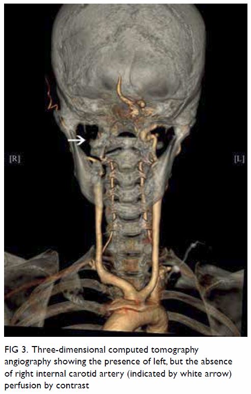
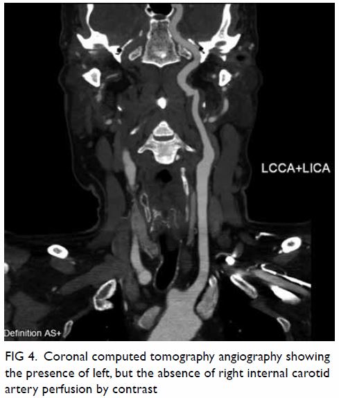
tomography scan revealed features suggestive of pyelonephritis at the lower pole of the right kidney (Fig 1). There was no obstructive urinary stone. In addition, the imaging demonstrated an atypical right pelvicalyceal system (Fig 2). A course of antibiotic was prescribed with good clinical response. Repeat urine microscopy was unremarkable 8 weeks after the initial presentation. Follow-up computed tomography scan confirmed resolution of right pyelonephritis but the unusual finding of the right pelvicalyceal system remained unchanged (Fig 3).
 

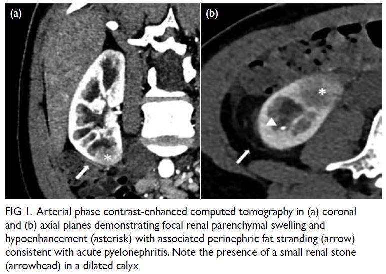
Figure 1. Arterial phase contrast-enhanced computed tomography in (a) coronal and (b) axial planes demonstrating focal renal parenchymal swelling and hypoenhancement (asterisk) with associated perinephric fat stranding (arrow) consistent with acute pyelonephritis. Note the presence of a small renal stone (arrowhead) in a dilated calyx
 

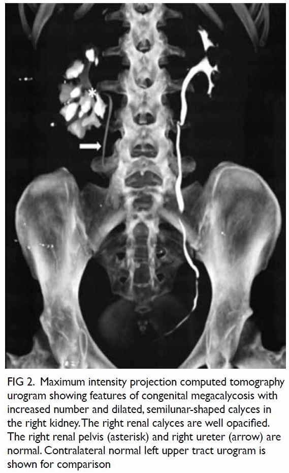
Figure 2. Maximum intensity projection computed tomography urogram showing features of congenital megacalycosis with increased number and dilated, semilunar-shaped calyces in the right kidney. The right renal calyces are well opacified. The right renal pelvis (asterisk) and right ureter (arrow) are normal. Contralateral normal left upper tract urogram is shown for comparison
 

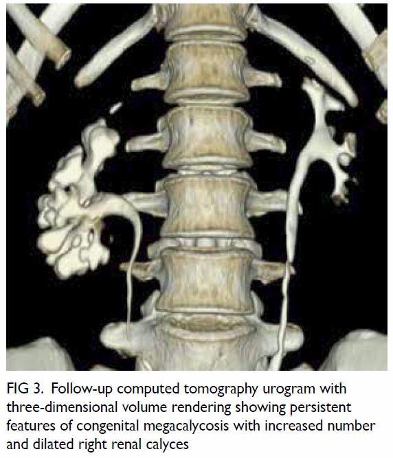
Figure 3. Follow-up computed tomography urogram with three-dimensional volume rendering showing persistent features of congenital megacalycosis with increased number and dilated right renal calyces
 
The radiological features were diagnostic of congenital megacalycosis. The anomaly is characterised by caliectasis with malformation. The classic triangular or conical shape of the renal calyces is replaced by a semilunar configuration. The pyramids of Malpighi are hypoplastic and the tip of each papilla is flat. The calyces have a rounded appearance with neither fornix nor papillae impressions. Polycalycosis is another feature of the condition and typically 20 to 25 calyces can be identified. The condition is differentiated from obstruction by the finding of a non-dilated renal pelvis, infundibulum, and ureter. The renal cortex was of normal thickness with good concentration of contrast medium in the distended calyces (Figs 2 and 3).
 
Congenital megacalycosis is a rare condition with approximately 100 cases reported in the literature. The anomaly is found predominantly in men and usually affects only one kidney. The exact pathogenesis remains unclear.1 2 3 The condition is usually asymptomatic and is detected during workup of urinary stone disease or urinary tract
 
infection in adults. On the contrary, reports of congenital megacalycosis in paediatric patients are on the rise with the increasing use of imaging in antenatal screening.4 Although urinary stasis in the distended calyces may predispose to infection and stone formation,1 2 renal function remains normal and neither anatomic nor functional deterioration occur over time.5 The benign nature suggests that megacalycosis should be considered a condition rather than a disease, and the term “megacalycose” has been suggested as an alternative term by some authors. Treatment should target complications only. Identification of the condition is important to avoid unnecessary investigation and intervention in a normally functioning kidney despite the inherent anatomic defect.2
 
Author contributions
Concept or design: CL Cho.
Acquisition of data: All authors.
Analysis or interpretation of data: All authors.
Drafting of the manuscript: All authors.
Critical revision of the manuscript for important intellectual content: All authors.
 
All authors had full access to the data, contributed to the study, approved the final version for publication, and take responsibility for its accuracy and integrity.
 
Conflicts of interest
The authors have disclosed no conflicts of interest.
 
Funding/support
This pictorial medicine paper received no specific grant from any funding agency in the public, commercial, or not-for-profit sectors.
 
Ethics approval
This study was conducted in accordance with the principles outlined in the Declaration of Helsinki. Verbal consent was obtained for the purpose of case study.
 
References
1. Puigvert A. Le megacalice. J Urol Nephrol 1964;70:321-6. Crossref
2. Kimche D, Lask D. Megacalycosis. Urology 1982;19:478-81. Crossref
3. Kalaitzis C, Patris E, Deligeorgiou E, et al. Radiological findings and the clinical importance of megacalycosis. Res Rep Urol 2015;7:153-5. Crossref
4. Kasap B, Kavukçu S, Soylu A, et al. Megacalycosis: report of two cases. Pediatr Nephrol 2005;20:828-30. Crossref
5. Redman JF, Neeb AD. Congenital megacalycosis: a forgotten diagnosis? Urology 2005;65:384-5. Crossref

Progressive diaphyseal dysplasia: a rare bone disorder with alarming radiographs

Hong Kong Academy of Medicine. CC BY-NC-ND 4.0
 
PICTORIAL MEDICINE
Progressive diaphyseal dysplasia: a rare bone disorder with alarming radiographs
Moses ML Li, MB, ChB, MHKICBSC1; KY Chung, FRCSEd (Orth), FHKAM (Orthopaedic Surgery)1; Alex WH Ng, FRCR, FHKAM (Radiology)2; KH Chiu, FRCS, FHKAM (Orthopaedic Surgery)1
1 Department of Orthopaedics and Traumatology, Prince of Wales Hospital, Hong Kong
2 Department of Imaging and Interventional Radiology, Prince of Wales Hospital, Hong Kong
 
Corresponding author: Dr Moses ML Li (moseslml@gmail.com)
 
 Full paper in PDF
 
In March 2017, a 57-year-old man was referred to our unit for left knee pain after a sprain, which resolved upon conservative treatment. Plain radiographs of the knees revealed diffuse sclerosis of bilateral femurs and tibias. A detailed history was sought to investigate the alarming skeletal pathology revealed on the plain radiographs. The patient had no history of bone pain, fever, weight loss, nor other constitutional symptoms. There was no history of malignancy. Physical examination revealed negative findings in the musculoskeletal, cardiac, pulmonary, renal, and neurological systems. Alkaline phosphatase was elevated (176 IU/L) but other blood parameters were normal, including white cell count, C-reactive protein, erythrocyte sedimentation rate, calcium, and phosphate. A skeletal survey revealed widespread sclerosis of tubular bone diaphysis of the lower extremities (Fig 1), upper extremities (Fig 2), and skull (Fig 3). Bone scintigraphy revealed increased osteoblastic activity corresponding to the locations of sclerosis (Fig 4). The characteristic radiographic features with the clinical and laboratory findings favoured a diagnosis of progressive diaphyseal dysplasia.
 

Figure 1. PPlain radiograph of bilateral lower limbs showing bilateral symmetrical diffuse sclerosis and cortical thickening with expansion of the diaphysis involving the femur, tibia, and fibula. Of note, the epiphyses of all the bones are spared which would be characteristic of progressive diaphyseal dysplasia
 

Figure 2. (a) Frontal radiographs of bilateral upper limbs showing symmetrical diffuse sclerosis and cortical thickening of the diaphysis involving the scapula, humerus, radius, and ulna. The epiphyses of these bones are also spared. (b) Frontal radiographs of the hands showing sclerosis with cortical thickening of the right second and left second to third metacarpal bones. Mild bony expansion with relative sparing of the medullary canals and epiphysis are noted
 

Figure 3. (a) Frontal radiograph of the skull showing sclerosis and cortical thickening of the calvaria and base of the skull. (a) Lateral radiograph of the skull showing severe sclerosis and thickening of the skull base and mandible. The cervical spine is not involved in this case
 

Figure 4. (a) Bone scintigraphy showing increased tracer uptake corresponding to the regions of sclerosis on plain radiographs, particularly in the skull vault and bilateral mandibles. (b) Lateral view of bone scintigraphy showing severe increased tracer uptake at the skull base
 
Sclerosing bone dysplasia is a heterogeneous group of rare bone disorders with pathognomonic radiological features, caused by a defective ossification pathway.1 2 Progressive diaphyseal dysplasia, also known as Camurati-Engelmann disease, is a disease belonging to this entity.3 4 5 It is an autosomal dominant disorder due to mutation in transforming growth factor–Β1. This in turn leads to a disorder of intramembranous ossification, and results in hyperostosis. Characteristic radiological features are bilateral symmetrical fusiform sclerosis involving the diaphyses of tubular bones. The epiphyses are typically spared as these regions are formed by endochondral ossification. The lower extremities are more affected than the upper extremities. In descending order of frequency, the tibia, femur, fibula, humerus, ulna, and radius are affected.3 Occasionally, the calvaria of the skull is involved. The affected bones show uneven cortical thickening of the diaphysis with hyperostosis extending in both periosteal and endosteal directions. Hyperostosis of the endosteal surface results in medullary canal narrowing.
 
Clinical manifestations of progressive diaphyseal dysplasia include limb pain, muscle weakness, and easy fatigability. However, our patient was completely asymptomatic. The disorder was picked up incidentally during investigation of an unrelated knee sprain. Detailed systemic review revealed an absence of extraosseous manifestations of Erdheim-Chester disease that shares similar radiological features.3 Ribbing disease (hereditary multiple diaphyseal sclerosis) usually presents with unilateral or asymmetrical involvement of the bones, and does not involve the skull vault. Osteopetrosis has epiphysis involvement and usually presents with fracture and extramedullary haematopoiesis. Although the differential diagnoses for bone sclerosis are extensive, characteristic radiographic distribution in association with clinical and laboratory findings can substantially narrow the possibilities and allow the diagnosis to be made. The age at which the diagnosis of progressive diaphyseal dysplasia is reached, the clinical manifestations of disease and the extent of radiological evidence of sclerosis are variable.4 5 Disease progression is slow and unpredictable. Treatment of the disease aims for symptomatic relief, with losartan reported to be effective in relieving limb pain based on the mechanism of down-regulation of transforming growth factor–Β1 receptor expression.
 
Author contributions
All authors contributed to the concept of the study, acquisition and analysis of the data, drafting of the manuscript, and critical revision of the manuscript for important intellectual content. All authors had full access to the data, contributed to the study, approved the final version for publication, and take responsibility for its accuracy and integrity.
 
Conflicts of interest
All authors have disclosed no conflicts of interest.
 
Funding/support
This pictorial medicine paper received no specific grant from any funding agency in the public, commercial, or not-for-profit sectors.
 
Ethics approval
The patient was treated in accordance with the principles outlined in the Declaration of Helsinki.
 
References
1. Boulet C, Madani H, Lenchik L, et al. Sclerosing bone dysplasias: genetic, clinical and radiology update of hereditary and non-hereditary disorders. Br J Radiol 2016;89:20150349. Crossref
2. Ihde LL, Forrester DM, Gottsegen CJ, et al. Sclerosing bone dysplasias: review and differentiation from other causes of osteosclerosis. Radiographics 2011;31:1865-82. Crossref
3. Uezato S, Dias G, Inada J, Valente M, Fernandes E. Imaging aspects of Camurati-Engelmann disease. Rev Assoc Med Bras (1992) 2016;62:825-7. Crossref
4. Van Hul W, Boudin E, Vanhoenacker FM, Mortier G. Camurati-Engelmann disease. Calcif Tissue Int 2019;104:554-60. Crossref
5. Yuldashev AJ, Shin CH, Kim YS, et al. Orthopedic manifestations of type I Camurati-Engelmann disease. Clin Orthop Surg 2017;9:109-15. Crossref

Skin testing for hypersensitivity and cross-reactivity between proton pump inhibitors

Hong Kong Academy of Medicine. CC BY-NC-ND 4.0
 
PICTORIAL MEDICINE
Skin testing for hypersensitivity and cross-reactivity between proton pump inhibitors
Philip H Li, MRCP, FHKCP
Division of Rheumatology and Clinical Immunology, Department of Medicine, The University of Hong Kong, Queen Mary Hospital, Pokfulam, Hong Kong
 
Corresponding author: Dr Philip H Li (liphilip@hku.hk)
 
 Full paper in PDF
 
A 44-year-old woman presented to our anaphylaxis clinic with first episode of anaphylaxis. She had no known prior food or drug allergies. She was previously prescribed esomeprazole 20 mg daily as required for dyspepsia by her private physician, but took it only very occasionally. Two months previously she took one tablet of esomeprazole (20 mg) and one of paracetamol (500 mg) together with a slice of plain bread for abdominal discomfort and influenza-like symptoms. Within 10 minutes, she experienced generalised pruritus and collapsed at home. She awoke after hitting the floor and telephoned for an ambulance. She was treated with intramuscular adrenaline in the ambulance because systolic blood pressure had fallen to <80 mm Hg. Initial investigations revealed a significant rise in acute tryptase (12.3 ng/mL) with a normal baseline level (2.1 ng/mL). Initially wheat or wheat-dependent cofactor augmented anaphylaxis was suspected. She was advised to avoid wheat, cyclooxygenase inhibitors, and proton pump inhibitors (PPI) until review.
 
Results for specific immunoglobulin E to wheat and omega-5-gliadin were negative. Skin prick test (SPT) to wheat solution (Inmunotek, Madrid, Spain) and prick-to-prick with the index bread slice was also negative. Skin prick test and intradermal tests (IDT) to paracetamol (both 100 mg/mL, GlaxoSmithKline, London, United Kingdom) were also negative. The patient tolerated an oral challenge with 500 mg of paracetamol together with a slice of the same index bread with no adverse reaction.
 
The SPT (8 mg/mL) and IDT (8 mg/mL, 0.8 mg/mL and 0.08 mg/mL; AstraZeneca, Bedfordshire, United Kingdom) were performed and are shown in the Figure. Skin prick test to histamine (positive control) and normal saline (negative control) was positive at 5 mm and negative at 0 mm, respectively. Skin prick test to esomeprazole was borderline positive with a 3-mm wheal and flare. Intradermal test to esomeprazole was positive at concentrations of 8 mg/mL, 0.8 mg/mL and 0.08 mg/mL; with 18-mm, 8-mm and 3-mm wheal expansion, respectively. The SPT and IDT with 8 mg/mL and 0.8 mg/mL were negative in a healthy control (the author) as shown in the Figure. To assess for potential cross-reactivity, SPT (4 mg/mL) and IDT (4 mg/mL and 0.4 mg/mL) to pantoprazole were also performed and are also shown in the Figure. The SPT was negative, but IDT was positive with 4 mg/mL and 0.4 mg/mL dilutions with 7-mm and 4-mm wheal expansion, respectively.
 

Figure. Left: Intradermal tests to esomeprazole in healthy control (neat, 1:10 dilution). Right: Skin prick and intradermal tests to esomeprazole (neat, 1:10 and 1:100 dilutions) and pantoprazole (neat and 1:10 dilution)
 
She was diagnosed with severe type I hypersensitivity to esomeprazole with cross-sensitisation to pantoprazole. Drug provocoation testing was not indicated in view of the compatible clinical history, strongly positive skin tests, and high risk of anaphylaxis. The patient declined testing with other PPI and was advised to avoid the entire class until further workup. She was prescribed famotidine 20 mg twice a day for her dyspepsia with no adverse effects.
 
This is the first reported case in Hong Kong of PPI anaphylaxis and demonstrates the utility of skin testing to assess potential cross-reactivity. Although relatively uncommon, reports of hypersensitivity to PPI are increasing, in parallel with their increasing use worldwide. The majority of hypersensitivity reactions appear to be of the immediate type.1 Previous exposure to esomeprazole may have been initial sensitising events. Despite common misconception, clinicians should bear in mind that prior tolerance of a certain drug does not preclude it as a future cause of drug allergy. Although patterns of cross-reactivity among various PPIs have been reported, these remain controversial and a thorough allergological workup should be performed for every patient.2 Clinicians are reminded to be vigilant of this uncommon cause of anaphylaxis and beware of potential cross-reactivity. The SPT and IDT have high specificity, but patients with suspected PPI hypersensitivity and negative skin tests should undergo drug provocation tests to confidently exclude this important diagnosis.3
 
Author contributions
The author contributed to the concept or design, acquisition of data, analysis or interpretation of data, drafting of the manuscript, and critical revision for important intellectual content. The author had full access to the data, contributed to the study, approved the final version for publication, and takes responsibility for its accuracy and integrity.
 
Conflicts of interest
The author has disclosed no conflicts of interest.
 
Funding/support
This pictorial medicine paper received no specific grant from any funding agency in the public, commercial, or not-forprofit sectors.
 
Ethics approval
The patient consented to this publication.
 
References
1. Otani IM, Banerji A. Immediate and delayed hypersensitivity reactions to proton pump inhibitors: evaluation and management. Curr Allergy Asthma Rep 2016;16:17. Crossref
2. Tourillon C, Mahe J, Baron A, et al. Immediate-type hypersensitivity cross-reactions to proton pump inhibitors: a descriptive study of data from the French National Pharmacovigilance Database. Int Arch Allergy Immunol 2019;178:159-66. Crossref
3. Kepil Özdemir S, Yilmaz I, Aydin Ö, et al. Immediate-type hypersensitivity reactions to proton pump inhibitors: usefulness of skin tests in the diagnosis and assessment of cross-reactivity. Allergy 2013;68:1008-14. Crossref

Clone of Clone of Primary hepatic schwannoma: imaging and histological findings

Hong Kong Academy of Medicine. CC BY-NC-ND 4.0
 
PICTORIAL MEDICINE
Primary hepatic schwannoma: imaging and histological findings
HL Tsui, MB, ChB, FRCR1; SM Yu, MB, ChB, FHKAM (Radiology)1; CH Lau, MB, ChB2; Sherman SM Lam, MB, BS, FHKAM (Surgery)3; PY Chu, MB, ChB, FHKAM (Radiology)1; YH Hui, MB, BS, FHKAM (Radiology)1; KL Lo, MB, ChB, FHKAM (Radiology)1
1 Department of Radiology and Organ Imaging, United Christian Hospital, Hong Kong
2 Department of Pathology, United Christian Hospital, Hong Kong
3 Department of Surgery, United Christian Hospital, Hong Kong
 
Corresponding author: Dr HL Tsui (karen.tsuihl@gmail.com)
 
 Full paper in PDF
 
Schwannoma is a rare tumour in the liver. It is likely to arise from the hepatobiliary nerves among the hepatic plexus in the liver hilum as well as interlobular connective tissues and hepatic arteries. To the best of our knowledge, no prior publications have reported cases in Hong Kong.
 
We report the case of a 64-year-old man with a history of nasopharyngeal carcinoma and colon carcinoma and a new liver lesion detected on follow-up imaging for surveillance in 2018 following detection of a slightly elevated serum MEDICINEcarcinoembryonic antigen level (4.7 μg/L). Alpha fetoprotein level was within the normal range (3.9 μg/L). Liver function tests were normal and he was asymptomatic with no history of neurofibromatosis.
 
Triphasic contrast computed tomography (CT) of the liver revealed a 5.2-cm ovoid hypodense lesion with heterogeneous enhancement in the caudate lobe of the liver (Fig 1). No washout of contrast was evident in the portal venous or delayed phases. Fluorodeoxyglucose-18 positron emission tomography–CT showed a moderately hypermetabolic lesion at the caudate lobe with a maximum standardised update value of 5.8 (Fig 2).
 

Figure 1. Triphasic computed tomography scans in (a) pre-contrast, (b) arterial, (c) portal venous, and (d) delayed phases, showing a well-defined, ovoid hypodense lesion with heterogeneous enhancement that persists in portal venous and delayed phases in the caudate lobe of the liver
 

Figure 2. Positron emission tomography–computed tomography scan in (a) coronal, (b) sagittal and (c) axial planes together with the (d) scout and (e) maximum intensity projection images, showing a moderately hypermetabolic lesion at the caudate lobe of the liver (red crosses)
 
Surgical resection of the lesion was performed (Fig 3). Pathology showed a schwannoma and degenerative changes. Histological examination revealed an encapsulated tumour consisting of highly ordered Antoni type A and B areas (Fig 4). Immunohistochemical analysis showed the tumour cells to be diffusely positive for S100, consistent with neural differentiation (Fig 4). HerPar1, a mitochondrial antigen of hepatocytes, was negative. The CD34, a cell surface glycoprotein that is positive in gastrointestinal stromal tumour, was also negative.
 

Figure 3. Gross specimen of hepatic resection with a well-circumscribed tumour mass and light tan colour cut surface
 

Figure 4. Micrographs of the tumour, showing (a) Antoni type A area, ×100; (b) Antoni type B area, ×100; (c) circumscribed and encapsulated tumour, ×20; and (d) immunostaining for S100 diffusely positive for the tumour cell, ×100
 
Schwannoma is most commonly found in the limbs and the head and neck region. A fifth of cases shows association with neurofibromatosis type 1. The mediastinum and retroperitoneum are other possible sites. It is uncommon in the gastrointestinal tract and extremely rare in the liver.1 It was first reported in 1978 by Pereira et al.2 A literature search through PubMed and MEDLINE revealed 32 reported cases. No cases have been published in Hong Kong.
 
The origin of hepatic schwannoma is the hepatobiliary nerves among the hepatic plexus in the liver hilum as well as interlobular connective tissues and the hepatic arteries.1 3 They are usually well-encapsulated and grow very slowly, usually smaller than 5 cm at the time of diagnosis. Larger schwannomas may undergo secondary degeneration with consequent pseudocystic regression, haemorrhage, and calcification. Malignant transformation is very rare.3
 
Pathologically, a schwannoma is an encapsulated tumour that arises within the nerve sheaths. It consists of a highly ordered cellular component (Antoni type A area) characterised by spindle cells with twisted nuclei arranged in short bundles, and a hypocellular area in a loose myxoid stroma (Antoni type B area) that comprises a loose meshwork of gelatinous and microcystic tissue.4
 
On imaging, hepatic schwannoma is usually well circumscribed with various signal characteristics, depending on the distribution of Antoni A and Antoni B areas.1 It is commonly of low density with heterogeneous enhancement on CT, hypointense on T1-weighted, and hyperintense on T2-weighted magnetic resonance imaging.5 There have also been reports of malignant tumours but there are no distinct radiological features that differentiate them from benign tumours.3 A hepatic schwannoma may be fluorodeoxyglucose-avid depending on inflammatory activity and cellularity. Fluorodeoxyglucose-18 positron emission tomography–CT alone may enable differentiation of a schwannoma from malignant lesions of the liver.1
 
Hepatic schwannoma is an extremely rare tumour and preoperative diagnosis with imaging is challenging. Biopsy or surgical resection is usually required for definitive diagnosis.
 
Author contributions
Concept or design: All authors.
Acquisition of data: HL Tsui and CH Lau.
Analysis or interpretation of data: HL Tsui, SM Yu, and CH Lau.
Drafting of the manuscript: HL Tsui, SM Yu, and CH Lau.
Critical revision of the manuscript for important intellectual content: All authors.
 
All authors had full access to the data, contributed to the study, approved the final version for publication, and take responsibility for its accuracy and integrity.
 
Conflicts of interest
All authors have disclosed no conflicts of interest.
 
Funding/support
This research received no specific grant from any funding agency in the public, commercial, or not-for-profit sectors.
 
Ethics approval
This study is approved by the cluster Research Ethics Committee (Ref KC/KE-19-0247/ER-3). Written patient consent was also obtained.
 
References
1. Hayashi M, Takeshita A, Yamamoto K, Tanigawa N. Primary hepatic benign schwannoma. World J Gastrointest Surg 2012;4:73-8. Crossref
2. Pereira Filho RA, Souza SA, Oliveira Filho JA. Primary neurilemmal tumour of the liver: case report. Arq Gastroenterol 1978;15:136-8.
3. Ozkan EE, Guldur M, Uzunkoy A. A case report of benign schwannoma of the liver. Intern Med 2010;49:1533-6. Crossref
4. Wan DL, Zhai ZL, Ren KW, Yang YC, Lin SZ, Zheng SS. Hepatic schwannoma: a case report and an updated 40-year review of the literature yielding 30 cases. Mol Clin Oncol 2016;4:959-64. Crossref
5. Yamamoto M, Hasegawa K, Arita J, et al. Primary hepatic schwannoma: a case report. Int J Surg Case Rep 2016;29:146-50. Crossref

Clone of Primary hepatic schwannoma: imaging and histological findings

Hong Kong Academy of Medicine. CC BY-NC-ND 4.0
 
PICTORIAL MEDICINE
Primary hepatic schwannoma: imaging and histological findings
HL Tsui, MB, ChB, FRCR1; SM Yu, MB, ChB, FHKAM (Radiology)1; CH Lau, MB, ChB2; Sherman SM Lam, MB, BS, FHKAM (Surgery)3; PY Chu, MB, ChB, FHKAM (Radiology)1; YH Hui, MB, BS, FHKAM (Radiology)1; KL Lo, MB, ChB, FHKAM (Radiology)1
1 Department of Radiology and Organ Imaging, United Christian Hospital, Hong Kong
2 Department of Pathology, United Christian Hospital, Hong Kong
3 Department of Surgery, United Christian Hospital, Hong Kong
 
Corresponding author: Dr HL Tsui (karen.tsuihl@gmail.com)
 
 Full paper in PDF
 
Schwannoma is a rare tumour in the liver. It is likely to arise from the hepatobiliary nerves among the hepatic plexus in the liver hilum as well as interlobular connective tissues and hepatic arteries. To the best of our knowledge, no prior publications have reported cases in Hong Kong.
 
We report the case of a 64-year-old man with a history of nasopharyngeal carcinoma and colon carcinoma and a new liver lesion detected on follow-up imaging for surveillance in 2018 following detection of a slightly elevated serum MEDICINEcarcinoembryonic antigen level (4.7 μg/L). Alpha fetoprotein level was within the normal range (3.9 μg/L). Liver function tests were normal and he was asymptomatic with no history of neurofibromatosis.
 
Triphasic contrast computed tomography (CT) of the liver revealed a 5.2-cm ovoid hypodense lesion with heterogeneous enhancement in the caudate lobe of the liver (Fig 1). No washout of contrast was evident in the portal venous or delayed phases. Fluorodeoxyglucose-18 positron emission tomography–CT showed a moderately hypermetabolic lesion at the caudate lobe with a maximum standardised update value of 5.8 (Fig 2).
 

Figure 1. Triphasic computed tomography scans in (a) pre-contrast, (b) arterial, (c) portal venous, and (d) delayed phases, showing a well-defined, ovoid hypodense lesion with heterogeneous enhancement that persists in portal venous and delayed phases in the caudate lobe of the liver
 

Figure 2. Positron emission tomography–computed tomography scan in (a) coronal, (b) sagittal and (c) axial planes together with the (d) scout and (e) maximum intensity projection images, showing a moderately hypermetabolic lesion at the caudate lobe of the liver (red crosses)
 
Surgical resection of the lesion was performed (Fig 3). Pathology showed a schwannoma and degenerative changes. Histological examination revealed an encapsulated tumour consisting of highly ordered Antoni type A and B areas (Fig 4). Immunohistochemical analysis showed the tumour cells to be diffusely positive for S100, consistent with neural differentiation (Fig 4). HerPar1, a mitochondrial antigen of hepatocytes, was negative. The CD34, a cell surface glycoprotein that is positive in gastrointestinal stromal tumour, was also negative.
 

Figure 3. Gross specimen of hepatic resection with a well-circumscribed tumour mass and light tan colour cut surface
 

Figure 4. Micrographs of the tumour, showing (a) Antoni type A area, ×100; (b) Antoni type B area, ×100; (c) circumscribed and encapsulated tumour, ×20; and (d) immunostaining for S100 diffusely positive for the tumour cell, ×100
 
Schwannoma is most commonly found in the limbs and the head and neck region. A fifth of cases shows association with neurofibromatosis type 1. The mediastinum and retroperitoneum are other possible sites. It is uncommon in the gastrointestinal tract and extremely rare in the liver.1 It was first reported in 1978 by Pereira et al.2 A literature search through PubMed and MEDLINE revealed 32 reported cases. No cases have been published in Hong Kong.
 
The origin of hepatic schwannoma is the hepatobiliary nerves among the hepatic plexus in the liver hilum as well as interlobular connective tissues and the hepatic arteries.1 3 They are usually well-encapsulated and grow very slowly, usually smaller than 5 cm at the time of diagnosis. Larger schwannomas may undergo secondary degeneration with consequent pseudocystic regression, haemorrhage, and calcification. Malignant transformation is very rare.3
 
Pathologically, a schwannoma is an encapsulated tumour that arises within the nerve sheaths. It consists of a highly ordered cellular component (Antoni type A area) characterised by spindle cells with twisted nuclei arranged in short bundles, and a hypocellular area in a loose myxoid stroma (Antoni type B area) that comprises a loose meshwork of gelatinous and microcystic tissue.4
 
On imaging, hepatic schwannoma is usually well circumscribed with various signal characteristics, depending on the distribution of Antoni A and Antoni B areas.1 It is commonly of low density with heterogeneous enhancement on CT, hypointense on T1-weighted, and hyperintense on T2-weighted magnetic resonance imaging.5 There have also been reports of malignant tumours but there are no distinct radiological features that differentiate them from benign tumours.3 A hepatic schwannoma may be fluorodeoxyglucose-avid depending on inflammatory activity and cellularity. Fluorodeoxyglucose-18 positron emission tomography–CT alone may enable differentiation of a schwannoma from malignant lesions of the liver.1
 
Hepatic schwannoma is an extremely rare tumour and preoperative diagnosis with imaging is challenging. Biopsy or surgical resection is usually required for definitive diagnosis.
 
Author contributions
Concept or design: All authors.
Acquisition of data: HL Tsui and CH Lau.
Analysis or interpretation of data: HL Tsui, SM Yu, and CH Lau.
Drafting of the manuscript: HL Tsui, SM Yu, and CH Lau.
Critical revision of the manuscript for important intellectual content: All authors.
 
All authors had full access to the data, contributed to the study, approved the final version for publication, and take responsibility for its accuracy and integrity.
 
Conflicts of interest
All authors have disclosed no conflicts of interest.
 
Funding/support
This research received no specific grant from any funding agency in the public, commercial, or not-for-profit sectors.
 
Ethics approval
This study is approved by the cluster Research Ethics Committee (Ref KC/KE-19-0247/ER-3). Written patient consent was also obtained.
 
References
1. Hayashi M, Takeshita A, Yamamoto K, Tanigawa N. Primary hepatic benign schwannoma. World J Gastrointest Surg 2012;4:73-8. Crossref
2. Pereira Filho RA, Souza SA, Oliveira Filho JA. Primary neurilemmal tumour of the liver: case report. Arq Gastroenterol 1978;15:136-8.
3. Ozkan EE, Guldur M, Uzunkoy A. A case report of benign schwannoma of the liver. Intern Med 2010;49:1533-6. Crossref
4. Wan DL, Zhai ZL, Ren KW, Yang YC, Lin SZ, Zheng SS. Hepatic schwannoma: a case report and an updated 40-year review of the literature yielding 30 cases. Mol Clin Oncol 2016;4:959-64. Crossref
5. Yamamoto M, Hasegawa K, Arita J, et al. Primary hepatic schwannoma: a case report. Int J Surg Case Rep 2016;29:146-50. Crossref

Primary hepatic schwannoma: imaging and histological findings

Hong Kong Academy of Medicine. CC BY-NC-ND 4.0
 
PICTORIAL MEDICINE
Primary hepatic schwannoma: imaging and histological findings
HL Tsui, MB, ChB, FRCR1; SM Yu, MB, ChB, FHKAM (Radiology)1; CH Lau, MB, ChB2; Sherman SM Lam, MB, BS, FHKAM (Surgery)3; PY Chu, MB, ChB, FHKAM (Radiology)1; YH Hui, MB, BS, FHKAM (Radiology)1; KL Lo, MB, ChB, FHKAM (Radiology)1
1 Department of Radiology and Organ Imaging, United Christian Hospital, Hong Kong
2 Department of Pathology, United Christian Hospital, Hong Kong
3 Department of Surgery, United Christian Hospital, Hong Kong
 
Corresponding author: Dr HL Tsui (karen.tsuihl@gmail.com)
 
 Full paper in PDF
 
Schwannoma is a rare tumour in the liver. It is likely to arise from the hepatobiliary nerves among the hepatic plexus in the liver hilum as well as interlobular connective tissues and hepatic arteries. To the best of our knowledge, no prior publications have reported cases in Hong Kong.
 
We report the case of a 64-year-old man with a history of nasopharyngeal carcinoma and colon carcinoma and a new liver lesion detected on follow-up imaging for surveillance in 2018 following detection of a slightly elevated serum carcinoembryonic antigen level (4.7 μg/L). Alpha fetoprotein level was within the normal range (3.9 μg/L). Liver function tests were normal and he was asymptomatic with no history of neurofibromatosis.
 
Triphasic contrast computed tomography (CT) of the liver revealed a 5.2-cm ovoid hypodense lesion with heterogeneous enhancement in the caudate lobe of the liver (Fig 1). No washout of contrast was evident in the portal venous or delayed phases. Fluorodeoxyglucose-18 positron emission tomography–CT showed a moderately hypermetabolic lesion at the caudate lobe with a maximum standardised update value of 5.8 (Fig 2).
 

Figure 1. Triphasic computed tomography scans in (a) pre-contrast, (b) arterial, (c) portal venous, and (d) delayed phases, showing a well-defined, ovoid hypodense lesion with heterogeneous enhancement that persists in portal venous and delayed phases in the caudate lobe of the liver
 

Figure 2. Positron emission tomography–computed tomography scan in (a) coronal, (b) sagittal and (c) axial planes together with the (d) scout and (e) maximum intensity projection images, showing a moderately hypermetabolic lesion at the caudate lobe of the liver (red crosses)
 
Surgical resection of the lesion was performed (Fig 3). Pathology showed a schwannoma and degenerative changes. Histological examination revealed an encapsulated tumour consisting of highly ordered Antoni type A and B areas (Fig 4). Immunohistochemical analysis showed the tumour cells to be diffusely positive for S100, consistent with neural differentiation (Fig 4). HerPar1, a mitochondrial antigen of hepatocytes, was negative. The CD34, a cell surface glycoprotein that is positive in gastrointestinal stromal tumour, was also negative.
 

Figure 3. Gross specimen of hepatic resection with a well-circumscribed tumour mass and light tan colour cut surface
 

Figure 4. Micrographs of the tumour, showing (a) Antoni type A area, ×100; (b) Antoni type B area, ×100; (c) circumscribed and encapsulated tumour, ×20; and (d) immunostaining for S100 diffusely positive for the tumour cell, ×100
 
Schwannoma is most commonly found in the limbs and the head and neck region. A fifth of cases shows association with neurofibromatosis type 1. The mediastinum and retroperitoneum are other possible sites. It is uncommon in the gastrointestinal tract and extremely rare in the liver.1 It was first reported in 1978 by Pereira et al.2 A literature search through PubMed and MEDLINE revealed 32 reported cases. No cases have been published in Hong Kong.
 
The origin of hepatic schwannoma is the hepatobiliary nerves among the hepatic plexus in the liver hilum as well as interlobular connective tissues and the hepatic arteries.1 3 They are usually well-encapsulated and grow very slowly, usually smaller than 5 cm at the time of diagnosis. Larger schwannomas may undergo secondary degeneration with consequent pseudocystic regression, haemorrhage, and calcification. Malignant transformation is very rare.3
 
Pathologically, a schwannoma is an encapsulated tumour that arises within the nerve sheaths. It consists of a highly ordered cellular component (Antoni type A area) characterised by spindle cells with twisted nuclei arranged in short bundles, and a hypocellular area in a loose myxoid stroma (Antoni type B area) that comprises a loose meshwork of gelatinous and microcystic tissue.4
 
On imaging, hepatic schwannoma is usually well circumscribed with various signal characteristics, depending on the distribution of Antoni A and Antoni B areas.1 It is commonly of low density with heterogeneous enhancement on CT, hypointense on T1-weighted, and hyperintense on T2-weighted magnetic resonance imaging.5 There have also been reports of malignant tumours but there are no distinct radiological features that differentiate them from benign tumours.3 A hepatic schwannoma may be fluorodeoxyglucose-avid depending on inflammatory activity and cellularity. Fluorodeoxyglucose-18 positron emission tomography–CT alone may enable differentiation of a schwannoma from malignant lesions of the liver.1
 
Hepatic schwannoma is an extremely rare tumour and preoperative diagnosis with imaging is challenging. Biopsy or surgical resection is usually required for definitive diagnosis.
 
Author contributions
Concept or design: All authors.
Acquisition of data: HL Tsui and CH Lau.
Analysis or interpretation of data: HL Tsui, SM Yu, and CH Lau.
Drafting of the manuscript: HL Tsui, SM Yu, and CH Lau.
Critical revision of the manuscript for important intellectual content: All authors.
 
All authors had full access to the data, contributed to the study, approved the final version for publication, and take responsibility for its accuracy and integrity.
 
Conflicts of interest
All authors have disclosed no conflicts of interest.
 
Funding/support
This research received no specific grant from any funding agency in the public, commercial, or not-for-profit sectors.
 
Ethics approval
This study is approved by the cluster Research Ethics Committee (Ref KC/KE-19-0247/ER-3). Written patient consent was also obtained.
 
References
1. Hayashi M, Takeshita A, Yamamoto K, Tanigawa N. Primary hepatic benign schwannoma. World J Gastrointest Surg 2012;4:73-8. Crossref
2. Pereira Filho RA, Souza SA, Oliveira Filho JA. Primary neurilemmal tumour of the liver: case report. Arq Gastroenterol 1978;15:136-8.
3. Ozkan EE, Guldur M, Uzunkoy A. A case report of benign schwannoma of the liver. Intern Med 2010;49:1533-6. Crossref
4. Wan DL, Zhai ZL, Ren KW, Yang YC, Lin SZ, Zheng SS. Hepatic schwannoma: a case report and an updated 40-year review of the literature yielding 30 cases. Mol Clin Oncol 2016;4:959-64. Crossref
5. Yamamoto M, Hasegawa K, Arita J, et al. Primary hepatic schwannoma: a case report. Int J Surg Case Rep 2016;29:146-50. Crossref

A common but often neglected source of emboli

© Hong Kong Academy of Medicine. CC BY-NC-ND 4.0
 
PICTORIAL MEDICINE
A common but often neglected source of emboli
SY Au, MB, BS, FHKAM (Medicine); KM Fong, MB, ChB, MRCP (UK); Jack KC Shek, MB, BS, FHKAM (Anaesthesiology); SK Yung, MB, BS, FHKAM (Medicine); George WY Ng, MB, BS, FHKAM (Medicine)
Intensive Care Unit, Queen Elizabeth Hospital, Hong Kong
 
Corresponding author: Dr SY Au (h0145237@gmail.com)
 
 Full paper in PDF
 
Case
An 82-year-old woman was admitted to the intensive care unit with acute abdomen. She was initially admitted for chest infection but complained of sudden-onset abdominal pain and abdominal distension. She was confused with decreased consciousness. She had a medical history of sick sinus syndrome with dual-chamber pacing performed 4 years previously. She was being treated for hypertension but was prescribed neither aspirin nor anticoagulation. Initial investigations revealed an increased white cell count, coagulopathy, and deranged liver and renal function, with severe mixed respiratory and lactic acidosis. She was commenced on a high-dose dopamine infusion and was intubated prior to transfer to the intensive care unit. Further imaging investigations were performed. Abdominal plain radiograph showed dilated bowel and subsequent computed tomography of the abdomen confirmed diffuse bowel ischaemia (Fig 1). There was no abdominal aortic aneurysm. Computed tomography was also performed in view of her confusion and showed left middle cerebral artery infarction. Electrocardiography performed to identify cardiac causes of systemic embolisation revealed a paced rhythm of 80 beats per minute and right bundle branch block pattern (Fig 2). A lateral chest radiograph subsequently confirmed that the ventricular lead was in the left ventricle as it was pointing posteriorly (Fig 3). Transthoracic echocardiography showed no intracardiac clots but confirmed that the pacing lead was positioned in the left heart through the atrial septum. Transoesophageal echocardiography confirmed that the lead entered the left heart through a patent foramen ovale. The ischaemic bowel was too extensive and not amenable to surgery and the patient finally succumbed.
 

Figure 1. Unusual course of the ventricular lead crossing the patent foramen ovale.The diffusely dilated bowel loops were due to ischaemic bowel secondary to cardiac embolisation
 

Figure 2. Right bundle branch block on electrocardiography
 

Figure 3. Ventricular lead in the left ventricle
 
Confirmation of placement of a pacing lead by a posterior-anterior chest radiograph can be difficult. A left bundle branch paced rhythm on echocardiography and a lateral radiograph that shows an anteriorly pointing ventricular lead help confirm placement of a pacing lead in the right ventricle. A pacing lead in the left ventricle and the absence of anticoagulation places a patient at risk of clot formation that may lead to systemic embolisation. In our patient, an intracardiac clot was not well demonstrated on either computed tomography or echocardiography, and 4 years had passed between pacemaker insertion and this systemic embolic episode. Only a probable, not a definite causal relationship could be established. We postulate that her chest infection may have impacted her clotting profile and resulted in this systemic embolic event.
 
Author contributions
Concept or design: All authors.
Acquisition of data: All authors.
Analysis or interpretation of data: SY Au.
Drafting of the manuscript: SY Au.
Critical revision of the manuscript for important intellectual content: All authors.
 
All authors had full access to the data, contributed to the study, approved the final version for publication and take responsibility for its accuracy and integrity.
 
Conflicts of interest
All authors have no conflicts of interest to disclose.
 
Funding/support
This pictorial medicine received no specific grant from any funding agency in the public, commercial or not-for-profit sectors.
 
Ethics approval
The study was conducted in accordance with the Declaration of Helsinki. Consent for publication was obtained from the patient’sfamily.
 

Vision loss due to ophthalmic artery occlusion secondary to spontaneous internal carotid artery dissection

© Hong Kong Academy of Medicine. CC BY-NC-ND 4.0
 
PICTORIAL MEDICINE
Vision loss due to ophthalmic artery occlusion secondary to spontaneous internal carotid artery dissection
Sunny CL Au, MB, ChB, AFCOphthHK1; Simon TC Ko, MB, BS, FHKAM (Ophthalmology)2
1 Department of Ophthalmology, Hong Kong East Cluster Ophthalmic Service, Tung Wah Eastern Hospital, Hong Kong
2 Department of Ophthalmology, Tung Wah Eastern Hospital, Hong Kong
 
Corresponding author: Dr Sunny CL Au (kilihcua@gmail.com)
 
 Full paper in PDF
 
Case
A 47-year-old male smoker with hypertension, diabetes mellitus, and hyperlipidaemia complained of right eye vision loss on waking. His best-corrected visual acuity was reduced to light perception only. Physical examination revealed anisocoria without ptosis and no extraocular movement deficit. The right eye pupil was larger than the left, and the difference was more obvious under a light environment. Direct and consensual pupil reflexes were both present but there was a marked right relative afferent pupillary defect. Slit lamp examination revealed normal anterior segments of the eyes, but a right pale optic disc without rim thinning, and a typical cherry-red spot over the right fovea surrounded by generalised whitened retina. Vessels were not tortuous and no emboli were seen nor retinal haemorrhage over different layers (Figs 1 and 2). Left eye posterior segment was normal. The patient also reported right-sided headache and left-sided numbness. Thorough physical examination revealed no other cranial nerve deficit or systemic focal neurological deficit. Temporal pulse was easily palpable and non-tender, and no carotid bruit was heard. The patient worked on a construction site and denied any trauma. He commenced hyperbaric oxygen therapy but no improvement was observed. Blood tests and brain imaging were all normal, but carotid Doppler showed significant obstruction >90% of the internal carotid artery, accounting for the absence of bruit. Computed tomography angiography confirmed dissection of a long segment of the right internal carotid artery (Figs 3 and 4), not amendable to stenting or bypass surgery. A diagnosis of ophthalmic artery occlusion was made and explained the ineffectiveness of hyperbaric oxygen therapy due to choroidal ischaemia. Symptomatic internal carotid artery dissection is a rare but major cause of young-onset stroke, itself uncommon.1 Neck trauma is a major aetiology, and there is a slight male predominance with mean age of onset in the 40s.2 Apart from the neurological signs and symptoms of stroke, the ophthalmological presentation of internal carotid artery dissection is more similar to that of painful Horner’s syndrome due to compression of the adjacent third-order sympathetic chain fibres; followed by cranial nerve palsy, caused by direct local compression or compromise of feeder vessels.3 Ophthalmic artery occlusion is rare. Computed tomography angiography has 80% sensitivity for diagnosis and patients need to be closely monitored for massive stroke that may occur weeks to months after first presentation.4 Hyperbaric oxygen therapy is indicated for central retinal artery occlusion, but not ophthalmic artery occlusion. It aims to reperfuse the ischaemic retina with oxygen by diffusion from the choroidal circulation, bypassing the obstructed retinal vasculature. The choroidal arteries are supplied by the posterior ciliary arteries that branch from the ophthalmic artery. If the ophthalmic artery is occluded, hyperbaric oxygen therapy has no means to tackle the compromised posterior ciliary vessels.5
 

Figure 1. Fundus fluorescein angiography of the right eye showing delayed and incomplete perfusion of the retina evidenced by the incomplete filling of arteries by fluorescein at around 6 minutes (normal is 10-13 s)
 

Figure 2. Optical coherence tomography of the right eye showing generalised retinal oedema secondary to retinal ischaemia that marks the typical clinical sign of cherry red spot on fundus examination
 

Figure 3. Three-dimensional computed tomography angiography showing the presence of left, but the absence of right internal carotid artery (indicated by white arrow) perfusion by contrast
 

Figure 4. Coronal computed tomography angiography showing the presence of left, but the absence of right internal carotid artery perfusion by contrast
 
In conclusion, vision loss due to internal carotid artery dissection is uncommon. Multidisciplinary care is essential.
 
Author contributions
Concept or design: SCL Au.
Acquisition of data: SCL Au.
Analysis or interpretation of data: SCL Au.
Drafting of the manuscript: SCL Au.
Critical revision of the manuscript for important intellectual content: All authors.
 
All authors had full access to the data, contributed to the study, approved the final version for publication, and take responsibility for its accuracy and integrity.
 
Conflicts of interest
All authors have disclosed no conflicts of interest.
 
Funding/support
This pictorial medicine received no specific grant from any funding agency in the public, commercial, or not-for-profit sectors.
 
Ethics approval
This study was conducted in accordance with the principles outlined in the Declaration of Helsinki. Relevant patient consent was obtained for the purpose of this case study.
 
References
1. Thanvi B, Munshi SK, Dawson SL, Robinson TG. Carotid and vertebral artery dissection syndromes. Postgrad Med J 2005;81:383-8. Crossref
2. Blum CA, Yaghi S. Cervical artery dissection: a review of the epidemiology, pathophysiology, treatment, and outcome. Arch Neurosci 2015;2:e26670. Crossref
3. Kasravi N, Leung A, Silver I, Burneo JG. Dissection of the internal carotid artery causing Horner syndrome and palsy of cranial nerve XII. CMAJ 2010;182:E373-7. Crossref
4. Borgman CJ. Horner syndrome secondary to internal carotid artery dissection after a short-distance endurance run: a case study and review. J Optom 2012;5:209-16. Crossref
5. Kim SH, Cha YS, Lee Y, Kim H, Yoon IN. Successful treatment of central retinal artery occlusion using hyperbaric oxygen therapy. Clin Exp Emerg Med 2018;5:278-81. Crossref

Temporal changes in computed tomography of COVID-19 pneumonia with perilobular fibrosis

Hong Kong Med J 2020 Jun;26(3):250–1.e1-2  |  Epub 29 Apr 2020
© Hong Kong Academy of Medicine. CC BY-NC-ND 4.0
 
PICTORIAL MEDICINE
Temporal changes in computed tomography of COVID-19 pneumonia with perilobular fibrosis
FH Ng, MB, ChB, FHKCR; SK Li, MB, ChB, FHKCR; YC Lee, MB, ChB, FHKCR; Johnny KF Ma, MB, BS, FHKCR
Department of Radiology, Princess Margaret Hospital, Hong Kong
 
Corresponding author: Dr FH Ng (ngphonehim@gmail.com)
 
 Full paper in PDF
 
As the designated tertiary referral centre for infectious diseases, Princess Margaret Hospital, Hong Kong, received the city’s first influx of patients diagnosed with coronavirus disease 2019 (COVID-19). In January 2020, a 57-year-old man with a history of travel to Wuhan, China, presented to Princess Margaret Hospital with a 4-day history of respiratory symptoms and fever. Laboratory studies showed lymphopoenia 0.7 × 10⁹/L (reference range, 1.1-2.9 109/L) and elevated inflammatory markers. The patient’s C-reactive protein level was 33.2 mg/L (reference range, <5 mg/L) on the day of admission and peaked (123 mg/L) on day 3 after admission. The diagnosis of COVID-19 was made by the detection of severe acute respiratory syndrome coronavirus 2 RNA in a nasopharyngeal aspirate using reverse transcription polymerase chain reaction. High-resolution computed tomography (HRCT) images were acquired on days 2, 14, and 22 after admission. On day 2 after admission, HRCT showed peripheral subpleural ground-glass opacities (GGOs) and consolidation without zonal predominance in the absence of centrilobular nodules, pleural effusions, or lymph node enlargement, compatible with organising pneumonia,1 which correlated with the known radiological pattern of COVID-19 pneumonia.2
 
Concurrent with improvement of clinical symptoms and normalising inflammatory markers, serial HRCTs showed reduced GGOs (Fig 1). By day 22 after admission, the patient’s C-reactive protein level had normalised (5.3 mg/L). Instead of complete resolution, consolidations became curved or arched consolidation bands, with shaded margins, distributed around the structures surrounding the secondary pulmonary lobules. It was likely the result of perilobular inflammation and took the form of an arcade (Fig 2). This arcade-like sign is one of the typical features of perilobular fibrosis found in secondary organising pneumonia.3
 

Figure 1. High-resolution computed tomography images of a 57-year-old man with a history of travel to Wuhan, China, with a 4-day history of respiratory symptoms and fever. On days 2, 14, and 22 after admission, peripheral subpleural ground-glass opacities and consolidation without zonal predominance are visible, compatible with an organising pneumonia pattern. Serial computed tomography scans show reduction of the ground-glass opacities with residual fibrosis
 

Figure 2. Computed tomography images of a 57-year-old man with a history of travel to Wuhan, China, with a 4-day history of respiratory symptoms and fever. Arcade-like signs (black arrows) are shown on coronal, axial and sagittal and reformatted computed tomography images. Consolidations became curved or arched consolidation bands, with shaded margins, distributed around the structures surrounding the secondary pulmonary lobules, suggestive of perilobular fibrosis
 
Residual GGOs with irregular lines and interfaces are the second most common pattern in COVID-19 pneumonia, suggesting the presence of secondary organising pneumonia, and hence pulmonary fibrosis.4 The gold standard to confirm pulmonary fibrosis requires lung biopsy or bronchoalveolar lavage, which are invasive. Lung biopsy was not performed in our case.
 
The present case highlights the computed tomography findings of pulmonary fibrosis, one of the sequelae of COVID-19 pneumonia.
 
Author contributions
All authors contributed to the concept or design of the study, acquisition of the data, drafting of the manuscript, and critical revision of the manuscript for important intellectual content. All authors had full access to the data, contributed to the study, approved the final version for publication, and take responsibility for its accuracy and integrity.
 
Conflicts of interest
All authors have disclosed no conflicts of interest.
 
Acknowledgement
We would like to express our gratitude to the Infectious Disease Team and “dirty team” physicians of Princess Margaret Hospital, Hong Kong, for their professional patient care and invaluable contribution to the understanding of a novel disease.
 
Funding/support
This pictorial medicine paper received no specific grant from any funding agency in the public, commercial, or not-for-profit sectors.
 
Ethics approval
This study was approved by the Kowloon West Cluster Research Ethics Committee (Ref 144-20). The patient provided written informed consent for all procedures and treatments.
 
References
1. Ujita M, Renzoni EA, Veeraraghavan S, Wells AU, Hansell DM. Organizing pneumonia: perilobular pattern at thin-section CT. Radiology 2004;232:757-61. Crossref
2. Chung M, Bernheim A, Mei X, et al. CT imaging features of 2019 novel coronavirus (2019-nCoV). Radiology 2020;295:202-7. Crossref
3. Chiarenza A, Ultimo LE, Falsaperla D, et al. Chest imaging using signs, symbols, and naturalistic images: a practical guide for radiologists and non-radiologists. Insights Imaging 2019;10:114. Crossref
4. Wang Y, Dong C, Hu Y, et al. Temporal changes of CT findings in 90 patients with COVID-19 pneumonia: A longitudinal study. Radiology 2020:200843. Crossref

High-resolution computed tomography in a patient with COVID-19 with non-diagnostic serial radiographs

Hong Kong Med J 2020 Jun;26(3):248–9.e1–3  |  Epub 29 Apr 2020
© Hong Kong Academy of Medicine. CC BY-NC-ND 4.0
 
PICTORIAL MEDICINE
High-resolution computed tomography in a patient with COVID-19 with non-diagnostic serial radiographs
HM Kwok, MB, BS# 1; SC Wong, MB, BS# 1TF Ng, MB, BS, FHKAM (Radiology)1; KS Yung, MB, BS1; WH Luk, MB, BS, FHKAM (Radiology)1KF Ma, MB, BS, FHKAM (Radiology)1; Thomas SH Chik, MRCP (UK), FHKAM (Medicine)2
1 Department of Radiology, Princess Margaret Hospital, Hong Kong
2 Infectious Disease Team, Princess Margaret Hospital, Hong Kong
# The first two authors contributed equally to the work
 
Corresponding author: Dr HM Kwok (hmkwok15@hotmail.com)
 
 Full paper in PDF
 
Case
A 63-year-old Chinese man from Wuhan, China, presented to the emergency department of Princess Margaret Hospital, Hong Kong, in January 2020 with coryzal symptoms.
 
He was afebrile with unremarkable respiratory examination results. He was admitted to an isolation ward for further investigation. He subsequently developed a fluctuating fever up to 38.5°C after admission and passed loose stool intermittently. His oxygen saturation levels were normal throughout hospitalisation. Supportive treatment and empirical antibiotics (amoxicillin-clavulanate) were administered.
 
Coronavirus disease 2019 (COVID-19) was suspected, and severe acute respiratory syndrome coronavirus 2 (SARS-CoV-2) infection was confirmed the next day by throat swab and nasopharyngeal aspirate tests. Stool culture was negative for SARS-CoV-2 and other pathogens.
 
Blood test results revealed lymphopoenia (0.6 × 10⁹/L). Total white blood cell count (4.4 × 10⁹/L), absolute neutrophil count (3.4 × 10⁹/L), monocyte count (0.3 × 10⁹/L), and eosinophil count (0.0 × 10⁹/L) were normal. Liver and renal function test results, C-reactive protein level, and serum calcium and phosphate levels were normal.
 
Daily portable chest radiographs over the next 5 days were unremarkable (Fig 1). Chest high-resolution computed tomography (HRCT) was performed to assess for pulmonary involvement. High-resolution computed tomography performed 6 days after admission (Fig 2) demonstrated a few patchy subpleural ground-glass opacities in both lungs. No other abnormalities were detected. Subsequent follow-up chest radiograph (Fig 3) remained clear.
 

Figure 1. A 63-year-old Chinese male with suspected coronavirus disease 2019 infection presented with coryzal symptoms without fever. A portable chest radiograph 6 days after admission was unremarkable despite changes on high-resolution computed tomography scans
 

Figure 2. High-resolution computer tomography 6 days after admission showing small patchy areas of subpleural ground-glass opacities in the posterior right lower lobe, the right middle lobe, and the left lower lobe (arrows). No other abnormalities are visible. Diagnosis of severe acute respiratory syndrome coronavirus 2 infection was confirmed with nasopharyngeal aspirate and throat swab samples
 

Figure 3. Follow-up chest radiograph 11 days after admission remained clear
 
The patient’s fever subsided 11 days after admission. No supplemental oxygen was required. Serial nasopharyngeal aspirate sample tests turned negative 22 days after admission. The patient was discharged from the hospital 27 days after admission.
 
Discussion
The SARS-CoV-2 infection is diagnosed with reverse transcription polymerase chain reaction results of respiratory specimens.
 
Prior to availability of confirmatory microbiological test results, challenges exist in decision making for patients with suspected COVID-19.
 
We advocate that early HRCT should be considered in suspected cases to aid in clinico-radiological diagnosis of SARS-CoV-2 infection by demonstrating compatible radiological features, before microbiological confirmation has been established.
 
This can be especially beneficial if patients have negative preliminary virology or radiographic findings, as in the previously reported HRCT features in severe acute respiratory syndrome (SARS) and Middle East respiratory syndrome (MERS),1 2 as well as recent reports on COVID-19 from China3 and Hong Kong.4
 
Findings on serial chest radiographs before and after HRCT remain occult without demonstrable airspace opacity, limiting its value in initial assessment, monitoring or exclusion of pulmonary involvement.
 
This is consistent with experience during the SARS epidemic that HRCT was useful for early radiological assessment for patients with negative chest radiographs.5
 
Our patient subsequently demonstrated a favourable outcome with recovery and did not require supplemental oxygen throughout hospitalisation.
 
Mirroring experiences in previous coronavirus outbreaks such as SARS and MERS, we believe that HRCT will likely play a key role in aiding diagnosis, assessing the extent of pulmonary involvement and risk in the current COVID-19 pandemic. Early HRCT can be beneficial in patients with obscure clinical presentation or negative preliminary virological or radiographic findings.
 
Author contributions
All authors contributed to the concept or design of the study, acquisition of the data, drafting of the manuscript, and critical revision of the manuscript for important intellectual content. All authors had full access to the data, contributed to the study, approved the final version for publication, and take responsibility for its accuracy and integrity.
 
Conflicts of interest
All authors have disclosed no conflicts of interest.
 
Acknowledgement
We would like to express our gratitude to the Infectious Disease Team and “dirty team” physicians of Princess Margaret Hospital, Hong Kong, for their professional patient care and invaluable contribution to the understanding of a novel disease.
 
Funding/support
This pictorial medicine paper received no specific grant from any funding agency in the public, commercial, or not-for-profit sectors.
 
Ethics approval
This study was approved by the Kowloon West Cluster Research Ethics Committee (Ref KW/EX-20-049(145-08)).
 
References
1. Koo HJ, Lim S, Choe J, Choi SH, Sung H, Do KH. Radiographic and CT features of viral pneumonia. Radiographics 2018;38:719-39. Crossref
2. Franquet T. Imaging of pulmonary viral pneumonia. Radiology 2011;260:18-39. Crossref
3. Lei J, Li J, Li X, Qi X. CT imaging of the 2019 novel coronavirus (2019-nCoV) pneumonia. Radiology 2020;295:18. Crossref
4. World Health Organization. Clinical management of severe acute respiratory infection when COVID-19 is suspected. Interim guidance. Available from: https:// www.who.int/publications-detail/clinical-management-of-severe-acute-respiratory-infection-when-novel-coronavirus-(ncov)-infection-is-suspected. Accessed 6 Feb 2020.
5. Hui JY, Hon TY, Yang MK, et al. High-resolution computed tomography is useful for early diagnosis of severe acute respiratory syndrome-associated coronavirus pneumonia in patients with normal chest radiographs. J Comput Assist Tomogr 2004;28:1-9. Crossref

Pages