A curious case of small vessel vascular dementia

© Hong Kong Academy of Medicine. CC BY-NC-ND 4.0
 
PICTORIAL MEDICINE
A curious case of small vessel vascular dementia
Whitney CT Ip, MRCP (UK)1; YF Shea, FHKAM (Medicine)1; HF Tong, FHKCPath2,3; CY Law, FHKAM (Pathology)2; CW Lam, FHKAM (Pathology)2,4; Patrick KC Chiu, FHKAM (Medicine)1
1 Department of Medicine, Queen Mary Hospital, The University of Hong Kong, Hong Kong SAR, China
2 Division of Chemical Pathology, Department of Pathology, Queen Mary Hospital, Hong Kong SAR, China
3 Department of Pathology, Princess Margaret Hospital, Hong Kong SAR, China
4 Department of Pathology, The University of Hong Kong, Hong Kong SAR, China
 
Corresponding author: Dr YF Shea (syf321@ha.org.hk)
 
 Full paper in PDF
 
A 63-year-old man was referred to the memory clinic of Queen Mary Hospital in December 2021 for early-onset dementia. He had stepwise deterioration in cognitive function over the previous 6 months, especially in short-term memory, poor judgement, and spatial and temporal disorientation. His home environment was poor with rotten food. Physical examination revealed symmetrical parkinsonism. Montreal Cognitive Assessment Hong Kong version score was 10/30 (<2nd percentile). Vitamin B12, folate and thyroid function tests were normal. A review of his medical history revealed three episodes of stroke since the age of 50 years. These episodes presented as left lower limb monoplegia, left-sided hemiplegia and slurring of speech 12 years, 8 years, and 1 year ago, respectively. Extensive workup including 24-hour Holter monitoring and transthoracic echocardiogram was unremarkable. He had hypertension and hyperlipidaemia and was prescribed amlodipine 5 mg and rosuvastatin 20 mg daily. His blood pressure was under control and the latest low-density lipoprotein was 1.7 mmol/L. A 
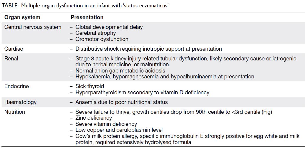
review of his computed tomography of the brain over the last 11 years showed a progressive increase in periventricular hypodensities (Fig 1). Brain magnetic 
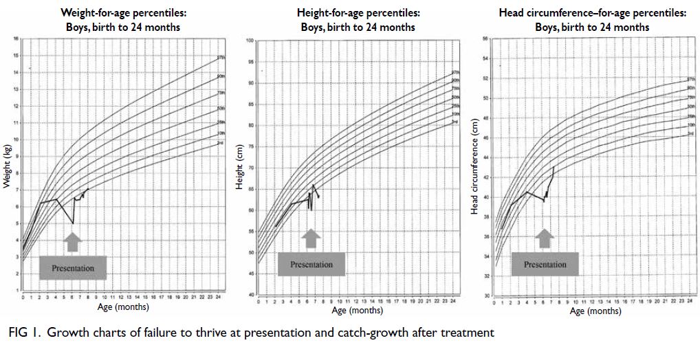
resonance imaging (1 year previously) showed extensive periventricular hyperintensities and an old ischaemic insult over bilateral external capsules 
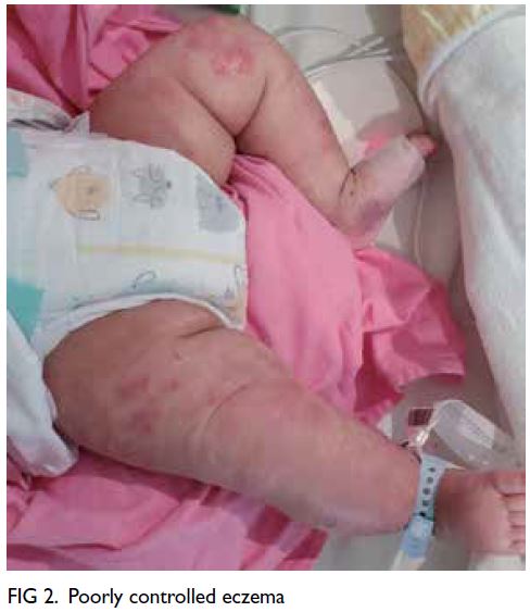
(Fig 2). Family history was notable for multiple first-degree relatives with young-onset stroke in their fifties and a suspected autosomal dominant inheritance pattern (Fig 3). Genetic testing of the neurogenic locus notch homolog protein 3 (NOTCH3) gene revealed a heterozygous mutation with a pathogenic variant (c.1630C>T, p.Arg544Cys), confirming the diagnosis of cerebral autosomal dominant arteriopathy with subcortical infarcts and leukoencephalopathy (CADASIL). The family was referred for genetic counselling.
 

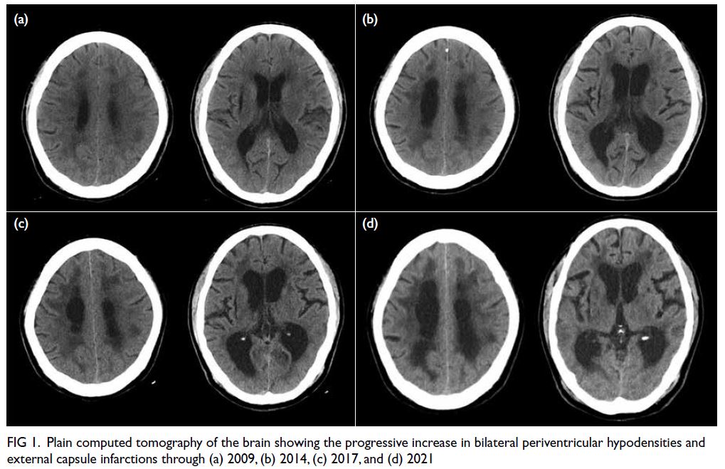
Figure 1. Plain computed tomography of the brain showing the progressive increase in bilateral periventricular hypodensities and external capsule infarctions through (a) 2009, (b) 2014, (c) 2017, and (d) 2021
 

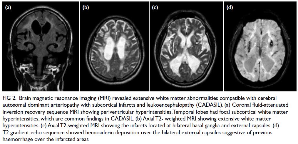
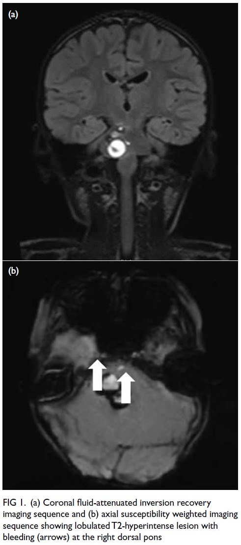
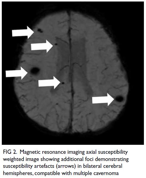
Figure 2. Brain magnetic resonance imaging (MRI) revealed extensive white matter abnormalities compatible with cerebral autosomal dominant arteriopathy with subcortical infarcts and leukoencephalopathy (CADASIL). (a) Coronal fluid-attenuated inversion recovery sequence MRI showing periventricular hyperintensities. Temporal lobes had focal subcortical white matter hyperintensities, which are common findings in CADASIL. (b) Axial T2-weighted MRI showing extensive white matter hyperintensities. (c) Axial T2-weighted MRI showing the infarcts located at bilateral basal ganglia and external capsules. (d) T2 gradient echo sequence showed hemosiderin deposition over the bilateral external capsules suggestive of previous haemorrhage over the infarcted areas
 

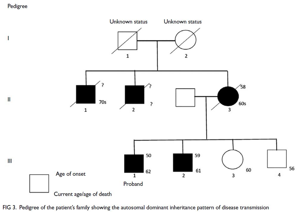
Figure 3. Pedigree of the patient’s family showing the autosomal dominant inheritance pattern of disease transmission
 
Cerebral autosomal dominant arteriopathy with subcortical infarcts and leukoencephalopathy is caused by cysteine-altering pathogenic variants in the NOTCH3 gene, with consequent vasculopathic changes, predominantly involving small penetrating arteries, arterioles, and brain capillaries.1 2 The mutation leads to an odd number of cysteine residues with deposition of osmiophilic material and progressive degeneration of vascular smooth muscle cells.1 2 The key to diagnosis includes a strong family history of young-onset stroke, an absence of strong vascular risk factors, and salient findings on brain magnetic resonance imaging, especially extensive white matter abnormalities and subcortical infarcts involving external capsules. Genetic testing for the NOTCH3 gene can be arranged after consultation with chemical pathologists in major public hospitals.3 4 The principle of management for symptomatic patients is similar to that of other patients with stroke, ie, antiplatelet therapy, lipid-lowering agents, and blood pressure control. There is no disease-modifying therapy currently available. Family members of affected individuals should be referred for genetic counselling with referral to tertiary centres for potential pre-implantation genetic testing to avoid transferring the mutation to offspring.5 There have been four other reported families with CADASIL in Hong Kong with different mutations. The mean age of symptom onset for index patients of these families was 51 years.3 4 The mutation in our patient has been commonly found in Fujian province and Taiwan, accounting for up to 14.5% to 70% of CADASIL cases.1
 
In summary, clinicians should obtain a detailed history and be alert to suspicious magnetic resonance imaging findings. Referral to chemical pathologists for genetic testing is key to the diagnosis of CADASIL.
 
Author contributions
All authors contributed to the concept or design of the study, acquisition of the data, analysis or interpretation of the data, drafting of the manuscript, and critical revision of the manuscript for important intellectual content. All authors had full access to the data, contributed to the study, approved the final version for publication, and take responsibility for its accuracy and integrity.
 
Conflicts of interest
The authors have no conflicts of interest to disclose.
 
Funding/support
This study received no specific grant from any funding agency in the public, commercial, or not-for-profit sectors.
 
Ethics approval
The patient was treated in accordance with the Declaration of Helsinki. Patient consent was obtained for all investigations.
 
References
1. Chen S, Ni W, Yin XZ, et al. Clinical features and mutation spectrum in Chinese patients with CADASIL: a multicenter retrospective study. CNS Neurosci Ther 2017;23:707-16. Crossref
2. Hu Y, Sun Q, Zhou Y, et al. NOTCH3 variants and genotype-phenotype features in Chinese CADASIL patients. Front Genet 2021;12:705284. Crossref
3. Au KM, Li HL, Sheng B, et al. A novel mutation (C271F) in the Notch3 gene in a Chinese man with cerebral autosomal dominant arteriopathy with subcortical infarcts and leukoencephalopathy. Clin Chim Acta 2007;376:229-32. Crossref
4. Hung LY, Ling TK, Lau NK, et al. Genetic diagnosis of CADASIL in three Hong Kong Chinese patients: a novel mutation within the intracellular domain of NOTCH3. J Clin Neurosci 2018;56:95-100. Crossref
5. Konialis C, Hagnefelt B, Kokkali G, Pantos C, Pangalos C. Pregnancy following pre-implantation genetic diagnosis of cerebral autosomal dominant arteriopathy with subcortical infarcts and leukoencephalopathy (CADASIL). Prenat Diagn 2007;27:1079-83. Crossref

A girl with acute-onset severe astigmatism and gaze palsy

© Hong Kong Academy of Medicine. CC BY-NC-ND 4.0
 
PICTORIAL MEDICINE
A girl with acute-onset severe astigmatism and gaze palsy
KHA Kwok, MB, BS, MRCPCH1; KL Hon, MB, BS, MD1; CC Au, MB, BS, MRCPCH1; Wilson WS Ho, MB, BS, MRCS2; Elaine YL Kan, MB, ChB, FRCR3
1 Department of Paediatrics and Adolescent Medicine, Hong Kong Children’s Hospital, Hong Kong SAR, China
2 Department of Neurosurgery, Hong Kong Children’s Hospital, Hong Kong SAR, China
3 Department of Radiology, Hong Kong Children’s Hospital, Hong Kong SAR, China
 
Corresponding author: Dr KL Hon (ehon@hotmail.com)
 
  A video clip demonstrating physical examination findings of bilateral fixed left lateral gaze and right lower-motor-neuron seventh cranial nerve palsy is available at www.hkmj.org
 
 Full paper in PDF
 
A previously healthy 4-year-old girl presented with a 4-week history of acute-onset visual disturbance. She was initially prescribed glasses for severe astigmatism but vision did not improve. It was later noted by her parents that her eyes could look only to the left side. She was then admitted to hospital. There was no history of headache, photophobia or vomiting but she exhibited drooling from the right side of her mouth and unsteady gait. She was afebrile and all other vital signs were normal with Glasgow Coma Scale score of 15. Physical examination revealed bilateral fixed left lateral gaze and right lower-motor-neuron seventh cranial nerve palsy. When asked to look to her right, she needed to compensate by head turning. Pupils were 3 mm equal and reactive to light. Ear canals were normal and there was no skin rash. Computed tomography scan of the brain showed a multilobulated mixed hyperdense and hypodense lesion at the dorsal pons with compression of the fourth ventricle. Magnetic resonance imaging (MRI) of the brain revealed bleeding from a tumour at the right dorsal pons (Fig 1); overall picture suggested multiple cavernoma (Fig 2). Neurosurgical decompression and stereotactic excision of a brainstem lesion was performed via suboccipital craniotomy. Histology showed features of a vascular lesion in keeping with cavernous haemangioma. Intracranial pressure was monitored via an external ventricular drain and remained normal postoperatively. Postoperative neurological examination showed bilateral fixed left lateral gaze, right lower-motor-neuron seventh cranial nerve palsy, and contralateral weakness of limbs. Genetic testing demonstrated a pathogenic mutation [heterozygous KRIT1 c.690C>A p.(Tyr230*)] for familial cerebral cavernous malformation (CM) syndrome. Her family was referred for further genetic testing and counselling.
 

Figure 1. (a) Coronal fluid-attenuated inversion recovery imaging sequence and (b) axial susceptibility weighted imaging sequence showing lobulated T2-hyperintense lesion with bleeding (arrows) at the right dorsal pons
 

Figure 2. Magnetic resonance imaging axial susceptibility weighted image showing additional foci demonstrating susceptibility artefacts (arrows) in bilateral cerebral hemispheres, compatible with multiple cavernoma
 
Acute-onset gaze disturbance in children is alarming and may signify serious cerebral abnormality. Almost all conjugate gaze palsies originate from a lesion in the midbrain or pons. These lesions can be caused by vascular or oncological space occupying lesions.
 
Cavernous malformations, also known as cavernous haemangiomas, are a type of benign, congenital malformation in which a cluster of dilated thin-walled capillaries form a characteristic ‘mulberry’ lesion with engorged purplish colour.1 They can be sporadic or inherited with an autosomal dominant pattern and incomplete penetrance, and can present as solitary or multiple lesions. Unlike capillary haemangiomas, CMs can be life-threatening and do not tend to regress. In all, 25% of cerebral CMs are infratentorial. They have a bleeding rate of 2% to 3% per year and recurrent bleeding rate of >20%. Due to the close proximity to multiple brainstem nuclei and fibre tracts, progressive neurological decline is observed in 39% patients with infratentorial CMs.2 Magnetic resonance imaging is the modality of choice, and susceptibility weighted imaging is the most sensitive means to detect blood products thus key to diagnosing cerebral CMs. Evolving blood products inside a CM appear as variable image intensities and give rise to the typical ‘popcorn’ appearance.3 Since MRI appearance is usually pathognomonic, biopsy is rarely needed. Angiography is indicated only if MRI cannot exclude arteriovenous malformation. Genetic testing should be arranged to screen for familial cerebral CM. Treatment approach depends on the site, size, symptoms, and history of haemorrhage.4 Indications for surgical resection of a cerebral CM include intracranial haemorrhage and epilepsy. Options include conventional surgery or stereotactic radiosurgery.4 5
 
Author contributions
All authors contributed to the concept or design, acquisition of data, analysis or interpretation of data, drafting of the manuscript, and critical revision of the manuscript for important intellectual content.
 
All authors had full access to the data, contributed to the study, approved the final version for publication, and take responsibility for its accuracy and integrity.
 
Conflicts of interest
As an editor of the journal, KL Hon was not involved in the peer review process. Other authors have disclosed no conflicts of interest.
 
Funding/support
This study received no specific grant from any funding agency in the public, commercial, or not-for-profit sectors.
 
Ethics approval
Ethics approval for this study was obtained from Hong Kong Children’s Hospital Ethics Committee, Hong Kong (Ref No.: HKCH REC 2019 009). The patient was treated in accordance with the tenets of the Declaration of Helsinki. Written consent for publication has been obtained from the patient’s parent.
 
References
1. Awad IA, Polster SP. Cavernous angiomas: deconstructing a neurosurgical disease. J Neurosurg 2019;131:1-13. Crossref
2. Fritschi JA, Reulen HJ, Spetzler RF, Zabramski JM. Cavernous malformations of the brain stem. a review of 139 cases. Acta Neurochir (Wien) 1994;130:35-46. Crossref
3. Lehnhardt FG, von Smekal U, Rückriem B, et al. Value of gradient-echo magnetic resonance-imaging in the diagnosis of familial cerebral cavernous malformation. Arch Neurol 2005;62:653-8. Crossref
4. Poorthuis MH, Klijn CJ, Algra A, Rinkel GJ, Al-Shahi Salman R. Treatment of cerebral cavernous malformations: a systematic review and meta-regression analysis. J Neurol Neurosurg Psychiatry 2014;85:1319-23. Crossref
5. Wang CC, Liu A, Zhang JT, Sun B, Zhao YL. Surgical management of brain-stem cavernous malformations: report of 137 cases. Surg Neurol 2003;59:444-54. Crossref

Depicting multiple schwannomas—a tale of two magnetic resonance neurogram techniques

© Hong Kong Academy of Medicine. CC BY-NC-ND 4.0
 
PICTORIAL MEDICINE
Depicting multiple schwannomas—a tale of two magnetic resonance neurogram techniques
HS Leung, MB, BS, FRCR1; JM Abrigo, FRCR, MD1; Eric HL Lau, FHKCORL, FHKAM (ORL)2; Winnie CW Chu, FHKCR, MD1
1 Department of Imaging and Interventional Radiology, The Chinese University of Hong Kong, Hong Kong
2 Department of Otorhinolaryngology, Head and Neck Surgery, The Chinese University of Hong Kong, Hong Kong
 
Corresponding author: Dr HS Leung (lhs655@ha.org.hk)
 
 Full paper in PDF
 
A 40-year-old female with unremarkable past health and no known family history of neurofibromatosis presented with a 3-year history of non-specific discomfort at the right submandibular region and upper neck. No facial nerve dysfunction was evident clinically. Ultrasound of the major salivary glands revealed a soft tissue nodule posterior to the right submandibular gland. Fine needle aspiration cytology findings were compatible with schwannoma. Subsequent magnetic resonance imaging (MRI) revealed a cluster of nodules within the deep lobe of the right parotid gland with a linear orientation. Further delineation with high-resolution MRI and neurography sequences with acquisition of double-echo steady state (DESS) [Fig 1] and post-contrast constructive interference in steady state (CISS) [Fig 2] sequences delineated these nodules arising eccentrically from the main trunk, inferior division and the marginal mandibular branch of the facial nerve, respectively. The patient continues with imaging surveillance while awaiting workup of underlying neurofibromatosis or schwannomatosis.
 

Figure 1. Double-echo steady state sequence of the right parotid gland with maximal intensity projection reconstruction, demonstrating a cluster of facial nerve schwannomas (dashed arrows 1-3) closely related to the facial nerve (solid arrows) at the main trunk (MT) and inferior division (Inf) of the facial nerve. The superior division (Sup) is not involved. A fourth schwannoma is identified more inferiorly (arrowheads) and shows a distinct low signal as a result of previous intratumoural haemorrhage. The facial nerve demonstrates positive contrast compared with the background low-signal parotid gland
 

Figure 2. Non-contrast constructive interference in steady state sequence of the right parotid with minimum intensity projection reconstruction, also demonstrating the facial nerve and its tributaries (solid arrows) and the facial nerve schwannomas (dashed arrows 1-4). A hyperintense cystic component is also depicted in the more inferior schwannoma (dashed arrow 3). The facial nerve demonstrates negative contrast compared with the background high-signal parotid gland
 
In the early era of MRI imaging, depiction of the extracranial intraparotid portion of the facial nerve was difficult, if not impossible, with conventional sequences. With advances in technology, higher resolution and newer imaging protocols allow clear delineation of intraparotid facial nerves. The use of high-resolution, three-dimensional and gradient echo–based techniques such as DESS1 and CISS2 allows clear delineation of the facial nerve from the parotid parenchyma and any adjacent facial nerve tumours, with a reasonable acquisition time of around 5 minutes. Both sequences have been proven useful in depicting the facial nerve and its branches,1 2 although there has not been any direct comparison of the efficacy of these sequences in facial nerve depiction. Nonetheless these two sequences are synergistic in the delineation of the facial nerve tributaries due to their differences in contrast characteristics. For DESS sequence, the facial nerve demonstrates positive contrast compared with the low-signal parotid gland; while in CISS, the facial nerve shows negative contrast compared with the high-signal parotid gland that is accentuated after contrast injection. The use of reconstruction techniques such as maximal intensity projection for DESS, or minimum intensity projection for CISS, can further improve visualisation of the peripheral nerve branches.
 
Dedicated magnetic resonance neurogram sequence for facial nerves is especially important in patients with salivary gland neoplasms, since facial nerve injury and consequent facial nerve weakness remain an important postoperative complication,3 and prior understanding of facial nerve anatomy is potentially helpful in reducing its incidence. It is also useful in predicting histology of nerve sheath tumours since schwannomas, as in our case, typically arise eccentrically from a nerve with displacement of its fascicles.4 Furthermore, such techniques can be applied to other extracranial nerves5 in the diagnosis of cranial neuropathies or in interventional planning for tumours in the head and neck region. Gradient echo–based DESS and CISS sequences partially overcome the problem of long acquisition time in conventional spin echo–based magnetic resonance neurogram; nonetheless images can be significantly compromised by susceptibility artefacts if adjacent implants are present. The latter remains a major obstacle that needs to be addressed.
 
Author contributions
Concept or design: All authors.
Acquisition of data: All authors.
Analysis or interpretation of data: HS Leung, JM Abrigo.
Drafting of the manuscript: HS Leung.
Critical revision of the manuscript for important intellectual content: JM Abrigo, EHL Lau, WCW Chu.
 
All authors had full access to the data, contributed to the study, approved the final version for publication, and take responsibility for its accuracy and integrity.
 
Conflicts of interest
All authors have disclosed no conflicts of interest.
 
Acknowledgement
The authors would like to acknowledge Mr Chi-kin Wong and Mr Chung-yee Cheung, radiographers of Gerald Choa Neuroscience Centre for their assistance in MRI scanning and devising MRI protocols.
 
Funding/support
This study received no specific grant from any funding agency in the public, commercial, or not-for-profit sectors.
 
Ethics approval
Ethics approval has been obtained from the Joint Chinese University of Hong Kong–New Territories East Cluster Clinical Research Ethics Committee (CREC Ref No.: 2021.639). Patient consent for publication was obtained.
 
References
1. Kim Y, Jeong HS, Kim HJ, Seong M, Kim Y, Kim ST. Three-dimensional double-echo steady-state with water excitation magnetic resonance imaging to localize the intraparotid facial nerve in patients with deep-seated parotid tumors. Neuroradiology 2021;63:731-9. Crossref
2. Guenette JP, Ben-Shlomo N, Jayender J, et al. MR imaging of the extracranial facial nerve with the CISS sequence. AJNR Am J Neuroradiol 2019;40:1954-9. Crossref
3. Jin H, Kim BY, Kim H, et al. Incidence of postoperative facial weakness in parotid tumor surgery: a tumor subsite analysis of 794 parotidectomies. BMC Surg 2019;19:199. Crossref
4. Soldatos T, Fisher S, Karri S, Ramzi A, Sharma R, Chhabra A. Advanced MR imaging of peripheral nerve sheath tumors including diffusion imaging. Semin Musculoskelet Radiol 2015;19:179-90. Crossref
5. Chhabra A, Bajaj G, Wadhwa V, et al. MR neurographic evaluation of facial and neck pain: normal and abnormal craniospinal nerves below the skull base. Radiographics 2018;38:1498-513. Crossref
 

A critically ill infant with multi-organ dysfunction due to eczema

© Hong Kong Academy of Medicine. CC BY-NC-ND 4.0
 
PICTORIAL MEDICINE
A critically ill infant with multi-organ dysfunction due to eczema
KL Hon, MB, BS, MD1; Karen KY Leung, MB, BS, MRCPCH1; WL Lin, PhD, BChinMed2; David CK Luk, MB, ChB, MRCPCH3
1 Department of Paediatrics and Adolescent Medicine, Hong Kong Children’s Hospital, Hong Kong
2 Hong Kong Institute of Integrative Medicine, Faculty of Medicine, The Chinese University of Hong Kong, Hong Kong
3 Department of Paediatrics and Adolescent Medicine, United Christian Hospital, Hong Kong
 
Corresponding author: Dr KL Hon (ehon@hotmail.com)
 
 Full paper in PDF
 
Eczema is the most prevalent childhood atopic illness routinely encountered by health professionals providing care to children.1 Some infants become critically ill and present to the paediatric intensive care unit (PICU) with this seemingly trivial condition.2 This anonymised (no clinical and laboratory data are presented) case of severe eczema and multi-organ dysfunction illustrates the extent of multi-organ involvement in eczema and the medical and psychosocial issues associated with the disease.1 3
 
A 6-month-old boy with eczema and septic shock presented to the emergency department. He was treated with fluid resuscitation and subsequently admitted to a PICU. Derangement of multiple organ functions were identified along with failure to thrive and poor development (Table, Figs 1 and 2). Dobutamine and broad-spectrum antibiotics were prescribed. Although his condition improved rapidly, it became apparent that his parents did not trust Western medicine and were reluctant to use emollient and topical steroid to manage their child’s eczema. The mother frankly admitted that since the development of eczema at 2 months of age, she had been giving the infant complementary herbal medicine remedies. She was also taking herbal medicine whilst breastfeeding so that her child would indirectly receive the more ‘effective’ herbal medicine. The parents were interviewed by healthcare workers from various disciplines but they remained resolute in their beliefs and phobias about Western medicine. The child was discharged from PICU after the acute medical issues were resolved. Extensive investigations have been performed and apart from persistent peripheral eosinophilia, slightly low immunoglobulin M level, borderline CD4:CD8 ratio and borderline low zinc level, all other investigations including metabolic workup remain unremarkable to date. Further paediatric dermatology, genetics and integrative medicine follow-ups have been arranged.
 

Table. Multiple organ dysfunction in an infant with ‘status eczematicus’
 

Figure 1. Growth charts of failure to thrive at presentation and catch-growth after treatment
 

Figure 2. Poorly controlled eczema
 
Our reported case is probably the youngest patient with critical ‘status eczematicus’ to survive. All growth parameters were compromised (Fig 1). Children with severe eczema rarely require admission to an ICU and there are very few such cases in the literature. Mortality due to eczema is rare, but we have previously reported a tragic case of an infant with eczema who died of group B streptococcus septicaemia and malnutrition despite expensive dietary supplements.2 Eczema is a chronic condition that can significantly affect a child’s quality of life as well as that of their family if it is not well controlled due to the potentially significant psychological toll.4 Although ICU is not an ideal setting to manage a family with multiple phobias, the child must first be stabilised, and other differential diagnoses of acute skin failure ruled out.2
 
Acute treatment of eczema is straightforward but long-term maintenance treatment is always challenging. Topical medications should be considered to prevent exacerbations and therapy should be proactive. Steroid phobia is prevalent and often leads to non-compliance.1
 
Recommendations about dietary avoidance should be specific and given only in confirmed cases of food allergy.1 The use of traditional and proprietary topical and herbal medicine is popular across many countries in Asia.1 Anxious food-avoiding parents may purchase multi-vitamin supplements, prebiotics, probiotic or symbiotics that claim to be effective. In an extreme case, death was reported to have a secondary association with extreme dietary practice.3 Physicians must be tactful when counselling these anxious parents who are steroid phobic and mistrusting of modern medicine. Indirect administration of herbal medicine to an infant through breastfeeding is not advocated even in the practice of integration medicine.
 
Management of eczema can sometimes be challenging. Apart from strong parental beliefs, cultural differences might also play a role in compliance with a prescribed treatment plan or a tendency to seek alternative therapies instead. These effects are likely to be underestimated in our community, especially in the primary care setting where consultation time might be limited. Physicians should endeavour to spend more time exploring parental beliefs. A practical solution may be to use an objective self/parental assessment to aid eczema control assessment, eg, ‘Traffic Light Control’ self-assessment system (Fig 3) and quality of life assessment, eg, Children’s Dermatology Life Quality Index.5 An ‘eczema action plan’ can also be given to parents to remind them of the prescribed eczema treatment. The treatment plan should be straightforward and easy to follow, especially if they perceive a worsening of eczema between clinic visits.5 A multidisciplinary and perhaps integrative medicine approach should be adopted where possible to manage patients and families with eczema, and education about the disease should be individualised to improve patient outcomes.
 

Figure 3. ‘Traffic Light Control’ self-assessment system on eczema control5
 
Healthcare providers must be aware of the mortality and morbidity associated with recalcitrant eczema and ‘status eczematicus’. These tragic cases of ‘status eczematicus’ serve to remind us of the grave consequences if eczema is inappropriately managed.
 
Author contributions
All authors contributed to the concept or design, acquisition of data, analysis or interpretation of data, drafting of the manuscript, and critical revision of the manuscript for important intellectual content.
 
All authors had full access to the data, contributed to the study, approved the final version for publication, and take responsibility for its accuracy and integrity.
 
Conflicts of interest
As an editor of the journal, KL Hon was not involved in the peer review process. Other authors have disclosed no conflicts of interest.
 
Funding/support
This study received no specific grant from any funding agency in the public, commercial, or not-for-profit sectors.
 
Ethics approval
The patient was treated in accordance with the tenets of the Declaration of Helsinki. Ethics approval for publication of patient information in the paediatric intensive care unit was obtained from the Hong Kong Children’s Hospital Research Ethics Committee (Ref No.: HKCH-REC-2019–0011).
 
References
1. Hon KL, Leong KF, Leung TN, Leung AK. Dismissing the fallacies of childhood eczema management: Case scenarios and an overview of best practices. Drugs Context 2018;7:212547. Crossref
2. Hon KL, Nip SY, Cheung KL. A tragic case of atopic eczema: malnutrition and infections despite multivitamins and supplements. Iran J Allergy Asthma Immunol 2012;11:267-70.
3. Hon KL, Kam WY, Leung TF, et al. Steroid fears in children with eczema. Acta Paediatr 2006;95:1451-5. Crossref
4. Hon KL, Kung JS, Wang M, Pong NH, Li AM, Leung TF. Clinical scores of sleep loss and itch, and antihistamine and topical corticosteroid usage for childhood eczema. Br J Dermatol 2016;175:1076-8. Crossref
5. Lam PH, Hon KL, Leung KK, Leong KF, Li CK, Leung TF. Self-perceived disease control in childhood eczema. J Dermatolog Treat 2022;33:1459-646. Crossref

Haemodynamic compensation in a patient with bilateral vertebral artery

© Hong Kong Academy of Medicine. CC BY-NC-ND 4.0
 
PICTORIAL MEDICINE
Haemodynamic compensation in a patient with bilateral vertebral artery
BD Ku, MD1; SS Yoon, MD, PhD2; SH Heo, MD, PhD2
1 Department of Neurology, International St Mary’s Hospital, Catholic Kwandong University College of Medicine, Incheon, South Korea
2 Department of Neurology, Kyung Hee University School of Medicine, Seoul, South Korea
 
Corresponding author: Prof BD Ku (bondku34@cku.ac.kr)
 
 Full paper in PDF
 
Bilateral vertebral artery dissection (VAD) is often related to sudden mechanical injury of the arteries such as that following direct trauma or rotational forces. The natural course of bilateral VAD is variable, from recanalisation to fatal subarachnoid haemorrhage.1 We report a patient with bilateral VAD and favourable clinical course due to adequate carotid haemodynamic compensation despite progressive vertebrobasilar insufficiency.
 
In February 2007, a 42-year-old man presented with acute, stabbing neck pain, radiating to his occipital area and persisting for 5 days after performing yoga exercises while standing on his head. He showed no neurological deficit except for limited neck motion due to pain. Serial magnetic resonance angiography (MRA) 5, 12, and 19 days later revealed progressive bilateral VAD with consequent vertebrobasilar flow insufficiency (Fig 1). Cerebral angiography 25 days after this dissection revealed compensated retrograde filling of the basilar artery (Fig 2). The MRA taken 50 days after dissection showed much more aggravated steno-occlusive changes in the entire vertebrobasilar circulation (Fig 3). Unlike the progressive deterioration of vertebrobasilar circulation, the patient’s neck pain gradually resolved without neurological deficit.
 

Figure 1. The serial magnetic resonance angiography taken (a) 5, (b) 12, and (c) 19 days after onset of neck pain showing progressive steno-occlusion of vertebral arteries and deterioration of vertebrobasilar flow (arrow heads). (a) Short segmental stenosis at the right vertebral artery at the distal portion of the right posterior inferior cerebellar artery. (b) Progressive segmental stenosis of the right vertebral artery and newly developed pseudoaneurysmal dilatation in the left vertebral artery. (c) Progressive aggravated stenosis of both vertebral arteries and insufficient vertebrobasilar flow
 

Figure 2. Cerebral angiography taken 25 days after dissection. (a, b) Tapered occlusion of the right vertebral artery just distal to the right posterior inferior cerebellar artery. (c, d) Pseudoaneurysmal dilatation with string-and-pearl sign in the left vertebral artery. (a-d) Steno-occlusive changes of bilateral vertebral arteries, and (e, f) compensated retrograde filling of basilar artery from compensated carotid circulation (arrows)
 

Figure 3. The magnetic resonance angiography taken 50 days after onset of neck pain, showing much more aggravated steno-occlusive changes in the entire vertebra-basilar circulation (arrow heads)
 
Cerebral angiography performed after the first three MRA studies showed nearly complete occlusion of the vertebral arteries just distal to the bilateral posterior inferior cerebellar artery. This protected the patient against neurological deficit. It is unclear why the fourth MRA at 1 month later showed further deterioration of vertebra-basilar circulation. The potential explanation for this finding may be related to the increased compensatory carotid artery flow through the collateral circulation. The presence of adequate haemodynamic compensation from the carotid artery territory constitutes an important positive prognostic factor of the low-flow ischaemia in a patient with bilateral VAD.1 2
 
Funaki et al3 reported that serial MRA could be of great use to monitor restorative bilateral VAD haemodynamics. In contrast, our patient showed progressive deterioration of vertebrobasilar circulation. This suggests that the haemodynamic status of bilateral VAD can variably alter, so serial MRA can help monitor the progression of dissection in some patients.2
 
The present case suggests that the haemodynamics of bilateral VAD are variable and adequate haemodynamic compensation may constitute a positive prognostic factor.
 
Author contributions
Concept or design: BD Ku.
Acquisition of data: All authors.
Analysis or interpretation of data: All authors.
Drafting of the manuscript: BD Ku.
Critical revision of the manuscript for important intellectual content: All authors.
 
All authors had full access to the data, contributed to the study, approved the final version for publication, and take responsibility for its accuracy and integrity.
 
Conflicts of interest
The authors have disclosed no conflicts of interest.
 
Acknowledgement
The authors would like to thank Harrisco (www.harrisco.com) for the English language review.
 
Funding/support
This study received no specific grant from any funding agency in the public, commercial, or not-for-profit sectors.
 
Ethics approval
This study was conducted in accordance with the principles outlined in the Declaration of Helsinki.
 
References
1. de Bray JM, Penisson-Besnier I, Dubas F, Emile J. Extracranial and intracranial vertebrobasilar dissections: diagnosis and prognosis. J Neurol Neurosurg Psychiatry 1997;63:46-51. Crossref
2. Bacci D, Valecchi D, Sgambati E, et al. Compensatory collateral circles in vertebral and carotid artery occlusion. Ital J Anat Embryol 2008;113:265-71.
3. Funaki T, Oshimoto T, Wataya T, et al. Bilateral vertebral artery dissection and its chronological changes detected by MR angiography: a case report [in Japanese]. No To Shinkei 2004;56:247-50.

Breast lymphoma: a rare breast malignancy mimicking benign breast disease and carcinoma

© Hong Kong Academy of Medicine. CC BY-NC-ND 4.0
 
PICTORIAL MEDICINE
Breast lymphoma: a rare breast malignancy mimicking benign breast disease and carcinoma
Carol PY Chien, MB, BS, FRCR1; Kimmy KM Kwok, FRCR, FHKAM (Radiology)2; Eliza PY Fung, FRCR, FHKAM (Radiology)2
1 Department of Radiology, Queen Mary Hospital, Hong Kong
2 Department of Diagnostic and Interventional Radiology, Kwong Wah Hospital, Hong Kong
 
Corresponding author: Dr Carol PY Chien (chienpyc@gmail.com)
 
 Full paper in PDF
 
In July 2020, a 52-year-old woman presented with a 1-month history of palpable right breast lump. Clinical examination revealed a hard mass at the upper inner quadrant of the right breast. No skin changes or nipple retraction were evident. Ultrasonography confirmed multiple oval circumscribed hypoechoic masses at the upper right breast measuring up to 2 cm that were considered likely benign (Fig 1). The lesions were mammographically occult (Fig 2).
 

Figure 1. Breast ultrasonogram showing multiple benign-looking lobulated and oval circumscribed hypoechoic masses measuring up to 2 cm at the upper inner and upper outer quadrants of the right breast
 

Figure 2. Bilateral mammograms are unremarkable. No focal mass or architectural distortion. No axillary lymphadenopathy
 
The patient also reported non-specific abdominal pain and distension for a few weeks. Positron emission tomography–computed tomography (PET-CT) showed multiple hypermetabolic masses at the right breast (maximum standardised uptake value [SUVmax] 19.0, 16 mm) and abdominal lymphadenopathy (Fig 3). Ultrasound-guided core biopsy of the right breast mass confirmed diffuse large B-cell lymphoma (Fig 4). The patient was diagnosed with stage IV lymphoma with diffuse extranodal involvement of the right breast. Reassessment PET-CT showed progressive disease despite R-CEOP chemotherapy with enlargement and increased metabolic activity of the right breast lymphoma (SUVmax 23.7, 41 mm) and abdominal lymphadenopathy (Fig 5). Second-line chemotherapy and radiotherapy of the breast was administered.
 

Figure 3. Positron emission tomography–computed tomography showing multiple hypermetabolic soft tissue masses at the upper outer, upper inner and lower inner quadrants of the right breast measuring up to 16 mm, with maximum standardised uptake value 19.0; and multiple hypermetabolic abdominal lymphadenopathies
 

Figure 4. (a) Haematoxylin and eosin stain, 400× magnification: dense sheets of medium-to-large-sized and mitotically active atypical lymphoid cells that overrun breast parenchyma. (b) CD20 immunostain, 400× magnification: The atypical lymphoid cells are diffusely positive for CD20
 

Figure 5. Reassessment positron emission tomography–computed tomography showing enlargement of biopsy-confirmed lymphoma at the upper inner quadrant of the right breast, with increased metabolic activity (maximum standardised uptake value 23.7, 41 × 21 mm)
 
The breast is an uncommon extranodal site of involvement by lymphoma because of the lack of lymphoid tissue. Breast lymphoma is a rare malignancy of the breast that accounts for <1% of all breast malignancies and <2% of all extranodal non-Hodgkin lymphoma.1 Diffuse large B-cell lymphoma is the most common histological type in both primary and secondary breast lymphoma with a median age at presentation of 60 to 70 years.1
 
The clinical presentation of breast lymphoma is highly variable, so diagnosis is challenging. It commonly presents as a mass that mimics carcinoma, or skin inflammatory changes that mimic benign inflammatory breast disease or inflammatory breast cancer.1 2 Axillary lymphadenopathy is uncommon.1 However, unlike with breast carcinoma, nipple retraction or discharge and other skin changes are rare with breast lymphoma.1 2
 
Mammographically, breast lymphoma commonly presents as a solitary mass (69%-76%) and asymmetry (20%). It may also present as skin thickening, architectural distortion or lymphedema.3 4 In contrast to breast carcinoma, spiculations, architectural distortion and microcalcifications are distinctively absent with breast lymphoma. Sonographically, breast lymphoma commonly presents as a parallel benign-looking hypoechoic mass without malignant features.3 4 Ultrasonography alone cannot distinguish breast lymphoma from breast carcinoma or benign breast lesions such as fibroadenoma. However, it can guide tissue diagnosis when mammogram is negative. This is particularly helpful in Asian patients with dense breast tissue that lowers mammogram sensitivity.
 
A recent study has demonstrated that 18F-fluorodeoxyglucose PET-CT can potentially differentiate between breast lymphoma and carcinoma.5 The authors found that the median SUVmax for breast lymphoma (10.96) was significantly higher than that for breast carcinoma (4.76; specificity up to 96.3%).5 Although the sonographic features of breast masses in our patient were absent, an SUVmax of 19.0 was unusually high for breast carcinoma. This raised the suspicion of secondary breast lymphoma rather than primary breast carcinoma with distant metastases. Histological diagnosis was required to guide appropriate treatment.
 
In conclusion, PET-CT can potentially differentiate between breast lymphoma and breast carcinoma. Increased awareness of breast lymphoma and correlation with radiological and pathological findings are essential for diagnosis.
 
Author contributions
Concept or design: All authors.
Acquisition of data: CPY Chien.
Analysis or interpretation of data: CPY Chien.
Drafting of the manuscript: CPY Chien.
Critical revision of the manuscript for important intellectual content: All authors.
 
All authors had full access to the data, contributed to the study, approved the final version for publication and take responsibility for its accuracy and integrity.
 
Conflicts of interest
All authors have disclosed no conflicts of interest.
 
Funding/support
This study received no specific grant from any funding agency in the public, commercial, or not-for-profit sectors.
 
Ethics approval
The patient was treated in accordance with the Declaration of Helsinki and provided informed consent for all investigations and procedures, and publication.
 
References
1. Raj SD, Shurafa M, Shah Z, Raj KM, Fishman MD, Dialani VM. Primary and secondary breast lymphoma: clinical, pathologic, and multimodality imaging review. Radiographics 2019;39:610-25. Crossref
2. Sabaté JM, Gómez A, Torrubia S, et al. Lymphoma of the breast: clinical and radiologic features with pathologic correlation in 28 patients. Breast J 2002;8:294-304. Crossref
3. Yang WT, Lane DL, Le-Petross HT, Abruzzo LV, Macapinlac HA. Breast lymphoma: imaging findings of 32 tumors in 27 patients. Radiology 2007;245:692-702. Crossref
4. Surov A, Holzhausen HJ, Wienke A, et al. Primary and secondary breast lymphoma: prevalence, clinical signs and radiological features. Br J Radiol 2012;85:e195-205. Crossref
5. Ou X, Wang J, Zhou R, et al. Ability of 18F-FDG PET/CT radiomic features to distinguish breast carcinoma from breast lymphoma. Contrast Media Mol Imaging 2019;2019:4507694. Crossref

Abdominal para-aortic ectopic thyroid tissue mimicking lymphadenopathy on computer tomography

© Hong Kong Academy of Medicine. CC BY-NC-ND 4.0
 
PICTORIAL MEDICINE
Abdominal para-aortic ectopic thyroid tissue mimicking lymphadenopathy on computer tomography
Charmaine HK Wong, MB, ChB, FRCR1; HL Tsui, MB, ChB, FHKAM (Radiology)1; CN Ling, MB, ChB2; Anthony WT Chin, MB, ChB, FHKAM (Radiology)1; PY Chu, MB, ChB, FHKAM (Radiology)1; CS Chan, MB, BS, FHKAM (Radiology)1
1 Department of Radiology and Organ Imaging, United Christian Hospital, Hong Kong
2 Department of Pathology, United Christian Hospital, Hong Kong
 
Corresponding author: Dr Charmaine HK Wong (charmainehwong@gmail.com)
 
 Full paper in PDF
 
A 70-year-old woman presented to our hospital for workup for chronic cough. A contrast computed tomography (CT) of the thorax and abdomen revealed an incidental finding of multilobulated soft tissue lesions in the abdomen along the left para-aortic region. The lesions measured up to 7.4 cm in diameter and demonstrated mild increased attenuation on pre-contrast images and vivid heterogenous enhancement on post-contrast study (Fig 1a and b). Appearance and location of these lesions raised concern about possible lymphadenopathy so fluorodeoxyglucose-18 positron emission tomography–CT was performed. The lesions demonstrated only mild metabolic activity (maximal standard uptake value=2.6) [Fig 1c]. They showed no interval change in appearance, and no other hypermetabolic lesion could be detected elsewhere. Tumour markers including alpha-fetoprotein, carcinoembryonic antigen, and cancer antigen 125 were not elevated.
 

Figure 1. (a) Pre-contrast and (b) post-contrast axial computed tomography images showing multilobulated lesions at left para-aortic region (white arrows). The lesions showing mild increased attenuation on pre-contrast images and vivid heterogenous enhancement on post-contrast images. (c) Axial fluorodeoxyglucose-18 positron emission tomography-computed tomography demonstrating mild metabolic activity over the lesions (maximum standard unit value=2.6)
 
The CT-guided biopsy of the para-aortic lesions subsequently showed cuboidal follicular cells arranged in architecture resembling thyroid parenchymal tissue (Fig 2). Immunostaining for thyroglobulin and thyroid transcription factor-1 were positive. There were no nuclear features of carcinoma.
 

Figure 2. (a, b) Histological images of the para-aortic lesions showing tissue consisting of colloid-containing follicles of different sizes, arranged in architecture resembling normal thyroid parenchymal tissue. Immunostaining for (c) thyroglobulin and (d) thyroid transcription factor-1 was positive
 
Thyroid scintigraphy using technetium-99m pertechnetate showed intense radiotracer uptake over the corresponding para-aortic lesions (Fig 3). Findings were consistent with ectopic thyroid tissue. The presence of an orthotopic thyroid at its normal pretracheal position was demonstrated on CT and on technetium-99m scan (Fig 4). The patient’s thyroid function was normal.
 

Figure 3. (a) Axial and (b) coronal thyroid scintigraphy images using technetium-99m pertechnetate showing intense uptake of radiotracer at the left side of the abdomen. Corresponding (c) axial and (d) coronal computed tomography fusion images showing the left para-aortic lesions
 

Figure 4. (a) Thyroid scintigraphy image and (b) corresponding computed tomography fusion image showing the presence of orthotopic thyroid gland, with a focal defect at the lower pole corresponding to a cold nodule
 
Ectopic thyroid gland is a rare developmental abnormality with a prevalence of approximately 1 per 100 000 to 300 000 population.1 It results from aberrant embryogenesis of the gland during its migration from the foramen cecum at the posterior aspect of the tongue to its final pretracheal position. Frequent locations include its path of embryological descent along the midline of the neck, with a lingual thyroid at the base of the tongue being most common, accounting for up to 90% of reported cases.2 Other locations include the lateral neck and superior mediastinum. More distant locations such as subdiaphragmatic locations have also been reported but are exceedingly rare. To the best of the authors’ knowledge, this is the first report of ectopic thyroid tissue occurring at the para-aortic region of the abdomen, mimicking the appearance of lymphadenopathy on CT.
 
Thyroid scintigraphy is the most important and sensitive imaging tool to detect ectopic thyroid tissue. It also has the advantage of being able to demonstrate the presence or absence of an orthotopic thyroid gland. In patients with ectopic thyroid tissue, it is important to evaluate for the presence of orthotopic thyroid and thyroid function. Hypothyroidism may be present, particularly in those without a normal thyroid gland, and more common in patients with lingual ectopic thyroid.2 Ultrasound is also useful to locate and evaluate the orthotopic thyroid gland.
 
Computed tomography and magnetic resonance imaging are important adjuncts in evaluation. Ectopic thyroid tissue may show similar imaging characteristics to normal thyroid gland with high attenuation on non-contrast CT due to its high iodine content and vivid post-contrast enhancement. When ectopic thyroid tissue is found in a location not consistent with embryologic development, the possibility of malignant metastasis needs to be considered, occurring in 7% to 23% of patients.3 Biopsy is invaluable and the orthotopic gland should be evaluated for possible malignant change. Pathological changes that affect a normal thyroid including malignant change have been reported in ectopic tissues,1 and should be considered in the management and follow-up of ectopic thyroid tissue.
 
Author contributions
Concept or design: All authors.
Acquisition of data: CHK Wong, HL Tsui, CN Ling.
Analysis or interpretation of data: CHK Wong, HL Tsui, CN Ling.
Drafting of the manuscript: CHK Wong, HL Tsui, CN Ling.
Critical revision of the manuscript for important intellectual content: All authors.
 
Conflicts of interest
All authors have disclosed no conflicts of interest.
 
Funding/support
This study received no specific grant from any funding agency in the public, commercial, or not-for-profit sectors.
 
Ethics approval
The patient was treated in accordance with the Declaration of Helsinki. The patient provided written informed consent for all treatments and procedures and consent for publication.
 
References
1. Noussios G, Anagnostis P, Goulis DG, Lappas D, Natsis K. Ectopic thyroid tissue: anatomical, clinical, and surgical implications of a rare entity. Eur J Endocrinol 2011:165:375-82. Crossref
2. Guerra G, Cinelli M, Mesolella M, et al. Morphological, diagnostic and surgical features of ectopic thyroid gland: a review of literature. Int J Surg 2014;12 Suppl 1:S3-11. Crossref
3. Ballard D, Patel P, Schild SD, Ferzli G, Gordin E. Ectopic thyroid presenting as supraclavicular mass: a case report and literature review. J Clin Transl Endocrinol Case Rep 2018;10:17-20. Crossref

Liver and kidney toxicity caused by wild mushroom poisoning

© Hong Kong Academy of Medicine. CC BY-NC-ND 4.0
 
PICTORIAL MEDICINE
Liver and kidney toxicity caused by wild mushroom poisoning
LF Yang, MD1; LP Zhu, MD1; YT Li, MD, PhD1; XB Zhong, MD1; ZG Chen, MD, PhD1; Karen KY Leung, MB, BS, MRCPCH2; KL Hon, MB, BS, MD2
1 Department of Pediatrics, The Third Affiliated Hospital of Sun Yat-sen University, Guangzhou, PR China
2 Department of Paediatrics and Adolescent Medicine, The Hong Kong Children’s Hospital, Hong Kong
 
Corresponding author: Dr KL Hon (ehon@hotmail.com)
 
 Full paper in PDF
 
In May 2020, a 2-year-old girl was admitted to our hospital with a 2-day history of persistent vomiting, diarrhoea, abdominal pain, and headache. Her family reported that she had eaten half of a large mushroom, about the size of a palm (Fig a) 17 hours before symptom onset.
 

Figure. Photographs of wild mushroom taken by the patient’s family and identified by mycologists as (a) possibly Amanita spp, which are hepatotoxic; and (b, c) Macrolepiota spp, which are edible
 
Physical examination was unremarkable. Laboratory investigations revealed liver disfunction, with elevated aspartate transaminase (AST) 228 U/L and alanine transaminase (ALT) 211 U/L rising to 2591 U/L and 2815 U/L, respectively. The patient was admitted to the paediatric intensive care unit for further treatment. After hospitalisation, AST and ALT further increased to 4005 U/L and 4335 U/L, respectively. Plasma exchange was performed along with treatments including 20% albumin, high-dose penicillin, N-acetylcysteine injection and Ganoderma lucidum. Liver function gradually normalised over the following week and the patient was discharged home.
 
The patient’s grandmother and aunt consumed the other half of the same mushroom (Fig a) and other mushrooms (Fig b and c), and both were admitted to hospital with serious vomiting, diarrhoea and mild abdominal pain. Her aunt had normal liver function and kidney function. The grandmother had mild liver and renal impairment with AST 169 U/L, ALT 147 U/L, blood urea nitrogen 12.1 mmol/L and creatinine 168 μmol/L, made a rapid and recovery following rehydration, a bicarbonate infusion, furosemide, and hepatoprotective drugs for 3 days.
 
Cyclopeptides are the major lethal toxins worldwide, often found in Amanita spp, Cyclospora and galea.1 2 In Guangzhou from 2007 to 2011, the most common species causing mushroom poisoning was Amanita spp. In mainland China 95% of fatal cases of mushroom poisoning are caused by amanitin. In Hong Kong, 90% of wild mushrooms are poisonous.
 
Ultra-performance liquid chromatography-mass spectrometry has high accuracy, and good sensitivity and specificity, and was used to identify the toxin in our patient. However, it was already 2 days after the girl ate the wild mushrooms, and the toxin might have been excreted so that the concentration was too low for detection.
 
Prognosis is often poor for patients with long incubation period of gastrointestinal symptoms (>6 hr), early manifestations of liver and kidney dysfunction, and multiple organ dysfunction.3 4 Plasma exchange effectively removes hepatotoxins and improves survival, drugs such as silybin, N-acetylcysteine, putatively ceftazidime/penicillin and Ganoderma lucidum can also have a significant benefit.5 Ganoderma lucidum contains triterpenoids that can protect the liver and reduce oxidative stress and apoptosis. Ganoderma lucidum decoction is effective in the treatment of amanita poisoning at a recommended dosage of 200 g/day decocted with 600 mL water, taking one-third of this decoction (200 mL per dose) 3 times per day, for 7 to 14 days. Liver transplantation is the final option for liver failure caused by mushroom poisoning.
 
Mushroom poisoning is usually the result of ingestion of wild mushrooms after misidentification of a toxic mushroom as an edible species. Public health education on identification of wild mushrooms and the dangers of wild mushroom consumption is particularly important to prevent mushroom poisoning.
 
Author contributions
Concept or design: KL Hon, LF Yang.
Acquisition of data: LF Yang, LP Zhu.
Analysis or interpretation of data: YT Li, XB Zhong.
Drafting of the manuscript: LF Yang.
Critical revision of the manuscript for important intellectual content: ZG Chen, KKY Leung, KL Hon.
 
All authors had full access to the data, contributed to the study, approved the final version for publication, and take responsibility for its accuracy and integrity.
 
Conflicts of interest
As an editor of the Journal, KL Hon was not involved in the peer review process. Other authors have disclosed no conflicts of interest.
 
Funding/support
This study received no specific grant from any funding agency in the public, commercial, or not-for-profit sectors.
 
Ethics approval
This study was approved by the Ethics Committee of The Third Affiliated Hospital of Sun Yat-sen University (Ref [2022]-02-064-01). Written informed consent was obtained from the patient’s guardian for the publication of this case report and all images.
 
References
1. Schenk-Jaeger KM, Rauber-Lüthy C, Bodmer M, Kupferschmidt H, Kullak-Ublick GA, Ceschi A. Mushroom poisoning: a study on circumstances of exposure and patterns of toxicity. Eur J Intern Med 2012;23:e85-91. Crossref
2. Brandenburg WE, Ward KJ. Mushroom poisoning epidemiology in the United States. Mycologia 2018;110:637-41. Crossref
3. Trabulus S, Altiparmak MR. Clinical features and outcome of patients with amatoxin-containing mushroom poisoning. Clin Toxicol (Phila) 2011;49:303-10. Crossref
4. Bonacini M, Shetler K, Yu I, Osorio RC, Osorio RW. Features of patients with severe hepatitis due to mushroom poisoning and factors associated with outcome. Clin Gastroenterol Hepatol 2017;15:776-9.Crossref
5. Lu Z, Hong G, Sun C, Chen X, Li H, Yu X. Chinese clinical guideline for the diagnosis and treatment of mushroom poisoning [in Chinese]. J Clin Emerg (China) 2019;20:583-98.

Unusual gallbladder disease: spontaneous gallbladder haematoma

© Hong Kong Academy of Medicine. CC BY-NC-ND 4.0
 
PICTORIAL MEDICINE
Unusual gallbladder disease: spontaneous gallbladder haematoma
Se-kook Kee, MD1; Dongwook Je, MD2; Woo-young Nho, MD1,2
1 Department of Surgery, CHA University, Republic of Korea
2 Department of Emergency Medicine, CHA University, Republic of Korea
 
Corresponding author: Prof WY Nho (wooyoung.nho@gmail.com)
 
 Full paper in PDF
 
In September 2020, a 29-year-old woman presented to the emergency department with a 2-day history of upper abdominal pain. The patient had no history of recent trauma around the abdominal area. The patient’s vital signs were stable upon arrival but physical examination revealed tenderness over the right upper quadrant area. The patient reported moderate to heavy alcohol consumption (10-15 standard drinks per week). Laboratory findings revealed a white blood cell count of 3030/mm3, haemoglobin level 12.8 g/dL, and low platelet count of 34 000/mm3. The prothrombin time was mildly prolonged to 14.2 s. Elevated levels of liver enzymes, aspartate transaminase (113 U/L), direct bilirubin (6.18 mg/dL), and gamma-glutamyltransferase (350 U/L), were reported. Computed tomography (CT) scans showed a distended gallbladder with several gallstones and bile duct stones. Moreover, the intraluminal space of the gallbladder was filled with heterogeneous material, and extravasation of the contrast agent was suspected in the arterial phase (Fig 1). Emergency laparoscopic exploration was planned with a preoperative diagnosis of gallbladder haematoma. At the first laparoscope view, cirrhotic change to the liver with ascites and an oedematous gallbladder were evident (Fig 2a). The surgeon tried gallbladder decompression by needle aspiration and made a small opening to remove the blood but it was unsuccessful (Fig 2b). Laparoscopic cholecystectomy was successful without complications such as perforation (Fig 2c and d). Endoscopic retrograde cholangiopancreatography with endoscopic sphincterotomy was performed 5 days after the initial surgery and remnant common bile duct stones were removed. Pathological evaluation revealed a gallbladder haematoma with 30 to 40 gallstones (Fig 3). Moreover, venous vessels were remarkably dilated in the vicinity of the haemorrhagic site on CD31 immunostaining. The patient was discharged on postoperative day 10 with no complications.
 

Figure 1. Computed tomography scans. (a) Non-enhanced image; heterogeneous shadow of intraluminal space (arrow). (b) Arterial phase image; extravasation of contrast was suspected (arrow). (c) Venous phase image. (d) Coronal section image (the arrow indicates contrast material)
 

Figure 2. Operative findings. (a) The first laparoscope view revealed a cirrhotic change to the liver with ascites. (b) A small opening to remove the blood. (c, d) Laparoscopic cholecystectomy was performed
 

Figure 3. Resected gallbladder with haematoma and gallstones
 
The gallbladder is normally physiologically empty or filled with bile juice. Gallbladder haematoma is defined as filling of the gallbladder with blood. The definition can vary from gallbladder bleeding that comprises active bleeding inside the gallbladder to gallbladder haematoma when already-formed blood clots are evident. Haemorrhagic cholecystitis may be an appropriate term in cases of a gallbladder showing inflammation with blood. Reported risk factors for spontaneous bleeding include atherosclerosis or aneurysm, biliary malignancy, renal failure, cirrhosis, and coagulopathy, or anticoagulant medication.1 2 3 Moreover, abdominal blunt trauma can lead to gallbladder injury and cause gallbladder bleeding.3 Clinical manifestations of gallbladder haematoma similarly represent acute gallbladder disease.2 Upper abdominal pain is frequent due to distension of the gallbladder, and melena or haematemesis may also be present. In cases of cholecystitis, fever may develop, and laboratory study will demonstrate inflammation. The conventional radiological methods to evaluate gallbladder disease like ultrasonography or CT scan have diagnostic value.3 Ultrasonography may reveal a blood clot in the lumen with wall thickening and fluid accumulation around the gallbladder.2 In the CT scan, hyperdense blood and bile shadow with or without fluid level may be suspicious of gallbladder haematoma. Extravasation of contrast material into the lumen of the gallbladder during the arterial phase of contrast-enhanced CT is related to active bleeding.3,4 Surgical treatment produces superior results and the minimally invasive method of a laparoscopic approach has been widely adopted.1 3 Other endoscopic or percutaneous approaches for decompression may be considered before surgery.1 Gallbladder haematoma is associated with high morbidity and mortality. Delayed management potentially increases the risk of perforation and concomitant unfavourable outcomes.4 5
 
Author contributions
Concept or design: WY Nho.
Acquisition of data: D Je.
Analysis or interpretation of data: SK Kee.
Drafting of the manuscript: SK Kee.
Critical revision of the manuscript for important intellectual content: WY Nho.
 
All authors had full access to the data, contributed to the study, approved the final version for publication, and take responsibility for its accuracy and integrity.
 
Conflicts of interest
All authors have disclosed no conflicts of interest.
 
Funding/support
This study received no specific grant from any funding agency in the public, commercial, or not-for-profit sectors.
 
Ethics approval
The study was reviewed and approved by the Institutional Review Board (IRB) of CHA Gumi Medical Center (GM20-11).
 
1. Leaning M. Surgical case report—acalculous hemorrhagic cholecystitis. J Surg Case Rep 2021;2021:rjab075. Crossref
2. Itagaki H, Katuhiko S. Gallbladder hemorrhage during orally administered edoxaban therapy: a case report. J Med Case Rep 2019;13:383. Crossref
3. Parekh J, Corvera CU. Hemorrhagic cholecystitis. Arch Surg 2010;145:202-4. Crossref
4. Kwon JN. Hemorrhagic cholecystitis: report of a case. Korean J Hepatobiliary Pancreat Surg 2012;16:120-2. Crossref
5. López V, Alconchel F. Hemorrhagic cholecystitis. Radiology 2018;289:316. Crossref

Transmural perforation by fish bone from stomach to liver

Hong Kong Academy of Medicine. CC BY-NC-ND 4.0
 
PICTORIAL MEDICINE
Transmural perforation by fish bone from stomach to liver
X Liu, MD1; J Shi, MD2
1 Department of Radiology, Hospital of Chengdu University of Traditional Chinese Medicine, Chengdu, China
2 Department of Nursing, Hospital of Chengdu University of Traditional Chinese Medicine, Chengdu, China
 
Corresponding author: Ms J Shi (267126966@qq.com)
 
 Full paper in PDF
 
A 46-year-old man was admitted to the emergency department with a 3-day history of epigastric abdominal pain associated with nausea. His temperature was 36.5°C, pulse 74 beats per minute and blood pressure 135/70 mm Hg. Physical examination revealed mild epigastric tenderness and rebound tenderness in the upper abdomen. Basic laboratory test results were normal. Abdominal computed tomography (CT) scan (Fig) revealed a radiodense linear foreign body measuring 3 cm, extending transmurally through the lesser curvature of the stomach and penetrating the liver capsule. Inflamed tissue surrounding the pylorus was also noted. Pneumoperitoneum was evident as free gas under the diaphragm. The patient was kept nil by mouth and transferred for emergency laparoscopy. An oesophagogastroduodenoscopy was performed and a fish bone penetrating the stomach wall was found. Clipping was performed after fish bone removal. Empiric intravenous antibiotic (tazobactam 4 g/8 h) was prescribed before and after the oesophagogastroduodenoscopy. The patient made an uneventful recovery and was discharged home 6 days after surgery.
 

Figure. (a, b) Axial and (c) coronal computed tomography images showing a linear hyperdense structure, extending transmurally through the lesser curvature of the stomach and penetrating the liver capsule, local fat stranding was identified (asterisk). (d) Axial computed tomography image showing pneumoperitoneum, evident as freeing gas (arrows) under the diaphragm, suggesting the possibility of perforation
 
After ingestion, most fish bones pass through the gastrointestinal tract within a week and cause no serious complications. Gastrointestinal perforation by fish bone is a rare medical emergency. Clinical symptoms are often atypical and may vary from mild to severe depending on the site of perforation and the degree of inflammation.1 In addition to a non-specific clinical presentation, a history of foreign body ingestion is rarely available. The patient in this report was unaware of fish bone ingestion. Laboratory findings are also non-specific and usually demonstrate elevated inflammatory markers. Thus, a diagnosis of fish bone perforation can be challenging and is often delayed. The radiologist plays an essential role in the detection of fish bone perforation and associated complications.
 
Computed tomography is considered the most effective means by which to identify foreign bodies and their associated complications due to its high resolution and high-quality multiplanar capabilities.2 The main imaging features of fish bone perforation on CT scan are a linear hyperdense structure in the gastrointestinal tract, inflammatory changes surrounding the perforation site, and pneumoperitoneum.2 3 Pneumoperitoneum is a rare yet important finding that can signal the possibility of perforation. In our case, multiplanar CT reconstructions revealed that the fish bone had partially perforated the stomach wall into the adjacent hepatic parenchyma, causing perigastric inflammatory changes. In addition, pneumoperitoneum was identified under the diaphragm. This report provides a reference for clinicians. Early endoscopic or surgical removal of a foreign body from the stomach causing complications is recommended.
 
Author contributions
Concept or design: X Liu.
Acquisition of data: All authors.
Analysis or interpretation of data: All authors.
Drafting of the manuscript: All authors.
Critical revision of the manuscript for important intellectual content: All authors.
 
All authors had full access to the data, contributed to the study, approved the final version for publication, and take responsibility for its accuracy and integrity.
 
Conflicts of interest
The authors declared no potential conflicts of interest.
 
Funding/support
This study received no specific grant from any funding agency in the public, commercial, or not-for-profit sectors.
 
Ethics approval
This study was approved by the Hospital of Chengdu University of Traditional Chinese Medicine Research Ethics Committee (Ref: 2021524). Informed consent was obtained from the patient.
 
References
1. Yi L, Cheng Z, Zhou Y, et al. Fishbone foreign body ingestion in duodenal papilla: a cause of abdominal pain resembling gastric ulcer. BMC Gastroenterol 2020;20:323. Crossref
2. Paixão TS, Leão RV, de Souza Maciel Rocha Horvat N, et al. Abdominal manifestations of fishbone perforation: a pictorial essay. Abdom Radiol (NY) 2017;42:1087-95. Crossref
3. Davarpanah AH, Eberhardt LW. Case 282: fishbone pylephlebitis. Radiology 2020;297:239-43. Crossref

Pages