Indocyanine green angiography and lymphography in microsurgical subinguinal varicocelectomy with evolving video microsurgery and fluorescence imaging platforms

Hong Kong Academy of Medicine. CC BY-NC-ND 4.0
 
PICTORIAL MEDICINE
Indocyanine green angiography and lymphography in microsurgical subinguinal varicocelectomy with evolving video microsurgery and fluorescence imaging platforms
CL Cho, FRCSEd (Urol), FHKAM (Surgery)1; Ringo WH Chu, FRCSEd (Urol), FHKAM (Surgery)2
1 SH Ho Urology Centre, Department of Surgery, The Chinese University of Hong Kong, Hong Kong
2 Private Practice
 
Corresponding author: Dr CL Cho (chochaklam@yahoo.com.hk)
 
 Full paper in PDF
 
Intra-operative use of indocyanine green (ICG) angiography and lymphography has been reported as a valuable adjunct during microsurgical subinguinal varicocelectomy (MSV).1 The development of a video microsurgery platform and fluorescence imaging technology further facilitates identification of testicular arteries and lymphatics. We report the intra-operative imaging of two patients who underwent varicocele repair for grade 3 left varicoceles in February and March 2021 using the new platform.
 
The operations were performed under three-dimensional (3D) optical magnification images on the television monitors using the video microsurgery platform with VITOM® 3D system (KARL STORZ SE, Tuttlingen, Germany) as previously described.2 A pack of 25 mg ICG (Diagnogreen; Daiichi Sankyo Co, Tokyo, Japan) was dissolved in 10 mL water. Identification of testicular arteries was assisted by ICG angiography with intravenous injection of 2 mL (5 mg) ICG solution. After preservation of the testicular arteries, the differentiation between lymphatics and small veins was facilitated by ICG lymphography performed following intra-parenchymal testicular injection of 0.5 mL (1.25 mg) ICG solution followed by gentle testicular massage.3 The setting of ICG fluorescence imaging consisted of an IMAGE1 S™ 4U RUBINA™ with 4K 3D monitor system (KARL STORZ SE, Tuttlingen, Germany) and a HOPKINS™ Straight Forward Telescope 0° (10-mm diameter/20-cm length) [KARL STORZ SE, Tuttlingen, Germany]. We demonstrate intra-operative ICG angiography of patient 1, a 35-year-old man with primary infertility and oligoasthenoteratozoospermia. The testicular artery appeared green on the overlay image mode with clear simultaneous visualisation of white light microscopy images in the background (Fig 1a and b). In addition to the testicular artery, the small (<1 mm) cremasteric artery could also be identified in the monochromatic mode that further enhanced the contrast (Fig 1c). After successful preservation of the testicular arteries in patient 2, a 28-year-old man with left scrotal pain, two probable lymphatics were identified under white light microscopy (Fig 2a). The strong green colouration seen in the overlay mode after ICG injection unambiguously confirmed the successful preservation of lymphatics in MSV (Fig 2b).
 

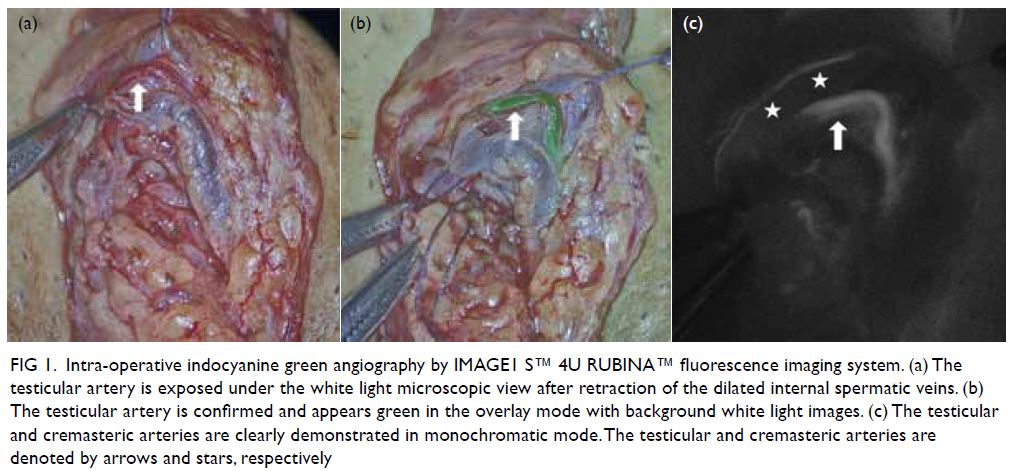
Figure 1. Intra-operative indocyanine green angiography by IMAGE1 S™ 4U RUBINA™ fluorescence imaging system. (a) The testicular artery is exposed under the white light microscopic view after retraction of the dilated internal spermatic veins. (b) The testicular artery is confirmed and appears green in the overlay mode with background white light images. (c) The testicular and cremasteric arteries are clearly demonstrated in monochromatic mode. The testicular and cremasteric arteries are denoted by arrows and stars, respectively
 

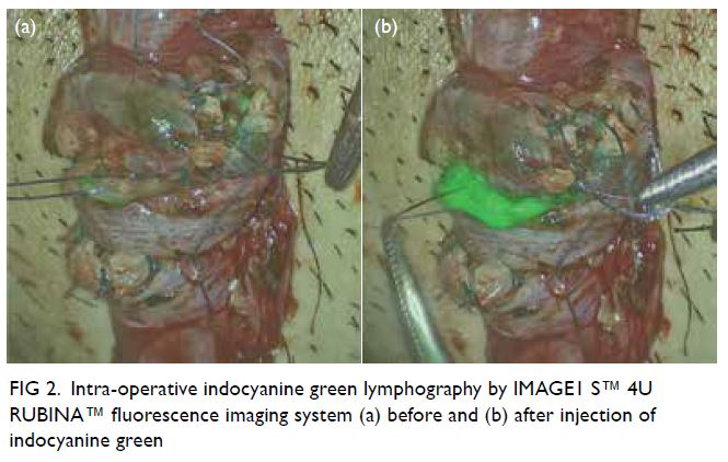
Figure 2. Intra-operative indocyanine green lymphography by IMAGE1 S™ 4U RUBINA™ fluorescence imaging system (a) before and (b) after injection of indocyanine green
 
Microsurgical subinguinal varicocelectomy is the standard for varicocele repair with excellent surgical outcomes reported.4 Nonetheless identification of testicular arteries and lymphatics under white light microscopy alone is operator-dependent and remains challenging for novice surgeons. Several adjuncts have been introduced to facilitate artery- and lymphatic-sparing procedures during MSV. One such adjunct is ICG fluorescence imaging.1 In our opinion, the new overlay mode provided by the latest platform is particularly useful for MSV. Without the need to switch between different modes, the combined regular white light image and near-infrared/ICG data allow accurate localisation of even the smallest vessel without ambiguity (Fig 3). Moreover, the display of ICG signal alone in white on a black background in the monochromatic mode maximises contrast and further improves identification of target vessels (Fig 3). We believe the advances of this surgical platform and imaging technology play a role in enhancing patient safety by increasing the success of arterial and lymphatic preservation in MSV.
 

Figure 3. Intra-operative indocyanine green (ICG) angiography with the previous fluorescence imaging platform utilising VITOM II ICG system with SPIES camera (KARL STORZ SE, Tuttlingen, Germany). Without the overlay mode, the images of (a) white light microscopy view and (b) Chroma mode can only be analysed separately. Correlation between the testicular arteries identified on ICG angiography and white light microscopy can be difficult
 
Author contributions
Concept or design: CL Cho.
Acquisition of data: All authors.
Analysis or interpretation of data: All authors.
Drafting of the manuscript: All authors.
Critical revision of the manuscript for important intellectual content: All authors.
 
All authors had full access to the data, contributed to the study, approved the final version for publication, and take responsibility for its accuracy and integrity.
 
Conflicts of interest
The authors have disclosed no conflict of interest.
 
Funding/support
This study received no specific grant from any funding agency in the public, commercial, or not-for-profit sectors.
 
Ethics approval
Patients were treated in accordance with the Declaration of Helsinki. Patients provided written informed consent for the procedures, and verbal consent for publication.
 
References
1. Cho CL. Is there any role for indocyanine green angiography in testicular artery preservation during microsurgical subinguinal varicocelectomy? In: Esteves SC, Cho CL, Majzoub A, Agarwal A, editors. Varicocele and Male Infertility: a Complete Guide. Switzerland AG: Springer Nature; 2019: 603-14. Crossref
2. Cho CL, Chu RW. Use of video microsurgery platform in microsurgical subinguinal varicocelectomy with indocyanine green angiography. Surg Prac 2019;23:20-4. Crossref
3. Cho CL, Chiu PK, Chu RW. Preliminary experience with indocyanine green lymphography during microsurgical subinguinal varicocelectomy. Surg Prac 2021;25:207-10. Crossref
4. Zini A. Varicocelectomy: microsurgical subinguinal technique is the treatment of choice. Can Urol Assoc J 2007;1:273-6.

Cullen sign in childhood malignancies

© Hong Kong Academy of Medicine. CC BY-NC-ND 4.0
 
PICTORIAL MEDICINE
Cullen sign in childhood malignancies
CC Au, MB, BS, MRCPCH1; Karen KY Leung, MB, BS, MRCPCH1; KL Hon, MB, BS, MD1; Junita KY Tung2; Carol LS Yan, MB, BS, MRCPCH1; WF Hui, MB, BS, MRCPCH1; WY Leung, MB, ChB, FRCSEd(Paed)3
1 Department of Paediatrics and Adolescent Medicine, The Hong Kong Children’s Hospital, Hong Kong
2 Faculty of Medicine, The Chinese University of Hong Kong, Hong Kong
3 Department of Surgery, The Hong Kong Children’s Hospital, Hong Kong
 
Corresponding author: Dr CC Au (aucc@ymail.com)
 
 Full paper in PDF
 
Introduction
We report four consecutive cases of young children with Cullen sign in a paediatric intensive care unit with abdominal malignancies and complications due to treatment. Two children had solid tumours with hepatic rupture and two had pancreatitis secondary to asparaginase use for leukaemia.
 
Case 1
A 23-month-old boy with ruptured liver metastasis and stage 4 neuroblastoma presented with abdominal distension due to a large retroperitoneal mass invading the liver and encasing the aorta and inferior vena cava. He required emergency laparotomy for haemorrhagic shock due to ruptured liver metastasis and endovascular embolisation of tumoural arteries. Subsequently there was an extensive subcutaneous haematoma around the surgical wound. Cullen sign was detected (Fig 1). Recurrent tumoural haemorrhage was excluded and surgical exploration performed to remove blood clots.
 

Figure 1. 23-month-old boy who required emergency laparotomy for haemorrhagic shock due to ruptured liver metastasis and endovascular embolisation of tumoural arteries. Subsequently there was an extensive subcutaneous haematoma around the surgical wound. Cullen sign was detected
 
Case 2
A 5-year-old girl had asparaginase-associated pancreatitis and common B-cell acute lymphoblastic leukaemia. She was 6 months into her treatment protocol and received re-induction phase chemotherapy including pegaspargase (Oncaspar). She presented with abdominal pain and vomiting due to severe acute necrotising pancreatitis. Cullen sign (peri-umbilicus), Grey Turner sign (flank), and Fox sign (inguinal ligament) were detected (Fig 2). She required vasopressor and non-invasive ventilation for systemic inflammatory response syndrome. She later developed pancreatic pseudocyst and underwent ultrasound-guided drainage.
 

Figure 2. 5-year-old girl presented with abdominal pain and vomiting due to severe acute necrotising pancreatitis. Cullen sign (peri-umbilicus), Grey Turner sign (flank), and Fox sign (inguinal ligament) were detected
 
Case 3
A 5-year-old boy with asparaginase-associated pancreatitis and B-cell acute lymphoblastic leukaemia had received re-induction phase chemotherapy with pegaspargase (Oncaspar) and presented with abdominal pain and vomiting. He had severe acute necrotising pancreatitis and systemic inflammatory response syndrome. Cullen sign (peri-umbilicus) and Bryant sign (scrotum) were detected (Fig 3). He required vasopressor support. Later he developed multiple intrapancreatic and peripancreatic fluid collections and required repeated computed tomography-guided aspiration and drainage.
 

Figure 3. 5-year-old boy presented with abdominal pain and vomiting due to severe acute necrotising pancreatitis and systemic inflammatory response syndrome. Cullen sign (peri-umbilicus) and Bryant sign (scrotum) were detected
 
Case 4
A 7-year-old boy with ruptured hepatoblastoma presented with abdominal distension. Hepatoblastoma involved all liver segments, extended into the inferior vena cava and had metastasised to the lungs. He required selective embolisation of left hepatic artery segments 2 and 3 for active haemorrhage. He had abdominal compartment syndrome and gross ascites that required drainage. Cullen sign was detected. (Fig 4) He was prescribed chemotherapy and consequently the tumour reduced in size.
 

Figure 4. 7-year-old boy with abdominal compartment syndrome and gross ascites that required drainage. Cullen sign was detected
 
Discussion
These four cases illustrate Cullen sign in paediatric malignancies. General practitioners and paediatricians should be aware of its diagnostic implications during clinical examination.
 
First described in 1918, Cullen sign referred to periumbilical ecchymosis due to retroperitoneal haemorrhage of ruptured ectopic pregnancy.1 It has become one of the classic abdominal signs in acute pancreatitis. Cullen sign or Grey Turner sign has been reported present in 3% of acute necrotising pancreatitis cases. In 1984, these signs were reported to have a significant mortality rate of 37%,2 but clinical outcomes of severe acute pancreatitis have since improved with intensive care. Nevertheless Cullen sign remains a pointer to serious intra-abdominal haemorrhage. Haemorrhage can originate anywhere along an anatomical pathway. Pathophysiologically, blood tracks from the retroperitoneum through the gastrohepatic ligament to the falciform ligament of the liver, then reaches the umbilicus through the round ligament of liver to form Cullen sign.3 Cullen sign has been reported in intra-abdominal malignancies, liver cirrhosis, and rectus sheath haematoma.4 5
 
Author contributions
All authors contributed to the concept or design of the study, interpretation of the data, drafting of the manuscript, and critical revision of the manuscript for important intellectual content. All authors had full access to the data, contributed to the study, approved the final version for publication, and take responsibility for its accuracy and integrity.
 
Conflicts of interest
As an editor of the journal, KL Hon was not involved in the peer review process for this article. Other authors have no conflicts of interest to disclose.
 
Funding/support
This study received no specific grant from any funding agency in the public, commercial, or not-for-profit sectors.
 
Ethics approval
The patients were treated in accordance with the Declaration of Helsinki. This study was approved by the Hong Kong Children’s Hospital Research Ethics Committee (Ref HKCH-REC-2019-009). Patients’ parents consented for publication of clinical photographs.
 
References
1. Cullen TS. A new sign in ruptured extrauterine pregnancy. Am J Obstet Gynecol 1918;78:457.
2. Dickson AP, Imrie CW. The incidence and prognosis of body wall ecchymosis in acute pancreatitis. Surg Gynecol Obstet 1984;159:343-7.
3. Bem J, Bradley EL 3rd. Subcutaneous manifestations of severe acute pancreatitis. Pancreas 1998;16:551-5. Crossref
4. Mabin TA, Gelfand M. Cullen’s sign, a feature in liver disease. Br Med J 1974;1:493-4. Crossref
5. Harris S, Naina HV. Cullen’s sign revisited. Am J Med 2008;121:682-3. Crossref

Emphysematous cystitis complicated by liver abscess

© Hong Kong Academy of Medicine. CC BY-NC-ND 4.0
 
PICTORIAL MEDICINE
Emphysematous cystitis complicated by liver abscess
L Zeng, MD1; Q Wang, MD2
1 Department of Radiology, Hospital of Chengdu University of Traditional Chinese Medicine, Sichuan, China
2 Department of Ultrasound, Hospital of Chengdu University of Traditional Chinese Medicine, Sichuan, China
 
Corresponding author: Dr Q Wang (444028177@qq.com)
 
 Full paper in PDF
 
In July 2019, a 54-year-old woman with type 2 diabetes mellitus and suboptimal drug compliance with metformin and gliclazide presented to the emergency department complaining of abdominal pain, progressive weakness, and confusion. Her body temperature was 39.1°C and blood tests revealed serum glucose 29.1 mmol/L; haemoglobin A1c 10.4%; serum glucose 29.1 mmol/L; haemoglobin A1c 10.4%; C-reactive protein 224.3 mg/L; and white cell count 11.9 × 10⁹/L with 92.8% segmented neutrophils; and serum creatinine 437.4 mmol/L. Computed tomography scan of the abdomen and pelvis without contrast revealed a cystic gas-producing liver lesion and diffuse intramural gas dissecting the urinary bladder wall with extension to the left kidney (Fig). A diagnosis was made of acute emphysematous cystitis complicated by liver abscess.
 

Figure. Computed tomography of the abdomen and pelvis. The patient had a gas-producing liver lesion (a) and diffuse intramural gas dissecting the urinary bladder wall (b) with extension to the left kidney
 
Percutaneous drainage of the liver abscess was performed under ultrasound guidance. Klebsiella pneumoniae, sensitive to meropenem, was cultured from the urine and drained pus. The patient was transferred to the intensive care unit and treated with resuscitation, glycaemic control, and broad-spectrum antibiotics with intravenous meropenem. Meropenem was continued for the initial 7 days and then changed to ceftazidime plus amikacin according to susceptibility testing results. Her symptoms subsided and blood parameters improved gradually. She made an uneventful recovery and was discharged from the hospital on day 20.
 
Emphysematous cystitis is an uncommon type of infection characterised by gas collections within the bladder wall and lumen. It is usually caused by gas-producing pathogens such as Escherichia coli and Klebsiella pneumoniae.1 Predisposing factors are diabetes, female sex, obstructive uropathy and possibly immunosuppression. Gas is believed to be produced by fermentation of albumin or glucose by the infecting organisms. Emphysematous cystitis combined with liver abscess is rare. The mechanisms of emphysematous cystitis are not well understood. Some reports emphasise that pathogenesis may be related to hematogenous transmission from the liver abscess.2 3 Immunosuppression related to poorly controlled diabetes is another contributing factor.
 
Clinical presentation of emphysematous cystitis varies. Our patient presented with systemic manifestations of septic shock and severe upper abdominal pain secondary to the liver abscess. She reported no lower urinary tract symptoms. Computed tomography is the best diagnostic modality with its high sensitivity when assessing the extent of gas patterns. Appropriate antibiotic therapy, correction of the underlying disorder, and adequate drainage is the recommended treatment.
 
Author contributions
Concept or design: Both authors.
Acquisition of data: L Zeng.
Analysis or interpretation of data: L Zeng.
Drafting of the manuscript: L Zeng.
Critical revision of the manuscript for important intellectual content: Q Wang.
 
Both authors had full access to the data, contributed to the study, approved the final version for publication, and take responsibility for its accuracy and integrity.
 
Conflicts of interest
The authors declared no potential conflicts of interest.
 
Funding/support
This study received no specific grant from any funding agency in the public, commercial, or not-for-profit sectors.
 
Ethics approval
This study was approved by the Hospital of Chengdu University of Traditional Chinese Medicine Research Ethics Committee. Informed consent was obtained from the patient.
 
References
1. Amano M, Shimizu T. Emphysematous cystitis: a review of the literature. Intern Med 2014;53:79-82. Crossref
2. Lai CC. Concomitant emphysematous cystitis and liver abscess. Korean J Intern Med 2018;33:839-40. Crossref
3. Su YC, Chen CC. Emphysematous cystitis complicating Klebsiella pneumoniae liver abscess. Am J Emerg Med 2006,24:256-7.Crossref

Gastric emphysema

© Hong Kong Academy of Medicine. CC BY-NC-ND 4.0
 
PICTORIAL MEDICINE
Gastric emphysema
G Liang, MMed; LC Zeng, MMed; MG Xie, BMed; MX Zhang, MMed (TCM); ZH Hou, MMed (TCM)
Department of Radiology, Hospital of Chengdu University of Traditional Chinese Medicine, Chengdu, Sichuan, China
 
Corresponding author: Prof MG Xie (xmg6806@163.com)
 
 Full paper in PDF
 
Case
A 47-year-old woman was admitted to the intensive care unit following craniectomy for intracranial haematoma. She reported tenderness in the epigastric region with no evidence of peritonitis. Enhanced computed tomography (CT) of the abdomen revealed a distended stomach and diffuse circumferential air in the stomach wall, with prominent enhancement of the stomach wall mucosa. Pneumoperitoneum was detected on identification of extraluminal stomach air along the greater curvature (Fig 1). Laboratory results showed no abnormality. The patient had no obvious predisposing factors or infection and inflammatory markers in blood cultures were normal. Based on the clinical presentation and relevant laboratory examinations, assessment of predisposing factors and CT findings, gastric emphysema (GE) was diagnosed and the patient was managed conservatively. She underwent gastric decompression with nasogastric tube placement and fluid resuscitation, and was prescribed a proton pump inhibitor and broad-spectrum antibiotics. Non-enhanced CT scan 11 days later revealed complete resolution of gas within the submucosal layer (Fig 2).
 

Figure 1. A 47-year-old woman with gastric emphysema. (a) Axial enhanced computed tomography and (b) sagittal multiplanar reconstruction images revealing a distended stomach and diffuse circumferential air in the stomach wall extending to the lower oesophagus, with prominent enhancement of the stomach wall mucosa. Pneumoperitoneum was evidenced by the presence of extraluminal stomach air along the greater curvature (white arrow)
 

Figure 2. Same patient 11 days after treatment. Non-enhanced computed tomography scan revealed complete resolution of gas within the submucosal layer
 
Discussion
Gastric pneumatosis is a rare finding identified by accumulation of gas within the stomach wall. Both GE and emphysematous gastritis (EG) are important differential diagnoses of intramural gastric air. They differ in their aetiology, clinical course, radiographic findings, management, and prognosis. However, it is important to differentiate the much more benign GE from the highly lethal EG.
 
Gastric emphysema is caused by a disruption in gastric mucosal integrity without underlying infection. Most patients with GE have no or mild symptoms, and the prognosis is excellent.1 Gastric emphysema is a relatively benign condition and usually self-limiting. The causes of the mucosal defect in GE include increased intragastric pressure, instrumentation such as gastroscopy, severe vomiting, and dissection of air from the mediastinum and ischaemia. The management of GE is usually non-surgical and includes bowel rest with nasogastric tube placement, fluid resuscitation and nutritional support.1 We think our case of GE was related to stress-related mucosal erosions of the stomach mucosa, and possibly increased gastric distension.
 
In contrast, EG, resulting from gas-forming organisms and associated with systematic toxicity, is a devastating infectious process with a mortality rate of 60%.2 3 Patients with EG usually display severe clinical signs including severe abdominal pain, severe abdominal tenderness, haematemesis, and occult gastric bleeding. The patient may need to be transferred to the intensive care unit and treated with broad-spectrum antibiotics if there is evidence of bacterial infection. Patients should undergo oesophagogastroduodenoscopy and enhancement CT as early as possible when EG is suspected. Surgical intervention is more commonly indicated for EG and is directed at removal of the septic organ, whereas the primary indication for surgical intervention in GE is uncertainty of diagnosis.3 4
 
In summary, despite similar radiographic findings, GE is typically secondary to mechanical injury of the stomach mucosa, whereas EG is an acute infection of the stomach wall. The differentiation of these two entities depends on the patient’s clinical presentation, assessment of predisposing factors, and CT findings.
 
Author contributions
Concept or design: All authors.
Acquisition of data: G Liang, LC Zeng.
Analysis or interpretation of data: All authors.
Drafting of the manuscript: G Liang.
Critical revision of the manuscript for important intellectual content: All authors.
 
All authors had full access to the data, contributed to the study, approved the final version for publication, and take responsibility for its accuracy and integrity.
 
Conflicts of interest
All authors have disclosed no conflicts of interest.
 
Acknowledgement
We would like to thank two anonymous reviewers and the journal editor, who have provided excellent comments and significantly contributed to the improvement of the article.
 
Funding/support
This study received no specific grant from any funding agency in the public, commercial, or not-for-profit sectors.
 
Ethics approval
This study was approved by the Hospital of Chengdu University of Traditional Chinese Medicine Research Ethics Committee. Informed consent was obtained from the patient.
 
References
1. Matsushima K, Won EJ, Tangel MR, Enomoto LM, Avaella DM, Soybel DI. Emphysematous gastritis and gastric emphysema: similar radiographic findings, distinct clinical entities. World J Surg 2015;39:1008-17. Crossref
2. Misro A, Sheth H. Diagnostic dilemma of gastric intramural air. Ann R Coll Surg Engl 2014;96:e11-3. Crossref
3. Guillén-Morales C, Jiménez-Miramón FJ, Carrascosa-Mirón T, Jover-Navalón JM. Emphysematous gastritis associated with portal venous gas: medical management to an infrequent acute abdominal pain. Rev Esp Enferm Dig 2015;107:455-6. Crossref
4. Inayat F, Zafar F, Zaman MA, Hussain Q. Gastric emphysema secondary to severe vomiting: a comparative review of 14 cases. BMJ Case Rep 2018;2018: bcr2018226594. Crossref

Picture-in-picture video demonstration of systematic transperineal prostate biopsy

© Hong Kong Academy of Medicine. CC BY-NC-ND 4.0
 
PICTORIAL MEDICINE
Picture-in-picture video demonstration of systematic transperineal prostate biopsy
KL Lo, FHKAM (Surgery); David Leung, MRCS; Zoe Lai, RN; Crystal Li, RN; SF Ma, MB, ChB; Julius Wong, MRCS; KK Yuen, FHKAM (Surgery); Joseph Li, FHKAM (Surgery); Peter Chiu, FHKAM (Surgery); SK Mak, FHKAM (Surgery); Joseph Wong, FHKAM (Surgery); CF Ng, FHKAM (Surgery)
Department of Surgery, The Chinese University of Hong Kong, Hong Kong
 
Corresponding author: Dr KL Lo (lokalun@surgery.cuhk.edu.hk)
 
  A video clip demonstrating systematic transperineal prostate biopsy is avaialble at www.hkmj.org
 
 Full paper in PDF
 
We first performed 10-core transperineal prostate biopsy at our institution in 2018. After having acquired the technique and experience, we developed a biopsy protocol that could be modified according to prostate size (Fig): 10 cores for prostate size <30 cc, 16 cores for prostate size 30 to 50 cc, and 20 cores for prostate size >50 cc.
 

Figure. Templates for prostate biopsy locations, longitudinal view: (a) 10-core template for prostate size <30 cc (5 cores in each lobe); (b) 16-core template for prostate size 30-50 cc (8 cores in each lobe); (c) 20-core template for prostate size >50 cc (10 cores in each lobe); (d) 24-core template (12 cores in each lobe)
 
We currently take a 24-core prostate biopsy (Fig d), irrespective of prostate size, to further improve the detection of prostate cancer. There is no gold standard protocol for total number of cores required in any patient; it is dictated by hospital policy, availability of resources, and the experience of the urologist.
 
This video demonstrates systematic transperineal prostate biopsy.
 
The instruments used during the procedure comprised an ultrasound machine with long side-fire sensor probe, a bed with two leg supports to facilitate lithotomy position, one disposable biopsy gun, two needles for local anaesthesia, two metal trocars, and eight specimen bottles.
 
Before the procedure and in the absence of any contraindication, patients are prescribed a single dose per oral 1 g Augmentin and 500 mg ciprofloxacin. One tube of per rectal 4.5 oz Fleet Enema is administered as bowel preparation. Local anaesthetic (EMLA) cream is applied to the patient’s perineum 1 hour before the procedure.
 
Step 1: the perineum is disinfected with chlorhexidine.
 
Step 2: 10 mL of 1% lidocaine is injected through each side of the perineum into the periprostatic plane as local anaesthesia, at an angle of 45° and 15 mm away from the anus as shown.
 
It is vital to maintain the needle parallel to the ultrasound probe to ensure continued visualisation of the needle.
 
Step 3: a 14-gauge metal trocar is inserted through the right side of the perineum under ultrasound guidance.
 
Step 4: anterior sector prostate biopsies are taken by tilting an 18-gauge disposable biopsy gun downwards.
 
It is important not to hit the urethra during the procedure.
 
Step 5: basal sector prostate biopsies are taken.
 
Step 6: central sector prostate biopsies are taken.
 
Step 7: posterior sector prostate biopsies are taken.
 
Step 8: Left lobe prostate biopsies are also taken using a technique similar to that for the right lobe.
 
Final step: After removing the metal trocars, haemostasis is achieved by compression. A transparent adhesive film dressing is sprayed over the two puncture sites.
 
No post-procedure antibiotic is required.
 
Author contributions
All authors contributed to the concept or design of the study, acquisition of the data, analysis or interpretation of the data, drafting of the manuscript, and critical revision of the manuscript for important intellectual content. All authors had full access to the data, contributed to the study, approved the final version for publication, and take responsibility for its accuracy and integrity.
 
Conflicts of interest
The authors have no conflicts of interest to disclose.
 
Funding/support
This study received no specific grant from any funding agency in the public, commercial, or not-for-profit sectors.
 
Ethics approval
This study was approved by the Joint Chinese University of Hong Kong–New Territories East Cluster Clinical Research Ethics Committee (Ref CREC 2018.323). The patient provided written informed consent.
 

Unilateral vocal cord palsy in a patient with jugular foramen schwannoma

© Hong Kong Academy of Medicine. CC BY-NC-ND 4.0
 
PICTORIAL MEDICINE
Unilateral vocal cord palsy in a patient with jugular foramen schwannoma
WK Wong, BDS, MD, CY Cheng, MD, WC Cheng, MD
Department of Neurosurgery, Chang Gung Memorial Hospital, Chiayi, Taiwan
 
Corresponding author: Dr WC Cheng (wancheng7511@yahoo.com.tw)
 
 Full paper in PDF
 
A 74-year-old man presented with a history of hoarseness of voice and choking for a few weeks. Laryngoscopy revealed left vocal cord palsy (Fig 1). Non-contrast computed tomography (CT) showed no pulmonary or neck lesion, but one lobulated, centrally low attenuating mass. T2-weighted magnetic resonance images confirmed a 3.8-cm soft tissue mass with heterogeneous enhancement in the left cerebellopontine angle. In both images there was extension of the mass through an expansion of the left jugular foramen, widened left internal acoustic meatus and skull base destruction (Fig 2), compatible with jugular foramen tumour. Subsequently the patient developed progressive globus sensation, tinnitus, left sternocleidomastoid muscle wasting, and left neurosensory hearing loss. The tumour was resected via a retrosigmoid approach. Surgical assessment of the tumour revealed its origin from cranial nerves IX and X and compression of cranial nerves VIII and XI. The histopathological diagnosis with immunohistochemistry staining was that of schwannoma. Postoperative imaging demonstrated adequate resection of the tumour. Improvement in swallowing and hearing function test from severe to moderate impairment were noted postoperatively.
 

Figure 1. Laryngoscopic examination showing (a) left vocal cord palsy before surgery and (b) improvement after surgery
 

Figure 2. (a) Non-contrast computed tomography and (b) T2-weighted magnetic resonance image showing a soft tissue mass in the left cerebellopontine angle. The arrows indicate the location of the normal right jugular foramen in both figures
 
Discussion
Schwannoma is a benign tumour of the nerve sheath. The most common intracranial schwannoma is vestibular schwannoma (acoustic neuroma) that arises from cranial nerve VIII.1 Jugular foramen schwannomas, which mainly arise from cranial nerves IX and X, account for around 3% to 4% of all intracranial schwannomas. They are more prevalent in women and occur between the third and sixth decades of life. Clinical presentation is variable because of their slow-growing nature and proximity to other cranial nerves. Symptoms appear when the tumour is sufficiently large and most commonly consist of hearing loss, tinnitus, dysphagia, ataxia, and hoarseness. Other symptoms include dysarthria, dysphonia, aspiration, vertigo, dizziness, shoulder weakness, and headache.2 Differential diagnoses of unilateral vocal cord palsy are usually divided into malignancies of the lung and the neck and non-malignant lesions of traumatic, neurological, inflammatory or infectious origin. A CT scan is indicated to evaluate the more common pulmonary malignancies and to ensure presence of a rarer lesion along the course of the recurrent laryngeal and vagus nerve up to the skull base is not missed, especially if signs and symptoms of multiple cranial nerve involvement are present.3
 
Imaging studies can help differentiate vestibular and lower cranial nerve schwannomas, meningioma, glomus jugulare paraganglioma, ependymomas, and metastatic tumour. Imaging findings of jugular foramen schwannoma include scalloped and sclerotic expansion of the temporal bone instead of a lytic pattern. On magnetic resonance imaging, the lesion is T1 iso- or hypo-intense or T2 iso- or hyper-intense with gadolinium enhancement, whereas on CT, it is isodense to brain parenchyma and enhanced with contrast.4 The site of origin is classified as cisternal, foraminal (intraosseous) or extracranial. However, it is difficult to ascertain the exact site radiologically and clinically because of the variable location and nerve involvement. Assessment of the nerve root during surgery is required to confirm the origin of the tumour. The selection of surgical approach for treatment is determined by the location, pattern and extension of the tumour, the degree of bone destruction/erosion and the neurological and preoperative hearing status.5 Risks of operation include damage to other cranial nerves, especially facial nerve palsy, and incomplete removal of the tumour leading to recurrence.
 
Author contributions
Concept or design: All authors.
Acquisition of data: WK Wong, CY Cheng.
Analysis or interpretation of data: All authors.
Drafting of the manuscript: WK Wong.
Critical revision of the manuscript for important intellectual content: All authors.
 
All authors had full access to the data, contributed to the study, approved the final version for publication, and take responsibility for its accuracy and integrity.
 
Conflicts of interest
All authors have disclosed no conflicts of interest.
 
Funding/support
This study received no specific grant from any funding agency in the public, commercial, or not-for-profit sectors.
 
Ethics approval
The patient was treated in accordance with the Declaration of Helsinki and provided consent for all investigations and procedures.
 
References
1. Suri A, Bansal S, Singh M, Mahapatra AK, Sharma BS. Jugular foramen schwannomas: a single institution patient series. J Clin Neurosci 2014;21:73-7. Crossref
2. Bakar B. The jugular foramen schwannomas: review of the large surgical series. J Korean Neurosurg Soc 2008;44:285-94. Crossref
3. Stimpson P, Patel R, Vaz F, et al. Imaging strategies for investigating unilateral vocal cord palsy: how we do it. Clin Otolaryngol 2011;36:266-71. Crossref
4. Lee M, Tong K. Jugular foramen schwannoma mimicking paraganglioma: case report and review of imaging findings. Radiol Case Rep 2016;11:25-8. Crossref
5. Samii M, Alimohamadi M, Gerganov V. Surgical treatment of jugular foramen schwannoma: surgical treatment based on a new classification. Neurosurgery 2015;77:424-32. Crossref

Primary orbital melanoma

© Hong Kong Academy of Medicine. CC BY-NC-ND 4.0
 
PICTORIAL MEDICINE
Primary orbital melanoma
Vivian WK Hui, MB, ChB1; TC Lau, PhD2; Lawrence PW Ng, MSc2; Hunter KL Yuen, FRCOphth1; W Cheuk, FHKCPath2
1 Department of Ophthalmology, Hong Kong Eye Hospital, Hong Kong
2 Department of Pathology, Queen Elizabeth Hospital, Hong Kong
 
Corresponding author: Dr W Cheuk (cwzz01@ha.org.hk)
 
 Full paper in PDF
 
A 68-year-old Chinese man presented with proptosis, epiphora and discomfort in the right eye associated with impaired visual acuity. Contrast computed tomography scan revealed a 3.3 cm × 2.1 cm heterogeneously enhancing mass at the superotemporal aspect of the right orbit with displacement of the lateral rectus muscle (Fig 1). A pigmented tumour with dense adhesions to the surrounding structures was found in orbitotomy with leakage of the pigmented content upon surgical exploration. The lesion was excised as much as possible. Detailed clinical examination showed no evidence of intraocular melanoma, conjunctival or eyelid melanoma, or melanoma anywhere else. Systemic examination and whole-body positron emission tomography-computed tomography scan were unremarkable. The patient was alive with no evidence of disease at 9-month follow-up.
 

Figure 1. Contrast computed tomography scan showing a contrast-enhancing tumour behind the right globe displacing the optic nerve
 
The tumour was a solid, dark brownish multinodular mass covered by fat and skeletal muscle, comprising lobulated sheets of epithelioid and spindly melanocytes with vesicular chromatin, prominent nucleoli, and variable amounts of melanin pigment (Figs 2, 3, and 4). The mitotic count was 1 per 10 HPFs. The neoplastic melanocytes were positive for S100 and SOX10 but negative for BRAF. BRCA1-associated protein 1 immunostaining was intact. Sanger sequencing revealed a GNA11 Q209L mutation. No mutation was found in GNAQ, NRAS, KRAS, BRAF or KIT.
 

Figure 2. Micrograph showing multinodular mass with variable amount of melanin pigment in different areas (×40)
 

Figure 3. Large sheets of epithelioid (left upper field) and spindly (right lower field) melanoma cells with pale chromatin and prominent nucleoli were found. There were scattered histiocytes with abundant cytoplasmic melanin pigment (×200)
 

Figure 4. Melanoma cells were positive on SOX10 (red chromogen) immunohistochemical staining (×200)
 
Ocular melanoma most frequently involves the uveal tract (choroid, ciliary body, and iris) and the conjunctiva, where melanocytes are normally present. According to The Cancer Genome Atlas uveal melanoma project, mutually exclusive mutations in GNAQ, GNA11, CYSLTR2 and PLCB4 are the tumour-initiating mutations whereas BAP1, EIF1AX and SF3B1/SRSF2 mutations are associated with prognosis in uveal melanoma.1 In contrast, conjunctival melanoma typically exhibits BRAF, NRAS and NF1 mutations of the MAPK pathway with UV-induced C→T mutation signature.2 Orbital soft tissue melanoma is uncommon with >90% representing secondary tumours as a result of contiguous spread or metastasis from uveal, conjunctival, cutaneous or sinonasal melanomas.3 Primary orbital melanoma is exceedingly rare and has been postulated to arise in embryological remnants of melanocytes arrested in the region since a substantial proportion of cases are associated with blue nevus, orbital melanocytosis or oculodermal melanocytosis (nevus of Ota). Alternatively, it may arise from isolated melanocytes found in the optic nerve sheath, orbital fat, extraocular muscles or orbital periosteum. The majority of patients are white Europeans in their fourth to fifth decade. Histologically, it comprises epithelioid, spindle or mixed populations of neoplastic melanocytes that are frequently pigmented but with a lesser degree of nuclear pleomorphism than their cutaneous and mucosal counterparts. The prognosis is generally poor but a small proportion of patients survive long term. The most frequent site of metastasis is the liver.3 4 The clinicopathological features of primary orbital melanoma are very similar to those of uveal melanoma. The findings of GNAQ, SF3B1, EIF1AX mutations5 and GNA11 mutation in this case provide further evidence that primary orbital melanoma has a close pathogenic relationship with uveal melanoma.
 
Author contributions
All authors contributed to the concept or design of the study, acquisition of the data, analysis or interpretation of the data, drafting of the manuscript, and critical revision of the manuscript for important intellectual content. All authors had full access to the data, contributed to the study, approved the final version for publication, and take responsibility for its accuracy and integrity.
 
Conflicts of interest
As an adviser of the journal, HKL Yuen was not involved in the peer review process. Other authors have disclosed no conflicts of interest.
 
Declaration
This case was presented as a poster at the Annual Scientific Meeting of The College of Ophthalmologists of Hong Kong and Hong Kong Ophthalmological Society on 14 to 15 December 2019.
 
Funding/support
This pictorial medicine paper received no specific grant from any funding agency in the public, commercial, or not-forprofit sectors.
 
Ethics approval
The patient was treated in accordance with the tenets of the Declaration of Helsinki. Informed consent was obtained from the patient.
 
References
1. Bakhoum MF, Esmaeli B. Molecular characteristics of uveal melanoma: insights from the Cancer Genome Atlas (TCGA) Project. Cancers (Basel) 2019;11:1061.Crossref
2. Swaminathan SS, Field MG, Sant D, et al. molecular characteristics of conjunctival melanoma using whole-exome sequencing. JAMA Ophthalmol 2017;135:1434-7. Crossref
3. Rose AM, Luthert PJ, Jayasena CN, Verity DH, Rose GE. Primary orbital melanoma: presentation, treatment, and long-term outcomes for 13 patients. Front Oncol 2017;7:316. Crossref
4. Shields JA, Shields CL. Orbital malignant melanoma: the 2002 Sean B Murphy lecture. Ophthalmic Plast Reconstr Surg 2003;19:262-9. Crossref
5. Rose AM, Luo R, Radia UK, et al. Detection of mutations in SF3B1, EIF1AX and GNAQ in primary orbital melanoma by candidate gene analysis. BMC Cancer 2018;18:1262. Crossref

Tattoo-associated uveitis

© Hong Kong Academy of Medicine. CC BY-NC-ND 4.0
 
PICTORIAL MEDICINE
Tattoo-associated uveitis
CY Mak, MB, BS, MRCSEd (Ophth)1; Mary Ho, FCOphth HK, FHKAM (Ophthalmology)1; Angela Z Chan, MB, BS2; Lawrence PL Iu, FRCSEd (Ophth), FHKAM (Ophthalmology)1; Christina MT Cheung, FHKCP, FHKAM (Medicine)3; Marten E Brelen, BMBCh (Oxon), FRCOphth1; Paul CL Choi, FRCPA, FHKAM (Pathology)2; Alvin L Young, FRCOphth, FHKAM (Ophthalmology)1
1 Department of Ophthalmology and Visual Sciences, Prince of Wales Hospital, Hong Kong
2 Department of Anatomical and Cellular Pathology, Prince of Wales Hospital, Hong Kong
3 Division of Dermatology, Department of Medicine and Therapeutics, Prince of Wales Hospital, Hong Kong
 
Corresponding author: Prof Alvin L Young (youngla@ha.org.hk)
 
 Full paper in PDF
 
A 19-year-old man with extensive body tattooing presented with recurrent episodes of reduced vision, bilateral photophobia and concomitant swelling of body tattoos. He had multiple tattoos over his entire body with mainly black pigment and occasional red and yellow pigment, performed over a period of 3 years prior to the presentation of ocular symptoms. He enjoyed good past health with no history of autoimmune diseases. There was no joint pain, skin rash or chest symptoms.
 
Ophthalmological examination revealed bilateral injection, anterior chamber cells, posterior synechiae (Fig 1) and marked vitritis, consistent with anterior and intermediate uveitis. There were no mutton-fat keratic precipitates or iris nodules. Presenting visual acuity was 20/200 in both eyes. Dermatological examination showed prominent induration of skin with mild tenderness in areas of body tattoo containing black pigment (Fig 2). Non-tattooed skin was unremarkable with no signs of inflammation. Incisional skin biopsy was taken from an area of prominently indurated tattoo. Histopathology showed marked non-caseating granulomatous reaction within the dermis and abundant black pigment deposition (Fig 3). Periodic acid-Schiff staining showed no fungal elements. Chest radiograph was clear with no hilar lymphadenopathy and interferon-gamma releasing assay was negative. Syphilis and human immunodeficiency virus serology was negative. Immune markers including antinuclear antibodies, antineutrophil cytoplasmic antibodies and anti-extractable nuclear antigens antibody were negative. The patient declined a blood test for angiotensin-converting enzyme level due to the associated cost. Serum calcium was not elevated.
 

Figure 1. Slit lamp photo of the right eye showing ciliary injection, posterior synechiae formation. There were no iris nodules
 

Figure 2. Prominent induration of part of a tattoo containing black pigment. Surrounding non-tattooed skin was unremarkable
 

Figure 3. Skin biopsy of indurated tattoo under haematoxylin and eosin staining showing dermal deposition of black pigment and marked non-caseating granulomatous reaction
 
He was treated with topical prednisolone and oral prednisolone 60 mg daily after exclusion of infectious uveitides. Body tattoo swelling subsided rapidly after systemic steroid and the uveitis was brought under control gradually with significant improvement in bilateral vision. He was maintained on mycophenolate mofetil 1g twice a day as a steroid-sparing agent for uveitis control. His oral prednisolone was tapered to below 15 mg daily. His visual acuity improved and maintained at 20/30 bilaterally. There were no features of systemic sarcoidosis. Overall clinicopathological features were compatible with tattoo-associated uveitis, a rare dermato-ophthalmological complication of body tattooing.
 
Systemic sarcoidosis, a rare disease in Asians, can occasionally cause tattoo granuloma and uveitis.1 Tattoo-associated uveitis without systemic sarcoidosis is a rare entity first described in a case series half a century ago.2 The disease is characterised by recurrent episodes of uveitis in conjunction with raised and indurated tattoo, while histology of affected skin demonstrates florid non-caseating granulomatous reaction indistinguishable from tattoo granuloma in systemic sarcoidosis.3 The exact pathogenesis is unknown, but it was believed to be a type of delayed hypersensitivity reaction to tattoo pigments.3 Treatment is mainly to control ocular inflammation by topical and systemic steroid, with or without steroid-sparing agent. Tattoo excision has been reported to be useful in limiting recurrences.1 3 However, the extensive tattoo involvement in our patient rendered excision impractical.
 
In summary, the clinical photos illustrate a rare case of tattoo-associated uveitis, highlighting the importance of inquiry into tattoo history and skin examination of tattoos in a patient with recurrent uveitis.
 
Author contributions
Concept or design: CY Mak, ME Brelen, AL Young.
Acquisition of data: CY Mak, AZ Chan, CMT Cheung.
Analysis or interpretation of data: CY Mak, M Ho, LPL Iu, PCL Choi.
Drafting of the manuscript: CY Mak, M Ho, AZ Chan.
Critical revision of the manuscript for important intellectual content: All authors.
 
All authors had full access to the data, contributed to the study, approved the final version for publication, and take responsibility for its accuracy and integrity.
 
Conflicts of interest
All authors have disclosed no conflicts of interest.
 
Funding/support
This pictorial medicine paper received no specific grant from any funding agency in the public, commercial, or not-forprofit sectors.
 
Ethics approval
This patient was treated in accordance with the Declaration of Helsinki. The patient provided written informed consent for all treatments and procedures and for publication of clinical photos.
 
References
1. Kluger N. Tattoo-associated uveitis with or without systemic sarcoidosis: a comparative review of the literature. J Eur Acad Dermatol Venereol 2018;32:1852-61. Crossref
2. Rorsman H, Brehmer-Andersson E, Dahlquist I, et al. Tattoo granuloma and uveitis. Lancet 1969;2:27-8. Crossref
3. Ostheimer TA, Burkholder BM, Leung TG, Butler NJ, Dunn JP, Thorne JE. Tattoo-associated uveitis. Am J Ophthalmol 2014;158:637-43.e1. Crossref

Eyelash trichomegaly induced by erlotinib for metastatic lung cancer

© Hong Kong Academy of Medicine. CC BY-NC-ND 4.0
 
PICTORIAL MEDICINE
Eyelash trichomegaly induced by erlotinib for metastatic lung cancer
Julia YY Chan, AFCOphth HK, MRCSEd (Ophth)1,2; Tracy YT Kwok, FCOphth HK, FHKAM (Ophthalmology)1,2; Hunter KL Yuen, FRCOphth, FRCSEd1,2
1 Department of Ophthalmology and Visual Sciences, The Chinese University of Hong Kong, Hong Kong
2 Hong Kong Eye Hospital, Hong Kong
 
Corresponding author: Dr Julia YY Chan (cyyj717@gmail.com)
 
 Full paper in PDF
 
A 65-year-old Chinese woman was referred to the ophthalmology clinic complaining of grittiness in both eyes for 1 month. She had been diagnosed with stage IV metastatic pulmonary adenocarcinoma with positive mutation in epidermal growth factor receptor (EGFR) gene (L858R) and had been prescribed erlotinib 4 months previously as palliative treatment (Table).
 

Table. Timeline of the patient
 
Physical examination revealed bilateral long thick curly eyelashes (Figs 1 and 2) affecting all four eyelids. A papulopustular rash over the periocular region was also evident. The patient denied use of topical prostaglandin analogue for either medical or cosmetic use. A diagnosis was made of drug-induced trichomegaly. Corneal punctate epithelial erosion as a result of the misdirected lashes was treated symptomatically with regular epilation and topical lubricants.
 

Figure 1. Clinical photographs of the (a) right and (b) left eyes of a 65-year-old woman who was taking palliative EGFR inhibitor erlotinib for 4 months. Trichomegaly with increased thickness, curling, pigmentation and length of the eyelashes affecting the right upper and lower lids is evident. The patient also has PRIDE syndrome with a papulopustular rash affecting the periocular skin (pharmacological mydriasis was induced as part of the complete ophthalmological examination)
 

Figure 2. Clinical photographs of the (a) right and (b) left eyes of the same patient 6 weeks after cessation of erlotinib treatment. Partial reversal of trichomegaly of the right eye is demonstrated. The curling and pigmentation of eyelashes had resolved although increased thickness and length of eyelashes persisted. Periocular skin inflammation had completely subsided
 
Upon follow-up, gradual partial reversal of trichomegaly was evident at 6 weeks after erlotinib cessation. There was also improvement of the periorbital papulopustular rash. The reversal of trichomegaly after EGFR inhibitor cessation is not well documented in the literature. This case illustrates the presentation and partial resolution of drug-induced trichomegaly.
 
Eyelash trichomegaly is a rare condition characterised by increase in length, thickness, pigmentation, and curling of the eyelashes. The eyelash follicle, just as the scalp hair follicle, undergoes the cycle of anagen, catagen, and telogen phase. The anagen phase of eyelashes (ie, the growth phase) typically spans 8 weeks in Asian patients and occurs in 18% of eyelashes at any given time.1
 
Drug-induced trichomegaly is the most common form of trichomegaly to present in a general ophthalmology setting. Prostaglandin analogues such as latanoprost and bimatoprost, although commonly used as antiglaucomatous drugs, are well known for their side-effect of eyelash trichomegaly. The effect is due to up-regulation of prostaglandin E2, D2 receptor expressed in hair follicles.2
 
The occurrence of EGFR inhibitor–induced trichomegaly is seen less often but is not uncommon in the ophthalmology setting. It has been postulated that EGFR inhibitors inactivate the nuclear factor of activated T-cells. This leads to activation of stem cell bulge and suprabulbar region of the eyelash hair follicles.3 In a typical course, trichomegaly develops between the second and fifth month of EGFR inhibitor treatment.4 An Asian study reported that 2% of patients prescribed EGFR inhibitor treatment had trichomegaly.5 The EGFR inhibitor is also well known to cause a series of cutaneous adverse effects known as the PRIDE syndrome (papulopustules and/or paronychia, regulatory abnormalities of hair growth, itching, and dryness).6 In this case, the patient exhibited PRIDE syndrome affecting the periocular skin as demonstrated in Figures 1 and 2.
 
Trichomegaly is a benign condition, but long misdirected lashes may lead to corneal punctate epithelial erosion and corneal abrasion. Frequent trimming, epilation, and topical lubricants serve as first-line treatment. Electro-epilation or cryoablation directed at the affected hair follicles may be considered in instances of recurrent misdirected lashes with corneal complications such as infective corneal ulcer.
 
This case highlights the clinical course of EGFR inhibitor–induced trichomegaly upon cessation of drug therapy, which is not well documented in the literature. Partial reversal of trichomegaly was achieved after stopping erlotinib for 6 weeks. It is evident that the time required for trichomegaly resolution follows the typical eyelash follicle cycle.
 
Author contributions
All authors contributed to the design, acquisition of data, analysis of data, drafting of the manuscript, and critical revision of the manuscript for important intellectual content. All authors had full access to the data, contributed to the study, approved the final version for publication, and take responsibility for its accuracy and integrity.
 
Conflicts of interest
As an adviser of the journal, HKL Yuen was not involved in the peer review process. Other authors have disclosed no conflicts of interest.
 
Funding/support
This pictorial medicine received no specific grant from any funding agency in the public, commercial, or not-for-profit sectors.
 
Ethics approval
The patient was treated in accordance with the Declaration of Helsinki. The patient provided written informed consent for the treatment/procedures, and consent for publication.
 
References
1. Na JI, Kwon OS, Kim BJ, et al. Ethnic characteristics of eyelashes: a comparative analysis in Asian and Caucasian females. Br J Dermatol 2006;155:1170-6. Crossref
2. Colombe L, Michelet JF, Bernard BA. Prostanoid receptors in anagen human hair follicles. Exp Dermatol 2008;17:63- 72. Crossref
3. Dalal A, Sharma S, Kumar A, Sharma N. Eyelash trichomegaly: a rare presenting feature of systemic lupus erythematosus. Int J Trichology 2017;9:79-81. Crossref
4. Jeon SH, Ryu JS, Choi GS, et al. Erlotinib induced trichomegaly of the eyelashes. Tuberc Respir Dis (Seoul) 2013;74:37-40. Crossref
5. Chanprapaph K, Pongcharoen P, Vachiramon V. Cutaneous adverse events of epidermal growth factor receptor inhibitors: A retrospective review of 99 cases. Indian J Dermatol Venereol Leprol 2015;81:547. Crossref
6. Lacouture ME, Lai SE. The PRIDE (Papulopustules and/or paronychia, Regulatory abnormalities of hair growth, Itching, and Dryness due to Epidermal growth factor receptor inhibitors) syndrome. Br J Dermatol 2006;155:852-4. Crossref

Popliteal artery entrapment syndrome: a rare diagnosis for calf pain

© Hong Kong Academy of Medicine. CC BY-NC-ND 4.0
 
PICTORIAL MEDICINE
Popliteal artery entrapment syndrome: a rare diagnosis for calf pain
Stephanie C Woo, MB, BS, FRCR; TS Chan, FHKCR, FHKAM (Radiology); NY Pan, FHKCR, FHKAM (Radiology); Johnny KF Ma, FRCR (UK), FHKAM (Radiology)
Department of Radiology, Princess Margaret Hospital, Hong Kong
 
Corresponding author: Dr SC Woo (stephaniecheriwoo@gmail.com)
 
 Full paper in PDF
 
An 18-year-old man presented with a long history of occasional right calf pain and fullness. He also complained that over the last year his right foot
 
An 18-year-old man presented with a long history of occasional right calf pain and fullness. He also complained that over the last year his right foot became pale and numb after exercising for few minutes. There was no history of trauma and the patient had no constitutional symptoms. On physical examination, the right calf was non-tender with no mass although right posterior tibial and dorsalis pedis pulses were weaker than the left. Radiograph of the right knee showed static right proximal tibial exostosis, which had been monitored since the patient was aged 10 years. Magnetic resonance arteriogram showed almost complete occlusion of the right popliteal artery and distal superficial femoral artery at the level of the right popliteal fossa (Fig 1). Collateral branches were seen feeding the distal right leg arteries trifurcation. Magnetic resonance imaging scan of the knee revealed the medial head of the gastrocnemius inserting at a more lateral position than usual (Fig 2). The popliteal artery was separated from the popliteal vein, passing medial to and underneath the medial head of the gastrocnemius, and was severely compressed between the medial head of the gastrocnemius and distal femur. Loss of normal flow-related signal void was noted in the right popliteal artery distal to the compression.
 

Figure 1. Magnetic resonance arteriogram of bilateral lower limbs showing almost complete occlusion of the right popliteal artery (white arrow) and distal superficial femoral artery at the level of the right popliteal fossa. Collateral branches are seen feeding the distal right leg arteries trifurcation
 

Figure 2. Magnetic resonance image of the knee. (a) T2-weighted, (b) T2-weight fat saturated and (c) T1-weighted fat-saturated post-contrast images showing popliteal artery (white arrows) passing medial to and underneath the medial head of the gastrocnemius. It is severely compressed between the medial head of the gastrocnemius (asterisk) and distal femur into a slitlike configuration. Findings are consistent with popliteal artery entrapment syndrome
 
Popliteal artery entrapment syndrome (PAES) is a rare1 and frequently underdiagnosed disease entity. It typically affects young male athletes who commonly have hypertrophied musculature without significant cardiovascular risk factors. The classic presentation of PAES is of symptoms related to vascular compression, which is intermittent lower limb claudication. Other symptoms can include numbness, pain, discoloration, or even paralysis.2 Symptoms in the early stages typically occur during or following physical activity but can progress to symptoms at rest if complications develop.
 
In addition to a careful history, proper physical examination aids in diagnosis. Usual findings include calf muscle hypertrophy,2 and reduced posterior tibial and dorsalis pedis pulses on passive dorsiflexion or active plantar flexion of the foot.3 In addition, resting ankle-brachial index tests will usually be normal but will show a decrease with exercise.4 Differential diagnoses include other vascular diseases such as atherosclerosis, exertional syndrome, and cystic adventitial disease. Further diagnostic testing is usually needed to make a confident diagnosis of PAES.
 
Doppler ultrasonography is one of the first-line imaging modalities. It may demonstrate popliteal artery stenosis, increased velocity, or reduced peak systolic velocity during stress manoeuvres. However, it plays a limited role since imaging findings with this modality are non-specific and show only the consequences of the abnormal anatomy.4
 
Conventional angiography has been long used for the diagnosis of PAES.1 Typical findings include medial deviation of the proximal segment, occlusion in the middle segment, and post-stenotic dilatation at the distal segment.5 However, it is invasive and is unable to demonstrate surrounding soft tissue structures leading to the occlusion of the popliteal artery. It has recently been replaced by diagnostic modalities that are non-invasive such as computed tomography angiography and magnetic resonance imaging with magnetic resonance arteriogram.
 
Computed tomography angiography offers good soft tissue contrast and can provide diagnostic evaluation of surrounding muscular anomalies.4 It may also be used to evaluate the contralateral limb to exclude bilateral entrapment.
 
Magnetic resonance imaging and MR angiography are promising imaging modalities for the diagnosis of PAES6 due to their superior capability to demonstrate surrounding anatomy and soft tissue compared with computed tomography angiography, with no ionising radiation required.
 
In this case, timely diagnosis was made and treatment given. However, delay in diagnosis and management may lead to irreversible effects of lower limb ischaemia. This case illustrates the importance of considering this rare diagnosis when encountering young patients with lower limb claudication or calf pain symptoms. This will facilitate early surgical intervention to minimise the risk of complications.
 
Author contributions
All authors contributed to the design, acquisition of data, analysis of data, drafting of the manuscript, and critical revision of the manuscript for important intellectual content. All authors had full access to the data, contributed to the study, approved the final version for publication, and take responsibility for its accuracy and integrity.
 
Conflicts of interest
All authors have disclosed no conflicts of interest.
 
Funding/support
This pictorial medicine received no specific grant from any funding agency in the public, commercial, or not-for-profit sectors.
 
Ethics approval
The patient was treated in accordance with the Declaration of Helsinki. The patient provided written informed consent for the treatment/procedures, and consent for publication.
 
References
1. Thanila AM, Johnson CM, Hallett JW Jr, Breen JF. Popliteal artery entrapment syndrome: role of imaging in the diagnosis. AJR Am J Roentgenol 2003;181:1259-65. Crossref
2. Davis DD, Shaw PM. Popliteal Artery Entrapment Syndrome. In: StatPearls [Internet]. Treasure Island (FL): StatPearls Publishing; 10 May 2019. Available from: https://www.ncbi.nlm.nih.gov/books/NBK441965/. Accessed 1 Apr 2020.
3. Tercan F, Oğuzkurt L, Kizilkiliç O, Yeniocak A, Gülcan O. Popliteal artery entrapment syndrome. Diagn Interv Radiol 2005;11:222-4.
4. Eliahou R, Sosna J, Bloom AI. Between a rock and a hard place: clinical and imaging features of vascular compression syndromes. Radiographics 2012;32:33-49. Crossref
5. Zhong H, Liu C, Shao G. Computed tomographic angiography and digital subtraction angiography findings in popliteal artery entrapment syndrome. J Comput Assist Tomogr 2010;34:254-9. Crossref
6. Atilla S, Ilgit ET, Akpek S, Yücel C, Tali ET, Işik S. MR imaging and MR angiography in popliteal artery entrapment syndrome. Eur Radiol 1998;8:1025-9. Crossref

Pages