Simultaneous pancreas and kidney transplantation as the standard surgical treatment for diabetes mellitus patients with end-stage renal disease

Hong Kong Med J 2016 Feb;22(1):62–9 | Epub 8 Jan 2016
DOI: 10.12809/hkmj154613
© Hong Kong Academy of Medicine. CC BY-NC-ND 4.0
 
REVIEW ARTICLE
Simultaneous pancreas and kidney transplantation as the standard surgical treatment for diabetes mellitus patients with end-stage renal disease
CM Chan; Thomas MY Chim; KC Leung; CH Tong; TF Wong; Gilberto KK Leung, MB, BS, FHKAM (Surgery)
Centre of Education and Training, Department of Surgery, Li Ka Shing Faculty of Medicine, The University of Hong Kong, Queen Mary Hospital, Hong Kong
 
Corresponding author: Dr Gilberto KK Leung (gilberto@hku.hk)
 
 Full paper in PDF
Abstract
Objectives: To review the outcome following simultaneous pancreas and kidney transplantation in patients with type 1 diabetes mellitus and end-stage renal disease, as well as those with type 2 diabetes mellitus, and to discuss the applicability of this treatment in this locality.
 
Methods: A systematic literature review was performed by searching the PubMed and Elsevier databases. The search terms used were “simultaneous pancreas and kidney transplantation”, “diabetes”, “pancreas transplant” and “SPK”. Original and major review articles related to simultaneous pancreas and kidney transplantation were reviewed. Papers published in English after 1985 were included. Clinical outcomes following transplantation were extracted for comparison between different treatment methods. Outcomes of simultaneous pancreas and kidney transplant and other transplantation methods were identified and categorised into patient survival, graft survival, diabetic complications, and quality of life. Patient survivals and graft survivals were also compared.
 
Results: Currently available clinical evidence shows good outcomes for type 1 diabetes mellitus in terms of patient survival, graft survival, diabetic complications, and quality of life. For type 2 diabetes mellitus, the efficacy and application of the procedure remain controversial but the outcomes are possibly comparable with those in type 1 diabetes mellitus.
 
Conclusions: Simultaneous pancreas and kidney transplantation is a technically demanding procedure that is associated with significant complications, and it should be regarded as a ‘last resort’ treatment in patients whose diabetic complications have become life-threatening or severely burdensome despite best efforts in maintaining good diabetic control through lifestyle modifications and medications.
 
 
Introduction
Simultaneous pancreas and kidney transplantation (SPK) has emerged as the worldwide standard for treatment of patients with end-stage renal disease (ESRD) resulting from type 1 diabetes mellitus (T1DM). Multiple studies have shown that SPK can significantly improve both their quality of life (QOL) and long-term survival. With the advances in surgical techniques, immunosuppression, management of graft rejection, and other related complications, SPK can now be performed successfully in the majority of patients, with the pancreatic graft survival rate comparable with those of kidney and liver transplants.1 It is currently the predominant type of pancreas transplantation for diabetic patients with ESRD.2 According to the International Pancreas Transplant Registry, among over 35 000 pancreas transplantations reported by the end of 2010, approximately 75% were SPK, 18% were pancreas after kidney transplantation (PAK), and 7% were pancreas transplantation alone.2 The vast majority of SPK performed in various countries used grafts from cadavers, while living donor pancreas and/or kidney grafts were used only in a minority of cases.
 
In this review, we first discuss the effectiveness of SPK in improving the outcome for diabetic patients with ESRD in comparison with other transplant options including kidney transplant alone (KTA). In addition, we address the controversy about whether patients with type 2 diabetes mellitus (T2DM)–associated ESRD, as with T1DM patients, should also receive SPK. This is followed by a discussion on the surgical risks, operative complications, future directions, and the application of this treatment approach in this locality. We reviewed original and review articles related to SPK. PubMed and Elsevier databases were searched using the keywords “simultaneous pancreas and kidney transplantation”, “diabetes”, “pancreas transplant”, and “SPK”. Articles published in English since 1985 were included.
 
Outcomes of type 1 diabetes with end-stage renal disease following simultaneous pancreas and kidney transplantation versus kidney transplant alone (living or deceased donor)
The outcomes of SPK can be assessed in terms of patient survival, graft survival, control of diabetic complications, and improvement in QOL. In the following, comparisons with KTA are made.
 
Patient survival
The most important parameter is patient survival. Lindahl et al3 reviewed 15 studies that compared the survival outcomes of SPK, living donor kidney alone (LDKA), and deceased donor kidney alone (DDKA). The authors included nine studies with short-term (up to 10 years) and six studies with long-term (beyond 10 years) follow-up (Table 14 5 6 7 8 9 10 11 12 13 14 15 16 17 18 19; one more latest short-term study in 2015 was included19). Overall, most large-scale studies agree that DDKA is inferior to SPK and LDKA. Nonetheless, whether SPK or LDKA achieves superior patient survival remains controversial. For short-term outcomes, the overall survival rate of SPK recipients has been shown to be almost equivalent to that of LDKA recipients, except in one large-scale study by Young et al4 which yielded a better survival rate in LDKA patients after adjustment for high-risk characteristics in this group of patients. Consistent with other studies, the unadjusted overall patient survival was equivalent for SPK and LDKA. For long-term outcomes, SPK recipients had a higher survival rate than LDKA recipients. This may be because the additional beneficial effects of pancreas transplantation on glycaemic control need time to manifest. Morath et al5 postulated that, over time, SPK would provide greater survival benefits since the initially higher associated operational mortalities would later be compensated by improved glycaemic control that reduced death from diabetic complications, particularly in terms of cardiovascular death. This view is further supported by the fact that the major cause of death in all these patients is primarily cardiovascular disease (62%), followed by infection (16%), malignancy (8%), and other causes (14%).6 It also potentially explained why in Young et al’s study,4 despite initial superior patient survival following LDKA compared with SPK (1-year survival of LDKA, SPK, and DDKA was 97%, 95%, and 93%, respectively), the results began to favour SPK by the end of the 72-month study period.
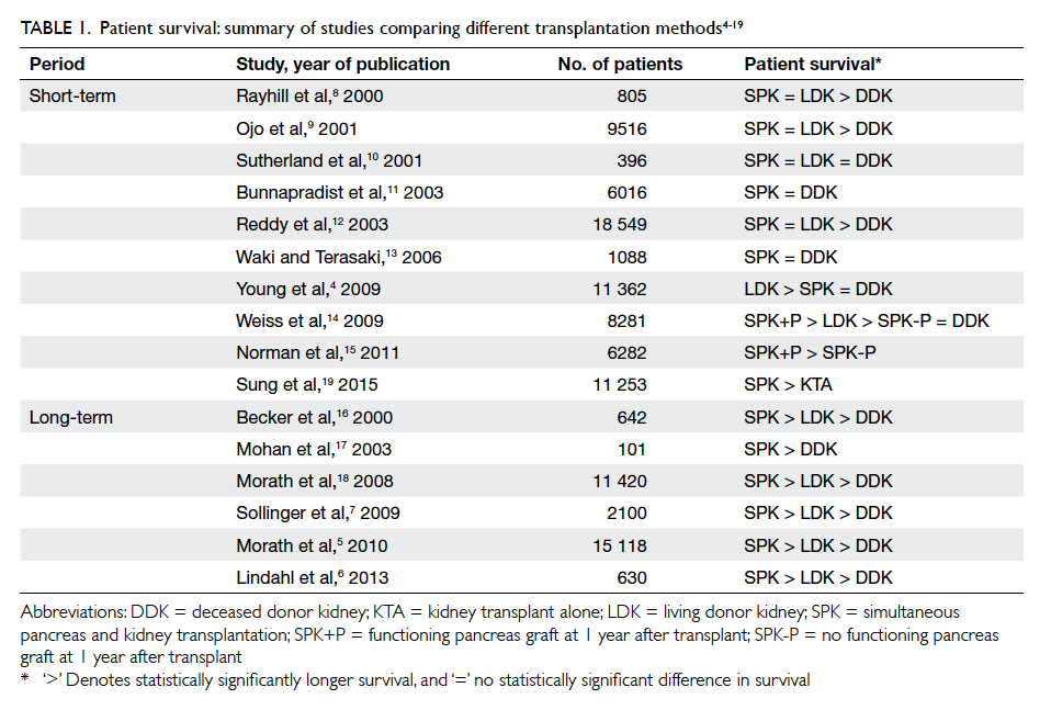
 

Table 1. Patient survival: summary of studies comparing different transplantation methods4 5 6 7 8 9 10 11 12 13 14 15 16 17 18 19
 
Graft survival
The assessment of graft survival in SPK includes that of the kidney and the pancreas. For kidney graft survival, the review by Lindahl et al3 found similar short-term (up to 10 years) kidney graft survival rates following SPK and LDKA; graft survival after DDKA was inferior. Long-term results (>10 years) showed that DDKA was inferior to both SPK and LDKA, and most long-term studies agreed that SPK was equivalent to LDKA, with the exception of a study by Morath et al5 that showed SPK to be superior to LDKA. As such, the current view is that SPK is at least non-inferior to LDKA in terms of long-term kidney graft survival, even though most SPK in the literature reviewed were from cadaver donors (Table 24 5 6 8 10 11 12 13 14 15 16 17 18 19 20).
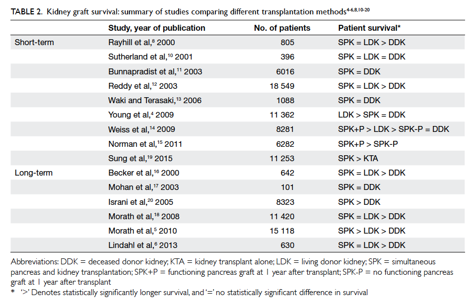
 

Table 2. Kidney graft survival: summary of studies comparing different transplantation methods4 5 6 8 10 11 12 13 14 15 16 17 18 19 20
 
For pancreas graft survival, the results from SPK have improved significantly over the past years due to improved surgical techniques and immunosuppressive regimens.3 Surgical techniques have evolved from the use of pancreatic duct occlusion (1983-1987) to that of exocrine drainage into the urinary bladder (1988-1999), and later to that of direct drainage into the proximal jejunum (2000 onwards). With the latter, the mean pancreas graft survival rates after 1 and 5 years have been reported to be 87% and 75%, respectively.3
 
Diabetic complications
In the present context of transplantation therapy, the important complications of T1DM include diabetic nephropathy, diabetic neuropathy, and an increased risk of cardiovascular diseases. For diabetic nephropathy, several studies have included kidney biopsy in graft assessment following SPK, so as to capture the early diabetes-related changes that might otherwise take time to manifest clinically. The common diabetes-related changes under electron microscopy include thickening of the glomerular basement membrane (GBM) and an increase in mesangial volume. The study by Bohman et al21 was the first to perform kidney biopsy in two SPK patients and six KTA patients. Diabetes-related changes were seen in five of the six KTA recipients but not in any of the SPK recipients. Wilczek et al22 included a larger sample size (20 SPK vs 30 KTA) with a mean postoperative biopsy time of 1 to 6.8 years. The associated changes under light and electron microscopy were significantly fewer in the SPK group than the KTA group. Bilous et al23 biopsied 12 PAK patients before and at least 1.9 years after the pancreas transplant, and found no glomerular disease progression. They also compared the 12 PAK with 13 KTA patients, and found lower mesangial volume in the PAK group. Although most studies showed that the outcomes would be better in patients with SPK relative to those with KTA, whether the pancreas graft can halt nephropathy progression remains controversial. Nyberg et al24 biopsied 11 SPK patients 2 to 4 years postoperatively, and found a mean increase in GBM thickness when compared with normal controls. While recurrence of diabetic nephropathy is still possible in the long run, current evidence nonetheless supports that SPK can at least delay the progression of diabetic nephropathy in comparison with KTA.
 
Apart from its effect on the kidney, a pancreas transplant in SPK may potentially improve other organ systems that can be affected by diabetes. Diabetic neuropathy is an example. Navarro et al25 compared 115 pancreas recipients with 92 patients prescribed standard insulin therapy. Neurological status was assessed by clinical examination, nerve conduction studies, and autonomic function test. Results up to 10 years showed significant improvement in nerve conduction studies and slight improvement on clinical examination and autonomic index in the SPK group. We proposed that pancreas transplantation could potentially halt the progression of diabetic neuropathy and may even lead to a degree of neurological improvement.
 
Besides microvascular complications, macrovascular complications are a major concern in diabetic patients. Cardiovascular diseases, in particular coronary artery disease, contribute to a significant portion of mortality in T1DM patients. Whether SPK can provide benefit in this respect has been studied. Jukema et al26 observed 32 SPK patients with 26 functioning pancreas grafts and six non-functioning pancreas grafts. Glycaemic control was measured by blood glucose level, and the progression of diffuse and focal coronary atherosclerosis was assessed by coronary angiography. It was found that in the presence of a functioning pancreas graft, glycaemic control was better and progression of coronary atherosclerosis was slower. This might also correlate with a lower risk of cardiovascular death and explain why long-term survival in SPK patients is superior to that of KTA patients.
 
Quality of life
In addition to survival benefit and reduced co-morbidity, improvement in QOL may also represent an important consideration. A functioning pancreas graft can potentially free a patient from the need for self-administered insulin and achieve more stable blood glucose levels.27 28 29 These outcomes may be associated with improvement in QOL. To directly quantify such improvement, some studies have used validated health-related quality of life (HRQOL) questionnaires to evaluate treatment outcomes. The latest cohort study by Martins et al30 compared the HRQOL scores of 126 patients before and after SPK with a follow-up duration of around 5 years. There were improvements in all domains under the Gastrointestinal Quality of Life Index post-transplantation, with a significant visual analogue scale health state improvement from 38% to 84%.30 Assessment by another tool, the EuroQol-5 Dimension questionnaire, also showed improvement in physical function, psychological status, social function, gastro-intestinal complaints, burden of medical treatment as well as the rate of unemployment.30 The majority of available studies have focused mainly on comparison of pre-transplant and post-transplant scores, or on transplanted and non-transplanted patients. Few studies have compared QOL outcomes between patients undergoing SPK and KTA. Sureshkumar et al31 conducted a case-control study involving 27 SPK patients and 27 KTA patients. The authors concluded that SPK patients had a significantly better diabetes-related QOL.
 
Considerations in type 1 versus type 2 diabetes mellitus
Indications (in type 1 versus type 2 diabetes mellitus)
The predominant indication for SPK is T1DM patients with ESRD and adequate cardiac reserve who have no opportunity for a living donor kidney transplantation.32 Currently, most centres will perform SPK mainly for T1DM, less commonly for T2DM patients. Nonetheless there has been pervasive controversy on whether the long-term outcomes of SPK in the two groups actually differ. This has important implications in organ allocation as T2DM is much more prevalent than T1DM. Several factors have to be considered. First, there are differences in pathogenesis between the two conditions—T2DM is attributed to insulin resistance in addition to insulin secretion defect; in T1DM, the pathogenesis involves the auto-destruction of islet cells, causing absolute insulin deficiency. This would theoretically render pancreas transplantation less efficacious in T2DM. Second, there is as yet no consensus on the distinction between the two conditions. For example, C-peptide, which is used in patient selection for SPK between T1DM and T2DM patients, has been shown by some studies to be unreliable in determining the outcomes of SPK.33 Third, it should be noted that there exists major differences between the two groups of diabetic patients that make meaningful comparison difficult. These include disproportionate sample sizes; the presence of confounders such as age, obesity, co-morbidities; and duration and treatment of the underlying diabetes.
 
Notwithstanding, there has been an increasing amount of evidence showing comparable results of SPK in selected T2DM and T1DM patients.34 In a large study using the data obtained from the Organ Procurement and Transplant Network/United Network for Organ Sharing (OPTN/UNOS) between 2000 and 2007, Sampaio et al35 showed no significant difference in 5-year survival rate between T1DM and T2DM recipients despite the fact that the latter group had a higher risk of death due to older age and longer pre-transplant dialysis time. In the same study, the 5-year pancreas graft survival in T2DM patients (69.8%) was comparable with that in T1DM patients (72.4%); an inferior 5-year kidney graft survival was found (77.8% vs 73.5%; P=0.007).35 After adjusting for other potential risk factors (eg time on dialysis, obesity), however, diabetes type was not identified as an independent prognostic factor.35
 
Concerning the role of C-peptide in defining T1DM and T2DM during patient selection, Stratta et al36 stratified 162 SPK recipients according to pre-transplant C-peptide levels into C-peptide ‘positive’ (≥2.0 ng/mL; n=30) and C-peptide ‘negative’ (<2.0 ng/mL; n=132) groups. With a mean follow-up duration of 5.6 years, the two groups showed no statistically significant differences in pancreas graft, kidney graft, or patient survival.36 In a similar study involving 80 SPK recipients, 10 were classified as T2DM and 70 as T1DM.37 On Cox regression survival analyses, no statistically significant difference in graft and patient survival was found between the two groups.37 The authors concluded that selected T2DM patients with ESRD should be considered potential candidates for SPK, and that the use of C-peptide as the predominant marker of the diabetes type was unreliable and potentially misleading.37
 
Simultaneous pancreas and kidney transplantation versus other transplant options in type 2 diabetes mellitus patients
At present, there is insufficient evidence to support the use of SPK over other kidney transplant options in patients with T2DM. In an early study that compared the outcomes of SPK, DDKA, and LDKA in patients with T2DM, Wiseman and Gralla38 concluded that both patient and graft survival rates were superior with LDKA transplantation, whereas patient but not graft survival rate was higher in SPK versus DDKA transplantation. After multivariable analysis, the survival advantage of SPK over DDKA was related not so much to the pancreas transplantation but other variables such as younger donor and recipient ages in the SPK cohort (Table 338). These findings, however, should not completely dismiss the consideration of SPK for selected T2DM patients who have little prospect for LDKA since other outcome measures such as the benefits of euglycaemia in terms of better QOL and secondary complications of diabetes will also have to be considered. Nonetheless, given the superior long-term outcome of SPK over LDKA in T1DM patients and that the same has yet to be demonstrated in T2DM patients, T1DM patients should still be given a stronger allocation priority of SPK grafts.3
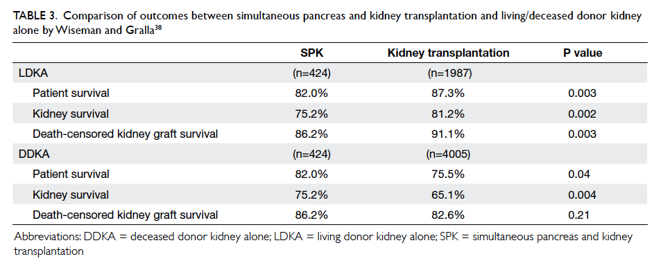
 

Table 3. Comparison of outcomes between simultaneous pancreas and kidney transplantation and living/deceased donor kidney alone by Wiseman and Gralla38
 
Complications of simultaneous pancreas and kidney transplantation
Most complications of SPK are related to the transplanted pancreas.39 Repeated laparotomy may be required in up to 50% of patients.40 41 The pancreatic graft survival at 1 year may range from 74% to 88%, with the sharpest drop during the first year.7 39 42 The reported 10-year and 20-year survival rates were 63% and 36%, respectively.7 Most complications occur within the first 60 days of operation and include graft pancreatitis (3%-12%), infection/abscess (1%-5%), focal/diffuse necrosis (12%), graft-vessel thrombosis (6%-17%), anastomotic leak (0.5%-2%), and intra-abdominal haemorrhage (0%-0.5%).39 40 41 42 43 Venous thrombosis has been reported to be the most common cause of graft failure; graft pancreatitis and focal/diffuse necrosis were the most common causes of mortality among graft-specific complications.39 40
 
Complications related to the transplanted kidney include acute tubular necrosis (ATN) or graft rejection, urinary complications, infection, and vascular thrombosis. According to a study of 112 SPK recipients by Grochowiecki et al,44 ATN and rejection were the most frequent (43.4%) causes leading to the loss of kidney graft function. Infections (28.6%) and vascular thrombosis due to atherosclerosis of the iliac arteries (28.6%) were the most common reasons for graft nephrectomy.44 The most severe complications were due to fungal infection.44 Overall, the 1-year survival rate for the kidney graft was over 90%.7 44 45 The 10-year and 20-year kidney survival rates were 63% and 38%, respectively.7 In terms of overall mortality, the most common causes following SPK have been reported to be cardiopulmonary (7.2%), followed by infection (3.4%), stroke (1.8%), and renal failure (1.5%). Patient survival rates at 1, 10, and 20 years were 97%, 80%, and 58%, respectively.7
 
Comparison of the complications of SPK versus KTA has revealed that SPK has a lower rate of ATN (8.9%)44 than KTA (15.3%),46 but a slightly higher rate of urological complications (4.5%)44 than KTA (3.7%).47 The incidence of vascular complications was comparable in SPK (1.8%)44 and KTA (0.5%-4%).48 49 50 As mentioned above, SPK has higher long-term patient and kidney survival rates than LDKA/DDKA.
 
Future directions and local applicability of simultaneous pancreas and kidney transplantation
Simultaneous pancreas and kidney transplantation has become an established treatment for patients with T1DM complicated by ESRD. The results have, so far, been promising. Furthermore, apart from SPK, newer techniques including pancreatic islet cell transplantation, and different combinations of living and/or deceased donor pancreas, islet cell and kidney graft transplantation are being evaluated. Current research aims to extend these techniques, still predominantly SPK, to the treatment of T2DM, for which LDKA remains the first-line treatment option. In situations where LDKA is not available for T2DM patients, DDKA remains the next best option. Regarding the ethical issues about graft allocation, the allocation of DDKA is based on an allocation system that takes account of patient age and waiting time as well as the degree of human leukocyte antigen (HLA) matching between the potential donor and the recipient.51 Difficulties may arise, however, when a deceased donor with both kidney and a pancreas graft has become available. Should the priority for these grafts be given to a patient on the SPK waiting list or a non–diabetes-related ESRD patient on the DDKA waiting list? And should the priority of SPK be given to a T2DM patient who is higher up on the allocation system (with a younger age, a longer waiting time, or lesser degree of HLA mismatch) or to a T1DM patient who is lower on the allocation system but is more likely to achieve a better outcome? The related ethical issues clearly deserve an informed discourse within the surgical community.
 
Simultaneous pancreas and kidney transplantation has not yet been performed in this locality. The main obstacle remains the shortage of cadaver organs. Here, the number of cadaveric renal transplantations performed in the Hospital Authority over the last decade ranges from 44 to 87 cases per year. As of 31 December 2014, the number of patients waiting for transplantation was close to 2000. This translates into significantly prolonged dialysis time for this specific group of patients, and hence a potentially suboptimal outcome after SPK. The implementation of SPK would not be readily feasible unless there is a significant improvement in organ availability.
 
Conclusions
Simultaneous pancreas and kidney transplantation has become a standard treatment worldwide for patients with T1DM and ESRD. There is a large volume of clinical evidence supporting good outcomes in patient survival, graft survival, diabetic complications, and QOL. For T2DM, the efficacy and application of the procedure remain controversial but the outcomes are possibly comparable to that in T1DM. Simultaneous pancreas and kidney transplantation is a technically demanding procedure that is associated with significant complications, and should be undertaken only in carefully selected patients. It should be regarded as a ‘last resort’ treatment for patients in whom diabetic complications have become life-threatening or severely burdensome despite best efforts in maintaining good diabetic control through lifestyle modification and medications. Continued efforts in patient education and the promotion of an altruistic culture of organ donation among the public are critical for the implementation of this treatment paradigm in this locality.
 
References
1. Redfield RR, Scalea JR, Odorico JS. Simultaneous pancreas and kidney transplantation: current trends and future directions. Curr Opin Organ Transplant 2015;20:94-102. Crossref
2. Gruessner AC. 2011 Update on pancreas transplantation: comprehensive trend analysis of 25,000 cases followed up over the course of twenty-four years at the International Pancreas Transplant Registry (IPTR). Rev Diabet Stud 2011;8:6-16. Crossref
3. Lindahl JP, Jenssen T, Hartmann A. Long-term outcomes after organ transplantation in diabetic end-stage renal disease. Diabetes Res Clin Pract 2014;105:14-21. Crossref
4. Young BY, Gill J, Huang E, et al. Living donor kidney versus simultaneous pancreas-kidney transplant in type I diabetics: an analysis of the OPTN/UNOS database. Clin J Am Soc Nephrol 2009;4:845-52. Crossref
5. Morath C, Zeier M, Döhler B, et al. Transplantation of the type 1 diabetic patient: the long-term benefit of a functioning pancreas allograft. Clin J Am Soc Nephrol 2010;5:549-52. Crossref
6. Lindahl JP, Hartmann A, Horneland R, et al. Improved patient survival with simultaneous pancreas and kidney transplantation in recipients with diabetic end-stage renal disease. Diabetologia 2013;56:1364-71. Crossref
7. Sollinger HW, Odorico JS, Becker YT, D’Alessandro AM, Pirsch JD. One thousand simultaneous pancreas-kidney transplants at a single center with 22-year follow-up. Ann Surg 2009;250:618-30. Crossref
8. Rayhill SC, D’Alessandro AM, Odorico JS, et al. Simultaneous pancreas-kidney transplantation and living related donor renal transplantation in patients with diabetes: is there a difference in survival? Ann Surg 2000;231:417-23. Crossref
9. Ojo AO, Meier-Kriesche HU, Hanson JA, et al. The impact of simultaneous pancreas-kidney transplantation on long-term patient survival. Transplantation 2001;71:82-90. Crossref
10. Sutherland DE, Gruessner RW, Dunn DL, et al. Lessons learned from more than 1,000 pancreas transplants at a single institution. Ann Surg 2001;233:463-501. Crossref
11. Bunnapradist S, Cho YW, Cecka JM, Wilkinson A, Danovitch GM. Kidney allograft and patient survival in type I diabetic recipients of cadaveric kidney alone versus simultaneous pancreas kidney transplants: a multivariate analysis of the UNOS database. J Am Soc Nephrol 2003;14:208-13. Crossref
12. Reddy KS, Stablein D, Taranto S, et al. Long-term survival following simultaneous kidney-pancreas transplantation versus kidney transplantation alone in patients with type 1 diabetes mellitus and renal failure. Am J Kidney Dis 2003;41:464-70. Crossref
13. Waki K, Terasaki PI. Kidney graft and patient survival with and without a simultaneous pancreas utilizing contralateral kidneys from the same donor. Diabetes Care 2006;29:1670-2. Crossref
14. Weiss AS, Smits G, Wiseman AC. Twelve-month pancreas graft function significantly influences survival following simultaneous pancreas-kidney transplantation. Clin J Am Soc Nephrol 2009;4:988-95. Crossref
15. Norman SP, Kommareddi M, Ojo AO, Luan FL. Early pancreas graft failure is associated with inferior late clinical outcomes after simultaneous kidney-pancreas transplantation. Transplantation 2011;92:796-801. Crossref
16. Becker BN, Brazy PC, Becker YT, et al. Simultaneous pancreas-kidney transplantation reduces excess mortality in type 1 diabetic patients with end-stage renal disease. Kidney Int 2000;57:2129-35. Crossref
17. Mohan P, Safi K, Little DM, et al. Improved patient survival in recipients of simultaneous pancreas-kidney transplant compared with kidney transplant alone in patients with type 1 diabetes mellitus and end-stage renal disease. Br J Surg 2003;90:1137-41. Crossref
18. Morath C, Zeier M, Döhler B, Schmidt J, Nawroth PP, Opelz G. Metabolic control improves long-term renal allograft and patient survival in type 1 diabetes. J Am Soc Nephrol 2008;19:1557-63. Crossref
19. Sung RS, Zhang M, Schaubel DE, Shu X, Magee JC. A reassessment of the survival advantage of simultaneous kidney-pancreas versus kidney-alone transplantation. Transplantation 2015;99:1900-6. Crossref
20. Israni AK, Feldman HI, Propert KJ, Leonard M, Mange KC. Impact of simultaneous kidney-pancreas transplant and timing of transplant on kidney allograft survival. Am J Transplant 2005;5:374-82. Crossref
21. Bohman SO, Tydén G, Wilczek H, et al. Prevention of kidney graft diabetic nephropathy by pancreas transplantation in man. Diabetes 1985;34:306-8. Crossref
22. Wilczek HE, Jaremko G, Tydén G, Groth CG. Evolution of diabetic nephropathy in kidney grafts. Evidence that a simultaneously transplanted pancreas exerts a protective effect. Transplantation 1995;59:51-7. Crossref
23. Bilous RW, Mauer SM, Sutherland DE, Najarian JS, Goetz FC, Steffes MW. The effects of pancreas transplantation on the glomerular structure of renal allografts in patients with insulin-dependent diabetes. N Engl J Med 1989;321:80-5. Crossref
24. Nyberg G, Holdaas H, Brekke IB, et al. Glomerular ultrastructure in kidneys transplanted simultaneously with a segmental pancreas to patients with type 1 diabetes. Nephrol Dial Transplant 1996;11:1029-33. Crossref
25. Navarro X, Sutherland DE, Kennedy WR. Long-term effects of pancreatic transplantation on diabetic neuropathy. Ann Neurol 1997;42:727-36. Crossref
26. Jukema JW, Smets YF, van der Pijl JW, et al. Impact of simultaneous pancreas and kidney transplantation on progression of coronary atherosclerosis in patients with end-stage renal failure due to type 1 diabetes. Diabetes Care 2002;25:906-11. Crossref
27. Robertson RP, Sutherland DE, Kendall DM, Teuscher AU, Gruessner RW, Gruessner A. Metabolic characterization of long-term successful pancreas transplants in type I diabetes. J Investig Med 1996;44:549-55.
28. Robertson RP, Holohan TV, Genuth S. Therapeutic controversy: Pancreas transplantation for type I diabetes. J Clin Endocrinol Metab 1998;83:1868-74. Crossref
29. Drognitz O, Benz S, Pfeffer F, et al. Long-term follow-up of 78 simultaneous pancreas-kidney transplants at a single-center institution in Europe. Transplantation 2004;78:1802-8. Crossref
30. Martins LS, Outerelo C, Malheiro J, et al. Health-related quality of life may improve after transplantation in pancreas-kidney recipients. Clin Transplant 2015;29:242-51. Crossref
31. Sureshkumar KK, Mubin T, Mikhael N, Kashif MA, Nghiem DD, Marcus RJ. Assessment of quality of life after simultaneous pancreas-kidney transplantation. Am J Kidney Dis 2002;39:1300-6. Crossref
32. Larsen JL. Pancreas transplantation: indications and consequences. Endo Rev 2004;25:919-46. Crossref
33. Knight RJ, Lawless A, Patel SJ, Gaber AO. Simultaneous kidney-pancreas transplantation for end-stage renal disease patients with insulin-dependent diabetes and detectable C-peptide. Transplant Proc 2010;42:4195-6. Crossref
34. Weems P, Cooper M. Pancreas transplantation in type II diabetes mellitus. World J Transplant 2014;4:216-21. Crossref
35. Sampaio MS, Kuo HT, Bunnapradist S. Outcomes of simultaneous pancreas-kidney transplantation in type 2 diabetic recipients. Clin J Am Soc Nephrol 2011;6:1198-206. Crossref
36. Stratta RJ, Rogers J, Farney AC, et al. Pancreas transplantation in C-peptide positive patients: does “type” of diabetes really matter? J Am Coll Surg 2015;220:716-27. Crossref
37. Chakkera HA, Bodner JK, Heilman RL, et al. Outcomes after simultaneous pancreas and kidney transplantation and the discriminative ability of the C-peptide measurement pretransplant among type 1 and type 2 diabetes mellitus. Transplant Proc 2010;42:2650-2. Crossref
38. Wiseman AC, Gralla J. Simultaneous pancreas kidney transplant versus other kidney transplant options in patients with type 2 diabetes. Clin J Am Soc Nephrol 2012;7:656-64. Crossref
39. Grochowiecki T, Gałązka Z, Madej K, et al. Surgical complications related to transplanted pancreas after simultaneous pancreas and kidney transplantation. Transplant Proc 2014;46:2818-21. Crossref
40. Sansalone CV, Maione G, Aseni P, et al. Surgical complications are the main cause of pancreatic allograft loss in pancreas-kidney transplant recipients. Transplant Proc 2005;37:2651-3. Crossref
41. Michalak G, Kwiatkowski A, Czerwinski J, et al. Surgical complications of simultaneous pancreas-kidney transplantation: a 16-year-experience at one center. Transplant Proc 2005;37:3555-7. Crossref
42. Gruessner AC, Sutherland DE, Gruessner RW. Pancreas transplantation in the United States: a review. Curr Opin Organ Transplant 2010;15:93-101. Crossref
43. Malaise J, Steurer W, Koenigsrainer A, et al. Simultaneous pancreas-kidney transplantation in a large multicenter study: surgical complications. Transplant Proc 2005;37:2859-60. Crossref
44. Grochowiecki T, Gałązka Z, Madej K, et al. Early complications related to the transplanted kidney after simultaneous pancreas and kidney transplantation. Transplant Proc 2014;46:2815-7. Crossref
45. Campos Hernández JP, Gómez Gómez E, Carrasco Valiente J, et al. Influence of surgical complications on kidney graft survival in recipients of simultaneous pancreas kidney transplantation. Transplant Proc 2015;47:112-6. Crossref
46. Ounissi M, Gargah T, Barbouch S, et al. Acute tubular necrosis in kidney transplantation [in French]. Tunis Med 2012;90:463-7.
47. Samhan M, Al-Mousawi M, Hayati H, Abdulhalim M, Nampoory MR. Urologic complications after renal transplantation. Transplant Proc 2005;37:3075-6. Crossref
48. Renoult E, Amicabile C, Jonon B, Kessler M, L’Hermite J. Early and recurrent venous graft thrombosis after kidney transplantation: benefit of an early surgery. Clin Nephrol 1990;34:236-7.
49. Bretan PN, Burke EC. Renal transplantation. In: Tanagho EA, Mc Annich JW, editors. Smith’s general urology. London: Appleton and Lange; 1995: 612-24.
50. Kribs SW, Rankin RN. Doppler ultrasonography after renal transplantation: value of reversed diastolic flow in diagnosing renal vein obstruction. Can Assoc Radiol J 1993;44:434-8.
51. Hawkins BR. A point score system for allocating cadaveric kidneys for transplantation based on patient age, waiting time and HLA match. Hong Kong J Nephrol 2004;2:79-83. Crossref

Use of insulin in diabetes: a century of treatment

Hong Kong Med J 2015 Dec;21(6):553–9 | Epub 6 Nov 2015
DOI: 10.12809/hkmj154557
© Hong Kong Academy of Medicine. CC BY-NC-ND 4.0
 
REVIEW ARTICLE
Use of insulin in diabetes: a century of treatment
Savita Shahani, MD1; Lokesh Shahani, MD2
1 Department of Pharmacology, MGM Medical College Mumbai (India), India
2 Department of Internal Medicine, Baylor College of Medicine, Houston, Texas, United States
 
Corresponding author: Dr Savita Shahani (drshahani@rediffmail.com)
 
 Full paper in PDF
Abstract
Insulin is a key player in the control of hyperglycaemia for patients with type 1 diabetes mellitus and selected patients with type 2 diabetes mellitus. There have been many advances in insulin drug delivery from its first administration as a crude pancreatic extract till today. The traditional and most predictable method for administration of insulin is by subcutaneous injection. Currently available insulin delivery systems include insulin syringes, infusion pumps, jet injectors, and pens. The major drawback of insulin therapy is its invasive nature. Non-invasive delivery of insulin has long been a major goal for the treatment of diabetes mellitus. Although there have been improvements in insulin therapy since it was first conceived, it is still far from mimicking the physiological secretion of pancreatic β-cells, and research to find new insulin formulations and new routes of administration continues. This article reviews the emerging technologies, including insulin inhalers, insulin buccal spray, insulin pill, islet cell transplant, and stem cell therapy, as treatment options for diabetes mellitus.
 
 
Introduction
Diabetes mellitus is a major public health concern worldwide. There is predicted to be an alarming increase in the population with type 2 diabetes mellitus both in developed and developing countries over the next two decades. The prevalence of diabetes among adults aged 20 to 70 years is expected to rise from 285 million in 2010 to 438 million by the year 2030.1 Prevalences of diabetes and impaired glucose tolerance are high in all Asian countries and are expected to increase further in the next 20 years. The present trend indicates that more than 60% of the world’s diabetic population will be in Asia.2 The prevalence of type 2 diabetes is particularly high in Asian Indians because of high genetic susceptibility and enhanced interaction with environmental triggers. Exposure to a high fat diet and low levels of physical activity are factors that can trigger the gene-environment interaction.2 Therapy with insulin is effective at lowering blood glucose in patients with diabetes. Insulin is a key treatment in the control of type 1 diabetes and it is required in the later stages by patients with type 2 diabetes mellitus; hyperglycaemia in type 1 diabetes is a result of insulin deficiency and, in type 2 diabetes, it is due to both impaired tissue response to insulin and insulin deficiency. The discovery of insulin has been hailed as one of the most dramatic events in the history of the treatment of diabetes.
 
Before the discovery of insulin, diabetes was a feared disease that led to death. Correlation of destruction of the pancreas with diabetes was observed in 1890 by von Mering and Minkowski,3 but internal secretion from the pancreas being responsible for control of sugar was not identified. In fact, the name ‘insulin’ was derived from the Latin word ‘insula’ (meaning ‘island’) much earlier than insulin was isolated.
 
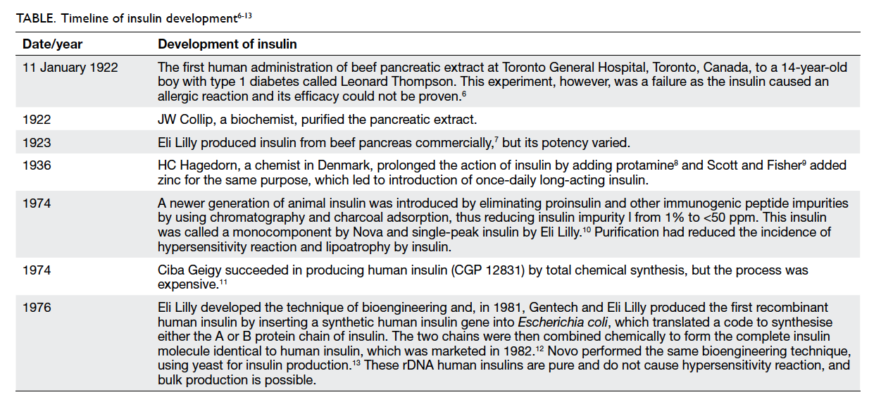
As described by Bliss,4 between 1914 and 1916, Paulesco and Zuelzer performed studies on dogs with experimentally induced diabetes, showing the antidiabetic effect of extract from the pancreas, but they had to give up their experiments due to lack of funds and the publication of their work was delayed until July 1921. In 1922, Banting and Best5 confirmed the antidiabetic effect of pancreatic extract in dogs with experimentally induced diabetes. To carry on with the experiments in a larger number of animals, a substantial quantity of insulin was needed so pancreatic extract from cows started being used. The timeline of insulin development is shown in the Table.6 7 8 9 10 11 12 13
 

Table. Timeline of insulin development6 7 8 9 10 11 12 13
 
Alternative devices
At this stage (1984), insulin was only available in vials and needed to be administered subcutaneously via a syringe and needle, making it possible to mix different types of insulin preparations. Patients, however, found it inconvenient to administer and there were mistakes in dosage measurement. Therefore, alternative devices were developed to improve drug delivery of insulin.
 
Insulin pen
The insulin pen combines an insulin container and syringe in a single modular unit that makes it convenient to administer insulin because of the ease of insulin cartridge replacement. The insulin pen has an inbuilt dial system that allows administration of an accurate amount of drug. Pens are available as a single premixed insulin administration unit.14
 
Insulin jet injector
The insulin jet injector has been designed to deliver a fine spray of insulin subcutaneously at high speed using a pressurised jet of air instead of a needle. The dose is controlled by the dial-a-dose operation through a single component design in comparison to a conventional multi-component syringe. However, the jet injector may cause bruising of the skin as well as having altered absorption levels. The size and cost of the jet injector further limit its routine use. This device can be considered for patients with needle phobia or those with severe insulin-induced lipoatrophy.15
 
Injection port
The injection port is another device that functions as a delivery channel directly into subcutaneous tissue. The injection port contains an insertion needle guiding a soft cannula into subcutaneous tissue. Once applied, the insertion needle is removed and a soft cannula remains in the subcutaneous tissue, acting as gateway through which insulin can be administered via a syringe or pen, thus avoiding multiple pricks. The injection port can be changed every third day. The device is available from Medtronic (Dublin, Ireland).16
 
External insulin pump
The external insulin pump is a small battery-operated device that is made up of an insulin reservoir connected to a tube ending in a cannula that is inserted under the skin of the abdomen and set to deliver continuous subcutaneous insulin infusion (CSII) throughout the day, and is programmed to deliver insulin in a larger quantity at meal times. This release pattern simulates physiological insulin secretion, but bypasses the liver. Such CSII release can be adjusted according to the specific needs of a patient. Insulin delivery, however, may be interrupted by infusion malfunction, needle displacement, pump dysfunction, and lack of insulin in the reservoir, therefore frequent blood glucose estimation is required.17
 
Implantable insulin pump
An implantable insulin pump is a combination of a continuous glucose sensor attached to a closed-loop insulin infusion pump. The device is also known as an artificial pancreas. Blood glucose control is achieved by using wireless communication of a continuous glucose monitor linked to an insulin infusion pump that facilitates automated data transfer and delivers insulin subcutaneously without the need for human intervention.18 The lag period of human insulin given subcutaneously by a pump is 60 to 90 minutes, which can be minimised by using newer fast-acting insulin analogues. Despite important developments in sensor and pump technology, this device has shown delays and inaccuracies in both glucose sensing and insulin delivery, which is a problem when controlling postprandial hyperglycaemia that occurs substantially faster than the time needed for insulin absorption and action. Suboptimal accuracy and reliability remain one of the biggest obstacles for closed-loop systems. Despite the substantial progress made in recent years, there remains a number of challenges to successful development of commercial implantable insulin pump devices. These challenges include the effect of exercise, concurrent illness, large carbohydrate meals, and the pharmacokinetics of current subcutaneous insulin.
 
Quick-acting insulin has a lag period of 60 to 90 minutes, therefore, it is required to be administered 30 minutes before meals to control postprandial hyperglycaemia and may cause late postprandial hypoglycaemia. To overcome this problem, rapid-acting insulin analogues such as aspart, lispro, and glulisine were synthesised by modifying amino acid sequences in the insulin chain so as to keep insulin in a monomeric form, which has a rapid onset of action of 10 to 15 minutes, peak of 30 to 90 minutes, and duration of 3 to 4 hours resembling physiological postprandial insulin secretion. Thus, these analogues are very efficient at controlling postprandial hyperglycaemia without the risk of delayed hypoglycaemia.19 Research has been done to enhance the onset of action of human insulin by combining it with human hyaluronidase.20
 
One approach to creating ultrarapid-acting insulin is use of a novel combination of excipients to modify the insulin hexamer complex resulting in more rapid dissociation of the hexamers into monomers and dimers following subcutaneous injection. Biodel Inc (Danbury [CT], US) has developed a technology that facilitates more rapid absorption of recombinant human insulin than that of current insulin products. Biodel is developing Linjeta (previously known as VIAject), an ultrarapid-acting, injectable recombinant human insulin formulation, which is currently under review by the US Food and Drug Administration (FDA) to compare its pharmacokinetic and pharmacodynamic characteristics with rapidly acting insulin analogues.21 BIOD-531 (Biodel Inc) has a more rapid onset and longer duration of action than combined prandial/basal insulins. The preclinical and clinical data demonstrate its unique and attractive pharmacokinetic and pharmacodynamic profiles.22 This formulation of recombinant human insulin has the potential to provide improved glucose control compared with insulin products designed to provide both prandial and basal coverage in a single injection. As BIOD-531 is a concentrated formulation (400 units/mL), the degree of glucose control can be achieved with a small volume of injection.
 
Long-acting insulin analogues were designed to obtain a steady basal insulin level compared with older intermediate-acting insulin, which has a risk of late-night hypoglycaemia. Insulin glargine and insulin detemir were designed by altering amino acid sequences in the human insulin chain to make a slow-release preparation when administered subcutaneously. Insulin degludec is another long-acting insulin approved by the US FDA in 2012 for basal blood sugar control. All these long-acting peakless insulins are given once a day to control basal blood sugar level without risk of producing hypoglycaemia.23 PEGylated insulin lispro (LY2605541) is a long-acting insulin whereby the insulin molecule is embedded in a polyethylene glycol (PEG) chain to increase the molecular size of the insulin, thus reducing its rate of absorption.24 LY2605541 has completed phase 2 clinical trials.
 
Newer trends in needle-free insulin delivery systems
None of the above formulations delivered insulin by needle-free technique, thus there is a continuing search for a novel insulin delivery to overcome the problem of needle prick. The newer trends for a needle-free insulin delivery system are discussed as follows.
 
Insulin inhaler
Owing to the large surface area, the lung is an ideal target for drug delivery, and inhaled insulin represents one of the most promising alternatives to injectable insulin. Insulin has been developed in powdered form. Exubera (Pfizer Inc, New York [NY], US) was the first inhalable insulin utilising recombinant human insulin. Exubera was approved by US FDA in 2006 for both type 1 and type 2 diabetics. As the inhaler contains short-acting insulin, it can only control postprandial hyperglycaemia. The powdered form of insulin can sometimes stick together making it difficult to inhale and reducing accuracy of the dose. A drawback of Exubera was that it was a bulky inhaler device and insulin was available in a blister packet that had to be loaded into the inhaler device for each dose.25 Since insulin is known to have growth-promoting properties by acting on insulin-like growth factor receptors, clinicians have been concerned about the possibility of long-term effects of intra-alveolar deposition of insulin, although safety data collected by Pfizer did not show any significant increase in the incidence of lung malignancy in clinical trials.26 A systematic review showed that Exubera was not superior to short-acting insulin in other formats and was not cost-effective, so Pfizer discontinued production.27 MannKind Corporation (Valencia [CA], US) received US FDA approval in June 2014 for its ultrarapid-acting inhalation human insulin powder, Afrezza, which contains recombinant human insulin using the technosphere concept and is administered via MannKind’s next-generation inhaler called Dreamboat. Technosphere technology is based on the pH-induced intermolecular self-assembly of a novel small-molecule excipient (fumaryl diketopiperazine). The technosphere drug delivery system creates insulin microparticles (2-3 µm), which form microspheres that are lyophilised into dry powder for inhalation, and dissolve immediately once they come in contact with alveoli. The peak plasma concentration is achieved at 12 to 15 minutes,28 resembling physiological postprandial insulin release, and thus it is required to be administered just before meals, and controls postprandial hyperglycaemia only. Afrezza was approved by the US FDA for both type 1 and type 2 diabetes, with a label restriction for patients with asthma, chronic obstructive pulmonary disease, or lung cancer.
 
Mouth spray and adhesives
The buccal route is another promising alternative for insulin delivery as this area has an abundant blood supply, thus offering the possibility of delivery of acid-labile insulin without undergoing first-pass metabolism. Spray insulin preparations deliver insulin in aerosol form, which is absorbed through the inside of the cheek and the back of the mouth. Generex Biotechnology Corporation (Toronto, Canada) developed the buccal insulin formulation Oral-lyn, which is a liquid formulation of regular human insulin with a spray propellant using rapid mist technology. Oral-lyn has an onset time of 5 minutes, with a peak of 30 minutes and duration of 2 hours, and can be used to control postprandial hyperglycaemia, having only 10% drug absorption.29 This formulation releases large micelles with a particle size of >10 µm, so insulin does not reach the lungs. The US FDA approved Oral-lyn for type 1 and type 2 diabetes in 2009.
 
MidaSol Therapeutics (Oxford, UK), a joint venture between nanotechnology firm Midatech Pharma (Abingdon, UK) and drug delivery specialist MonoSol Rx (Warren [NJ], US), has cleared its first clinical hurdle in its bid to develop a gold-based nanoparticle formulation of insulin that offers a novel delivery route. A phase 1 trial of MidaForm insulin, which is administered in a soluble strip that adheres to the inside of the mouth, demonstrated good safety and tolerability. The strip creates an osmotic gradient across the buccal mucosa resulting in rapid systemic delivery of insulin, with a peak plasma concentration time of 5 to 8 minutes.30
 
Transdermal insulin
Transdermal insulin delivery could provide diabetic patients with a sustained physiological level of basal insulin in a pain-free manner. Dermal permeation is limited to small lipophilic molecules, as the stratum corneum is the major barrier to penetration. Several physical enhancement techniques such as iontophoresis, ultrasound, microneedles, electroporation, lesser ablation, and chemical enhancement have been explored to increase the permeability of transdermal drugs.31 The U-Strip (Ultrasonic Strip) Insulin Patch (Transdermal Specialties, Inc, Broomall [PA], US) was designed to deliver insulin lispro through the dermis using alternating sonic waveforms to enlarge the diameter of skin pores, enabling large-diameter molecules to penetrate the stratum corneum.32 The U-Strip is at an advanced stage of clinical trial, but is not yet approved by the US FDA.
 
Oral insulin
The oral route of administration is considered to be most acceptable and convenient for treatment of chronic diseases. The concept of oral insulin delivery has always been a challenge as the insulin has to be protected from the acidic environment of stomach and various metabolising enzymes, and has poor permeability due to its hydrophilicity. Oral insulin delivery has the advantage that it delivers the drug through the portal circulation, thus distributing a high concentration in the liver resembling physiological insulin secretion. Various attempts have been made to overcome the obstacles of oral insulin therapy. Insulin has been complexed with cyclodextrins in order to improve its solubility and stability in the form of dry powder, after encapsulation into poly(D,L-lactic-co-glycolic acid) microspheres. Other attempts at oral insulin delivery include incorporating insulin with a delivery agent (sodium N-[8-(2-hydroxybenzoyl)amino] caprylate), preparation of hyaluronan-insulin complex and calcium phosphate-PEG-insulin-casein particles for oral delivery.33 The physiological barriers to absorption of oral insulin are its low bioavailability and high inter-patient variability.
 
Biocon Ltd (Bangalore, India) has entered into a research collaboration with Nobex Corporation (Research Triangle Park [NC], US) to jointly develop the oral insulin analogue IN-105. The recombinant human insulin molecule has been modified by linking a single short-chain amphiphilic oligomer, through a covalent non-hydrolysable amide bond, to the free amino acid group on the Lys-β29 residue. This improved the solubility, stability, and systemic absorption.34 From the results published so far,35 it appears that IN-105 is a rapid-acting oral insulin that could potentially have a place in the control of postprandial hyperglycaemia. Biocon is collaborating with Bristol-Myers Squibb (New York [NY], US) for global clinical trials. In India, IN-105 has completed a phase 2 clinical trial with promising results.36
 
Oramed Pharmaceuticals (Jerusalem, Israel) was granted patent approval for its oral insulin technology by the Japan Patent Office in April 2013. The POD (Protein Oral Delivery) technology combines adjuvants that are capable of significantly enhancing the absorption of peptides and proteins across the intestinal wall when delivered orally without modifying the active compounds. These adjuvants are intended to protect the active peptide or protein while in transit through the harsh chemical environment of the gastro-intestinal tract and promote its transport across the intestinal wall into the general blood circulation. The tablet is enteric-coated, thus preventing insulin release in the stomach, and the increased pH of the intestines signals it to open and the technology to start functioning.37 The technology is in phase 2 clinical trial, and preliminary data in patients with type 1 and type 2 diabetes are encouraging.38
 
Another oral insulin analogue under development by Novo Nordisk (Bagsværd, Denmark) incorporates Merrion Pharmaceuticals’ (Wilmington [NC], US) Gastrointestinal Permeation Enhancement Technology. This technology uses specially designed oral formulations of absorption enhancers that activate micelle formation, facilitating transport of drug and substantially increasing absorption, with good reproducibility. The specially coated tablets are targeted to dissolve in the duodenum releasing both drug and absorption enhancer, which may pass through the duodenal cell membrane. Novo Nordisk has completed a phase 1 study of oral insulin NN1952 as well as another molecule, NNC0148-0000-0362 (NN1954), in healthy participants and patients with type 1 and type 2 diabetes.39
 
There is renewed hope for the treatment of type 1 diabetes with gel capsules. The Norwegian University of Science and Technology has developed a new type of capsule called Trondheim Alginate Microcapsule, which is designed to camouflage the insulin-producing cells from the body’s immune system. If this becomes a medical reality, diabetic patients with transplanted insulin-producing donor cells in their abdominal cavity might not have to take immunosuppressants for the rest of their lives.40
 
Pancreas transplantation and stem cell therapy
Intensified exogenous insulin therapy rarely attains normal blood glucose levels without risk of major hypoglycaemic episodes, and cannot approximate normal physiological pulsatile insulin secretary patterns with complete integrity. Pancreas transplantation is the only therapy shown to stop the progression of diabetic complications without increasing the incidence of hypoglycaemic events. Whole pancreas transplantation was first performed for treatment of diabetes in 1966.41 While percutaneous islet cell transplantation is a minimally invasive cellular replacement therapy that was developed to avoid the surgical complications of whole pancreas transplantation,42 both procedures require immunosuppressant therapy.
 
Developments in this fast-moving area of research have focused on the principle of generating insulin-expressing cells from stem cells, with the possibility of generating unlimited numbers of functional beta cells for transplantation therapy. The adoption of a stem cell–based therapy for diabetes, however, will depend on it being shown to be as safe and effective as the current therapy of administration of exogenous insulin.
 
The stem cell populations that have been used in experimental studies can be tissue stem cells, defined as multipotent progenitor cells found in fetal and adult tissues; embryonic stem cells, defined as pluripotent, undifferentiated cells generated from the inner cell mass of a developing blastocyst; or induced pluripotent stem cells, defined as pluripotent cells generated by reprogramming differentiated adult cells by forced expression of pluripotency genes. The pluripotency and proliferative potential of stem cell populations raises the undesirable possibility of uncontrolled cellular proliferation and formation of teratomas after transplantation, which has been reported in animal studies.43 Autologous grafting of insulin-secreting cells derived from the patient’s own tissue stem cells is attractive, but experimental studies have not yet translated into clinically useful material, mainly because of problems with restricted proliferative capacity, causing low levels of insulin. Thus, in spite of the impressive promise of stem cells, no proven benefits have been demonstrated by stem cell therapy in the treatment of diabetes.
 
Xenotransplantation refers to the transplantation of tissue or organs from one species to another. This therapy offers the ability to overcome the problem of transplant organ shortage. At present, pigs are thought to be the best candidates for xenotransplant donation, as they are plentiful, quick to mature, and breed well. Transplantation of porcine islet cells into non-human primates has been successfully performed with encouraging results, including longer graft survival,44 but the benefits of xenotransplantation must be weighed against the potential for interspecies transmission of viral infection and issues related to incongruity of tissue ageing between humans and swine.
 
Conclusion
Insulin has saved the lives of countless people since its discovery as a pancreatic crude extract. The advances made in insulin delivery could surely provide intensive insulin therapy regimens that can reduce the multiple daily subcutaneous injections and heavy burden of compliance on patients. Research and development in insulin delivery technology has opened new avenues that can be explored for the cure and control of insulin-dependent diabetes mellitus. The day does not seem to be far away when the parenteral route of insulin administration, which has been the only suitable route, will be outdated and patients will be using alternative routes with ease and comfort. Alternative technologies for the delivery of insulin will be a major breakthrough in changing the lifestyles of millions of diabetic patients around the globe. Therefore, research and investigation into the development of safer and more effective systems for delivery of insulin must continue.
 
Declaration
No conflicts of interests were declared by authors.
 
References
1. Unwin N, Whiting D, Gan D, Jacqmain O, Ghyoot G, editors. IDF Diabetes Atlas. 4th ed. Brussels: International Diabetes Federation; 2009.
2. Ramachandran A, Snehalatha C, Shetty AS, Nanditha A. Trends in prevalence of diabetes in Asian countries. World J Diabetes 2012;3:110-7. Crossref
3. von Mering J, Minkowski O. Diabetes mellitus nach pankreas extirpation. Arch Exp Pathol Pharmacol 1890;26:371-87. Crossref
4. Bliss M. The history of insulin. Diabetes Care 1993;16 Suppl 3:4-7. Crossref
5. Banting FG, Best CH. Internal secretion of the pancreas. J Lab Clin Med 1922;7:251-66.
6. Banting FG. The history of insulin. Edinburg Med J 1929;36:1-18.
7. Discovery of insulin: A medical marvel for the sugar sickness. Available from: http://www.trumanlibrary.org/histday/insulin/eli-lilly-and-company.html. Accessed Mar 2015.
8. Hagedorn HC, Jensen BN, Krarup NB, Wodstrup I. Protamine insulinate. JAMA 1936;106:177-80. Crossref
9. Scott DA, Fisher AM. The effect of zinc salts on the action of insulin. J Pharmacol Exp Ther 1935;55:206-21.
10. Deckert T, Andersen OO, Poulsen JE. The clinical significance of highly purified pig-insulin preparations. Diabetologia 1974;10:703-8. Crossref
11. Teuscher A. The biological effect of purely synthetic human insulin in patients with diabetes mellitus. Schweiz Med Wochenschr 1979;109:743-7.
12. Keen H, Glynne A, Pickup JC, et al. Human insulin produced by recombinant DNA technology: safety and hypoglycaemic potency in healthy men. Lancet 1980;2:398-401. Crossref
13. Markussen J, Damgaard U, Pingel M, Snel L, Sørensen AR, Sørensen E. Human insulin (Novo) chemistry and characteristics. Diabetes Care 1983;6 Suppl 1:4-8.
14. Magwire ML. Addressing barriers to insulin therapy: the role of insulin pens. Am J Ther 2011;18:392-402. Crossref
15. Logwin S, Conget I, Jansa M, Vidal M, Nicolau C, Gomis R. Human insulin-induced lipoatrophy. Successful treatment using a jet-injection device. Diabetes Care 1996;19:255-6. Crossref
16. Blevins T, Schwartz SL, Bode B, et al. A study assessing an injection port for administration of insulin. Diabetes Spectr 2008;21:197-201. Crossref
17. Dandona P, Foster M, Healey F, Greenbury E, Beckett AG. Low-dose insulin infusions in diabetic patients with high insulin requirements. Lancet 1978;2:283-5. Crossref
18. Peyser T, Dassau E, Breton M, Skyler JS. The artificial pancreas: current status and future prospects in the management of diabetes. Ann N Y Acad Sci 2014;1311:102-23. Crossref
19. Powers AC, D’Alessio D. Endocrine pancreas and pharmacotherapy of diabetes mellitus and hypoglycemia. In: Goodman and Gilman’s: The pharmacological basis of therapeutics. 12th ed. New Delhi: McGraw Hill; 2011: 1237-73.
20. Hompesch M, Muchmore DB, Morrow L, Vaughn DE. Accelerated insulin pharmacokinetics and improved postprandial glycemic control in patients with type 1 diabetes after coadministration of prandial insulins with hyaluronidase. Diabetes Care 2011;34:666-8. Crossref
21. Steiner S, Hompesch M, Pohl R, et al. A novel insulin formulation with a more rapid onset of action. Diabetologia 2008;51:1602-6. Crossref
22. Morrow L, Hompesch M, Canney L, Pichotta P, Krasner A, de Souza E. Biphasic pharmacokinetic and pharmacodynamic profiles associated with concentrated insulin BIOD-531 show rapid onset and basal duration of action. Poster presented at the EASD Annual Meeting 2014; 15-19 September 2014; Vienna, Austria. Abstract #937.
23. Tahrani AA, Bailey CJ, Barnett AH. Insulin degludec: a new ultra-longacting insulin. Lancet 2012;379:1465-7. Crossref
24. Rosenstock J, Bergenstal RM, Blevins TC, et al. Better glycemic control and weight loss with the novel long-acting basal insulin LY2605541 compared with insulin glargine in type 1 diabetes: a randomized, crossover study. Diabetes Care 2013;36:522-8. Crossref
25. Barnett AH. Exubera inhaled insulin: a review. Int J Clin Pract 2004;58:394-401. Crossref
26. Barnett AH, Lange P, Dreyer M, Serdarevic-Pehar M; Exubera Phase 3 Study Group. Long-term tolerability of inhaled human insulin (Exubera) in patients with poorly controlled type 2 diabetes. Int J Clin Pract 2007;61:1614-25. Crossref
27. Black C, Cummins E, Royle P, Philip S, Waugh N. The clinical effectiveness and cost-effectiveness of inhaled insulin in diabetes mellitus: a systematic review and economic evaluation. Health Technol Assess 2007;11:1-126. Crossref
28. Rosenstock J, Lorber DL, Gnudi L, et al. Prandial inhaled insulin plus basal insulin glargine versus twice daily biaspart insulin for type 2 diabetes: a multicentre randomised trial. Lancet 2010;375:2244-53. Crossref
29. Modi P, Mihic M, Lewin A. The evolving role of oral insulin in the treatment of diabetes using a novel RapidMist System. Diabetes Metab Res Rev 2002;18 Suppl 1:S38-42. Crossref
30. Soares S, Costa A, Sarmento B. Novel non-invasive methods of insulin delivery. Expert Opin Drug Deliv 2012;9:1539-58. Crossref
31. Park EJ, Dodds J, Smith NB. Dose comparison of ultrasonic transdermal insulin delivery to subcutaneous insulin injection. Int J Nanomedicine 2008;3:335-41.
32. Insulin patch offers hope of needle free diabetes management. Unveiled at American Diabetes Association’s 72nd Annual Scientific Meeting on June 9-11 2012 in Philadelphia. Heritage. Eli Lilly and Company. Available from: http://srxawordonhealth.com/2012/06/01/insulin-patch-offers-hope-of-needle-free-diabetes-management/. Accessed Sep 2015.
33. Semalty A, Semalty M, Singh R, Saraf SK, Saraf S. Properties and formulation of oral drug delivery systems of protein and peptides. Indian J Pharm Sci 2007;69:741-7. Crossref
34. Kinesh VP, Neelam DP, Punit BP, Bhavesh SB, Pranga KS. Novel approach for oral delivery of insulin and current status of oral insulin products. Int J Pharm Sci Nanotechnology 2010;3:1057-64.
35. Heinemann L, Jacques Y. Oral insulin and buccal insulin: a critical reappraisal. J Diabetes Sci Technol 2009;3:568-84. Crossref
36. Khedkar A, Iyer H, Anand A, et al. A dose range finding study of novel oral insulin (IN-105) under fed conditions in type 2 diabetes mellitus subjects. Diabetes Obes Metab 2010;12:659-64. Crossref
37. Kidron M, Arbit E, Shushlav Y. Comparative assessment of the glucose-lowering effect of multiple oral insulin (ORMD-0801) formulation variant in pigs. Paper presented at the 74th Scientific Sessions of the American Diabetes Association; 13-17 June 2014; San Francisco, California, USA.
38. Neutel J, Kidron M, Arbit E, Homer K. Bedtime oral insulin lowers fasting blood glucose levels in T2DM patients. Poster presented at the 74th Scientific Sessions of the American Diabetes Association; 13-17 June 2014; San Francisco, California, USA.
39. A trial investigating the safety, tolerability, pharmacokinetics and pharmacodynamics of NNC0148-0000-0362 in healthy subjects. Available from: https://clinicaltrials.gov/ct2/show/NCT01597713. Accessed Sep 2015.
40. de Vos P, Lazarjani HA, Poncelet D, Faas MM. Polymers in cell encapsulation from an enveloped cell perspective. Adv Drug Deliv Rev 2014;67-68:15-34. Crossref
41. Kelly WD, Lillehei RC, Merkel FK, Idezuki Y, Goetz FC. Allotransplantation of the pancreas and duodenum along with the kidney in diabetic nephropathy. Surgery 1967;61:827-37.
42. Hatipoglu B, Benedetti E, Oberholzer J. Islet transplantation: current status and future directions. Curr Diab Rep 2005;5:311-6. Crossref
43. Parnaud G, Bosco D, Berney T, et al. Proliferation of sorted human and rat beta cells. Diabetologia 2008;51:91-100. Crossref
44. Hering BJ, Wijkstrom M, Graham ML, et al. Prolonged diabetes reversal after intraportal xenotransplantation of wild-type porcine islets in immunosuppressed nonhuman primates. Nat Med 2006;12:301-3. Crossref

Clinicopathological effects of pepper (oleoresin capsicum) spray

Hong Kong Med J 2015 Dec;21(6):542–52 | Epub 6 Nov 2015
DOI: 10.12809/hkmj154691
© Hong Kong Academy of Medicine. CC BY-NC-ND 4.0
 
REVIEW ARTICLE    CME
Clinicopathological effects of pepper (oleoresin capsicum) spray
MF Yeung, MB, ChB, MRCP (UK)1; William YM Tang, FRCP (Edin), FHKAM (Medicine)2
1 Department of Medicine and Geriatrics, Tuen Mun Hospital, Tuen Mun, Hong Kong
2 Private practice, Hong Kong
 
Corresponding author: Dr MF Yeung (wmfyeung@hotmail.com); (yeungmf1@ha.org.hk)
 
 Full paper in PDF
Abstract
Objectives: Pepper (oleoresin capsicum) spray is one of the most common riot-control measures used today. Although not lethal, exposure of pepper spray can cause injury to different organ systems. This review aimed to summarise the major clinicopathological effects of pepper spray in humans.
 
Data sources: MEDLINE, EMBASE database, and Cochrane Database of Systematic Reviews were used to search for terms associated with the clinicopathological effects of pepper spray in humans and those describing the pathophysiology of capsaicin. A phone interview with two individuals recently exposed to pepper spray was also conducted to establish clinical symptoms.
 
Study selection: Major key words used for the MEDLINE search were “pepper spray”, “OC spray”, “oleoresin capsicum”; and other key words as “riot control agents”, “capsaicin”, and “capsaicinoid”. We then combined the key words “capsaicin” and “capsaicinoid” with the major key words to narrow down the number of articles. A search with other databases including EMBASE and Cochrane Database of Systematic Reviews was also conducted with the above phrases to identify any additional related articles.
 
Data extraction: All article searches were confined to human study. The bibliography of articles was screened for additional relevant studies including non-indexed reports, and information from these was also recorded. Non-English articles were included in the search.
 
Data synthesis: Fifteen articles were considered relevant. Oleoresin capsicum causes almost instantaneous irritative symptoms to the skin, eyes, and respiratory system. Dermatological effects include a burning sensation, erythema, and hyperalgesia. Ophthalmic effects involve blepharospasm, conjunctivitis, peri-orbital oedema, and corneal pathology. Following inhalation, a stinging or burning sensation can be felt in the nose with sore throat, chest tightness, or dyspnoea. The major pathophysiology is neurogenic inflammation caused by capsaicinoid in the pepper spray. There is no antidote for oleoresin capsicum. Treatment consists of thorough decontamination, symptom-directed supportive measures, and early detection and treatment of systemic toxicity. Decontamination should be carefully carried out to avoid contamination of the surrounding skin and clothing.
 
Conclusion: Pepper (oleoresin capsicum) spray is an effective riot-control agent and does not cause life-threatening clinical effects in the majority of exposed individuals. Early decontamination minimises the irritant effects.


Introduction
Pepper spray has been commonly employed for decades by government agencies or military forces worldwide as a non-lethal incapacitating agent against interpersonal violence or civil unrest, and for law enforcement, criminal incapacitation, personal self-defence, and sometimes control of wild animals. It was first used by Federal Bureau of Investigation personnel in the US in 1973. It is usually used when someone is under threat and wants to defend himself from an attacker. It is also known as a riot-control agent or harassing agent. Pepper spray is also used by the Hong Kong Police Force. In this article, the related clinicopathological effects of pepper spray are discussed with a review of the medical literature.
 
Chemistry of oleoresin capsicum
The active ingredient in pepper spray is oleoresin capsicum (OC), an oily concentrated extract from pepper plants of the genus Capsicum, commonly referred as chilli pepper (Capsicum annuum). For centuries, people have used pepper extracts to prepare spicy foods. Other uses include neurobiological research, local anaesthesia, and the production of self-defence products. Oleoresin capsicum comprises a group of fat-soluble pungent chemical phenols described as ‘capsaicinoids’. There are five naturally occurring capsaicinoids: capsaicin, dihydrocapsaicin, nordihydrocapsaicin, homocapsaicin, and homodihydrocapsaicin. The most pungent capsaicinoid analogues are capsaicin (trans-8-methyl-N-vanillyl-6-nonenamide, C18H27NO3 with molecular weight of 305.4) and dihydrocapsaicin: together they constitute 80% to 90% of the total concentration in pepper spray products.1 2 The absolute and relative abundance of each capsaicinoid analogue varies in fresh peppers and OC spray products. Nonivamide, or ‘synthetic’ capsaicin, exhibits the same pungency as capsaicin.3
 
Methods
A literature review was conducted using the MEDLINE database (from 1946 to present), and major key words of “pepper spray”, “OC spray”, “oleoresin capsicum” and other key words of “riot control agents”, “capsaicin”, and “capsaicinoid”. We then combined the key words “capsaicin” and “capsaicinoid” with the major key words to narrow down the number of articles. All article searches were limited to human study. Search with other databases including EMBASE and Cochrane Database of Systematic Reviews was also conducted with the above phrases to look for any additional related articles. The bibliography of relevant articles was screened for additional relevant studies including non-indexed reports. Non-English articles were included in the search. Phone interview was conducted with individuals recently exposed to pepper spray in Hong Kong to obtain information about exposure history and symptomatology.
 
Results
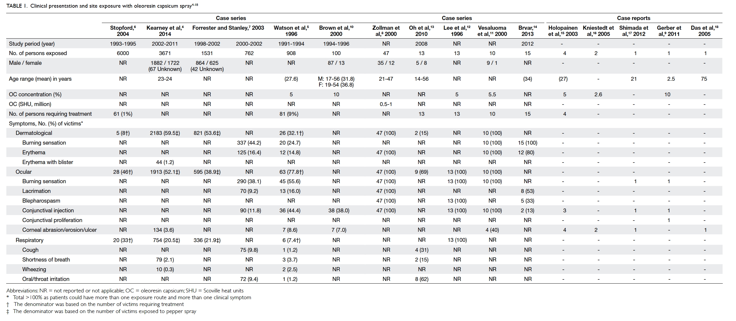
A total of 111 articles were identified of which 15 were considered relevant to our review. The details are summarised in Table 1.4 5 6 7 8 9 10 11 12 13 14 15 16 17 18 Our search identified 10 studies published formally in peer-reviewed medical and scientific journals that addressed the clinicopathological effects of OC spray in humans. The remaining five papers were case reports of the clinical effects of OC spray on a particular organ system.
 

Table 1. Clinical presentation and site exposure with oleoresin capsicum spray4 5 6 7 8 9 10 11 12 13 14 15 16 17 18
 
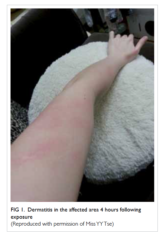
One of the authors (MF Yeung) conducted a telephone interview with two pepper spray victims. One 22-year-old man sustained direct lateral spray to his neck and forearm from a distance of 1 to 2 m. He experienced an almost immediate burning sensation over the sprayed areas, described as a very strong mint feeling. He then flushed himself with water but the chemical contaminated his clothes and other skin areas resulting in persistent burning pain for 30 minutes. Pain gradually resolved after an hour. Another 29-year-old woman sustained spray to her arm and forearm from a distance of approximately 5 m. Immediately she experienced a severe burning sensation over the affected areas (Fig 1), described as touching chilli seeds. She irrigated herself with water in the public toilet. The upper chest was also involved due to contamination of her clothing. Following irrigation, pain reduced over the next few hours to a mild hot sensation but still continued. She took a bath with warm water and baby shampoo 3 hours after exposure but experienced increased pain over the sprayed area. The pain resolved completely around 5 hours post-exposure.
 

Figure 1. Dermatitis in the affected area 4 hours following exposure
(Reproduced with permission of Miss YY Tse)
 
Clinical features
Exposure to OC can be via direct contact with skin, eyes or mucous membranes, or as a result of inhalation or ingestion. The irritant effects result in almost instantaneous onset of symptoms: within seconds it can induce a burning sensation on skin, involuntary closure of eyes, and diminished hand-eye coordination. Victims are usually rapidly incapacitated although most symptoms resolve within 30 to 60 minutes. The use of pepper spray by the US police has been reported to be successful in subduing aggressive individuals in 90% of cases.19
 
Dermatological injury
Initial contact of capsaicin with skin or mucous membranes produces a violent irritation with subsequent desensitisation to irritant chemicals. Victims experience acute burning pain, tingling, erythema, oedema, and pruritus (the pain can be prolonged for several hours in persons not adequately decontaminated).4 20 In prolonged exposure and in severe cases, persistent dermatitis with severe erythema and/or blister formation may occur.4 20
 
Kearney et al4 made a retrospective chart review of all human exposures to pepper spray recorded in the electronic database of the California Poison Control System during 2002-2011 (Table 1). Of the 3671 victims recorded, the most common type of exposure was dermal (2183 victims, 59.5%) with 2080 (56.7%) victims reporting minor/self-limiting symptoms, and 103 (2.8%) reporting more severe symptoms that required medical evaluation, including persistent dermatitis, dermal burns, and/or blister formation.
 
Watson et al5 conducted a retrospective study based on medical record review of 81 patients who presented to an emergency department after exposure to OC (908 individuals in total exposed during law enforcement) in 1991 to 1994 in Kansas City, Missouri, US. In 26 (32.1%) cases the chief symptoms upon arrival to casualty were pain or a burning sensation in exposed skin. In the US, a retrospective case review of approximately 6000 officers was conducted at the Department of Corrections; these officers were exposed to pepper spray during training in 1993 to 1995.6 21 Only 61 (1%) required medical treatment: five (8%) complained of dermatological symptoms of whom two had hives. Forrester and Stanley7 used data derived from telephone calls to the Texas Poison Center Network to investigate the epidemiology of OC exposure. Of 762 human exposures during 2000 to 2002, 337 (44.2%) experienced dermal irritation/pain and 125 (16.4%) had erythema on exposed skin.
 
Ophthalmic injury
Contact of the eye with OC causes redness, swelling, severe burning pain, tingling, lacrimation, and involuntary or reflex closing of the eyelids. More severe symptoms include persistent pain, foreign body sensation, photophobia, discharge or exudate, or peri-orbital oedema. Zollman et al8 showed that in police cadet volunteers sprayed in the face with OC, the most significant symptoms were conjunctival and scleral irritation and blepharospasm. Symptoms usually resolved within 1.5 to 2 hours of decontamination although mild chemosis, corneal oedema, or hyphaema could persist.
 
Conjunctival proliferation has been reported in a young child following mild spray injury and was refractory to steroid therapy with subsequent need for surgical excision.9 Decreased tear production as evidenced by a lower Schirmer test score was found. An impaired corneal reflex lacrimation and corneal blink reflex have also been observed.22
 
Oleoresin capsicum spray can affect both morphology and sensitivity of the cornea. Brown et al10 demonstrated that of 100 exposed individuals (10% OC spray), 7% (n=7) had corneal abrasion. Watson et al5 found 8.6% (n=7) of 81 affected officers who required medical treatment actually had corneal abrasion. Epstein and Majmudar23 described a case with keratopathy, and the victim’s eye showed a corneal epithelial defect after OC spray to the eyes. Although the defects healed within 5 days of treatment with polymyxin-bacitracin ophthalmic ointment, smouldering inflammation persisted. Further treatment with tobramycin-dexamethasone drops every 4 hours caused the inflammation to subside but superficial stromal opacity remained, resulting in irregular astigmatism.23
 
Zollman et al8 demonstrated that corneal sensation was severely affected after 10 minutes and 1 hour of exposure. At 1 week, sensation returned to baseline and corneal abnormalities disappeared. Apart from capsaicin, the carrier vehicle in which the active ingredient is dissolved may be toxic to corneal epithelial cells and cause temporary ocular irritation or superficial keratitis or erosion.12
 
Oral, nasal, and respiratory toxicity
Exposure may occur through inhalation, causing immediate inflammation of mucous membranes. Throat irritation results in a burning sensation, cough, choking, and inability to speak (due to laryngospasm or laryngeal paralysis). In the nasal mucosa, OC produces irritation, burning pain, sneezing, and a dose-dependent serous discharge.24 Other respiratory symptoms have also been reported, including severe coughing, mucus secretion, shortness of breath, bronchoconstriction presenting as wheeze, and chest tightness. Direct contact of capsaicinoids with the vocal cords causes laryngospasm lasting 45 seconds.21 Duration or magnitude of bronchoconstriction did not differ among normal subjects, smokers, and asthmatics.21
 
A review by Watson et al5 of 81 victims of law enforcement action revealed that 7.4% (n=6) had respiratory symptoms after inhalation of OC spray, and 3.7% (n=3) complained of shortness of breath. Oh et al13 studied an incident of OC gas leak in an urban shopping mall that affected 13 victims, of whom two (15%) experienced shortness of breath and eight (62%) complained of oral/throat irritation.
 
A controlled clinical trial involving 35 healthy subjects exposed to OC spray did not detect significant changes in predicted forced expiratory volume in 1 second (FEV1) or oxygen saturation when compared with population norms.25 The prone maximum restraint position reduced forced vital capacity (FVC) and FEV1 by 15% compared with a sitting position, but there was no statistically significant difference in these parameters when use of OC and controls were compared in the restraint position.25
 
Systemic toxicity and death
Inhalation of OC spray can cause laryngeal and pulmonary oedema and chemical pneumonitis but this is rare.21 An 11-year-old child who directly inhaled a jet spray from a pressurised container ultimately recovered but was reported to develop subglottic obstruction of the trachea and bilateral pulmonary infiltration that required intubation.26 Billmire et al27 described a 4-week-old healthy infant exposed to 5% OC when a self-defence device was accidentally discharged; the infant developed respiratory failure and hypoxaemia requiring extracorporeal membrane oxygenation. The patient was discharged and 12-month follow-up revealed several episodes of viral respiratory infections.27
 
Some studies have reported systemic symptoms including disorientation, fear, loss of body motor control (eg diminished hand-eye coordination),28 hyperventilation, tachycardia, and pulmonary oedema.21 The acute increase in blood pressure could cause headache, increased stroke risk and heart attack.21 In a review of approximately 6000 (police) officers directly exposed to OC, eight (0.1%) trainees reported headache and chest problems that persisted for more than 1 week.6
 
Since 1993, over 70 in-custody deaths have involved the use of OC spray during the arrest process.29 At the same time, Amnesty International has claimed that since the early 1990s more than 100 people in the US have died following exposure to pepper spray.30 Steffee et al31 reported an in-custody death in a known asthmatic who had been sprayed 10 to 15 times with OC spray (Table 2). O’Halloran and Frank29 reported 21 cases of restraint in-custody death, of which 10 were preceded by use of OC spray. Granfield et al32 reported 30 cases of in-custody death following OC exposure in the US but this review concluded that pepper spray was not the cause of death in any of the cases. Pollanen et al33 reported 21 in-custody restraint deaths in which four of the subjects had been sprayed with OC. Details are shown in Table 3.29 31 32 33
 

Table 2. Relative “heat” of edible peppers and capsaicin31
 

Table 3. Reported deaths with pepper spray use identified as one of the circumstances surrounding death29 31 32 33
 
Decontamination
The pre-hospital management of an individual exposed to pepper spray should include prompt on-scene removal of exposure to reduce the source of irritation. First, victims should move away from any continuing source of exposure. The presence of any signs or symptoms of serious systemic distress such as cardiovascular or respiratory problems should be ascertained.13 The immediate and most readily available method of decontamination at the scene of exposure is likely to be water. The affected skin and mucous membranes should be irrigated thoroughly with copious amounts of cool water to help soothe the burning sensation and flush away any spray residue.12
 
Some authors suggest that eyes exposed to OC should be irrigated with copious amounts of room-temperature water or normal saline for at least 15 minutes.4 If the victim is wearing contact lenses, they should be removed as quickly as possible.12 Washing the eyelids with a mild, oil-free soap will help break down the OC resin and speed up its removal.12 If a high level of spray residue is present in clothing, it should be removed and placed in sealed plastic bags until it can be cleaned or discarded.12 Removal of contaminated clothing will aid in preventing secondary contamination. Contamination by used towels should be avoided.12 Showering with soap and water is advised to remove the irritant from skin that should be blotted dry, not rubbed.12
 
For acute relief of burning dermal pain, one study suggested application of magnesium-aluminium hydroxide suspension.34 Vegetable oil immersion and vinegar have been reported to be more effective than water in relief of burning sensation.35 36 One study of 49 volunteer adult law enforcement trainees exposed to OC during a routine training exercise, who were randomised to one of five treatment groups (aluminium hydroxide–magnesium hydroxide, 2% lidocaine gel, baby shampoo, milk, or water), reported no significant difference in pain relief. Time after exposure appeared to be the best predictor for decrease in pain.37 Medical treatment for dermatitis may include topical steroids, oral antihistamines, and topical antibiotics.
 
Discussion
Pepper sprays are generally regarded as immediately effective and less toxic than other riot-control agents such as chloroacetophenone (CN) and o-chlorobenzylidene malononitrile (CS). Both CN and CS are effective lacrimating agents with CN the most lethal. The estimated lethal dose (LCt50) of CN, CS, and OC is 8500, 25 000 and >100 000 mg/min/m3, respectively.38
 
Oleoresin capsicum spray contains the active ingredient capsaicin, which is obtained from chilli pepper. Typically, pepper spray weapons contain a 10% to 20% solution of OC. The capsaicinoid content determines the ‘hotness’ of preparation, commonly referred to as Scoville Heat Unit (SHU; Table 2). The scale is named after its creator, American pharmacist Wilbur Scoville. Sprays used in police work typically fall between 0.5 and 2 million SHUs.8 The SHU governs the efficacy and pungency of pepper spray, thus the higher the SHU, the greater the inflammatory effect on skin and mucous membranes. A higher concentration of OC lengthens the necessary recovery period, thus affecting decontamination.39 Greater than 5% OC might not atomise well into a fine spray (may clog the aerosol spray).40 The capsaicin and related capsaicinoid content in most law enforcement OC sprays is between 1.3% and 2%. Areas of exposure can be dermatological, ocular, inhalational, ingestion or mixed, thus symptoms often involve more than one system. The prevalence of severe symptoms observed in various epidemiological studies varies from 2.7% to 15%.4 5 7 10
 
We found dermatological manifestation as the most common symptom in the case series (32%-100%),5 8 11 although another study reported dermatological symptoms in only 8% of cadet officers exposed to OC.6 The predominant site of exposure will be affected by any protective measures adopted: use of protective goggles or shields will greatly diminish exposure of skin to OC. Circumstances of OC spray use will also affect the predominant sites of exposure and severity of symptoms. Faced with confrontation during a public demonstration, officers will target OC spray at the eyes of protestors to quickly incapacitate them. This will result in a greater degree of both dermatological (face) and ocular exposure. Kearney et al4 analysed the risk factors with the largest independent associations with more severe symptoms among 3671 cases exposed to direct OC spray designed to incapacitate during law enforcement crowd-control activities.
 
Multiple exposure of skin or mucous membranes over a period of seconds or minutes exaggerate the inflammatory response. Capsaicin exposure may diminish sensitivity to heat- or chemical-induced pain, and thus increase the risk and severity of dermal burns. Some authors have suggested that capsaicin can powerfully stimulate heat receptors to cause reflex sweating and vasodilatation, and activate hypothalamus-mediated cooling; this may result in an increased risk of hypothermia if victims are decontaminated with cold water on cold days.41 42 Further, capsaicin-related cutaneous sensation may be heightened by perspiration, lacrimation, high humidity, and bathing at a warm temperature.43
 
The dermatological effects of capsaicin in OC spray have been further described in some case reports. ‘Hunan hand’ syndrome describes painful contact dermatitis in people preparing chilli peppers (containing capsaicin) by direct handling,44 while Sweet’s syndrome has been described following exposure to jalapeño peppers.45
 
Respiratory effects of OC spray involve cough reflex stimulation via capsaicin-sensitive nerves and bronchoconstriction. Although OC spray exposure causes cough and transient increase in airway resistance, experimental studies and clinical case reports show no evidence that patients with bronchial hypersensitivity are any more susceptible to the irritant effects of OC spray than those without.5 No cases of occupational asthma due to capsaicin have been reported.21 A cross-sectional study of workers exposed to capsaicin detected a statistically significant increase in complaints of cough in capsaicin-exposed workers, although there was no significant difference in FEV1 and FVC between the two groups.46 Not all asthmatics are sensitive to the bronchoconstrictive effects of OC spray.21
 
As capsaicinoids are lipophilic and have limited water solubility (16 µg/mL),47 alcohols or other organic solvents are commonly used as a base to facilitate aerosolisation in pepper spray products. Suitable solvents include methylene chloride, isopropanol, propylene glycol, ethanol, and methanol. The solvent enhances capsaicinoid solubility to enable delivery to the intended target tissue. A gaseous propellant (usually nitrogen or carbon dioxide) is incorporated in the spray to discharge the canister contents.1 Inhalation of high doses of some of these chemicals may produce adverse cardiac, respiratory, and neurological effects, including arrhythmia.21 The health effects of solvent and propellants are beyond the scope of our study, but their potential hazards and effects need to be considered.
 
Little is known about the long-term effects of pepper spray. Concern has been raised about their mutagenic and carcinogenic effects considering the organic solvent content, but the findings of numerous studies are inconclusive. Genotoxic effects have been demonstrated for capsaicin, but usually in cultured cells and at micromolar concentrations.48 These levels would not normally be achieved in field use. Some authors suggest that single or incidental relatively low doses of OC spray, as used by the police, will not produce a significant cancer risk.19 Nonetheless the long-term risks for those sprayed on multiple occasions are unknown.21
 
In the cases of fatality that we identified, a causal connection between OC spray exposure and death remained controversial. A number of deaths in custody occurred after exposure to pepper spray but the cause was not thoroughly investigated. In a report by Steffee et al,31 autopsy findings and toxicology results were analysed alongside pre-mortem chain of events, symptomatology, and degree of natural disease process. One asthmatic victim died because of severe acute bronchospasm, probably precipitated by the use of OC spray.31 Another review of various fatalities49 found that positional asphyxia (subjects are placed prone, typically handcuffed behind the back, hog-tied, and breathing becomes more difficult), drugs (alcohol intoxication, excited delirium [cocaine-induced], methamphetamine), pre-existing respiratory or cardiovascular disease, obesity and other conditions caused or contributed to almost all deaths.21 Exposure to OC spray was not judged to be a precipitating factor in any of these cases.50 Some authors have concluded that OC spray is not inherently lethal or dangerous.51 From the literature search, most cases of death associated with the use of pepper spray involved other factors and there was no convincing evidence that OC spray was implicated.
 
Although OC is lipophilic and many substances have been recommended for decontamination, water is easily available and of no harm. In the presence of ocular exposure, it is helpful to irrigate the upper and lower palpebral cul-de-sac because spray residue tends to collect in these locations and becomes entrapped.12 In one individual whose eyes were exposed to OC, the best-corrected visual acuity with spectacles did not improve beyond 20/40 after 7 months because of irregular astigmatism associated with persistent corneal opacity.23 Therefore prompt ocular irrigation is important to avoid potentially permanent ocular sequelae.
 
The severity of health effects from pepper spray exposure may be contingent on several product-, dispersal-, and patient-related factors. Various handheld pressurised canisters of OC spray are available and at least three variables affect their overall effectiveness. The first is the type, level of ‘hotness’, or concentration of pepper spray. Since the concentration of extract in pepper sprays varies (5%-15%), the potential risks associated with capsaicinoid exposure may vary up to 30-fold among different brands of OC spray.21 Second, the physiochemical properties of the solvent, that is, the vehicle type and its chemical solubility, can influence skin bioavailability.52 It increases the effectiveness of spray by improving penetration, removing skin oils, or prolonging contact time.12 Lastly, the delivery device or propellant—liquid spray, powder, or aerosolisation—dictates particle size and penetration into mucosal membranes and airways.
 
The severity of injury also depends on the circumstances of exposure. Direct exposure may be accidental or intentional.4 The latter involves a higher-risk situation that might require long OC application and tactics with consequent higher potential for injury. Concurrent disease and health of victims, varying doses, and distance being sprayed also determine different degrees of toxicity. Environmental conditions (windy, rainy weather), and crowd size and characteristics should also be taken into account when assessing level of exposure. Eyeglasses, sunglasses, and other protective eyewear and clothing can greatly reduce the effectiveness of OC sprays, as can the behaviour of the victim. For example, raising the hands in a defensive measure may block the spray.39 This was exemplified by the use of goggles and umbrellas at recent altercations with police reinforcement in Hong Kong.
 
Neurogenic inflammation
Capsaicin-induced neurogenic inflammation (Fig 221 53) is associated with heat allodynia (decreased heat pain threshold), thermal and mechanical hyperalgesia and cold hypoesthesia at the site of challenge.54 Neurophysiological study has demonstrated increased contact heat pain and shortened latencies of contact heat-evoked potentials following capsaicin-induced heat hyperalgesia.55 It is possible that capsaicin causes a decrease in the A-delta-fibre threshold giving rise to heat allodynia.55 Therefore, the ongoing pain and discharges of the sensitised nociceptors following capsaicin sensitisation depend on skin surface temperature: mild cooling of skin results in reduction of the burning pain sensation.55 This is why Lee et al12 suggested that during the decontamination process, allowing fresh air to circulate or even fanning the exposed area could assist in recovery. Cool water soothes the burning sensation while hot water increases it.
 

Figure 2. Schematic diagram of capsaicin-related cutaneous neurogenic inflammation
Skin is densely innervated by nociceptive nerve fibres for sensing stimuli in external environment. A large proportion of these afferent somatic nerves are fine polymodal unmyelinated C fibres or myelinated A delta fibres derived from dorsal root ganglia. Both respond to a range of physiological stimuli such as heat, cold, nociception, and mechanical distension. Upon stimulation by capsaicinoids, active neuropeptides are released resulting in an inflammatory response known as cutaneous neurogenic inflammation. This is characterised by transient burning pain, localised erythema, and circular oedema. In the final stage, a flared rim or edge appears around the circle with concomitant axon reflex vasodilatation. The axon reflex hypothesis of neurogenic inflammation suggests that damage to tissues triggers the immediate signal through the sensory nerves to the dorsal root ganglia and the central nervous system (orthodromic reflex), which transmits the sensation of pain. There is rapid depolarisation of the nociceptive terminals. The signal in the opposite direction, antidromic sensory nerve response induces release of vasodilatative neuropeptides (substance P and calcitonin gene-related peptides) in peripherally innervated tissues.53
The capsaicinoids contained within the pepper spray activate the TRPV1 receptor—the capsaicin receptor, a ligand-gated transmembrane calcium channel expressed at high levels by peripheral sensory nerves. These receptors are also expressed by keratinocytes (skin), tongue, and respiratory epithelium. Meanwhile, neurogenic inflammation in the airway blood vessels, epithelium, glands, and smooth muscles leads to vasodilatation, increased vascular permeability, neutrophil chemotaxis, mucus secretion, and bronchoconstriction.21
 
Pharmacological use
While our review focuses on OC spray, the most active ingredient capsaicin can be used therapeutically at an appropriate dilution. Prolonged topical application of capsaicin has been shown to induce a localised reversible loss of >60% of nociceptive nerve fibre terminals in the epidermis and dermis56 and attenuate heat pain sensitivity (defunctionalisation of TRPV1-expressing nociceptive fibres), resulting in relief in peripheral neuropathic pain syndromes.57
 
Limitations
There are limited large-scale case studies in the English literature of the clinicopathological effects of OC spray exposure. In some case series, data were reviewed retrospectively (from poison control centre registries) and may not have been collected using a standardised format, including exposure circumstances, thus some data might be missing. There was little information about the exact method, effectiveness, and precise timing of decontamination procedures or other interventions. Data on the concentration and especially SHU of OC spray, which could affect the strength and efficacy of spray to produce injury, were largely lacking, including particle size of OC spray and dose administered. The circumstances of exposure (riot control, crowd control, police cadet training, or domestic) and the scale of canister (distance of spray jet) would have affected the severity of clinicopathological effects of OC spray but were not recorded in detail. As most studies were observational or retrospective, a definite causal relationship between exposure and symptoms could not be drawn. There are likely variables and confounders present. In some studies, the reporting of symptoms was largely voluntary and thus likely to be biased. Most studies did not address chronic toxicity or residual disability in people exposed to OC spray.
 
Conclusion
Based on our review, OC spray appears to be an effective incapacitating riot-control agent. Significant adverse clinical effects or life-threatening conditions are not anticipated in the majority of exposures and death caused by OC spray exposure is unlikely. Dermatological and ophthalmic effects are commonly seen after exposure to OC spray in humans due to neurogenic inflammation. Careful early decontamination can minimise the irritant effects. Victims should be monitored for any evidence of serious adverse effects, with prompt medical intervention for any life-threatening symptoms. Large prospective cohort studies or case-control studies would strengthen the ability to infer a causal relationship between pepper spray exposure and health outcome.
 
References
1. Reilly CA, Crouch DJ, Yost GS. Quantitative analysis of capsaicinoids in fresh peppers, oleoresin capsicum and pepper spray products. J Forensic Sci 2001;46:502-9. Crossref
2. Reilly CA, Crouch DJ, Yost GS, Fatah AA. Determination of capsaicin, dihydrocapsaicin, and nonivamide in self-defense weapons by liquid chromatography–mass spectrometry and liquid chromatography–tandem mass spectrometry. J Chromatogr A 2001;912:259-67. Crossref
3. Cordell GA, Araujo OE. Capsaicin: identification, nomenclature, and pharmacotherapy. Ann Pharmacother 1993;27:330-6.
4. Kearney T, Hiatt P, Birdsall E, Smollin C. Pepper spray injury severity: ten-year case experience of a poison control system. Prehosp Emerg Care 2014;18:381-6. Crossref
5. Watson WA, Stremel KR, Westdorp EJ. Oleoresin capsicum (Cap-Stun) toxicity from aerosol exposure. Ann Pharmacother 1996;30:733-5.
6. Stopford W. Occupational exposures to riot control agents. In: Olajos EJ, Stopford W, editors. Riot control agents. Issues in toxicology, safety and health. New York: CRC Press; 2004: 277. Crossref
7. Forrester MB, Stanley SK. The epidemiology of pepper spray exposures reported in Texas in 1998-2002. Vet Hum Toxicol 2003;45:327-30.
8. Zollman TM, Bragg RM, Harrison DA. Clinical effects of oleoresin capsicum (pepper spray) on the human cornea and conjunctiva. Ophthalmology 2000;107:2186-9. Crossref
9. Gerber S, Frueh BE, Tappeiner C. Conjunctival proliferation after a mild pepper spray injury in a young child. Cornea 2011;30:1042-4. Crossref
10. Brown L, Takeuchi D, Challoner K. Corneal abrasions associated with pepper spray exposure. Am J Emerg Med 2000;18:271-2. Crossref
11. Vesaluoma M, Müller L, Gallar J, et al. Effects of oleoresin capsicum pepper spray on human corneal morphology and sensitivity. Invest Ophthalmol Vis Sci 2000;41:2138-47.
12. Lee RJ, Yolton RL, Yolton DP, Schnider C, Janin ML. Personal defense sprays: Effects and management of exposure. J Am Optom Assoc 1996;67:548-60.
13. Oh JJ, Yong R, Ponampalam R, Anantharman V, Lim SH. Mass casualty incident involving pepper spray exposure: impact on the emergency department and management of casualties. Hong Kong J Emerg Med 2010;17:325-9.
14. Brvar M. Pepper spray (capsaicin) exposure: decontamination with amphoteric, chelating and hypertonic solution [abstract]. Clin Toxicol 2013;51:314.
15. Holopainen JM, Moilanen JA, Hack T, Tervo TM. Toxic carriers in pepper sprays may cause corneal erosion. Toxicol Appl Pharmacol 2003;186:155-62. Crossref
16. Kniestedt C, Fleischhauer J, Stürmer J, Thiel MA. Pepper spray injuries of the anterior segment of the eye [in German]. Klin Monbl Augenheilkd 2005;222:267-70. Crossref
17. Shimada M, Young C, Tanen DA. Corneal ulcer associated with pepper spray exposure during military training. J Emerg Med 2012;43:e149. Crossref
18. Das S, Chohan A, Snibson GR, Taylor HR. Capsicum spray injury of the eye. Int Ophthalmol 2005;26:171-3. Crossref
19. Busker RM, van Helden HP. Toxicologic evaluation of pepper spray as a possible weapon for the Dutch police force: risk assessment and efficacy. Am J Forensic Med Pathol 1998;19:309-16. Crossref
20. Broadstock M. What is the safety of ‘pepper spray’ use by law enforcement or mental health service staff? New Zealand Health Technology Assessment Tech Brief Series. Available from: http://nzhta.chmeds.ac.nz/publications/pepper_spray.pdf. Accessed Oct 2015.
21. Smith CG, Stopford W. Health hazards of pepper spray. Available from: http://duketox.mc.duke.edu/pepper%20spray.pdf. Accessed 1 Dec 2014.
22. Rasier R, Kukner AS, Sengul EA, Yalcin NG, Temizsoylu O, Bahcecioglu HO. The decrease in aqueous tear production associated with pepper spray. Curr Eye Res 2015;40:429-33. Crossref
23. Epstein RJ, Majmudar PA. Pepper spray in the eye. Ophthalmology 2001;108:1712-3. Crossref
24. Geppetti P, Fusco BM, Marabini S, Maggi CA, Fanciullacci M, Sicuteri F. Secretion, pain and sneezing induced by the application of capsaicin to the nasal mucosa in man. Br J Pharmacol 1988;93:509-14. Crossref
25. Chan TC, Vilke GM, Clausen J, et al. The effect of oleoresin capsicum “pepper” spray inhalation on respiratory function. J Forensic Sci 2002;47:299-304. Crossref
26. Winograd HL. Acute croup in an older child. An unusual toxic origin. Clin Pediatr (Phila) 1977;16:884-7. Crossref
27. Billmire DF, Vinocur C, Ginda M, et al. Pepper-spray–induced respiratory failure treated with extracorporeal membrane oxygenation. Pediatrics 1996;98:961-3.
28. Smith J, Greaves I. The use of chemical incapacitant sprays: a review. J Trauma 2002;52:595-600. Crossref
29. O’Halloran RL, Frank JG. Asphyxial death during prone restraint revisited: a report of 21 cases. Am J Forensic Med Pathol 2000;21:39-52. Crossref
30. USA: Excessive and lethal force? Amnesty International’s concerns about deaths and ill-treatment involving police use of Tasers. Available from: http://www.amnestyusa.org/node/55449?page=72. Accessed 7 Dec 2014.
31. Steffee CH, Lantz PE, Flannagan LM, Thompson RL, Jason DR. Oleoresin capsicum (pepper) spray and “in-custody deaths.” Am J Forensic Med Pathol 1995;16:185-92. Crossref
32. Granfield J, Onnen J, Petty CS. Pepper spray and in-custody deaths: international association of chiefs of police executive brief. RCMP Gazette 1994;56:12-7.
33. Pollanen MS, Chiasson DA, Cairns JT, Young JG. Unexpected death related to restraint for excited delirium: a retrospective study of deaths in police custody and in the community. CMAJ 1998;158:1603-7.
34. Lee DC, Ryan JR. Magnesium-aluminum hydroxide suspension for the treatment of dermal capsaicin exposures. Acad Emerg Med 2003;10:688-90. Crossref
35. Jones L, Tandberg D, Troutman WG. A controlled evaluation for household treatments for “chile burns”. Vet Hum Toxicol 1986;28:486.
36. Vogl TP. Treatment of Hunan hand. N Engl J Med 1982;306:178. Crossref
37. Barry JD, Hennessy R, McManus JG Jr. A randomized controlled trial comparing treatment regimens for acute pain for topical oleoresin capsaicin (pepper spray) exposure in adult volunteers. Prehosp Emerg Care 2008;12:432-7. Crossref
38. Schep LJ, Slaughter RJ, McBride DI. Riot control agents: the tear gases CN, CS and OC—a medical review. J R Army Med Corps 2015;161:94-9. Crossref
39. Adkins LD. Oleoresin capsicum: an analysis of the implementation of pepper spray into the law enforcement use of force continuum in a selected police department. Available from: http://dc.etsu.edu/etd/779. Accessed 23 Jan 2015.
40. National Institute of Justice. Oleoresin capsicum: pepper spray as a force alternative. Available from: https://www.ncjrs.gov/pdffiles1/nij/grants/181655.pdf. Accessed 23 Jan 2015.
41. Tominack RL, Spyker DA. Capsicum and capsaicin—a review: case report of the use of hot peppers in child abuse. J Toxicol Clin Toxicol 1987;25:591-601. Crossref
42. Monsereenusorn Y, Kongsamut S, Pezalla PD. Capsaicin—a literature survey. Crit Rev Toxicol 1982;10:321-39. Crossref
43. Hilmas CJ, Poole MJ, Katos AM, Williams PT. Chapter 12: Riot control agents. In: Handbook of toxicology of chemical warfare agents. Available from: http://www.dtic.mil/dtic/tr/fulltext/u2/a539686.pdf. Accessed Oct 2015.
44. Weinberg RB. Hunan hand. N Engl J Med 1981;305:1020. Crossref
45. Greer JM, Rosen T, Tschen JA. Sweet’s syndrome with an exogenous cause. Cutis 1993;51:112-4.
46. Blanc P, Liu D, Juarez C, Boushey HA. Cough in hot pepper workers. Chest 1991;99:27-32. Crossref
47. Kasting GB. Kinetics of finite dose absorption through skin 1. Vanillylnonanamide. J Pharm Sci 2001;90:202-12. CrossRef
48. Durnford JM. Chapter 10: Genetic toxicity of riot control agents. In: Olajos EJ, Stopford W, editors. Riot control agents. Issues in toxicology, safety and health. New York: CRC Press; 2004: 183-200. Crossref
49. The American Civil Liberties Union of Southern California. Pepper spray update: more fatalities, more questions; 1995 Jun. Available from: https://www.aclusocal.org/issues/police-practices/pepper-spray/. Accessed 19 Nov 2014.
50. International Association of Chiefs of Police. Pepper spray evaluation project; 1995 Jun 22. Available from: http://www.theiacp.org/portals/0/pdfs/IntroPepperSpray.pdf. Accessed 19 Nov 2014.
51. Christensen RG, Frank DE. Preliminary investigation of oleoresin capsicum. National Institute of Justice Report 100-95, Gaithersburg, MD: National Institute of Standards and Technology; 1995.
52. Pershing LK, Corlett JL, Nelson JL. Comparison of dermatopharmacokinetic vs. clinical efficacy methods for bioequivalence assessment of miconazole nitrate vaginal cream, 2% in humans. Pharm Res 2002;19:270-7. Crossref
53. Caterina MJ, Schumacher MA, Tominaga M, Rosen TA, Levine JD, Julius D. The capsaicin receptor: a heat-activated ion channel in the pain pathway. Nature 1997;389:816-24. Crossref
54. LaMotte RH, Shain CN, Simone DA, Tsai EF. Neurogenic hyperalgesia: psychophysical studies of underlying mechanisms. J Neurophysiol 1991;66:190-211.
55. Madsen CS, Johnsen B, Fuglsang-Frederiksen A, Jensen TS, Finnerup NB. Increased contact heat pain and shortened latencies of contact heat evoked potentials following capsaicin-induced heat hyperalgesia. Clin Neurophysiol 2012;123:1429-36. Crossref
56. Kennedy WR, Vanhove GF, Lu SP, et al. A randomized, controlled, open-label study of the long-term effects of NGX-4010, a high-concentration capsaicin patch, on epidermal nerve fiber density and sensory function in healthy volunteers. J Pain 2010;11:579-87. Crossref
57. Anand P, Bley K. Topical capsaicin for pain management: therapeutic potential and mechanisms of action of the new high-concentration capsaicin 8% patch. Br J Anaesth 2011;107:490-502. Crossref

Using the script concordance test to assess clinical reasoning skills in undergraduate and postgraduate medicine

Hong Kong Med J 2015 Oct;21(5):455–61 | Epub 28 Aug 2015
DOI: 10.12809/hkmj154572
© Hong Kong Academy of Medicine. CC BY-NC-ND 4.0
 
REVIEW ARTICLE
Using the script concordance test to assess clinical reasoning skills in undergraduate and postgraduate medicine
SH Wan, MB, ChB, MRCP (Edin)
School of Medicine Sydney, University of Notre Dame, 160 Oxford Street, Darlinghurst, NSW 2010, Australia
Corresponding author: Dr SH Wan (michael.wan@nd.edu.au)
 
 Full paper in PDF
Abstract
The script concordance test is a relatively new format of written assessment that is used to assess higher-order clinical reasoning and data interpretation skills in medicine. Candidates are presented with a clinical scenario, followed by the reveal of a new piece of information. The candidates are then asked to assess whether this additional information increases or decreases the probability or likelihood of a particular diagnostic, investigative, or management decision. To score these questions, the candidate’s decision in each question is compared with that of a reference panel of expert clinicians. This review focuses on the development of quality script concordance questions, using expert panellists to score the items and set the passing score standard, and the challenges in the practical implementation (including pitfalls to avoid) of the written assessment.


Introduction
Script concordance test (SCT) is a relatively new format of written assessment to assess higher-order clinical reasoning and data interpretation skills of medical candidates.1
 
In recent years, universities and postgraduate colleges worldwide have used SCT for both formative and summative assessment of clinical reasoning in various medical disciplines including paediatric medicine, paediatric emergency medicine, neurology, orthopaedics, surgery, and radiology.2 3 4 5 6 7 8 In the classic written assessment, multiple-choice questions (MCQ) and short-answer questions (SAQ) usually examine the candidates’ simple knowledge recall at the lowest ‘knows’ level of the Miller’s Pyramid (Fig 1).9 10 Questions in SCT are able to test candidates at the higher order of thinking at the ‘knows how’ and even ‘shows how’ level. It is a unique assessment tool that targets the essential clinical reasoning and data interpretation skills in a very authentic way that reflects the element of ‘uncertainty’ in real-world clinical scenarios prevalent in clinical practice. This is the key aspect of clinical competency that enables medical graduates or fellows in training to link and transfer their mastery of declarative clinical knowledge and skills into clinical practice in a real clinical setting. Recent literature reports the value of using SCT to assess other areas of disciplines where classic questions are difficult to develop, for example, in assessing medical ethical principles and professionalism.11
 

Figure 1. Miller’s Pyramid9 10
 
The structure and format of script concordance test
In SCT, candidates are presented with a clinical vignette/scenario, followed by the reveal of a new piece of information. The candidates are then asked to assess whether this additional information increases or decreases the probability or likelihood of the suggested provisional diagnosis, and increases or decreases the usefulness/appropriateness of a proposed investigation or management option. The process reflects everyday real-world decision-making processes where clinicians retrieve their ‘illness scripts’ or network of knowledge (about similar patient problems and presentations stored in their memory) when faced with uncertainty in a clinical presentation. This enables them to determine the follow-on diagnosis and management options most appropriate to the situation. As further clinical encounters are experienced, the scripts are updated and refined.12 Script concordance test assesses the candidates’ clinical reasoning and data interpretation ability in the context of uncertainty, particularly involving ill-defined patient problems in clinical practice.13 Sample SCT questions in Table 1 illustrate the structure and format of the SCT questions. As the clinical scenario unfolds, additional data such as clinical photos, radiological images, or audiovisual material can also be presented to enhance the authenticity of the scenarios.5 14 15
 

Table 1. Sample questions of script concordance test
 
In scenario A in Table 1, the ‘clinical vignette’ is that of a 22-year-old woman who presents to the Emergency Department with severe abdominal pain. A piece of ‘new information’ is then revealed that her serum beta–human chorionic gonadotropin (β-HCG) is normal. The candidate is asked whether this additional information makes the ‘diagnosis’ of ectopic pregnancy: much less likely (-2), less likely (-1), neither more nor less likely (0: no effect on the likelihood), more likely (+1), or much more likely (+2). The next piece of new information (independent of the first one) is that the examination shows marked guarding and rigidity of the abdomen and the candidate is asked to determine the likelihood of a diagnosis of acute appendicitis.
 
In scenario B in Table 1, a similar format is used to assess the appropriateness of ordering an investigation in relation to the respective piece of additional information. The first question asks for the appropriateness of ordering a computed tomographic scan of the abdomen for a 16-year-old girl who presents with acute abdominal pain if her last menstrual period was 8 weeks ago.
 
In scenario C in Table 1, the focus is on the usefulness of different management options after being presented with different pieces of new information related to the clinical vignette.
 
In preparing candidates to answer the questions, it is crucial to emphasise that each piece of new information is independent of the previous piece but in the same clinical setting. For example, in scenario A, when answering the second question given that she has guarding and rigidity in the abdomen, she does NOT have a serum β-HCG test done.
 
With respect to the likelihood descriptors used in the SCT questions for the diagnosis type of scenario, the preference is to use the option of “much less likely (-2)” rather than “ruling out the diagnosis”; and “much more likely (+2)” rather than “almost certain/definite diagnosis”. This will allow candidates to use the full range of the five options. In the practice of medicine, there are usually few situations wherein a diagnosis can be confidently excluded or definitely diagnosed with a few pieces of information provided.3
 
There are nonetheless limitations to the design and format of SCT. Candidates cannot seek additional information to that given in the question; the scenario is only a snapshot of the clinical encounter without the comprehensive history, physical examination, and investigations that would be particularly desirable in an ambiguous clinical situation.16
 
Scoring script concordance test
To score these questions, the candidate’s decision in each question is compared with that of a reference panel of expert clinicians. Each member of the panel attempts the same set of questions and the answers are recorded. As there is no single best correct answer to the question, a full (1) mark will be awarded if the candidate’s decision concurs (hence the name ‘concordance’) with the majority of the expert panel. A proportional (partially credited or weighted) score (<1) will be given if the candidate’s decision concurs with the minority of the panel. The candidate will score a ‘0’ if no panellist chooses this option.3 The formula to calculate the weighted scores is shown in Table 2.
 

Table 2. The formula to calculate the weighted scores
 
There are other scoring methods reported in the literature where a consensus-based single-answer scoring method or 3-point Likert scale scoring method is employed to determine the candidate scores.4 17
 
Selecting the reference panel
In general, a panel of 10 to 15 expert members relevant to the discipline is recommended to produce credible and reliable scores.18 The inter- and intra-rater reliability in the SCT panel have been shown to be good.19
 
The composition of the panel should include clinical teachers and academics who are familiar with the curriculum and experts in the field relevant to the discipline tested. Studies have shown that using general practitioners (GPs) in the panel may produce similar mean scores to specialists but with a wider standard deviation.3
 
A recent study, however, raised concerns about the reference standard and judgement of the expert panel. The study compared 15 emergency medicine consultants’ judgement scores with evidence-based likelihood ratios. The results showed that 73.3% of the mean judgement was significantly different to the corresponding likelihood ratios, with 30% overestimation, 30% underestimation, and 13.3% with diagnostic values in the opposite direction.20 Other studies raised concerns about the possibility of outdated clinical knowledge and cognitive bias in the experts’ decision-making.21 22 Evidence of context specificity has also been highlighted whereby the agreement between SCT scores derived using different scoring keys with expert reference panels from a different context (hospitals and specialty) was poor.23
 
Implementation of script concordance test in formative and summative assessments
The structure and layout of the SCT questions can easily be implemented in the usual pen and paper-based or online electronic format. Candidates answer each question (with five options) using a standardised answer sheet to facilitate computer scanning and scoring or directly online using the computer.
 
It is often difficult to get busy clinicians to meet together face-to-face to answer the questions. By uploading the questions online, the panellists can attempt them anytime and make the questions available through a secure online platform. The response data can then be collated and the weighted scores for responses on each score scale calculated.3
 
After capturing the candidates’ responses for all items, scoring of responses for each question can then be performed using the formula described above. This will ensure a rapid turnaround time that will be very effective in the assessment process.
 
For formative assessment purposes, expert panel consensus scores are provided to the candidates, followed by expert clinicians explaining and discussing the options in each scenario with the candidates for constructive feedback. Script concordance test can also be used to identify borderline students with suboptimal clinical reasoning skills for appropriate remedial measures such as bedside teaching, tutorials, or clinical simulations.24
 
For summative assessment purposes, particularly where there is not a large pool of SCT items, it is important to avoid constructing irrelevant variance in SCT scores, by not releasing or discussing post-examination, the expected responses (based on expert panel’s responses), and the associated score for each of the answer options in SCT items.
 
Unlike MCQ where there is only one single best answer that candidates could memorise and disseminate after the examination, the partial credit scoring model applied in SCT, where multiple answer options are accepted and each carries a fraction or all of the allocated mark, has to a certain extent rendered sharing of ‘correct’ answers after the examination difficult.
 
Developing quality script concordance test questions
Each clinical scenario has to be authentic and the presentation represents a realistic clinical encounter that is relevant to the specific discipline, preferably with a certain degree of uncertainty. The new information presented needs to stimulate the candidate to re-consider and re-evaluate how that particular piece of new information will affect the likelihood of the initial diagnosis, or appropriateness of initial planned investigation or management option. This will ensure the content validity in the SCT questions.
 
Particular care should be taken to develop options that will attract the full range of the five options available for the candidate to choose from. In other words, the additional pieces of new information should result in the consideration of -2 and +2 as well as -1, 0, and +1 options. A test-wise candidate might choose to consider only the options of -1, 0 and +1 if they notice that most panel consensus answers with a full score of 1 mark usually fall within these three options rather than also covering the -2 and +2.25 As a result, developing good-quality SCT questions is not easy. Care should be taken to develop clinical scenarios that do not focus solely on factual recall but involve a reasoning process with elements of uncertainty that will likely attract responses that spread across the 5-point Likert scale.26
 
Reliability and validity of script concordance test as an assessment tool
The reliability of SCT as an assessment tool has been investigated.2 6 A 60- to 90-minute examination will produce a Cronbach’s alpha of 0.70 to 0.85.7 25 27 28 There are concerns, however, about inter-panellist errors in SCT; the use of Cronbach’s alpha in measuring the reliability of the test where partial credit model of scoring is used, ie multiple options/responses are awarded either a full or fraction of allocated mark; and case scenarios that could create inconsistencies among items.
 
As an assessment tool, SCT has been shown to be valid in assessing clinical reasoning.13 14 19 28 Studies have shown that SCT scores correlate well with other assessment scores from the clinical years of the candidates.2
 
The construct validity of SCT questions can be examined by correlating the scores with the level of training to predict future performance on clinical reasoning. A recent study has compared the progression of clinical reasoning skills of medical students with those of a group of practising GPs who are also their Problem Based Learning group tutors.29 Another study showed that there was a statistically significant gain in SCT performance over a 2-year period in two different cohorts of medical students using the same set of 75-item SCT.26 There was significant progression of clinical reasoning skills from medical students at the novice level through to practising GP clinicians, reflected by the higher scores in the GP group attempting the SCT questions. Empirical evidence supporting the construct validity based on progression of SCT scores with clinical experience from undergraduate students to postgraduate training has also been reported.2 5 24 30 31 The construct validity of SCT has been questioned because of the logical inconsistencies as a result of partial credit scoring methodology making it possible for a hypothesis to be simultaneously more likely and less likely.32 Nonetheless, a certain degree of variability in panel scores has been shown to be a key determinant of the discriminatory power of the test and allows richness of thinking about clinical cases.33 34 Another study found that 27% of residents in one SCT administration scored above the expert panel’s mean, which may indicate issues with the construct validity, particularly in the credibility and validity of the scoring key and hence the resulting SCT scores interpretation.33
 
Test-wise candidates would select the answers to be around ‘0’ rather than ‘-2’ or ‘+2’ if they noticed that most panellist scores did not fall in the ‘extreme’ (-2 or +2) range due to the construct of the SCT questions and options. This could be avoided by first using the option descriptor of “much less likely (-2)” and “much more likely (+2)” rather than “ruling out the diagnosis” and “almost certain/definite diagnosis” as described in the format of SCT section above.19 Second, when collating the SCT questions into an examination paper, one could select a relatively equal number of items with both ‘extreme’ answers as well as median answers. Recent data have shown that by employing the above strategies in developing the paper, candidates who chose ‘0’ for all the questions would score only around 25% in the SCT examination and would gain no advantage (unpublished data). This is in contrast to the finding of another study wherein candidates who chose the midpoint scale (‘0’) performed better than the average candidate.32
 
The correlation of SCT scores with other modalities of assessment would be expected to be low as SCT is designed to measure clinical reasoning rather than factual or knowledge recall. The correlation coefficient between SCT and MCQ was poor (r=0.22), and that between SCT and extended matching questions (EMQ) was 0.46.4
 
Collating and moderating the expert reference panellist responses
In collating the SCT questions for use in a summative examination, appropriate clinical scenarios/vignettes with the related diagnoses, investigations, and management should be selected according to the blueprint of the assessment. The clinical topics should have a good spread and represent core areas of learning that are relevant to the curriculum and appropriate to the level of training of the candidates.
 
In reviewing the expert panel responses to each question, bi-modal and uniform divergence responses should trigger a detailed scrutiny of the clinical vignette and the options. In the case of bi-modal response (Fig 2a), the panel has an equal split of the best option between -2 and +2. This usually results from an error in the question or a controversial investigation or management option with discordant ‘expert opinions’. A modification of the question and re-scoring will usually solve this issue. If re-scoring results in the same bi-modal response, the question should be discarded for scoring in the examination. In the case of uniform divergence responses (Fig 2b), there is an equal spread in the number of members choosing all the five options. This usually signifies a non-discriminating question and the item should again be discarded. A discrete outlier response (Fig 2c) usually represents an error in the particular panellist’s decision or ‘clicking the wrong answer accidentally’ when the member should have answered -2 instead of +2. The ideal pattern would be relatively close convergence with some variation (Fig 2d).3
 

Figure 2. Number of responses from the expert panel to script concordance test questions
(a) Bi-modal response, (b) uniform divergence response, (c) discrete outlier response, and (d) ideal response
 
As mentioned previously, the set of questions in the SCT examination should be selected in such a way that there are similar numbers of full marks in each option across the five options. This will avoid the test-wise candidates being advantaged by selecting only the -1, 0, or +1 options and avoiding the extreme options of -2 and +2.3 By employing this strategy to select questions that cover the full 5-point Likert scale, test-wise students will only score 25% in the SCT examination if they choose the response of ‘0’ for all questions (unpublished data) compared with 57.6% in another cohort sitting a SCT test without the specific question selection process.32
 
Standard setting the pass/fail cutoff score
In setting the pass/fail cutoff score of the SCT questions, the expert panels’ mean scores and standard deviations are chosen to guide the process. This is calculated by asking all the members of the panel to attempt the same set of SCT questions and their responses are then scored accordingly. The borderline score of the undergraduate students is usually set at 3 to 4 standard deviations below the expert panel’s mean score.3 35 Studies have shown that using recent graduates or fellows in training might result in a mean score that is closer to the students’ mean and therefore a smaller number of standard deviations would be more appropriate.3
 
Other methods of standard setting include using the single correct answer method.29 36 Standard setting of a pass/fail cutoff score is an area that warrants ongoing research to inform and improve the practice of using SCT as a summative assessment tool for clinical data interpretation and decision-making skills.
 
The use of script concordance test in the Asia-Pacific region and its limitations
Examinations using SCT have been successfully implemented in the school-entry medical schools in Indonesia, Singapore, Taiwan, and Australia3 7 36 37; and in graduate-entry medical schools in Australia.29 38 Such test has the potential to supplement MCQ and SAQ to test the higher-order thinking of medical candidates to allow a more robust overall written assessment in the assessment programme. In fact, SCT is one of the few currently available assessment tools for clinical reasoning in a written format.28 It can be implemented relatively easily in the paper-based format or online. Initial pilot examinations can be set as a formative exercise to enhance candidates’ feedback and learning.24 Further collaboration with other institutions to develop, score, and share question items can ensure effective and efficient delivery of such examinations.
 
Limitations to the widespread usage of SCT could be due to: difficulties in developing good-quality SCT clinical scenarios, concerns about the validity of the test, recruiting a sufficient number of appropriate expert clinicians for the reference panel, lack of a general consensus in setting the borderline pass mark, and the candidates’ familiarity with the question format.3 24 25 28 32 34
 
Conclusions
This article attempts to review the current use of SCT in assessing clinical reasoning and data interpretation skills in undergraduate and postgraduate medicine. The empirical evidence reported for the reliability and validity of SCT scores from existing literature seems encouraging. Approaches to develop quality items, moderation of expert panel scoring and these post-hoc quality assurance measures, and optimisation of scoring scale will to a certain extent mitigate the threat to the validity of SCT score interpretation and its use for summative examination purposes. Combining SCT (testing the clinical reasoning and data interpretation skills with authentic written simulations of ill-defined clinical problems set at the ‘knows how’ level) with MCQ/SAQ/EMQ (testing the ‘knows’ and ‘knows how’), objective structured clinical examination (testing ‘shows how’), and workplace-based assessment (testing the ‘does’) in the medical curriculum will enhance the robustness and the credibility of the assessment programme.
 
Further research into the use of SCT in both undergraduate and postgraduate medical education is warranted, particularly on standard setting for the pass/fail cutoff score and best practices that may help reduce the threat to the validity of SCT scores.
 
References
1. Charlin B, Roy L, Brailovsky C, Goulet F, van der Vleuten C. The Script Concordance test: a tool to assess the reflective clinician. Teach Learn Med 2000;12:189-95. Crossref
2. Carrière B, Gagnon R, Charlin B, Downing S, Bordage G. Assessing clinical reasoning in pediatric emergency medicine: validity evidence for a Script Concordance Test. Ann Emerg Med 2009;53:647-52. Crossref
3. Duggan P, Charlin B. Summative assessment of 5th year medical students’ clinical reasoning by Script Concordance Test: requirements and challenges. BMC Med Educ 2012;12:29. Crossref
4. Kelly W, Durning S, Denton G. Comparing a script concordance examination to a multiple-choice examination on a core internal medicine clerkship. Teach Learn Med 2012;24:187-93. Crossref
5. Lubarsky S, Chalk C, Kazitani D, Gagnon R, Charlin B. The Script Concordance Test: a new tool assessing clinical judgement in neurology. Can J Neurol Sci 2009;36:326-31. Crossref
6. Meterissian S, Zabolotny B, Gagnon R, Charlin B. Is the script concordance test a valid instrument for assessment of intraoperative decision-making skills? Am J Surg 2007;193:248-51. Crossref
7. Soon D, Tan N, Heng D, Chiu L, Madhevan M. Neurologists vs emergency physicians: reliability of a neurological script concordance test in a multi-centre, cross-disciplinary setting. Neurology 2014;82(10 Suppl):327.
8. Talvard M, Olives JP, Mas E. Assessment of medical students using a script concordance test at the end of their internship in pediatric gastroenterology [in French]. Arch Pediatr 2014;21:372-6. Crossref
9. Miller GE. The assessment of clinical skills/competence/performance. Acad Med 1990;65(9 Suppl):S63-7. Crossref
10. Mehay R. The essential handbook for GP training and education. Chapter 29. Assessment and competence. Available from: http://www.essentialgptrainingbook.com/chapter-29.php. Accessed 10 May 2015.
11. Foucault A, Dubé S, Fernandez N, Gagnon R, Charlin B. Learning medical professionalism with the online concordance-of-judgment learning tool (CJLT): A pilot study. Med Teach 2014 Oct 22:1-6. Epub ahead of print. Crossref
12. Schmidt HG, Norman GR, Boshuizen HP. A cognitive perspective on medical expertise: theory and implications. Acad Med 1990;65:611-21. Crossref
13. Lubarsky S, Charlin B, Cook DA, Chalk C, van der Vleuten CP. Script concordance testing: a review of published validity evidence. Med Educ 2011;45:329-38. Crossref
14. Brazeau-Lamontagne L, Charlin B, Gagnon R, Samson L, Van Der Vleuten C. Measurement of perception and interpretation skills during radiology training: utility of the script concordance approach. Med Teach 2004;26:326-32. Crossref
15. Collard A, Gelaes S, Vanbelle S, et al. Reasoning versus knowledge retention and ascertainment throughout a problem-based learning curriculum. Med Educ 2009;43:854-65. Crossref
16. Lineberry M, Kreiter CD, Bordage G. Script concordance tests: strong inferences about examinees require stronger evidence. Med Educ 2014;48:452-3. Crossref
17. Williams RG, Klamen DL, White CB, et al. Tracking development of clinical reasoning ability across five medical schools using a progress test. Acad Med 2011;9:1148-54. Crossref
18. Gagnon R, Charlin B, Coletti M, Sauvé E, van der Vleuten C. Assessment in the context of uncertainty: how many members are needed on the panel of reference of a script concordance test? Med Educ 2005;39:284-91. Crossref
19. Dory V, Gagnon R, Vanpee D, Charlin B. How to construct and implement script concordance tests: a systematic review. Med Educ 2012;46:552-63.
20. Ahmadi SF, Khoshkish S, Soltani-Arabshahi K, et al. Challenging script concordance test reference standard by evidence: do judgments by emergency medicine consultants agree with likelihood ratios? Int J Emerg Med 2014;7:34. Crossref
21. Ramos K, Linscheid R, Schafer S. Real-time information-seeking behavior of residency physicians. Fam Med 2003;35:257-60.
22. Norman GR, Eva KW. Diagnostic error and clinical reasoning. Med Educ 2010;44:94-100. Crossref
23. Tan N, Tan K, Ponnamperuma G. Expert clinical reasoning is not just local but hyperlocal—insights into context specificity from a multicentre neurology script concordance test. Neurology 2015;84(14P4):191.
24. Ducos G, Lejus C, Sztark F, et al. The Script Concordance Test in anesthesiology: Validation of a new tool for assessing clinical reasoning. Anaesth Crit Care Pain Med 2015;34:11-5. Crossref
25. See KC, Tan KL, Lim TK. The script concordance test for clinical reasoning: re-examining its utility and potential weakness. Med Educ 2014;48:1069-77. Crossref
26. Humbert AJ, Miech EJ. Measuring gains in the clinical reasoning of medical students: longitudinal results from a school-wide script concordance test. Acad Med 2014;89:1046-50. Crossref
27. Gagnon R, Charlin B, Lambert C, Carrière B, Van der Vleuten C. Script concordance testing: more cases or more questions? Adv Health Sci Educ Theory Pract 2009;14:367-75. Crossref
28. Nouh T, Boutros M, Gagnon R, et al. The script concordance test as a measure of clinical reasoning: a national validation study. Am J Surg 2012;203:530-4. Crossref
29. Wan SH. Using Script Concordance Testing (SCT) to assess clinical reasoning—the progression from novice to practising general practitioner. Proceedings of the 11th Asia Pacific Medical Education Conference; 2014 Jan 15-19; Singapore.
30. Charlin B, van der Vleuten C. Standardized assessment in contexts of uncertainty: The script concordance approach. Eval Health Prof 2004;27:304-19. Crossref
31. Lambert C, Gagnon R, Nguyen D, Charlin B. The script concordance test in radiation oncology: validation study of a new tool to assess clinical reasoning. Radiat Oncol 2009;4:7. Crossref
32. Lineberry M, Kreiter C, Bordage G. Threats to validity in the use and interpretation of script concordance test scores. Med Educ 2013;47:1175-83. Crossref
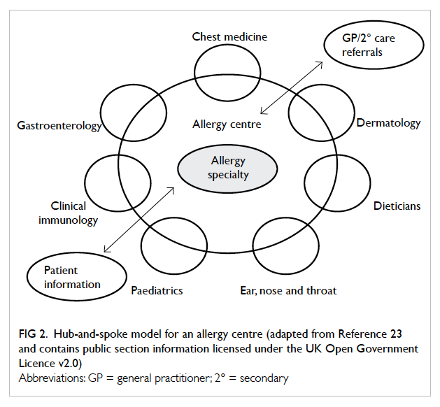
33. Charlin B, Gagnon R, Lubarsky S, et al. Assessment in the context of uncertainty using the script concordance test: more meaning for scores. Teach Learn Med 2010;22:180-6. Crossref
34. Lubarsky S, Gagnon R, Charlin B. Scoring the Script Concordance Test: not a black and white issue. Med Educ 2013;47:1159-61. Crossref
35. Wan SH, Clarke R. Using a clinician panel to set the borderline mark for Script Concordance Testing (SCT) to assess clinical reasoning for graduating medical candidates. Proceedings of the 8th International Medical Education Conference; 2013 March 13-15; Kuala Lumpur, Malaysia.
36. Irfannuddin I. Knowledge and critical thinking skills increase clinical reasoning ability in urogenital disorders: a Universitas Sriwijaya Medical Faculty experience. Med J Indonesia 2009;18:53-9. Crossref
37. Tsai TC, Chen DF, Lei SM. The ethics script concordance test in assessing ethical reasoning. Med Educ 2012;46:527. Crossref
38. Ingham AI. The great wall of medical school: a comparison of barrier examinations across Australian medical schools. Aus Med Student J 2011;2:5-8.

Avoiding hypoglycaemia: a new target of care for elderly diabetic patients

Hong Kong Med J 2015 Oct;21(5):444–54 | Epub 5 Jun 2015
DOI: 10.12809/hkmj144494
© Hong Kong Academy of Medicine. CC BY-NC-ND 4.0
 
REVIEW ARTICLE    CME
Avoiding hypoglycaemia: a new target of care for elderly diabetic patients
CW Wong, FHKCP, FHKAM (Medicine)
Department of Medicine and Geriatrics, Caritas Medical Centre, Shamshuipo, Hong Kong
Corresponding author: Dr CW Wong (chitwaiwong@hotmail.com)
 
 Full paper in PDF
Abstract
Optimising glycaemic control to prevent diabetes-associated complications has received much attention. The associated risk of iatrogenic hypoglycaemia, however, is inevitable and can have a significant impact on health. The prevalence of iatrogenic hypoglycaemia tends to increase with advancing age. Elderly people are intrinsically prone to hypoglycaemia. Ageing attenuates the glucose counter-regulatory and symptomatic response to hypoglycaemia, particularly in the presence of a longer duration of diabetes. Multiple co-morbidities and polypharmacy correlated with advancing age also increase the hypoglycaemic risk. In addition to the acute adverse effects of hypoglycaemia, such as fall with injury, cardiovascular events and mortality, a hypoglycaemic episode can have long-term consequences. Repeated episodes may have a significant psychological impact and are also a risk factor for dementia. Because of the heterogeneous health status of the elderly, not all will benefit from optimal glycaemic control. Setting an individual glycaemic target and formulating a management plan that takes account of the patient’s circumstances combined with balancing the benefit and risk of diabetes intervention to avoid hypoglycaemia is a more practical approach to the management of elderly diabetic patients.
 
 
Introduction
Diabetes mellitus is a prevalent health problem in the elderly and contributes to significant morbidity and mortality due to its acute and long-term complications. Optimising diabetic control to prevent or delay microvascular complications is well-established and it may also reduce macrovascular events. However, it takes time for good diabetic control to come into effect. Given the heterogeneous health status of elderly people, diabetes intervention strategies designed for long-term benefit may not be appropriate for all elderly patients, especially those who are frail or who have a limited life expectancy. Furthermore, hypoglycaemia is an inevitable complication of good diabetic control and elderly people are particularly vulnerable, with both acute and long-term detrimental effects. Thus, avoidance of hypoglycaemia is important. This review will discuss the risk factors for hypoglycaemia in the elderly, the impact of hypoglycaemia, and whether the elderly can benefit from stringent glycaemic control. The recently promoted patient-centred approach to diabetes management in older people will also be reviewed.
 
In this article, hypoglycaemia refers to treatment-induced or iatrogenic hypoglycaemia in patients with diabetes. It is defined as any episode of an abnormally low plasma glucose level that exposes the individual to potential harm,1 2 confirmed by the documentation of Whipple’s triad: symptoms and signs consistent with hypoglycaemia, a low blood glucose level, and resolution of symptoms and signs after blood glucose concentration is raised.3 There is no definitive blood glucose level to define hypoglycaemia as the glycaemic threshold for activation of the physiological defences against hypoglycaemia and the hypoglycaemic symptom response are dynamic. It may be shifted to a higher or lower value depending on the degree of diabetic control and the prior hypoglycaemic episode.1 The American Diabetes Association recommends that diabetic patients with a plasma glucose level of 3.9 mmol/L (70 mg/dL) should be alert to the possibility of developing hypoglycaemia.1 2 4 5 This value (3.9 mmol/L, 70 mg/dL) approximates the glycaemic threshold for activation of physiological glucose counter-regulatory mechanisms6 7 and is the upper limit of plasma glucose level to blunt the counter-regulatory response to subsequent hypoglycaemia8; it is also suggested as the cutoff value in the classification of hypoglycaemia in diabetes.1 Severe hypoglycaemia is usually defined as episode requiring external assistance, while a mild episode can be self-treated.
 
Prevalence of hypoglycaemia in type 2 diabetes
The prevalence of diabetes mellitus increases with age. A local large-scale population-based epidemiological study using 1985 World Health Organization (WHO) diagnostic criteria showed that the prevalence of type 2 diabetes was 26% in people aged 65 to 74 years, compared with approximately 10% in those aged 35 to 64 years.9 10 Another local study of 1467 elderly subjects using a fasting plasma glucose (FPG) level of >7.8 mmol/L for diabetes screening showed a prevalence of 15% in people aged 60 to 80 years and 17% in those older than 80 years.11 These figures would be higher if an oral glucose tolerance test was performed. Likewise, the prevalence of diabetes in these studies would be further increased if the 1999 WHO diagnostic criteria for diabetes mellitus were used, in which the cutoff value of FPG is 7 mmol/L.
 
A high prevalence of diabetes in the elderly and a corresponding increased consumption of anti-diabetic therapies implies that the incidence of iatrogenic hypoglycaemia increases with age. Nonetheless, the prevalence of hypoglycaemia is difficult to estimate because it is often under-recognised and under-reported in clinical practice. Severe hypoglycaemia is a dramatic event that is more likely to be reported but the recall of mild hypoglycaemia is unreliable. The wide variation in reporting of hypoglycaemia prevalence is due to the absence of a standardised definition among studies.
 
In general, the frequency of hypoglycaemia is substantially lower in type 2 than in type 1 diabetes.12 Event rates for severe hypoglycaemia in type 2 diabetes range from 3 to 73 episodes per 100 patient-years compared with 62 to 320 episodes per 100 patient-years in type 1 diabetes.4 A number of variables correlate with hypoglycaemic episodes. Risk of hypoglycaemia appears to increase with advancing age even when glycaemic control is comparable. A prospective observational registry of 3810 patients with type 2 diabetes prescribed oral anti-diabetic therapy and with comparable glycated haemoglobin (HbA1c) level (7.3%-7.6%) showed that 12.8% of those aged ≥70 years, 10.1% aged 60 to 69 years, and 9% aged <60 years experienced hypoglycaemia, of any severity, in a year.13 Data from interventional trials showed that the incidence of hypoglycaemia was higher in the intensive treatment group. In the ADVANCE (Action in Diabetes and Vascular Disease: Preterax and Diamicron Modified Release Controlled Evaluation) trial, 2.7% and 52% of patients in the intensive treatment group experienced severe or minor hypoglycaemic episodes, respectively during 5 years of follow-up, compared with 1.5% and 37.3% of patients in the standard treatment group.14 In the ACCORD (Action to Control Cardiovascular Risk in Diabetes) trial, the annual rate of hypoglycaemic episodes requiring medical assistance was 3.1% in the intensive treatment group and 1% in the standard treatment group.15 The prevalence of hypoglycaemia also varies with different treatment regimens. In the UKPDS (UK Prospective Diabetes Study), 2.4% of patients using metformin, 3.3% using sulfonylurea, and 11.2% using insulin experienced hypoglycaemia requiring medical attention or hospital admission over 6 years of follow-up.16 The incidence of hypoglycaemia also increases with longer duration of diabetes and insulin treatment.17 It has been reported that the frequency of severe hypoglycaemia is comparable in type 1 and 2 diabetes matched for duration of insulin therapy.18 These findings indicate that as type 2 diabetic patients become insulin-deficient, hypoglycaemia becomes more frequent as in type 1 diabetes.
 
In a local study, drug-induced hypoglycaemia accounted for approximately 0.5% of admissions to a local hospital.19 The most vulnerable group was elderly people with co-morbidities including macrovascular complications, renal insufficiency, and concurrent infection, and those living in an institution or with high dependency.
 
Elderly people are at risk of hypoglycaemia
Risk factors for hypoglycaemia in type 2 diabetes are excessive exogenous insulin or insulin secretagogue, exercise, erratic meal intake with respect to anti-diabetic therapy intake, treatment with insulin for more than 10 years, prior hypoglycaemia, renal insufficiency, alcohol intake, and polypharmacy.4 18 20 21 22 Advanced age is an independent risk factor for serious hypoglycaemia. In the Tennessee Medicaid study, diabetic patients aged ≥80 years treated with insulin or sulfonylureas had a 1.8-times increased risk of developing serious hypoglycaemia when compared with those aged 65 to 70 years.23 In a survey of emergency department visits for hypoglycaemia, diabetic patients aged ≥75 years had twice the number of visits as the general diabetic population.24 Why are elderly diabetic patients prone to hypoglycaemia?
 
Defective glucose counter-regulation and lack of awareness of hypoglycaemia
A fall in blood glucose level initiates a sequence of hierarchical counter-regulatory responses to limit the hypoglycaemia.6 7 25 Prior to a blood glucose level fall below the physiological range (ie 4.7-4.4 mmol/L), insulin secretion from pancreatic islet β-cells is decreased. A further fall in blood glucose level below the physiological range (ie 3.9-3.6 mmol/L) precipitates increased glucagon secretion from pancreatic islet α-cells and epinephrine secretion from the adrenal medulla. These counter-regulatory hormones are important to protect against acute hypoglycaemia. With prolonged hypoglycaemia, growth hormone and cortisol secretion are increased to restore the normal blood glucose level. With a further fall in blood glucose level to 3.1-2.8 mmol/L, hypoglycaemic warning symptoms develop to prompt a behavioural defence of food ingestion. Neurogenic or autonomic symptoms—such as palpitations, tremor, and sweating—that enable a subjective awareness of hypoglycaemia, are the result of autonomic activation (both sympathoadrenal and parasympathetic) and begin at 3.2-3 mmol/L. Neuroglycopenic symptoms—such as confusion, seizure, and loss of consciousness—are the result of brain glucose deprivation and begin at 2.8 mmol/L. The higher blood glucose level for initiation of neurogenic warning symptoms than that of onset of neuroglycopenic symptoms allows time to take measures to avoid neuroglycopenia and severe hypoglycaemia.
 
Glucose counter-regulatory mechanisms are intact in the early stage of type 2 diabetes and can effectively protect against hypoglycaemia, thus the frequency of hypoglycaemic episodes is low. With progression to insulin deficiency in type 2 diabetes and in insulin-deficient type 1 diabetes, however, all three physiological defences of glucose counter-regulation (decreased insulin secretion, increased glucagon and epinephrine secretion) are compromised.12 26 In the absence of endogenous insulin, the insulin level is unregulated and depends on the interplay of absorption and clearance of exogenous insulin. In the absence of an intra-islet insulin signal, the glucagon response to hypoglycaemia is lost. In the absence of both endogenous insulin and glucagon response, epinephrine is the main defence against hypoglycaemia in patients with type 1 and advanced type 2 diabetes requiring insulin. Nonetheless, the epinephrine response is often attenuated with the glycaemic threshold for response shifts to a lower blood glucose level.12 26 This attenuated epinephrine response is the result of an attenuated sympathoadrenal response to the falling blood glucose level that also causes a decrease or loss of neurogenic warning symptoms, and leads to hypoglycaemia unawareness.12 26 Compromised glucose counter-regulation and hypoglycaemia unawareness increase the risk of severe iatrogenic hypoglycaemia by 25-fold and 6-fold, respectively.27 28
 
In addition, recent hypoglycaemia induces hypoglycaemia-associated autonomic failure in type 1 and advanced type 2 diabetes.12 20 26 29 It causes further defective glucose counter-regulation and hypoglycaemia unawareness by reducing the epinephrine and symptomatic response to subsequent hypoglycaemia, and thus, a vicious cycle of recurrent hypoglycaemia ensues.
 
Since a longer duration of diabetes with endogenous insulin deficiency often correlates with advancing age, elderly people are at risk of compromised glucose counter-regulation and hypoglycaemia.
 
Ageing and the physiological response to hypoglycaemia
With increasing age, hypoglycaemic warning symptoms are less intense and reduced hypoglycaemic awareness becomes more common, even with intact counter-regulatory responses.30 31 32 33 This is reflected by the lower autonomic symptom scores in older people compared with younger people in the hypoglycaemic clamp studies,30 31 32 33 and that is independent of the presence or absence of diabetes.34 In addition, half of the middle-aged (39-64 years) diabetic patients but only one of 13 older (≥65 years) diabetic patients were aware that their blood glucose level was low during a hypoglycaemic episode.30 This diminished hypoglycaemia symptom intensity may be the consequence of the impaired counter-regulatory response,32 or the impaired end-organ response to counter-regulatory hormones (catecholamine) that occurs with advancing age.31 Impaired perception of the warning symptoms of hypoglycaemia put elderly diabetic patients at a high risk of severe hypoglycaemic episodes.
 
The glycaemic threshold gap between the development of neurogenic and neuroglycopenic symptoms becomes narrower or even lost in older people. In a study that compared the response of healthy younger diabetic men (aged 22-36 years) with older men (aged 60-70 years) to stepped reduction in the blood glucose level, neurogenic symptoms began at a lower blood glucose level in older men than in younger men (3 ± 0.2 mmol/L vs 3.6 ± 0.1 mmol/L) and their symptoms were less intense.33 Furthermore, the difference between the blood glucose level for subjective awareness of hypoglycaemia and the onset of cognitive dysfunction was lost in the older men but retained in the younger men (0 ± 0.2 mmol/L vs 0.8 ± 0.1 mmol/L). This narrower or absent glycaemic threshold gap between the onset of neurogenic and neuroglycopenic symptoms may limit the time available for self-treatment and increase the risk of evolving into neuroglycopenia and severe hypoglycaemia in the elderly.
 
Older people have an increased susceptibility to cognitive impairment during a hypoglycaemic episode. In normal situations, they tend to have a longer reaction time for cognitive performance (a measure of cognitive function) than younger people and this reaction time is further prolonged by hypoglycaemia.30 Psychomotor incoordination is also more marked and occurs earlier during the course of hypoglycaemia in older people.33 Thus, the earlier onset and greater degree of cognitive dysfunction in the elderly during an episode of hypoglycaemia may impair their ability to perceive the warning symptoms to prompt corrective action.
 
The effect of ageing on the physiological response to hypoglycaemia indicates that elderly people are intrinsically at greater risk for asymptomatic serious hypoglycaemia even before the start of anti-diabetic therapy.
 
Co-morbidities and polypharmacy
The prevalence of multiple chronic conditions increases with advancing age: as many as 40% of elderly people with diabetes have four or more chronic conditions.35 In a population-based study, advanced age and multiple co-morbidities, especially renal impairment, were the most frequent contributing factors to severe hypoglycaemia in type 2 diabetic patients.36 In another study of hypoglycaemia-associated mortality in patients admitted to general wards, those who developed hypoglycaemia were older and had more co-morbidities, regardless of whether they had diabetes.37 Renal insufficiency and hepatic disease affecting glucose homeostasis and drug metabolism increase the hypoglycaemic risk. Cognitive dysfunction and depression affecting self-care ability and functionality may cause erratic timing of medication intake, irregular eating, inability to self-monitor blood glucose, and failure to recognise hypoglycaemic symptoms to enable prompt management, and thus increase the risk of hypoglycaemia.38 39 40 Elderly people with multiple co-morbidities are also likely to be admitted to hospital for worsening or developing complications of underlying medical illness, which is another risk factor for subsequent hypoglycaemia. Recent hospitalisation (first 30 days after discharge) was associated with a 4.5-times increased risk of developing serious hypoglycaemia compared with the risk of ≥366 days after discharge.23 The risk was further increased with advancing age when patients aged ≥80 years were compared with those aged 65 to 69 years.
 
Medications prescribed for co-morbidities make patients prone to the impact of polypharmacy and increase the risk of drug side-effects and drug-to-drug interactions. Adverse effects are further exacerbated in the elderly because of the age-related changes in pharmacokinetics and pharmacodynamics that affect drug deposition. Elderly diabetic patients using four or more concomitant medications have been found to be at increased risk of developing serious hypoglycaemia.23 36 In addition to the insulin and insulin secretagogues, a number of drugs (beta-blockers, angiotensin-converting enzyme inhibitors, quinine, indomethacin, lithium, levofloxacin) have been reported to cause hypoglycaemia although evidence for the associated hypoglycaemia is poor.4 41 Non-selective beta-blockers exert a potential hypoglycaemic effect by blunting the signs and symptoms of hypoglycaemia, diminishing the physiological response to hypoglycaemia and direct potentiation of the effect of insulin. However, the evidence to support the increased hypoglycaemic risk among those prescribed non-selective beta-blockers is weak despite a 2-fold increased risk reported in patients on insulin.42 Selective beta-blockers appear to be safe.42 43 Angiotensin-converting enzyme inhibitors may increase insulin sensitivity in diabetic patients. They have been found to increase the risk of hypoglycaemia by nearly 3-fold in patients prescribed insulin or oral antidiabetic drugs44 although other studies have failed to show any effect.42 43 Overall, the combination of these commonly used drugs with insulin and insulin secretagogues in daily practice may potentiate the hypoglycaemic risk in the elderly diabetic patients.
 
Impact of hypoglycaemia
Patients with a hypoglycaemic attack are at risk of adverse outcomes. The immediate adverse effects range from unpleasant symptoms, to significant morbidities such as fall and accident with fracture and injury,45 46 47 cardiovascular events,48 49 transient cognitive impairment,50 51 seizure, coma and death. Some of these adverse effects can endure for a period of time after the hypoglycaemic episode or have long-term sequelae. In addition to physical morbidity, hypoglycaemia has a long-term psychological impact. It is associated with lower health-related quality of life and greater burden of depression and fear of hypoglycaemia.52 53 These may preclude patients from adherence to treatment in order to prevent hypoglycaemic attacks.
Fall and fracture
Elderly people with diabetes are at risk of fall even in the absence of hypoglycaemic episodes. Diabetes complications (such as autonomic dysfunction with orthostatic hypotension, peripheral neuropathy with gait disorder, and diabetes retinopathy with poor vision54) and the treatment complications (such as metformin-associated vitamin B12 deficiency with resultant neuropathy55) increase the susceptibility of diabetic patients to fall. Diabetes is itself a factor for increased fracture risk and elderly diabetic patients are at even higher risk.56 The underlying mechanisms are complex.57 Nevertheless, the frail elderly with multiple co-morbidities including osteoporosis are vulnerable to bone fracture after fall; both thiazolidinediones and insulin administration are found to be associated with increased risk of fracture.47 56 57 58 In this way, a hypoglycaemic episode precipitates the pre-existing increased fall and fracture risk in the elderly diabetic patients.
 
Cardiovascular complications
Severe hypoglycaemia is a potential risk factor for cardiovascular disease in people with type 2 diabetes. A meta-analysis of six studies with 903 510 participants and mean age of 60 to 67 years revealed that severe hypoglycaemia was associated with approximately twice the risk of cardiovascular disease, including myocardial infarction, congestive heart failure, stroke, and cardiovascular death.48 A study of 21 type 2 diabetic patients treated with insulin with good glycaemic control but concomitant coronary artery disease showed that significantly more patients experienced chest pain and demonstrated ischaemic electrocardiogram changes when blood glucose level was <3.8 mmol/L compared with blood glucose level of normal range during 72 hours of continuous glucose monitoring.59 Hypoglycaemia can also cause alteration of ventricular repolarisation with prolongation of the QT interval that can precipitate ventricular arrhythmia and result in sudden death.49 60 Although the causal link between hypoglycaemia and cardiovascular disease is unknown, hypoglycaemia can trigger a series of responses with detrimental effects on the cardiovascular system. The responses include sympathoadrenal activation, inflammation, endothelial dysfunction, and increased platelet activation and coagulability.61 62 63 These pose an adverse effect on the myocardium and vascular system, and may induce a cardiovascular event. Thus, elderly diabetic patients at risk of cardiovascular disease are particularly prone to hypoglycaemia-associated cardiovascular events.
 
Dementia
Many epidemiological studies have demonstrated that patients with diabetes are at increased risk of dementia. The underlying pathophysiology linking diabetes to cognitive impairment is potentially complex and is not well understood. Diabetes is known to cause cerebrovascular disease that can in turn cause vascular cognitive impairment. Hyperglycaemia with hyperinsulinaemia, and increased formation of advanced glycation end products and reactive oxygen species may play a role in causing cognitive impairment.50 51
 
Recently, hypoglycaemia has been increasingly recognised to be associated with subsequent dementia in elderly patients. During an acute hypoglycaemic episode, numerous aspects of cognition—such as immediate verbal and visual memory, working memory, delayed memory, visual-motor skills, visual-spatial skills, and global cognitive function—are impaired.50 51 These transient deficits might translate into long-term cognitive deficits, especially if the hypoglycaemic episode is severe or recurrent. The exact mechanism is not completely understood although it has been proposed that hypoglycaemia reduces the brain’s supply of sugar causing neuronal damage or death that in turn accelerates the development of dementia.64 The elderly patients are particularly vulnerable because of less brain reserve.
 
A large-scale longitudinal cohort study from 1980 to 2007 in the United States, based on the electronic hospital records of 16 667 type 2 diabetic patients with a mean age of 65 years, showed that severe hypoglycaemic episodes (requiring hospitalisation or an emergency department visit) were associated with increased risk of dementia.65 Patients with a history of hypoglycaemia had a 2.4% increase in absolute risk of dementia per year when compared with patients without a hypoglycaemic history. There was also a graded increase in the dementia risk according to the number of severe hypoglycaemia episodes experienced, such that the risk was almost double with three or more episodes. Another similar study in Taiwan involved 15 404 type 2 diabetic patients with a mean age of 64 years and over 7 years of follow-up found that prior hypoglycaemia had a significant increased risk of dementia with a risk ratio of 1.6 after adjustment for age and sex.66 In a cross-sectional study of 1066 type 2 diabetic patients aged 60 to 75 years, self-reported history of severe hypoglycaemia was associated with poorer late-life cognitive ability after adjustment for the estimated prior cognitive ability.67
 
A recently published prospective study gives further support to the association between hypoglycaemia and dementia in elderly patients with diabetes. Since 1997, a total of 783 diabetic patients without dementia and with a mean age of 74 years have been followed up for at least 12 years.39 In contrast to previous studies, this study showed a bidirectional association of hypoglycaemia with dementia; patients who experienced at least one episode of significant hypoglycaemia were twice as likely to develop dementia compared with those who did not have a hypoglycaemic event. Furthermore, demented patients were 2.2 times more likely than those diabetic patients without dementia to become hypoglycaemic. The results suggest that hypoglycaemia and dementia can create a vicious cycle in which hypoglycaemia damages the brain that in turn decreases the ability to manage diabetes or recognise hypoglycaemic symptoms, and thus leads to increased risk for hypoglycaemia.
 
The association of hypoglycaemia with cognitive dysfunction has implications for clinical practice. Detecting and avoiding hypoglycaemia is important to prevent or delay cognitive impairment. Cognitive function should be taken into account in the clinical management of elderly diabetic patients to minimise the risk of hypoglycaemic complications.
 
Management of hypoglycaemia
The primary aim of diabetes management is optimisation of glycaemic control to avoid acute hyperglycaemia complications and prevent long-term diabetes complications, both microvascular and macrovascular, and at the same time to minimise the treatment side-effect of hypoglycaemia. In younger people with new-onset diabetes, stringent glycaemic control before the establishment of long-term complications is of paramount importance. On the other hand, in those with advanced age and limited life expectancy or with longer duration of diabetes and established complications and multiple co-morbidities, the benefit of stringent glycaemic control is dubious. Diabetes management guidelines, mainly based on studies of a younger population, may not be appropriate for the older population with heterogeneous health status. A more patient-centred approach for type 2 diabetes management that takes account of the potential benefits and risks of treatment, health and functional state, and social background for an individual patient has been emphasised recently.
 
Does stringent glycaemic control benefit the elderly diabetic patients?
The long-term benefit of good glycaemic control seems to be affected by the duration of type 2 diabetes and the presence or absence of the established macrovascular and microvascular complications.
 
In the UKPDS, newly diagnosed type 2 diabetic patients (mean age, 53 years) who received intensive therapy (HbA1c achieved, 7%) with a median follow-up of 11 years showed borderline significance for reduction in risk of myocardial infarction but a 25% risk reduction in microvascular complications, mainly due to fewer cases of retinal photocoagulation.68 During 10 years of post-trial follow-up, the previous intensive therapy group continued to show fewer microvascular complications and had emergent macrovascular benefit in terms of a 15% risk reduction for myocardial infarction.69
 
In three more recent large-scale trials, ACCORD, ADVANCE and VADT (Veterans Affairs Diabetes Trial) that recruited older people (mean age, 60-66 years) with longer type 2 diabetes duration (8-11.5 years) and of whom 32% to 40% had a history of cardiovascular events, the intensive therapy group (HbA1c achieved, 6.4%-6.9%) showed no benefit in reduction of overall major cardiovascular events and death over 5 years of follow-up,15 70 71 but only a lower rate of non-fatal myocardial infarction in the ACCORD trial.72 Instead, there was a higher mortality rate in the intensive therapy group of the ACCORD trial that led to premature discontinuation of intensive therapy after 3.5 years of follow-up. In the VADT subtype analysis of the effect of calcified coronary atherosclerosis on the cardiovascular outcomes of intensive therapy, patients with a higher coronary calcium score (>100; associated with more advanced vascular disease or atherosclerosis) on intensive therapy showed no reduction in cardiovascular events.73 On the other hand, those with lower scores (≤100) showed benefit from intensive therapy but the benefit diminished progressively with increasing coronary calcium score. For microvascular renal outcomes, most renal benefits derived from reduced development of macroalbuminuria. Both ADVANCE and ACCORD trials showed that the intensive therapy group had 30% lower macroalbuminuria development whilst VADT showed only borderline significant reduction in any worsening of albumin excretion in the intensive therapy group.70 71 74 None of them showed any effect on the progression of renal impairment. The ACCORD trial showed that intensive therapy was associated with decreased progression of retinopathy by 33%75 and modest risk reduction in development of peripheral neuropathy.74 The ADVANCE trial and VADT showed no such effect.
 
Treatment benefit is also affected by underlying co-morbidity. In a 5-year observational study of Italian patients with type 2 diabetes and baseline HbA1c of ≤6.5% to 7%, those with low-to-moderate co-morbidity (mean age, 61.7 years) had a lower incidence of cardiovascular events than those with high co-morbidity (mean age, 64.3 years).76
 
Targeting HbA1c to a low level may increase mortality. A retrospective study from the UK General Practice Research Database showed a U-shaped relationship between HbA1c level and mortality in type 2 diabetic patients (mean age, 64 years) who received intensified treatment, with the lowest hazard ratio for mortality at HbA1c level of 7.5%.77 An HbA1c level higher or lower than 7.5% was associated with higher all-cause mortality and cardiac events, which was independent of treatment regimen.
 
These findings imply that good glycaemic control does not always have a positive effect: most benefit appears to be derived if such control commences earlier, before the establishment of long-term complications. It is important to note, deduced from the studies, that it takes over 5 years of intensive glycaemic control to reap microvascular benefit68 70 71 74 75 and over 10 to 20 years for macrovascular benefit.69 For those with a limited life expectancy and multiple co-morbidities, the adverse effects are likely to outweigh the benefit.
 
Individualised glycaemic targets
There are various frameworks or guidelines based on patient characteristics and health status to assist in determining glycaemic treatment goals in elderly patients with type 2 diabetes.78 79 80 81 82 In generally healthy young and active elderly people without significant co-morbidities, the same glycaemic target as for young adults may be worthwhile to prevent long-term complications. For the frail elderly with multiple illnesses and limited life expectancy, the aim of glycaemic control is to prevent acute hyperglycaemic complications (polyuria, dehydration, hyperglycaemic hyperosmolar syndrome, infection, and poor wound healing) and to avoid treatment adverse effects, but not to gain long-term benefit. Thus glycaemic control can be less stringent.
 
The suggested target HbA1c level varies from <7.5% for healthy young elderly people to 8%-9% for those with very poor health and limited life expectancy. The Table79 shows a framework for considering treatment goals in elderly patients with diabetes produced by the American Diabetes Association and American Geriatrics Society. It classifies elderly patients into three groups: (1) relatively healthy with longer life expectancy; (2) multiple co-morbidities and decreased self-care ability; (3) very poor health with significant co-morbidities and functional impairment and limited life expectancy. Although the framework may not address the health status of all elderly patients, it gives an idea of individualised treatment decisions. Further studies are needed to guide the glycaemic target and clinical care plan for heterogeneous elderly patients.
 

Table. American Diabetes Association/American Geriatrics Society consensus guideline for glycaemia goal in elderly people with diabetes79
 
Pharmacotherapy
Metformin, which is associated with a low risk for hypoglycaemia, is the preferred initial therapy in the elderly with type 2 diabetes.79 80 The dosage must be reduced in chronic kidney disease and should be avoided in patients with an estimated glomerular filtration rate of <30 mL/min per 1.73 m2 or in patients at risk of lactic acidosis. Long-acting sulfonylurea, such as chlorpropamide and glibenclamide, is associated with a high risk of hypoglycaemia83 84 and is not recommended for the elderly.85 It should be replaced by a short-acting sulfonylurea such as glipizide as it is less associated with hypoglycaemia.84 An α-glucosidase inhibitor for postprandial hyperglycaemia has a low risk of hypoglycaemia and can be considered for the elderly.79 Thiazolidinedione—with side-effects of weight gain, water retention with oedema and heart failure, bone fractures, and possible bladder cancer—may not be suitable for elderly people.58 86 87 More recently approved therapy, the incretin-based therapies such as glucagon-like peptide receptor agonists and dipeptidyl peptidase-IV inhibitors, are useful for postprandial hyperglycaemia. They have a low risk of hypoglycaemia, are well tolerated without weight gain, and may be beneficial for the elderly people.88
 
In those treated with insulin, substitution of long-acting basal insulin analogue (glargine and detemir) for intermediate-acting insulin and substitution of preprandial rapid-acting insulin analogue (lispro and aspart) for short-acting (regular) insulin are associated with lower overall and nocturnal hypoglycaemia, less weight gain, and greater reduction in postprandial blood glucose level.89 90 91 The higher cost, however, may limit their popularity in the elderly.
 
Properties of anti-diabetic medications, their adverse effects, and patient’s tolerability should be considered when planning treatment. Management of other cardiovascular risk factors (eg smoking, hypertension, and hypercholesterolaemia) is also important. Patients should be assessed for glycaemic control and any hypoglycaemic events during follow-up with the treatment regimen adjusted accordingly. Adequate diabetes education should be offered to patients or their caregiver. It includes goal setting, self-monitoring of blood glucose level, regular meal intake in relation to drug intake, recognising risk factors and symptoms of hypoglycaemia and self-management.
 
Recent hypoglycaemia
Patients with recent episodes of hypoglycaemia are at risk of a blunted counter-regulatory response to subsequent hypoglycaemia within a short period of time. This may lead to recurrent hypoglycaemia of a severe degree and hypoglycaemia unawareness.8 Patients with episodic severe hypoglycaemia or hypoglycaemia unawareness, which indicates underlying defective glucose counter-regulation, are particularly at risk. A 2-to-3-week period of scrupulous avoidance of hypoglycaemia with loose glycaemic control for restoring the glucose counter-regulatory response and hypoglycaemia awareness is advised in order to prevent the recurrent hypoglycaemia.4
 
Conclusion
Elderly people are potentially at risk of hypoglycaemia. Longer duration of diabetes with endogenous insulin deficiency, which is often linked with advancing age, compromises the glucose counter-regulation. Together with the decreased hypoglycaemia symptomatic response with ageing, elderly people are prone to hypoglycaemia unawareness and severe hypoglycaemic episodes. Multiple co-morbidities and polypharmacy further exacerbate this hypoglycaemic risk. Hypoglycaemia can have a significant acute and long-term impact on the elderly. Accident, fall with injuries, or a cardiovascular event following hypoglycaemia can all be life-threatening. Dementia risk in the long term can compromise self-management of diabetes and further increase the hypoglycaemic risk. More importantly, stringent glycaemic control offers only modest benefit to the elderly; it takes over 5 years for microvascular and over 10 to 20 years for macrovascular benefits to appear; and in patients with established complications and multiple co-morbidities, the additional benefit of stringent control is in doubt. An individualised treatment target that takes account of the heterogeneous health status with the intention of avoiding hypoglycaemia and acute hyperglycaemic complications should be emphasised, especially in the frail elderly. Multifactorial and multidisciplinary approaches to integrate a patient’s needs, preference, and social supportive network should be considered; management of other cardiovascular risk factors, nutritional assessment and nutritional plan and serving meal strategies, physical activity and exercise advice, and education are all useful. Finally, studies on elderly diabetic patients, especially the older old, are limited. Further studies are needed to reach a clear consensus on the management of the elderly diabetic patients.
 
References
1. Seaquist ER, Anderson J, Childs B, et al. Hypoglycemia and diabetes: a report of a workgroup of the American Diabetes Association and the Endocrine Society. Diabetes Care 2013;36:1384-95. Crossref
2. Workgroup on Hypoglycemia, American Diabetes Association. Defining and reporting hypoglycemia in diabetes: a report from American Diabetes Association Workgroup on Hypoglycemia. Diabetes Care 2005;28:1245-9. Crossref
3. Whipple AO. The surgical therapy of hyperinsulinism. J Int Chir 1938;3:237-76.
4. Cryer PE, Axelrod L, Grossman AB, et al. Evaluation and management of adult hypoglycemic disorders: an Endocrine Society Clinical Practice Guideline. J Clin Endocrinol Metab 2009;94:709-28. Crossref
5. Cryer PE. Preventing hypoglycaemia: what is the appropriate glucose alert value? Diabetologia 2009;52:35-7. Crossref
6. Schwartz NS, Clutter WE, Shan SD, Cryer PE. Glycemic thresholds for activation of glucose counterregulatory systems are higher than the threshold for symptoms. J Clin Invest 1987;79:777-81. Crossref
7. Mitrakou A, Ryan C, Veneman T, et al. Hierarchy of glycemic thresholds for counterregulatory hormone secretion, symptoms and cerebral dysfunction. Am J Physiol 1991;260:E67-74.
8. Davis SN, Shavers C, Mosqueda-Garcia R, Costa F. Effects of differing antecedent hypoglycemia on subsequent counterregulation in normal humans. Diabetes 1997;46:1328-35. Crossref
9. Janus ED. Epidemiology of cardiovascular risk factors in Hong Kong. Clin Exp Pharmacol Physiol 1997;24:987-8. Crossref
10. Lam TH, Liu LJ, Janus ED, Lam KS, Hedley AJ; Hong Kong Cardiovascular Risk Factor Prevalence Study Steering Committee. Fibrinogen, other cardiovascular risk factors and diabetes mellitus in Hong Kong: a community with high prevalence of type 2 diabetes mellitus and impaired glucose tolerance. Diabet Med 2000;17:798-806. Crossref
11. Kung AW, Janus ED, Lau C. The prevalence of diabetes mellitus and its effect in elderly subjects in Hong Kong. Hong Kong Med J 1996;1:26-33.
12. Cryer PE, Davis SN, Shamoon H. Hypoglycemia in diabetes. Diabetes Care 2003;26:1902-12. Crossref
13. Bramlage P, Gitt AK, Binz C, Krekler M, Deeg E, Tschöpe D. Oral antidiabetic treatment in type-2 diabetes in the elderly: balancing the need for glucose control and the risk of hypoglycemia. Cardiovasc Diabetol 2012;11:122. Crossref
14. Zoungas S, Patel A, Chalmers J, et al. Severe hypoglycemia and risks of vascular events and death. N Engl J Med 2010;363:1410-8. Crossref
15. Action to Control Cardiovascular Risk in Diabetes Study Group, Gerstein HC, Miller ME, Byington RP, et al. Effects of intensive glucose lowering in type 2 diabetes. N Engl J Med 2008;358:2545-59. Crossref
16. U.K. prospective diabetes study 16. Overview of 6 years’ therapy of type II diabetes: a progressive disease. U.K. Prospective Diabetes Study Group. Diabetes 1995;44:1249-58. Crossref
17. Henderson JN, Allen KV, Deary IJ, Frier BM. Hypoglycaemia in insulin-treated type 2 diabetes: frequency, symptoms and impaired awareness. Diabet Med 2003;20:1016-21. Crossref
18. Hepburn DA, MacLeod KM, Pell AC, Scougal IJ, Frier BM. Frequency and symptoms of hypoglycaemia experienced by patients with type 2 diabetes treated with insulin. Diabet Med 1993;10:231-7. Crossref
19. So WY, Chan JC, Yeung VT, et al. Sulphonylurea-induced hypoglycaemia in institutionalized elderly in Hong Kong. Diabet Med 2002;19:966-8. Crossref
20. Cryer PE. Diverse causes of hypoglycemia-associated autonomic failure in diabetes. N Engl J Med 2004;350:2272-9. Crossref
21. Zammitt NN, Frier BM. Hypoglycemia in type 2 diabetes: pathophysiology, frequency, and effects of different treatment modalities. Diabetes Care 2005;28:2948-61. Crossref
22. Donnelly LA, Morris AD, Frier BM, et al. Frequency and predictors of hypoglycaemia in type 1 and insulin-treated type 2 diabetes: a population-based study. Diabet Med 2005;22:749-55. Crossref
23. Shorr RI, Ray WA, Daugherty JR, Griffin MR. Incidence and risk factors for serious hypoglycemia in older persons using insulin or sulfonylureas. Arch Intern Med 1997;157:1681-6. Crossref
24. Centers for Disease Control and Prevention. Emergency department visit rates for hypoglycaemia as first-listed diagnosis per 1,000 diabetic adults aged 18 years or older, by age, United States, 2006-1009. Available from: http://www.cdc.gov/diabetes/statistics/hypoglycemia/fig5.htm. Accessed May 2015.
25. Fanelli C, Pampanelli S, Epifano L, et al. Relative roles of insulin and hypoglycaemia on induction of neuroendocrine responses to, symptoms of, and deterioration of cognitive function in hypoglycaemia in male and female humans. Diabetologia 1994;37:797-807. Crossref
26. Cryer PE. Hypoglycaemia: the limiting factor in the glycaemic management of type I and type II diabetes. Diabetologia 2002;45:937-48. Crossref
27. White NH, Skor DA, Cryer PE, Levandoski LA, Bier DM, Santiago JV. Identification of type 1 diabetic patients at increased risk for hypoglycemia during intensive therapy. N Engl J Med 1983;308:485-91. Crossref
28. Gold AE, MacLeod KM, Frier BM. Frequency of severe hypoglycemia in patients with type 1 diabetes with impaired awareness of hypoglycemia. Diabetes Care 1994;17:697-703. Crossref
29. Cryer PE. Mechanisms of hypoglycemia-associated autonomic failure and its component syndromes in diabetes. Diabetes 2005;54:3592-601. Crossref
30. Bremer JP, Jauch-Chara K, Hallschmid M, Schmid S, Schultes B. Hypoglycemia unawareness in older compared with middle-aged patients with type 2 diabetes. Diabetes care 2009;32:1513-7. Crossref
31. Brierley EJ, Broughton DL, James OF, Alberti KG. Reduced awareness of hypoglycaemia in the elderly despite an intact counter-regulatory response. QMJ 1995;88:439-45.
32. Menilly GS, Cheung F, Tuokko H. Altered response to hypoglycemia of healthy elderly people. J Clin Endocrinol Metab 1994;78:1341-8. Crossref
33. Matyka K, Evans M, Lomas J, Cranston I, Macdonald I, Amiel SA. Altered hierarchy of protective responses against severe hypoglycemia in normal aging in healthy men. Diabetes Care 1997;20:135-41. Crossref
34. Meneilly GS, Cheung E, Tuokko H. Counterregulatory hormone responses to hypoglycemia in the elderly patient with diabetes. Diabetes 1994;43:403-10. Crossref
35. Wolff JL, Starfield B, Andersen G. Prevalence, expenditures, and complications of multiple chronic conditions in the elderly. Arch Intern Med 2002;162:2269-76. Crossref
36. Holstein A, Plaschke A, Egberts EH. Clinical characterisation of severe hypoglycaemia—a prospective population-based study. Exp Clin Endocrinol Diabetes 2003;111:364-9. CrossRef
37. Boucai L, Southern WN, Zonszein J. Hypoglycemia-associated mortality is not drug-associated but linked to comorbidities. Am J Med 2011;124:1028-35. Crossref
38. de Galan BE, Zoungas S, Chalmers J, et al. Cognitive function and risks of cardiovascular disease and hypoglycaemia in patients with type 2 diabetes: the Action in Diabetes and Vascular disease: preterAx and diamicroN modified-release Controlled Evaluation (ADVANCE) trial. Diabetologia 2009;52:2328-36. Crossref
39. Yaffe K, Falvey CM, Hamilton N, et al. Association between hypoglycemia and dementia in a biracial cohort of older adults with diabetes mellitus. JAMA Intern Med 2013;173:1300-6. Crossref
40. Katon WJ, Young BA, Rysso J, et al. Association of depression with increased risk of severe hypoglycemic episodes in patients with diabetes. Ann Fam Med 2013;11:245-50. Crossref
41. Murad MH, Coto-Yglesias F, Wang AT, et al. Clinical review: Drug-induced hypoglycemia: a systematic review. J Clin Endocrinol Metab 2009;94:741-5. Crossref
42. Shorr RI, Wayne AR, Daugherty JR, Griffin MR. Antihypertensives and the risk of serious hypoglycemia in older persons using insulin or sulfonylurea. JAMA 1997;278:40-3. Crossref
43. Efficacy of atenolol and captopril in reducing risk of macrovascular and microvascular complications in type 2 diabetes: UKPDS 39. UK Prospective Diabetes Study Group. BMJ 1998;317:713-20. Crossref
44. Herings RM, de Boer A, Stricker BH, Leufkens HG, Porsius A. Hypoglycaemia associated with use of inhibitors of angiotensin converting enzyme. Lancet 1995;345:1195-8. Crossref
45. Signorovitch JE, Macaulay D, Diener M, et al. Hypoglycaemia and accident risk in people with type 2 diabetes mellitus treated with non-insulin antidiabetes drugs. Diabetes Obes Metab 2013;15:335-41. Crossref
46. Johnston SS, Conner C, Aagren M, Ruiz K, Bouchard J. Association between hypoglycaemic events and fall-related fractures in Medicare-covered patients with type 2 diabetes. Diabetes Obes Metab 2012;14:634-43. Crossref
47. Schwartz AV, Vittinghoff E, Sellmeyer DE, et al. Diabetes-related complications, glycemic control, and falls in older adults. Diabetes Care 2008;31:391-6. Crossref
48. Goto A, Arah OA, Goto M, Terauchi Y, Noda M. Severe hypoglycaemia and cardiovascular disease: systematic review and meta-analysis with bias analysis. BMJ 2013;347:f4533. Crossref
49. Landstedt-Hallin L, Englund A, Adamson U, Lins PE. Increased QT dispersion during hypoglycaemia in patients with type 2 diabetes mellitus. J Intern Med 1999;246:299-307. Crossref
50. McCrimmon RJ, Ryan CM, Frier BM. Diabetes and cognitive dysfunction. Lancet 2012;379:2291-9. Crossref
51. Kodl CT, Seaquist ER. Cognitive dysfunction and diabetes mellitus. Endocr Rev 2008;29:494-511. Crossref
52. Irvine AA, Cox D, Gonder-Frederick L. Fear of hypoglycemia: relationship to physical and psychological symptoms in patients with insulin-dependent diabetes mellitus. Health Psychol 1992;11:135-8. Crossref
53. Green AJ, Fox KM, Grandy S; SHIELD Study Group. Self-reported hypoglycemia and impact on quality of life and depression among adults with type 2 diabetes mellitus. Diabetes Res Clin Pract 2012;96:313-8. Crossref
54. Mayne D, Stout NR, Aspray TJ. Diabetes, falls and fractures. Age Ageing 2010;39:522-5. Crossref
55. Bauman WA, Shaw S, Jayatilleke E, Spungen AM, Herbert V. Increased intake of calcium reverses vitamin B12 malabsorption induced by metformin. Diabetes Care 2000;23:1227-31. Crossref
56. Schneider AL, Williams EK, Brancati FL, Blecker S, Coresh J, Selvin E. Diabetes and risk of fracture-related hospitalization: the Atherosclerosis Risk in Communities Study. Diabetes Care 2013;36:1153-8. Crossref
57. Montagnani A, Gonnelli S, Alessandri M, Nuti R. Osteoporosis and risk of fracture in patients with diabetes: an update. Aging Clin Exp Res 2011;23:84-90. Crossref
58. Loke YK, Singh S, Furberg CD. Long-term use of thiazolidinediones and fractures in type 2 diabetes: a meta-analysis. CMAJ 2009;180:32-9. Crossref
59. Desouza C, Salazar H, Cheong B, Murgo J, Fonseca V. Association of hypoglycemia and cardiac ischemia: a study based on continuous monitoring. Diabetes Care 2003;26:1485-9. Crossref
60. Marques JL, George E, Peacey SR, et al. Altered ventricular repolarization during hypoglycaemia in patients with diabetes. Diabet Med 1997;14:648-54. Crossref
61. Wright RJ, Frier BM. Vascular disease and diabetes: is hypoglycaemia an aggravating factor? Diabetes Metab Res Rev 2008;24:353-63. Crossref
62. Collier A, Patrick AW, Hepburn DA, et al. Leucocyte mobilization and release of neutrophil elastase following acute insulin-induced hypoglycaemia in normal humans. Diabet Med 1990;7:506-9. Crossref
63. Fisher BM, Hepburn DA, Smith JG, Frier BM. Responses of peripheral blood cells to acute insulin-induced hypoglycaemia in humans: effect of alpha-adrenergic blockade. Horm Metab Res Suppl 1992;26:109-10.
64. Auer RN. Hypoglycemic brain damage. Metab Brain Dis 2004;19:169-75. Crossref
65. Whitmer RA, Karter AJ, Yaffe K, Quesenberry CP Jr, Selby JV. Hypoglycemic episodes and risk of dementia in older patients with type 2 diabetes mellitus. JAMA 2009;301:1565-72. Crossref
66. Lin CH, Sheu WH. Hypoglycaemic episodes and risk of dementia in diabetes mellitus: 7-year follow-up study. J Intern Med 2013;273:102-10. Crossref
67. Aung PP, Strachan MW, Frier BM, et al. Severe hypoglycaemia and late-life cognitive ability in older people with type 2 diabetes: the Edinburgh Type 2 Diabetes Study. Diabet Med 2012;29:328-36. Crossref
68. Intensive blood-glucose control with sulphonylureas or insulin compared with conventional treatment and risk of complications in patients with type 2 diabetes (UKPDS 33). UK Prospective Diabetes Study (UKPDS) Group. Lancet 1998;352:837-53. Crossref
69. Holman RR, Paul SK, Bethel MA, Matthews DR, Neil HA. 10-year follow-up of intensive glucose control in type 2 diabetes. N Engl J Med 2008;359:1577-89. Crossref
70. ADVANCE Collaborative Group, Patel A, MacMahon S, Chalmers J, et al. Intensive blood glucose control and vascular outcomes in patients with type 2 diabetes. N Engl J Med 2008;358:2560-72. Crossref
71. Duckworth W, Abraira C, Moritz T, et al. Glucose control and vascular complications in veterans with type 2 diabetes. N Engl J Med 2009;360:129-39. Crossref
72. ACCORD Study Group, Gerstein HC, Miller ME, Genuth S, et al. Long-term effects of intensive glucose lowering on cardiovascular outcomes. N Engl J Med 2011;364:818-28. Crossref
73. Reaven PD, Moritz TE, Schwenke DC, et al. Intensive glucose-lowering therapy reduces cardiovascular disease events in veterans affairs diabetes trial participants with lower calcified coronary atherosclerosis. Diabetes 2009;58:2642-8. Crossref
74. Ismail-Beigi F, Craven T, Banerji MA, et al. Effect of intensive treatment of hypoglycaemia on microvascular outcomes in type 2 diabetes: an analysis of the ACCORD randomised trial. Lancet 2010;376:419-30. Crossref
75. ACCORD Study Group; ACCORD Eye Study Group, Chew EY, Ambrosius WT, Davis MD, et al. Effects of medical therapies on retinopathy progression in type 2 diabetes. N Engl J Med 2010;363:233-44. Crossref
76. Greenfield S, Billimek J, Pellegrini F, et al. Comorbidity affects the relationship between glycemic control and cardiovascular outcomes in diabetes: a cohort study. Ann Intern Med 2009;151:854-60. Crossref
77. Currie CJ, Peters JR, Tynan A, et al. Survival as a function of HbA(1c) in people with type 2 diabetes: a retrospective cohort study. Lancet 2010;375:481-9. Crossref
78. Ismail-Beigi F, Moghissi E, Tiktin M, Hirsch IB, Inzucchi SE, Genuth S. Individualizing glycemic targets in type 2 diabetes mellitus: implications of recent clinical trials. Ann Intern Med 2011;154:554-9. Crossref
79. Kirkman MS, Briscoe VJ, Clark N, et al. Diabetes in older adults. Diabetes Care 2012;35:2650-64. Crossref
80. American Geriatrics Society Expert Panel on Care of Older Adults with Diabetes Mellitus, Moreno G, Mangione CM, Kimbro L, Vaisberg E. Guidelines abstracted from the American Geriatrics Society Guidelines for Improving the Care of Older Adults with Diabetes Mellitus: 2013 update. J Am Geriatr Soc 2013;61:2020-6. Crossref
81. Sinclair AJ, Paolisso G, Castro M, et al. European Diabetes Working Party for Older People 2011 clinical guidelines for type 2 diabetes mellitus. Executive summary. Diabetes Metab 2001;37 Suppl 3:S27-38. Crossref
82. Inzucchi SE, Bergenstal RM, Buse JB, et al. Management of hyperglycemia in type 2 diabetes: a patient-centered approach: position statement of the American Diabetes Association (ADA) and the European Association for the Study of Diabetes (EASD). Diabetes Care 2012;35:1364-79. Crossref
83. Gerich JE. Oral hypoglycemic agents. N Engl J Med 1989;321:1231-45. Crossref
84. Shorr RI, Ray WA, Daugherty JR, Griffin MR. Individual sulfonylureas and serious hypoglycemia in older people. J Am Geriatr Soc 1996;44:751-5. Crossref
85. American Geriatrics Society 2012 Beers Criteria Update Expert Panel. American Geriatrics Society updated Beers Criteria for potentially inappropriate medication use in older adults. J Am Geriatr Soc 2012;60:616-31.
86. Nesto RW, Bell D, Bonow RO, et al. Thiazolidinedione use, fluid retention, and congestive heart failure: a consensus statement from the American Heart Association and American Diabetes Association. October 7, 2003. Circulation 2003;108:2941-8. Crossref
87. Colmers IN, Bowker SL, Majumdar SR, Johnson JA. Use of thiazolidinediones and the risk of bladder cancer among people with type 2 diabetes: a meta-analysis. CMAJ 2012;184:E675-83. Crossref
88. Drucker DJ, Sherman SI, Gorelick FS, Bergenstal RM, Sherwin RS, Buse JB. Incretin-based therapies for the treatment of type 2 diabetes: evaluation of the risks and benefits. Diabetes Care 2010;33:428-33. Crossref
89. Gough SC. A review of human and analogue insulin trials. Diabetes Res Clin Pract 2007;77:1-15. Crossref
90. Hirsch IB. Insulin analogues. N Engl J Med 2005;352:174-83. Crossref
91. Horvath K, Jeitler K, Berghold A, et al. Long-acting insulin analogues versus NPH insulin (human isophane insulin) for type 2 diabetes mellitus. Cochrane Database Sys Rev 2007;(2):CD005613.

Strategies and solutions to alleviate access block and overcrowding in emergency departments

Hong Kong Med J 2015 Aug;21(4):345–52 | Epub 19 Jun 2015
DOI: 10.12809/hkmj144399
© Hong Kong Academy of Medicine. CC BY-NC-ND 4.0
 
REVIEW ARTICLE
Strategies and solutions to alleviate access block and overcrowding in emergency departments
Stewart SW Chan, MSc, FHKAM (Emergency Medicine); NK Cheung, MB, ChB, FHKAM (Emergency Medicine); Colin A Graham, MD, FCEM; Timothy H Rainer, MD, FIFEM
Emergency Department, Prince of Wales Hospital; Accident and Emergency Medicine Academic Unit, The Chinese University of Hong Kong, Shatin, Hong Kong
Corresponding author: Dr Stewart SW Chan (stewart_chan@hotmail.com)
 
 Full paper in PDF
 
Abstract
Objectives: Access block refers to the delay caused for patients in gaining access to in-patient beds after being admitted. It is almost always associated with emergency department overcrowding. This study aimed to identify evidence-based strategies that can be followed in emergency departments and hospital settings to alleviate the problem of access block and emergency department overcrowding; and to explore the applicability of these solutions in Hong Kong.
 
Data sources: A systematic literature review was performed by searching the following databases: CINAHL, Cochrane Database of Systematic Reviews, EMBASE, MEDLINE (OVID), NHS Evidence, Scopus, and PubMed.
 
Study selection: The search terms used were “emergency department, access block, overcrowding”. The inclusion criteria were full-text articles, studies, economic evaluations, reviews, editorials, and commentaries. The exclusion criteria were studies not based in the emergency departments or hospitals, and abstracts.
 
Data extraction: Abstracts of identified papers were screened, and papers were selected if they contained facts, data, or scientific evidence related to interventions that aimed at improving outcome measures for emergency department overcrowding and/or access block. Papers identified were used to locate further references.
 
Data synthesis: All relevant scientific studies were evaluated for strengths and weaknesses using appraisal tools developed by the Critical Appraisal Skills Programme. We identified solutions broadly classified into the following categories: (1) strategies addressing emergency department overcrowding: co-locating primary care within the emergency department, and fast-track and emergency nurse practitioners; and (2) strategies addressing access block: holding units, early discharge and patient flow, and political action—management and resource priority.
 
Conclusion: Several evidence-based approaches have been identified from the literature and effective strategies to overcome the problem of access block and overcrowding of emergency departments may be formulated.


Introduction
In the past 20 years, access block and emergency department (ED) overcrowding have emerged and given rise to major problems, affecting the health care systems of developed countries, including those in the US, UK, and Australia.1 2 3 4 In Australia, the term ‘access block’ is defined as the situation where patients in EDs are unable to gain access to in-patient beds within 8 hours of presentation to the ED.4 In the UK, it is defined as 4 hours or more from arrival to admission, transfer, or discharge.3 Although ED overcrowding may be due to many factors other than access block—such as increased ED attendances, inappropriate use of ED services, or deficiencies in ED medical and nursing staffing levels or patterns—access block is almost always associated with overcrowding, and significantly leads to poor quality of care outcomes.5 Numerous studies from the US, UK, Canada, and Australia have shown that access block causes ED overcrowding and affects quality of care.6 7 8 9 10 Bullard et al8 reported that admitted patients “boarding” in the ED (ie access block) was the number one advocacy issue for the Canadian Association of Emergency Physicians. Richardson9 conducted a survey from 83 EDs in Australia and concluded that overall, caring for patients waiting for beds represented 40% of the workload of ED staff in major hospitals. Apart from this, ED overcrowding affects the outcomes of admitted patients. Sun et al10 performed a retrospective cohort analysis of 995 379 ED visits resulting in admissions in 187 acute care hospitals in California, US, and found that periods of ED overcrowding were associated with increased mortality, longer length of stay, and higher costs for admitted patients.
 
Access block and ED overcrowding are detrimental to the morale of ED staff members.11 Moreover, a survey of 400 admitted ED patients by Bartlett and Fatovich12 in Perth, Australia reported that patients preferred waiting in ward corridors for a ward bed if there were no ED cubicles available. This study alone showed that access block, independent of overcrowding, affects patients. Jelinek et al13 also showed that overcrowding in the ED affects the supervision of junior doctors.
 
Overcrowding and access block in EDs are rapidly becoming problematic areas for the health system even in Hong Kong. Figure 1 shows a photograph depicting the crowded ED with literally dozens of patients waiting for admission, which is almost an everyday scenario in a major teaching hospital in Hong Kong. At the time of drafting of this manuscript, the hospital management had set a target that patients waiting for admission should not have to wait longer than 24 hours (Fig 2), a target that is ludicrous by international standards. This review therefore aimed to answer the question: what are evidence-based strategies and solutions that can be applied within the ED and the hospital setting that are shown to be effective in alleviating ED overcrowding and access block? Further, we explored the relevance and applicability of these solutions identified with respect to the Hong Kong setting.
 

Figure 1. Emergency department overcrowding from access block in Hong Kong
 

Figure 2. Hospital management target for access block, as displayed in the intranet portal for communication to all staff: patients should not have to wait in the emergency department for admission for longer than 24 hours
 
Methods
A search for English language papers was performed on the following electronic databases: CINAHL, Cochrane Database of Systematic Reviews, EMBASE, MEDLINE (OVID), NHS Evidence, Scopus, and PubMed. The search terms used were: “emergency department, access block, overcrowding”. The inclusion criteria were: full-text articles, randomised controlled trials, systematic reviews, cohort studies, case-control studies, qualitative studies, economic evaluations, narrative reviews, editorials, and commentaries. The exclusion criteria were studies with interventions that were not primarily based in the ED or hospital (eg primary care or pre-hospital strategies) and abstracts.
 
Abstracts of identified papers were screened, and papers were selected if they contained facts, data, or scientific evidence related to interventions that aimed at improving outcome measures for ED overcrowding and/or access block. References identified from these articles were used to locate further references. All relevant scientific studies were evaluated for strengths and weaknesses by using appraisal tools developed by the Critical Appraisal Skills Programme, which can be accessed through the link: <http://www.casp-uk.net/>.
 
Results
The number of citations returned from the search was as follows: CINAHL (11), Cochrane Database of Systematic Reviews (0), EMBASE (39), MEDLINE (OVID) [11], NHS Evidence (166), Scopus (32), and PubMed (20). These citations were screened for relevance and fulfilment of inclusion and exclusion criteria. A total of 22 papers were selected which included one systematic review, 12 cohort studies, seven reviews, one qualitative study, and one expert opinion article. From these article references, more papers that were relevant to the subject were found and studied.
 
The review identified numerous management interventions that were likely to be effective in improving outcome measures to prevent ED overcrowding and access block. They are organised and listed below:
(1) Strategies primarily addressing ED overcrowding:
(a) Co-locating primary care within the ED; and
(b) Fast-track and emergency nurse practitioners (ENPs).
(2) Strategies primarily addressing access block:
(a) Holding units;
(b) Early discharge and patient flow; and
(c) Political action—management and resource priority.
 
Discussion
Strategies primarily addressing overcrowding in emergency departments
Co-locating primary care within the emergency departments
Overcrowding of ED has been attributed to primary care attenders inappropriately utilising the ED.14 15 If this were true, then the provision for additional number of primary care practitioners to the ED physician workforce would follow as a possible logical solution to counter ED overcrowding.
 
There is a wide variation in the incidence of primary care attendance in the ED, with figures ranging from 6% to 60% among hospitals in the UK.16 Variations in these studies may be due to differences in concept as to what primary care problems need or need not be treated in the ED.17 It was shown that expanding primary care and out-of-hour services may lead to decreased primary care attendance at EDs.18 A considerable proportion of patients attending the ED could be adequately looked after by general practitioners (GPs) or primary care physicians. Although it is considered cost-effective, one study did find that GPs tend to utilise more resources and another study showed that providing primary care services in the ED actually increased the number of primary care attendances, resulting in increased waiting time.19 20 21
 
The synthesis of all these studies suggests that co-locating primary care within the ED is a workable solution in most instances, but the extent of the benefits will depend on the relative importance of primary care attendance as a cause for overcrowding, which differs from country to country and from one hospital to another. In Hong Kong, the principle behind this solution has already been applied in several hospitals, but is yet to be developed into a territory-wide systematic strategy. In the past several years, some hospitals have started to employ part-time GPs to provide regular session-based services in the ED. Their duty is to handle cases of low acuity and this has considerably helped in reducing congestion at EDs. Although there have not been any published data to show that these arrangements improve waiting times in Hong Kong, our experience is that these locum doctors do alleviate overcrowding whenever they are present. In our hospital’s ED, the GPs are encouraged to discuss difficult cases with senior doctors in the EDs, who would advise on treatment or disposition, or even take over the patient for further management. In this way, quality of care can be ensured. This approach seems to be promising and a model that can be improved and developed further. Based on the cited evidence, this may have implications beyond just solving the manpower number issue unto a more comprehensive strategy and direction in health care planning.
 
Fast-track and emergency nurse practitioners
The concept of fast-track service stems from the fact that most of the crowding in an ED may actually involve low acuity patients like those with minor injuries or minor illnesses. Therefore, fast-track services for such patients may be an important front-end operational strategy to relieve congestion. If the fast-track services are efficiently designed and provided by dedicated staff at a designated area in the ED, we can expect improvement in flow and elimination of wastes, which may result in shorter overall waiting times. A review by Yoon et al,22 commissioned by the Canadian Health Technology Assessment, concluded that fast-track systems in EDs are efficient, cost-effective, safe, and satisfactory for patients. Since 2002, the UK National Health Service (NHS) has also encouraged the national use of fast-track systems under the ‘see and treat’ principle.3 The introduction of fast-track systems has been investigated in a wide variety of clinical settings.23 24 25 26 These studies found that fast-track systems decreased patient waiting times and shortened the overall length of stay in EDs. The rate of patients “left without being seen” was also reduced. Further, quality of care was not compromised, as shown by data on patient satisfaction, unscheduled reattendance, and mortality rates. A key principle of using fast-track systems is to have experienced and competent staff designated to ‘see and treat’ the patients.
 
Studies also suggest that having ENPs incorporated into these systems for seeing and treating front-end strategies may further increase efficiency in relieving overcrowding. Emergency nurse practitioners have been increasingly used in EDs in the UK since the 1990s.27 Carter and Chochinov28 performed a systematic review of ENPs working in the ED by looking at the key outcome measures of waiting times, patient satisfaction, quality of care, and cost-effectiveness. They found that ENPs can reduce waits, lead to high patient satisfaction, and provide quality of care equivalent to a mid-grade resident doctor, although the costs of resident doctors are higher. A recent Australian study which included ENPs and physicians working in an ED fast-track unit of a tertiary hospital showed that while the quality of care was high in both groups, patient satisfaction score was significantly higher with the ENP group than with the physician group.29
 
In Hong Kong, formal training of ENPs has been developed only recently. This started when two experienced emergency nurses from the authors’ institution were funded by a charitable foundation to receive ENP training in the UK in 2006. They subsequently started their ENP practice in June 2007 with a scope of practice focusing primarily on minor injuries. In 2010, a university master’s programme for advanced ENPs was first established in Hong Kong to provide education and training for emergency nurses. Currently in our ED, ENPs are on roster for 5 days a week to ‘see and treat’ patients, numbering up to 20 patients per 8-hour shift, with holistic responsibilities which include performing minor procedures such as suturing in conjunction with a vast array of nursing care services for the patients they have seen. A retrospective study from our department reported that ENP services reduced waiting time and processing time without compromising quality of care.30 The future training and development of more ENPs into the workforce, and their incorporation into an ED fast-track service, are promising strategies for alleviating ED overcrowding in Hong Kong.
 
Nevertheless, there are certain issues that are still stumbling blocks. Currently, nurses in Hong Kong are not legally allowed to prescribe medications and issue sick leave certificates. Therefore, ENPs are not completely independent although they carry a good amount of clinical load. The ENPs also need autonomy to refer patients for X-rays and allied health services (eg physiotherapy) and these are important barriers for effective development of their service. Finally, there is a debate as to how cost-effective it is to designate an ENP (at least at the grade of Advanced Practising Nurse) to perform duties that can be performed by a junior resident doctor. The answer to this question will depend very much on the relative supply of each of these categories of staff prevailing at that point of time.
 
Strategies primarily addressing access block
The possible causes of access block include (1) the disinclination for clinicians to discharge patients, (2) inefficient flow in the discharge process, and (3) genuinely insufficient bed capacity. These causes of access block are discussed further and addressed under the following three possible solutions.
 
Holding units
Holding units are clinical decision units or observation units within the ED. In the US, reviews by the Institute of Medicine Committee found that such units were able to reduce the need for boarding or ambulance diversion, which means these were able to alleviate access block and ED overcrowding.31 They also contribute to reduction in hospitalisation and improvements in ambulatory care. In 2007, 1746 EDs in the US reported having observation units and this constituted about 36% of the total number of EDs.32 Among them, 56% were administratively managed by ED staff. In the UK, Cooke et al3 also reviewed the use of observation units and found that they might reduce length of stay in the ED and possibly in the hospital too. Nevertheless, the review concluded that results of the studies were variable and confounded by methodological issues. Experience in an ED in Spain in 2009 showed that opening of a 16-bed holding unit in the ED of a 900-bed teaching hospital led to improvement in access block.33 Observation units have also been shown to play a role in selected clinical conditions, like acute exacerbation of heart failure, which is known to be a very common cause for hospital admission.34 Another condition in which observation units can be helpful is acute pyelonephritis.35 A retrospective cohort study was performed reviewing 633 patients with pyelonephritis before and after the opening of the observation unit. The proportion of patients admitted to hospital from the ED decreased significantly from 36% to 26% after the opening of the observation unit.35
 
The functions and setup of these holding units may differ from one institution to another. If the setup is more like a short-stay ward or even an in-patient ward, then its favourable effect on access block may be attributed simply and chiefly from an increase in the number of beds, as opposed to the streamlining of management. In Hong Kong, the idea of an ED observation unit is not new and many EDs, including ours, have been running observation units for the past 15 to 20 years. The difference is that, over the past 3 to 4 years, many observation units have been expanded with increased number of beds and broader case-mix, and renamed as ‘Emergency Medicine Wards’. For example, in our ED, the 20-bed observation unit was expanded to a 40-bed Emergency Medicine Ward with introduction of formal care protocols and pathways for managing conditions such as congestive heart failure, chronic obstructive airway diseases, deep venous thrombosis, cellulitis, and pyelonephritis. From our experience, we are skeptical if the Emergency Medicine Ward has contributed significantly in alleviating access block or ED overcrowding. This ward has provided extra beds and obviously alleviated some of the bed access problems. Therefore, it just means that the duty and workload are shifted to the ED with a cost involved, which includes space and human resources at the minimum. Within the holding unit context, further contributions over and above this would require adherence to management protocols that have been proven to safely reduce length of stay or hospitalisation rates.
 
In Canada, Schull et al36 retrospectively evaluated the effect of ED clinical decision units on overall ED patient flow, comparing outcomes (including length of stay, admission rate, etc) between seven EDs which had implemented clinical decision units and nine control EDs without clinical decision units. They concluded that the benefits of clinical decision units were just marginal and that the potentials for gains in efficiency were limited.
 
In summary, there is some evidence for the role of holding units for alleviating access block and overcrowding but this needs to be incorporated together with carefully planned clinical management protocols and adequate support staff.
 
Early discharge and patient flow
In a recent study based on ED presentations, inpatient admission, and discharge data from 23 hospitals in Queensland, Australia, it was shown that during the days when ‘discharge peak’ lags behind the peak in in-patient admissions, hospitals exhibit increased levels of occupancy, in-patient and ED length of stay, and increased access block.37 38 Initiatives directed at early in-patient discharges would effectively mitigate the problem of ED overcrowding and access block.
 
Since access block increases the clinical risk of patients who might be deprived of timely attention, assessment and management by various specialty medical teams, considering earlier discharge of low-risk, almost fully recovered in-patients in order to create bed capacity for the incoming sick patients would be an important principle worth putting into practice, and for clinicians and managers to balance the risks and benefits. This involves researching and refining prediction rules to categorise the levels of risk of discharging in-patients earlier.2 39 This process is described as ‘reverse triage’, and described by Kelen et al39 as to select patients who can be discharged safely with little risk of serious consequence, in the event of disasters that demand increased hospital bed capacity. In 2012, an anecdotal report was published describing how this reverse triage system was put to effective use in an unexpected event resulting in a sudden demand for beds.40 Although initially described for use during disasters, this system is also considered suitable even for everyday hospital use to ensure safe management of hospital capacity or to reduce access block.2 39
 
There is some evidence from a systematic review of nine studies which showed that involving social workers to support discharge of elderly patients was able to reduce the readmission rate within 6 to 12 months without apparent increase in mortality. However, the effect on length of hospital stay was uncertain.41 Expanding social work services may help to prevent re-attendance, overcrowding, and access block.
 
Discharge lounges are areas in the hospital for patients to wait until transport and other administrative discharge arrangements are completed. A study found that after the introduction of such lounges, there can be substantial savings in bed hours (early discharge and reduced length of stay).42 However, further studies focusing more on economic evaluations are needed. In hospital wards, delay in discharges may also be associated with attitudes of staff members who think that quicker discharges would result in more admissions, and hence increased workload. Incentives and reward programmes to motivate in-patient staff members to speed up the discharge process are therefore important.
 
Some studies have shown that modelling methods using patient flow systems or bed management techniques may improve the flow of patients by identifying bottlenecks and key factors driving access block. For example, Martin et al43 found that the greatest source of delay in patient flow was the waiting time from an admission request for bed to the actual time of exit from the ED for admission. Some researchers have developed a mathematical model using the ED census to predict crowding, daily surge and operational efficiency, the basic pattern of the ED census comprising input, throughput and output.44 King et al45 showed that through the application of “lean thinking” and by process mapping followed by identification of value streams in the ED, they were able to significantly improve waiting times and length of stay in EDs. Strategies can also be focused on addressing demand on in-patient beds by gathering predictable data on daily or weekly peaks and valleys, and be able to distribute admissions more smoothly and evenly across the weeks.2 46
 
Political action—management and resource priority
One logical and clear solution for managing access block is to increase bed capacity, increase the number of acute beds and corresponding staff strength in the hospitals. In reality, this is an issue of resource availability and prioritising, a problem which the hospital management constantly grapples with, in an attempt to find the best balance. Therefore, health care institutional funders need to be convinced that ED overcrowding and access block are issues of significant importance compared to other areas of health care, in which resource distribution is also needed. Ultimately, when all other solutions fail simply because the root cause of the problem is a system capacity matter, then effective responses need to come from the institutional and system-wide level to increase capacity. There are many avenues and methods by which the attention of the government and health authority can be drawn to help focus on increasing the number of acute hospital beds and thereby reducing access block. Health care professionals can press for changes by taking collective actions, organising information campaigns, lobbying, drawing attention of the press and media, negotiations, and even by public demonstrations.
 
The review by Moskop et al2 presented an anecdotal report of an information campaign conducted by doctors in Canada that effectively influenced changes in the government funding policies.47 In April 2005, emergency physicians at Vancouver General Hospital, frustrated by their ongoing failure to persuade hospital administration to address their access block crisis, gave selected patients a statement expressing their “non-confidence in the ability of the Vancouver General Hospital ED to provide safe, timely, and appropriate emergency medical care.”47 Emergency physicians at other hospitals in Vancouver expressed similar concerns publicly.47 As a result of this campaign, the provincial Ministry of Health injected significant funding to address the problem during that period.47
 
In order to relieve access block, governments can also set targets and performance measures for hospitals and the most well known among them is the 4-hour rule introduced by the UK NHS in 2004.48 By this measure, 98% of ED patients are to be seen and either admitted, discharged, or transferred within 4 hours from the time of triage. This makes hospital administration take more responsibility for the problem of access block, which becomes a ‘hospital-wide issue’ rather than purely an ED problem. As a result, emergency care was prioritised, government funding was increased, facilities were upgraded, and more staff employed in order that hospital EDs can achieve the target.48 Before implementation of the 4-hour rule, as many as 23% of patients waited longer than 4 hours in the ED, but the 2007 statistics show that 97.7% patients were assessed, treated, and discharged within 4 hours.48 49 In 2009 in Perth, the Western Australian government also introduced a similar 4-hour rule for hospitals, whereby initially 85% and eventually 98% of patients presenting to the ED should be either discharged home or admitted into hospital within 4 hours.50 A study was performed by retrieving hospital and patient data which looked at outcome measures like mortality rates, access block, and overcrowding rates pre- and post-introduction of the 4-hour rule.50 It was found that reversal of overcrowding coincided with significant improvements in mortality rate in three tertiary hospitals in which the rule was introduced.50 Although targets are intended to improve the quality of care, sometimes patient care can be compromised in order to meet stringent time targets, as seen with Mid Staffordshire NHS Trust in the UK which was reported to have neglected clinical needs and safety of patients in order to achieve time targets.48 In 2011, the 4-hour target in England was replaced by other clinical indicators.
 
Limitations
Due to differences in health systems, some of the solutions discussed in the Hong Kong perspective may be less practicable in other countries. For example, in some countries the number of primary care physicians from the public system available to participate in ED work may be limited; and the private practitioners may generally be less motivated to provide part-time services.
 
Conclusion
Several possible strategies and management approaches effective in dealing with the complex problem of access block and ED overcrowding in hospitals have been identified and discussed. Some of these solutions have been developed for many years and are supported by current evidence, while others are promising, warranting more detailed investigation. The strategy to co-locate primary care in the ED is worth evaluating more, the extent of the benefits being dependent on the relative predominance of ‘primary care attendance’ as a cause of ED overcrowding, and the availability of GPs or other primary care providers in the workforce. Further development of fast-track or minor injury units, the use of ‘see and treat’ strategies, and further training of more ENPs are also important directions to take. Holding units have been quite extensively studied and there is some evidence for their role, although these need to be incorporated together with well-planned management protocols and adequate staff support. ‘Reverse triage’ is a relatively new concept and needs to be well formulated. Prediction rules to select patients who can be discharged safely with little risk of serious consequences have been derived and can be used in the event of vast surges in demand for hospital bed capacity. This may be applicable for accelerating discharge of patients safely in times of access block.
 
References
1. Moskop JC, Sklar DP, Geiderman JM, Schears RM, Bookman KJ. Emergency department crowding, part 1—concepts, causes, and moral consequences. Ann Emerg Med 2009;53:605-11. Crossref
2. Moskop JC, Sklar DP, Geiderman JM, Schears RM, Bookman KJ. Emergency department crowding, part 2—barriers to reform and strategies to overcome them. Ann Emerg Med 2009;53:612-7. Crossref
3. Cooke M, Fisher J, Dale J, et al. Reducing attendances and waits in emergency departments. A systematic review of present innovations. London: The National Coordinating Centre for the Service Delivery and Organisation, London School of Hygiene and Tropical Medicine; 2004.
4. Forero R, McCarthy S, Hillman K. Access block and emergency department overcrowding. Available from: http://ccforum.com/content/15/2/216. Accessed 1 Feb 2013.
5. Dunn R. Reduced access block causes shorter emergency department waiting times: An historical control observational study. Emerg Med 2003;15:232-8. Crossref
6. Forero R, Hillman KM, McCarthy S, Fatovich DM, Joseph AP, Richardson DB. Access block and ED overcrowding. Emerg Med Australas 2010;22:119-35. Crossref
7. Gilligan P, Winder S, Ramphul N, O’Kelly P. The referral and complete evaluation time study. Eur J Emerg Med 2010;17:349-53. Crossref
8. Bullard MJ, Villa-Roel C, Bond K, Vester M, Holroyd B, Rowe B. Tracking emergency department overcrowding in a tertiary care academic institution. Healthc Q 2009;12:99-106. Crossref
9. Richardson D. 2008 – 2. Access block point prevalence survey. The Australasian College for Emergency Medicine 2008. Available from: https://www.acem.org.au/getattachment/e6442562-06f7-4629-b7f9-8102236c8b9d/Access-Block-2009-point-prevalence-study.aspx. Accessed 1 Feb 2013.
10. Sun BC, Hsia RY, Weiss RE, et al. Effect of emergency department crowding on outcomes of admitted patients. Ann Emerg Med 2013;61:605-11.e6. Crossref
11. Kilcoyne M, Dowling M. Working in an overcrowded accident and emergency department: nurses’ narratives. Aust J Adv Nurs 2007;25:21-7.
12. Bartlett S, Fatovich D. Emergency department patient preferences for waiting for a bed. Emerg Med Australas 2009;21:25-30. Crossref
13. Jelinek GA, Weiland TJ, Mackinlay C. Supervision and feedback for junior medical staff in Australian emergency departments: findings from the emergency medicine capacity assessment study. BMC Med Educ 2010;10:74. Crossref
14. Rajpar SF, Smith MA, Cooke MW. Study of choice between accident and emergency departments and general practice centres for out of hours primary care problems. J Accid Emerg Med 2000;17:18-21. Crossref
15. Bianco A, Pileggi C, Angelillo IF. Non-urgent visits to a hospital emergency department in Italy. Public Health 2003;117:250-5. Crossref
16. Murphy AW. ‘Inappropriate’ attenders at accident and emergency departments I: definition, incidence and reasons for attendance. Fam Pract 1998;15:23-32. Crossref
17. Gill JM, Reese CL 4th, Diamond JJ. Disagreement among health care professionals about the care needs of emergency department patients. Ann Emerg Med 1996;28:474-9. Crossref
18. Roberts E, Mays N. Accident and emergency care at the primary-secondary interface: a systematic review of the evidence on substitution. London: King’s Fund Commission; 1997.
19. Dale J, Lang H, Roberts JA, Green J, Glucksman E. Cost effectiveness of treating primary care patients in accident and emergency: a comparison between general practitioners, senior house officers, and registrars. BMJ 1996;312:1340-4. Crossref
20. Gibney D, Murphy AW, Smith M, Bury G, Plunkett PK. Attitudes of Dublin accident and emergency department doctors and nurses towards the services offered by local general practitioners. J Accid Emerg Med 1995;12:262-5. Crossref
21. Krakau I, Hassler E. Provision for clinic patients in the ED produces more nonemergency visits. Am J Emerg Med 1999;17:18-20. Crossref
22. Yoon P, Steiner I, Reinhardt G. Analysis of factors influencing length of stay in the emergency department. CJEM 2003;5:155-61.
23. Sanchez M, Smally AJ, Grant RJ, Jacobs LM. Effects of a fast-track area on emergency department performance. J Emerg Med 2006;31:117-20. Crossref
24. Rodi SW, Grau MV, Orsini CM. Evaluation of a fast track unit: alignment of resources and demand results in improved satisfaction and decreased length of stay for emergency department patients. Qual Manag Health Care 2006;15:163-70. Crossref
25. O’Brien GM, Shapiro MJ, Woolard RW, O’Sullivan PS, Stein MD. “Inappropriate” emergency department use: a comparison of three methodologies for identification. Acad Emerg Med 1996;3:252-7. Crossref
26. Kwa P, Blake D. Fast track: has it changed patient care in the emergency department? Emerg Med Australas 2008;20:10-5. Crossref
27. Neades BL. Expanding the role of the nurse in the Accident and Emergency department. Postgrad Med J 1997;73:17-22. Crossref
28. Carter AJ, Chochinov AH. A systematic review of the impact of nurse practitioners on cost, quality of care, satisfaction and wait times in the emergency department. CJEM 2007;9:286-95.
29. Dinh M, Walker A, Parameswaran A, Enright N. Evaluating the quality of care delivered by an emergency department fast track unit with both nurse practitioners and doctors. Australas Emerg Nurs J 2012;15:188-94. Crossref
30. Chung J, Way R, Rainer TH. Development of an emergency nurse practitioner service in Hong Kong through international collaboration: evaluation of the service development. 1st Global Conference on Emergency and Trauma Care; 2014 Sep 18-21; Dublin, Ireland. Int Emerg Nurs 2014;22:237-60. Available from: http://www.internationalemergencynursing.com/article/S1755-599X(14)00280-8/fulltext. Accessed 6 Nov 2014.
31. Institute of Medicine Committee on the Future of Emergency Care in the United States Health System. Hospital-based emergency care: At the Breaking Point (2007). Washington, DC: National Academies Press; 2006. Available from: http://www.nap.edu/catalog/11621.html. Accessed 15 Feb 2013.
32. Wiler JL, Ross MA, Ginde AA. National study of emergency department observation services. Acad Emerg Med 2011;18:959-65. Crossref
33. Gómez-Vaquero C, Soler AS, Pastor AJ, Mas JR, Rodriguez JJ, Virós XC. Efficacy of a holding unit to reduce access block and attendance pressure in the emergency department. Emerg Med J 2009;26:571-2. Crossref
34. Collins SP, Schauer DP, Gupta A, Brunner H, Storrow AB, Eckman MH. Cost-effectiveness analysis of ED decision making in patients with non-high-risk heart failure. Am J Emerg Med 2009;27:293-302. Crossref
35. Schrock JW, Reznikova S, Weller S. The effect of an observation unit on the rate of ED admission and discharge for pyelonephritis. Am J Emerg Med 2010;28:682-8. Crossref
36. Schull MJ, Vermeulen MJ, Stukel TA, et al. Evaluating the effect of clinical decision units on patient flow in seven Canadian emergency departments. Acad Emerg Med 2012;19:828-36. Crossref
37. Khana S, Boyle J, Good N, Lind J. Early discharge and its effect on ED length of stay and access block. In: Meader AJ, Matin-sanchez FJ, editors. Health informatics: building a healthcare future through trusted information. Amsterdam: IOS Press; 2012: 92-8.
38. Khana S, Boyle J, Good N, Lind J. Unravelling relationships: Hospital occupancy levels, discharge timing and emergency department access block. Emerg Med Australas 2012;24:510-7. Crossref
39. Kelen GD, Kraus CK, McCarthy ML, et al. Inpatient disposition classification for the creation of hospital surge capacity: a multiphase study. Lancet 2006;368:1984-90. Crossref
40. Satterthwaite PS, Atkinson CJ. Using ‘reverse triage’ to create hospital surge capacity: Royal Darwin Hospital’s response to the Ashmore Reef disaster. Emerg Med J 2012;29:160-2. Crossref
41. Hyde CJ, Robert IE, Sinclair AJ. The effects of supporting discharge from hospital to home in older people. Age Ageing 2000;29:271-9. Crossref
42. Cowdell F, Lees B, Wade M. Discharge planning. Armchair fan. Health Serv J 2002;112:28-9.
43. Martin M, Champion R, Kinsman L, Masman K. Mapping patient flow in a regional Australian emergency department: a model driven approach. Int Emerg Nurs 2011;19:75-85. Crossref
44. Flottemesch TJ, Gordon BD, Jones SS. Advanced statistics: developing a formal model of emergency department census and defining operational efficiency. Acad Emerg Med 2007;14:799-809. Crossref
45. King DL, Ben-Tovim DI, Bassham J. Redesigning emergency department patient flows: application of Lean Thinking to health care. Emerg Med Australas 2006;18:391-7. Crossref
46. Litvak E, Buerhaus PI, Davidoff F, Long MC, McManus ML, Berwick DM. Managing unnecessary variability in patient demand to reduce nursing stress and improve patient safety. Jt Comm J Qual Patient Saf 2005;31:330-8.
47. Abu-Laban RB. The junkyard dogs find their teeth: addressing the crisis of admitted patients in Canadian emergency departments. CJEM 2006;8:388-91.
48. Letham K, Gray A. The four-hour target in the NHS emergency departments: a critical comment. Emergencias 2012;24:69-72.
49. Department of Health (UK). Hospital waiting times and waiting lists. 2010. Available from: http://webarchive.nationalarchives.gov.uk/20100406182906/http://www.dh.gov.uk/en/Publicationsandstatistics/Statistics/Performancedataandstatistics/HospitalWaitingTimesandListStatistics/index.htm. Accessed 1 Mar 2013.
50. Geelhoed GC, de Klerk NH. Emergency department overcrowding, mortality and the 4-hour rule in Western Australia. Med J Aust 2012;196:122-6. Crossref

Eczema therapeutics in children: what do the clinical trials say?

Hong Kong Med J 2015 Jun;21(3):251–60 | Epub 23 Apr 2015
DOI: 10.12809/hkmj144474
© Hong Kong Academy of Medicine. CC BY-NC-ND 4.0
 
REVIEW ARTICLE    CME
Eczema therapeutics in children: what do the clinical trials say?
Theresa NH Leung, FHKCPaed, FRCPCH; KL Hon, MD, FCCM
Department of Paediatrics, The Chinese University of Hong Kong, 6/F, Clinical Sciences Building, Prince of Wales Hospital, Shatin, Hong Kong
Corresponding author: Prof KL Hon (ehon@cuhk.edu.hk)
 Full paper in PDF
Abstract
Eczema or atopic dermatitis is a common childhood atopic disease associated with chronicity and impaired quality of life. As there is no cure for the disease, treatment relies on topical and systemic anti-allergic or immunomodulating therapies. Topical corticosteroid, macrolide immunosuppressants, and oral immunomodulating drugs for recalcitrant disease have been the mainstay of therapy. Management of atopic dermatitis must consider the individual symptomatic variability of the disease. Basic therapy is focused on patient/family education, hydrating topical treatment, and avoidance of specific and non-specific provocative factors. Anti-inflammatory treatment based on topical glucocorticosteroids and topical calcineurin inhibitors is used for exacerbation management and more recently in selective cases for proactive therapy. Systemic immunosuppressive treatment is an option for severe refractory cases. Microbial colonisation and superinfection may induce disease exacerbation and justify additional antimicrobial treatment. Adjuvant therapy includes ultraviolet (UV) irradiation preferably with UVA1 or narrowband UVB. Dietary recommendations should be specific and given only when food allergy is confirmed. Allergen-specific immunotherapy against aeroallergens may be useful in selected cases. Parallel use of traditional and proprietary topical and herbal medicine has also been popular in China and many cities in Asia. Complementary and alternative medicine may have a place but evidence-based data are lacking.


Introduction
Eczema or atopic dermatitis (AD) is a chronically relapsing dermatosis that affects 9% to 20% of children in the US and is more prevalent in children who belong to upper socio-economic classes, smaller family sizes, and families with overzealous hygiene.1 2 3 4 5 6 7 The disease affects 5.6% of young children and 3.8% of school children and adolescents in Hong Kong.8 9
 
Children with AD may suffer from lack of sleep, irritability, daytime tiredness, emotional stress, lowered self-esteem, and psychological disturbance.10 11 The disruption of school, family life, and social interactions can severely impair the quality of life and extends beyond childhood. Parents may experience guilt, frustration, resentment, exhaustion, and helplessness due to their child’s condition.12
 
The diagnosis of AD is predominantly clinical, based on a constellation of clinical features. Firm criteria to define AD were first established by Hanifin and Rajka.2 13 The UK working diagnostic criteria are also concise and practical.14
 
Atopic dermatitis involves defective cell-mediated immunity related to an imbalance in two subsets of CD4-T cells that creates a predominance of T-memory cells in the T-helper 2 pathways and preferential apoptosis of interferon-gamma producing T-helper 1 memory and effector T-cells. 15 16 Recently, we demonstrated that AD also involves many cellular and humoral immune mediators in addition to an aberration of cell-mediated immunity.17
 
There is a strong genetic predisposition. It has been shown that loss-of-function mutations in the filaggrin (filament-aggregating protein) [FLG] gene predispose to AD.18 19 20 21 22 Recent findings have shown that the affected skin of atopic individuals is deficient in filaggrin degradation products.23 24
 
In summary, AD is an atopic/allergic disease that involves complex interactions among susceptible genes, immunological factors, skin barrier defects, infections, neuroendocrine factors, and environmental factors.2 Using clinical guidelines primarily but not exclusively from Europe, the US, Australia, Canada, Hong Kong, Korea and Japan, this paper summarises the contemporary therapeutics in management of childhood AD.
 
Overview of atopic dermatitis therapeutic guidelines
Reviews and guidelines on management of AD have been published by various professional organisations worldwide.25 26 27 28 29 30 31 32 33 34 35 36 37 Most of the guidelines have provided recommendations for both children and adults while the National Institute for Health and Care Excellence (NICE) Guidelines focus on management of AD for children of 12 years and younger.36 38 These evidence-based guidelines are regularly reviewed and updated. The NICE guidelines were reviewed in 2011 and 2014 with no new recommendations or changes. The American Academy of Dermatology first published the guidelines in 2004 and recently updated them in 2014 with more a comprehensive review that was divided into four sections with paediatric considerations highlighted in the section on management and treatment with phototherapy and systemic agents.29 30 31 32 33 39 The new European Guidelines published in 201227 28 evaluated existing evidence-based guidelines from Germany, the Health Technology Assessment Report, and the position statement of the European Task Force on Atopic Dermatitis26 together with an appraisal of updated literature to provide consensus recommendations for management. In 2013, the Hong Kong College of Paediatricians formed a panel group with paediatricians and dermatologists to agree on management guidelines of AD in children based on the NICE guidelines for children with recommendations for local practice.40
 
Overview of individual treatment
Emollient
Although emollients are widely recommended as the foundation in management of AD, their use is supported by only limited evidence.41 42 43 44 45 There is no evidence to show that any emollient is superior to their counterparts. A small-scale randomised controlled trial (RCT) showed that over-the-counter petroleum-based skin protectant moisturiser for mild-to-moderate AD in children has similar clinical efficacy and much higher cost-efficacy than the glycyrrhetinic acid–containing barrier repair cream or a ceramide-dominant barrier repair cream.46 Recently concern has been raised about the possible adverse effects of sodium lauryl sulphate (SLS), a surfactant commonly found in many emollients like aqueous cream and emulsifying ointments. Aqueous cream has been shown to cause skin irritation, thinning of the cornea stratum, and increased transepidermal water loss following twice daily application for a few weeks.47 48 49 Hence, SLS-containing emollients are more suitable as a soap substitute rather than left-on emollients. There is a lack of evidence for other bathing practices like addition of emollients to bathing water, while use of emollients immediately after bathing as ‘soak and seal’ can help maintain hydration.30 45 Generally, liberal use of emollients is recommended but it is uncertain whether their use in between periods of eczema flare-up helps to prevent further deterioration and how different methods and timing of application of emollients influence their efficacy.
 
The Barrier Enhancement for Eczema Prevention research study (http://beepstudy.org) aims to find out whether skin care advice including application of emollients can prevent eczema in newborns. A pilot study identified 124 infants with high-risk factors for AD including more than one first-degree relative with a history of asthma, hay fever, or AD.50 Subjects were randomised to receive once-daily application of an emollient before the age of 3 weeks and continuing for 6 months. The 6-month cumulative incidence of investigator-diagnosed eczema was 22% in the daily emollient group compared with 43% in controls corresponding to a relative risk reduction of 50%.50 Another prospective RCT to investigate whether protecting the skin barrier with a moisturiser during the neonatal period could prevent development of AD and allergic sensitisation showed that daily application of moisturiser during the first 32 weeks of life reduces the risk of AD/eczema in infants.51 These reports suggest that use of emollients might prevent development of AD in high-risk patients.
 
Topical corticosteroids
Evidence for the use of topical corticosteroids (TCS) in the management of AD is well established.52 Topical corticosteroids are recommended as first-line anti-inflammatory therapy for AD in children and adults.27 30 35 40 The TCS are classified in order of relative potency according to their vasoconstrictive effect.30 40 Different formulations of the same agent affect the potency, with ointment, cream, and lotion in descending order of clinical efficacy. There are limited clinical studies to compare the different types of TCS and evidence for recommendations on dosage, frequency, and duration of application is sparse. The choice of type and formulation of TCS depends on a number of factors including severity and site of lesions, patient’s age and preference. The NICE guidelines for children recommend use of the corresponding potency of TCS for severity of atopic eczema; mild potency for the face and neck and moderate potency only for short-term (3-5 days) use in severe flares; moderate or potent preparations for short periods only (7-14 days) for flares in vulnerable sites such as axillae and groin.36 53 Potent fluorinated corticosteroids should be avoided for infants and sensitive skin areas.34 Systematic reviews of studies that compared the frequency of application of newer-generation moderately potent to very potent steroids identified no benefit in outcome for more frequent applications over once-daily application.54 In general, TCS should be applied once daily and no more than twice daily.36 38 53 During acute flares, it is advisable to use the strength and potency of steroids appropriate to the severity of the eczema daily to gain rapid control of inflammation, reducing gradually with a less-potent steroid or less-frequent application.
 
Topical corticosteroids are generally safe with few serious reported adverse effects.55 56 Risks of side-effects increase with higher potency, occlusion, thinner skin areas, severity of AD, young age, and longer duration of use. Local adverse effects include skin atrophy, telangiectasia, hypertrichosis, and striae.56 Skin absorption of TCS sufficient to cause clinical significant systemic adverse effects are rare. A systematic review of small-scale studies on the effects of hypothalamic-pituitary-adrenal (HPA) axis in children using TCS showed an overall good safety profile with a few cases demonstrating HPA suppression associated with use of potent TCS. Reports on effects of growth delay were inconclusive.57 Clinical monitoring of potential side-effects of TCS is sufficient and routine monitoring of systemic side-effects is not recommended. Steroid phobia is a common cause of failure of treatment.58 59 Parental concerns should be addressed to ensure adherence to treatment.60 Systematic review of seven case-control and cohort studies on pregnant women using TCS did not demonstrate an association of TCS with congenital abnormality or adverse outcome of pregnancy.61
 
Wet-wrap
Use of wet-wrap treatment was first described in detail in the early 1990s. Wet-wrap treatment involved application of TCS, with or without dilution, and emollients under layers of wet dressings. The principles of action included increase in TCS effects under occlusion, maintenance of skin hydration, cooling of inflamed skin, and reduction of scratching.62 Devillers and Oranje63 reviewed 10 small-scale studies with two RCTs and eight observation studies of wet-wrap treatment in children with moderate-to-severe eczema. All except one study applied undiluted mild TCS (hydrocortisone) or diluted mild-to-potent steroid (1:5 to 1:50 dilution) cream or ointment under two layers (one wet layer and one dry layer) of tubular bandages, maintained for 3 to 24 hours a day for a period of 2 to 14 days. There were variations in the type of steroid, emollient, and dressing used for wet-wrap, but all studies reported improvement in eczema scores.63 One study used emollients combined with 0.5% chlorhexidine and also showed mild improvement in SCORAD (SCORing Atopic Dermatitis) score after 3 days of treatment. A recent RCT showed that wet-wrap with diluted 1% mometasone ointment had a better outcome and acted faster than emollients only.64 The most common reported adverse effects include discomfort, mostly due to chills, and folliculitis more commonly caused by ointment. Six out of the 10 studies reported a temporary decrease in early morning serum cortisol that normalised afterwards.63 Most experts recommended wet-wrap with diluted steroids as a short-term second-line treatment for severe eczema after infection was controlled. A set of practice guidelines on wet-wrap treatment was published by the same group of authors with a detailed description of methodology and materials used.62 Nonetheless climacteric and personal issues limit the usefulness of wet-wrap treatment outside of the hospital setting.65
 
Topical immunomodulants
Topical calcineurin inhibitors (TCI) are ascomycin macrolactam derivatives produced by Streptomyces strains of bacterium. Topical calcineurin inhibitors act through inhibition of calcineurin function and hence reduce the production and release of proinflammatory cytokines in AD. Pimecrolimus cream 1% and tacrolimus ointment 0.03% are licensed for use in patients older than 2 years. Tacrolimus ointment (0.1%) is recommended for use only over the age of 15 years and it remains part of the current recommendations by the US Food and Drug Administration (FDA).66 67 68 69 Short-term (3-12 weeks) and long-term (up to 1 year) studies of both TCIs showed that TCIs were significantly more effective than vehicle.40 41 42 Tacrolimus studies included mostly patients with moderate-to-severe AD and pimecrolimus studies focused on mild-to-moderate AD.66 67 68 There is strong evidence that TCIs have a steroid-sparing effect and long-term use of up to 12 months can prevent flares.66 Topical calcineurin inhibitors are particularly useful for sensitive sites including the face, neck, and skin flexures. Tacrolimus has been objectively shown to reduce itch and ameliorate sleep disturbance.70
 
Three studies that directly compared the efficacy of pimecrolimus and tacrolimus involved mainly children with mild-to-severe AD and showed that tacrolimus was more effective than pimecrolimus.66 68 In comparing TCI and TCS, a few short-term paediatric studies showed that 0.03% and 0.1% tacrolimus ointment was more effective than 1% hydrocortisone acetate cream or ointment.66 68 Two paediatric studies showed that 1% pimecrolimus is more effective than 1% hydrocortisone acetate and 0.1% triamcinolone acetate.68 One adult study showed that pimecrolimus was less effective than moderate TCS.67
 
In long-term studies of children and adults, TCIs were well tolerated. The most common side-effects were local irritation, erythema, or pruritus.66 There was no evidence of increase in skin infections or skin atrophy even when TCIs were applied under occlusion. Systemic absorption was low and serum level did not accumulate following application for up to 12 months. Nonetheless data from animal studies on the risks of cancer associated with TCIs and post-marketing reports of lymphoma and skin cancer led the US FDA to issue recommendations for TCIs as short-term non-continuous therapy of AD in non-immunocompromised patients aged 2 years or above,69 and approved a black-box warning label for the two TCIs pimecrolimus cream (1%) and tacrolimus ointment (0.03% and 0.1%) in 2006.66 Subsequently, the efficacy and safety of TCIs have been systematically reviewed by various authors and professional groups: most concluded that TCIs are safe without evidence of increased risk of malignancy.55 66 69 71 72 At the moment, data from available human case-control and cohort studies have identified no causal association of TCIs with malignancies although the long-term potential carcinogenic risks of TCIs remain uncertain. Most international guidelines recommend that clinicians alert patients/parents to the black-box warning before starting treatment.26 30 36 The latest guidelines of the American Academy of Dermatology recommend off-label use of 0.03% tacrolimus ointment and 1% pimecrolimus for patients younger than 2 years with mild-to-severe AD.30
 
Proactive approach with topical anti-inflammatory therapy
Atopic dermatitis is a chronic relapsing inflammatory condition. Use of topical anti-inflammatory medications (TCS or TCI) intermittently only during acute flares and maintenance therapy with emollients during periods of clinical remission has been a common treatment strategy. Nonetheless, visually normal skin may have subclinical inflammation and be prone to exacerbations. There has been a paradigm shift in the approach to prevent flares through low-dose topical maintenance and proactive therapy after stabilisation of an acute exacerbation.32 73 A systematic review identified eight RCTs of proactive treatment with four trials of tacrolimus, three trials of fluticasone propionate, and one of methylprednisolone aceponate in paediatric and adult patients suffering from moderate-to-severe AD.74 Study medications were applied once daily twice a week (except one study of tacrolimus with application 3 times a week) over a period up to 16 weeks and 40 weeks for TCS and TCI, respectively. The data showed that the proactive treatment approach was more efficacious in prevention of flares during the treatment period but long-term safety data are lacking.74 The results suggested that for a patient with moderate-to-severe eczema and chronic relapsing lesions, maintenance treatment with topical anti-inflammatory therapy twice a week may be a better strategy to prevent AD flares and TCS may be more effective than TCIs.74
 
Topical sodium cromoglicate
Sodium cromoglicate (SCG) has anti-allergic, anti-itch, and anti-inflammatory properties. It has been used topically in the treatment of asthma, allergic rhinitis, and allergic conjunctivitis. Recently, the effect of a new formulation of 4% SCG cutaneous emulsions was evaluated as treatment for children with AD.75 A meta-analysis of three multicentre RCTs involving 490 children with AD and a mean age of 5.3 years compared the outcomes of application of SCG emulsion against vehicle over a 12-week period.75 There was significant improvement in SCORAD score within the treatment group and 4% SCG produced a more significant but clinically mild difference in SCORAD score of -2.82 (-5.36; -0.29), P=0.03.75 Another RCT of 208 children aged 2 to 12 years over 12 weeks showed that 4% SCG was more effective in terms of greater improvement in SCORAD and SASSAD (Six Area, Six Sign Atopic Dermatitis) severity scores and reduced use of topical steroids during treatment periods.76 These results suggested that 4% SCG emulsion could be an effective treatment for children with AD.
 
Antimicrobials and antiseptics
There is no doubt that Staphylococcus aureus is linked to AD and treatment with short-course oral antibiotics in infected AD is a common practice despite the lack of supportive evidence.77 78 A systematic review included 26 short-term low-quality studies of 1229 AD participants and compared various interventional strategies to reduce S aureus infection, including oral antibiotics, topical antibiotics, or topical anti-septics.79 The authors concluded that anti-staphylococcal strategies can reduce the amount of bacteria in skin but the clinical benefit, especially in non-infected AD, remains uncertain. Large-scale long-term good-quality studies are needed to study clinical outcomes and adverse effects. One study involved 31 children (6 months to 17 years) with moderate-to-severe AD and evidence of bacterial infection who were randomised to use diluted bleach bath of 0.005% sodium hypochlorite twice per week plus nasal mupirocin 5 days per month for 3 months.80 Bacteria count was reduced and there was significant improvement in mean eczema EASI (Eczema Area and Severity Index) scores compared with placebo at 1 month and 3 months.80 Recent guidelines have included diluted bleach bath and nasal mupirocin as a recommended treatment for patients with moderate-to-severe AD and clinical signs of secondary bacterial infection.30 34 40
 
Antihistamines
Relief of itch will help to break the ‘itch-scratch cycle’ in AD. Despite the widespread use of first- and second-generation H1 antihistamines in AD, there is no strong evidence that oral antihistamines are effective anti-pruritics.81 They are safe to use and their sedative effects, where present, may be useful to promote better sleep quality.
 
Systemic immunomodulating therapy
In patients with moderate-to-severe eczema who fail to respond to topical therapy, systemic immunomodulating agents have been prescribed as off-label medications. The European Treatment of Severe Atopic Eczema in Children Taskforce conducted a survey on the use of systemic therapy across eight European countries.82 Overall, 343 consultant physicians responded with 89.2% dermatologists: 71% would initiate systemic immunosuppression for children with severe atopic eczema. Cyclosporin A (CsA; 43.0%) was the most popular first-line drug followed by oral corticosteroids (30.7%) and azathioprine (AZA; 21.7%); CsA (33.6%) and AZA (28.7%) were the two most commonly used as second-line medication, while methotrexate (MTX) was ranked as the most popular third-line drug (26.2%).82 Around half of the respondents (53.7%) replied that they routinely test and treat cutaneous infection prior to starting systemic treatment and 78.3% used penicillins as the first choice of antibiotics.82 Roekevisch et al83 recently published an updated systematic review based on Grading of Recommendations, Assessment, Development and Evaluation (GRADE) methodology, on the efficacy and safety of 12 systemic treatment interventions for moderate-to-severe AD. Overall, 34 RCTs were selected comprising a total of 1653 paediatric and adult patients. It concluded that CsA, AZA, and MTX could be recommended as systemic treatment for refractory AD. There were 14 short-term and long-term studies on CsA that all demonstrated improvement in clinical outcomes, and short-term studies had the largest number of subjects for the strongest recommendations. Although the authors recommended CsA as a first-line therapy, AZA as second-line, and MTX as third-line systemic treatment for moderate-to-severe AD, there was a lack of data on direct comparison of the efficacy of these three agents.83 In addition, paediatric data were scant and the long-term safety profile of these medications in AD patients was uncertain. Clinicians should consider patient factors and potential risks when prescribing any one of the above systemic agents. Recommendations were not possible for other systemic treatment including mycophenolate mofetil, intravenous gamma globulin, montelukast, systemic calcineurin inhibitors, and systemic corticosteroids due to limited or poor-quality data. Systemic oral glucocorticosteroids were used for years but the high risk of flares after cessation of treatment and unfavourable risk-benefit profile limited their clinical use, especially in children.
 
Cyclosporin A
Cyclosporin A inhibits activation of T cells and has been originally used to control graft rejection in transplant patients. With accumulating experience and data about its role in the treatment of severe AD, international guidelines recommend CsA as a systemic immune therapy to consider for chronic severe eczema in children and adults who have failed to respond to topical therapy.28 33 34 Five RCTs with 146 participants compared CsA with placebo and revealed an improvement in clinical eczema severity scores by 53% to 95% after treatment from 10 days to 8 weeks.84 One study also showed a decrease in surface area of involvement.84 In a systematic review of 15 controlled and uncontrolled studies including 602 patients, cyclosporin consistently decreased the severity of atopic eczema.83 Analysis of 12 studies found a dose-related response at 2-week treatment with a pooled mean decrease in disease severity of 22% (95% confidence interval [CI], 8-36%) with low-dose cyclosporin (<3 mg/kg) and 40% (95% CI, 29-51%) at dosages of ≥4 mg/kg. After 6 to 8 weeks, the relative effectiveness was 55% (95% CI, 48-62%).83 A few studies compared CsA with other agents and showed it to be more effective than oral prednisolone, intravenous immunoglobulin (Ig), and phototherapy.81 There were four open-label paediatric and adult long-term studies for CsA of up to 12 months showing sustained effects with continuous use.81 The potential side-effects of CsA in AD are similar to those in other conditions that require CsA therapy. They include infection, nephrotoxicity, hypertension, hypertrichosis, gingival hyperplasia, increased risks of skin cancer and lymphoma. Patients should have their blood pressure checked at every visit with renal and liver function tests, and blood cell counts performed every 2 weeks for the first 2 to 3 months of treatment, then monthly thereafter. The usual dosage recommendations of CsA ranged from 2.5 to 6 mg/kg/day although a higher initial starting dosage of 5 mg/kg/day demonstrated a more rapid response and adverse effects were not reported to be dose-related. Nonetheless on balance, it is advisable to start at a lower dose of 2.5 to 3 mg/kg/day and titrate stepwise to ascertain the lowest maintenance dose for control with serum creatinine within 25% of baseline.28 33 As the long-term safety profile and relapse rate of CsA use in AD patients, in particular paediatric patients, is uncertain, it may be more suitable as a short-term therapy. Concomitant use of other immunosuppressants or phototherapy is not recommended.
 
Azathioprine
Azathioprine is a purine analogue with immunomodulating properties. Similar to CsA, it was initially developed in the 1960s for treatment of graft-versus-host disease. In the past 20 years evidence has accumulated for its use as a steroid-sparing agent in the treatment of severe recalcitrant AD. Two RCTs in 98 adult patients found better improvement in SASSAD score following AZA treatment for up to 12 weeks compared with placebo.83 A study comparing AZA with MTX showed similar improvement in symptom scores and quality of life after 12 weeks of treatment.83 Paediatric data were limited but a few observational studies showed promising results. A local retrospective study of 17 children and young adults showed improved SCORAD score at 3 months and 6 months of treatment.85 Dosage recommendations ranged from 1 to 3 mg/kg/day. Mild side-effects such as gastro-intestinal intolerance and headache are common. Serious side-effects have been linked to genetic polymorphism of thiopurine methyltransferase (TPMT), an important enzyme in the metabolism of AZA. Individuals with homozygous TPMT mutations (low or absent TPMT activity) are prone to develop life-threatening myelosuppression while those with supra-normal TPMT activity may respond less well to treatment but could be at higher risk of hepatotoxicity.33 The dosing of AZA can also be guided by TPMT activity as patients with TPMT activity at the heterozygous range respond at the low dosage range with no increase in toxicity.86 Measurement of TPMT level before initiation of treatment with AZA is generally recommended and regular monitoring of blood cell counts and liver function tests is indicated for all patients during the course of treatment.28 33
 
Methotrexate
Methotrexate is a folic acid antagonist with T-cell suppression, approved for use in two dermatological conditions including psoriasis and advanced mycosis fungoides. A few small-scale retrospective studies showed that MTX was effective in the treatment of AD adult patients.33 83 There were only a few paediatric retrospective case series demonstrating that MTX treatment in AD was safe, effective, and well tolerated.33 Recently, El-Khalawany et al87 published the first RCT study comparing low-dose MTX (7.5 mg/week) and CsA (2.5 mg/kg/week) in 40 Egyptian children with severe AD. At the end of 12 weeks, there was a similar reduction in mean SCORAD score for MTX and CsA. The study showed 20% to 30% of patients on MTX had minor adverse effects of anaemia, fatigue and diarrhoea, and 20% had abnormal liver but none had significant liver toxicity or other adverse effects that required adjustment in treatment.
 
Phototherapy
Garritsen et al88 performed the first systematic review of phototherapy in AD patients using GRADE system. Nineteen RCT studies of various modalities of photo(chemo)therapies with treatment duration of 10 days to 40 weeks involving 905 participants (age range, 8-83 years) were included. Meta-analysis could not be performed as most studies were small scale with wide heterogeneity in quality and methodology. The authors concluded that narrowband ultraviolet B (NB-UVB) and ultraviolet A1 (UVA1) seemed to be the most effective forms of phototherapy.88 Psoralen plus UVA (PUVA) was also an effective option but generally not the first choice due to its association with side-effects. Other treatment modalities including full-spectrum UVA, broadband UVB, and full-spectrum light therapy were considered less effective treatment. Most forms of phototherapy were mainly used for treatment of chronic lichenified forms of moderate-to-severe AD, except for high-dose UVA1 that was also effective for treatment of acute flares.88 In general, long-term exposure to UV light increases the risk of skin cancer. One report highlighted an increased risk of non-melanoma skin cancer in children with psoriasis on PUVA treatment.33 There are, however, no data about the long-term risk of developing cancer in children undergoing NB-UVB.89 Common reported side-effects included xerosis cutis, erythema, burning sensations, and skin pigmentation. Ultraviolet A treatment, particularly with the addition of oral psoralen (PUVA), was more likely to be associated with side-effects of photosensitivity, cataract formation, folliculitis, and photo-onycholysis. Side-effects of oral psoralen include headache, nausea, vomiting, and, rarely, hepatotoxicity. Concomitant therapy with emollients and TCS may be used for control of acute flare and maintenance therapy but TCI should be avoided. In current guidelines, phototherapy is considered a safe and effective second-line treatment for moderate-to-severe AD.28 33 34 Available evidence supports NB-UVB as the preferred treatment with moderate-dose UVA1 as an alternative treatment, while high-dose UVA1 may be considered in the acute phase. Clinical response to the type, dose, and duration of phototherapy varied widely and treatment regimens have to be individualised.
 
Dietary interventions
The relationship of food allergy with AD is complex.90 91 Immunoglobulin E sensitisation to food has been reported in about 40% to 90% of patients with moderate-to-severe AD, but positive skin prick testing and specific IgE levels have poor predictive value for immediate or delayed eczematous reactions.91 92 Dietary avoidance based on food-specific IgA or IgG testing is not efficacious in ameliorating disease severity.93 Food allergens trigger eczema exacerbations especially in infants and young children but there is a lack of evidence that food allergens cause AD. A systematic review of the effects in nine trials of an elimination diet on existing eczema showed that there was no evidence to support the use of egg-free or milk-free dietary exclusion for unselected AD patients and no benefit for elemental or food-restricted diet in general.94 An exclusion diet is more likely to be useful in patients with a clinical history of IgE-mediated allergic reactions and young patients with severe disease.94 An elimination diet should not be continued if no improvement is observed after 3 to 4 weeks.92
 
Allergen-specific immunotherapy
Administration of allergen-specific immunotherapy (ASIT) through giving repeated incremental subcutaneous (SCIT) or sublingual (SLIT) doses of allergen may induce immune tolerance in patients sensitised to allergens. In patients with AD, the best evidence for use of ASIT is with the house dust mite (HDM) allergen. In a systematic review, eight RCTs that comprised a total of 385 AD subjects sensitised to HDM were given ASIT: six studies used SCIT and two studies used SLIT for a treatment period of 4 to 36 months. It was found that ASIT had a significant positive effect on clinical outcome of AD.95 The effect was significant in subgroup analysis for long-term treatment longer than 1 year, severe AD and SCIT, but insignificant for children only and SLIT.95 Nonetheless with the limitations of small-scale heterogeneous studies, it is not possible to make specific recommendations on the use of ASIT. Current evidence suggests that ASIT may be one of the treatment options in patients with severe AD sensitised to HDM.28 34
 
Chinese herbal medicine
The beneficial effects of Chinese herbal medicine (CHM) in children with AD have not been consistently demonstrated.96 97 Meta-analyses have been performed by Cochrane reviews and show no conclusive evidence that oral intake of most Chinese herbs or Chinese herbal formulae used in the included studies could improve eczema.96 Further, they could not find convincing evidence that topical application of CHM, used alone or in conjunction with oral administration of Chinese herbal formula, could reduce the severity of eczema in children or adults. Even though the included studies claimed that there were statistically significant differences in the outcome measures for the CHM treatment groups compared with the control groups, these claims could not be substantiated due to a low strength of evidence and high risk of bias.98
 
In a series of studies and a RCT, Hon et al99 100 101 102 demonstrated that a PentaHerbs concoction improved quality of life in children with moderate-to-severe eczema. Experts in the Cochrane reviews have suggested that well-designed, adequately powered trials are needed to evaluate the efficacy and safety of CHM for managing eczema.96 103
 
Psychological and educational interventions
Psychological symptoms of stress, anxiety, and depression are prevalent among eczema patients and correlate with quality-of-life impairment.104 Psychological and educational interventions are complementary to other therapeutic approaches to help patients and carers to cope with this chronic condition. These interventions were reviewed systematically and recently updated by the Cochrane Skin Group in 2014.105 Ten studies were included with nine studies reviewing predominantly parent-focused educational interventions and one focused on child-centred psychological interventions.105 One large study of 992 subjects revealed that the study group who received age-appropriate group education in six standardised sessions had significant improvement in disease severity score and quality of life.105 Due to the heterogeneity of interventions and outcome, the authors concluded that it was impossible to draw definitive conclusions and there was a need for further research, in particular standalone psychological interventions.
 
Conclusion
Management of AD has remained challenging, often not because of the lack of effective treatment but of non-adherence and unrealistic expectations on the part of patients or their parents. Detailed evaluation of disease severity, treatment history, and its impact on patients’ and parents’ quality of life will determine the individual success of management for this complex atopic disease.
 
References
1. Simpson EL, Hanifin JM. Atopic dermatitis. J Am Acad Dermatol 2005;53:115-28. Crossref
2. Leung AK, Hon KL, Robson WL. Atopic dermatitis. Adv Pediatr 2007;54:241-73. Crossref
3. Leung R, Wong G, Lau J, et al. Prevalence of asthma and allergy in Hong Kong schoolchildren: an ISAAC study. Eur Respir J 1997;10:354-60. Crossref
4. Leung DY, Nicklas RA, Li JT, et al. Disease management of atopic dermatitis: an updated practice parameter. Joint Task Force on Practice Parameters. Ann Allergy Asthma Immunol 2004;93(3 Suppl 2):S1-21. Crossref
5. Simpson EL. Atopic dermatitis: a review of topical treatment options. Curr Med Res Opin 2010;26:633-40. Crossref
6. Shaw TE, Currie GP, Koudelka CW, Simpson EL. Eczema prevalence in the United States: data from the 2003 National Survey of Children’s Health. J Invest Dermatol 2011;131:67-73. Crossref
7. Leung AK, Barber KA. Managing childhood atopic dermatitis. Adv Ther 2003;20:129-37. Crossref
8. Wong GW, Leung TF, Ko FW, et al. Declining asthma prevalence in Hong Kong Chinese schoolchildren. Clin Exp Allergy 2004;34:1550-5. Crossref
9. Wong GW, Leung TF, Ma Y, Liu EK, Yung E, Lai CK. Symptoms of asthma and atopic disorders in preschool children: prevalence and risk factors. Clin Exp Allergy 2007;37:174-9. Crossref
10. Chamlin SL, Frieden IJ, Williams ML, Chren MM. Effects of atopic dermatitis on young American children and their families. Pediatrics 2004;114:607-11. Crossref
11. Hon KL, Leung TF, Wong KY, Chow CM, Chuh A, Ng PC. Does age or gender influence quality of life in children with atopic dermatitis? Clin Exp Dermatol 2008;33:705-9. Crossref
12. Grillo M, Gassner L, Marshman G, Dunn S, Hudson P. Pediatric atopic eczema: the impact of an educational intervention. Pediatr Dermatol 2006;23:428-36. Crossref
13. Hanifin J, Rajka G. Diagnostic features of atopic dermatitis. Acta Derm Venereol (Stockh) 1980;2:44-7.
14. Williams HC, Burney PG, Pembroke AC, Hay RJ. The U.K. Working Party’s Diagnostic Criteria for Atopic Dermatitis. III. Independent hospital validation. Br J Dermatol 1994;131:406-16. Crossref
15. Leung DY, Jain N, Leo HL. New concepts in the pathogenesis of atopic dermatitis. Curr Opin Immunol 2003;15:634-8. Crossref
16. Abramovits W. Atopic dermatitis. J Am Acad Dermatol 2005;53(1 Suppl 1):S86-93. Crossref
17. Hon KL, Wang SS, Leung TF. The atopic march: from skin to the airways. Iran J Allergy Asthma Immunol 2012;11:73-7.
18. Marenholz I, Nickel R, Ruschendorf F, et al. Filaggrin loss-of-function mutations predispose to phenotypes involved in the atopic march. J Allergy Clin Immunol 2006;118:866-71. Crossref
19. Palmer CN, Irvine AD, Terron-Kwiatkowski A, et al. Common loss-of-function variants of the epidermal barrier protein filaggrin are a major predisposing factor for atopic dermatitis. Nat Genet 2006;38:441-6. Crossref
20. Sandilands A, Terron-Kwiatkowski A, Hull PR, et al. Comprehensive analysis of the gene encoding filaggrin uncovers prevalent and rare mutations in ichthyosis vulgaris and atopic eczema. Nat Genet 2007;39:650-4. Crossref
21. Sandilands A, Smith FJ, Irvine AD, McLean WH. Filaggrin’s fuller figure: a glimpse into the genetic architecture of atopic dermatitis. J Invest Dermatol 2007;127:1282-4. Crossref
22. Ching G, Hon KL, Np PC, Leung TF. Filaggrin null mutations in childhood atopic dermatitis among the Chinese. Int J Immunogenet 2009;36:251-4. Crossref
23. Jungersted JM, Scheer H, Mempel M, et al. Stratum corneum lipids, skin barrier function and filaggrin mutations in patients with atopic eczema. Allergy 2010;65:911-8. Crossref
24. Hon KL, Leung AK, Barankin B. Barrier repair therapy in atopic dermatitis: an overview. Am J Clin Dermatol 2013;14:389-99. Crossref
25. Akdis CA, Akdis M, Bieber T, et al. Diagnosis and treatment of atopic dermatitis in children and adults: European Academy of Allergy and Clinical Immunology/American Academy of Allergy, Asthma and Immunology/PRACTALL Consensus Report. Allergy 2006;61:969-87. Crossref
26. Darsow U, Wollenberg A, Simon D, et al. ETFAD/EADV eczema task force 2009 position paper on diagnosis and treatment of atopic dermatitis. J Eur Acad Dermatol Venereol 2010;24:317-28. Crossref
27. Ring J, Alomar A, Bieber T, et al. Guidelines for treatment of atopic eczema (atopic dermatitis) Part I. J Eur Acad Dermatol Venereol 2012;26:1045-60. Crossref
28. Ring J, Alomar A, Bieber T, et al. Guidelines for treatment of atopic eczema (atopic dermatitis) Part II. J Eur Acad Dermatol Venereol 2012;26:1176-93. Crossref
29. Hanifin JM, Cooper KD, Ho VC, et al. Guidelines of care for atopic dermatitis, developed in accordance with the American Academy of Dermatology (AAD)/American Academy of Dermatology Association “Administrative Regulations for Evidence-Based Clinical Practice Guidelines”. J Am Acad Dermatol 2004;50:391-404. Crossref
30. Eichenfield LF, Tom WL, Berger TG, et al. Guidelines of care for the management of atopic dermatitis: section 2. Management and treatment of atopic dermatitis with topical therapies. J Am Acad Dermatol 2014;71:116-32. Crossref
31. Eichenfield LF, Tom WL, Chamlin SL, et al. Guidelines of care for the management of atopic dermatitis: section 1. Diagnosis and assessment of atopic dermatitis. J Am Acad Dermatol 2014;70:338-51. Crossref
32. Sidbury R, Tom WL, Bergman JN, et al. Guidelines of care for the management of atopic dermatitis: section 4. Prevention of disease flares and use of adjunctive therapies and approaches. J Am Acad Dermatol 2014;71:1218-33. Crossref
33. Sidbury R, Davis DM, Cohen DE, et al. Guidelines of care for the management of atopic dermatitis: section 3. Management and treatment with phototherapy and systemic agents. J Am Acad Dermatol 2014;71:327-49. Crossref
34. Schneider L, Tilles S, Lio P, et al. Atopic dermatitis: a practice parameter update 2012. J Allergy Clin Immunol 2013;131:295-9.e1-27.
35. Saeki H, Furue M, Furukawa F, et al. Guidelines for management of atopic dermatitis. J Dermatol 2009;36:563-77. Crossref
36. Lewis-Jones S, Mugglestone MA; Guideline Development Group. Management of atopic eczema in children aged up to 12 years: summary of NICE guidance. BMJ 2007;335:1263-4. Crossref
37. Katayama I, Kohno Y, Akiyama K, et al. Japanese Guideline for Atopic Dermatitis 2014. Allergol Int 2014;63:377-98. Crossref
38. Baker M. NICE guidance points the way to tackling eczema in children. Community Pract 2013;86:40.
39. Eichenfield LF. Consensus guidelines in diagnosis and treatment of atopic dermatitis. Allergy 2004;59 Suppl 78:86-92. Crossref
40. Leung TN, Chow CM, Chow MP, et al. Clinical guidelines on management of atopic dermatitis in children. Hong Kong J Paediatr 2013;18:96-104.
41. Hon KL, Ching GK, Leung TF, Choi CY, Lee KK, Ng PC. Estimating emollient usage in patients with eczema. Clin Exp Dermatol 2010;35:22-6. Crossref
42. Hon KL, Wang SS, Lau Z, et al. Pseudoceramide for childhood eczema: does it work? Hong Kong Med J 2011;17:132-6.
43. Hon KL, Wang SS, Pong NH, Leung TF. The ideal moisturizer: a survey of parental expectations and practice in childhood-onset eczema. J Dermatolog Treat 2013;24:7-12. Crossref
44. Hon KL, Leung AK. Use of ceramides and related products for childhood-onset eczema. Recent Pat Inflamm Allergy Drug Discov 2013;7:12-9. Crossref
45. Hon KL, Pong NH, Wang SS, Lee VW, Luk NM, Leung TF. Acceptability and efficacy of an emollient containing ceramide-precursor lipids and moisturizing factors for atopic dermatitis in pediatric patients. Drugs R D 2013;13:37-42. Crossref
46. Miller DW, Koch SB, Yentzer BA, et al. An over-the-counter moisturizer is as clinically effective as, and more cost-effective than, prescription barrier creams in the treatment of children with mild-to-moderate atopic dermatitis: a randomized, controlled trial. J Drugs Dermatol 2011;10:531-7.
47. Tsang M, Guy RH. Effect of Aqueous Cream BP on human stratum corneum in vivo. Br J Dermatol 2010;163:954-8. Crossref
48. Mohammed D, Matts PJ, Hadgraft J, Lane ME. Influence of Aqueous Cream BP on corneocyte size, maturity, skin protease activity, protein content and transepidermal water loss. Br J Dermatol 2011;164:1304-10. Crossref
49. Danby SG, Al-Enezi T, Sultan A, Chittock J, Kennedy K, Cork MJ. The effect of aqueous cream BP on the skin barrier in volunteers with a previous history of atopic dermatitis. Br J Dermatol 2011;165:329-34. Crossref
50. Simpson EL, Chalmers JR, Hanifin JM, et al. Emollient enhancement of the skin barrier from birth offers effective atopic dermatitis prevention. J Allergy Clin Immunol 2014;134:818-23. Crossref
51. Horimukai K, Morita K, Narita M, et al. Application of moisturizer to neonates prevents development of atopic dermatitis. J Allergy Clin Immunol 2014;134:824-30.e6. Crossref
52. Hoare C, Li Wan PA, Williams H. Systematic review of treatments for atopic eczema. Health Technol Assess 2000;4:1-191.
53. Baumer JH. Atopic eczema in children, NICE. Arch Dis Child Educ Pract Ed 2008;93:93-7.
54. Green C, Colquitt JL, Kirby J, Davidson P. Topical corticosteroids for atopic eczema: clinical and cost effectiveness of once-daily vs. more frequent use. Br J Dermatol 2005;152:130-41. Crossref
55. Callen J, Chamlin S, Eichenfield LF, et al. A systematic review of the safety of topical therapies for atopic dermatitis. Br J Dermatol 2007;156:203-21. Crossref
56. Hengge UR, Ruzicka T, Schwartz RA, Cork MJ. Adverse effects of topical glucocorticosteroids. J Am Acad Dermatol 2006;54:1-15; quiz 16-8. Crossref
57. Haeck IM, Rouwen TJ, Timmer-de ML, de Bruin-Weller MS, Bruijnzeel-Koomen CA. Topical corticosteroids in atopic dermatitis and the risk of glaucoma and cataracts. J Am Acad Dermatol 2011;64:275-81. Crossref
58. Charman CR, Morris AD, Williams HC. Topical corticosteroid phobia in patients with atopic eczema. Br J Dermatol 2000;142:931-6. Crossref
59. Hon KL, Kam WY, Leung TF, et al. Steroid fears in children with eczema. Acta Paediatr 2006;95:1451-5. Crossref
60. Hon KL, Tsang YC, Pong NH, et al. Correlations among steroid fear, acceptability, usage frequency, quality of life and disease severity in childhood eczema. J Dermatolog Treat 2015 Apr 20:1-8. Epub ahead of print. Crossref
61. Chi CC, Wang SH, Kirtschig G, Wojnarowska F. Systematic review of the safety of topical corticosteroids in pregnancy. J Am Acad Dermatol 2010;62:694-705. Crossref
62. Oranje AP, Devillers AC, Kunz B, et al. Treatment of patients with atopic dermatitis using wet-wrap dressings with diluted steroids and/or emollients. An expert panel’s opinion and review of the literature. J Eur Acad Dermatol Venereol 2006;20:1277-86. Crossref
63. Devillers AC, Oranje AP. Efficacy and safety of ‘wet-wrap’ dressings as an intervention treatment in children with severe and/or refractory atopic dermatitis: a critical review of the literature. Br J Dermatol 2006;154:579-85. Crossref
64. Janmohamed SR, Oranje AP, Devillers AC, et al. The proactive wet-wrap method with diluted corticosteroids versus emollients in children with atopic dermatitis: a prospective, randomized, double-blind, placebo-controlled trial. J Am Acad Dermatol 2014;70:1076-82. Crossref
65. Hon KL, Wong KY, Cheung LK, et al. Efficacy and problems associated with using a wet-wrap garment for children with severe atopic dermatitis. J Dermatolog Treat 2007;18:301-5. Crossref
66. Kalavala M, Dohil MA. Calcineurin inhibitors in pediatric atopic dermatitis: a review of current evidence. Am J Clin Dermatol 2011;12:15-24. Crossref
67. El-Batawy MM, Bosseila MA, Mashaly HM, Hafez VS. Topical calcineurin inhibitors in atopic dermatitis: a systematic review and meta-analysis. J Dermatol Sci 2009;54:76-87. Crossref
68. Chen SL, Yan J, Wang FS. Two topical calcineurin inhibitors for the treatment of atopic dermatitis in pediatric patients: a meta-analysis of randomized clinical trials. J Dermatolog Treat 2010;21:144-56. Crossref
69. Berger TG, Duvic M, Van Voorhees AS, VanBeek MJ, Frieden IJ; American Academy of Dermatology Association Task Force. The use of topical calcineurin inhibitors in dermatology: safety concerns. Report of the American Academy of Dermatology Association Task Force. J Am Acad Dermatol 2006;54:818-23. Crossref
70. Hon KL, Lam MC, Leung TF, Chow CM, Wong E, Leung AK. Assessing itch in children with atopic dermatitis treated with tacrolimus: objective versus subjective assessment. Adv Ther 2007;24:23-8. Crossref
71. Ring J, Barker J, Behrendt H, et al. Review of the potential photo-cocarcinogenicity of topical calcineurin inhibitors: position statement of the European Dermatology Forum. J Eur Acad Dermatol Venereol 2005;19:663-71. Crossref
72. Segal AO, Ellis AK, Kim HL. CSACI position statement: safety of topical calcineurin inhibitors in the management of atopic dermatitis in children and adults. Allergy Asthma Clin Immunol 2013;9:24. Crossref
73. Wollenberg A, Ehmann LM. Long term treatment concepts and proactive therapy for atopic eczema. Ann Dermatol 2012;24:253-60. Crossref
74. Schmitt J, von Kobyletzki L, Svensson A, Apfelbacher C. Efficacy and tolerability of proactive treatment with topical corticosteroids and calcineurin inhibitors for atopic eczema: systematic review and meta-analysis of randomized controlled trials. Br J Dermatol 2011;164:415-28. Crossref
75. Stevens MT, Edwards AM. The effect of 4% sodium cromoglicate cutaneous emulsion compared to vehicle in atopic dermatitis in children—A meta-analysis of total SCORAD scores. J Dermatolog Treat 2014 Jul 18:1-7. Epub ahead of print. Crossref
76. Berth-Jones J, Pollock I, Hearn RM, et al. A randomised, controlled trial of a 4% cutaneous emulsion of sodium cromoglicate in treatment of atopic dermatitis in children. J Dermatolog Treat 2014 Aug 7:1-6. Epub ahead of print. Crossref
77. Hon KL, Lam MC, Leung TF, et al. Clinical features associated with nasal Staphylococcus aureus colonisation in Chinese children with moderate-to-severe atopic dermatitis. Ann Acad Med Singapore 2005;34:602-5.
78. Hon KL, Wang SS, Lee KK, Lee VW, Fan LT, Ip M. Combined antibiotic/corticosteroid cream in the empirical treatment of moderate to severe eczema: friend or foe? J Drugs Dermatol 2012;11:861-4.
79. Bath-Hextall FJ, Birnie AJ, Ravenscroft JC, Williams HC. Interventions to reduce Staphylococcus aureus in the management of atopic eczema: an updated Cochrane review. Br J Dermatol 2010;163:12-26. Crossref
80. Huang JT, Abrams M, Tlougan B, Rademaker A, Paller AS. Treatment of Staphylococcus aureus colonization in atopic dermatitis decreases disease severity. Pediatrics 2009;123:e808-14. Crossref
81. Apfelbacher CJ, van Zuuren EJ, Fedorowicz Z, Jupiter A, Matterne U, Weisshaar E. Oral H1 antihistamines as monotherapy for eczema. Cochrane Database Syst Rev 2013;(2):CD007770. Crossref
82. Proudfoot LE, Powell AM, Ayis S, et al. The European TREatment of severe Atopic eczema in children Taskforce (TREAT) survey. Br J Dermatol 2013;169:901-9. Crossref
83. Roekevisch E, Spuls PI, Kuester D, Limpens J, Schmitt J. Efficacy and safety of systemic treatments for moderate-to-severe atopic dermatitis: a systematic review. J Allergy Clin Immunol 2014;133:429-38. Crossref
84. Schmitt J, Schmitt N, Meurer M. Cyclosporin in the treatment of patients with atopic eczema—a systematic review and meta-analysis. J Eur Acad Dermatol Venereol 2007;21:606-19. Crossref
85. Hon KL, Ching GK, Leung TF, Chow CM, Lee KK, Ng PC. Efficacy and tolerability at 3 and 6 months following use of azathioprine for recalcitrant atopic dermatitis in children and young adults. J Dermatolog Treat 2009;20:141-5. Crossref
86. Meggitt SJ, Gray JC, Reynolds NJ. Azathioprine dosed by thiopurine methyltransferase activity for moderate-to-severe atopic eczema: a double-blind, randomised controlled trial. Lancet 2006;367:839-46. Crossref
87. El-Khalawany MA, Hassan H, Shaaban D, Ghonaim N, Eassa B. Methotrexate versus cyclosporine in the treatment of severe atopic dermatitis in children: a multicenter experience from Egypt. Eur J Pediatr 2013;172:351-6. Crossref
88. Garritsen FM, Brouwer MW, Limpens J, Spuls PI. Photo(chemo)therapy in the management of atopic dermatitis: an updated systematic review with implications for practice and research. Br J Dermatol 2014;170:501-13. Crossref
89. Grundmann SA, Beissert S. Modern aspects of phototherapy for atopic dermatitis. J Allergy (Cairo) 2012;2012:121797.
90. Hon KL, Leung TF, Kam WY, Lam MC, Fok TF, Ng PC. Dietary restriction and supplementation in children with atopic eczema. Clin Exp Dermatol 2006;31:187-91. Crossref
91. Hon KL, Chan IH, Chow CM, et al. Specific IgE of common foods in Chinese children with eczema. Pediatr Allergy Immunol 2011;22:50-3. Crossref
92. Campbell DE. Role of food allergy in childhood atopic dermatitis. J Paediatr Child Health 2012;48:1058-64. Crossref
93. Hon KL, Poon TC, Pong NH, et al. Specific IgG and IgA of common foods in Chinese children with eczema: friend or foe. J Dermatolog Treat 2013;25:462-6. Crossref
94. Bath-Hextall F, Delamere FM, Williams HC. Dietary exclusions for improving established atopic eczema in adults and children: systematic review. Allergy 2009;64:258-64. Crossref
95. Bae JM, Choi YY, Park CO, Chung KY, Lee KH. Efficacy of allergen-specific immunotherapy for atopic dermatitis: a systematic review and meta-analysis of randomized controlled trials. J Allergy Clin Immunol 2013;132:110-7. Crossref
96. Zhang W, Leonard T, Bath-Hextall F, et al. Chinese herbal medicine for atopic eczema. Cochrane Database Syst Rev 2005;(2):CD002291.
97. Hon KL, Leung TF, Yau HC, Chan T. Paradoxical use of oral and topical steroids in steroid-phobic patients resorting to traditional Chinese medicines. World J Pediatr 2012;8:263-7. Crossref
98. Gu S, Yang AW, Xue CC, et al. Chinese herbal medicine for atopic eczema. Cochrane Database Syst Rev 2013;(9):CD008642.
99. Hon KL, Leung TF, Wong Y, et al. A pentaherbs capsule as a treatment option for atopic dermatitis in children: an open-labeled case series. Am J Chin Med 2004;32:941-50. Crossref
100. Hon KL, Leung TF, Ng PC, et al. Efficacy and tolerability of a Chinese herbal medicine concoction for treatment of atopic dermatitis: a randomized, double-blind, placebo-controlled study. Br J Dermatol 2007;157:357-63. Crossref
101. Hon KL, Lo W, Cheng WK, et al. Prospective self-controlled trial of the efficacy and tolerability of a herbal syrup for young children with eczema. J Dermatolog Treat 2012;23:116-21. Crossref
102. Hon KL, Chan BC, Leung PC. Chinese herbal medicine research in eczema treatment. Chin Med 2011;6:17. Crossref
103. Ernst E. Homeopathy for eczema: a systematic review of controlled clinical trials. Br J Dermatol 2012;166:1170-2. Crossref
104. Hon KL, Pong NH, Poon TC, et al. Quality of life and psychosocial issues are important outcome measures in eczema treatment. J Dermatolog Treat 2015;26:83-9. Crossref
105. Ersser SJ, Cowdell F, Latter S, et al. Psychological and educational interventions for atopic eczema in children. Cochrane Database Syst Rev 2014;(1):CD004054. Crossref

Vitamin B12 deficiency in the elderly: is it worth screening?

Hong Kong Med J 2015 Apr;21(2):155–64 | Epub 10 Mar 2015
DOI: 10.12809/hkmj144383
© Hong Kong Academy of Medicine. CC BY-NC-ND 4.0
 
REVIEW ARTICLE    CME
Vitamin B12 deficiency in the elderly: is it worth screening?
CW Wong, FHKCP, FHKAM (Medicine)
Department of Medicine and Geriatrics, Caritas Medical Centre, Shamshuipo, Hong Kong
Corresponding author: Dr CW Wong (chitwaiwong@hotmail.com)
 Full paper in PDF
Abstract
Vitamin B12 deficiency is common among the elderly. Elderly people are particularly at risk of vitamin B12 deficiency because of the high prevalence of atrophic gastritis–associated food-cobalamin (vitamin B12) malabsorption, and the increasing prevalence of pernicious anaemia with advancing age. The deficiency most often goes unrecognised because the clinical manifestations are highly variable, often subtle and non-specific, but if left undiagnosed the consequences can be serious. Diagnosis of vitamin B12 deficiency, however, is not straightforward as laboratory tests have certain limitations. Setting a cut-off level to define serum vitamin B12 deficiency is difficult; though homocysteine and methylmalonic acid are more sensitive for vitamin B12 deficiency, it may give false result in some conditions and the reference intervals are not standardised. At present, there is no consensus or guideline for diagnosis of this deficiency. It is most often based on the clinical symptoms together with laboratory assessment (low serum vitamin B12 level and elevated serum homocysteine or methylmalonic acid level) and the response to treatment to make definitive diagnosis. Treatment and replacement with oral vitamin B12 can be as effective as parenteral administration even in patients with pernicious anaemia. The suggested oral vitamin B12 dose is 1 mg daily for a month, and then maintenance dose of 125 to 250 µg for patients with dietary insufficiency and 1 mg daily for those with pernicious anaemia. Vitamin B12 replacement is safe and without side-effects, but prompt treatment is required to reverse the damage before it becomes extensive or irreversible. At present, there is no recommendation for mass screening for vitamin B12 in the elderly. Nevertheless, the higher prevalence with age, increasing risk of vitamin B12 deficiency in the elderly, symptoms being difficult to recognise, and availability of safe treatment options make screening a favourable option. However, the unavailability of reliable diagnostic tool or gold standard test makes screening difficult to carry out.


Introduction
Vitamin B12 deficiency is a common condition affecting the elderly and tends to increase with age. Acquirement of vitamin B12 into our body for cell metabolism involves dietary intake of vitamin B12–enriched foods and the absorption of vitamin B12 into our body for utilisation. The main dietary sources of vitamin B12 are animal products because animals obtain vitamin B12 through microbial symbiosis. The subsequent release of vitamin B12 from food for absorption into the body is complex and requires intact function of stomach, pancreas, and ileum. Pathophysiological changes, multiple co-morbidities, coupled with multiple drug intake, and increasing dependency associated with ageing can lead to malnutrition due to inadequate intake and malabsorption of vitamin B12, resulting in deficiency. Vitamin B12 is essential for the normal metabolism and functioning of all cells in the body. Vitamin B12 deficiency can pose significant adverse effects to organ systems with high cell turnover and metabolism like the bone marrow, gastro-intestinal tract, and brain. Fortunately, vitamin B12 deficiency can be readily treated by vitamin B12 replacement. Nevertheless, prompt diagnosis and treatment are required to prevent extensive and irreversible damage to the body.
 
Prevalence of vitamin B12 deficiency among the elderly
In general, vitamin B12 level declines with age and therefore prevalence of vitamin B12 deficiency increases with age.1 Studies have shown that prevalence of vitamin B12 deficiency among elderly can range between 5% and 40% depending on the definition of vitamin B12 deficiency used.1 2 3 4 5 6 7 Many studies have used serum vitamin B12 level with or without additional tests for its metabolites like homocysteine and methylmalonic acid (MMA) to estimate the prevalence of vitamin B12 in the population. The most frequent serum vitamin B12 cut-off to diagnose vitamin B12 deficiency is 150 pmol/L (203 pg/mL). Using this serum vitamin B12 cut-off alone, the prevalence of vitamin B12 deficiency is estimated to be in the range of 5% to 15%.3 4 5 6 However, when higher serum vitamin B12 cut-off at 258 pmol/L (350 pg/mL) or using elevated serum homocysteine or MMA level in addition to a low or low-to-normal serum vitamin B12 level to diagnose vitamin B12 deficiency, the prevalence of deficiency increases to 40.5%.1 3 Also, the prevalence of vitamin B12 deficiency appears to increase with age among the elderly population.4 5 Furthermore, reports have indicated that institutionalised elderly with multiple co-morbidities and with increasing dependency are more prone to vitamin B12 deficiency than non-institutionalised (free-living) elderly. In such individuals, the prevalence of vitamin B12 deficiency has been reported to reach 30% to 40%.8 9 In our unpublished study on 2096 institutionalised elderly residents aged >65 years, the prevalence of serum vitamin B12 level of <150 pmol/L was 34.9%, whilst in another local study conducted on non-institutionalised (free-living) elderly residents aged over 70 years, the prevalence of vitamin B12 level of <140 pmol/L was only 6.6%.7
 
Diagnosis of vitamin B12 deficiency
There is no precise or ‘gold standard’ test to diagnose vitamin B12 deficiency. The diagnosis is usually based on identifying a low level of serum vitamin B12 with clinical evidence of deficiency, which responds to vitamin B12 replacement therapy. When there is a clinical suspicion of vitamin B12 deficiency, the initial laboratory assessment includes serum vitamin B12 levels, complete blood count, and blood film examination.10 11 12 Although the blood picture and classical finding of vitamin B12 is megaloblastic anaemia, often times this is not seen especially in mild cases of vitamin B12 deficiency. The investigations for vitamin B12 deficiency are traditionally recommended for patients with macrocytosis, but macrocytosis with or without anaemia is neither specific nor sensitive to confirm the diagnosis.10 11 12 The reason for this is that macrocytosis can also be found in other conditions like folate deficiency and myelodysplastic disorders, and up to 84% of cases would be missed if macrocytosis is used as the only parameter to screen for vitamin B12 deficiency.13
 
Tests to measure and quantify serum vitamin B12 levels in the body are readily available and inexpensive. However, the screening test has some limitations and drawbacks. The main drawback is that there is no universally accepted serum vitamin B12 cut-off to define deficiency although the value of <150 pmol/L (200 pg/mL) is often used, and at this serum vitamin B12 level or below, metabolites like serum homocysteine, serum and urine MMA, become elevated. The World Health Organization has suggested to use this cut-off to define vitamin B12 deficiency since the year 2008.14 However, some have argued that the cut-off value of 150 pmol/L is too low and inevitably does not reflect a sufficient level of vitamin B12 in the body, and more so the clinical symptoms of vitamin B12 deficiency like neurological symptoms can occur even if serum vitamin B12 is above 150 pmol/L. Thus, a higher cut-off value of 220 to 258 pmol/L (298-350 pg/mL) based on more sensitive indicators of vitamin B12 status like elevated serum homocysteine and MMA levels has been suggested.3 15 It should be noted that not all the vitamin B12 circulating in the blood is in metabolically active form and a low serum vitamin B12 level is not necessarily equivalent to tissue deficiency. The falsely low vitamin B12 level can be related to the disturbance in vitamin B12 metabolism but may not be associated with any tissue vitamin B12 deficiency. Such situations can occur in people with folate deficiency, multiple myeloma, and transcobalamin I deficiency.10 11 12 On the other hand, falsely normal serum vitamin B12 level may occur in the presence of liver disease, myeloproliferative disorder, congenital transcobalamin II deficiency, and intestinal bacterial overgrowth.10 11 12
 
When serum vitamin B12 results are normal but still the clinical suspicion of deficiency exists, additional ‘confirmatory testing’ may help to identify vitamin B12 deficiency. There is compensatory elevation of homocysteine and MMA levels preceding the drop in serum vitamin B12 level and these are regarded as more sensitive indicators of vitamin B12 deficiency than just low serum vitamin B12 level.11 12 16 17 Elevated serum homocysteine and MMA level (>3 standard deviations above the mean in normal subjects) has a sensitivity of 95.9% and 98.4%, respectively to diagnose vitamin B12 deficiency.16 However, the reference intervals for serum MMA and homocysteine are variable among different laboratories. Serum MMA of 100 to 750 nmol/L, urine MMA of 1 to 4 nmol/L, and serum homocysteine of 6 to 29 µmol/L are the reference ranges for most methods.10 If the normalisation of elevated serum homocysteine and MMA levels in response to vitamin B12 replacement therapy is used as a diagnosis of deficiency, up to 50% of patients may be missed when the diagnosis is based on low vitamin B12 level (150 pmol/L) alone.18 19 Rise in homocysteine level before increase in MMA is an early indicator of vitamin B12 deficiency. However, this is less specific than elevated MMA level for vitamin B12 deficiency, since such elevated homocysteine levels can occur even in vitamin B6 and folate deficiency states. Both homocysteine and MMA levels can be elevated in renal insufficiency, hypovolaemia, and inherited metabolic defects.12 Although elevated homocysteine and MMA levels can aid in the diagnosis of vitamin B12 deficiency in people with ‘normal’ serum vitamin B12 levels, there are concerns about these metabolite assays. Some have reported that serum MMA and homocysteine levels increase with age and the prevalence of elevated MMA and homocysteine levels is higher than the prevalence of low vitamin B12 or clinically evident vitamin B12 deficiency in the elderly.19 20 21 22 In this regard, using the assay for metabolites alone may result in overdiagnosis and overtreatment. The rationale for these findings is uncertain and some have suggested that it may be related to the increased prevalence of subclinical vitamin B12 deficiency in the elderly. Moreover, these add to the controversies about whether to use metabolite estimation as the initial test to diagnose vitamin B12 deficiency. Besides, other important considerations are that they are more expensive, not readily available, and reference intervals are not standardised. Currently, the initial test for vitamin B12 deficiency is to assess serum vitamin B12 levels, and only when there is low normal vitamin B12 level, metabolite assay is most often suggested.11 12 However, the consensus for vitamin B12 threshold levels for ordering the additional tests has not yet been reached.
 
In addition to elevation in homocysteine and MMA levels, a decrease in serum holotranscobalamin level is also considered an early marker for vitamin B12 deficiency. Holotranscobalamin is composed of vitamin B12 attached to a transport protein, transcobalamin II. It is a biologically active fraction of vitamin B12 that can be readily taken up by all cells and represents only 6% to 20% of total serum vitamin B12.23 In vitamin B12 deficiency, serum level of holotranscobalamin decreases even before elevation in homocysteine and MMA levels occurs.24 It has been shown that holotranscobalamin is the most sensitive marker for vitamin B12 deficiency, followed by MMA.23 25 Like homocysteine and MMA, holotranscobalamin cannot be tested in renal patients as its level increases in renal impairment.23 Furthermore, higher cost and lesser availability than homocysteine and MMA testing make it difficult to acquire wide clinical acceptance.
 
Causes of vitamin B12 deficiency in the elderly
As we know elderly people are particularly at risk of vitamin B12 deficiency. The main aetiologies can be divided under two main categories: inadequate dietary intake and impaired absorption of vitamin B12 (Table 1).
 

Table 1. Causes of vitamin B12 deficiency
 
It is believed that in developed countries, the most common cause for vitamin B12 deficiency in the elderly is inadequate dietary intake.1 15 However, studies have shown that this is far from real. A French study showed that among 172 elderly patients with vitamin B12 deficiency, only 2% accounted for inadequate intake,26 while in a hospital-based Chinese study on 52 patients, only 3.8% (median age, 73.5 years) with megaloblastic anaemia (98% had vitamin B12 deficiency) had inadequate dietary intake.27 However, this can be a problem in strict vegans because animal products are the only dietary source of vitamin B12. Usually, 2 to 3 mg of vitamin B12 reserves are stored in the body primarily in the liver, and our daily requirement of vitamin B12 is only about 2 to 3 µg. Thus, even with vegan diets, deficiency generally takes several years to develop. According to a local study on 119 older Chinese vegetarian women, the prevalence of deficiency was 42%.28 Besides, factors like poor health conditions, especially in those living in institutions, lead to inadequate nutritional intake and vitamin B12 deficiency.
 
Often, vitamin B12 deficiency can be seen even among the elderly consuming meat and animal proteins and this is because of malabsorption. Vitamin B12 in animal food is bound to a protein, and after ingestion, it is broken down in the stomach by pepsin and hydrochloric acid to release free vitamin B12 (Fig 129). The free vitamin B12 is then bound to R-protein (transcobalamin I) found in saliva and gastric juice. The vitamin B12-R-protein complex is also secreted in bile from the enterohepatic circulation. These complexes are then degraded by pancreatic enzyme to release free vitamin B12 in the duodenum. The free vitamin B12 is then bound to intrinsic factor secreted by the gastric parietal cells, and then they travel undisturbed until the distal 80 cm of ileum where they bind to mucosal cell receptors. Subsequently, vitamin B12 is carried by transport protein, transcobalamin, via the portal system to all cells in the body for utilisation. About 60% of vitamin B12 from food is absorbed through this pathway, and any pathophysiological changes in stomach, pancreas, and intestine result in disturbance of vitamin B12 absorption. Food-cobalamin (vitamin B12) malabsorption, first described by Carmel in 1995,30 is the most common cause of vitamin B12 deficiency in the elderly and accounts for about 40% to 70% of cases.26 29 31 It is characterised by the inability to release vitamin B12 from food or from its binding protein and thus, preventing vitamin B12 from being taken up by intrinsic factor for absorption. It is defined by vitamin B12 deficiency in the presence of sufficient dietary vitamin B12 intake, negative Schilling test, and lack of anti-intrinsic factor antibodies.30 Clinically, it is diagnosed by exclusion of other disorders or factors causing vitamin B12 deficiency. It can be corrected simply with oral vitamin B12 supplement since free vitamin B12 absorption is not affected.31 Any process that interferes with the release of free vitamin B12, such as decreased production of gastric acid and pepsin for releasing vitamin B12 from food, and impaired secretion of pancreatic enzyme for releasing vitamin B12 from vitamin B12-R-protein complex, can lead to malabsorption. Atrophic gastritis is the main cause of food-cobalamin malabsorption in the elderly. In the stomach, hypochlorhydria associated with atrophic gastritis interferes with vitamin B12 release from the food and causes intestinal bacterial overgrowth to compete for vitamin B12 uptake, resulting in a decline in vitamin B12 in the body. The prevalence of atrophic gastritis in the elderly ranges from 20% to 50% and generally increases with age.26 32 According to Framingham Heart Study, the prevalence in age-group of 60 to 69 years was 24% and increased to 37% in people aged >80 years.33 Chronic Helicobacter pylori infection is strongly associated with atrophic gastritis,34 35 and a study reported that H pylori was found in 56% of people with vitamin B12 deficiency.35 Other causes of food-cobalamin malabsorption include long-term consumption of proton pump inhibitors,36 histamine H2 blockers,36 chronic alcohol consumption, gastric bypass surgery, and pancreatic insufficiency in patients with alcohol abuse and cystic fibrosis. Food-cobalamin malabsorption often produces a slow, progressive depletion of vitamin B12. Clinical manifestations tend to be subtle and mild,2 although progression to more severe form, like pernicious anaemia (PA), can still occur in a minority of patients.26
 

Figure 1. Sites of vitamin B12 absorption and causes of deficiency
 
Pernicious anaemia, a result of autoimmune atrophic gastritis (type A atrophic gastritis), is most often diagnosed in the elderly. Earlier studies suggested that PA was restricted to Northern Europeans, but subsequent studies indicate that PA affects virtually all ethnic groups.37 Pernicious anaemia was considered a classical cause of vitamin B12 deficiency before food-cobalamin malabsorption was described, and accounted for 15% to 25% of vitamin B12 deficiency in the elderly in studies.9 In a local study on 296 Chinese patients, definite PA was diagnosed in 61% of patients having megaloblastic anaemia with vitamin B12 or folate deficiency.38 Pernicious anaemia is characterised by destruction of gastric mucosa, especially fundal mucosa, primarily by a cell-mediated mechanism.39 There is progressive destruction and eventual loss of intrinsic factor producing gastric parietal cells. Moreover, auto-antibodies in gastric juices bind and block the vitamin B12–binding site of intrinsic factor and prevents the uptake of vitamin B12. The end result is gastric atrophy and depletion of intrinsic factor leading to poor absorption of food-bound, free, and biliary vitamin B12.2 Malabsorption is more complete and severe in PA compared to food-cobalamin malabsorption which is more partial in nature,2 and so the manifestations are more overt and severe in PA. Two antibodies, anti-parietal cell antibody and anti-intrinsic factor antibody, have been described in PA. Anti-parietal cell antibody is more sensitive (90%) but less specific (50%) for diagnosis of PA as it can also be found in other autoimmune diseases.29 39 On the other hand, anti-intrinsic factor antibody is less sensitive (50%) but more specific (98%), and its presence is almost diagnostic of PA.29 39 Schilling test, traditionally used to diagnose intrinsic factor–related malabsorption, is now rarely performed. Although PA is associated with excess risk of gastric carcinoma and gastric carcinoid tumour,40 the benefit of endoscopic surveillance has still not been established. Once the patient is diagnosed with PA, single endoscopic screening for gastric cancer or carcinoid tumours is recommended, but subsequent routine endoscopic surveillance recommendation is inconclusive.41
 
In the elderly, long-term use of medications for co-morbidities can interfere or reduce vitamin B12 absorption. These include proton pump inhibitors and histamine H2 blockers, which suppress gastric acid secretion and prevent release of vitamin B12 from food.42 Other drugs like metformin reduces intestinal availability of free calcium ions for vitamin B12–intrinsic factor complex uptake by ileal cell membrane receptors,43 and cholestyramine interferes with vitamin B12 absorption from intestine.44
 
Clinical manifestations of vitamin B12 deficiency
Vitamin B12 is essential for metabolism of all cells in our body. In humans, two enzymatic reactions are dependent on vitamin B12—methylmalonyl coenzyme A mutase (MUT) reaction and 5-methyltetrahydrofolate-homocysteine methyltransferase (MTR) reaction (Fig 2). The MUT reaction is an important step in the extraction of energy from protein and fat in the mitochondrial citric acid cycle. In the MTR reaction, vitamin B12 and folic acid are required for the conversion of homocysteine to methionine that is important for maintaining the integrity of nervous system. Tetrahydrofolate is also regenerated via the MTR reaction for DNA synthesis. Hence, in vitamin B12 deficiency, multi-organ systems can be affected and hence associated with wide spectrum of clinical manifestations. However, clinically overt vitamin B12 deficiency with classical feature of macrocytic anaemia and neuropathy is infrequently seen in the elderly.2 Very often they have mild, subclinical deficiency, which are usually asymptomatic.2
 

Figure 2. Metabolism of vitamin B12
 
Clinical manifestations of vitamin B12 deficiency are usually non-specific and are highly variable according to severity or organ systems involved.9 There is no one clinical feature unique to all patients with vitamin B12 deficiency. Non-specific symptoms and signs are loss of appetite, diarrhoea, fatigue and weakness, shortness of breath, low blood pressure, confusion, and change in mental states.9 29 Classical manifestations include Hunter’s glossitis, megaloblastic anaemia, and subacute combined degeneration of spinal cord (Table 29).
 

Table 2. Clinical manifestations of vitamin B12 deficiency
 
Vitamin B12 deficiency and atherosclerotic vascular disease
Hyperhomocysteinaemia, as an independent risk factor for cardiovascular disease, has been receiving increased attention. Elevated homocysteine level is associated with an increased risk for atherosclerotic and thrombotic events.45 Meta-analysis of 30 studies involving 5073 ischaemic heart disease (IHD) events suggested that elevated homocysteine level was at most a modest independent predictor of IHD and stroke risk in healthy populations, and a 25% reduction in homocysteine levels was associated with 11% and 19% reduction in IHD and stroke, respectively.46 Another meta-analysis also provided a strong evidence of the causal association between homocysteine and cardiovascular disease, and showed that lowering homocysteine level by 3 µmol/L could reduce the risk of IHD by 16% and stroke by 24%.47
 
Vitamin B12, folic acid, and vitamin B6 are required for homocysteine metabolism, and often nutritional deficiency of these vitamins can cause hyperhomocysteinaemia. In contrast to severe hyperhomocysteinaemia associated with genetic disorders, hyperhomocysteinaemia resulted from nutritional deficiency is mild but is still associated with increased risk of atherothrombosis. The proposed mechanism for hyperhomocysteinaemia on inducing endothelial dysfunction and thus atherosclerosis includes homocysteine-induced endoplasmic reticulum stress, oxidative stress, and proinflammatory response.48 Animal models of hyperhomocysteinaemia have confirmed the causal relationship between hyperhomocysteinaemia and the development of endothelial dysfunction and accelerated atherosclerosis.48
 
Although meta-analyses have shown reduction of cardiovascular risk with reduction of homocysteine levels,46 47 vitamin supplementation (with vitamin B6, vitamin B12, and folic acid) to lower homocysteine in the body may not be transformed into clinically beneficial vascular outcomes. In a double-blind, randomised controlled trial of 3680 adults with non-disabling cerebral infarction, subjects who received a combination of vitamin B6, vitamin B12, and folic acid showed moderate reduction in total homocysteine levels, but there was no effect on vascular outcomes (recurrent ischaemic stroke and coronary heart disease) during 2 years of follow-up.49 Probably a longer duration of treatment may be necessary or there may be other factors governing the clinical response. Therefore, we need more controlled trials to explore the vascular benefits of vitamin supplementation.
 
Vitamin B12 deficiency and neuropsychiatric illness
Neuropsychiatric manifestations in the absence of haematological abnormalities are commonly seen in the elderly.2 50 These include paraesthesia, weakness, gait abnormalities, and cognitive or behavioural changes. Although the exact mechanism of how vitamin B12 deficiency causes neuropsychiatric disorder is unclear, the disruption of both MUT and MTR vitamin B12–dependent reactions seem to play a role. Vitamin B12 deficiency disrupts MUT reaction with accumulation of MMA; MMA is a myelin destabiliser and can affect normal myelin formation. Besides, disruption of MTR reaction leads to insufficient supply of methionine and S-adenosylmethionine (SAM), which is essential for the myelination of myelin sheath, phospholipids and neurotransmitter synthesis, for maintaining brain and nervous system function.51 Furthermore, high levels of homocysteine due to vitamin B12 deficiency are associated with an increased risk of atherosclerotic vascular disease, and this in turn may increase the risk of cognitive impairment or dementia. It has been shown that low serum vitamin B12 is associated with a 2- to 4-fold higher risk of cognitive impairment.50 The prevalence of low serum vitamin B12 has been reported to be significantly higher in the people with Alzheimer’s disease (AD).52 However, the causal relationship between vitamin B12 deficiency and the development of AD remains controversial. Amyloid deposition and hyperphosphorylation of tau protein are believed to be involved in the mechanism of AD. The SAM-dependent methylation is involved in the regulation of mechanism of presenilin I expression, γ-secretase activity, and thus amyloid levels; SAM is also involved in the regulation of tau phosphorylation.51 Moreover, hyperhomocysteinaemia has been shown to be associated with a significant increase in amyloid level and amyloid deposition on cortex and hippocampus in mouse models of AD.53 Overall, vitamin B12 deficiency may have implications in the neuropathological process of AD.
 
Depression is a common psychiatric manifestation of vitamin B12 deficiency. Involved in the synthesis of neurotransmitters, SAM may be implicated in mood disorders. In a population-based study of 3884 elderly people, deficiency of vitamin B12 was associated with almost 70% more likelihood of having a depressive disorder.54 In another cross-sectional study of 700 community-dwelling, physically disabled women aged ≥65 years, vitamin B12–deficient women were twice more likely to have severe depressive symptoms.55 Although controlled studies to show response to vitamin B12 replacement therapy in depression are lacking, it is recommended that all patients with vitamin B12 deficiency should be managed as part of depression treatment. Psychosis, including delusion and hallucination, has also been reported in vitamin B12–deficient patients. Although the exact mechanism is unknown, vitamin B12 replacement even after a prolonged period (at least up to 2 years) has shown good outcomes in patients with psychosis.56
 
Therapeutic management
In general, vitamin B12 replacement therapy helps to reverse the haematological abnormalities and psychiatric disorders. However, even after correcting serum vitamin B12 and its metabolite levels, or haematological abnormalities, the ability to reverse cognitive impairment (dementia) and neurological disorders is not promising.50 51 52 The longer the time the neurological disorder or cognitive impairment presents before treatment, the less likely it can be reversed. It is suggested that prompt correction of deficiency should be done within 6 to 12 months of cognitive impairment in order to obtain maximum response.57 Nevertheless, continuous replacement therapy may still help to prevent symptoms from deteriorating. Treatment for subtle or subclinical deficiency is still debatable although prompt diagnosis and treatment might prevent the progress to clinically overt deficiency.
 
Classical treatment for vitamin B12 deficiency is parenteral administration, usually intramuscular injection, to correct the deficiency and build up tissue storage. There are two forms of vitamin B12 for parenteral administration: cyanocobalamin and hydroxocobalamin. It is believed that hydroxocobalamin is converted to active enzyme more easily and retained in the body for a longer period of time than cyanocobalamin, and therefore be administered in intervals of 3 months. The regimen for vitamin B12 therapy varies across countries and between individual practices. Generally, the schedule for vitamin B12 replacement is 1 mg daily for a week or 1 mg 3 times a week for 2 weeks, followed by 1 mg per week for 1 month, and then 1 mg per month as maintenance dose.9
 
Around 1% to 5% of free vitamin B12 can be absorbed along the entire intestine by passive diffusion. Oral vitamin B12 replacement is theoretically as effective as parenteral administration even in patients with PA or ileal disease, provided that the dosage is high. However, the unpredictable absorption by passive diffusion makes recommendation of a standard dose difficult. A Cochrane review supports the use of high-dose vitamin B12 (1 mg and 2 mg daily) in elevating serum vitamin B12 level and achieving haematological and neurological responses, even in patients with PA or with ileal resection.58 The recommendation for oral replacement is 1 mg daily for a month, and then 125 to 250 µg daily as maintenance dose for patients with dietary insufficiency and food-cobalamin malabsorption, while for PA the maintenance dose is 1 mg daily.29
 
Vitamin B12 does not have side-effects even when prescribed in large doses.59 However, hypokalaemia, resulting from uptake of circulating potassium by newly growing and dividing haematopoietic cells, can be severe or sometimes life-threatening. Transient potassium replacement at the initial stage of vitamin B12 replacement, especially in those with low-normal serum potassium, can prevent subsequent hypokalaemia.
 
Correction of risk factors associated with vitamin B12 deficiency, like antibiotics for H pylori infection and intestinal bacterial overgrowth, stopping or replacing offending medications are also important in the management and prevention of vitamin B12 deficiency. Some institutions have even recommended universal vitamin B12 supplement for people aged ≥60 years in view of the high prevalence of vitamin B12 deficiency among this popualation.15
 
Conclusion
Vitamin B12 deficiency is prevalent among the elderly. Elderly people are particularly at risk of deficiency because of the increasing prevalence with increasing age of atrophic gastritis–associated food-cobalamin malabsorption, PA, and due to drug intake for co-morbidities. Symptoms and signs of vitamin B12 deficiency are usually vague and unrecognised. Treatment may always be useful to correct clinical abnormalities like vitamin B12–related haematological abnormalities, psychiatric and depressive symptoms. For neurological disease and dementia, prompt vitamin replacement is necessary before it becomes irreversible or permanent. Both oral and parenteral administration of vitamin B12 are effective and without untoward side-effects. Overall, we are in support of screening for vitamin B12 deficiency in the elderly. However, accurate diagnosis of vitamin B12 deficiency remains controversial. To diagnose vitamin B12 deficiency, laboratory tests have their limitations, and this makes it difficult to choose a reliable and easily available tool for screening. Although there is no formal recommendation for screening for vitamin B12 deficiency in asymptomatic elderly people, the high prevalence, higher risk of deficiency in the elderly, easy and safe treatment availability warrant more liberal testing and vitamin supplementation in the elderly.
 
References
1. Baik HW, Russell RM. Vitamin B12 deficiency in the elderly. Annu Rev Nutr 1999;19:357-77. Crossref
2. Carmel R. Current concepts in cobalamin deficiency. Annu Rev Med 2000;51:357-75. Crossref
3. Lindenbaum J, Rosenberg IH, Wilson PW, Stabler SP, Allen RH. Prevalence of cobalamin deficiency in the Framingham elderly population. Am J Clin Nutr 1994;60:2-11.
4. Clarke R, Refsum H, Birks J, et al. Screening for vitamin B-12 and folate deficiency in older persons. Am J Clin Nutr 2003;77:1241-7.
5. Clarke R, Grimley Evans J, Schneede J, et al. Vitamin B12 and folate deficiency in later life. Age Ageing 2004;33:34-41. Crossref
6. Loikas S, Koskinen, Irjala K, et al. Vitamin B12 deficiency in the aged: a population-based study. Age Ageing 2007;36:177-83. CrossRef
7. Chui CH, Lau FY, Wong R, et al. Vitamin B12 deficiency—need for a new guideline. Nutrition 2001;17:917-20. Crossref
8. Matthews JH. 12 Cobalamin and folate deficiency in the elderly. Baillieres Clin Haematol 1995;8:679-97. Crossref
9. Dali-Youcef N, Andrè E. An update on cobalamin deficiency in adults. QJM 2009;102:17-28. Crossref
10. Guidelines on the investigation and diagnosis of cobalamin and folate deficiencies. A publication of the British Committee for Standards in Haematology. BCSH General Haematology Test Force. Clin Lab Haematol 1994;16:101-15.
11. Klee GG. Cobalamin and folate evaluation: measurement of methylmalonic acid and homocysteine vs vitamin B12 and folate. Clin Chem 2000;46:1277-83.
12. Snow CF. Laboratory diagnosis of vitamin B12 and folate deficiency: a guide for the primary care physician. Arch Intern Med 1999;159:1289-98. Crossref
13. Oosterhuis WP, Niessen RW, Bossuyt PM, Sanders GT, Sturk A. Diagnostic value of the mean corpuscular volume in the detection of vitamin B12 deficiency. Scand J Clin Lab Invest 2000;60:9-18. Crossref
14. Conclusions of a WHO technical consultation on folate and vitamin B12 deficiencies. Food Nutr Bull 2008;29:S238-44.
15. Wolters M, Ströhle A, Hahn A. Cobalamin: a critical vitamin in the elderly. Prev Med 2004;39:1256-66. Crossref
16. Savage DG, Lindenbaum J, Stabler SP, Allen RH. Sensitivity of serum methylmalonic acid and total homocysteine determinations for diagnosing cobalamin and folate deficiencies. Am J Med 1994;96:239-46. Crossref
17. Lindenbaum J, Savage DG, Stabler SP, Allen RH. Diagnosis of cobalamin deficiency: II. Relative sensitivities of serum cobalamin, methylmalonic acid, and total homocysteine concentrations. Am J Hematol 1990;34:99-107. Crossref
18. Stabler SP. Screening the older population for cobalamin (vitamin B12) deficiency. J Am Geriatr Soc 1995;43:1290-7.
19. Pennypacker LC, Allen RH, Kelly JP, et al. High prevalence of cobalamin deficiency in elderly outpatients. J Am Geriatr Soc 1992;40:1197-204.
20. Joosten E, Lesaffre R, Riezler R. Are different reference intervals for methylmalonic acid and total homocysteine necessary in elderly people? Eur J Haematol 1996;57:222-6. Crossref
21. Joosten E, van den Berg A, Riezler R, et al. Metabolic evidence that deficiencies of vitamin B-12 (cobalamin), folate, and vitamin B-6 occur commonly in elderly people. Am J Clin Nutr 1993;58:468-76.
22. Chanarin I, Metz J. Diagnosis of cobalamin deficiency: the old and the new. Br J Haematol 1997;97:695-700. Crossref
23. Herrmann W, Obeid R, Schorr H, Geisel J. Functional vitamin B12 deficiency and determination of holotranscobalamin in populations at risk. Clin Chem Lab Med 2003;41:1478-88. Crossref
24. Herbert V. Staging vitamin B-12 (cobalamin) status in vegetarians. Am J Clin Nutr 1994;59(5 Suppl):1213S-22S.
25. Nexo E, Hoffmann-Lücke E. Holotranscobalamin, a marker of vitamin B-12 status: analytical aspects and clinical utility. Am J Clin Nutr 2011;94:359S-65S. Crossref
26. Andrès E, Affenberger S, Vinzio S, et al. Food-cobalamin malabsorption in elderly patients: clinical manifestations and treatment. Am J Med 2005;118:1154-9. Crossref
27. Chan CW, Liu SY, Kho CS, et al. Megaloblastic anaemia in Chinese patients: a review of 52 cases. Hong Kong Med J 1998;4:296-74.
28. Kwok T, Cheng G, Woo J, Lai WK, Pang CP. Independent effect of vitamin B12 deficiency on hematological status in older Chinese vegetarian women. Am J Hematol 2002;70:186-90. Crossref
29. Andrès E, Loukili NH, Noel E, et al. Vitamin B12 (cobalamin) deficiency in elderly patients. CMAJ 2004;171:251-9. Crossref
30. Carmel R. Malabsorption of food cobalamin. Baillieres Clin Haematol 1995;8:639-55. Crossref
31. Carmel R. Cobalamin, the stomach, and aging. Am J Clin Nutr 1997;66:750-9.
32. Selhub J, Bagley LC, Miller J, Rosenberg IH. B vitamins, homocysteine, and neurocognitive function in the elderly. Am J Clin Nutr 2000;71:614S-20S.
33. Krasinski SD, Russell RM, Samloff IM, et al. Fundi atrophic gastritis in an elderly population. Effect on hemoglobin and several serum nutritional indicators. J Am Geriatr Soc 1986;34:800-6.
34. Blaser MJ, Parsonnet J. Parasitism by the “slow” bacterium Helicobacter pylori leads to altered gastric homeostasis and neoplasia. J Clin Invest 1994;94:4-8. Crossref
35. Kaptan K, Beyan C, Ural AU, et al. Helicobacter pylori—is it a novel causative agent in Vitamin B12 deficiency? Arch Intern Med 2000;160:1349-53. Crossref
36. Valuck RJ, Ruscin JM. A case-control study on adverse effects: H2 blocker or proton pump inhibitor use and risk of vitamin B12 deficiency in older adults. J Clin Epidemiol 2004;57:422-8. Crossref
37. Carmel R. Ethnic and racial factors in cobalamin metabolism and its disorders. Semin Hematol 1999;36:88-100.
38. Chan CW, Liu SY, Kho CS, et al. Pernicious anemia in Chinese: a study of 181 patients in a Hong Kong hospital. Medicine (Baltimore) 2006;85:129-38. Crossref
39. Toh BH, van Driel IR, Gleeson PA. Pernicious anemia. N Engl J Med 1997;337:1441-8. Crossref
40. Hsing AW, Hansson LE, McLaughlin JK, et al. Pernicious anemia and subsequent cancer. A population-based cohort study. Cancer 1993;71:745-50. Crossref
41. Hirota WK, Zuckerman MJ, Adler DG, et al. ASGE guideline: the role of endoscopy in the surveillance of premalignant conditions of upper GI tract. Gastrointest Endosc 2006;63:570-80. Crossref
42. Schuümann K. Interactions between drugs and vitamins at advanced age. Int J Vitam Nutr Res 1999;69:173-8. Crossref
43. Bauman WA, Shaw S, Jayatilleke E, Spungen AM, Herbert V. Increased intake of calcium reverses vitamin B12 malabsorption induced by metformin. Diabetes Care 2000;23:1227-31. Crossref
44. Desouza C, Keebler M, McNamara DB, Fonseca V. Drugs affecting homocysteine metabolism: impact on cardiovascular risk. Drugs 2002;62:605-16. Crossref
45. Welch GN, Loscalzo J. Homocysteine and atherothrombosis. N Engl J Med 1998;338:1042-50. Crossref
46. Homocysteine Studies Collaboration. Homocysteine and risk of ischemic heart disease and stroke: a meta-analysis. JAMA 2002;288:2015-22. Crossref
47. Wald DS, Law M, Morris JK. Homocysteine and cardiovascular disease: evidence on causality from a meta-analysis. BMJ 2002;325:1202. Crossref
48. Austin RC, Lentz SR, Werstuck GH. Role of hyperhomocysteinemia in endothelial dysfunction and atherothrombotic disease. Cell Death Differ 2004;11 Suppl 1:S56-64. Crossref
49. Toole JF, Malinow MR, Chambless LE, et al. Lowering homocysteine in patients with ischemic stroke to prevent recurrent stroke, myocardial infarction, and death: the Vitamin Intervention for Stroke Prevention (VISP) randomized controlled trial. JAMA 2004;291:565-75. Crossref
50. Lindenbaum J, Healton EB, Savage DG, et al. Neuropsychiatric disorders caused by cobalamin deficiency in the absence of anemia or macrocytosis. N Engl J Med 1988;318:1720-8. Crossref
51. Vogel T, Dali-Youcef N, Kaltenbach G, Andrès E. Homocysteine, vitamin B12, folate and cognitive functions: a systematic and critical review of the literature. Int J Clin Pract 2009;63:1061-7. Crossref
52. Malouf R, Areosa Sastre A. Vitamin B12 for cognition. Cochrane Database Syst Rev 2003;(3):CD004326.
53. Zhou JM, Praticò D. Acceleration of brain amyloidosis in an Alzheimer’s disease mouse model by a folate, vitamin B6 and B12-deficiency diet. Exp Gerontol 2010;45:195-201. Crossref
54. Tiemeier H, van Tuijl HR, Hofman A, Meijer J, Kiliaan AJ, Breteler MM. Vitamin B12, folate, and homocysteine in depression: the Rotterdam study. Am J Psychiatry 2002;159:2099-101. Crossref
55. Penninx BW, Guralnik JM, Ferrucci L, Fried LP, Allen RH, Stabler SP. Vitamin B12 deficiency and depression in physically disabled older women: epidemiologic evidence from Women’s Health and Aging Study. Am J Psychiatry 2000;157:715-21. Crossref
56. Sabeen S, Holroyd S. Vitamin B12 and psychiatric illness. Ann Longterm Care 2009;17:32-6.
57. Martin DC, Francis J, Protech J, Huff FJ. Time dependency of cognitive recovery with cobalamin replacement: report of a pilot study. J Am Geriatr Soc 1992;40:168-72.
58. Vidal-Alaball J, Butler CC, Cannings-John R, et al. Oral vitamin B12 versus intramuscular vitamin B12 for vitamin B12 deficiency. Cochrane Database Syst Rev 2005;(3):CD004655.
59. Food and Nutrition Board, Institute of Medicine. Dietary reference intakes for thiamin, riboflavin, niacin, vitamin B6, folate, vitamin B12, pantothenic acid, biotin, and choline. Washington, DC: National Academy Press; 1998.

Risks and benefits of citrate anticoagulation for continuous renal replacement therapy

Hong Kong Med J 2015 Apr;21(2):149–54 | Epub 5 Dec 2014
DOI: 10.12809/hkmj144330
© Hong Kong Academy of Medicine. CC BY-NC-ND 4.0
 
REVIEW ARTICLE
Risks and benefits of citrate anticoagulation for continuous renal replacement therapy
HP Shum, FHKCP, FHKAM (Medicine)1; WW Yan, FHKCP, FHKAM (Medicine)1; TM Chan, MD, FHKAM (Medicine)2
1 Department of Intensive Care, Pamela Youde Nethersole Eastern Hospital, Chai Wan, Hong Kong
2 Department of Medicine, The University of Hong Kong, Queen Mary Hospital, Pokfulam, Hong Kong
 
Corresponding author: Dr HP Shum (shumhp@ha.org.hk)
 Full paper in PDF
Abstract
Heparin, despite its significant side-effects, is the most commonly used anticoagulant for continuous renal replacement therapy in critical care setting. In recent years, citrate has gained much popularity by improving continuous renal replacement therapy circuit survival and decreasing blood transfusion requirements. However, its complex metabolic consequences warrant modification in the design of the citrate-based continuous renal replacement therapy protocol. With thorough understanding of the therapeutic mechanism of citrate, a simple and practicable protocol can be devised. Citrate-based continuous renal replacement therapy can be safely and widely used in the clinical setting with appropriate clinical staff training.


Introduction
Continuous renal replacement therapy (CRRT) is commonly used for the management of acute kidney injury in the intensive care unit (ICU) worldwide. Anticoagulation is necessary to prevent clotting of the extracorporeal circuit. Unfractionated heparin has the advantages of acceptable circuit life, low cost, easy monitoring and simple reversal, and hence, remains a popular choice.1 2 However, critically ill patients have various bleeding risks that may contra-indicate the use of heparin. The contra-indications may include recent surgical procedures, multiple trauma, thrombocytopenia, and coagulation defects.3 Moreover, binding of heparin to endothelial antithrombin inhibits its anti-inflammatory actions and prevents local prostacyclin formation that, in turn, jeopardises micro-circulation.4 5 Hence, heparin use may be disadvantageous in patients with Gram-negative sepsis.4 Although implementation of an anticoagulation-free regimen is a safer alternative, circuit clotting occurs frequently, which not only increases treatment cost and downtime, but also increases the need of blood transfusion and the nursing workload. Among the various alternative anticoagulants like low-molecular-weight heparin, serine proteinase inhibitor nafamostat, prostacyclin, hirudin and direct thrombin inhibitor, citrate has gained popularity in recent years. This paper provides a general review on citrate use in CRRT and focuses on studies published in the past decade.
 
Mechanism of action
Citrate just mixes with the blood before it enters the CRRT circuit as illustrated in the commonly used citrate-based CRRT regimens (Figures 1 to 3). Citrate chelates the ionised calcium, which is essential for the normal coagulation cascade and results in inhibition of thrombin generation. An extracorporeal, post-filter, ionised calcium concentration of 0.25 to 0.35 mmol/L is effective to achieve the anticoagulation effect. The majority of citrate is removed by either filtration or dialysis with a sieving coefficient of one in both processes.6 The removal fraction varies from 20% to 80%, depending on the blood flow rate, effluent flow rate, and CRRT modality.7 8 The remaining calcium-citrate product enters the systemic circulation and is metabolised in the liver, muscles, and kidneys to produce three molecules of bicarbonate for every molecule of citrate. Replacement infusion of calcium is commonly given to compensate the extracorporeal loss and to normalise a patient’s systemic calcium level.8 9 The relative contra-indications for citrate-based CRRT include liver failure with or without cirrhosis, severe hypoxaemia, and after massive blood transfusion.
 

Figure 1. Citrate-based continuous venovenous haemodialysis

Figure 2. Citrate-based continuous venovenous haemodiafiltration

Figure 3. Citrate-based continuous venovenous haemofiltration
 
Circuit survival
Factors affecting the circuit life include a patient’s clinical condition and coagulation status, the position and patency of the vascular access, the choice of anticoagulant, modality of CRRT, and filtration fraction.10 11 Most of the published randomised controlled trials (RCTs) indicate improved circuit survival with citrate versus heparin (Table 112 13 14 15 16 17). Three meta-analyses have been published recently that summarise this issue.18 19 20 While Liao et al20 only focused on RCTs that compare unfractionated heparin with citrate, Zhang and Hongying18 and Wu et al19 also included studies on low-molecular-weight heparin and regional heparin. They concluded that circuit life with citrate was comparable19 20 or better (by a mean difference of 23.03 hours; 95% confidence interval, 0.45-45.61 hours) than that with heparin.18 The main reason for the discrepancy between the findings by Zhang and Hongying18 (which showed more favourable circuit life for citrate group vs heparin group) and Wu et al19 (which showed comparable circuit life between citrate and heparin groups) was attributed to the study by Betjes et al,17 which did not report interquartile ranges of circuit survival. While Zhang and Hongying18 estimated the survival time in this study by Kaplan-Meier analysis, Wu et al19 excluded it from their circuit survival analysis. With the improved circuit life, citrate can decrease circuit downtime and minimise discrepancy between prescribed and delivered CRRT dose, achieve lower treatment cost, avoid unnecessary blood loss, and reduce nursing workload. In fact, the latest Kidney Disease Improving Global Outcomes Clinical Practice Guidelines recommended citrate as the anticoagulant of choice in patients requiring CRRT.21
 

Table 1. Characteristics and major findings of randomised controlled trials comparing regional citrate- and heparin-based continuous renal replacement therapy
 
Metabolic control
Concerning reversal of metabolic acidosis and control of uraemia, most RCTs12 13 17 reported similar efficacy when comparing citrate- with heparin-based CRRT, except one study16 which was in favour of citrate. This might be explained by the longer circuit lifetime, which offered better uraemic toxin clearance.
 
Bleeding events
All RCTs,12 13 15 16 17 except one,14 showed a higher bleeding risk with heparin-based CRRT when compared with citrate anticoagulation (Table 1). All the three meta-analyses18 19 20 demonstrated a significantly lower incidence of bleeding in citrate-based CRRT compared with heparin, with a pooled risk ratio ranging from 0.25 to 0.34. However, the definition of bleeding events varied in all the included trials. Blood transfusion requirement was lower in the citrate group when compared with the unfractionated heparin group.20
 
Mortality
Three studies provided outcome information on mortality (Table 1).12 13 15 The study by Kutsogiannis et al15 was relatively small (n=30, mortality rate of citrate group vs heparin group = 81% vs 71%; P=0.69) and was not powered to detect survival difference. The single-centre study by Oudemans-van Straaten et al13 showed a mortality benefit at 3 months (48% vs 63% respectively; P=0.03) when comparing citrate with low-molecular-weight heparin. Mortality was mainly reduced in surgical patients, those with sepsis, those younger (<73 years), and in patients with higher Sepsis-related Organ Failure Assessment scores (>11 points). The study speculated that citrate, apart from being an excellent anticoagulant for CRRT, might have immunomodulatory actions that suppress inflammation, thus, leading to a survival benefit in critically ill patients.13 However, a subsequent multicentre study by Hetzel et al12 failed to demonstrate such benefit. While Hetzel et al12 used unfractionated heparin, Oudemans-van Straaten et al13 used a low-molecular-weight heparin preparation. Hetzel et al12 also included subjects who were younger and more septic. The discrepancy in clinical background and the different mode of anticoagulation might explain the difference in the observed results. It was important to note that both studies were not sufficiently powered to ascertain survival benefit of regional citrate anticoagulation, and mortality rate was not the primary end-point. A properly powered, well-designed RCT is required to clarify this issue.
 
Side-effects
Citrate anticoagulation has complex metabolic consequences due to its physiochemical property. Apart from being an anticoagulant, it also serves as a pH buffer, chelating agent, and a source of energy and sodium.
 
Citrate toxicity
A recent pharmacokinetic study showed that citrate clearance is not impaired in critically ill patients.22 However, reduced metabolism of citrate occurs in patients with chronic liver disease, ischaemic hepatitis, hypoxia and impaired muscle perfusion, which are commonly found in the ICU setting. Citrate accumulation results in ionised hypocalcaemia and acidosis, which cause hypotension due to decreased myocardial contractility and vascular hypotonia. A paradoxical increase in total calcium concentration often occurs due to increased citrate-bound calcium, as well as the increased use of calcium replacement in response to ionised hypocalcaemia. The total-to-ionised calcium ratio is, therefore, an important marker to detect citrate accumulation.23 24 An elevated ratio of >2.25 should alert the clinician to the possibility of citrate accumulation. Impaired citrate metabolism is an independent risk factor for mortality. A ratio of ≥2.4 independently predicted a 33.5-fold increase in 28-day mortality rate in critically ill patients.25 There was also a significant correlation between total-to-ionised calcium ratio and the severity of critical illness. Citrate toxicity should be suspected, based on the presence of the following three observations, namely, elevated total-to-ionised calcium ratio of >2.25, increased use of calcium replacement, and increasing metabolic acidosis. Clinically, patients may present with symptoms of hypocalcaemia like circumoral paraesthesia, carpopedal spasm, generalised tetany, and hyper-reflexia.26 Prolonged QT interval may follow by the development of Torsades de pointes or ventricular arrhythmia in untreated patients.26 Confirmation of citrate intoxication can only be done by checking citrate concentration in blood, which is not readily available in most hospital laboratories. Therefore, clinical symptoms and signs are suggestive but not diagnostic of citrate toxicity. In highly suspicious cases, citrate infusion should be stopped immediately, followed by intravenous calcium injection. Continuous renal replacement therapy could be resumed using citrate-free regimen after initial stabilisation. Despite the potential risks associated with citrate toxicity, CRRT with citrate anticoagulation is considered feasible in patients with liver impairment,27 provided that careful monitoring of calcium level and meticulous titration are ensured. Citrate accumulation should be minimised with the reduction in citrate infusion, increase in effluent flow to promote citrate clearance, adequate calcium replacement to counteract hypocalcaemia, and supplementation with extra bicarbonate to correct acidosis.
 
Metabolic derangement
Recent meta-analyses showed no significant difference in the incidence of metabolic alkalosis in citrate groups compared with heparin groups in patients with normal metabolism.19 20 Hypernatraemia is a problem commonly associated with the use of concentrated citrate solution (4% trisodium citrate solution has 544 mmol/L sodium while Anticoagulant Citrate Dextrose Solution A [ACDA] has 224 mmol/L sodium). Adoption of a slightly hyponatraemic replacement or dialysate solution in the CRRT regimen may be a remedy to this problem. Alternatively, normonatraemic citrate solution may be used, for instance, Prismocitrate 18/0 or Prismocitrate 10/2 (Gambro Hospal, Stockholm, Sweden), which serves as both an anticoagulant and a source of buffer for predilutional treatment.28 29 The drawback of this method is that it does not guarantee a fixed relationship between citrate and blood flow. This is due to the fact that the amount of replacement fluid entering the circuit is dependent on filtrate flow and the desired amount of fluid removal, which are prone to variations. The varying citrate concentration may consequently exert a negative effect on the circuit survival time. This issue can be resolved by fixing the flow ratio between blood and the citrate-containing substitution fluid. Citrate binds to magnesium, resulting in excessive loss in filtrate and causing hypomagnesaemia, which in turn decreases the release of parathyroid hormone, promotes hypokalaemia, and induces tetany as well as cardiac arrhythmia. Monitoring and replacement of magnesium should be done regularly.
 
Energy gain
Citrate also serves as a source of energy with 0.59 kcal/mmoL and can enter cells without insulin. The bioenergetic gain of citrate-anticoagulated CRRT is not limited to citrate itself, but is also contributed by glucose (in ACDA) and lactate (in replacement or dialysate solution). The energy delivered can differ substantially between modalities, even with comparable doses.30 Such information should, therefore, be taken into account when nutritional needs are being calculated.
 
Treatment cost
The composition of commonly used fluid for CRRT is shown in Table 2. Using average filter life (citrate vs heparin = 52 hours vs 30 hours) calculated based on previously published RCTs (Table 1), the total treatment cost for citrate-based CRRT lasting for 72 hours is around HK$10 000 (using continuous venovenous haemodiafiltration with 1250 mL/h pre-filter citrate containing replacement solution plus 1250 mL/h dialysate flow rate and two haemofilter/circuit changes within 72 hours of treatment). The cost is similar to that of heparin-based regimen (using continuous venovenous haemodiafiltration with 1250 mL/h post-filter replacement solution plus 1250 mL/h dialysate flow rate and three haemofilter/circuit changes within 72 hours of treatment).
 

Table 2. Composition of fluids available in Hong Kong for continuous renal replacement therapy
 
Future development
Development of fluid for citrate-based continuous renal replacement therapy
The setup of regional citrate anticoagulant with conventional CRRT machines was more complicated compared with other anticoagulants. Since 1995, citrate in the form of ACDA had been the default anticoagulation method for CRRT in our unit, as described by Leung and Yan.31 The solutions for replacement are customised (with low bicarbonate and sodium level) as a concentrated citrate solution will provide extra sodium and bicarbonate load. Reconstitution of this customised replacement fluid was tedious, time-consuming, and error-prone. The next evolution was to use online replacement fluid generated from Gambro AK200 Ultra S (Gambro Hospal, Stockholm, Sweden) together with post-dilution continuous venovenous haemofiltration with citrate anticoagulation using ACDA. The advantage of this system was the flexibility of adjusting sodium and bicarbonate concentrations with the replacement solution generated online. It also came at a lower treatment cost due to generation of sterile replacement fluid via the online system.32 However, generation of ultra-pure water is a prerequisite for implementation, and ICUs without pre-installed water delivery and treatment systems will be precluded from this treatment technology. In the recent 10 years, commercially prepared citrate-containing replacement solution and tailor-made dialysate have become widely available (Table 2), and citrate-based CRRT has been widely adopted in different ICUs.
 
Machine and protocol development
Previous CRRT machines were not specifically designed for citrate-based treatment. Additional infusion pumps for continuous citrate administration were required during treatment, and posed major safety problems. When the CRRT machine alarm sounds for bag exchange or for other mechanical problems, all CRRT infusion pumps will stop except for the pump used to infuse the citrate solution, which may result in the direct infusion of citrate solution into the patient. Nowadays, the new CRRT machines have incorporated integrated citrate modules and specific protocols. Continuous monitoring of citrate and ionised calcium levels together with computerised algorithms may further improve patient safety and minimise potential side-effects. 33 34 Yet, successful implementation of the protocol requires focused and continuous training for the involved clinical staff.
 
Conclusion
Citrate is a safe and effective anticoagulant for CRRT. Its advantages can be fully appreciated with a simple, well-devised and practicable protocol, and appropriate clinical staff training.
 
References
1. Uchino S, Bellomo R, Morimatsu H, et al. Continuous renal replacement therapy: a worldwide practice survey. The beginning and ending supportive therapy for the kidney (B.E.S.T. kidney) investigators. Intensive Care Med 2007;33:1563-70. Crossref
2. Davies H, Leslie G. Anticoagulation in CRRT: agents and strategies in Australian ICUs. Aust Crit Care 2007;20:15-26. Crossref
3. van de Wetering J, Westendorp RG, van der Hoeven JG, Stolk B, Feuth JD, Chang PC. Heparin use in continuous renal replacement procedures: the struggle between filter coagulation and patient hemorrhage. J Am Soc Nephrol 1996;7:145-50.
4. Hoffmann JN, Vollmar B, Laschke MW, et al. Adverse effect of heparin on antithrombin action during endotoxemia: microhemodynamic and cellular mechanisms. Thromb Haemost 2002;88:242-52.
5. Oudemans-van Straaten HM, Kellum JA, Bellomo R. Clinical review: anticoagulation for continuous renal replacement therapy—heparin or citrate? Crit Care 2011;15:202. Crossref
6. Chadha V, Garg U, Warady BA, Alon US. Citrate clearance in children receiving continuous venovenous renal replacement therapy. Pediatr Nephrol 2002;17:819-24. Crossref
7. Hartmann J, Strobl K, Fichtinger U, Schildböck C, Falkenhagen D. In vitro investigations of citrate clearance with different dialysis filters. Int J Artif Organs 2012;35:352-9. Crossref
8. Oudemans-van Straaten HM, Ostermann M. Bench-to-bedside review: Citrate for continuous renal replacement therapy, from science to practice. Crit Care 2012;16:249. Crossref
9. Tolwani A, Wille KM. Advances in continuous renal replacement therapy: citrate anticoagulation update. Blood Purif 2012;34:88-93. Crossref
10. Joannidis M, Oudemans-van Straaten HM. Clinical review: Patency of the circuit in continuous renal replacement therapy. Crit Care 2007;11:218. Crossref
11. Baldwin I. Factors affecting circuit patency and filter ‘life’. Contrib Nephrol 2007;156:178-84. Crossref
12. Hetzel GR, Schmitz M, Wissing H, et al. Regional citrate versus systemic heparin for anticoagulation in critically ill patients on continuous venovenous haemofiltration: a prospective randomized multicentre trial. Nephrol Dial Transplant 2011;26:232-9. Crossref
13. Oudemans-van Straaten HM, Bosman RJ, Koopmans M, et al. Citrate anticoagulation for continuous venovenous hemofiltration. Crit Care Med 2009;37:545-52. Crossref
14. Fealy N, Baldwin I, Johnstone M, Egi M, Bellomo R. A pilot randomized controlled crossover study comparing regional heparinization to regional citrate anticoagulation for continuous venovenous hemofiltration. Int J Artif Organs 2007;30:301-7.
15. Kutsogiannis DJ, Gibney RT, Stollery D, Gao J. Regional citrate versus systemic heparin anticoagulation for continuous renal replacement in critically ill patients. Kidney Int 2005;67:2361-7. Crossref
16. Monchi M, Berghmans D, Ledoux D, Canivet JL, Dubois B, Damas P. Citrate vs. heparin for anticoagulation in continuous venovenous hemofiltration: a prospective randomized study. Intensive Care Med 2004;30:260-5. Crossref
17. Betjes MG, van Oosterom D, van Agteren M, van de Wetering J. Regional citrate versus heparin anticoagulation during venovenous hemofiltration in patients at low risk for bleeding: similar hemofilter survival but significantly less bleeding. J Nephrol 2007;20:602-8.
18. Zhang Z, Hongying N. Efficacy and safety of regional citrate anticoagulation in critically ill patients undergoing continuous renal replacement therapy. Intensive Care Med 2012;38:20-8. Crossref
19. Wu MY, Hsu YH, Bai CH, Lin YF, Wu CH, Tam KW. Regional citrate versus heparin anticoagulation for continuous renal replacement therapy: a meta-analysis of randomized controlled trials. Am J Kidney Dis 2012;59:810-8. Crossref
20. Liao YJ, Zhang L, Zeng XX, Fu P. Citrate versus unfractionated heparin for anticoagulation in continuous renal replacement therapy. Chin Med J (Engl) 2013;126:1344-9.
21. Khwaja A. KDIGO Clinical Practice Guidelines for Acute Kidney Injury. Nephron Clin Pract 2012;120:179-84. Crossref
22. Zheng Y, Xu Z, Zhu Q, et al. Citrate pharmacokinetics in critically ill patients with acute kidney injury. PLoS One 2013;8:e65992. Crossref
23. Meier-Kriesche HU, Gitomer J, Finkel K, DuBose T. Increased total to ionized calcium ratio during continuous venovenous hemodialysis with regional citrate anticoagulation. Crit Care Med 2001;29:748-52. Crossref
24. Bakker AJ, Boerma EC, Keidel H, Kingma P, van der Voort PH. Detection of citrate overdose in critically ill patients on citrate-anticoagulated venovenous haemofiltration: use of ionised and total/ionised calcium. Clin Chem Lab Med 2006;44:962-6. Crossref
25. Link A, Klingele M, Speer T, et al. Total-to-ionized calcium ratio predicts mortality in continuous renal replacement therapy with citrate anticoagulation in critically ill patients. Crit Care 2012;16:R97. Crossref
26. Ward DM. The approach to anticoagulation in patients treated with extracorporeal therapy in the intensive care unit. Adv Ren Replace Ther 1997;4:160-73.
27. Saner FH, Treckmann JW, Geis A, et al. Efficacy and safety of regional citrate anticoagulation in liver transplant patients requiring post-operative renal replacement therapy. Nephrol Dial Transplant 2012;27:1651-7. Crossref
28. Leung AK, Shum HP, Chan KC, Chan SC, Lai KY, Yan WW. A retrospective review of the use of regional citrate anticoagulation in continuous venovenous hemofiltration for critically ill patients. Crit Care Res Pract 2013;2013:349512.
29. Shum HP, Chan KC, Yan WW. Regional citrate anticoagulation in predilution continuous venovenous hemofiltration using prismocitrate 10/2 solution. Ther Apher Dial 2012;16:81-6. Crossref
30. Balik M, Zakharchenko M, Leden P, et al. Bioenergetic gain of citrate anticoagulated continuous hemodiafiltration—a comparison between 2 citrate modalities and unfractionated heparin. J Crit Care 2013;28:87-95. Crossref
31. Leung AK, Yan WW. Renal replacement therapy in critically ill patients. Hong Kong Med J 2009;15:122-9.
32. Takatori M, Yamaoka M, Nogami S, et al. Online CHDF system: excellent cost-effectiveness for continuous renal replacement therapy with high efficacy and individualization. Contrib Nephrol 2010;166:173-80. Crossref
33. Szamosfalvi B, Frinak S, Yee J. Automated regional citrate anticoagulation: technological barriers and possible solutions. Blood Purif 2010;29:204-9. Crossref
34. Brandl M, Strobl K, Hartmann J, Kellner K, Posnicek T, Falkenhagen D. A target-orientated algorithm for regional citrate-calcium anticoagulation in extracorporeal therapies. Blood Purif 2012;33:7-20. Crossref

Allergy in Hong Kong: an unmet need in service provision and training

Hong Kong Med J 2015 Feb;21(1):52–60 | Epub 2 Jan 2015
DOI: 10.12809/hkmj144410
© Hong Kong Academy of Medicine. CC BY-NC-ND 4.0
 
REVIEW ARTICLE
Allergy in Hong Kong: an unmet need in service provision and training
YT Chan, MB, BS, FHKAM (Pathology)1; HK Ho, MB, BS, FHKAM (Paediatrics)2; Christopher KW Lai, DM, FRCP3; CS Lau, FRCP, FHKAM (Medicine)4; YL Lau, MD, FHKAM (Paediatrics)2; TH Lee, ScD, FRCP5; TF Leung, MD, FRCPCH6; Gary WK Wong, MD, FRCPC6; YY Wu, MB, ChB, DABA&I3; The Hong Kong Allergy Alliance
1Division of Clinical Immunology, Department of Pathology and Clinical Biochemistry, Queen Mary Hospital, Pokfulam, Hong Kong
2Department of Paediatrics and Adolescent Medicine, Queen Mary Hospital, The University of Hong Kong, Pokfulam, Hong Kong
3Private practice, Hong Kong
4Division of Rheumatology and Clinical Immunology, Department of Medicine, The University of Hong Kong, Pokfulam, Hong Kong
5Allergy Centre, Hong Kong Sanatorium & Hospital, Hong Kong
6Department of Paediatrics, The Chinese University of Hong Kong, Prince of Wales Hospital, Shatin, Hong Kong
Corresponding author: Dr TH Lee (thlee@hksh.com)
 Full paper in PDF
Abstract
Many children in Hong Kong have allergic diseases and epidemiological data support a rising trend. Only a minority of children will grow out of their allergic diseases, so the heavy clinical burden will persist into adulthood. In an otherwise high-quality health care landscape in Hong Kong, allergy services and training are a seriously unmet need. There is one allergy specialist for 1.5 million people, which is low not only compared with international figures, but also compared with most other specialties in Hong Kong. The ratio of paediatric and adult allergists per person is around 1:460 000 and 1:2.8 million, respectively, so there is a severe lack of adult allergists, while the paediatric allergists only spend a fraction of their time working with allergy. There are no allergists and no dedicated allergy services in adult medicine in public hospitals. Laboratory support for allergy and immunology is not comprehensive and there is only one laboratory in the public sector supervised by accredited immunologists. These findings clearly have profound implications for the profession and the community of Hong Kong and should be remedied without delay. Key recommendations are proposed that could help bridge the gaps, including the creation of two new pilot allergy centres in a hub-and-spoke model in the public sector. This could require recruitment of specialists from overseas to develop the process if there are no accredited allergy specialists in Hong Kong who could fulfil this role.


Introduction
There is a global epidemic of allergic diseases in the developed world and Hong Kong has not been spared. This review provides an overview of the epidemiology of allergic diseases in Hong Kong and matches it to the provision of local health care services as well as training in allergy.
 
How common are allergic diseases in Hong Kong?
The International Study of Asthma and Allergies in Childhood (ISAAC)1 2 3 4 5 6 and other local studies7 8 9 10 11 provide data on the prevalence and changing trends for some allergic diseases in Hong Kong. The ISAAC was a multi-country cross-sectional survey that provided a global epidemiological map of eczema, asthma, and rhinoconjunctivitis in 1995, 2000, and 2003. Children aged 6 to 7 years and 13 to 14 years were studied. The ISAAC Phase Three was a repetition of the ISAAC Phase One that aimed to evaluate the possible trend of disease prevalence after a period of 5 to 10 years.
 
In 2001, the prevalence of those who had ever been diagnosed with asthma in 6- to 7-year-olds was 7.9%. This reflected an increase of about 0.04% compared with 1995. In 2002 the prevalence of having asthma ever in 13- to 14-year-olds was 10.1%, representing a decrease in prevalence of 0.15% per year since 1995.
 
In 2001, the prevalence of lifetime eczema in 6- to 7-year-olds was 30.7% and current eczema was 4.6%. This reflected an increase in the current eczema prevalence of 0.12% per year since 1995. In 13- to 14-year-olds, the prevalence of lifetime eczema was 13.4% and that of current eczema was 3.3%. This reflected an increase of 0.08% per year since 1995.
 
In 2002, the rhinoconjunctivitis prevalence in 13- to 14-year-olds was 22.6% and showed a decrease of 0.2% per year since 1995. Similar figure for 6- to 7-year-olds was 17.7% in 2001 and this represented an increase of 0.7% per year since 1995.
 
The Asthma Insights and Reality in Asia-Pacific Study was conducted twice in Asian countries, including mainland China, Hong Kong, Korea, Malaysia, The Philippines, Taiwan, and Vietnam, to gain insight into asthma management.12 In the first survey of more than 3000 adults and children, more than 40% of asthmatic patients had at least one hospitalisation or visit to the emergency department for acute exacerbation. Inhaled corticosteroid use was reported by only 13.6% of the respondents. Another study performed 10 years after the first survey showed that less than 5% of patients achieved a level of complete asthma control, while more than one third were in the uncontrolled asthma category. Patients tended to overestimate their level of control and tolerated a high degree of impairment of their daily activities.12 13 14 Most participants younger than 16 years had inadequately controlled asthma (53.4% ‘uncontrolled’ and 44.0% ‘partly controlled’). The demands for urgent health care services (51.7%) and use of short-acting β-agonists (55.2%) were high.15
 
There are little epidemiological data on asthma and allergy in Hong Kong adults. In a review of data from local public hospitals in 2005, asthma ranked fourth and fifth highest as a cause of respiratory hospitalisations (5.7%) and respiratory inpatient bed-days (2.6%), respectively.16 The overall crude hospitalisation rate for asthma in 2005 was 76/100 000, and was high at both extremes of age. The age-standardised mortality rate of asthma increased between 1997 (1.33/100 000) and 1998 (1.82/100 000), but decreased thereafter to 1.4/100 000 in 2005. The overall annual change in asthma mortality was not significantly different between 1997 and 2005. The prevalence of current wheeze increased from 7.5% in 1991/1992 to 12.1% in 2003/2004 among people older than 70 years; the corresponding figures for asthma were 5.1% and 5.8%.17
 
A number of studies have examined the prevalence of food allergies and adverse food reactions in a wide age range of Hong Kong children.9 10 11 18 In 2009, parent-reported adverse reactions in 2- to 7-year-olds was 8.1%. A study involving children aged 7 to 10 years reported in 2010 that ‘probable’ food allergy in Hong Kong was 2.8%.10 In 2012, the prevalence of food allergy in children from birth to 14 years was 4.8%, of which shellfish was by far the commonest food causing allergic symptoms, alongside egg, milk, peanuts, and fruits.10
 
Children with food allergies have 2 to 4 times higher rates of co-morbid conditions, including asthma, rhinoconjunctivitis, and eczema. Strikingly 700/100 000 of the population (15.6% of children with food allergies) aged 14 years or younger are estimated to be at risk for anaphylaxis, which is high relative to other countries. Almost 50% of cases are estimated to be caused by foods, with drug allergy also being a cause.11
 
Regarding other allergic diseases, Leung et al19 reported glove-related symptoms in nearly one third of 1472 employees in a teaching hospital in Hong Kong. Most of these allergies could be classified as glove dermatitis, whereas only 3.3% had symptoms suggestive of latex allergy. About 7% of 133 participants had positive skin prick testing to one or more of the five latex extracts.
 
How does Hong Kong compare with the rest of the world in the number of allergy specialists?
There are only four immunology and allergy specialists (Medical Council Specialist Registration S34) in Hong Kong. Two of these clinicians, both of whom were trained abroad, are in private practice and the other two are not involved in allergy practice. There are no registered allergy specialists in adult medicine in public hospitals.
 
There are six specialists in Paediatric Immunology and Infectious Diseases (PIID; Medical Council Specialist Registration S56) of whom two work full time and four work part time, mainly in an allergy/immunology practice in the Hospital Authority (HA) hospitals/university sector. There is another PIID specialist working in private practice. Most of these clinicians only work part time on allergy.
 
Many patients with allergic diseases in Hong Kong are treated by non-allergy specialists, such as general practitioners, or specialists in dermatology, respiratory medicine, ear nose and throat medicine, and paediatrics. While these excellent clinicians undoubtedly have experience in looking after patients with allergies, it is unclear how many have received formal training in managing complex multi-system allergies or whether their continuous professional development (CPD) activities include allergy.
 
If one assumes that PIID specialists spend on average 40% of their working week (5.5 days) on allergy, irrespective of whether they are full or part time (a generous estimate), then Hong Kong has 2 full-time equivalent (FTE) adult allergists and 2.8 FTE PIID specialists consulting for allergy. The overall ratio is therefore estimated to be around one allergist to 1.46 million population in Hong Kong, which is near the bottom of the world league table published by the World Allergy Organization (Table20).
 

Table. Allergists per head of population20
 
There is a stark contrast in Hong Kong between the level of service provision for children and that for adults. The ratio of paediatric and adult allergists per head of population is around 1:460 000 (assuming there are about 1.3 million children in Hong Kong who are younger than 18 years) and 1:2.8 million (assuming there are about 5.7 million adults), respectively. There are no allergists for adult patients in public hospitals. The very low numbers of allergists (4.8 FTE) compares unfavourably with other specialties in Hong Kong, for example, there are 226 cardiologists, 164 gastroenterologists, 162 respiratory physicians, 190 otorhinolaryngologists, and 92 dermatologists. These data, combined with the average waiting times at public hospitals of 6 to 9 months for a new allergy appointment, clearly indicate that the demands for allergy services are unmet. The disease burden cannot be absorbed by the private sector as there are also very few private allergists.
 
Laboratory support is essential for the good practice of allergy and immunology. There are two Hong Kong Medical Council–registered immunologists (S44) who have also received some allergy training. One of them directs a public laboratory service in immunology and allergy as well as providing a limited service for drug allergy, while the other is not involved with allergy. Their budget does not allow a comprehensive menu of relevant tests to support the specialty.
 
In countries where there are more allergists per head of population than in Hong Kong, patients still consult non-allergy specialists instead of an allergist, even for a condition that often has an allergic cause such as rhinitis (Fig 120).This suggests that there is a relative global lack of understanding of what allergists can offer in health care. Clearly much needs to be done in public and professional education.
 

Figure 1. Who sees rhinitis patients?
 
In the absence of allergists, patients may suffer because they may find it hard to get state-of-the-art medicine and diagnostics. Pharmaceutical companies are less likely to register their products in a country where the drugs will be prescribed only rarely. Furthermore, there is a high probability that unproven diagnostic procedures and therapies could be introduced if mainstream medicine is unavailable, or conventional tests are used inappropriately.21 Finally, with a lack of allergy specialists, it becomes difficult to train future generation of clinicians, researchers, and teachers in allergy.
 
What is the provision of allergy services in public hospitals in Hong Kong?
Current situation for children
There are 12 acute paediatric units admitting children and adolescents for various acute exacerbations of diseases, including systemic allergic reactions and acute asthmatic attacks. The level of acute care is comprehensive and includes intensive care unit support when indicated. Data from the HA suggest that one in 10 patients with anaphylaxis attending acute emergency departments has been admitted to a paediatric intensive care unit.22
 
Ambulatory and out-patient follow-ups, however, are sometimes fragmented, especially for the prevention of anaphylaxis and investigation to identify allergens. Adrenaline auto-injector (eg EpiPen [Dey LP, Napa, California, US]) availability is very limited, although much improved recently, probably as the result of an audit report identifying the unmet need.22
 
Only four hospitals have designated allergy clinics to investigate food and drug allergy (Queen Mary Hospital [QMH], Prince of Wales Hospital [PWH], Princess Margaret Hospital [PMH], and Queen Elizabeth Hospital [QEH]). Some paediatricians with gastro-intestinal training look after patients with non-immunoglobulin E (IgE)–mediated food allergy with predominant gastro-intestinal symptoms.
 
Most of the paediatric units in Hong Kong have asthma clinics that are run by paediatricians with respiratory training. Care of patients with allergic rhinoconjunctivitis is largely provided by general paediatricians in conjunction with other organ specialists such as ear, nose and throat surgeons and ophthalmologists; there are long waiting times, ranging from 6 to 12 months.
 
Five hospitals have dermatology clinics (QMH, PWH, United Christian Hospital, Caritas Medical Centre, and Pamela Youde Nethersole Eastern Hospital) run by paediatricians with dermatology training, but also treating some patients with allergies.
 
Currently, there is one single immunology/allergy public laboratory service in QMH that provides limited numbers of specific IgE, human leukocyte antigen, and tryptase tests. Clinical investigational laboratories for in-vivo allergen skin tests and challenge protocols are run by specialists in PIID or paediatrics. Food and drug challenges are provided at QMH and PWH on a regular basis, but may be occasionally provided by other hospitals such as QEH, PMH, and Kwong Wah Hospital. The waiting time is generally about 6 months. There are no commonly accepted local challenge protocols, so those used by internationally recognised paediatric allergy centres are followed.
 
Currently, the four PIID centres have two FTE staff plus four part-time staff who work mainly on food and drug allergies. The trained dietician and nurses work only on a part-time basis. There is also one private PIID specialist.
 
Clinical guidelines
Local anaphylaxis guidelines have been drafted and are pending approval and implementation. The Hong Kong College of Paediatricians has issued guidelines to improve care for atopic dermatitis. Different hospitals may have different in-house guidelines for asthma or they may be adopted from international guidelines.
 
Allergen immunotherapy
Allergen immunotherapy is very limited in public service. The reasons are multifactorial and include affordability, availability, and accessibility.
 
Resources
Lack of central funding may seem to be a hindrance to provision of allergy services, but the real problem is the shortage of skilled staff. Many trained nurses experienced in skin testing and allergy education have left their jobs or have been redeployed to other areas. To cater for the current service demands and to achieve reasonable waiting times, more staff need to be trained urgently, for instance, a resident trainee, advanced practice nurses, and even clerical support.
 
Current situation for adults
There is currently no formal allergy clinical service provided in the public sector for adults. Clinicians, including a few dermatologists, respiratory physicians and otolaryngologists, who have an interest in allergy provide an ad-hoc service to patients with various allergic disorders. Limited skin prick tests are provided. There are, however, no specialty nurses or technicians specially trained in this area.
 
In 2013, the Division of Rheumatology and Clinical Immunology, together with the Division of Clinical Immunology (Pathology) set up a Drug Allergy Clinic at QMH to provide consultations for Hong Kong West Cluster patients. Because of resource restrictions, these consultations are limited to the diagnosis and confirmation of general anaesthetic and antibiotic allergies.
 
Hong Kong has produced only one locally trained immunologist who is an HIV (human immunodeficiency virus) specialist currently working in the Department of Health. There have been no trainees in allergy and immunology since 1998.
 
Drug allergies
Data retrieved on 30 June 2013 from an analysis of HA data (personal communication) indicate that almost 400 000 patients have drug allergy, with 44 018 having three or more drug allergies. Almost 5000 patients (mainly adults) have three or more antibiotic allergies. This is a huge potential clinical workload that impacts on many other specialties, and is a growing area of allergic disease that needs to be addressed urgently.
 
Laboratory support services for allergy/immunology
Only one laboratory service for allergy/immunology in Hong Kong is directed by accredited immunologists in the public sector (at QMH). The service cannot offer a complete portfolio of tests because of budgetary constraints.
 
Training
Paediatric Immunology and Infectious Diseases
The first Fellowships of the subspecialty of PIID were conferred in 2012 (Medical Council Registration S56). Among the first 12 Fellows, five work principally in the field of immunology and allergy. Paediatric units of four regional hospitals (QMH/The University of Hong Kong [HKU], PWH/The Chinese University of Hong Kong [CUHK], PMH, and QEH) are accredited to be the training centres and they have formed a training network. Allergy is an integral part of the PIID programme.
 
Higher training in allergy in adult medicine
Allergy and hypersensitivity is one of the five areas of knowledge requirement for the training in allergy and immunology under the Hong Kong College of Physicians (HKCP). The other four areas include autoimmune and immune complex diseases, primary and secondary immunodeficiency, transplantation, and lymphoproliferative diseases. Training in adult allergy is hampered by the lack of trainers and the lack of an allergy clinical service in the public sector.
 
Immunology
Training of immunology is under the Hong Kong College of Pathologists (HKCPath). The goal of training is to produce specialist immunologists who are able to direct a laboratory service in clinical immunology and tissue typing, to advise clinicians on the management of immunological disorders, including allergy, autoimmunity, immunodeficiency, and malignancy of the immune system. At present such training is only available at QMH where there are two immunologists.
 
Recommendations
With the introduction of potent targeted biologics, greater understanding of the genetics and epigenetics determining allergic disease expression, improved strategies and vaccines for allergen-specific desensitisation, novel approaches to allergy prevention, and the advent of an era of stratified medicine, the need for more allergists, allergy services, research, and trainees in the specialty have never been more urgently required. In an otherwise high-quality health care landscape in Hong Kong, allergy services and training are a seriously unmet need. The deficiencies should be remedied without delay for the benefit of the patient community.
 
The recommendations described below are adapted from a recent authoritative report about allergy23 and should also be seen in the context of the declaration of the World Allergy Organization in 2013.24
 
Model and location
(1) We recommend that urgent advice is sought from the major stakeholders on how one might remedy the unmet need for allergy services and training in Hong Kong.
(2) We recommend that the best model for allergy service delivery is a ‘hub-and-spoke’ model (Fig 223). The ‘hub’ would act as a central point of expertise with outreach clinical services, education, and training provided to doctors, nurses, and allied health care professionals in primary and secondary care (the ‘spokes’). In this way, knowledge regarding the diagnosis and management of allergic conditions could be disseminated throughout the region. The hub and spokes in its entirety forms the ‘allergy centre’. The hub should lead and coordinate the activities of the entire centre.
(3) Each hub should have an allergy service both for adults and for children to share in knowledge transfer and resources. In addition to hubs, a network of satellite allergy services could be established at different hospitals (for instance by changing the emphasis of one or two existing clinics a week designated for respiratory medicine, otorhinolaryngology, and/or dermatology to become allergy clinics), which can then link to one of the allergy hubs for academic, clinical, and educational support. This solution might not incur substantially more resources, as the complex multi-system allergy cases could be transferred from the other clinics and managed in a new dedicated allergy service.
(4) We recommend that paediatric and adult services in an allergy centre should each be led by an allergy specialist and each should be supported by at least one other clinical colleague (another allergy specialist or an organ specialist with a special interest in allergy), at least one trainee, specialist dietician and nursing support, and a technician for routine allergy testing, counselling, and education.
(5) The hub forming the allergy centre will be collaborating, not competing, with single organ specialists or with general paediatricians, internists, and general practitioners. It is envisaged that the tertiary allergy centres would work together with other colleagues to provide joint, integrated, and holistic care for the most complex allergy cases, which are often characterised by multi-system involvement. To facilitate this interaction, it is recommended that clear criteria are defined for the types of patients that could be referred to tertiary specialist allergy centres.
 

Figure 2. Hub-and-spoke model for an allergy centre (adapted from Reference 23 and contains public section information licensed under the UK Open Government Licence v2.0)
 
Adult allergy
(1) We recommend that two pilot allergy centres are created by recruiting allergy specialists (from overseas if necessary) to start the services and to oversee a training programme.
(2) We recommend that each of the new appointees is a joint appointment between the HA and a university. Each appointee should be supported by three trainees, a specialist nurse, and a dietician.
(3) We recommend that the two pilot allergy centres should be located at QMH/HKU and PWH/CUHK (hubs), so that Hong Kong, Kowloon, and the New Territories are covered. Two pilot centres are required because of the heavy burden of allergic disease and the capacity of a solitary centre in Hong Kong would very soon become overextended. Both QMH and PWH have a long distinguished history of looking after children with allergic and immunological diseases, but both lack a dedicated allergist in adult medicine. Creation of an allergy centre that integrates existing strengths in paediatric clinical/academic/education in allergy with a new adult clinical/academic/education allergy service would be a major catalyst to bridging the obvious gaps in service and academic provision. Formal designation of both hospitals as pilot allergy centres could also provide formal encouragement to hospital and university management for some internal realignment of resources. Finally creating these innovative allergy centres could provide significant opportunities to attract private funding from benefactors to grow the discipline subsequently.
(4) We recommend that metrics for success of each pilot centre be predefined and progress in the first 5 years be assessed against those goals. If the pilot is successful, then the model should be continued and could even be extended to other suitable clinical/academic centres.
(5) We recommend that the HKCP training curriculum for immunology and allergy is updated as soon as possible. In addition, we suggest that the HKCP and HKCPath consider creating an intercollegiate training programme in immunology and allergy to produce clinical immunologists who will direct allergy/immunology laboratories and consult for allergic patients. This can be extended to other colleges and cross-college training is encouraged by the Hong Kong Academy of Medicine. In due course, a core curriculum in allergy could be shared by all interested colleges in addition to a college-specific curriculum. This model is already being explored for subspecialty training in genetics and genomics among some academy colleges.
(6) We recommend the training of allergy as a main subject to be included in the college training guidelines in allergy and immunology and four allergy and immunology trainees majoring in allergy are recruited every 4 years.
 
Paediatric allergy
(1) It is envisaged to develop an immunology and allergy centre at the Hong Kong Children’s Hospital (HKCH) for management of complex allergy cases from 2018 onwards (a hub). A team of core medical and nursing staff will be based at HKCH, but will also run an outreach programme by linking up with other network hospitals.
(2) When the immunology and allergy centre at the HKCH is operational, there will need to be some reorganisation with parts of the top-tier paediatric allergy services in the ‘hub’ hospitals being moved to the HKCH (which will then become the new hub), leaving satellite services in the previous ‘hub’ hospitals (the spokes) in situ.
(3) To facilitate a smooth transition to the HKCH, we recommend at least four to five FTE PIID specialists majoring in allergy/immunology to be appointed to run the top-tier service at the HKCH, to provide training and conduct local relevant audit/research (hub). A further 12 PIID specialists will be required to provide step-down and secondary services in both the training (PMH, PWH, QEH, QMH) and non-training (other HA paediatric units) posts for the specialty and general paediatrics (spokes).
(4) We recommend that common protocols, guidelines, care pathway, and a referral network, especially for complex cases, should be agreed and formally created.
(5) We recommend that four PIID trainees are recruited every 3 years, of which at least two resident specialists majoring in allergy/immunology should be trained. This will maintain a sustainable public workforce for specialty development and cover for normal turnover. It should then be possible to produce 12 PIID specialists in three cycles (around 9 years), of whom 50% will have majored in allergy/immunology with the rest majoring in infectious diseases. Therefore, the estimated total required workforce for PIID in the public sector for the hub-and-spoke model could be 18 to 20 with eight in the hub (four to five in allergy and immunology and two to three in infectious diseases) and about 12 in the spokes working in both the specialty and general paediatrics.
(6) We recommend that allergy is added to the title of the PIID training programme so it will become a PIAID (paediatric immunology allergy and infectious diseases) programme and the paediatric discipline should also be so named.
 
Drug allergy
Drug allergy is common and constitutes a major clinical problem, which needs to be managed by allergy specialists. We recommend resources to be made available to establish two separate supraregional drug allergy services at QMH and PWH (as they already have a limited service) to cover Hong Kong Island and Kowloon/New Territories. This could be part of the new pilot centres.
 
Laboratory support
We recommend that two supraregional laboratories for Hong Kong should be created with a focus on drug and food allergy that are directed by accredited immunologists. The laboratories should be adequately funded so that they have sufficient manpower, equipment, and budget for reagents to widen the scope of routine laboratory services to include tests for specific IgE to a wide spectrum of whole allergen extracts and to allergen components, basophil activation tests, and lymphocyte function tests. This can be incorporated into existing laboratory support at QMH and PWH with only a relatively modest increase in resources. These laboratories could then support the new pilot centres.
 
Education
We recommend that collaboration is established between the Hong Kong Institute of Allergy (as the professional platform) and Hong Kong Allergy Association (as the allergy charity) to create an agenda for professional CPD (such as regular workshops) as well as engaging and educating the public about allergy. These organisations are strongly encouraged to involve other professional societies and charities as appropriate when designing their strategies.
 
Schools
(1) We recommend that the appropriate Government department should audit the level of allergy training staff in schools receive, and consider taking urgent remedial action to improve this training where required.
(2) We recommend that the Government should review the desirability of schools holding one or two generic auto-injectors.
 
Air quality
Solving the urban air pollution problem is a huge challenge. Bold, realistic, and moral leadership by national leaders is required to address this increasingly important public health issue. We recommend that it is essential to develop effective strategies to reduce pollution, to engage the public, and to monitor whether the strategies result in a significant improvement in the prevalence of pollution-related diseases in Hong Kong and mainland China.
 
Conclusions
Epidemiological data support a rising trend in many allergic diseases. The provision of services and training for specialists in allergy is mismatched with disease burden and there is a large unmet need that should be remedied without delay. Key recommendations are proposed that could help bridge the gaps, including the creation of two pilot allergy centres in a hub-and-spoke model in the public sector. This could require recruitment of specialists from overseas to start the process in the likely event that there are no accredited allergy specialists in Hong Kong who could fulfil this role in the short term.
 
Declarations of interest
Drs CKW Lai, CS Lau, YY Wu, and Prof TF Leung are consultants or serve on advisory boards and/or receive travel expenses and lecture fees to attend international meetings from various pharmaceutical companies including GlaxoSmithKline, AstraZeneca, Takeda, Mundipharma, Boehringer, ALK-Abello, AbbVie, Bristol-Myers Squibb, Celltrion, Janssen, Novartis, Pfizer, Roche, Sanofi, and Union Chimique Belge. Dr TH Lee is President of the Hong Kong Institute of Allergy and Honorary Clinical Professor, The University of Hong Kong. Dr Marco Ho is Chairman of the Hong Kong Allergy Association.
 
The Hong Kong Allergy Alliance is a group of individuals with an interest in allergy drawn from academia, HA hospitals, private practitioners, representatives from the HA, Hong Kong Institute of Allergy, Hong Kong Thoracic Society, Hong Kong Allergy Association, patients, and drug company representatives from ALK.
 
References
1. Worldwide variation in prevalence of symptoms of asthma, allergic rhinoconjunctivitis, and atopic eczema: ISAAC. The International Study of Asthma and Allergies in Childhood (ISAAC) Steering Committee. Lancet 1998;351:1225-32. CrossRef
2. Worldwide variations in the prevalence of asthma symptoms: the International Study of Asthma and Allergies in Childhood (ISAAC). Eur Respir J 1998;12:315-35. CrossRef
3. Asher MI, Montefort S, Björkstén B, et al. Worldwide time trends in the prevalence of symptoms of asthma, allergic rhinoconjunctivitis and eczema in childhood: ISAAC Phases One and Three repeat multicountry cross-sectional surveys. Lancet 2006;368:733-43. CrossRef
4. Pearce N, Aït-Khaled N, Beasley R, et al. Worldwide trends in the prevalence of asthma symptoms: phase III of the International Study of Asthma and Allergies in Childhood (ISAAC). Thorax 2007;62:758-66. CrossRef
5. Williams H, Stewart A, von Mutius E, Cookson W, Anderson HR; International Study of Asthma and Allergies in Childhood (ISAAC) Phase One and Three Study Groups. Is eczema really on the increase worldwide? J Allergy Clin Immunol 2008;121:947-54.e15.
6. Lai CK, Beasley R, Crane J, Foliaki S, Shah J, Weiland S; International Study of Asthma and Allergies in Childhood Phase Three Study Group. Global variation in the prevalence and severity of asthma symptoms: phase three of the International Study of Asthma and Allergies in Childhood (ISAAC). Thorax 2009;64:476-83. CrossRef
7. Wong GW, Leung TF, Ko FW. Changing prevalence of allergic diseases in the Asia-pacific region. Allergy Asthma Immunol Res 2013;5:251-7. CrossRef
8. Wong GW, Leung TF, Ma Y, Liu EK, Yung E, Lai CK. Symptoms of asthma and atopic disorders in preschool children: prevalence and risk factors. Clin Exp Allergy 2007;37:174-9. CrossRef
9. Smit DV, Cameron PA, Rainer TH. Anaphylaxis presentations to an emergency department in Hong Kong: incidence and predictors of biphasic reactions. J Emerg Med 2005;28:381-8. CrossRef
10. Leung TF, Yung E, Wong YS, Lam CW, Wong GW. Parent-reported adverse food reactions in Hong Kong Chinese preschoolers: epidemiology, clinical spectrum and risk factors. Pediatr Allergy Immunol 2009;20:339-46. CrossRef
11. Ho MH, Lee SL, Wong WH, Ip P, Lau YL. Prevalence of self-reported food allergy in Hong Kong children and teens—a population survey. Asian Pac J Allergy Immunol 2012;30:275-84.
12. Lai CK, De Guia TS, Kim YY, et al. Asthma control in the Asia-Pacific region: the Asthma Insights and Reality in Asia-Pacific Study. J Allergy Clin Immunol 2003;111:263-8. CrossRef
13. Rabe KF, Adachi M, Lai CK, et al. Worldwide severity and control of asthma in children and adults: the global asthma insights and reality surveys. J Allergy Clin Immunol 2004;114:40-7. CrossRef
14. Zainudin BM, Lai CK, Soriano JB, Jia-Horng W, De Guia TS; Asthma Insights and Reality in Asia-Pacific (AIRIAP) Steering Committee. Asthma control in adults in Asia-Pacific. Respirology 2005;10:579-86. CrossRef
15. Wong GW, Kwon N, Hong JG, Hsu JY, Gunasekera KD. Pediatric asthma control in Asia: phase 2 of the Asthma Insights and Reality in Asia-Pacific (AIRIAP 2) survey. Allergy 2013;68:524-30. CrossRef
16. Chan-Yeung M, Lai CK, Chan KS, et al. The burden of lung disease in Hong Kong: a report from the Hong Kong Thoracic Society. Respirology 2008;13 Suppl 4:S133-65. CrossRef
17. Vital statistics on mortality. Department of Health website: http://www.healthyhk.gov.hk/phisweb/en/health_info/vit_stat/mortality/. Accessed Sep 2014.
18. Wong GW, Mahesh PA, Ogorodova L, et al. The EuroPrevall-INCO surveys on the prevalence of food allergies in children from China, India and Russia: the study methodology. Allergy 2010;65:385-90. CrossRef
19. Leung R, Ho A, Chan J, Choy D, Lai CK. Prevalence of latex allergy in hospital staff in Hong Kong. Clin Exp Allergy 1997;27:167-74. CrossRef
20. Warner JO, Kaliner MA, Crisci CD, et al. Allergy practice worldwide: a report by the World Allergy Organization Specialty and Training Council. Int Arch Allergy Immunol 2006;139:166-74. CrossRef
21. Wüthrich B. Unproven techniques in allergy diagnosis. J Investig Allergol Clin Immunol 2005;15:86-90.
22. Ho MH, Wong LM, Ling SC, et al. Identifying the service gaps in the management of severe systemic allergic reaction/anaphylaxis by Paediatrics Departments of the Hospital Authority. HK J Paediatr (new series) 2010;15:186-97.
23. House of Lords Science and Technology Committee. Allergy: HL 166-I, 6th Report of Session 2006-07—Volume 1. The Stationery Office Ltd; 2007.
24. Pawankar R, Canonica GW, Holgate ST, Lockey RF. WAO White book on allergy 2011-2012: Executive summary. WAO White book on allergy. Milwaukee, Wisconsin: World Allergy Organization; 2011.
 
Find HKMJ in MEDLINE:
 

Pages