Use of viscoelastic haemostatic assay in emergency and elective surgery

Hong Kong Med J 2015 Feb;21(1):45–51 | Epub 1 Aug 2014
DOI: 10.12809/hkmj134147
© Hong Kong Academy of Medicine. CC BY-NC-ND 4.0
 
REVIEW ARTICLE
Use of viscoelastic haemostatic assay in emergency and elective surgery
Maximus CF Yeung, MB, BS; Steven YT Tong, MB, BS; Paul YW Tong, MB, BS; Billy HH Cheung, MB, BS; Joanne YW Ng, MB, BS; Gilberto KK Leung, MB, BS, FHKAM (Surgery)
Department of Surgery, Li Ka Shing Faculty of Medicine, The University of Hong Kong, Queen Mary Hospital, Pokfulam, Hong Kong
 
Corresponding author: Dr Gilberto KK Leung (gilberto@hku.hk)
 Full paper in PDF
Abstract
Objectives: To review the current evidence for the use of viscoelastic haemostatic assays in different surgical settings including trauma, cardiac surgery, liver transplantation, as well as the monitoring of antiplatelet agents and anticoagulants prior to surgery.
 
Data sources: PubMed database.
 
Study selection: Key words for the literature search were “thromboelastography” or “ROTEM” in combination with “trauma”, “antiplatelet”, “cardiac surgery”, “liver transplantation” or “anticoagulants”.
 
Data extraction: Original and major review articles related to the use of viscoelastic haemostatic assays.
 
Data synthesis: Haemostatic function is a critical factor determining patient outcomes in emergency or elective surgery. The increasing use of antiplatelet agents and anticoagulants has potentially increased the risks of haemorrhages and the need for transfusion. Conventional coagulation tests have limitations in detecting haemostatic dysfunctions in subgroups of patients and are largely ineffective in diagnosing hyperfibrinolysis. The viscoelastic haemostatic assays are potentially useful point-of-care tools that provide information on clot formation, clot strength, and fibrinolysis, as well as to guide goal-directed transfusion and antifibrinolytic therapy. They may also be used to monitor antiplatelet and anticoagulant therapy. However, standardisation of techniques and reference ranges is required before these tests can be widely used in different clinical settings.
 
Conclusions: Viscoelastic haemostatic assays, as compared with conventional coagulation tests, are better for detecting coagulopathy and are the only tests that can provide rapid diagnosis of hyperfibrinolysis. Goal-directed administration of blood products based on the results of viscoelastic haemostatic assays was associated with reduction in allogeneic blood product transfusions in trauma, cardiac surgery, and liver transplantation cases. However, there is currently no evidence to support the routine use of viscoelastic haemostatic assays for monitoring platelet function prior to surgery.
 
 
Introduction
Conventional coagulation tests (CCTs) including prothrombin time (PT), activated partial thromboplastin time (aPTT), fibrinogen level, and D-dimer have long been the standard laboratory indicators of patients’ coagulation status. With the introduction of the cell-based model of haemostasis,1 the role of platelets for intact thrombin generation, and the limitations of CCTs have become increasingly recognised. First of all, CCTs only measure individual clotting components but do not take into account the important interactions between platelets, clotting factors, and other cellular components in the generation of thrombin, nor the balance between coagulation and fibrinolysis. Consequently, results from CCTs may not correlate with clinically significant coagulopathies or guide transfusion. Secondly, the quantity of individual elements does not necessarily indicate how well haemostasis is functioning, and qualitative dysfunction of platelets and clotting factors is not taken into account. In addition, PT/aPTT do not assess the overall strength and stability of clots as they are read at the initiation of fibrin polymerisation, which occurs when only <5% of total thrombin has been generated. Last but not least, CCTs are not point-of-care assays and the long processing time may lead to treatment delay with associated morbidity and mortality.2
 
In recent years, there has been an increasing use of viscoelastic haemostatic assays (VHAs), mainly thrombelastography (TEG; Hemoscope Corporation, Niles [IL], US) and the rotational thromboelastometry (ROTEM; Tem International GmbH, Munich, Germany), to provide global and functional assessment of coagulation. Both tests can be performed at the point-of-care during both emergency and elective surgery, and may be used to target transfusion at specific abnormalities with a positive impact on minimising unnecessary transfusion of allogeneic blood products.3 4 5
 
In the following sections, we shall describe the basic principles of VHAs, followed by a literature review of current evidence for its use. We searched the PubMed database for all original and major review articles published in the English language, using the keywords “thromboelastography” or “ROTEM” in combination with “trauma”, “antiplatelet”, “cardiac surgery”, “liver transplantation” or “anticoagulants”. Abstracts were screened for eligibility, and the reference lists of eligible articles were searched for further related studies. The date of the last search was 31 May 2013. All original studies and major review articles concerning the use of TEG and/or ROTEM in trauma, cardiac surgery, liver transplantation, and monitoring of antiplatelet agents and anticoagulants were included.
 
Principles of viscoelastic haemostatic assays
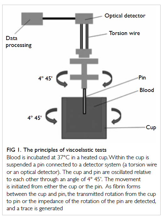
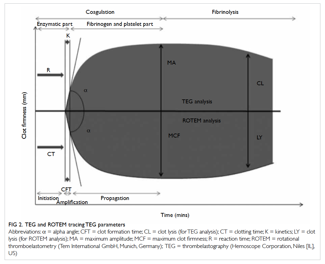
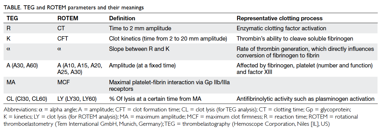
Thrombelastography was first described in 1948 by Hartert6 as a method to assess the viscoelastic properties of coagulation in whole blood under low shear conditions. The VHAs give a graphic presentation of clot formation and subsequent lysis.3 They measure the viscoelastic properties of a clot as it forms in a cup after the addition of activators. As the cup or pin oscillates, the torque of the rotational cup, which is directly related to the strength of the formed clot, is transmitted via the pin to a mechanoelectrical transducer (Fig 1). The signals are then analysed by a computer that produces a trace graph. The latter is divided into several parts with each reflecting different stages of the haemostatic process (Fig 2). Although the nomenclatures differ between TEG and ROTEM, both provide information on the speed of coagulation initiation (coagulation factor activation), kinetics of clot growth (thrombin generation and cleavage of fibrinogen), clot strength, and fibrinolysis (Table). Different tracing morphology and parameters will indicate which component of the coagulation process may be dysfunctional.
 

Figure 1. The principles of viscoelastic tests
 

Figure 2. TEG and ROTEM tracing TEG parameters
 

Table. TEG and ROTEM parameters and their meanings
 
Theoretical advantages of viscoelastic haemostatic assays over conventional coagulation tests
Viscoelastic haemostatic assays assess the combined influence of circulating plasmatic and cellular (platelets, erythrocytes, leukocytes, microparticles) elements. Results are available within 10 minutes, which can be utilised at the point-of-care in different clinical settings. The end-points are clinically relevant, allowing goal-directed coagulation factor transfusions,7 8 9 with a reduction in allogeneic blood product usage in trauma, cardiac, and liver surgery cases.10 11 12 For example, fibrinogen concentrates could be given in situations with decreased maximum amplitude (MA)/maximum clot firmness (MCF). Last but not least, as fibrinolysis plays an important role in both trauma-associated coagulopathy as well as bleeding in patients undergoing elective cardiac and liver surgery, VHAs serve as the only tests available for rapid identification and quantification of hyperfibrinolysis in those situations.
 
Moreover, VHAs can be used in diagnosing surgical bleeding when the results of CCTs are normal, and also play an important role in the assessment of hypercoagulable states, prediction of bleeding risks in patients with liver and renal failures, and as a research tool to improve the understanding of the impact of various interventions on coagulation.
 
Trauma surgery
Uncontrolled haemorrhage accounts for more than 50% of all trauma-related deaths within the first 48 hours after hospitalisation.13 14 15 16 Current evidence suggests that haemodilution, hypothermia, acidaemia, and the consumption of clotting factors all play roles in the pathogenesis of coagulopathy in trauma.17 Hyperfibrinolysis is associated with more severe injury, greater coagulation abnormalities, and a higher mortality rate. Kashuk et al18 suggested that primary fibrinolysis occurred early in severely injured patients, leading to rapid clot dissolution.
 
Massive transfusion of blood products in trauma is not without risks, and may be associated with increased risks of acute respiratory distress syndrome, multi-organ failure, volume overload, and transfusion-related infections. In particular, the risk of transfusion-related acute lung injury following fresh frozen plasma transfusion is recognised as the most important cause of transfusion-associated mortality and morbidity.19 Currently, the decision of what, when, and how much blood products to transfuse in massive trauma-related bleeding is mainly empirical, though often supplemented by results from CCTs. The laboratory diagnosis of fibrinolysis, unfortunately, is often a difficult one, requiring multiple tests such as euglobulin lysis time (ELT), platelet count, fibrinogen, protein S, protein C, and antithrombin levels, all of which may not be readily available in the acute phase of trauma management. Moreover, D-dimer (a marker of fibrinolysis) is elevated in virtually all trauma patients and is, therefore, not a useful indicator.
 
The VHAs, on the other hand, may provide distinct advantages by promptly diagnosing trauma coagulopathy (especially hyperfibrinolysis), guiding blood transfusions, and providing potential prognostic indicators. In a systematic review of 12 reported studies, Sankarankutty et al20 found that TEG/ROTEM could be used to diagnose hyperfibrinolysis accurately. There were otherwise inconsistent correlations between VHAs and CCTs; the most consistent correlation identified was between TEG MA and ROTEM MCF with platelet count and aPTT. Davenport et al21 showed that ELT, which was used as a gold standard test for the detection of fibrinolysis, was correlated with clot amplitude at 10 minutes and 15 minutes, MCF, and clot lysis index at 60 minutes. This has important implications in the use of antifibrinolytic agents and fibrinogen for targeted management.22 As demonstrated in the CRASH-2 study, trauma patients who received an antifibrinolytic agent within 3 hours of trauma experienced a significant reduction in mortality.23
 
Other studies also provided evidence to support VHA-guided transfusion practice. Schöchl et al24 25 reported that ROTEM-guided haemostatic therapy with coagulation factor concentrates (eg fibrinogen concentrate, prothrombin complex concentrate) could facilitate early and effective correction of coagulopathy, as well as reduce transfusion of allogeneic blood products. Kashuk et al26 27 showed that goal-directed resuscitation based on rapid-TEG (rTEG) could reduce the use of fresh frozen plasma. In terms of prognostication, TEG MA and ROTEM MCF have been found to be associated with mortality, while excessive fibrinolysis diagnosed by either TEG clot lysis (CL) or ROTEM maximum lysis (ML) was an independent predictor of mortality.
 
Cardiac surgery
Heparin is used during cardiopulmonary bypass in open-heart surgery, and excessive postoperative bleeding has been attributed to the insufficient reversal of heparinisation with protamine sulfate. This is partly due to the fact that conventional monitoring by means of activated clotting time may fail to differentiate between the contributions from heparinisation, dilution, and platelet dysfunction.28 29 Johansson et al30 reviewed over 3250 cardiac patients reported in 16 studies, and demonstrated superiority of VHAs over CCTs in predicting bleeding and the need for re-operation. The total number of blood transfusions was reduced with VHA-guided transfusion compared with CCT-based practices. The degree of heparinisation could be evaluated with assays that neutralise the heparin (heparinase in TEG and HepTEM [heparinase-modified thromboelastometry] in ROTEM), so that non-heparin–related haemostatic problems could be detected. These findings were corroborated in a recent Cochrane analysis.11
 
Liver transplantation
A temporary hypercoagulable state is common after liver transplantation due to imbalance between the procoagulant and anticoagulant systems, as well as fibrinolytic shutdown.31 This may play a role in the early development of hepatic artery thrombosis.32 The use of CCT monitoring alone in the first week post-transplantation may lead to significant bleeding complications in certain patients.33 34 In contrast, VHAs can accurately assess postoperative hypercoagulability and thrombosis. Cerutti et al35 showed that TEG monitoring could demonstrate hypercoagulability in the majority of the subjects after living donor liver transplantation and may, therefore, be used to guide antithrombotic treatment in the perioperative period.
 
Monitoring antiplatelet therapy
With the rising prevalence of atherosclerotic diseases such as ischaemic heart disease and stroke, it is not uncommon for surgical patients to be on antiplatelet drugs. The latter may worsen bleeding in trauma, increase transfusion requirement, and increase the need for re-operation after elective surgery.36 37 Unfortunately, CCTs do not test for platelet inhibition due to antiplatelet therapy, and tests for platelet function such as platelet aggregometry do not provide results rapidly enough for intra-operative monitoring and assessment of coagulopathy in emergency surgery. Furthermore, although antiplatelet drugs are thought to work primarily by decreasing platelet aggregation, they may also have anticoagulant effects.36 In this respect, the MA/MCF from TEG/ROTEM may serve to reveal the overall platelet function and fibrinogen levels. In differentiating whether reduced clot strength is due to low fibrinogen or low platelet concentration/functionality, both TEG and ROTEM have specific assays (functional fibrinogen and FIBTEM [fibrin-based clotting], respectively) that block platelet contributions with the addition of platelet inhibitors. At present, however, there is no clinical evidence to support the routine use of VHAs for monitoring platelet function prior to surgery. Further studies are required to demonstrate the clinical benefits of these tests in reducing transfusion requirement and improving surgical outcomes.
 
Monitoring anticoagulation therapy
Anticoagulation therapy has similar negative impact on emergency and elective surgery as antiplatelet therapy. Point-of-care VHAs have been shown to be useful in monitoring treatments with low-molecular-weight heparin (LMWH), heparinoids (eg danaparoid), and unfractionated heparin.38 However, to increase the sensitivity of VHAs for the effects of LMWH and heparinoids, both standard and heparinase-modified tests have to be carried out. Recently, direct thrombin inhibitors such as dabigatran are being used increasingly for the prevention and treatment of venous thromboembosis, acute coronary syndrome, and heparin-induced thrombocytopenia.39 The monitoring of this group of patients is facilitated by VHAs with the use of ecarin clotting time, in which ecarin is used to activate thrombin directly.40 41
 
Limitations of viscoelastic haemostatic assays
There is at present a lack of universally agreed algorithms for the use of VHAs. In a recent systematic review of nine randomised clinical trials (eight in cardiac surgery and one in liver transplantation) comparing VHA-based algorithms with standard treatments, the only supporting evidence identified for the use of VHAs was for detection of hyperfibrinolysis.2
 
On an operational level, VHAs have been criticised for not having undergone the same evaluation process as CCTs. There are wide technical variations in how VHAs are performed, and the machine requires calibrations 2 to 3 times a day which causes significant inconvenience in daily point-of-care usage.42 While originally designed for fresh whole blood with no additional activators, subsequent modifications have included sample anticoagulation and the use of different activators to standardise the initiation of coagulation. Patients’ gender, age, and alcohol drinking may also affect the result. Moreover, the normal reference ranges for VHAs were derived from hospitalised surgical patients in one study, and from a small sample of 12 healthy volunteers in another.43 Hence, each centre is recommended to generate its own reference range by specially trained personnel according to the guidelines from the Clinical Laboratory Improvement Amendments.44 The latter, which regulates all laboratory tests in the United States, demands a minimum of 30 to 40 subjects for special coagulation tests.45 All these necessitate an active and tightly controlled quality assurance programme.42
 
Despite its proposed role as a point-of-care test, a complete viscoelastic test can take up to 60 minutes although useful information can be obtained in approximately 10 minutes, when MCF is reached. This may be further increased by a 30- to 40-minute wait for anticoagulated samples.42 To compensate for this, a modified rTEG has been developed. It uses tissue factors as activators in addition to kaolin, and can provide results on MCF more rapidly. However, information on coagulation and clot formation time is limited. Although there is a strong correlation between rTEG and conventional TEG in terms of overall clot strength and platelet function, there is only moderate correlation in the degree of fibrin cross-linking and poor correlation in evaluating thrombolysis.46
 
There are conditions in which VHAs may fail to detect haemostatic dysfunction. The test setting is at 37°C. Therefore, the effect of hypothermia, which has a well-recognised negative impact on coagulation, may not be recognised.47 48 Viscoelastic haemostatic assays do not assess the endothelial contribution to haemostasis since an activator is directly added to initiate coagulation during the test. This means that the diagnosis of certain conditions such as von Willebrand disease is not possible with VHAs.48
 
Lastly, the interchangeability of results between TEG and ROTEM has been questioned. Although they share the same fundamental principles, and similar parameters, hardware and techniques, the results generated may not be directly comparable, possibly due to the use of different activators. Consistent correlations are limited to that between TEG MA and ROTEM MCF measurements, and that between TEG CL and ROTEM ML in diagnosing hyperfibrinolysis and predicting mortality.
 
Conclusions
Haemostatic function is a critical factor determining patient outcomes in emergency or elective surgery. The increasing use of antiplatelet agents and anticoagulants has potentially increased the risks of haemorrhages and need for transfusion. Conventional coagulation tests have limitations in detecting haemostatic dysfunction in subgroups of patients and are largely ineffective in diagnosing hyperfibrinolysis. The VHAs are potentially useful point-of-care tools to provide information on clot formation, clot strength, and fibrinolysis, as well as to guide goal-directed transfusion and antifibrinolytic therapy. They may also be used to monitor antiplatelet and anticoagulant therapy. However, standardisation of techniques and reference ranges is required before these tests can be widely used in different clinical settings. Further studies are needed to validate different algorithms, and address coagulopathies in situations like hypothermia or endothelial dysfunction.
 
Declaration
No conflicts of interest were declared by the authors.
 
References
1. Roberts HR, Hoffman M, Monroe DM. A cell-based model of thrombin generation. Semin Thromb Hemost 2006;32 Suppl 1:32-8. CrossRef
2. Wikkelsoe AJ, Afshari A, Wetterslev J, Brok J, Moeller AM. Monitoring patients at risk of massive transfusion with thrombelastography or thromboelastometry: a systematic review. Acta Anaesthesiol Scand 2011;55:1174-89. CrossRef
3. Ganter MT, Hofer CK. Coagulation monitoring: current techniques and clinical use of viscoelastic point-of-care coagulation devices. Anesth Analg 2008;106:1366-75. CrossRef
4. Kaufmann CR, Dwyer KM, Crews JD, Dols SJ, Trask AL. Usefulness of thrombelastography in assessment of trauma patient coagulation. J Trauma 1997;42:716-20. CrossRef
5. Rugeri L, Levrat A, David JS, et al. Diagnosis of early coagulation abnormalities in trauma patients by rotation thrombelastography. J Thromb Haemost 2007;5:289-95. CrossRef
6. Hartert H. Blutgerinnungsstudien mit der thrombelastographie, einem neuen untersuchungsverfahren [in German]. Klin Wochenschr 1948;26:577-83. CrossRef
7. Bontempo FA, Lewis JH, Van Thiel DH, et al. The relation of preoperative coagulation findings to diagnosis, blood usage and survival in adult liver transplantation. Transplantation 1985;39:532-6. CrossRef
8. Steib A, Gengenwin N, Freys G, Boudjema K, Levy S, Otteni JC. Predictive factors of hyperfibrinolytic activity during liver transplantation in cirrhotic patients. Br J Anaesth 1994;73:645-8. CrossRef
9. Dzik WH, Arkin CF, Jenkins RL, Stump DC. Fibrinolysis during liver transplantation in humans: role of tissue-type plasminogen activator. Blood 1988;71:1090-5.
10. Afshari A, Wikkelsø A, Brok J, Møller AM, Wetterslev J. Thrombelastography (TEG) or thromboelastometry (ROTEM) to monitor haemotherapy versus usual care in patients with massive transfusion. Cochrane Database Syst Rev 2011;(3):CD007871.
11. Wang SC, Shieh JF, Chang KY, et al. Thromboelastography-guided transfusion decreases intraoperative blood transfusion during orthotopic liver transplantation: randomized clinical trial. Transplant Proc 2010;42:2590-3. CrossRef
12. Aoki K, Sugimoto A, Nagasawa A, Saito M, Ohzeki H. Optimization of thromboelastography-guided platelet transfusion in cardiovascular surgery. Gen Thorac Cardiovasc Surg 2012;60:411-6. CrossRef
13. Sauaia A, Moore FA, Moore EE, et al. Epidemiology of trauma deaths: a reassessment. J Trauma 1995;38:185-93. CrossRef
14. Brohi K, Cohen MJ, Davenport RA. Acute coagulopathy of trauma: mechanism, identification and effect. Curr Opin Crit Care 2007;13:680-5. CrossRef
15. Maegele M, Lefering R, Yucel N, et al. Early coagulopathy in multiply injury: an analysis from the German Trauma Registry on 8724 patients. Injury 2007;38:298-304. CrossRef
16. MacLeod JB, Lynn M, McKenney MG, Cohn SM, Murtha M. Early coagulopathy predicts mortality in trauma. J Trauma 2003;55:39-44. CrossRef
17. Hess JR, Brohi K, Dutton RP, et al. The coagulopathy of trauma: a review of mechanisms. J Trauma 2008;65:748-54. CrossRef
18. Kashuk JL, Moore EE, Sawyer M, et al. Primary fibrinolysis is integral in the pathogenesis of the acute coagulopathy of trauma. Ann Surg 2010;252:434-4.
19. Stainsby D, Jones H, Asher D, et al. Serious hazards of transfusion: a decade of hemovigilance in the UK. Transfus Med Rev 2006;20:273-82. CrossRef
20. Sankarankutty A, Nascimento B, Teodoro da Luz L, Rizoli S. TEG® and ROTEM® in trauma: similar test but different results. World J Emerg Surg 2012;7 Suppl 1:S3. CrossRef
21. Davenport R, Manson J, De’Ath H, et al. Functional definition and characterization of acute traumatic coagulopathy. Crit Care Med 2011;39:2652-8.
22. Henry DA, Carless PA, Moxey AJ, et al. Anti-fibrinolytic use for minimising perioperative allogeneic blood transfusion. Cochrane Database Syst Rev 2011;(1):CD001886.
23. Roberts I, Shakur H, Coats T, et al. The CRASH-2 trial: a randomised controlled trial and economic evaluation of the effects of tranexamic acid on death, vascular occlusive events and transfusion requirement in bleeding trauma patients. Health Technol Assess 2013;17:1-79.
24. Schöchl H, Nienaber U, Maegele M, et al. Transfusion in trauma: thromboelastometry-guided coagulation factor concentrate-based therapy versus standard fresh frozen plasma-based therapy. Crit Care 2011;15:R83. CrossRef
25. Schöchl H, Nienaber U, Hofer G, et al. Goal-directed coagulation management of major trauma patients using thromboelastometry (ROTEM®)-guided administration of fibrinogen concentrate and prothrombin complex concentrate. Crit Care 2010;14:R55. CrossRef
26. Kashuk JL, Moore EE, Le T, et al. Noncitrated whole blood is optimal for evaluation of postinjury coagulopathy with point-of-care rapid thrombelastography. J Surg Res 2009;156:133-8. CrossRef
27. Kashuk JL, Moore EE, Wohlauer M, et al. Initial experiences with point-of-care rapid thrombelastography for management of life-threatening postinjury coagulopathy. Transfusion 2012;52:23-33. CrossRef
28. Koster A, Fischer T, Praus M, et al. Hemostatic activation and inflammatory response during cardiopulmonary bypass: impact of heparin management. Anesthesiology 2002;97:837-41. CrossRef
29. Koster A, Despotis G, Gruendel M, et al. The plasma supplemented modified activated clotting time for monitoring of heparinization during cardiopulmonary bypass: a pilot investigation. Anesth Analg 2002;95:26-30. CrossRef
30. Johansson PI, Sølbeck S, Genet G, Stensballe J, Ostrowski SR. Coagulopathy and hemostatic monitoring in cardiac surgery: an update. Scand Cardiovasc J 2012;46:194-202. CrossRef
31. Stahl RL, Duncan A, Hooks MA, Henderson JM, Millikan WJ, Warren WD. A hypercoagulable state follows orthotopic liver transplantation. Hepatology 1990;12:553-8. CrossRef
32. Lisman T, Porte RJ. Hepatic artery thrombosis after liver transplantation: more than just a surgical complication? Transpl Int 2009;22:162-3. CrossRef
33. Francoz C, Belghiti J, Vilgrain V, et al. Splanchnic vein thrombosis in candidates for liver transplantation: usefulness of screening and anticoagulation. Gut 2005;54:691-7. CrossRef
34. Widen A, Rolando N, Manousou P, et al. Anticoagulation after liver transplantation: a retrospective audit and case-control study. Blood Coagul Fibrinolysis 2009;20:615-8. CrossRef
35. Cerutti E, Stratta C, Romagnoli R, et al. Thromboelastogram monitoring in the perioperative period of hepatectomy for adult living liver donation. Liver Transpl 2004;10:289-94. CrossRef
36. Jacob M, Smedira N, Blackstone E, Williams S, Cho L. Effect of timing of chronic preoperative aspirin discontinuation on morbidity and mortality in patients having combined coronary artery bypass grafting and valve surgery. Am J Cardiol 2012;109:824-30. CrossRef
37. Jacob M, Smedira N, Blackstone E, Williams S, Cho L. Effect of timing of chronic preoperative aspirin discontinuation on morbidity and mortality in coronary artery bypass surgery. Circulation 2011;123:577-83. CrossRef
38. Coppell JA, Thalheimer U, Zambruni A, et al. The effects of unfractionated heparin, low molecular weight heparin and danaparoid on the thromboelastogram (TEG): an in-vitro comparison of standard and heparinase-modified TEGs with conventional coagulation assays. Blood Coagul Fibrinolysis 2006;17:97-104. CrossRef
39. Di Nisio M, Middeldorp S. Büller HR. Direct thrombin inhibitors. N Engl J Med 2005;353:1028-40. CrossRef
40. Nielsen VG, Steenwyk BL, Gurley WQ, Pereira SJ, Lell WA, Kirklin JK. Argatroban, bivalirudin, and lepirudin do not decrease clot propagation and strength as effectively as heparin-activated antithrombin in vitro. J Heart Lung Transplant 2006;25:653-63. CrossRef
41. Carroll RC, Chavez JJ, Simmons JW, et al. Measurement of patients’ bivalirudin plasma levels by a thrombelastograph ecarin clotting time assay: a comparison to a standard activated clotting time. Anesth Analg 2006;102:1316-9. CrossRef
42. da Luz LT, Nascimento B, Rizoli S. Thrombelastography (TEG): practical considerations on its clinical use in trauma resuscitation. Scand J Trauma Resusc Emerg Med 2013;21:29. CrossRef
43. Scarpelini S, Rhind SG, Nascimento B, et al. Normal range values for thromboelastography in healthy adult volunteers. Braz J Med Biol Res 2009;42:1210-7. CrossRef
44. Chan KL, Summerhayes RG, Ignjatovic V, Horton SB, Monagle PT. Reference values for kaolin-activated thromboelastography in healthy children. Anesth Analg 2007;105:1610-3. CrossRef
45. Centers for Disease Control and Prevention. Considering the clinical laboratory improvement amendment. CDC website: http://wwwn.cdc.gov/cliac/pdf/Addenda/cliac0210/Addendum%20Y.pdf. Accessed 9 Feb 2010.
46. Lee TH, McCully BH, Underwood SJ, Cotton BA, Cohen MJ, Schreiber MA. Correlation of conventional thrombelastography and rapid thrombelastography in trauma. Am J Surg 2013;205:521-7. CrossRef
47. Rundgren M, Engström M. A thromboelastometric evaluation of the effects of hypothermia on the coagulation system. Anesth Analg 2008;107:1465-8. CrossRef
48. Johansson PI, Stissing T, Bochsen L, Ostrowski SR. Thrombelastography and thromboelastometry in assessing coagulopathy in trauma. Scand J Trauma Resusc Emerg Med 2009;17:45. CrossRef
 
Find HKMJ in MEDLINE:
 

Management of secondary lymphoedema

Hong Kong Med J 2014;20(6):519–28 | Epub 29 Aug 2014
DOI: 10.12809/hkmj134116
© Hong Kong Academy of Medicine. CC BY-NC-ND 4.0
 
REVIEW ARTICLE     CME 
Management of secondary lymphoedema
TW Chiu, FHKAM (Surgery)
Division of Plastic Aesthetic and Reconstructive Surgery, Prince of Wales Hospital, Shatin, New Territories, Hong Kong
 
Corresponding author: Dr TW Chiu (torchiu@surgery.cuhk.edu.hk)
 Full paper in PDF
Abstract
Lymphoedema is a chronic, progressive condition. There is no cure but it is most easily managed with early recognition and therapy; those who do not have treatment tend to worsen rapidly and advanced disease is more difficult to treat than early disease. Surgery for lymphoedema is often regarded as a last resort but traditional excisional techniques that have been slightly modified for modern practice have shown good results, whilst newer microsurgical reconstruction techniques show promise although long-term results are lacking. This report provides an update on the therapy of lymphoedema.


Introduction
Lymphoedema is characterised by an imbalance of lymphatic flow leading to accumulation of protein-rich fluid in the interstitium of subcutaneous tissues. The consequent swelling may cause cosmetic and functional impairment, with significant physical and psychological morbidity. There is progressive damage to the lymphatics with inflammation, fibrosis and more swelling, eventually leading to elephantiasis. Recurrent infection is a common complication whilst lymphangiosarcomas are rare, occurring in 0.03% of patients surviving more than 10 years after mastectomy.1
 
It is traditional to classify lymphoedema into primary or secondary forms—in the former there is congenital lymphatic dysfunction related to dysplasia/malformation whilst in the latter there is disruption of lymphatic outflow related to another disease process or due to iatrogenic mechanisms.
 
Primary lymphoedema
The traditional subdivision of primary lymphoedema according to the time of onset has little clinical significance. There is confusion in the literature regarding the terms used, particularly, the various eponymous syndromes that have been described; most cases of primary lymphoedema are not associated with specific syndromes.
  • Congenita (10% of cases): the swelling is often present at birth (any swelling that begins before the age of 2 years is included in this group).
  • Praecox (80% of cases): patients present with swelling before the age of 35 years.
  • Tarda (10% of cases): patients present after the age of 35 years with swelling that is usually bilateral. Lymphatic vessels tend to be hyperplastic.
  •  
    It is important to appreciate that primary lymphoedema is a heterogeneous group with many subtypes occurring due to many different causes which are generally poorly understood. The term Milroy’s disease is often used interchangeably with lymphoedema congenita—it should only be applied to the group of inherited congenital lymphoedemas that demonstrates autosomal dominant inheritance; it is linked to vascular endothelial growth factor (VEGF) receptor–3 mutations (FLT4 gene on locus 5q35.3)2 3 and some evidence reveals that there is a functional defect of absorption rather than a gross structural defect.
     
    Lymphoedema praecox is the commonest type (80% of cases) and the swelling may be unilateral and limited to the foot/calf region. Patients lack distal lymphatics (hypoplasia) and often have a strong family history, some demonstrating autosomal dominant inheritance with different mutations in FOXC2 gene on chromosome 16.4 Some patients with praecox have distichiasis (double row of eyelashes) and form a distinct syndromic entity—lymphoedema distichiasis (mutation on 16q24.3).5 Patients with Meige’s disease, which is a subset of lymphoedema, usually present at puberty and the term should be reserved for those with the familial form of the disease (with as-yet unknown mutation).
     
    Secondary lymphoedema
    Worldwide, the commonest cause of secondary lymphoedema is filariasis, caused by infection with Wuchereria bancrofti. However, this condition is rare in developed countries such as Hong Kong where lymphoedema is most commonly related to malignant disease and, particularly, its treatment with surgery and/or irradiation.6 Upper limb lymphoedema most often follows the treatment of breast cancer, typically after a latent period of variable duration—77% of cases present within 3 years of surgery7 but the condition can arise at any time.
     
    Although the exact mechanism of the response of lymphatic channels to trauma is unknown, in general, the more extensive the surgery the greater is the damage. Thus, a formal axillary node dissection carries a risk of up to 20% compared to 4% to 10% following sentinel node biopsy.8 9 10 11 Irradiation causes fibrosis and inhibits lymphangiogenesis and, approximately, doubles the risk of developing lymphoedema after nodal surgery. Lymph vessels do have some regenerative capacity12 and not all patients undergoing surgery and/or radiotherapy develop lymphoedema; it may be related to some underlying susceptibility that is as yet undefined, and may be, possibly, genetic.13 Patients with lymphoedema often have subtle lymphatic anomalies in the contralateral limb.14
     
    Staging
    The International Society of Lymphology staging is the most commonly used system (Table 115), but it is somewhat flawed in that it is based only on physical findings. Some experts suggest incorporating measures of quality of life (QOL) to improve its usefulness.16 The classification offered by Campisi et al17 shows congruence with indocyanine green (ICG) dermal backflow patterns, which provides an indication of lymphatic function (Table 2).17 18 19 Bioimpedance spectroscopy uses electric current to measure the degree of tissue fluid retention and is useful in detecting early-stage lymphoedema including stage 0 disease.
     

    Table 1. International Society of Lymphology (ISL) staging 2003 with ratification in 2009 consensus document; stage 0 (latent lymphoedema) is a recent addition15
     

    Table 2. Campisi staging 201017
     
    The most commonly used method of objectively assessing the swelling is some form of conal measurement, such as measuring the circumference at 4 cm intervals, which is more practical than water displacement methods20 21 but is, supposedly, similar in accuracy.22 A perometer uses infrared rays to measure limb cross-sections at multiple intervals and, thus, determine the volume of the limb.23 The author has been exploring the idea of using Kinect (part of the Microsoft Xbox game system) as a three-dimensional scanner to gauge volume in collaboration with the Department of Computer Science and Engineering at The Chinese University of Hong Kong.
     
    Imaging
    In most cases, diagnosis can be made from the clinical history and examination, although some co-morbidities may confound the clinical picture. Lymphoedema can be assessed by common imaging techniques including computed tomography, magnetic resonance imaging, and duplex ultrasonography that can reliably show volume differences between the affected and normal limbs, the presence of subcutaneous fatty fibrosis, and help exclude proximal obstruction in late-onset unilateral lymphoedema which may be due to an occult visceral tumour.
     
    Lymphangiography involves direct cannulation of a lymphatic vessel on the dorsum of the foot or hand (under magnification). An oil-based contrast material is then injected through this vessel and serial plain radiographs of the limb are taken, allowing the lymphatics to be precisely delineated. Due to the potential of damage to the lymphatic vessels, it can theoretically worsen lymphoedema and, thus, is not commonly used.
     
    Radionuclide lymphoscintigraphy is the current standard investigation for evaluation of lymphatic function. Technetium-labelled colloid is injected into the web spaces of the toes or fingers, and drainage of the colloid from the injection site and the time taken to move proximally are recorded using a gamma camera. It is minimally invasive and enables making both qualitative and quantitative analyses. It does not require dye injection, a method that has been occasionally complicated by allergic skin reactions or anaphylaxis.
     
    Magnetic resonance lymphangiography may replace lymphoscintigraphy as it does not require direct injection of contrast and avoids the use of ionising radiation using a magnetic contrast medium to provide a sharp image of lymphatic vessels. In our early experience with this investigation, reproducibility has been a potential issue and needs further evaluation.
     
    Near-infrared fluorescence imaging with ICG is a promising emerging imaging modality that allows dynamic study of even small lymphatics in the skin.
     
    Treatment
    There is no cure for lymphoedema but it is easily managed with early recognition and therapy. Those who do not have treatment tend to worsen rapidly and advanced disease tends to be more difficult to treat than early disease. Patients are best treated in a specialised clinic24; inexperienced staff may delay treatment, or worse, advocate inappropriate treatments.
     
    The standard of care for lymphoedema is, what is commonly referred as, complex (or combined or complete) physical therapy (CPT, sometimes called complex decongestive therapy [CDT]), which is a staged combination of various components in two phases. The actual treatment regimen varies significantly by locality but, in general, 60% to 70% of compliant patients will respond to CPT when administered by specially trained therapists, with an average volume reduction of 50%.25 26 However, the time and effort involved, as well as the associated moderate discomfort, lead to decreased patient compliance, particularly in the long term.
    (1) Phase 1, also known as the ‘decongestive phase’, often requires the patient to be admitted as an in-patient as the regimen is intensive. Treated 5 days a week, it may take 4 to 6 weeks or more to have an effect on the limb volume, depending on the severity of the disease. A lack of effect may be due to improper technique, non-compliance, or incorrect diagnosis.
  • Manual lymph drainage (MLD) is not the same as massage; it is a much lighter, slow and specific action that aims to promote lymph movement in the superficial tissues away from the swelling.
  • Compression bandage is applied to reduce limb size but the bandages used are not of the standard variety—they are short and non-stretch, and applied with more pressure distally than proximally. This aims to move fluid out of the limb (hence the proximal portions need to be emptied with MLD first). It is only really effective within a CPT programme; the efficacy is reduced considerably in advanced disease.
  • Nail/skin care and exercises with bandaging/pressure garments as tight as tolerated to oppose the filtration pressure and provide counterforce to muscle contractions. Concerns that exercise may exacerbate or trigger lymphoedema have not been proven in studies.
  • (2) Phase 2, or the maintenance phase, can be out-patient–based or as self-care in selected trained patients. Patients should wear pressure garments during the day and compression bandages at night (alternatives include specifically designed garments such as Reid sleeves). Intermittent pneumatic compression machines are sometimes used but care needs to be taken to ensure that they have appropriate design, action, and pressures (usually 30-60 mm Hg though pressures of >45 mm Hg can cause lymphatics to rupture). Pneumatic compression is to be avoided in those with chronic non-pitting disease or those with active infection.
     
    Systematic reviews support the use of CDT and MLD. Conservative therapy can give good results but the effects are temporary without the maintenance and continued compression. In Hong Kong, the climate may reduce patient compliance with pressure garments. General care is also important; patients are advised to avoid even minor degrees of trauma such as venepuncture, insect bites, and acupuncture. The evidence for some of the following preventive practices is low but they are simple to follow.
  • Air travel: maintain hydration and mobilisation, whilst some experts suggest wearing pressure garments.
  • Avoid overuse of the affected limb.
  • Avoid trauma; take care when cutting nails, avoid needlestick/venepuncture, and blood pressure measurement. Patients can wear medical alert bracelets to inform others of the condition.
  • Avoid extremes of heat/cold and overtight clothing.
  •  
    Medications such as benzopyrenes and diuretics are not useful and will not be discussed any further. Oral penicillins such as amoxicillin and dicloxacillin should be started early when there is evidence of infection in a lymphoedematous limb and continued until the signs of inflammation resolve. There is borderline support for the use of prophylactic antibiotics in those who have more than two to three episodes of infection a year. Many modalities have been promoted for the non-surgical treatment of lymphoedema but at present only low-level laser therapy has approval from the US Food and Drug Administration (FDA), although evidence of its long-term effects is lacking—it generates low-intensity light (650-1000 nm) that is believed to promote lymph vessel regeneration and increase lymph pumping. The typical response is moderate and slow, and requires repeated treatments. Near-infrared light therapy aims to increase nitric oxide in the tissues to improve tissue repair and lymph regeneration but has not received FDA approval, whilst electrical stimulation is not recommended based on current evidence.27
     
    Surgery
    For reasons such as concerns of scarring or perceived lack of effect, surgery has often been regarded as a ‘last resort’, meaning that there is often a delay before patients are referred28 by which time they may only be suited for ‘salvage’ procedures. Whilst initial attempts at ‘physiological’ reconstruction were met with disappointing late results, improved understanding of the pathophysiology accompanied by improved microsurgical techniques have seen the development of newer techniques that seem to potentially offer better outcomes.
     
    Physiological/reconstructive techniques
    Physiological techniques aim to repair the damage and increase the return of lymph to the circulation, by reconnecting the lymphatic pathways above and beyond the obstruction, either directly (lymphatic to lymphatic) or indirectly via another segment such as veins/venules. Campisi et al17 29 30 have been a pioneer of these microsurgical techniques and classified them into ‘derivative’ techniques (essentially a lymphovenous bypass) and ‘reconstructive’ techniques (lymphatic-to-lymphatic connections).
     
    Lymphovenous anastomosis or bypass
    When interpreting the literature describing lymphovenous bypass, it is important to note the fundamental difference between early (lymphovenous) and newer (lymphaticovenular) techniques. The bypass concept was first described in 1963 in a rat model31 whilst Yamada32 and O’Brien et al33 were the first to use it in patients. O’Brien et al33 anastomosed lymphatics to veins measuring approximately 1 to 3 mm diameter and reported an average volume reduction of 44% in this series with 14 years’ follow-up. Campisi et al30 reported an average of 67% reduction in volume and 87% reduction in cellulitis in this large long-term series. Overall, 85% of patients were able to stop conservative management. However, one criticism of these retrospective studies was patient heterogeneity.
     
    However, Damstra et al34 found no improvement in patients with postmastectomy lymphoedema who were treated with lymphovenous anastomosis/bypass; a prospective study of 10 patients demonstrated only a 4.8% volume reduction at 3 months that was further reduced to 2% after 1 year, with minimal improvement in reported QOL. Boccardo et al35 36 performed lymphovenous bypass at the time of breast surgery with the aim of preventing lymphoedema—they anastomosed tributaries of the axillary vein to lymphatics with a patency rate of 95.6% but found no difference in limb volume compared with controls at up to 18 months’ follow-up.
     
    Lymphaticovenular anastomoses
    In Koshima et al’s opinion,37 38 39 there were inherent problems in lymphovenous anastomoses: (1) it is difficult to find larger lymphatic trunks, most are 0.8 mm or under in diameter; (2) reduced lymphatic pumping function; and (3) increased venous pressure and high rate of thrombosis due to blood at anastomotic site. According to these authors, lymphaticovenular anastomosis (LVA) offered better vessel size match compared with lymphovenous anastomoses. In 52 patients with an average of 2.1 LVAs per patient, there was a mean reduction of 41.8% in leg circumference at a mean follow-up of 14.5 months. Benefit could be demonstrated even in patients with stage III/IV disease, with recurrent lymphangitis and fibrosis. Koshima et al37 38 39 have also applied the technique in early disease for prophylaxis against development of fibrotic disease.
     
    The Tokyo group uses ICG fluorescent lymphography to stage the disease severity based on the amount of dermal backflow, and thus, select patients for surgery.40 41 Subdermal vessels are explored through small (<3 cm) skin incisions, aiming to use lymphatic vessels of approximately 0.2 mm in diameter with venules sized 0.5 mm or less on the basis that smaller veins will have lower pressures. Some call this ‘supermicrosurgery’ although the use of the term is rather arbitrary. The procedure can be performed under local anaesthesia with short hospital stay, which, in the author’s opinion, is one of the biggest advantages of this technique.
     
    The studies from Koshima et al’s unit37 38 39 have demonstrated an average volume reduction of 82.5% in those not responding to CPT. The Chang’s series42 demonstrated a 35% reduction of lymphoedema in breast cancer patients 1 year after LVA. Cormier et al43 reviewed eight studies and calculated a mean volume reduction of 54.0%. It is generally accepted that microsurgery will offer better results in early disease when patients have some healthy lymphatics before progressing to the fibrotic stage when damage is irreversible.
     
    Lymph node transfer
    Wongtrungkapun44 performed lymph node transfer in patients with filariasis; one to two groin nodes were partially decapsulated and anastomosed to the saphenous vein. Becker et al45 46 described the transfer of lymph nodes from the groin to the axilla in 24 patients with postmastectomy lymphoedema; 62.5% of the patients were said to be ‘cured’ and able to discontinue physiotherapy. Overall, in Becker et al’s experience,45 46 98% of the patients had some improvement; only 2% had repeat infections. Whilst 40% of patients with stage 1 or 2 lymphoedema had complete normalisation and did not require additional conservative therapy, those with stage 3 lymphoedema needed conservative therapy. Lin et al47 anastomosed superficial circumflex iliac nodes at the wrist to treat upper limb lymphoedema and reported a 55% reduction in volume at 56 months, with fewer episodes of cellulitis.
     
    Proponents of node surgery say that LVA or lymphatic-lymphatic anastomosis eventually become occluded (possibly due to the effect of interstitial pressure on low-pressure thin vessels) whilst lymph nodes are supposedly less susceptible. The lymphatic vessels are not actually anastomosed; it seems that the transplanted nodes develop new drainage pathways—proposed theories for this drainage include nodes acting as suction pumps whilst others suggest they are a source of VEGF-C that promotes lymphangiogenesis.
     
    Some experts have developed a procedure to transfer lymph nodes as part of a breast reconstruction procedure48; there may be several other benefits of such a procedure including release of scar tissue in the axilla and the provision of vascularised tissue as a lymphatic bridge.49 Isotope scans at 3 and 6 months demonstrated improved function in all but one patient.48 With this type of surgery, it is important to only harvest nodes of lower abdomen and not the leg; axillary reverse mapping may help to spare limb lymphatics.50
     
    Overall, good long-term functional data are lacking and some authors have had difficulty reproducing published results,25 possibly, due to the significant learning curve. Some have found 38% risk of complications, although mostly transient (eg lymphocele and hydrocele); however, some complications such as iatrogenic lymphoedema and chronic pain may be more persistent.51 They also found no volume difference after a median follow-up of 40 months with interval CDT and pressure garments. Viitanen et al52 found reduced lymphatic flow in the donor site/limb with lymphoscintigraphy without overt clinical lymphoedema in a small group of 10 patients.
     
    Preliminary results suggest that node transfer is more successful if performed sooner after nodal dissection surgery.45 53 Saaristo et al48 regard it as largely experimental but can be justified if performed at the time of breast reconstruction. Some surgeons have looked at animal models, combining node transfer with additional VEGF-C and -D,54 55 and this may offer a new type of treatment; however, the effect on lymphatic metastasis is unknown and deserves attention.
     
    Lymphatic grafting/transplantation
    Other less commonly used procedures include lymphatic grafting. Baumeister et al56 57 58 59 have significant experience in using this technique to treat secondary lymphoedema with an average volume reduction of 65%.60 61 In the lower limb, a long segment of lymphatic vessel is dissected out from the upper inner thigh and tunnelled over to affected contralateral leg for a lymphatic-lymphatic anastomosis. The efficacy in the upper limb where the lymphatic tissue is used as a free graft has also been demonstrated with significant improvement in over 90% of patients, with a mean volume reduction of 22% to 31% whilst scintigraphy shows continued graft function.62 63 64 The dissection of lymphatics is technically challenging; Campisi65 used veins instead of lymphatic vessels to bridge lymphatics.
     
    Some experts have used flaps for lymphatic bridging; for instance, the inclusion of random lymphatics in tissues such as the omentum66 or an axial flap such as the deltopectoral.67 The use of omentum has largely been abandoned as there is no evidence that it actually promotes lymph drainage, and the surgery is associated with high rates of morbidity. Similarly, enteromesenteric bridge operations and tube/thread implants should be avoided, based on current evidence.
     
    Debulking or excision
    Early attempts at treating lymphoedema involved techniques that were mainly based on debulking, that is, removing the oedematous tissue to restore form. The underlying lymphatic dysfunction is not addressed and may actually lead to further deterioration of the condition.
     
    Charles procedure
    The Charles procedure used in 1912 has been wrongly attributed to Sir Richard Charles for the treatment of leg lymphoedema by McIndoe.68 The surgeon had primarily treated scrotal swelling69 and had only described one unsuccessful case involving the lower limb. In simple terms, the lymphoedematous tissue is excised down to the fascial level and the defect is covered with skin grafts. The grafts can be taken from the excised tissue if it is not grossly abnormal70; otherwise, it has to be harvested from another site. Complications include graft breakdown/ulceration, scarring, and recurrence. The aesthetic results are rather poor and, thus, usually reserved for cases with severe skin changes.
     
    Homans-Miller procedure
    The Homans-Miller procedure, first used in 1936, is based on a multistage procedure initially described by Sistrunk71—skin flaps are elevated along one border of the limb, and after the deeper swollen tissue has been excised along with the fascia, the skin flaps are replaced.72 73 It is a traditional practice to avoid surgery at or below ankle, taking care to avoid damage to the common peroneal and sural nerves. A study with 14 years’ follow-up showed that this type of surgery was capable of long-lasting reduction in size in 80% of the treated patients, and associated with improved function and reduced cellulitis.73 It is the most common excisional surgery for lymphoedema. Others have refined this further by preserving the perforator vessels during flap elevation, allowing the flaps to be thinner (5 mm) and, potentially, for both sides to be treated in one stage.74
     
    The Thompson technique used in 1962 involves similar tissue resection whilst also harvesting a dermal flap75 that is buried into the muscle next to the neurovascular bundle, with the aim of creating a bridge for lymphatic return. However, long-term results were similar to excision alone and did not support the theoretical aim of a physiological effect and, thus, has largely been abandoned.
     
    The proven benefits of excisional surgery are often ignored, in part due to misconceptions of morbidity and complications; these were mostly related to early aggressive use of the technique leading to almost total abandonment of procedures in the mid-20th century. Recently Karri et al76 demonstrated that good results are possible with modern application of the Charles procedure that may also be combined with negative-pressure wound therapy.77 Similarly, surgeons have modified the Homans-Miller procedure25 and combined this with postoperative pressure garments to achieve good results.
     
    Liposuction
    Liposuction is a relatively recently described debulking technique based on the observation that there is adipose hypertrophy in lymphoedema. The fat accumulation may be related to altered lipid transport78 79 and corresponds to a non-pitting type of swelling that is not responsive to compression.
     
    O’Brien et al80 were one of the first surgeons to use liposuction to treat lymphoedema, and reported a volume reduction of 23%. Brorson et al81 used liposuction to treat patients with post–breast cancer lymphoedema that had been resistant to conservative therapy with reasonable effects that were confirmed with volume measurements, computed tomographic scans, and plethysmography. Liposuction for lymphoedema is similar but not the same as cosmetic liposuction. The technique has evolved in several ways, particularly, with the adoption of a tumescent technique with injection of adrenaline combined with use of a tourniquet that causes less bleeding—13% versus 25% without tourniquet.82 More recently, Schaverien et al83 used the Brorson technique (1997) and demonstrated a 101% reduction compared to normal limb at 1 year, and that was maintained at 5 years.
     
    The technique seems to be straightforward and safe, and produces consistent results84 85 86 with treated patients reporting improved QOL and suffering from decreased episodes of cellulitis. Preliminary results with laser Doppler scanning seem to support the theory that liposuction can reduce the lymphatic load87 without damaging lymph function.88 The National Institute for Health and Care Excellence89 suggests that liposuction may be considered in those patients with severe disease (massive incapacitating disease, unresponsive to conservative therapy). The morbidity may be less than traditional debulking surgeries and, thus, it can be regarded as the first choice of a debulking operation, if the skin is normal. Its main disadvantage is that patients are required to wear lifelong compression garments90; otherwise, the treated limb enlarges again; this may make the modality less suited in regions such as Hong Kong.
     
    Lymphoedema surgery in Hong Kong
    Since 1993, breast cancer has become the most common cancer in women in Hong Kong with an incidence of 79.4/100 000 in 2009.91 A local study presented at the 2010 Hospital Authority Convention found that 11.3% of patients had lymphoedema at 3 months after breast cancer surgery with axillary dissection.92 Mak et al93 found that previous infection-inflammation (odds ratio [OR]=4.49), surgery on the side of the dominant hand (OR=2.97), increased body mass index, and older age at the time of axillary dissection were significant risk factors for the development of moderate-to-severe lymphoedema in our local population.
     
    Despite the significant number of patients liable to suffer from breast cancer–related lymphoedema (BCRL), the general awareness of lymphoedema in Hong Kong is low among both health care professionals and patients. It is a common misconception that nothing can be done for the condition; thus, patients tend to be diagnosed late with symptomatic moderate-to-severe disease, and salvage-type surgical procedures are often the only therapeutic option.
     
    Medical costs for women with BCRL are substantially higher than for those without,94 with the difference mostly accounted for by the costs of treating infections. A local study95 found that instituting effective and standardised primary intervention for BCRL in the form of CDT/MLD would be beneficial to both patients and the health care institute, with savings of as much as HK$444 200 per year in a local hospital.
     
    Improvements in care for lymphoedema patients in Hong Kong require establishment of integrated treatment protocols which may include the following:
    (1) Education for caregivers and patients is important. Public/teaching hospitals should take the lead; in addition, support groups such as the Hong Kong Breast Cancer Foundation have a particularly important role. Medical staff with an interest in treating lymphoedema should keep themselves updated.
    (2) Establishment of multidisciplinary care units focusing around nurse-led clinics with formalised protocols following proven MLD/CDT programmes such as Foldi or Vodder that should be offered as a first option for lymphoedema patients. These clinics should be supported by medical staff who would offer medical advice, discuss surgical options, as well as treat complications when they occur.
    (3) Although CDT/MLD remains the mainstay of treatment for patients with lymphoedema, surgery may be considered in those patients who do not respond to conservative therapy. Newer microsurgical techniques (LVA and lymph node transfer) may be useful in early-stage disease though they have a significant learning curve and, therefore, should be undertaken only by experienced microsurgeons.
    (a) The author prefers offering LVA under local anaesthesia as a first option in stage I/II lymphoedema, with lymph node transfer for stage II/III disease.
    (b) Debulking surgery (Homans-Miller procedure with perforator preservation or Charles procedure with negative pressure dressings) may be considered in those with severe disabling swelling; the role of liposuction is likely to be minor in Hong Kong but can be offered to those willing to wear pressure garments continuously.
     
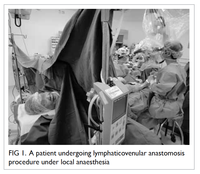
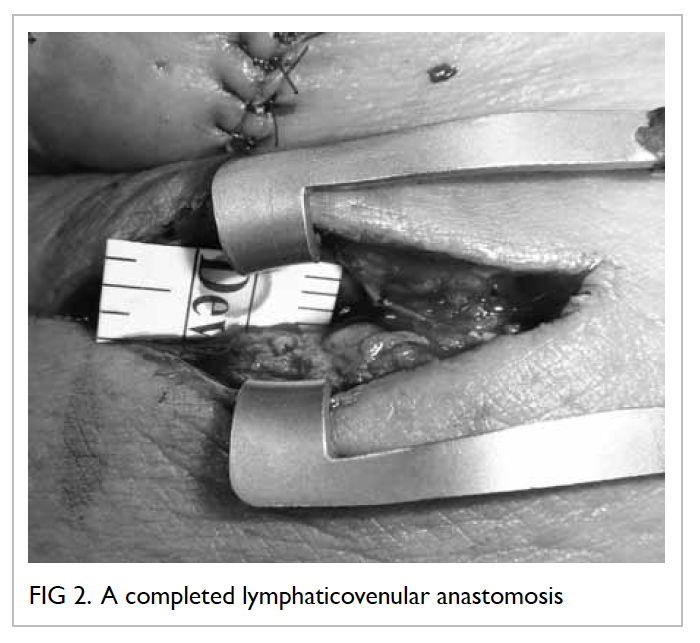
    A number of local surgeons have travelled to regional centres of excellence for training and our unit has organised courses in supermicrosurgery and lymph node transfer to encourage the uptake of these techniques among other surgeons. Lymphatic venous anastomosis has been performed in Hong Kong since 2012 (Figs 1 and 2) and the preliminary results are encouraging, with patients often describing early relief, particularly from symptoms of ‘tightness’. Given the short period of experience and small number of patients, it is too early to comment on local results; however, there is no reason to suggest that the results would not be comparable with international findings in the long term.
     

    Figure 1. A patient undergoing lymphaticovenular anastomosis procedure under local anaesthesia
     

    Figure 2. A completed lymphaticovenular anastomosis
     
    References
    1. de Jong MA, Oldenborg S, Bing Oei S, et al. Reirradiation and hyperthermia for radiation-associated sarcoma. Cancer 2012;118:180-7. CrossRef
    2. Karkkainen MJ, Ferrell RE, Lawrence EC, et al. Missense mutations interfere with VEGFR-3 signalling in primary lymphoedema. Nat Genet 2000;25:153-9. CrossRef
    3. Irrthum A, Karkkainen MJ, Devriendt K, Alitalo K, Vikkula M. Congenital hereditary lymphoedema caused by a mutation that inactivates VEGFR3 thyrosine kinase. Am J Hum Genet 2000;67:295-301. CrossRef
    4. Cederberg A, Grønning LM, Ahrén B, Taskén K, Carlsson P, Enerbäck S. FOXC2 is a winged helix gene that counteracts obesity, hypertriglyceridemia, and diet-induced insulin resistance. Cell 2001;106:563-73. CrossRef
    5. Yildirim-Toruner C, Subramanian K, El Manjra L, Chen E, Goldstein S, Vitale E. A novel frameshift mutation of FOXC2 gene in a family with hereditary lymphoedema-distichiasis syndrome associated with renal disease and diabetes mellitus. Am J Med Genet A 2004;131:281-6. CrossRef
    6. Warren AG, Brorson H, Borud LJ, Slavin SA. Lymphedema: a comprehensive review. Ann Plast Surg 2007;59:464-72. CrossRef
    7. Petrek JA, Senie RT, Peters M, Rosen PP. Lymphedema in a cohort of breast carcinoma survivors 20 years after diagnosis. Cancer 2001;92:1368-77. CrossRef
    8. Langer I, Guller U, Berclaz G, et al. Morbidity of sentinel lymph node biopsy (SLN) alone versus SLN and completion axillary lymph node dissection after breast cancer surgery: a prospective Swiss multicenter study on 659 patients. Ann Surg 2007;245:452-61. CrossRef
    9. Lucci A, McCall LM, Beitsch PD, et al. Surgical complications associated with sentinel lymph node dissection (SLND) plus axillary lymph node dissection compared with SLND alone in the American College of Surgeons Oncology Group Trial Z0011. J Clin Oncol 2007;25:3657-63. CrossRef
    10. Veronesi U, Paganelli G, Viale G, et al. A randomized comparison of sentinel-node biopsy with routine axillary dissection in breast cancer. N Engl J Med 2003;349:546-53. CrossRef
    11. Wilke LG, McCall LM, Posther KE, et al. Surgical complications associated with sentinel lymph node biopsy: results from a prospective international cooperative group trial. Ann Surg Oncol 2006;13:491-500. CrossRef
    12. Tammela T, Alitalo K. Lymphangiogenesis: molecular mechanisms and future promise. Cell 2010;140:460-76. CrossRef
    13. Newman B, Lose F, Kedda MA, et al. Possible genetic predisposition to lymphedema after breast cancer. Lymphat Res Biol 2012;10:2-13. CrossRef
    14. Aldrich MB, Guilliod R, Fife CE, et al. Lymphatic abnormalities in the normal contralateral arms of subjects with breast cancer–related lymphedema as assessed by near-infrared fluorescent imaging. Biomed Opt Express 2012;3:1256-65. CrossRef
    15. International Society of Lymphology. The diagnosis and treatment of peripheral lymphedema. 2009 Concensus Document of the International Society of Lymphology. Lymphology 2009;42:51-60.
    16. Lee BB, Laredo J, Neville RF. Combined clinical and laboratory (lymphoscintigraphic) staging. In: Lee B, Bergan J, Rockson SG, editors. Lymphedema: a concise compendium of theory and practice. London: Springer; 2011: 97-104. CrossRef
    17. Campisi C, Bellini C, Campisi C, Accogli S, Bonioli E, Boccardo F. Microsurgery for lymphoedema: clinical research and long-term results. Microsurgery 2010;30:256-60.
    18. Yamamoto T, Narushima M, Doi K, et al. Characteristic indocyanine green lymphography findings in lower extremity lymphedema: the generation of a novel lymphedema severity staging system using dermal backflow patterns. Plast Reconstr Surg 2011;127:1979-86. CrossRef
    19. Yamamoto T, Matsuda N, Doi K, et al. The earliest finding of indocyanine green lymphography in asymptomatic limbs of lower extremity lymphedema patients secondary to cancer treatment: the modified dermal backflow stage and concept of subclinical lymphedema. Plast Reconstr Surg 2011;128:314e-321e. CrossRef
    20. Stanton A, Modi S, Mellor R, Levick R, Mortimer P. Diagnosing breast cancer–related lymphoedema in the arm. J Lymphoedema 2006;1:12-5.
    21. Sander AP, Hajer NM, Hemenway K, Miller AC. Upper-extremity volume measurements in women with lymphedema: a comparison of measurements obtained via water displacement with geometrically determined volume. Phys Ther 2002;82:1201-12.
    22. Taylor R, Jayasinghe UW, Koelmeyer L, Ung O, Boyages J. Reliability and validity of arm volume measurements for assessment of lymphedema. Phys Ther 2006;86:205-14.
    23. Stanton AW, Northfield JW, Holroyd B, Mortimer PS, Levick JR. Validation of an optoelectronic limb volumeter (Perometer). Lymphology 1997;30:77-97.
    24. Garfein ES, Borud LJ, Warren AG, Slavin SA. Learning from a lymphedema clinic: an algorithm for the management of localized swelling. Plast Reconstr Surg 2008;121:521-8. CrossRef
    25. Lee BB, Kim YW, Kim DI, Hwang JH, Laredo J, Neville R. Supplemental surgical treatment to end stage (stage IV-V) of chronic lymphedema. Int Angiol 2008;27:389-95.
    26. Ko DS, Lerner R, Klose G, Cosimi AB. Effective treatment of lymphedema of the extremities. Arch Surg 1998;133:452-8. CrossRef
    27. Moseley AL, Carati CJ, Piller NB. A systematic review of common conservative therapies for arm lymphoedema secondary to breast cancer treatment. Ann Oncol 2007;18:639-46. CrossRef
    28. Lee BB. Contemporary issues in management of chronic lymphedema: personal reflection on an experience with 1065 patients. Lymphology 2005;38:28-31.
    29. Campisi C, Davini D, Bellini C, et al. Lymphatic microsurgery for the treatment of lymphoedema. Microsurgery 2006;26:65-9. CrossRef
    30. Campisi C, Boccardo F, Zilli A, Macciò A, Napoli F. Long-term results after lymphatic-venous anastomoses for the treatment of obstructive lymphoedema. Microsurgery 2001;21:135-9. CrossRef
    31. Laine JB, Howard JM. Experimental lymphatico-venous anastomosis. Surg Forum 1963;14:111-2.
    32. Yamada Y. The studies on lymphatic venous anastomosis in lymphoedema. Nagoya J Med Sci 1969;32:1-21.
    33. O’Brien BM, Sykes P, Threlfall GN, Browning FS. Microlymphaticovenous anastomoses for obstructive lymphoedema. Plast Reconstr Surg 1977;60:197-211. CrossRef
    34. Damstra RJ, Voesten HG, van Schelven WD, van der Lei B. Lymphatic venous anastomosis (LVA) for treatment of secondary arm lymphedema. A prospective study of 11 LVA procedures in 10 patients with breast cancer related lymphedema and a critical review of the literature. Breast Cancer Res Treat 2009;113:199-206. CrossRef
    35. Boccardo F, Casabona F, De Cian F, et al. Lymphedema microsurgical preventive healing approach: a new technique for primary prevention of arm lymphedema after mastectomy. Ann Surg Oncol 2009;16:703-8. CrossRef
    36. Boccardo FM, Casabona F, Friedman D, et al. Surgical prevention of arm lymphedema after breast cancer treatment. Ann Surg Oncol 2011;18:2500-5. CrossRef
    37. Koshima I, Nanba Y, Tsutsui T, Takahashi Y, Itoh S. Long-term follow-up after lymphaticovenular anastomosis for lymphoedema in the leg. J Reconstr Microsurg 2003;19:209-15. CrossRef
    38. Koshima I, Nanba Y, Tsutsui T, Tahahashi Y, Itoh S, Fujitsu M. Minimal invasive lymphaticovenular anastomosis under local anesthesia for leg lymphedema: is it effective for stage III and IV? Ann Plast Surg 2004;53:261-6. CrossRef
    39. Koshima I, Kawada S, Moriguchi T, Kajiwara Y. Ultrastructural observations of lymphatic vessels in lymphedema in human extremities. Plast Reconstr Surg 1996;97:397-405; discussion 406-7. CrossRef
    40. Mihara M, Uchida G, Hara H, et al. Lymphaticovenous anastomosis for facial lymphoedema after multiple courses of therapy for head-and-neck cancer. J Plast Reconstr Aesthet Surg 2011;64:1221-5. CrossRef
    41. Furukawa H, Osawa M, Saito A, et al. Microsurgical lymphaticovenous implantation targeting dermal lymphatic backflow using indocyanine green fluorescence lymphography in the treatment of postmastectomy lymphedema. Plast Reconstr Surg 2011;127:1804-11. CrossRef
    42. Chang DW. Lymphaticovenular bypass for lymphedema management in breast cancer patients: a prospective study. Plast Reconstr Surg 2010;126:752-8. CrossRef
    43. Cormier JN, Rourke L, Crosby M, Chang D, Armer J. The surgical treatment of lymphedema: a systematic review of the contemporary literature (2004-2010). Ann Surg Oncol 2012;19:642-51. CrossRef
    44. Wongtrungkapun R. Microsurgical lymphonodovenous implantation for chronic lymphedema. J Med Assoc Thai 2004;87:877-82.
    45. Becker C, Assouad J, Riquet M, Hidden G. Postmastectomy lymphedema: long-term results following microsurgical lymph node transplantation. Ann Sur 2006;243:313-5. CrossRef
    46. Becker C, Vasile JV, Levine JL, et al. Microlymphatic surgery for the treatment of iatrogenic lymphedema. Clin Plast Surg 2012;39:385-98. CrossRef
    47. Lin CH, Ali R, Chen SC, et al. Vascularized groin lymph node transfer using the wrist as a recipient site for management of postmastectomy upper extremity lymphedema. Plast Reconstr Surg 2009;123:1265-75. CrossRef
    48. Saaristo AM, Niemi TS, Viitanen TP, Tervala TV, Hartiala P, Suominen EA. Microvascular breast reconstruction and lymph node transfer for postmastectomy lymphedema patients. Ann Surg 2012;255:468-73. CrossRef
    49. Card A, Crosby MA, Liu J, Lindstrom WA, Lucci A, Chang DW. Reduced incidence of breast cancer–related lymphedema following mastectomy and breast reconstruction versus mastectomy alone. Plast Reconstr Surg 2012;130:1169-78. CrossRef
    50. Klimberg VS. A new concept toward the prevention of lymphedema: axillary reverse mapping. J Surg Oncol 2008;97:563-4. CrossRef
    51. Vignes S, Blanchard M, Yannoutsos A, Arrault M. Complications of autologous lymph-node transplantation for limb lymphoedema. Eur J Vasc Endovasc Surg 2013;45:516-20. CrossRef
    52. Viitanen TP, Mäki MT, Seppänen MP, Suominen EA, Saaristo AM. Donor-site lymphatic function after microvascular lymph node transfer. Plast Reconstr Surg 2012;130:1246-53. CrossRef
    53. Slavin SA, Van den Abbeele AD, Losken A, Swartz MA, Jain RK. Return of lymphatic function after flap transfer for acute lymphedema. Ann Surg 1999;229:421-7. CrossRef
    54. Tammela T, Saaristo A, Holopainen T, et al. Therapeutic differentiation and maturation of lymphatic vessels after lymph node dissection and transplantation. Nat Med 2007;13:1458-66. CrossRef
    55. Lähteenvuo M, Honkonen K, Tervala T, et al. Growth factor therapy and autologous lymph node transfer in lymphedema. Circulation 2011;123:613-20. CrossRef
    56. Baumeister RG, Seifert J, Hahn D. Autotransplantation of lymphatic vessels. Lancet 1981;1:147. CrossRef
    57. Baumeister RG, Seifert J, Wiebecke B, Hahn D. Experimental basis and first application of clinical lymph vessel transplantation of secondary lymphedema. World J Surg 1981;5:401-7. CrossRef
    58. Baumeister RG. Microsurgery of the lymphatic system [in German]. Chirurg 1983;54:374-8.
    59. Baumeister RG, Siuda S. Treatment of lymphedemas by microsurgical lymphatic grafting: what is proved? Plast Reconstr Surg 1990;85:64-74; discussion 75-6. CrossRef
    60. Baumeister RG, Siuda H, Bohmert H, Moser E. A microsurgical method for reconstruction of interrupted lymphatic pathways: autologous lymph-vessel transplantation for treatment of lymphedemas. Scand J Plast Reconstr Surg 1986;20:141-6. CrossRef
    61. Kleinhans E, Baumeister RG, Hahn D, Siuda S, Büll U, Moser E. Evaluation of transport kinetics in lymphoscintigraphy: follow-up study in patients with transplanted lymphatic vessels. Eur J Nucl Med 1985;10:349-52. CrossRef
    62. Weiss M, Baumeister RG, Hahn K. Post-therapeutic lymphedema: scintigraphy before and after autologous lymph vessel transplantation: 8 years of long-term follow-up. Clin Nucl Med 2002;27:788-92. CrossRef
    63. Weiss M, Baumeister RG, Hahn K. Dynamic lymph flow imaging in patients with oedema of the lower limb for evaluation of the functional outcome after autologous lymph vessel transplantation: an 8-year follow-up study. Eur J Nucl Med Mol Imaging 2003;30:202-6. CrossRef
    64. Baumeister RG, Frick A. The microsurgical lymph vessel transplantation [in German]. Handchir Mikrochir Plast Chir 2003;35:202-9.
    65. Campisi C. Use of autologous interposition vein graft in management of lymphedema: preliminary experimental and clinical observations. Lymphology 1991;24:71-6.
    66. Goldsmith HS, De los Santos R. Omental transposition in primary lymphoedema. Surg Gynecol Obstet 1967;125:607-10.
    67. Withey S, Pracy P, Wood S, Rhys-Evans P. The use of a lymphatic bridge in the management of head and neck lymphoedema. Br J Plast Surg 2001;54:716-9. CrossRef
    68. Dumanian GA, Futrell JW. The Charles procedure: misquoted and misunderstood since 1950. Plast Reconstr Surg 1996;98:1258-63. CrossRef
    69. Charles R. Elephantiasis scroti. In: Latham A, English TC, editors. A system of treatment. London: Churchill; 1912.
    70. Gibson T, Tough JS. A simplified one-stage operation for the correction of lymphedema of the leg. AMA Arch Surg 1955;71:809-17. CrossRef
    71. Sistrunk WE. Further experience with the Kondoleon operation for elephantiasis. JAMA 1918;71:800-6. CrossRef
    72. Homans J. The treatment of elephantiasis of the legs. A preliminary report. N Engl J Med 1936;215:1099-104. CrossRef
    73. Miller TA, Wyatt LE, Rudkin GH. Staged skin and subcutaneous excision for lymphedema: a favorable report of long-term results. Plast Reconstr Surg 1998;102:1486-98; discussion 1499-501. CrossRef
    74. Salgado CJ, Mardini S, Spanio S, Tang WR, Sassu P, Chen HC. Radical reduction of lymphedema with preservation of perforators. Ann Plast Surg 2007;59:173-9. CrossRef
    75. Thompson N. Surgical treatment of chronic lymphoedema of the lower limb. With preliminary report of new operation. Br Med J 1962;2:1566-73. CrossRef
    76. Karri V, Yang MC, Lee IJ, et al. Optimizing outcome of Charles procedure for chronic lower extremity lymphoedema. Ann Plast Surg 2011;66:393-402. CrossRef
    77. van der Walt JC, Perks TJ, Zeeman BJ, Bruce-Chwatt AJ, Graewe FR. Modified Charles procedure using negative pressure dressings for primary lymphedema: a functional assessment. Ann Plast Surg 2009;62:669-75. CrossRef
    78. Brorson H. Surgical treatment of postmastectomy lymphedema—liposuction. In: Lee BB, Bergan J, Rockson SG, editors. Lymphedema: a concise compendium of theory and practice. London: Springer; 2011: 409-18. CrossRef
    79. Dixon JB. Lymphatic lipid transport: sewer or subway? Trends Endocrinol Metab 2010;21:480-7. CrossRef
    80. O’Brien BM, Khazanchi RK, Kumar PA, Dvir E, Pederson WC. Liposuction in the treatment of lymphoedema; a preliminary report. Br J Plast Surg 1989;42:530-3. CrossRef
    81. Brorson H, Ohlin K, Svensso B. The facts about liposuction as a treatment for lymphoedema. J Lymphoedema 2008;3:38-47.
    82. Wojnikow S, Malm J, Brorson H. Use of tourniquet with and without adrenaline reduces blood loss during liposuction for lymphoedema of the arm. Scand J Plast Reconstr Surg Hand Surg 2007;41:243-9. CrossRef
    83. Schaverien MV, Munro KJ, Baker PA, Munnoch DA. Liposuction for chronic lymphoedema of the upper limb: 5 years of experience. J Plast Reconstr Aesthet Surg 2012;65:935-42. CrossRef
    84. Brorson H. Liposuction in arm lymphoedema treatment. Scand J Surg 2003;92:287-95.
    85. Brorson H, Svensson H. Complete reduction of lymphoedema of the arm by liposuction after breast cancer. Scand J Plast Reconstr Surg Hand Surg 1997;31:137-43. CrossRef
    86. Brorson H, Svensson H. Liposuction combined with controlled compression therapy reduces arm lymphoedema more effectively than controlled compression therapy alone. Plast Reconstr Surg 1998;102:1058-67; discussion 1068. CrossRef
    87. Brorson H. Liposuction gives complete reduction of chronic large arm lymphedema after breast cancer. Acta Oncol 2000;39:407-20. CrossRef
    88. Brorson H, Svensson H, Norrgren K, Thorsson O. Liposuction reduces arm lymphoedema without significantly altering the already impaired lymph transport. Lymphology 1998;31:156-72.
    89. National Institute for Health and Clinical Excellence. Liposuction for chronic lymphoedema, February 2008. Available from: http://www.nice.org.uk/guidance/ipg251/resources/guidance-liposuction-for-chronic-lymphoedema-pdf. Accessed Jan 2014.
    90. Brorson H, Ohlin K, Olsson G, Långström G, Wiklund I, Svensson H. Quality of life following liposuction and conservative treatment of arm lymphoedema. Lymphology 2006;39:8-25.
    91. Hong Kong Cancer Registry. Available from: http://www3.ha.org.hk/cancereg/statistics.html. Accessed Jan 2014.
    92. Leung SY. A prospective cohort study on physiotherapy service to identify service gaps for post-operative breast cancer patients. Proceedings of the Hospital Authority Convention 2010; 2010 May 10-11; Hong Kong.
    93. Mak SS, Yeo W, Lee YM, et al. Predictors of lymphedema in patients with breast cancer undergoing axillary lymph node dissection in Hong Kong. Nurs Res 2008;57:416-25. CrossRef
    94. Shih YC, Xu Y, Cormier JN, et al. Incidence, treatment costs, and complications of lymphedema after breast cancer among women of working age: a 2-year follow-up study. J Clin Oncol 2009;27:2007-14. CrossRef
    95. Tang KS. A clinical guideline for management of lymphoedema using nurse-led manual lymphatic drainage therapy. Hong Kong: The University of Hong Kong; 2013.

    Use of cephalosporins in patients with immediate penicillin hypersensitivity: cross-reactivity revisited

    DOI: 10.12809/hkmj144327
    © Hong Kong Academy of Medicine. CC BY-NC-ND 4.0
     
    REVIEW ARTICLE     CME 
    Use of cephalosporins in patients with immediate penicillin hypersensitivity: cross-reactivity revisited
    QU Lee, MB, ChB, FHKAM (Paediatrics)
    Department of Paediatrics and Adolescent Medicine, Princess Margaret Hospital, Laichikok, Hong Kong
     
    Corresponding author: Dr QU Lee (leequnui@gmail.com)
     Full paper in PDF
    Abstract
    A 10% cross-reactivity rate is commonly cited between penicillins and cephalosporins. However, this figure originated from studies in the 1960s and 1970s which included first-generation cephalosporins with similar side-chains to penicillins. Cephalosporins were frequently contaminated by trace amount of penicillins at that time. The side-chain hypothesis for beta-lactam hypersensitivity is supported by abundant scientific evidence. Newer generations of cephalosporins possess side-chains that are dissimilar to those of penicillins, leading to low cross-reactivity. In the assessment of cross-reactivity between penicillins and cephalosporins, one has to take into account the background beta-lactam hypersensitivity, which occurs in up to 10% of patients. Cross-reactivity based on skin testing or in-vitro test occurs in up to 50% and 69% of cases, respectively. Clinical reactivity and drug challenge test suggest an average cross-reactivity rate of only 4.3%. For third- and fourth-generation cephalosporins, the rate is probably less than 1%. Recent international guidelines are in keeping with a low cross-reactivity rate. Despite that, the medical community in Hong Kong remains unnecessarily skeptical. Use of cephalosporins in patients with penicillin hypersensitivity begins with detailed history and physical examination. Clinicians can choose a cephalosporin with a different side-chain. Skin test for penicillin is not predictive of cephalosporin hypersensitivity, while cephalosporin skin test is not sensitive. Drug provocation test by experienced personnel remains the best way to exclude or confirm the diagnosis of drug hypersensitivity and to find a safe alternative for future use. A personalised approach to cross-reactivity is advocated.
     
     
    The ten per cent myth about beta-lactam cross-reactivity
    Penicillins and cephalosporins are two groups of widely prescribed antibiotics. They belong to the class of beta-lactam (BL) antibiotics because both possess the same BL nucleus. Allergic reactions are common side-effects of BL antibiotics. Studies in the 1960s and 1970s frequently estimated 10% cross-reactivity between penicillins and cephalosporins.1 2 However, at least two recent reviews showed much lower cross-reactivity.3 4 Notably, cross-reactivity is higher between penicillins and first- and second-generation cephalosporins compared with third- and fourth-generation cephalosporins.5 The latter two groups are considered safe alternatives for patients with penicillin hypersensitivity.6 The 10% cross-reactivity rate has recently been questioned as a medical myth.4 7 Yet until 2005, an influential drug reference such as the British National Formulary (BNF) abided by the “10% rule”.8 Faced with such recommendation, an ordinary physician naturally avoids all BL antibiotics in patients with a history suggestive of penicillin hypersensitivity.9 The implications are far-reaching as physicians often resort to expensive, broad-spectrum antibiotics, which may induce antibiotic resistance by selecting out resistant organisms.10 In order to minimise unnecessary exposure to expensive broad-spectrum antibiotics with higher toxicities and to preserve patients’ right to receive commonly prescribed antibiotics, a better understanding of BL cross-reactivity is needed. In the following discussion, the author will review the use of cephalosporins in patients with immediate hypersensitivity to penicillins. Mechanism and epidemiology of cross-reactivity will be discussed, followed by a suggestion for a pragmatic approach.
     
    By definition, ‘cross-reaction’ between two substances is “the interaction of an antigen with an antibody formed against a different antigen with which the first antigen shares identical or closely related antigenic determinants”.11 Hence, antigenic similarity forms the basis of cross-reactivity. Public hospitals often suggest avoiding all cephalosporins indiscriminately for patients with penicillin hypersensitivity, as exemplified by a recent antibiotic guideline.12 What remains unsettled is how far the BL nucleus also acts as a common antigenic determinant. In other words, does structural similarity in the drug nucleus translate into clinically relevant allergic reaction?
     
    Mechanism of beta-lactam hypersensitivity
    The BL nucleus is probably the only structure common to penicillins and cephalosporins. What differentiates between them is that penicillins possess a 5-membered thiazolidine ring attached to the BL nucleus while cephalosporins have a 6-membered dihydrothiazine ring. Secondly, while penicillins have a single 6-positioned side-chain, cephalosporins have a 3-positioned as well as a 7-positioned side-chain.3
     
    When a BL antibiotic is absorbed into the body, the BL nucleus undergoes spontaneous opening. Covalent bonding between the drug and endogenous protein results in a hapten-protein conjugate. In case of penicillins, stable protein conjugates formed include penicilloyl (major) determinants and other minor determinants.13 For cephalosporin, however, haptenic determinants are less clear.14 Once inside the body, cephalosporins undergo rapid fragmentation of the BL nucleus and dihydrothiazine rings. The resulting unstable metabolites do not allow haptenisation of proteins.15 In subjects with BL hypersensitivity, the hapten-protein conjugate has the capability to activate T-cells and the ensuing B-cell response. Specific immunoglobulin (Ig) E antibodies produced by B-cells become attached to the surface of effector cells such as mast cells and basophils. Subsequent exposure to the same drug induces formation of hapten-protein conjugates. Immediate hypersensitivity is the result of cross-linking of adjacent surface IgE molecules by the hapten-protein conjugates that culminates in rapid degranulation of preformed inflammatory mediators such as histamine and tryptase.16
     
    Mechanism of cross-reactivity and the side-chain hypothesis
    Early cephalosporins before 1980s were contaminated with trace amounts of penicillin during the manufacturing process by the cephalosporium mould. That partly accounted for the higher cross-reactivity rate between penicillins and first-generation cephalosporins.14 Cross-reactivity within penicillins is based on common antigenic determinants. Antibody binding against basic structures such as BL ring or penicilloyl frequently results in higher cross-reactivity rate. More complex motifs, such as side-chains found only in certain sub-groups, are associated with lower cross-reactivity. An in-vitro experiment has identified two types of T-cells responsible for penicillin hypersensitivity. The restricted type is immunologically reactive against a combined penicilloyl and side-chain structure but exhibits little cross-reactivity with penicillins with different side-chains such as amoxicillin or ampicillin. The broad type does react against different penicillins, but not against cephalosporins.17
     
    Epitopes (antibody-binding sites) on penicillin molecules may involve the BL nucleus, the thiazolidine ring, the side-chain or even the new antigenic determinant. Side-chain antigenic determinants account for 42% to 92% of the penicillin hypersensitivity.18 19 Epitopes on cephalosporin molecules are even more heterogeneous than penicillin, and involve the whole molecule.20 R1 side-chain and BL fragment protein conjugates appear to be the major determinants of cephalosporin hypersensitivity.21 R2 side-chain makes little contribution to cephalosporin hypersensitivity, as it disappears when the BL ring is opened.22
     
    Human studies have provided insight into the role of similarity in the R1 side-chains in causing BL cross-reactivity.15 For instance, the 2-amino-2-phenylacetic acid side-chain in ampicillin is also present in first- or second-generation cephalosporins like cephalexin and cefaclor, respectively, but is absent in third- or fourth-generation cephalosporins. Similarly, the same 2-amino-2-(4-hydroxyphenyl) acetic acid side-chain is present in amoxicillin and cefadroxil but not in new generations of cephalosporins.16 In another study on selective amoxicillin hypersensitivity, Miranda et al23 have shown that oral challenge with cephadroxil, a first-generation cephalosporin that shared the same side-chain mentioned above, resulted in a cross-reactivity of 38%. On the other hand, use of cefamandole, a second-generation cephalosporin with a different side-chain from amoxicillin and cephadroxil, did not result in any cross-reactivity.23 Notwithstanding, other authors do not accord with the side-chain hypothesis.24 Fine structure within the side-chain such as methylene group has also been suggested as an antigenic determinant common to penicillins and cephalosporins.25
     
    Background and co-existing drug hypersensitivity
    When dealing with potential cross-reactivity between penicillins and cephalosporins, one should take into account the background hypersensitivity rates in unselected population, which range between 0.7% and 10% for penicillins.26 However, among patients with a history of penicillin allergy, only 10% to 20% exhibit positive allergic reaction to skin test or challenge test.27 28 A non-urticarial, maculopapular skin rash is the most common allergic reaction with a frequency of 1% to 2%. The frequency of anaphylaxis per penicillin course is 0.01% to 0.05%.29 Similarly, background hypersensitivity rates for cephalosporins range between 1% and 10%, while anaphylaxis occurs in less than 0.02%.30 In other words, patients with penicillin hypersensitivity may develop non–cross-reacting allergic response to cephalosporins by coincidence. They are also at increased risk of non-BL hypersensitivity, with a reported rate of 16% to 23%.31 32 A caveat is that, as local prevalence data are lacking, epidemiological data can only be applied to the Hong Kong situation by extrapolation.
     
    Cross-reactivity based on cephalosporin skin testing
    Skin test is an in-vivo method used to diagnose IgE-mediated allergic response. Substantial cross-reactivity in terms of skin testing exists between penicillins and first-generation cephalosporins. In the 1970s, Assem and Vickers33 studied 24 patients with penicillin hypersensitivity of which 11 (46%) showed positive intradermal test to cephaloridine; however, this reaction was not observed in any of the patients without penicillin hypersensitivity.33 Dash2 studied 100 patients with clinical reaction to penicillin and demonstrated positive cephalosporin skin test in 11 (11%) patients. However, seven (9.3%) of 75 control subjects without penicillin hypersensitivity also tested positive.2 In another study in the 1980s, Sullivan et al34 recruited 74 patients with penicillin hypersensitivity confirmed by positive skin prick test (SPT). Of these, 38 (50%) also exhibited a positive SPT to cephalothin, another first-generation cephalosporin.34 Audicana et al35 studied 34 patients allergic to penicillin and found that five (14%) had positive skin test to cephalexin, a first-generation cephalosporin, but none to ceftazidime, a third-generation cephalosporin. Romano et al36 studied 128 adult subjects with a history of immediate penicillin hypersensitivity; positive cephalosporin skin test was observed in 11% of them. Of the 128 subjects, 101 (94 skin test negative and 7 skin test positive for cephalosporins) who accepted the challenge could tolerate oral cefuroxime axetil and intramuscular ceftriaxone.36 Although controlled trial is not possible, the implication is that cephalosporins can be safely given to patients with a history of penicillin hypersensitivity but who have negative cephalosporin skin test.
     
    Cross-reactivity based on in-vitro tests
    Substantial in-vitro cross-reactivity also exists between penicillins and first-generation cephalosporins. In an early study in 1960s, Abraham et al37 were able to demonstrate haemagglutination antibody against cephalothin (titre of 1:8 or greater) in 22 (69%) of 32 patients who had been given penicillin but denied a history of cephalothin therapy. A subsequent adsorption study using penicilloic acid-solid phase by Zhao et al25 further identified cross-reacting specific IgE antibodies against both benzylpenicillin and cephalothin. Recently, Liu et al38 employed radioallergosorbent test to identify specific IgE antibodies against penicillins and cephalosporins in 420 subjects with penicillin hypersensitivity; cross-reacting specific IgE antibodies occurred in 22.6% of the subjects. Specific cephalosporin IgE antibodies were present in 27.1% of those with specific penicillin IgE antibodies, compared with 14.6% in those without specific penicillin IgE antibodies.38 However, in the absence of cross-linking, demonstration of antibodies cannot be equated with clinical reactivity.2
     
    Clinical reaction to cephalosporins in patients with a history of penicillin hypersensitivity
    As skin test and in-vitro test are often inadequate for confirmation of cephalosporin hypersensitivity, one has to rely on a provocation test or the result of drug exposure. In an early review of 701 patients with a history of penicillin hypersensitivity, Petz39 reported an 8.1% reactivity rate to first- or second-generation cephalosporins, compared with 1.9% among those without penicillin hypersensitivity. In another cohort study in the 1980s by Solley et al,40 178 patients with a history of penicillin allergy were given cephalosporins. Positive reaction resulted in two patients, equivalent to a clinical cross-reactivity rate of 1.1%.40 Goodman et al41 reviewed the medical records of 413 patients with a self-reported history of penicillin allergy who underwent anaesthetic procedures that included antibiotic therapy. Only one patient (0.24%) probably developed cross-reactivity to cephalexin, a first-generation cephalosporin.41 Despite the retrospective nature and the lack of confirmatory tests, the low clinical cross-reactivity is reassuring.
     
    Fonacier et al42 reviewed 83 patients with penicillin hypersensitivity who were subsequently given cephalosporins. Seven (8.4%) of them developed an adverse drug reaction. A definite history of penicillin hypersensitivity was found in six (85.7%) of the seven patients. Eleven (13.3%) patients with penicillin hypersensitivity also reported hypersensitivity reaction to other drugs such as non-BL antibiotics and codeine. Regarding the types of cephalosporin, clinical cross-reactivity rates between penicillin and first-, second-, third-, and fourth-generation cephalosporins are 4.6%, 50%, 10.5% and 0%, respectively. Small sample size and potential recall bias undermine the reliability of the study. The role of side-chain is highlighted by a 4-fold increase in the cross-reactivity rate between penicillins and cephalosporins with similar amino-benzyl ring side-chain.
     
    In a large prospective study by Atanasković-Marković et al43 that included 644 children with a history of hypersensitivity reaction to penicillins, rate of cross-reactivity to cephalosporins was 31.5%. If the generations of cephalosporins were taken into account, the cross-reactivity rate with aminopenicillins differed by 100-fold, ranging from 0.3% to 0.7% in third-generation cephalosporins to around 32.4% to 38.5% in first- or second-generation cephalosporins, respectively. This, again, illustrates the relevance of side-chain in cross-reactivity. An interesting corollary is that, in patients with negative skin test to penicillins or cephalosporins, 0% to 1.8% of patients showed positive drug challenge to the test drug. Hence the false-negative rate of skin test is quite low. On the other hand, as patients with positive skin test were not further challenged with drugs to confirm clinical hypersensitivity, the true-positive rate cannot be ascertained.
     
    A 5-year retrospective study by Apter et al32 reviewed 534 810 patients in the United Kingdom who received a penicillin followed by cephalosporin of which 64% were tested with first-generation cephalosporins. The authors compared 3920 patients with allergy-like events (ALE) within 30 days of receiving penicillin with 530 890 patients without ALE. Among 3920 patients with ALE after receiving penicillin, 1% cross-reacted with cephalosporins. The unadjusted risk ratios for ALE after the subsequent cephalosporin and sulphonamide challenges were 10.0 (95% confidence interval [CI], 7.4-13.6) and 7.2 (95% CI, 3.8-13.5), respectively, suggesting that patients allergic to penicillin may have an increased tendency for drug hypersensitivity via a mechanism other than cross-reactivity.
     
    In another retrospective study, Daulat et al44 reviewed medical records of 606 patients with a history of penicillin allergy who were subsequently given a cephalosporin. Confirmatory penicillin skin testing was not reported. Clinical allergy occurred in only one patient given cefazolin, a first-generation cephalosporin. This is tantamount to a cross-reactivity rate of 0.165%. As drug allergy was suspected from diagnostic coding only, true penicillin allergy, and hence cephalosporin cross-reactivity, might have been higher.
     
    In a landmark meta-analysis in 2007, Pichichero and Casey15 reviewed nine studies that compared allergic reaction rate to cephalosporins in patients with or without penicillin allergy. Among 47 284 patients with a history of penicillin allergy alone, the odds ratio (OR) for cephalosporin cross-reactivity in general was 2.63 (95% CI, 2.11-3.28; P<0.00001). However, the increased cross-reactivity rate was mainly due to first-generation cephalosporins, as the corresponding ORs for first-, second-, and third-generation cephalosporins were 4.79 (95% CI, 3.71-6.17; P<0.00001), 1.13 (95% CI, 0.61-2.12; P=0.70), and 0.45 (95% CI, 0.18-1.13; P=0.09), respectively. There was actually a trend towards decreased risk of cross-reactivity to third-generation cephalosporins, although the result did not reach statistical significance.
     
    Clinical reaction to cephalosporins in patients with penicillin hypersensitivity confirmed by investigations
    In a cohort study by Solley et al,40 none of the 27 patients with a history of penicillin allergy and a positive penicillin skin test developed clinical reactivity to cephalosporins. On the contrary, two of the 151 patients with allergic history but negative penicillin skin test reacted to cephalosporins, putting to doubt the value of penicillin skin test in predicting cross-reactivity to cephalosporins.40 In another small cohort study by Blanca et al,45 19 patients with confirmed penicillin hypersensitivity were given parenteral cephamandole, a second-generation cephalosporin, followed by oral cephaloridine, a first-generation cephalosporin, if the former was tolerated. Two (10.5%) of the 19 patients cross-reacted with cephamandole while all the remaining 17 patients tolerated cephaloridine.
     
    Sastre et al24 subjected 16 patients with selective amoxicillin hypersensitivity confirmed by skin test or drug challenge with cephadroxil, a first-generation cephalosporin. Two (12%) were found to be cross-reactors.24 Novalbos et al46 recruited 41 patients with a history of penicillin hypersensitivity confirmed by either skin test or drug challenge. Patients were then challenged with three cephalosporins (cephazoline, cefuroxime and ceftriaxone) with side-chains which were different from that of penicillin. None of them cross-reacted with the cephalosporins.46 Hameed and Robinson47 recruited 158 patients with positive penicillin test. Seven (4.4%) of them developed immediate hypersensitivity when given cephalosporins. None of the cephalosporins was from the third generation.47 There is a lack of published reports on anaphylactic reaction to cephalosporins in children with a history of anaphylaxis to penicillins, and only a few such reports in adults have been published.47
     
    Macy and Burchette48 studied 83 patients with a history of adverse reaction to penicillin confirmed by skin test. Post–skin test exposure to cephalosporins in 42 resulted in adverse reaction in one, amounting to 2.4% cross-reactivity rate. The corresponding figure for non-BL was eight (10.8%) out of 74, suggesting that in patients allergic to penicillin, cross-reactivity for non-BLs may be even higher than that for BLs.48 This study and the one by Apter et al32 have significant implications for practitioners who routinely employ non-BL antibiotics for patients with penicillin hypersensitivity.
     
    In a preoperative assessment clinic, Park et al49 recruited 1072 patients with a history of BL allergy for penicillin skin testing. Among the 999 patients who underwent the skin test, 43 had a positive skin test for penicillin and three of those 43 eventually received cefazolin. None developed cross-reactivity.49 Ahmed et al50 reviewed 173 children with a history of penicillin hypersensitivity, with or without a skin test, who underwent cephalosporin challenge. None among those with positive skin test showed reactivity. However, one (0.7%) of the 152 patients with negative skin test had an immediate allergic reaction after cephalexin, underscoring the lack of predictive power of the penicillin skin test.50
     
    In a meta-analysis by Pichichero and Casey,15 1831 patients with a history of penicillin allergy also received penicillin skin test. Compared with patients with negative skin test, the OR for cross-reactivity to any cephalosporin for patients with positive results was 1.48 (95% CI, 0.64-3.41; P=0.36). Corresponding ORs for first-, second-, and third-generation cephalosporins were 4.13 (95% CI, 0.70-24.51; P=0.11), 1.33 (95% CI, 0.32-5.52; P=0.69), and 0.75 (95% CI, 0.15-3.66; P=0.72), respectively.15 There was a trend towards increased risk for first-generation cephalosporins, although the result did not reach statistical significance.
     
    Studies on cephalosporin drug challenge in patients with a history of penicillin hypersensitivity have several inherent limitations. Firstly, retrospective studies are subjected to recall bias. Secondly, the so-called ‘positive reaction’ may include ‘nocebo effects’, ie untoward effects after administration of an inert substance, which may occur in around 27% of subjects.51 Thirdly, as most studies excluded patients with positive penicillin skin test, investigators had no way to tell whether these patients could actually tolerate cephalosporins. Lastly, most studies of cephalosporin challenge were performed in an open, uncontrolled manner. Patients with penicillin allergy who may have underlying multiple drug allergy syndrome will be missed in the absence of a control arm, such as non-BL group.52
     
    Ten per cent cross-reactivity: an over-estimation
    Review of published studies, as described above, shows that cross-reactivity between penicillins and cephalosporins, if restricted to clinical reaction or positive drug challenge, varies between 0% and 31.5% (Fig 1). Among the 14 studies that included a total of 6464 patients with penicillin hypersensitivity, 279 showed clinical reactivity or positive challenge to cephalosporins, resulting in an average cross-reactivity rate of 4.32%. Corresponding figures for patients with a history of penicillin allergy alone and those confirmed by investigations are 4.34% and 3.76%, respectively. Studies reporting rates higher than 10% are mainly those involving first- or second-generation cephalosporins, especially when in-vitro or skin tests were employed. It must be emphasised that although cross-reactivity is substantial with first-generation cephalosporins (up to 32%), it is less than 1% for third- and fourth-generation cephalosporins.
     

    Figure 1. Studies on cross-reactivity between penicillins and cephalosporins
     
    A probable reason for the low cross-reactivity stems from the fact that, despite having the same BL nucleus, penicillins and cephalosporins are immunologically different. If the BL nucleus is the common antigenic determinant, one should expect a very high cross-reactivity. However, this is not the case because the BL ring opens in the process of metabolism to form major or minor determinants. Secondly, newer generations of cephalosporin do not share similar side-chains with penicillins, hence cross-reactivity will, generally, not occur. Thirdly, among patients with alleged penicillin hypersensitivity, less than 10% show genuine hypersensitivity. The majority of cases may suffer from transient adverse reaction followed by subsequent tolerance to cephalosporins.7
     
    Despite current recognition of the low cross-reactivity rate, international guidelines are not unanimous in their recommendations regarding the use of cephalosporins in patients with penicillin hypersensitivity. A recent practice parameter from a Joint Task Force in the United States stated that “most patients with a history of penicillin allergy tolerate cephalosporins”.10 If patients with positive penicillin skin test are given cephalosporins, around 2% may cross-react, including some with anaphylactic reactions. If a clinician chooses not to skin test a patient with a history of penicillin allergy but directly prescribe a cephalosporin, the chance of developing a reaction is probably less than 1%. In treating otitis media in children with penicillin allergy, the American Academy of Pediatrics simply suggested prescribing, rather than avoiding, either second- or third-generation cephalosporins.53 Basing on dissimilarity in chemical structures, the Academy considered cross-reactivity between penicillin and second- or third-generation cephalosporins to be ‘highly unlikely’.
     
    A relatively conservative approach is adopted by the Infectious Diseases Society of America (IDSA). In the 2012 clinical practice guideline for bacterial rhinosinusitis, the IDSA recommended third-generation cephalosporins only for patients with non–type I penicillin allergy. Non-BL antibiotics were recommended for those with type I penicillin allergy.54 Even more conservative is the British Medical Association; in the 2014 edition, the BNF advised against using cephalosporins in patients with penicillin hypersensitivity. Nevertheless, if no other alternatives are available, third- and fourth-generation cephalosporins can be used, albeit with caution.55
     
    Skepticism still lingers within the medical community in Hong Kong. For instance, a recent public hospital antibiotic guideline does not differentiate between different generations of cephalosporins, but treats all cephalosporins as having the potential to cross-react with penicillins.56 The IMPACT guideline in Hong Kong has aptly pointed out a deep-rooted preoccupation with cross-reactivity among the medical profession. The guideline suggests that “second, third and fourth generation cephalosporins have negligible cross-reactivity with penicillin”. However, it also raises a common concern that contra-indications indicated in product inserts have resulted in “medico-legal implications when using cephalosporins in patients with penicillin allergy”.57 This concern is understandable but unfounded, for two reasons. Firstly, a legal case appealed to the New Jersey Supreme Court in 1998 has come to the final decision that product inserts alone do not establish a standard of care.3 Secondly, a review of the inserts shows that, rather than contra-indicating the use of cephalosporins, pharmaceutical companies only issue words of caution in patients with penicillin allergy.58 59
     
    Pragmatic approach to cross-reactivity
    For patients with suspected penicillin hypersensitivity, one should begin with careful history and physical examination to establish the likelihood of adverse drug reaction. Clinicians will not do justice by simply avoiding all cephalosporins in patients with so-called penicillin hypersensitivity. Injudicious use of non-BL antibiotics without precaution is falsely reassuring and will expose patients with allergic tendency to further drug hypersensitivity.
     
    Allergological investigations should preferably be done 4 to 6 weeks after resolution of adverse drug reaction.60 One should start from skin testing to confirm penicillin hypersensitivity. Ideally, skin test reagents should include penicilloyl polylysine (PPL) and minor determinant mixture (MDM). Unfortunately, the two major manufacturers, Allergopharma (Hamburg, Germany) and Hollister-Stier (Spokane, WA, US) ceased production of PPL and MDM in 2004. Although Diater (Madrid, Spain) has launched the production of PPL and MDM since 2003, the reagents have not gained widespread popularity in Hong Kong.61 Besides, diagnosis of selective reaction to a single BL requires a long algorithm, which begins testing with PPL and MDM, followed by the culprit drug.5 This may be tedious and time-consuming in daily clinical practice. For pragmatic purposes, it is often the culprit drug and/or a potentially safe alternative that will be tested and prescribed. Non-irritating concentration of the culprit drug should be employed for skin testing.62
     
    Patients with negative penicillin skin test may undergo supervised drug provocation test (DPT) of the culprit drug.3 The aim of DPT is to exclude or confirm the diagnosis of drug hypersensitivity and to find a safe alternative for future use. Drug provocation test generally has a high negative predictive value of 94% to 98%. A caveat is that anaphylactic reactions can still occur among a few cross-reacting patients. Hence, DPT must be performed by experienced personnel in a setting with resuscitation facilities.60 Patients with remote or severe hypersensitivity may be re-tested 2 to 4 weeks later to exclude a small but possible risk of re-sensitisation after initial negative testing. Contra-indications to DPT include a history of severe cutaneous drug reactions (eg Stevens-Johnson syndrome or toxic epidermal necrolysis), severe anaphylaxis or certain medical conditions (eg uncontrolled asthma or pregnancy).63 In the absence of severe or recent immediate penicillin hypersensitivity, patients may choose to receive penicillin directly without skin testing.10 To further ensure drug safety, the first dose may be divided into incremental steps similar to DPT.
     
    Patients who have a history of severe penicillin hypersensitivity, a positive penicillin skin test or DPT may resort to cephalosporins. However, a positive penicillin skin test does not predict cross-reactivity with cephalosporin.30 50 Clinicians may perform skin tests using a cephalosporin with a different side-chain to guide clinical use.10 Unfortunately, the diagnostic accuracy of cephalosporin skin test is difficult to establish.5 Studies generally have shown low sensitivity and positive predictive value.64 65 Nevertheless, skin test for BL hypersensitivity is still considered ‘good’ by the International Consensus in 2014.60 Patients with negative cephalosporin skin test should pass a DPT before finally receiving a cephalosporin. Patients who fail the DPT may be given a non-BL antibiotic or undergo desensitisation, if the cephalosporin is essential.60 A suggested algorithm for investigation and management of suspected immediate penicillin hypersensitivity is summarised in Figure 2.
     

    Figure 2. Suggested algorithm for investigation and management of suspected penicillin hypersensitivity
     
    Conclusion
    The available evidence to date does not support the notion of a 10% cross-reactivity rate between penicillins and cephalosporins. Above all, 10% is an oversimplified and indiscriminate generalisation of cross-reactivity. Scientific evidence supports the side-chain hypothesis and a low cross-reactivity rate. Clinicians should adopt a personalised approach towards BL cross-reactivity. Finally, future research on the local prevalence of BL hypersensitivity and cross-reactivity is needed.
     
    Declaration
    No conflicts of interests were declared by the author.
     
    References
    1. Petz LD, Fudenberg HH. Coombs-positive hemolytic anemia caused by penicillin administration. N Engl J Med 1966;274:171-8. CrossRef
    2. Dash CH. Penicillin allergy and the cephalosporins. J Antimicrob Chemother 1975;1(3 Suppl):107-18. CrossRef
    3. DePestel DD, Benninger MS, Danziger L, et al. Cephalosporin use in treatment of patients with penicillin allergies. J Am Pharm Assoc (2003) 2008;48:530-40. CrossRef
    4. Campagna JD, Bond MC, Schabelman E, Hayes BD. The use of cephalosporins in penicillin-allergic patients: a literature review. J Emerg Med 2012;42:612-20. CrossRef
    5. Torres MJ, Blanca M, Fernandez J, et al. Diagnosis of immediate allergic reactions to beta-lactam antibiotics. Allergy 2003;58:961-72. CrossRef
    6. Prematta T, Shah S, Ishmael FT. Physician approaches to beta-lactam use in patients with penicillin hypersensitivity. Allergy Asthma Proc 2012;33:145-51. CrossRef
    7. Herbert ME, Brewster GS, Lanctot-Herbert M. Medical myth: ten percent of patients who are allergic to penicillin will have serious reactions if exposed to cephalosporins. West J Med 2000;172:341. CrossRef
    8. British Medical Association and the Royal Pharmaceutical Society of Great Britain. British National Formulary BNF 49 March 2005. London: BMJ Books; 2005.
    9. Solensky R, Earl HS, Gruchalla RS. Clinical approach to penicillin-allergic patients: a survey. Ann Allergy Asthma Immunol 2000;84:329-33. CrossRef
    10. Joint Task Force on Practice Parameters; American Academy of Allergy, Asthma and Immunology; American College of Allergy, Asthma and Immunology; Joint Council of Allergy, Asthma and Immunology. Drug allergy: an updated practice parameter. Ann Allergy Asthma Immunol 2010;105:259-73. CrossRef
    11. Cross reaction. In: Anderson DM, editor. Dorland’s illustrated medical dictionary. 31st ed. Philadelphia: W.B. Saunders; 2007.
    12. Kowloon West Cluster Antibiotic Subcommittee. KWC Antimicrobial Therapy Guide 2012, Second Edition [cited 2014 June 5]. Available from: https://gateway.ha.org.hk/f5-w-687474703a2f2f706d682e686f6d65$$/sites/mg/General/KWC%20Antimicrobial%20Thearpy%20 Guide%202012.pdf. Accessed Jun 2014.
    13. Park B, Sanderson J, Naisbitt D. Drugs as haptens, antigens, and immunogens. In: Pichler WJ, editor. Drug hypersensitivity. Basel: Karger; 2007: 55-65. CrossRef
    14. Kelkar PS, Li JT. Cephalosporin allergy. N Engl J Med 2001;345:804-9. CrossRef
    15. Pichichero ME, Casey JR. Safe use of selected cephalosporins in penicillin-allergic patients: a meta-analysis. Otolaryngol Head Neck Surg 2007;136:340-7. CrossRef
    16. Torres M, Mayorga C, Blanca M. Urticaria and anaphylaxis due to betalactams (penicillins and cephalosporins). In: Pichler WJ, editor. Drug hypersensitivity. Basel: Karger; 2007: 190-203. CrossRef
    17. Mauri-Hellweg D, Zanni M, Frei E, et al. Cross-reactivity of T cell lines and clones to beta-lactam antibiotics. J Immunol 1996;157:1071-9.
    18. Mayorga C, Obispo T, Jimeno L, et al. Epitope mapping of beta-lactam antibiotics with the use of monoclonal antibodies. Toxicology 1995;97:225-34. CrossRef
    19. Torres MJ, Romano A, Mayorga C, et al. Diagnostic evaluation of a large group of patients with immediate allergy to penicillins: the role of skin testing. Allergy 2001;56:850-6. CrossRef
    20. Pham NH, Baldo BA. beta-Lactam drug allergens: fine structural recognition patterns of cephalosporin-reactive IgE antibodies. J Mol Recognit 1996;9:287-96. CrossRef
    21. Sánchez-Sancho F, Perez-Inestrosa E, Suau R, et al. Synthesis, characterization and immunochemical evaluation of cephalosporin antigenic determinants. J Mol Recognit 2003;16:148-56. CrossRef
    22. Blanca M, Romano A, Torres MJ, et al. Update on the evaluation of hypersensitivity reactions to betalactams. Allergy 2009;64:183-93. CrossRef
    23. Miranda A, Blanca M, Vega JM, et al. Cross-reactivity between a penicillin and a cephalosporin with the same side chain. J Allergy Clin Immunol 1996;98:671-7. CrossRef
    24. Sastre J, Quijano LD, Novalbos A, et al. Clinical cross-reactivity between amoxicillin and cephadroxil in patients allergic to amoxicillin and with good tolerance of penicillin. Allergy 1996;51:383-6. CrossRef
    25. Zhao Z, Baldo BA, Rimmer J. beta-Lactam allergenic determinants: fine structural recognition of a cross-reacting determinant on benzylpenicillin and cephalothin. Clin Exp Allergy 2002;32:1644-50. CrossRef
    26. Idsoe O, Guthe T, Willcox RR, de Weck AL. Nature and extent of penicillin side-reactions, with particular reference to fatalities from anaphylactic shock. Bull World Health Organ 1968;38:159-88.
    27. Graff-Lonnevig V, Hedlin G, Lindfors A. Penicillin allergy—a rare paediatric condition? Arch Dis Child 1988;63:1342-6. CrossRef
    28. Park MA, Li JT. Diagnosis and management of penicillin allergy. Mayo Clin Proc 2005;80:405-10. CrossRef
    29. Shepherd GM. Allergy to β-lactam antibiotics. Immunol Allergy Clin N Am 1991;11:611-33.
    30. Annè S, Reisman RE. Risk of administering cephalosporin antibiotics to patients with histories of penicillin allergy. Ann Allergy Asthma Immunol 1995;74:167-70.
    31. Khoury L, Warrington R. The multiple drug allergy syndrome: a matched-control retrospective study in patients allergic to penicillin. J Allergy Clin Immunol 1996;98:462-4. CrossRef
    32. Apter AJ, Kinman JL, Bilker WB, et al. Is there cross-reactivity between penicillins and cephalosporins? Am J Med 2006;119:354.e11-9.
    33. Assem ES, Vickers MR. Tests for penicillin allergy in man. II. The immunological cross-reaction between penicillins and cephalosporins. Immunology 1974;27:255-69.
    34. Sullivan TJ, Wedner HJ, Shatz GS, Yecies LD, Parker CW. Skin testing to detect penicillin allergy. J Allergy Clin Immunol 1981;68:171-80. CrossRef
    35. Audicana M, Bernaola G, Urrutia I, et al. Allergic reactions to betalactams: studies in a group of patients allergic to penicillin and evaluation of cross-reactivity with cephalosporin. Allergy 1994;49:108-13. CrossRef
    36. Romano A, Guéant-Rodriguez RM, Viola M, Pettinato R, Guéant JL. Cross-reactivity and tolerability of cephalosporins in patients with immediate hypersensitivity to penicillins. Ann Intern Med 2004;141:16-22. CrossRef
    37. Abraham GN, Petz LD, Fudenberg HH. Immunohaematological cross-allergenicity between penicillin and cephalothin in humans. Clin Exp Immunol 1968;3:343-57.
    38. Liu XD, Gao N, Qiao HL. Cephalosporin and penicillin cross-reactivity in patients allergic to penicillins. Int J Clin Pharmacol Ther 2011;49:206-16. CrossRef
    39. Petz LD. Immunologic cross-reactivity between penicillins and cephalosporins: a review. J Infect Dis 1978;137 Suppl:S74-S79. CrossRef
    40. Solley GO, Gleich GJ, Van Dellen RG. Penicillin allergy: clinical experience with a battery of skin-test reagents. J Allergy Clin Immunol 1982;69:238-44. CrossRef
    41. Goodman EJ, Morgan MJ, Johnson PA, Nichols BA, Denk N, Gold BB. Cephalosporins can be given to penicillin-allergic patients who do not exhibit an anaphylactic response. J Clin Anesth 2001;13:561-4. CrossRef
    42. Fonacier L, Hirschberg R, Gerson S. Adverse drug reactions to a cephalosporins in hospitalized patients with a history of penicillin allergy. Allergy Asthma Proc 2005;26:135-41.
    43. Atanasković-Marković M, Velicković TC, Gavrović-Jankulović M, Vucković O, Nestorović B. Immediate allergic reactions to cephalosporins and penicillins and their cross-reactivity in children. Pediatr Allergy Immunol 2005;16:341-7. CrossRef
    44. Daulat S, Solensky R, Earl HS, Casey W, Gruchalla RS. Safety of cephalosporin administration to patients with histories of penicillin allergy. J Allergy Clin Immunol 2004;113:1220-2. CrossRef
    45. Blanca M, Fernandez J, Miranda A, et al. Cross-reactivity between penicillins and cephalosporins: clinical and immunologic studies. J Allergy Clin Immunol 1989;83:381-5. CrossRef
    46. Novalbos A, Sastre J, Cuesta J, et al. Lack of allergic cross-reactivity to cephalosporins among patients allergic to penicillins. Clin Exp Allergy 2001;31:438-43. CrossRef
    47. Hameed TK, Robinson JL. Review of the use of cephalosporins in children with anaphylactic reactions from penicillins. Can J Infect Dis 2002;13:253-8.
    48. Macy E, Burchette RJ. Oral antibiotic adverse reactions after penicillin skin testing: multi-year follow-up. Allergy 2002;57:1151-8. CrossRef
    49. Park M, Markus P, Matesic D, Li JT. Safety and effectiveness of a preoperative allergy clinic in decreasing vancomycin use in patients with a history of penicillin allergy. Ann Allergy Asthma Immunol 2006;97:681-7. CrossRef
    50. Ahmed KA, Fox SJ, Frigas E, Park MA. Clinical outcome in the use of cephalosporins in pediatric patients with a history of penicillin allergy. Int Arch Allergy Immunol 2012;158:405-10. CrossRef
    51. Liccardi G, Senna G, Russo M, et al. Evaluation of the nocebo effect during oral challenge in patients with adverse drug reactions. J Investig Allergol Clin Immunol 2004;14:104-7.
    52. American Academy of Allergy Asthma & Immunology. Work group report: cephalosporin administration to patients with a history of penicillin allergy May, 2009 [cited 2014 April 6]. Available from: https://www.aaaai.org/Aaaai/media/MediaLibrary/PDF%20Documents/Practice%20and%20Parameters/Cephalosporin-administration-2009. pdf. Accessed Apr 2014.
    53. Lieberthal AS, Carroll AE, Chonmaitree T, et al. The diagnosis and management of acute otitis media. Pediatrics 2013;131:e964-99. CrossRef
    54. Chow AW, Benninger MS, Brook I, et al. IDSA clinical practice guideline for acute bacterial rhinosinusitis in children and adults. Clin Infect Dis 2012;54:e72-e112. CrossRef
    55. British Medical Association and the Royal Pharmaceutical Society of Great Britain. British National Formulary BNF 66 September 2013. London: BMJ Books; 2014.
    56. Ching B. Hospital Authority guideline on known drug allergy checking. Hong Kong: Hospital Authority; 2013.
    57. Reducing bacterial resistance with IMPACT—Interhospital Multi-disciplinary Programme on Antimicrobial ChemoTherapy. 4th ed. Hong Kong: Centre for Health Protection; 2012.
    58. Ceftriaxone for Injection, USP [package insert] Lake Forest, IL: Hospira; [updated Dec 2010; cited 2014 June 22]. Available from: http://www.hospira.com/Images/EN-2726_32-91495_1.pdf. Accessed Jun 2014.
    59. Cefotaxime for Injection, USP [package insert] Bridgewater, NJ: Sanofi; [updated Feb 2014; cited 2014 June 22]. Available from: http://products.sanofi.us/claforan/claforan.pdf. Accessed Jun 2014.
    60. Demoly P, Adkinson NF, Brockow K, et al. International Consensus on drug allergy. Allergy 2014;69:420-37. CrossRef
    61. Romano A, Viola M, Bousquet PJ, et al. A comparison of the performance of two penicillin reagent kits in the diagnosis of beta-lactam hypersensitivity. Allergy 2007;62:53-8. CrossRef
    62. Brockow K, Garvey LH, Aberer W, et al. Skin test concentrations for systemically administered drugs—an ENDA/EAACI Drug Allergy Interest Group position paper. Allergy 2013;68:702-12. CrossRef
    63. Aberer W, Bircher A, Romano A, et al. Drug provocation testing in the diagnosis of drug hypersensitivity reactions: general considerations. Allergy 2003;58:854-63. CrossRef
    64. Romano A, Gaeta F, Valluzzi RL, Alonzi C, Viola M, Bousquet PJ. Diagnosing hypersensitivity reactions to cephalosporins in children. Pediatrics 2008;122:521-7. CrossRef
    65. Yoon SY, Park SY, Kim S, et al. Validation of the cephalosporin intradermal skin test for predicting immediate hypersensitivity: a prospective study with drug challenge. Allergy 2013;68:938-44. CrossRef

    Preventing aspiration pneumonia in older people: do we have the 'know-how'?

    Hong Kong Med J 2014 Oct;20(5):421–7 | Epub 4 Jul 2014
    DOI: 10.12809/hkmj144251
    © Hong Kong Academy of Medicine. CC BY-NC-ND 4.0
     
    REVIEW ARTICLE
    Preventing aspiration pneumonia in older people: do we have the 'know-how'?
    James KH Luk, FHKCP, FHKAM (Medicine)1; Daniel KY Chan, MD, FACP2,3
    1 The University of Hong Kong; Department of Medicine and Geriatrics, Fung Yiu King Hospital, 9 Sandy Bay Road, Pokfulam, Hong Kong
    2 Faculty of Medicine, University of New South Wales, High Street, Kensington NSW 2052, Australia
    3 Aged Care and Rehabilitation, Bankstown-Lidcombe Hospital, Eldridge Road, Bankstown NSW 2200, Australia
     
    Corresponding author: Dr James KH Luk (lukkh@ha.org.hk)
     Full paper in PDF
    Abstract
    Aspiration pneumonia is common in older people. To reduce the risk of aspiration pneumonia, maintenance of good oral hygiene is important and medications affecting salivary flow or causing sedation are best avoided, if possible. The use of H2 blockers and proton-pump inhibitors should be minimised. Different compensatory and facilitation techniques can be applied during oral feeding. Hand feeding should be tried before consideration of tube feeding. The use of tube feeding is the last resort and is mainly for improving nutrition and hydration. Prevention of aspiration pneumonia and increasing survival rates should not be the rationales for tube feeding. Feeding via both gastrostomy and nasogastric tube has similar risks for aspiration pneumonia, and continuous pump feeding is not better than intermittent feeding. Jejunal feeding might decrease the chance of aspiration pneumonia in selected high-risk patients. If older patients are on angiotensin-converting enzyme inhibitors without intolerable cough, continuing the drug may be beneficial. Folate deficiency, if present, needs to be promptly corrected. Further better-designed studies are warranted to find the best ways for prevention of aspiration pneumonia.
     
     
    Introduction
    In Hong Kong, as in many other countries, there has been an increasing trend of older patients, especially from residential care homes for the elderly (RCHE), getting admitted to medical wards due to pneumonia. Pneumonia is the second commonest cause of death in Hong Kong.1 An overseas study revealed that a significant proportion of pneumonia in RCHE is related to aspiration pneumonia (AP).2 In the US, annual hospitalisation due to AP in the older population nearly doubled from 1991 to 1998.3 The true incidence of AP is hard to ascertain because the criteria for diagnosis are not standardised. Often, an aspiration event is not witnessed; rather, it is suspected from a history of difficulty in swallowing.
     
    Pathophysiology of aspiration
    Aspiration is defined as inhalation of oropharyngeal or gastric contents into the pulmonary tree. Depending on the composition of the aspirates, three complications have been described.4 Chemical pneumonitis is the first complication and is the result of aspiration of acidic gastric contents. The severity depends on the pH value and quantity of aspirate. A pH of <2.5 and gastric volume of >0.3 L per kg of body weight are needed to develop chemical pneumonitis.5 In addition to acid, stomach may have other particulate contents and aspiration of these contents may also lead to lung damage. There are two phases of lung changes after acid aspiration. In the first phase, there is direct toxic damage to the respiratory epithelium resulting in interstitial pulmonary oedema with ventilation/perfusion mismatch.4 The second phase occurs 2 to 3 hours later, and is characterised by inflammatory response with production of cytokines, neutrophil infiltration, and macrophage activation.6 Oxygen-free radicals are generated which, in turn, lead to further lung damage.
     
    Some patients may remain asymptomatic after acid aspiration. Others may develop dyspnoea, pleuritic chest pain, cough, fever, bronchospasm, bloody or frothy sputum, respiratory failure and/or acute respiratory distress syndrome. Management of uncomplicated cases is usually supportive with airway clearance, oxygen, and positive pressure ventilation. Antibiotics do not alter the clinical outcome unless bacterial infection occurs.7 Bronchodilators can be used to treat bronchospasm if present, but there is little role for corticosteroids.8 Not uncommonly, it may not be clear whether the patient has chemical pneumonitis or bacterial pneumonia. In these situations, it is prudent to start empirical broadspectrum antibiotics.
     
    The second complication of aspiration is AP which is either secondary bacterial infection following chemical pneumonitis or primary bacterial aspiration due to inhalation of oropharyngeal secretions or gastric contents contaminated with bacteria.4 Acid-associated pneumonitis favours the secondary development of AP by superinfection with bacteria following destruction of the epithelium. Bacterial infection is more common in older patients because of their impaired immunity, poor oral hygiene, and bacterial colonisation in the stomach due to low-acidity environment, especially when they are on histamine H2-receptor blockers (H2 blocker) or proton-pump inhibitors (PPI).9
     
    The third complication of aspiration is particle-associated aspiration which is the inhalation of particulate matters of the gastric content resulting in acute obstruction of smaller and, possibly, larger airways. The patients will have sudden arterial hypoxaemia with development of lung atelectasis.
     
    Aspiration pneumonia
    The most common areas of the lung affected are the posterior segment of the upper lobe and superior segment of the lower lobe, if aspiration occurs in a supine position. The lower segments of the lower lobe are often involved if aspiration occurs in a sitting position.10 The patients may have an insidious course with increased respiratory rate, foul-smelling sputum, haemoptysis, and fever.7 There is usually leukocytosis and raised C-reactive protein, and complications like exudative pleural effusion, empyema, and lung abscesses can occur. However, atypical presentation of AP is also common.11 Older patients may have impaired T cell function and hence, they may be unable to mount a febrile response. The mucociliary clearance of older people is also impaired, resulting in diminished sputum production and cough. Therefore, they can present non-specifically with different geriatric syndromes such as falls, incontinence, delirium, and decreased mobility or activity of daily living.
     
    Risk factors for aspiration pneumonia
    Oral hygiene and aspiration pneumonia
    Bad oral hygiene is one of the important risk factors for AP.7 Indeed, about half of healthy adults aspirate saliva during sleep.12 If the person has normal immunity, good cough reflex, normal respiratory ciliary movement and good oral hygiene, there are usually no harmful effects of aspirating saliva. Unfortunately, oral hygiene is the least-considered aspect in hospitalised and institutionalised patients.13 Reduction in saliva flow leads to an increased concentration of bacteria in the oral cavity. Medications such as diuretics, anticholinergics, anxiolytics, and antipsychotics reduce salivary flow. In a Japanese study14 involving 71 edentulous older people, direct relationship between the number of decayed teeth and AP was reported, and tongue coating was a risk indicator for AP. All these point to the fact that bacterial flora in the oral cavity are related to AP. The microflora of the oral cavity include Gram-positive, Gram-negative, and anaerobic organisms. Hence, broad-spectrum antibiotics with good aerobic and anaerobic coverage are usually needed for AP treatment.
     
    Dysphagia and gag reflex
    Some studies show an association between swallowing dysfunction and AP, while others show that dysphagia is not sufficient to cause AP unless other risk factors are present.15 A recent meta-analysis of four cohort studies concluded that dysphagia was a serious risk factor for AP in frail older people, particularly in those suffering from cerebrovascular disease.16 The most common cause for oropharyngeal dysphagia in older people is neurological diseases including stroke, advanced dementia, and Parkinson’s disease.17 Different diseases have different dysphagia trajectories and prognosis. Dysphagia resulting from stroke is mostly temporary; however, it is progressive in older patients with advanced dementia or Parkinson’s disease.
     
    Some studies report that abnormal gag reflex and cough reflex are associated with aspiration.18 However, other studies fail to confirm this correlation.19 Hence, abnormal gag reflex may not be a reliable predictor of AP risk. Similarly, a study involving 107 in-patients from a rehabilitation hospital showed that bedside evaluation for risk of aspiration tends to underestimate the risk of aspiration with positive and negative predictive values of 0.75 and 0.7, respectively.20
     
    Medications and aspiration pneumonia
    Medications such as diuretics, anticholinergics, anxiolytics, antipsychotics, and levodopa reduce salivary flow and increase bacterial flora in the oral cavity. Some medications, such as antipsychotics and anxiolytics, lead to impaired swallowing function due to the effects on the central nervous system and can increase the risk of AP.21 Barium is considered to be inert but barium aspiration has been reported during barium swallowing studies with severe chemical pneumonitis and death.22 A low pH environment is needed to kill the organisms that colonise the gastro-intestinal tract. Histamine H2 blockers and PPI increase the pH of the stomach, favouring the growth of bacteria. Increased risk of pneumonia in acute hospital patients using H2 blockers and PPI has been reported in a meta-analysis of eight observational studies.23
     
    Tube feeding and aspiration pneumonia: the all-time controversy
    In Hong Kong, many older patients are put on tube feeding when they are dysphagic or feeding inadequately. Risk of AP and decreased survival are the most frequently cited reasons by health care teams for starting tube feeding in older patients.24 To date, evidence has clearly proven that tube feeding, be it nasogastric (NG) or via gastrostomy, does not prevent AP. On the contrary, it has been shown that the incidence or risk of AP may be increased by the use of tube feeding.25 Placement of NG tube weakens the ability of the lower oesophageal sphincter to prevent gastro-oesophageal reflux.26 The use of tube feeding also leads to neglect of the oral hygiene. Moreover, partial pulling of the NG tube by confused elderly whilst the feeding is running can cause the feed to drip into the trachea, leading to AP. To date, studies have failed to demonstrate the survival benefits of tube feeding in older people. One study with gastrostomy revealed that only 38% of nursing home patients were alive at 1 year after feeding tube placement.27 In a local study performed by our group involving 312 RCHE residents with advanced dementia, 164 (53%) were on enteral feeding.28 The 1-year mortality rate was 38% and enteral feeding was one of the important risk factors for 2-year mortality.28
     
    Ethical issues of tube feeding in frail older patients
    One may ask why tube feeding remains prevalent in Hong Kong among older patients. There are complex factors in the ‘conceptual framework’ behind this decision-making. Firstly, there are the family factors. Family members may think that they cannot let the demented relative starve to death. They may be too optimistic concerning the clinical course of their demented relative, and often, they are informed by physicians that the tube can be removed if patients regain their ability to eat normally.29 However, the chance of weaning off tube feeding is actually lower than 20% among all indications for tube placement.30 Secondly, physician factors may come into play. It is shown that 80% of physicians believe that clinical outcomes are improved by tube feeding while 40% think that tube feeding should continue even if other life-sustaining measures such as mechanical ventilation are ceased.31 Many physicians are under high pressure from family members and RCHE staff when they discuss about tube feeding. Some physicians recommend gastrostomy feeding as they believe it to have a lower risk of AP versus NG tube feeding. In addition, they may be afraid of legal consequences if demented patients are not fed with a feeding tube. Thirdly, cultural taboo of palliation as an option and lack of the concept of advance care planning are also important factors. In Hong Kong, advance directive is not commonly practised and hence, very few people have laid down their options for tube feeding in an advance directive.32 Fourthly, in Hong Kong, there are practical issues if we do not use tube feeding in some patients. If older patients are feeding poorly, there are difficulties in discharging them from hospitals, especially if they are going to RCHE. Many RCHE in Hong Kong do not have enough manpower to hand-feed their residents. Often, the RCHE staff will send their older residents back to the hospitals if they are eating poorly, leading to a ‘revolving door syndrome’ with high readmission rates after discharge from hospitals. Alternative ways of hydration including hypodermoclysis (subcutaneous fluid infusion) are not practised in local RCHE.33 In dysphagic patients, it is also very difficult to give oral medications to patients without a NG or gastrostomy tube.
     
    Assessment for aspiration
    Since bedside clinical evaluation alone may underestimate the frequency of aspiration, two reliable tests have been used to assess the risk of AP.34 Videofluoroscopic swallowing study (VFSS) is the most commonly used test for this purpose.35 In VFSS, a patient’s response to aspiration, silent aspiration, and effectiveness of airway protection manoeuvres with various types of barium-coated food can be assessed. In addition, oesophageal phase of swallowing can be visualised. It is more sensitive than bedside testing and is the ‘gold standard’ of swallowing assessment. The other procedure is the fibreoptic endoscopic evaluation of swallowing (FEES) which involves passing an endoscope through the nose to the level of the soft palate to directly visualise the pharynx and larynx, and observe the pharyngeal phase of swallowing.36 When FEES includes sensory testing using air pulse to trigger a swallowing response, it is named fibreoptic endoscopic evaluation of swallowing with sensory testing (FEEST). Both VFSS and FEES/FEEST are equally accurate in detecting dysphagia and they have their independent advantages—VFSS is more widely available, non-invasive, and shows all phases of swallowing; while FEES/FEEST does not involve exposure to radiation, can be performed more frequently, and allows examiners to view the swallowing process in real time. Hence, the choice between VFSS and FEES/FEEST depends on the availability of the investigation, patient condition, and the expertise of the centre.
     
    Measures to prevention of aspiration pneumonia
    Oral hygiene
    Maintaining a good oral hygiene is one of the important measures to prevent AP. Older people tend to have more dental caries, as well as gingival and periodontal diseases.37 Oral care in both edentate and dentate patients is equally important. A recent systematic review38 showed that tooth brushing, cleaning dentures, and oral health care were the best interventions to reduce AP. However, older people, in general, do not like to receive dental care.39 In Hong Kong, few older patients, especially those living in RCHE, receive regular dental assessment and management. The reasons behind this include cost, lack of awareness in the general population about the importance of oral hygiene, and nonavailability of dentists for older patients in RCHE.
     
    Adjustment of medications
    Reducing the total number of medications and avoiding drugs that are known to cause decreased salivary flow can reduce the risk of AP.7 If possible, sedating medications are best avoided. Moreover, withholding medications that can raise the gastric pH seems to be useful in preventing AP. A recent meta-analysis40 showed increased risk of pneumonia in patients receiving ranitidine compared with those receiving sucralfate. Since PPI also reduce acid secretion, we may need to consider stopping PPI as soon as these are not necessary.
     
    Hand feeding and eating environment
    Assisted hand feeding provides a viable alternative to tube feeding, especially among demented patients. Indeed, patients with dysphagia who are orally fed have fewer episodes of aspiration than those fed by gastrostomy.41 Older patients with dementia can still form a relationship with their carers and touching, kissing, hugging, and responding to non-verbal cues by carers can influence the food intake of older people.42 The environment can also affect feeding.43 In an environment highly distracted by television, loud noises or conversations, older people are less likely to complete their meal. In addition, older patients are prone to delirium in noisy environments and this may further impair feeding. Unfortunately, the medical wards in Hong Kong public hospitals are often crowded and noisy, and distraction is very common during feeding. In the local setting of RCHE, manpower issues and crowded environment also affect feeding among the RCHE residents.
     
    Dietary modification
    Food thickening, pureeing, and blending are usually recommended for dysphagic patients to reduce their risk of AP. However, the effectiveness in preventing AP has not been proven clinically. Diet modification with thickeners is expensive, while pureeing and blending are labour-intensive. Thickening agents affect bioavailability of medications, reduce the flavour of food, increase oral processing time, and increase feelings of fullness resulting in poor appetite.44 The Chinese believe that to eat well is to have good fortune in life, and these alternative ‘foods’ reduce the joy of eating. The authors’ personal experience is that many older Chinese patients dislike the texture of thickened fluid and purees. This may lead to poor feeding among the older patients resulting in unintended side-effects of malnutrition, dehydration, and sub-therapeutic medication levels. In addition, we may have a false sense of security after dietary modifications as dysphagic patients who are given a pureed diet may still aspirate.
     
    Compensatory techniques
    It is important for older patients to sit upright when eating. For those who cannot get out of bed, raising the head of the bed by at least 30 degrees is the usual recommended practice.45 The chin-down or chin-tuck manoeuvre is widely used in dysphagia treatment.46 Tilting the body to the non-paralysed side (side-lying) and turning the head towards the paralysed side makes it easier to bring a food bolus downward and pass through the non-paralysed side. Speech therapists often teach dysphagic patients to use double or multiple swallowing. Other techniques include hard swallow, supraglottic swallow, and Mendelsohn manoeuvre.
     
    Facilitation techniques, exercises, and neurostimulation
    Mechanical or thermal stimulation at the anterior oropharynx can lead to reflex swallowing action.47 Based on this theory, cold, tactile, and pressure stimulation are used to facilitate swallowing. Different exercises have been advocated to improve the range of movement and strength of jaw, tongue, lip, palate, and vocal cords.48
     
    Recently, two new neurostimulation approaches are being developed to improve the swallowing function by stimulating cortical neuroplasticity.49 These include stimulation of the peripheral oropharyngeal sensory system by chemical, physical or electrical stimuli, and direct stimulation of the pharyngeal motor cortex using repetitive transcranial magnetic stimulation.50 At this moment, we are awaiting more studies evaluating these new treatments in dysphagic patients.
     
    Aspiration pneumonia when using tube feeding
    As mentioned previously, tube feeding does not prevent AP, instead, it serves as an alternative to deliver nutrition. Raising the head of the bed to 30 degrees during tube feeding is usually practised. Although not proven by clinical trials, it is desirable to start feeding at a slow rate and with small amounts in the initial phase. Most clinical studies cannot find any differences in AP between patients receiving continuous pump feeding and those receiving intermittent feeding.51 A local study in 178 infirmary patients showed no difference in either pneumonia or death rates between the modes of feeding.52 Continuous pump feeding has a higher operating cost than intermittent feeding. Although individual older patients may tolerate continuous pump feeding, based on the evidence so far, we cannot recommend the use of continuous pump feeding solely for the purpose of preventing AP.
     
    Checking gastric residual volume (RV) every 4 hours, and maintaining a residual level of less than 100 mL are usually advocated during tube feeding. However, it has been shown that the frequencies of regurgitation and aspiration are similar over a wide range of RV from 0 to 400 mL. Hence, RV is not a reliable marker of aspiration.53 On the other hand, large-bore NG tubes may increase the risk of aspiration of gastric contents by interfering with normal lower oesophageal sphincter.54
     
    In contrast to western countries, gastrostomy is not commonly performed in Hong Kong for various reasons, including the need for a gastro-intestinal specialist for the procedure, small risk of surgical complications, lack of knowledge about and confidence among RCHE staff in taking care of the tube, and cost in buying replacement tube if dislodged. In the authors’ personal experience, many local clinicians believe gastrostomy feeding to be better than NG tube feeding in preventing AP. However, the current evidence fails to suggest any advantage of the former in AP prevention. A review comparing NG tube feeding with gastrostomy feeding could not find any differences in mortality and pneumonia between the two groups.55 On the other hand, feeding via percutaneous endoscopic gastrostomy with jejunal extension (PEGJ) might decrease the chance of aspiration in the selected high-risk group such as those with severe gastroparesis.56 Nevertheless, the use of PEGJ feeding is associated with many problems such as dumping syndrome, dislodgement and movement of the tube up to stomach. These complications limit its use in older patients.
     
    Pharmacological prevention of aspiration pneumonia
    Impaired dopamine metabolism may affect swallowing reflexes. It follows that drugs like amantadine and levodopa may be useful to improve swallowing. Administration of levodopa to patients with stroke can improve the swallowing reflex, while amantadine seems to be able to reduce the risk of pneumonia by 20% in stroke patients.57 58 However, both amantadine and levodopa have their own side-effects and additional trials are warranted to confirm their role in AP prevention. Cilostazol is a phosphodiesterase inhibitor which inhibits platelet aggregation and is a direct arterial vasodilator used in peripheral vascular diseases. Cilostazol may increase dopamine and substance P concentrations in the brain, which are important for the swallowing reflex. Studies on cilostazol for pneumonia prevention were mainly conducted in Japanese stroke patients.59 60 61 Cilostazol has a grade C1 recommendation in the Japanese Guidelines for the Management of Stroke 2009.62 Since potential serious complications like bleeding can occur, it is not recommended in other parts of the world for AP prevention. Folate, which affects dopamine metabolism, may lead to an impaired swallowing reflex when it is deficient. A 2-year non-randomised trial63 showed that folic acid can improve the latency of the swallowing reflex. Since folic acid supplement has minimal side-effects, it should be given to older patients with folate deficiency. Angiotensin-converting enzyme inhibitor (ACEI) reduces the degradation of bradykinin and tachykinin, including substance P. Accumulation of substance P has a sensitising effect on cough and swallowing reflex.64 This effect is particularly prominent in Asians. Previous studies,65 66 mainly in stroke patients, suggest that ACEIs could reduce the risk of pneumonia. However, we still do not know the optimum dose of ACEI for preventing AP and its long-term efficacy. Metoclopramide can delay the development of pneumonia but has no effect on the frequency of pneumonia and the overall mortality in tube-feeding patients.67 Other prokinetic agents, including cisapride and erythromycin, can increase gastric emptying.68 However, no reduction in pneumonia frequency has been reported and we cannot conclude whether these medications can help prevent AP.
     
    Conclusion
    We do not have the complete ‘know-how’ in preventing AP among older patients. Aspiration pneumonia continues to occur commonly in our older patients, implying that our AP prevention strategies are not entirely effective. At present, we should provide an individual care plan to each elderly patient, based on our current knowledge. It is important to maintain a satisfactory oral hygiene and withhold unnecessary medications which will affect the salivary flow and cause sedation. The use of H2 blockers and PPI has to be minimised, if possible. Dietary modifications and compensatory and facilitation techniques can be used during feeding. Instead of starting tube feeding immediately, careful hand feeding may be tried in a quiet environment. It is best to keep the use of tube feeding as the last resort, mainly for improving nutrition and hydration. Prevention of AP and improving the chances of survival are not the rationales for placing feeding tubes. If tube feeding is deemed necessary, the ‘start-low-and-go-slow’ principle may be used. Both gastrostomy and NG tube feeding have similar risk for AP, and continuous pump feeding is not better than intermittent feeding. Feeding via PEGJ might decrease the chance of aspiration in selected high-risk patients. If older patients are on ACEIs without intolerable cough, continuing the drug may be beneficial. Folate deficiency, if present, needs to be promptly corrected. The use of other medications to prevent AP remains controversial and is not routinely practised. Further large-scale international and local studies are warranted to find the best ways of AP prevention among the older patients.
     
    References
    1. Department of Health, Hong Kong. Public Health Information and Statistics of Hong Kong. Available from: http://www.healthyhk.gov.hk/phisweb/plain/en/healthy_ facts/disease_burden/major_causes_death/major_causes_ death. Accessed 1 Mar 2014.
    2. Marrie TJ. Pneumonia in the long-term-care facility. Infect Control Hosp Epidemiol 2002;23:159-64. CrossRef
    3. Baine WB, Yu W, Summe JP. Epidemiologic trends in the hospitalization of elderly Medicare patients for pneumonia, 1991-1998. Am J Public Health 2001;91:1121-3. CrossRef
    4. Janda M, Scheeren TW, Nöldge-Schomburg GF. Management of pulmonary aspiration. Best Pract Res Clin Anaesthesiol 2006;20:409-27. CrossRef
    5. James CF, Modell JH, Gibbs CP, Kuck EJ, Ruiz BC. Pulmonary aspiration—effects of volume and pH in the rat. Anesth Analg 1984;63:665-8. CrossRef
    6. Beck-Schimmer B, Rosenberger DS, Neff SB, et al. Pulmonary aspiration: new therapeutic approaches in the experimental model. Anesthesiology 2005;103:556-66. CrossRef
    7. Marik PE. Aspiration pneumonitis and aspiration pneumonia. N Engl J Med 2001;344:665-71. CrossRef
    8. Gates S, Huang T, Cheney FW. Effects of methylprednisolone on resolution of acid-aspiration pneumonitis. Arch Surg 1983;118:1262-5. CrossRef
    9. Oh E, Weintraub N, Dhanani S. Can we prevent aspiration pneumonia in the nursing home? J Am Med Dir Assoc 2005;6(3 Suppl):S76-80. CrossRef
    10. Finegold SM. Aspiration pneumonia. Rev Infect Dis 1991;13 Suppl 9:S737-42. CrossRef
    11. Fein AM. Pneumonia in the elderly. Special diagnostic and therapeutic considerations. Med Clin North Am 1994;78:1015-33.
    12. Gleeson K, Eggli DF, Maxwell SL. Quantitative aspiration during sleep in normal subjects. Chest 1997;111:1266-72. CrossRef
    13. Yoneyama T, Yoshida M, Ohrui T, et al. Oral care reduces pneumonia in older patients in nursing homes. J Am Geriatr Soc 2002;50:430-3. CrossRef
    14. Abe S, Ishihara K, Adachi M, Okuda K. Tongue-coating as risk indicator for aspiration pneumonia in edentate elderly. Arch Gerontol Geriatr 2008;47:267-75. CrossRef
    15. Langmore SE, Terpenning MS, Schork A, et al. Predictors of aspiration pneumonia: how important is dysphagia? Dysphagia 1998;13:69-81. CrossRef
    16. van der Maarel-Wierink CD, Vanobbergen JN, Bronkhorst EM, Schols JM, de Baat C. Meta-analysis of dysphagia and aspiration pneumonia in frail elders. J Dent Res 2011;90:1398-404. CrossRef
    17. Ertekin C, Aydogdu I. Neurophysiology of swallowing. Clin Neurophysiol 2003;114:2226-44. CrossRef
    18. Horner J, Brazer SR, Massey EW. Aspiration in bilateral stroke patients: a validation study. Neurology 1993;43:430- 3. CrossRef
    19. Terré R, Mearin F. Oropharyngeal dysphagia after the acute phase of stroke: predictors of aspiration. Neurogastroenterol Motil 2006;18:200-5. CrossRef
    20. Splaingard ML, Hutchins B, Sulton LD, Chaudhuri G. Aspiration in rehabilitation patients: videofluoroscopy vs bedside clinical assessment. Arch Phys Med Rehabil 1988;69:637-40.
    21. Aparasu RR, Chatterjee S, Chen H. Risk of pneumonia in elderly nursing home residents using typical versus atypical antipsychotics. Ann Pharmacother 2013;47:464-74. CrossRef
    22. Kawsar HI, Shahnewaz J, Ricaurte B, Daw HA. Barium aspiration. BMJ Case Rep 2012;2012. pii: bcr0220125891.
    23. Eom CS, Jeon CY, Lim JW, Cho EG, Park SM, Lee KS. Use of acid-suppressive drugs and risk of pneumonia: a systematic review and meta-analysis. CMAJ 2011;183:310- 9. CrossRef
    24. Li I. Feeding tubes in patients with severe dementia. Am Fam Physician 2002;65:1605-11.
    25. Vergis EN, Brennen C, Wagener M, Muder RR. Pneumonia in long-term care: a prospective case-control study of risk factors and impact on survival. Arch Intern Med 2001;161:2378-81. CrossRef
    26. Gomes GF, Pisani JC, Macedo ED, Campos AC. The nasogastric feeding tube as a risk factor for aspiration and aspiration pneumonia. Curr Opin Clin Nutr Metab Care 2003;6:327-33. CrossRef
    27. Mitchell SL, Tetroe JM. Survival after percutaneous endoscopic gastrostomy placement in older persons. J Gerontol A Biol Sci Med Sci 2000;55:M735-9. CrossRef
    28. Luk JK, Chan WK, Ng WC, et al. Mortality and health services utilisation among older people with advanced cognitive impairment living in residential care homes. Hong Kong Med J 2013;19:518-24.
    29. Carey TS, Hanson L, Garrett JM, et al. Expectations and outcomes of gastric feeding tubes. Am J Med 2006;119:527. e11-6.
    30. Wolfsen HC, Kozarek RA, Ball TJ, Patterson DJ, Botoman VA, Ryan JA. Long-term survival in patients undergoing percutaneous endoscopic gastrostomy and jejunostomy. Am J Gastroenterol 1990;85:1120-2.
    31. Solomon MZ, O’Donnell L, Jennings B, et al. Decisions near the end of life: professional views on life-sustaining treatments. Am J Public Health 1993;83:14-23. CrossRef
    32. Chu LW, Luk JK, Hui E, et al. Advance directive and endof- life care preferences among Chinese nursing home residents in Hong Kong. J Am Med Dir Assoc 2011;12:143- 52. CrossRef
    33. Luk JK, Chan FH, Chu LW. Is hypodermoclysis suitable for frail Chinese elderly? Asian J Gerontol Geriatr 2008;3:49-50.
    34. Teramoto S, Fukuchi Y. Detection of aspiration and swallowing disorder in older stroke patients: simple swallowing provocation test versus water swallowing test. Arch Phys Med Rehabil 2000;81:1517-9. CrossRef
    35. Kim SY, Kim TU, Hyun JK, Lee SJ. Differences in videofluoroscopic swallowing study (VFSS) findings according to the vascular territory involved in stroke. Dysphagia 2014 Mar 29. Epub ahead of print.
    36. Leder SB. Serial fiberoptic endoscopic swallowing evaluations in the management of patients with dysphagia. Arch Phys Med Rehabil 1998;79:1264-9. CrossRef
    37. Palmer LB, Albulak K, Fields S, Filkin AM, Simon S, Smaldone GC. Oral clearance and pathogenic oropharyngeal colonization in the elderly. Am J Respir Crit Care Med 2001;164:464-8. CrossRef
    38. van der Maarel-Wierink CD, Vanobbergen JN, Bronkhorst EM, Schols JM, de Baat C. Oral health care and aspiration pneumonia in frail older people: a systematic literature review. Gerodontology 2013;30:3-9. CrossRef
    39. Kiyak HA, Reichmuth M. Barriers to and enablers of older adults’ use of dental services. J Dent Educ 2005;69:975-86.
    40. Huang J, Cao Y, Liao C, Wu L, Gao F. Effect of histamine- 2-receptor antagonists versus sucralfate on stress ulcer prophylaxis in mechanically ventilated patients: a metaanalysis of 10 randomized controlled trials. Crit Care 2010;14:R194. CrossRef
    41. DiBartolo MC. Careful hand feeding: a reasonable alternative to PEG tube placement in individuals with dementia. J Gerontol Nurs 2006;32:25-33; quiz 34-5.
    42. Lange-Alberts ME, Shott S. Nutritional intake. Use of touch and verbal cuing. J Gerontol Nurs 1994;20:36-40. CrossRef
    43. Amella EJ. Factors influencing the proportion of food consumed by nursing home residents with dementia. J Am Geriatr Soc 1999;47:879-85.
    44. Cichero JA. Thickening agents used for dysphagia management: effect on bioavailability of water, medication and feelings of satiety. Nutr J 2013;12:54. CrossRef
    45. Carnaby G, Hankey GJ, Pizzi J. Behavioural intervention for dysphagia in acute stroke: a randomised controlled trial. Lancet Neurol 2006;5:31-7. CrossRef
    46. Shanahan TK, Logemann JA, Rademaker AW, Pauloski BR, Kahrilas PJ. Chin-down posture effect on aspiration in dysphagic patients. Arch Phys Med Rehabil 1993;74:736- 9. CrossRef
    47. Teismann IK, Steinsträter O, Warnecke T, et al. Tactile thermal oral stimulation increases the cortical representation of swallowing. BMC Neurosci 2009;10:71. CrossRef
    48. Hägg M, Anniko M. Lip muscle training in stroke patients with dysphagia. Acta Otolaryngol 2008;128:1027-33. CrossRef
    49. Rofes L, Vilardell N, Clavé P. Post-stroke dysphagia: progress at last. Neurogastroenterol Motil 2013;25:278-82. CrossRef
    50. Rhee WI, Won SJ, Ko SB. Diagnosis with manometry and treatment with repetitive transcranial magnetic stimulation in dysphagia. Ann Rehabil Med 2013;37:907-12. CrossRef
    51. MacLeod JB, Lefton J, Houghton D, et al. Prospective randomized control trial of intermittent versus continuous gastric feeds for critically ill trauma patients. J Trauma 2007;63:57-61. CrossRef
    52. Lee JS, Kwok T, Chui PY, et al. Can continuous pump feeding reduce the incidence of pneumonia in nasogastric tube-fed patients? A randomized controlled trial. Clin Nutr 2010;29:453-8. CrossRef
    53. McClave SA, Lukan JK, Stefater JA, et al. Poor validity of residual volumes as a marker for risk of aspiration in critically ill patients. Crit Care Med 2005;33:324-30. CrossRef
    54. Ibáñez J, Peñafiel A, Marsé P, Jordá R, Raurich JM, Mata F. Incidence of gastroesophageal reflux and aspiration in mechanically ventilated patients using smallbore nasogastric tubes. JPEN J Parenter Enteral Nutr 2000;24:103-6. CrossRef
    55. Gomes CA Jr, Lustosa SA, Matos D, et al. Percutaneous endoscopic gastrostomy versus nasogastric tube feeding for adults with swallowing disturbances. Cochrane Database Syst Rev 2010;(11):CD008096.
    56. Lin F, Luk JK, Ng MM, Chan FH. Jejunal feeding for an elderly man with advanced Parkinson’s disease. Asian J Gerontol Geriatr 2013;8:50-3.
    57. Kobayashi H, Nakagawa T, Sekizawa K, Arai H, Sasaki H. Levodopa and swallowing reflex. Lancet 1996;348:1320-1. CrossRef
    58. Nakagawa T, Wada H, Sekizawa K, Arai H, Sasaki H. Amantadine and pneumonia. Lancet 1999;353:1157. CrossRef
    59. Teramoto S, Yamamoto H, Yamaguchi Y, et al. Antiplatelet cilostazol, an inhibitor of type III phosphodiesterase, improves swallowing function in patients with a history of stroke. J Am Geriatr Soc 2008;56:1153-4. CrossRef
    60. Shinohara Y. Antiplatelet cilostazol is effective in the prevention of pneumonia in ischemic stroke patients in the chronic stage. Cerebrovasc Dis 2006;22:57-60. CrossRef
    61. Yamaya M, Yanai M, Ohrui T, Arai H, Sekizawa K, Sasaki H. Antithrombotic therapy for prevention of pneumonia. J Am Geriatr Soc 2001;49:687-8. CrossRef
    62. Uchiyama S. Japanese Guidelines for the Management of Stroke 2009 [in Japanese]. Nihon Ronen Igakkai Zasshi 2011;48:633-6. CrossRef
    63. Sato E, Ohrui T, Matsui T, Arai H, Sasaki H. Folate deficiency and risk of pneumonia in older people. J Am Geriatr Soc 2001;49:1739-40. CrossRef
    64. Tomaki M, Ichinose M, Miura M, et al. Angiotensin converting enzyme (ACE) inhibitor-induced cough and substance P. Thorax 1996;51:199-201. CrossRef
    65. Harada J, Sekizawa K. Angiotensin-converting enzyme inhibitors and pneumonia in elderly patients with intracerebral hemorrhage. J Am Geriatr Soc 2006;54:175-6. CrossRef
    66. Caldeira D, Alarcão J, Vaz-Carneiro A, Costa J. Risk of pneumonia associated with use of angiotensin converting enzyme inhibitors and angiotensin receptor blockers: systematic review and meta-analysis. BMJ 2012;345:e4260. CrossRef
    67. Yavagal DR, Karnad DR, Oak JL. Metoclopramide for preventing pneumonia in critically ill patients receiving enteral tube feeding: a randomized controlled trial. Crit Care Med 2000;28:1408-11. CrossRef
    68. MacLaren R, Kuhl DA, Gervasio JM, et al. Sequential single doses of cisapride, erythromycin, and metoclopramide in critically ill patients intolerant to enteral nutrition: a randomized, placebo-controlled, crossover study. Crit Care Med 2000;28:438-44. CrossRef

    Helicobacter pylori infection and skin disorders

    Hong Kong Med J 2014 Aug;20(4):317–24 | Epub 18 July 2014
    DOI: 10.12809/hkmj134174
    © Hong Kong Academy of Medicine. CC BY-NC-ND 4.0
     
    REVIEW ARTICLE
    Helicobacter pylori infection and skin disorders
    Zekayi Kutlubay, MD1; Tuba Zara, MD1; Burhan Engin, MD1; Server Serdaroğlu, MD1; Yalçın Tüzün, MD1; Erkan Yilmaz, MD2; Bülent Eren, MD3
    1 Dermatology Department, Cerrahpasa Faculty of Medicine, Istanbul University, Cerrahpaşa, 34099 Istanbul, Turkey
    2 Blood Bank, Tissue Typing Laboratory, Cerrahpasa Faculty of Medicine, Istanbul University, Cerrahpaşa, 34099 Istanbul, Turkey
    3 Council of Forensic Medicine of Turkey, Bursa Morgue Department, 16010, Bursa, Turkey
     
    Corresponding author: Dr Bülent Eren (drbulenteren@gmail.com)
     Full paper in PDF
    Abstract
    Helicobacter pylori is a Gram-negative bacterium that has been linked to peptic ulcer disease, gastric lymphoma, and gastric carcinoma. Apart from its well-demonstrated role in gastroduodenal diseases, some authors have suggested a potential role of Helicobacter pylori infection in several extra-intestinal pathologies including haematological, cardiovascular, neurological, metabolic, autoimmune, and dermatological diseases. Some studies suggest an association between Helicobacter pylori infection and skin diseases such as chronic idiopathic urticaria and rosacea. There have also been few case reports documenting association between Helicobacter pylori and psoriasis vulgaris, Behçet’s disease, alopecia areata, Henoch-Schönlein purpura, and Sweet’s syndrome. However, more systematic studies are required to clarify the proposed association between Helicobacter pylori and skin diseases; most of the studies do not show relevant relationships of these diseases with Helicobacter pylori infections. This review discusses skin diseases that are believed to be associated with Helicobacter pylori.
     
     
    Introduction
    Helicobacter pylori (HP) is a frequent gastro-intestinal infectious agent having worldwide distribution. It is a Gram-negative, microaerophilic, spiral bacterium that shows particular tropism for the gastric mucosa, and induces a strong inflammatory response with release of various bacterial and host-dependent cytotoxic substances. In 1984, Marshall and Warren1 first described HP. At first, they named the bacterium as Campylobacter pyloridis. Later, it was renamed as Helicobacter pylori.1 2 3 4 Helicobacter pylori has been linked to different forms of gastritis, peptic ulcer disease, low-grade gastric lymphoma arising from mucosa-associated lymphoid tissue, and gastric adenocarcinoma.5 The host, and environmental and bacterial factors are important in the clinical manifestations of infections with this bacillus.6 Apart from its well-demonstrated role in gastroduodenal diseases, some authors have suggested a potential role of HP infection in several extra-intestinal pathologies including haematological, cardiovascular, neurological, metabolic, autoimmune, and skin diseases.2 7 8 The immunological response caused by this bacterium is oriented locally as well as systemically. This immunological response may cause local damage as well as influence the clinical course of other diseases, including those outside the stomach, thus, opening the field of extragastric manifestations of HP infection.9
     
    Gastric colonisation by HP is accompanied by production of large quantities of various pro-inflammatory substances such as cytokines, eicosanoids, and acute-phase proteins. This inflammatory response may lead to the development of antigen-antibody complexes or cross-reactive antibodies (by molecular mimicry) resulting in damage to other organs. In addition, increased permeability of the gastric and intestinal mucosa in infected patients may result in increased exposure to alimentary antigens. The key pathophysiological events in HP infection include initiation and continuance of an inflammatory response.4 6 10 Helicobacter pylori strains have been divided into types I and II. Type I strains express cytotoxin-associated antigen (CagA) and vacuolating cytotoxin antigen (VacA), whereas type II strains do not express any antigens.7 Helicobacter pylori infection is associated with mucosal inflammation due to infiltration by neutrophils and monocytes in the gastric mucosa. Urease, catalase, protease, lipase, and phospholipase are produced by HP, and these enzymes may be involved in the pathogenesis of gastric inflammation.11 Translocation of CagA into the gastric epithelial cells leads to increased levels of pro-inflammatory cytokines such as tumour necrosis factor–α, interleukin (IL)-6, IL-10, and IL-8. The VacA protein interacts with macrophages, B- and T-lymphocytes and causes reduced IL-2 production with resultant suppression of IL-2–mediated T-lymphocyte proliferation. The interaction between HP and B-lymphocytes results in uncontrolled growth and proliferation of predominantly CD5+ B-cells that produce polyreactive and autoreactive immunoglobulin (Ig) M and IgG3 antibodies. The antibodies produced do not result in clearance of the pathogen and may result in further production of autoreactive antibodies, such as anti-H/KATPase antibodies. These autoantibodies have been implicated in the development of gastric atrophy.12 Franceschi et al13 found an epidemiological link between CagA- and VacA-positive HP strains and idiopathic dysrhythmias in a study with 54 dysrhythmic patients.
     
    Helicobacter pylori infection has been considered a potential inducer of several immune-mediated skin disorders. A considerable number of reports have attempted to link HP infection with the development of skin disorders, with numerous studies showing either negative or positive results. Most animal models of diseases do not provide data to support the role of HP in skin disease development. Most of the mechanisms discussed in the literature remain as hypotheses that require more extensive investigation. We need to emphasise that these studies investigating the role of HP, speculate, rather than demonstrate, a pathogenic role for this pathogen. These disorders can be manifestations of systemic vasculitides (Behçet’s disease [BD]) or may be related to skin disorders with presumed autoimmune origin (urticaria, psoriasis, alopecia areata [AA], lichen planus, etc).4
     
    The results of studies investigating HP seropositivity in skin diseases and the effect of eradication therapy (amoxycillin and clarithromycin in triple therapy) are conflicting. Helicobacter pylori infection triggers a marked local inflammatory response and a chronic systemic immune response. It is possible that inflammatory mediators released during the immune response to HP infection may play a role in the pathogenesis of skin diseases. Helicobacter pylori eradication may result in total or partial remission of clinical symptoms in at least some cases of skin diseases with itching. There are also some studies in which certain patients achieved complete remission after successful eradication with appropriate treatment. Helicobacter pylori eradication has no effect on psoriasis, and there is no exact evidence of an association between HP infection and psoriasis. In addition to this, eradication therapy is not always effective for treating chronic urticaria.7 14 Thus, long-term, randomised, placebo-controlled systematic studies on the potential effects of HP eradication in patients with skin diseases are needed.
     
    In this review, skin diseases that are thought to be associated with HP and the results of eradication therapies will be discussed.
     
    Helicobacter pylori and chronic urticaria
    Approximately, 15% to 25% of the population will experience at least one episode of urticaria in their lifetime, and an estimated one fourth of these people will have chronic urticaria.14 15 Chronic urticaria is a skin disorder characterised by recurrent, transitory, itchy wheals, which occur daily or almost daily, and persist for longer than 6 weeks in the absence of a physical cause. The clinical symptoms are caused by the release of histamine and other vasoactive mediators induced by the binding of an allergen to the specific receptor on mast cells.16 The factors that have been identified as possibly being important in the pathogenesis of chronic urticaria include infections, food additives, medications, malignancy, physical factors, and vasculitis.14 17 The aetiology of chronic urticaria is unknown in 50% to 60% of cases, and this group is defined as chronic idiopathic urticaria (CIU). Patients having demonstrable histamine-releasing autoantibodies are classified as CIU and they have very strong association with autoimmune diseases such as thyroiditis, vitiligo, insulin-dependent diabetes mellitus, rheumatoid arthritis, and pernicious anaemia.18 An association between HP and CIU has been proposed. One of the suggested pathogenic mechanisms is an increase in gastric vascular permeability during infection resulting in increased exposure of the host to alimentary allergens. The other one is immunological stimulation by chronic infection leading to, through mediator release, a non-specific increase in sensitivity of the cutaneous vasculature to vasopermeability-enhancing agents. Another hypothesis is that infection with HP may induce production of pathogenetic antibodies, possibly, by molecular mimicry.3 6 7 Helicobacter pylori infection might be a source of circulating immune complexes and these immune complexes may trigger urticaria.5 19 20
     
    The results of studies investigating HP prevalence in patients with chronic urticaria and the effect of eradication are conflicting. Fukuda et al21 performed a study to assess the prevalence of HP infection and effect of bacterial eradication on skin lesions in patients with CIU (n=50). They found that 52% of the patients (n=26) with CIU were HP-seropositive, while 48% of the control subjects were HP-seropositive (statistically non-significant). Of the 26 patients with CIU infected with HP, 19 received eradication therapy, and eradication was successful in 17 of them. Of these 17 patients, six (35%) had complete remission and 11 (65%) had partial remission. On the other hand, of the nine patients without HP eradication, only two (22%) showed partial remission and seven (78%) had no improvement.21 According to this study, eradication of HP would be a valid choice for the treatment of CIU if patients were infected with HP.
     
    Wedi et al22 studied prevalence of HP-associated gastritis in chronic urticaria. A potential infectious trigger could be identified in 43 (43%) of 100 patients with chronic urticaria. Of patients with focal lesions, 26 (60%) had HP-associated gastritis. Elevated HP IgA and/or IgG antibodies were found in 47 (47%) patients. Of the 47 seropositive subjects, 25 underwent endoscopy with biopsies. Gastritis of antrum (100%) and/or corpus (46%) was confirmed histologically in all these patients. In 91% of subjects, urticaria disappeared or improved after eradication treatment, whereas only 50% of untreated HP-seropositive subjects improved spontaneously. The reported association between HP and urticaria is consistent with an aetiological role of HP but does not prove it. However, the disappearance or improvement of urticaria in almost all subjects (91%) after HP eradication provides strong evidence for a causal relationship between HP gastritis and urticaria.22
     
    Schnyder et al23 identified 46 patients with CIU. Infected patients were treated in a double-blind placebo-controlled crossover study with amoxycillin and lansoprazol. They assessed HP status by enzyme-linked immunosorbent assay (ELISA) IgG and 13C urea breath test (13C-UBT). Of the 50 patients, 14 (28%) had a positive serology for HP and 12 (24%) had active HP infection, as demonstrated by 13C-UBT. Of the 46 (92%) patients with CIU followed up for 6 months, 19 (41%) had CIU resolved within 6 months without any or only symptomatic treatment (mainly non-sedating antihistamines). Eleven of 12 patients with active HP infection participated in the double-blind crossover study. Eradication could be achieved in three (27%) subjects and in four (36%) individuals a resolution of the chronic urticaria was observed. Urticaria resolved in only one patient after successful eradication treatment, whereas the urticaria disappeared without eradication of HP in three patients. Thus, in this study, neither the frequency of HP infection nor the response to treatment indicated a causal relationship between chronic urticaria and HP infection.23
     
    Moreira et al24 evaluated 21 patients with CIU using 13C-UBT. Triple therapy (amoxycillin, clarithromycin, and omeprazole) were given to infected patients for 7 days. The results of therapy were assessed by 13C-UBT 1 month after therapy. Urticaria and gastro-intestinal symptoms were assessed on enrolment and for 6 months after eradication. Prevalence of HP infection was 71% (15/21); HP eradication rate was 86% (12/14). Three patients had clinical improvement with total resolution of urticaria, starting immediately after eradication therapy.
     
    In another study,25 78 patients with chronic urticaria were checked for the positivity of autologous serum skin test (ASST) and 13C-UBT; 21 patients had both positive ASST and positive 13C-UBT (group A), and 24 patients had negative ASST and positive 13C-UBT (group B). All patients with positive 13C-UBT received eradication treatment. The effect of HP eradication on chronic urticaria was evaluated by urticaria activity score (UAS), measured at study entry, and at 8 and 16 weeks. At week 8, baseline UAS reduced from 4.7 ± 1.1 to 2.4 ± 1.4 (P=0.027) in group A and from 4.3 ± 1.5 to 2.3 ± 1.2 (P=0.008) in group B, but there was no statistical significance between the two groups. In control group and in six patients with HP eradication failure, no changes in UAS were noted. The authors concluded that an improvement in UAS was related to HP eradication, irrespective of ASST positivity.25
     
    In a cohort of 42 patients with CIU, Di Campli et al16 found that 55% were infected by HP; 88% of infected patients, in whom the bacterium was eradicated after therapy, showed a total or partial remission of urticaria symptoms. Conversely, symptoms remained unchanged in all uninfected patients. According to this study, HP eradication was associated with remission of urticaria symptoms, suggesting a possible role of HP in the pathogenesis of this skin disorder.
     
    Daudén et al26 evaluated 25 patients with chronic urticaria using 13C-UBT to find a 68% prevalence of HP infection; this value did not differ from that in the general population. After eradication therapy, only one patient showed complete remission of urticaria and two patients showed partial remission. These results support a lack of relationship between HP infection and the course of CIU.
     
    There are many studies in the literature showing the interactions between HP and chronic urticaria. However, the results of studies investigating HP prevalence in patients with urticaria and the effect of eradication are conflicting. For this reason, more randomised and case-control studies are necessary to prove an association between HP and CIU.
     
    Helicobacter pylori and rosacea
    Rosacea is a common chronic facial dermatosis in adults which primarily affects those aged 30 to 60 years, with women being more often affected than men, especially in the early disease stages.27 28 It is characterised by transient or persistent central facial erythema, visible blood vessels, and often, papules and pustules.4 29 The skin manifestations progress in stages. The disease lasts for years, with episodes of improvement or exacerbation. Alcohol, sun exposure, and consumption of coffee and other products containing caffeine, as well as hot or spicy food, may precipitate exacerbation. Four subtypes of the disease have been recognised: erythematotelangiectatic, papulopustular, phymatous, and ocular rosacea.28 Although rosacea is a common disease, its cause remains a mystery. Endocrinological, pharmacological, immunological, infectious, climatic, thermal, and alimentary factors are implicated as triggers in its aetiology.27 30 There is no laboratory benchmark test and aetiopathogenesis and physiology of the condition are not exactly understood. The role of microorganisms in the development of rosacea has been addressed in a variety of studies, but clear evidence for their pathogenic role has not been demonstrated. The relationship between rosacea and HP infection has previously been investigated by a number of researchers. Rosacea has often been related to hypochlorhydria, gastritis, and abnormalities in jejunal mucosa. The seasonal behaviour of peptic ulcer and of rosacea is similar. Moreover, metronidazole benefits both rosacea and peptic ulcer; in the latter case, the effects are due to its activity on HP. It is proposed that the bacterium, through the production of specific cytotoxins and the release of vascular mediators like histamines, might be the triggering factor for the development of rosacea. These clues suggest that HP may be actively involved in the pathogenesis of rosacea.31 32
     
    Diaz et al33 examined a series of 49 patients to assess the potential association between the severity of rosacea and direct and serological evidences of HP infection. Patients with rosacea were classified by severity into non-inflammatory/erythematotelangiectatic or inflammatory/papulopustular rosacea, and were tested for current HP infection and evidence of previous exposure by using 13C-UBT and ELISA test for IgG antibodies to the 120 kDa (CagA) antigen of HP. Positive 13C-UBT and ELISA tests were more likely to be observed in patients with inflammatory rosacea, although not statistically significant (odds ratio [OR]=3.0; P=0.15 and OR=2.9, P=0.16, respectively). However, when the two assays were interpreted in series, patients with inflammatory/papulopustular rosacea were 4.5 times more likely to exhibit positive test results on both 13C-UBT and ELISA versus at least one negative result (OR=4.5; P=0.06). This study provides sufficient evidence suggestive of a positive association between the severity of rosacea and the presence of HP to warrant further research.33
     
    Utaş et al31 evaluated 25 rosacea patients and 87 age- and sex-matched healthy controls to investigate the effect of HP eradication therapy in patients with rosacea. They detected IgG and IgA antibodies against HP in both groups. An upper gastro-intestinal endoscopy and a rapid urease test were performed on the 13 patients with rosacea. Amoxycillin 500 mg 3 times daily, metronidazole 500 mg 3 times daily, and bismuth subcitrate 300 mg 4 times daily were administered to patients positive for HP. There was no statistical difference in seropositivity between the two groups. In HP-positive rosacea patients, there was a significant decrease in the severity of rosacea after eradication. These findings suggest that HP may be involved in rosacea, and that eradication treatment may be beneficial.31
     
    Bamford et al34 evaluated 320 patients with rosacea using the rapid whole blood test and the UBT in a randomised, double-blind, placebo-controlled clinical trial. A total of 145 patients were seropositive for HP; 50 patients had a positive UBT for HP and 44 patients were enrolled in the study (the rest did not complete the study or had side-effects). The treatment group received a 14-day therapy including clarithromycin 500 mg orally 3 times a day, and omeprazole 40 mg orally once a day. There was no statistical difference when the results of active treatment were compared with those of placebo. It was concluded that treating HP infection had no short-term beneficial effect on the symptoms of rosacea to support the suggested causal association between HP infection and rosacea.34
     
    In a prospective study, Boixeda de Miquel et al35 studied 44 patients diagnosed with rosacea; HP infection was determined, and infected patients were treated with eradication therapy. A subgroup of 29 infected patients in whom eradication had been achieved was followed up for a mean (± standard deviation [SD]) duration of 16.8 ± 17.8 months. Complete improvement was observed in 10 (34.5%) patients, relevant improvement in nine (31.0%), poor improvement in five (17.2%), and absence of improvement in five (17.2%) cases. Regarding subtype of rosacea, there was a relevant improvement in 83.3% of cases with papulopustular type as opposed to 36.5% of cases with erythematous predominance (P=0.02). This study suggests a correlation between HP and rosacea, and that it is worthwhile to investigate for HP infections as an appreciable percentage of patients diagnosed with rosacea and HP infection benefited from eradication therapy, predominantly in the papulopustular subtype.35
     
    Argenziano et al36 evaluated serum IgG, IgA anti-HP and anti-CagA antibodies by means of ELISA and immunoenzymatic method (RADIM) in a group of 48 patients with rosacea. They found IgG antibodies in 81% of the rosacea patients with dyspepsia and 16% of the rosacea patients without dyspeptic symptoms. The IgA anti-HP antibodies were present in 62% of patients with dyspepsia and in 6% of patients with no upper gastro-intestinal symptoms. Anti-CagA antibodies were seen to be present in 75% of patients with both rosacea and gastric symptomatology, and were prevalent in patients affected by rosacea with papular symptoms versus rosacea with erythematous symptoms. This study, thus, suggested a correlation between rosacea and HP infection.36
     
    Szlachcic37 studied 60 patients (aged 30-70 years) with visible cutaneous rosacea symptoms and 60 age- and gender-matched controls without skin diseases but with dyspeptic symptoms similar to those of rosacea and without endoscopic changes in gastroduodenal mucosa (non-ulcer dyspepsia [NUD]) to examine the prevalence of HP infection verified by 13C-UBT, Campylobacter-like organism test (CLO-test), HP culture, and serology (IgG and IgA). All the subjects underwent gastroscopy during which mucosal biopsy samples were taken from the stomach (antrum and corpus) and tested for rapid urease CLO-test (Jartoux-H.p.-test; Procter & Gamble Pharmaceuticals, Weiterstadt, Germany) and bacterial culture on special agar plates with the addition of 5% horse serum and antibiotics that blocked the growth of non-HP bacteria. Furthermore, the biopsy samples of antral and fundal mucosa were taken for histological evaluation using the Sydney classification. To confirm HP infection in the stomach, the 13C-UBT was performed. Additionally, the levels of IgG and IgA anti-HP antibodies were measured in plasma and saliva by ELISA, CLO-tests were performed, and bacterial cultures were performed with material from the oral cavity (saliva, dental plaque, and gingival pocket fluid). The tests were conducted before and 4 weeks after anti-HP therapy. All subjects with diagnosed HP infection received 1-week triple therapy with omeprazole (2×30 mg), clarithromycin (2×500 mg), and metronidazole (2×500 mg). In the group of 60 subjects with skin lesions typical of rosacea, 53 (88.3%) were diagnosed as having HP infection in the stomach, compared with only 39 (65%) of the NUD controls, confirmed by at least two HP tests (13C-UBT, CLO-test, or culture). The overall difference in HP prevalence between rosacea patients and NUD controls was statistically significant. A large proportion of the rosacea subjects (72%) had chronic active gastritis, involving predominantly the antral portion of the stomach (antritis). About 10% of the subjects had chronic active multifocal inflammation of the stomach (gastritis multifocalis), and the remaining 18% had both antritis and chronic inflammation of the body of the stomach (corpusitis). Only antral chronic active gastritis without involvement of the fundal gland area was histologically documented in the NUD controls. The titres of the anti-HP IgG antibodies in the plasma were significantly lower in the NUD controls than in the rosacea subjects. After application of the systemic and local therapy in the oral cavity, HP was eradicated from the stomach in 97% and from the oral cavity in 73% of treated patients. Of the 53 subjects with cutaneous rosacea symptoms and HP infection, 51 showed disappearance or improvement of skin lesions after eradication of HP, and the best results were seen in cases with mild or moderate skin symptoms. This study proposed that rosacea is a disorder with various gastro-intestinal symptoms closely related to gastritis, especially involving the antral mucosa, and that eradication of HP leads to improvement of symptoms of rosacea and reduction in related gastro-intestinal symptoms.37
     
    Thus, many studies have suggested that HP is involved in the aetiology of rosacea, at least as a triggering factor, and that eradication treatment provides symptomatic relief.
     
    Helicobacter pylori and psoriasis vulgaris
    Psoriasis vulgaris is a chronic, debilitating skin disease that affects millions of people worldwide. The underlying pathophysiology of psoriasis involves Th1 and Th17 cells, and most likely, their interaction with cells involved in innate immunity. It is characterised by periods of exacerbation and remission. Clinically, red plaques (due to dilatation of blood vessels) with silver- or white-coloured scales (due to rapid keratinocyte proliferation) that are clearly demarcated from adjacent, normal-appearing, non-lesional skin are usually seen. Individuals with psoriasis have areas of involved skin (lesional skin) as well as areas of normal-appearing uninvolved skin (non-lesional skin). Lesions often occur at sites of epidermal trauma, such as the elbows and knees, but can appear anywhere on the body. Psoriatic arthropathy is seen approximately in 7% to 8% of psoriatic patients. Other co-morbidities observed in individuals with psoriasis can include cardiovascular disease, diabetes mellitus (mainly type II), metabolic syndrome, obesity, impaired quality of life, and depression.38 A number of bacterial and fungal pathogens have been proposed as causal for psoriasis.5 Several recent reports have pointed to a possible relationship between HP infection of gastric mucosa and psoriasis, and have suggested that HP may be one of the organisms capable of triggering psoriasis.39 Qayoom and Ahmad40 detected HP antibodies in 20 (40%) psoriatic patients and five (10%) patients of control group. Healthy individuals without any gastro-intestinal complaints were taken as controls. Patients taking antibiotics or medications for upper gastro-intestinal problems were excluded from the study. Helicobacter pylori serology was done in both groups using ELISA test. As the number of seropositive individuals was significantly different in the two groups, the authors concluded that the data supported a causal role of HP in the pathogenesis of psoriasis.40 Fabrizi et al41 conducted a study with 49 patients (age range, 5-19 years): 20 patients with psoriasis and a control group of 29 patients without skin disorders. Patients who had an equivocal clinical picture or who were taking antibiotics, antacids, or other medications for their gastric complaints were excluded. All patients were tested for HP infection with 13C-UBT. Of the 20 patients with psoriasis, two (10%) had a positive test result. Of the 29 patients without skin disorders, five (17%) had a positive test result. This study showed that there was low prevalence of HP infection in children and teenagers with psoriasis, and that this relationship was not different from that in children without skin disorders. These data did not support a relationship between HP infection and psoriasis, at least in childhood.41 Fathy et al42 compared 20 patients with chronic plaque–type psoriasis with 20 healthy, age- and sex-matched controls for HP infection by using HP IgG quantitative enzyme immunoassay (ELISA test). The mean (± SD) prevalence of HP IgG seropositivity in psoriatic patients was significantly higher compared with controls (67.7 ± 32.5 vs 33.9 ± 15.1; P<0.05), and higher values were correlated with severe psoriasis. Based on these results, the link between HP and psoriasis might be supported. Large-scale studies and further investigation for the eradication of HP in psoriatic patients with HP seropositivity are required for a definite confirmation.42 In a study by Onsun et al,43 300 patients with plaque-type psoriasis and 150 non-psoriatic healthy controls were evaluated to determine the prevalence of HP seropositivity in psoriasis, the relationship between Psoriasis Area and Severity Index (PASI) scores and HP infection, and the impact of HP infection on the response to treatment. A stool antigen test for HP was performed in both patients and controls. Severity of disease was assessed using PASI scores in all patients. Fifty patients were selected at random from 184 psoriatic patients infected with HP. These 50 were assigned to one of two groups: the first group (n=25) received HP treatment and acitretin, while the second group (n=25) received acitretin monotherapy. Additionally, 25 patients who received only HP treatment without any systemic treatment were also compared with the two groups. Eight weeks later, the patients’ PASI scores were measured and compared. The prevalence of HP infection was 61.3% in psoriatic patients (n=184) and 59.3% in controls (n=89/150; P>0.05). The mean PASI score was 5.92 ± 5.50 in patients with psoriasis who were HP-positive while it was 0.79 ± 0.54 in patients with psoriasis who were HP-negative (P<0.001). Patients who received acitretin and who were also treated for HP infection showed more rapid improvement than those who received acitretin alone (mean decrease in PASI score, 3.38 ± 1.99; P<0.001 vs 1.22 ± 0.77; P<0.05). Patients who received only HP treatment also showed significant improvement versus controls (decrease in mean PASI score, 2.85 ± 1.25; P<0.001). This study suggests that HP infection plays a role in the severity of psoriasis, and that eradicating such infections enhances the effectiveness of psoriasis treatment.43
     
    Helicobacter pylori and other dermatological disorders
    Behçet disease, first described by Hulusi Behçet in 1937, is a multisystemic, chronic, relapsing vasculitis of unknown origin that affects nearly all organs and systems. Involvement of the gastro-intestinal system is called Entero-Behçet disease.44 Cakmak et al45 studied 40 patients with BD using fibre-optic oesophagogastroduodenoscopy and UBT. Helicobacter pylori was found in 26 (65%) patients with BD and in 28 (70%) controls (no statistical significance by Chi squared test, P>0.05). In a study by Avci et al,46 anti-HP IgG was positive in 41 (83.7%) patients with BD and in 35 (71.4%) controls. The difference was not statistically significant (P=0.22).46 Ersoy et al47 evaluated 45 BD patients and 40 controls to study the prevalence of HP. They found no significant difference between the two groups in terms of prevalence of HP (73% vs 75%) and eradication rate (75% vs 70%).47
     
    Henoch-Schönlein purpura (HSP), also known as a leukocytoclastic vasculitis of small vessels, primarily involves the skin, gastro-intestinal tract, joints, and kidneys. The pathogenesis of HSP remains unclear, but a wide variety of conditions such as bacterial or viral infections, vaccinations, drugs, and other environmental exposures may be responsible for the onset. Hoshino48 reported a 33-year-old man who presented with HSP accompanied by gastric HP infection. The gastro-intestinal manifestations and purpuric rashes were dramatically resolved after HP eradication therapy.48
     
    Mytinger et al49 reported a 13-year-old boy with HSP associated with HP infection. Treatment of the HP infection was accompanied by prompt resolution of the HSP.
     
    Alopecia areata is a disease of the hair follicles, with strong evidence supporting an autoimmune origin, although the exact pathogenesis of the disease is not clear.50 It affects 1% to 2% of the population,5 and occurs in all ethnic groups, ages, and both sexes.51 The pattern of hair loss can vary and can affect any part of the body. Alopecia areata frequently occurs in association with other autoimmune diseases such as autoimmune thyroiditis, lichen planus, psoriasis, Sjögren syndrome, and idiopathic thrombocytopenic purpura.50 Abdel-Hafez et al51 compared 31 patients with AA and 24 healthy volunteers of similar gender ratio for the presence of HP surface antigen (HpSAg) in stool. Optical density values for HP infection were positive in 18 (58.1%) of all 31 patients evaluated, while these were negative in 13 (41.9%) patients. In the control group, 10 (41.7%) of 24 yielded positive results. Although the mean HpSAg level was higher in the AA group, the difference was not statistically significant. This study did not support the relationship between HP and AA.51 Campuzano-Maya50 reported the case of a 43-year-old man with an 8-month history of AA of the scalp and beard areas. He had a history of dyspepsia and the UBT confirmed HP infection. The patient went into remission from AA after HP eradication therapy. This association, however, must be confirmed with epidemiological studies.50 Rigopoulos et al52 also reported no increased prevalence of HP infection in patients with AA.
     
    Sweet’s syndrome, or acute febrile neutrophilic dermatosis, is characterised by the acute onset of fever, leukocytosis, and erythematous plaques infiltrated with neutrophils. It has been associated with inflammatory and neoplastic diseases, but most cases are idiopathic. An association between HP and Sweet’s syndrome has been proposed. Kürkçüoğlu and Aksoy53 reported a 42-year-old woman with Sweet’s syndrome. Her endoscopic biopsy specimens from gastric mucosa disclosed chronic active gastritis and showed HP organisms. After eradication therapy, the skin lesions subsided. New case reports and clinical trials are necessary to confirm this association.
     
    Conclusion
    Although some studies have shown that HP has a role in the pathogenesis of some dermatological diseases, it is not known whether HP is a trigger or the causative agent for the disease. The results of studies investigating HP seropositivity in skin diseases and the effect of eradication are conflicting. For this reason, systematic studies examining the relationship between dermatological entities and infection with HP, and documentation of the effect of HP eradication are needed to further our understanding on this topic.
     
    References
    1. Marshall BJ, Warren JR. Unidentified curved bacilli in the stomach of patients with gastritis and peptic ulceration. Lancet 1984;1:1311-5. CrossRef
    2. Franceschi F, Gasbarrini A. Helicobacter pylori and extragastric diseases. Best Pract Res Clin Gastroenterol 2007;21:325-34. CrossRef
    3. Realdi G, Dore MP, Fastame L. Extradigestive manifestations of Helicobacter pylori infection: fact and fiction. Dig Dis Sci 1999;44:229-36. CrossRef
    4. Kutlubay Z, Karakus O, Engin B, Serdaroğlu S, Tüzün Y. Helicobacter pylori infection and skin diseases. In: Manfredi M, de’Angelis GL, editors. Helicobacter pylori: detection methods, diseases and health implications. New York: Nova Science Publishers; 2013: 323-35.
    5. Leontiadis GI, Sharma VK, Howden CW. Nongastrointestinal tract associations of Helicobacter pylori infection. Arch Intern Med 1999;159:925-40. CrossRef
    6. Hernando-Harder AC, Booken N, Goerdt S, Singer MV, Harder H. Helicobacter pylori infection and dermatologic diseases. Eur J Dermatol 2009;19:431-44.
    7. Shiotani A, Okada K, Yanaoka K, et al. Beneficial effect of Helicobacter pylori eradication in dermatologic diseases. Helicobacter 2001;6:60-5. CrossRef
    8. Bohr UR, Annibale B, Franceschi F, Roccarina D, Gasbarrini A. Extragastric manifestations of Helicobacter pylori infection—other Helicobacters. Helicobacter 2007;1:45-53. CrossRef
    9. Figura N, Franceschi F, Santucci A, Bernardini G, Gasbarrini G, Gasbarrini A. Extragastric manifestations of Helicobacter pylori infection. Helicobacter 2010;15 Suppl 1:60-8. CrossRef
    10. Cremonini F, Gasbarrini A, Armuzzi A, Gasbarrini G. Helicobacter pylori–related diseases. Eur J Clin Invest 2001;31:431-7. CrossRef
    11. Tsang KW, Lam SK. Helicobacter pylori and extra-digestive diseases. J Gastroenterol Hepatol 1999;14:844-50. CrossRef
    12. Hasni S, Ippolito A, Illei GG. Helicobacter pylori and autoimmune diseases. Oral Dis 2011;17:621-7. CrossRef
    13. Franceschi F, Brisinda D, Buccelletti F, et al. Prevalence of virulent Helicobacter pylori strains in patients affected by idiopathic dysrhythmias. Intern Emerg Med 2013;8:333-7. CrossRef
    14. Federman DG, Kirsner RS, Moriarty JP, Concato J. The effect of antibiotic therapy for patients infected with Helicobacter pylori who have chronic urticaria. J Am Acad Dermatol 2003;49:861-4. CrossRef
    15. Kocatürk E, Kavala M, Kural E, Sarıgul S, Zındancı I. Autologous serum skin test vs autologous plasma skin test in patients with chronic urticaria: evaluation of reproducibility, sensitivity and specificity and relationship with disease activity, quality of life and anti-thyroid antibodies. Eur J Dermatol 2011;21:339-43.
    16. Di Campli C, Gasbarrini A, Nucera E, et al. Beneficial effects of Helicobacter pylori eradication on idiopathic chronic urticaria. Dig Dis Sci 1998;43:1226-9. CrossRef
    17. Magen E, Mishal J. Possible benefit from treatment of Helicobacter pylori in antihistamine-resistant chronic urticaria. Clin Exp Dermatol 2013;38:7-12. CrossRef
    18. Sianturi GN, Soebaryo RW, Zubier F, Syam AF. Helicobacter pylori infection: prevalence in chronic urticaria patients and incidence of autoimmune urticaria (study in Dr. Cipto Mangunkusumo Hospital, Jakarta). Acta Med Indones 2007;39:157-62.
    19. Ben Mahmoud L, Ghozzi H, Hakim A, Sahnoun Z, Zeghal K. Helicobacter pylori associated with chronic urticaria. J Infect Dev Ctries 2011;5:596-8. CrossRef
    20. Yadav MK, Rishi JP, Nijawan S. Chronic urticaria and Helicobacter pylori. Indian J Med Sci 2008;62:157-62. CrossRef
    21. Fukuda S, Shimoyama T, Umegaki N, Mikami T, Nakano H, Munakata A. Effect of Helicobacter pylori eradication in the treatment of Japanese patients with chronic idiopathic urticaria. J Gastroenterol 2004;39:827-30. CrossRef
    22. Wedi B, Wagner S, Werfel T, Manns MP, Kapp A. Prevalence of Helicobacter pylori–associated gastritis in chronic urticaria. Int Arch Allergy Immunol 1998;116:288-94. CrossRef
    23. Schnyder B, Helbling A, Pichler WJ. Chronic idiopathic urticaria: natural course and association with Helicobacter pylori infection. Int Arch Allergy Immunol 1999;119:60-3. CrossRef
    24. Moreira A, Rodrigues J, Delgado L, Fonseca J, Vaz M. Is Helicobacter pylori infection associated with chronic idiopathic urticaria? Allergol Immunopathol (Madr) 2003;31:209-14. CrossRef
    25. Magen E, Mishal J, Schlesinger M, Scharf S. Eradication of Helicobacter pylori infection equally improves chronic urticaria with positive and negative autologous serum skin test. Helicobacter 2007;12:567-71. CrossRef
    26. Daudén E, Jiménez-Alonso I, García-Díez A. Helicobacter pylori and idiopathic chronic urticaria. Int J Dermatol 2000;39:446-52. CrossRef
    27. Abram K, Silm H, Maaroos HI, Oona M. Risk factors associated with rosacea. J Eur Acad Dermatol Venereol 2010;24:565-71. CrossRef
    28. Lazaridou E, Giannopoulou C, Fotiadou C, Vakirlis E, Trigoni A, Ioannides D. The potential role of microorganisms in the development of rosacea [in English, German]. J Dtcsh Dermatol Ges 2011;9:21-5. CrossRef
    29. Crawford GH, Pelle MT, James WD. Rosacea: I. Etiology, pathogenesis, and subtype classification. J Am Acad Dermatol 2004;51:327-41; quiz 342-4. CrossRef
    30. Del Rosso JQ. Advances in understanding and managing rosacea: part 1: connecting the dots between pathophysiological mechanisms and common clinical features of rosacea with emphasis on vascular changes and facial erythema. J Clin Aesthet Dermatol 2012;5:16-25.
    31. Utaş S, Ozbakir O, Turasan A, Utaş C. Helicobacter pylori eradication treatment reduces the severity of rosacea. J Am Acad Dermatol 1999;40:433-5. CrossRef
    32. Baz K, Cimen MY, Kokturk A, et al. Plasma reactive oxygen species activity and antioxidant potential levels in rosacea patients: correlation with seropositivity to Helicobacter pylori. Int J Dermatol 2004;43:494-7. CrossRef
    33. Diaz C, O’Callaghan CJ, Khan A, Ilchyshyn A. Rosacea: a cutaneous marker of Helicobacter pylori infection? Results of a pilot study. Acta Derm Venereol 2003;83:282-6. CrossRef
    34. Bamford JT, Tilden RL, Blankush JL, Gangeness DE. Effect of treatment of Helicobacter pylori infection on rosacea. Arch Dermatol 1999;135:659-63. CrossRef
    35. Boixeda de Miquel D, Vázquez Romero M, Vázquez Sequeiros E, et al. Effect of Helicobacter pylori eradication therapy in rosacea patients [in English, Spanish]. Rev Esp Enferm Dig 2006;98:501-9. CrossRef
    36. Argenziano G, Donnarumma G, Iovene MR, Arnese P, Baldassarre MA, Baroni A. Incidence of anti–Helicobacter pylori and anti-CagA antibodies in rosacea patients. Int J Dermatol 2003;42:601-4. CrossRef
    37. Szlachcic A. The link between Helicobacter pylori infection and rosacea. J Eur Acad Dermatol Venereol 2002;16:328-33. CrossRef
    38. Johnson-Huang LM, Lowes MA, Krueger JG. Putting together the psoriasis puzzle: an update on developing targeted therapies. Dis Model Mech 2012;5:423-33. CrossRef
    39. Martin Hübner A, Tenbaum SP. Complete remission of palmoplantar psoriasis through Helicobacter pylori eradication: a case report. Clin Exp Dermatol 2008;33:339-40. CrossRef
    40. Qayoom S, Ahmad QM. Psoriasis and Helicobacter pylori. Indian J Dermatol Venereol Leprol 2003;69:133-4.
    41. Fabrizi G, Carbone A, Lippi ME, Anti M, Gasbarrini G. Lack of evidence of relationship between Helicobacter pylori infection and psoriasis in childhood. Arch Dermatol 2001;137:1529.
    42. Fathy G, Said M, Abdel-Raheem SM, Sanad H. Helicobacter pylori infection: a possible predisposing factor in chronic plaque-type psoriasis. J Egypt Women Dermatol Soc 2010;7:39-43.
    43. Onsun N, Arda Ulusal H, Su O, Beycan I, Biyik Ozkaya D, Senocak M. Impact of Helicobacter pylori infection on severity of psoriasis and response to treatment. Eur J Dermatol 2012;22:117-20.
    44. Tüzün Y, Keskin S, Kote E. The role of Helicobacter pylori infection in skin diseases: facts and controversies. Clin Dermatol 2010;28:478-82. CrossRef
    45. Cakmak SK, Cakmak A, Gül U, Sulaimanov M, Bingöl P, Hazinedaroğlu MS. Upper gastrointestinal abnormalities and Helicobacter pylori in Behçet’s disease. Int J Dermatol 2009;48:1174-6. CrossRef
    46. Avci O, Ellidokuz E, Simşek I, Büyükgebiz B, Güneş AT. Helicobacter pylori and Behçet’s disease. Dermatology 1999;199:140-3. CrossRef
    47. Ersoy O, Ersoy R, Yayar O, Demirci H, Tatlican S. H pylori infection in patients with Behcet’s disease. World J Gastroenterol 2007;13:2983-5.
    48. Hoshino C. Adult onset Schönlein-Henoch purpura associated with Helicobacter pylori infection. Intern Med 2009;48:847-51. CrossRef
    49. Mytinger JR, Patterson JW, Thibault ES, Webb J, Saulsbury FT. Henoch-Schönlein purpura associated with Helicobacter pylori infection in a child. Pediatr Dermatol 2008;25:630-2. CrossRef
    50. Campuzano-Maya G. Cure of alopecia areata after eradication of Helicobacter pylori: a new association? World J Gastroenterol 2011;17:3165-70.
    51. Abdel-Hafez HZ, Mahran AM, Hofny ER, Attallah DA, Sayed DS, Rashed HA. Is Helicobacter pylori infection associated with alopecia areata? J Cosmet Dermatol 2009;8:52-5. CrossRef
    52. Rigopoulos D, Katsambas A, Karalexis A, Papatheodorou G, Rokkas T. No increased prevalence of Helicobacter pylori in patients with alopecia areata. J Am Acad Dermatol 2002;46:141. CrossRef
    53. Kürkçüoğlu N, Aksoy F. Sweet’s syndrome associated with Helicobacter pylori infection. J Am Acad Dermatol 1997;37:123-4. CrossRef

    An update on irreversible electroporation of liver tumours

    Hong Kong Med J 2014 Aug;20(4):313–6 | Epub 6 Jun 2014
    DOI: 10.12809/hkmj134190
    © Hong Kong Academy of Medicine. CC BY-NC-ND 4.0
     
    REVIEW ARTICLE
    An update on irreversible electroporation of liver tumours
    Enoch SL Yeung, BDS, MB, BS; Max WY Chung, MB, BS; Keedon Wong, MB, BS; Clement YK Wong, MB, BS; Enoch CT So, BEng (UNSW), MB, BS; Albert CY Chan, FCSHK, FHKAM (Surgery)
    Department of Surgery, Queen Mary Hospital, The University of Hong Kong, Pokfulam, Hong Kong
     
    Corresponding author: Dr Albert CY Chan (acchan@hku.hk)
     Full paper in PDF
    Abstract
    Objective: To investigate the clinical efficacy and safety of irreversible electroporation for ablation of liver tumour in humans.
     
    Data sources: The PubMed and MEDLINE databases were systematically searched.
     
    Study selection: Clinical research published in English in the last 10 years until October 2013 that address clinical issues related to irreversible electroporation of human liver tumours were selected. “Liver tumour”, “local ablative therapy”, and “irreversible electroporation” were used as the search terms.
     
    Data extraction and synthesis: The data extracted for this review was analysed by the authors, with a focus on the clinical efficacy and the safety of irreversible electroporation. The complete response rates look promising, ranging from 72% to 100%, except in one study in a subgroup of liver tumours in which the complete response rate was only 50% that was likely due to the inclusion of larger-size tumours. In one study, the local recurrence rate at 12 months was approximately 40%. As for the safety of irreversible electroporation, there were only a few reported complications (cardiac arrhythmia, pneumothorax, and electrolyte disturbance) that were mostly transient and not serious. There was no reported mortality related to the use of irreversible electroporation.
     
    Conclusion: Irreversible electroporation is a potentially effective liver tumour ablative therapy that gives rise to only mild and transient side-effects. Further studies with better patient selection criteria and longer follow-up are needed to clarify its role as a first-line liver tumour treatment modality.
     
     
    Introduction
    Local ablative therapies are frequently employed for the treatment of primary and secondary malignancies in the liver. Common choices include radiofrequency and microwave ablation. These treatment modalities, however, may cause thermal injury to major bile ducts within periductal tumours. In addition, the efficacy of thermal ablative therapy is often undermined by the heat sink effect, whereby the delivered thermal energy is dissipated via a continuous high blood flow in nearby major portal pedicles and hepatic veins. Recently, the use of irreversible electroporation (IRE) has been introduced into clinical practice. The aim of this article was to provide an updated review of the latest developments of this new technology in the management of liver tumours.
     
    Methods
    A search on the medical literature was performed to identify the relevant studies and reviews regarding the use of IRE as a treatment for primary liver neoplasm. Both PubMed and MEDLINE databases were searched for clinical studies published in English in the last 10 years (until October 2013) that involved the use of IRE for liver tumours. Key words used for the literature search were: “liver tumour”, “local ablative therapy”, and “irreversible electroporation”.
     
    Mechanism and history of electroporation
    Electroporation utilises electrical fields to induce changes to plasma membrane permeability. More specifically, multiple rapid direct current electrical pulses are applied to the tissue of interest. These electrical pulses induce nano-scale pores within the phospholipid bilayer, thus changing cell permeability.1 There are two types: reversible electroporation (RE) and IRE.
     
    As the name suggests, tissues subjected to RE remain viable after the procedure. The lesser electrical strength and duration of the applied pulses during the procedure allow pores in the membrane to spontaneously seal by themselves. Researchers exploited this unique effect of a transient increase in cell permeability to enable foreign materials that were previously deemed impermeable to pass through the phospholipid bilayer. A prominent example would be electrochemotherapy. Since the 1990s, multiple human clinical trials2 3 4 have shown that RE applied for this purpose enhances the delivery of chemotherapy (eg bleomycin) to the desired tissue (eg skin cancer, breast cancer).
     
    By contrast, IRE relies on delivering electrical pulses whose strength and/or duration exceeds the threshold of spontaneous cell membrane repair. The permanent permeability of the cell membrane that they induce disrupts the homeostasis of the cells, leading to cell death. Interestingly, this technique was largely ignored by the medical community until 2005, when Davalos et al5 proved its theoretical basis via a mathematical analysis. This predicted that “irreversible electroporation can ablate substantial volumes of tissue, comparable to those achieved with other ablation techniques, without causing any detrimental thermal effects and without the need of adjuvant drugs.”5
     
    Rubinsky et al6 performed the first experimental IRE in 2007 by performing 35 ablations on 14 swine livers. Interestingly, all animals survived till the electrical pulse applications ceased, but reversible chemical paralysis was necessary to prevent unwanted muscular contractions during the procedure. Histologically there was haemorrhagic necrosis of the liver, but preservation of the vessels and bile ducts within the zones of ablation.7
     
    Since then, many other animal studies have been performed in various organs and tissues, including liver, prostate, pancreas, small bowel, kidney, carotid artery, atrial appendages, and lung. The results were encouraging, in that IRE conferred three key advantages in all of these studies:
    (1) It was effective in ablating tissues of interest, including tumours.
    (2) It lacked the heat-sink effect. Traditional thermal ablation relied on tissue temperature reaching a certain threshold (60°C) in order to induce cell death. Cells near the large vessels were therefore prone to the continuously cooling effect from the flowing fluids within the vessels. This leads to incomplete necrosis, making local recurrence of tumours more likely.1
    (3) It was capable of preserving critical structures (blood vessels, bile ducts, urethra, and nerves) within the zone of ablation. Maor et al8 performed an experiment by applying IRE directly onto the carotid arteries of six rats. All rats survived with no apparent side-effects. Carotid arteries remained intact with no evidence of aneurysm, thrombus, or necrosis 28 days later. Histologically, there was a statistically significant decrease in mean vascular smooth muscle cell density (24 ± 11 vs 139 ± 14; P<0.001) but with no apparent damage to extracellular matrix components and structure. This may explain why critical structures appear to be preserved with this new technology.
     
    Clinical efficacy of irreversible electroporation on human liver tumours
    To date, four published case series9 10 11 12 have evaluated the safety and efficacy of IRE on human liver tumours. All of them adopted the NanoKnife system (AngioDynamics, New York, US), which consists of a footswitch, a control panel with a screen and a cardiac synchroniser, and a direct current generator connected with unipolar or bipolar needle electrodes. The number and placement of electrodes are determined by the size of the target tumour. The current, the applied voltage, and duration of ablation can be varied according to tumour characteristics. The procedure can be performed via percutaneous, laparoscopic, or open surgical approaches.9 10 11
     
    The Table9 10 11 12 illustrates the efficacy and postoperative outcomes after IRE in various case series. The complete response rate ranged from 72% to 100%,9 10 11 12 except in Thomson et al’s study9 in which the complete response rate was only 50% for colorectal liver metastasis. That study also demonstrated lack of significant tumour response when the size of the liver metastatic lesion was larger than 5 cm.
     

    Table. Clinical efficacy of irreversible electroporation for liver tumours
     
    In Cannon et al’s study,10 however, the complete response rates were the same for hepatocellular carcinoma (HCC) and colorectal liver metastasis. The lower complete response rate in Thomson et al’s study9 could be partially attributed to the inclusion of larger-size tumours and greater use of the percutaneous approach. Consistent with Thomson et al’s study,9 they also showed a trend towards higher recurrence rates for tumours exceeding 4 cm in diameter.
     
    The longest follow-up among these studies was 12 months, which was in Cannon et al’s study,10 by which time the local recurrence rate was about 40% for both HCC and colorectal liver metastases. To date, no randomised controlled trial comparing the efficacy of IRE and other ablation modalities has been published. The latest quoted figures for recurrence rates are approximately 2% to 15% 2 years after radiofrequency ablation, and 11% to 35% 2 years after percutaneous ethanol injection.13
     
    The complete response rate of liver tumour to IRE looked promising, but a 40% local recurrence rate after 1 year was too high to justify its use as first-line treatment. Moreover, tumour size seems to be an important consideration affecting the outcome. The inclusion of large tumours may have contributed to the high local recurrence rate in Cannon et al’s study.10 More prospective studies are warranted to define standard selection criteria in order to achieve satisfactory outcomes.
     
    Adverse effects of irreversible electroporation
    Like any other local ablative therapy, IRE is also associated with a few other adverse effects, be they general or procedure-related.
     
    General intra-operative complications
    As with all operations, IRE carries the risk of general anaesthesia9 and positional neuropraxia.14 In two retrospective studies, such effects occurred in an isolated number of patients but were transient and self-limiting, and resolved without any long-term disability.9 14
     
    Specific intra-operative complications
    One of the specific complications related to IRE is unintended injury to other organs and structures during manipulation of the electrodes. One instance of pneumothorax due to direct injury caused by an electrode was reported by Thomson et al.9 That pneumothorax resolved spontaneously and did not result in a delayed discharge. The same authors9 reported another instance of direct damage by an instrument due to an unplanned tip insertion during an attempt to treat a renal tumour. That particular patient had transient acute hypotension, and subsequent mild hypotension for a further 2 months.
     
    Cardiac arrhythmia is a potential life-threatening complication associated with IRE, which is presumed due to application of a large current close to the heart, especially for liver tumours situated below the right hemidiaphragm. Thomson et al9 and Ball et al14 reported cases of ventricular bigeminy, ventricular tachycardia, and atrial fibrillation during the procedure, in which a drop in blood pressure was associated with the arrhythmia. Furthermore, IRE has been incorporated with electrocardiographic synchronisation, rendering the possibility of intra-operative arrhythmia less common. Although none of the studies reported mortality due to cardiac arrhythmia, this potentially devastating effect should not be ignored.
     
    In addition to cardiac arrhythmia, discharges from the electrode could cause muscle stimulation. In one case, insufficient muscle relaxant was used, resulting in an upper body contraction similar to what ensues during a grand mal seizure.14 Hence, IRE treatment should be performed under general anaesthesia with deep neuromuscular blockade, in order to prevent excessive body movement during treatment.
     
    Since IRE involves the disruption of the cellular membrane, it results in the release of intracellular contents whenever tumour cells are electroporated. Ball et al14 reported four instances of hyperkalaemia in 21 patients treated with IRE, but without significant sequelae. Early postoperative arterial blood gas sampling and electrocardiographic monitoring during the procedure may help to prevent the lethal consequence of severe hyperkalaemia.
     
    Premature termination of the procedure for technical reasons has also been reported, but detailed explanations were not given.11 This complication subjects patients to further IRE treatments, but under more controlled conditions.
     
    Postoperative complications
    The postoperative complications of IRE have been reported in several studies. Postoperative pain was of primary concern for most of the operations. About half of the patients who underwent IRE had some degree of postoperative pain but those treated with either IRE or radiofrequency ablation reported similar pain scores.15 Notably, there have been no reported instance of vascular or biliary complications after IRE for periductal tumours or tumours abutting major vessels. This means that for tumours in difficult locations, it is a promising local ablative treatment modality compared with other local ablative therapies. Besides, hitherto there has not been any mortality directly related to IRE. One study reported that the mortality rate at 30 days was 0%.9 However, another study available in abstract form reported one fatality 1 month after the operation, though no other details were provided.16 As of 2012, IRE has been performed 158 times in 106 patients with liver tumours, with no attributed mortality.7 At the time of writing, a prospective multicentre phase II study on the efficacy and safety profile of the NanoKnife System (AngioDynamics) for early-stage HCC has just been completed and the outcome of this study is eagerly awaited.
     
    Conclusion
    The tumour ablative effect of IRE appears promising. In particular, it seems effective for small tumours (<3 cm), periductal tumours, and tumours abutting major hepatic vessels, where conventional local ablative treatments for such difficult tumour locations could be risky and less effective.
     
    References
    1. Rubinsky B. Irreversible electroporation in medicine. Technol Cancer Res Treat 2007;6:255-60.
    2. Benevento R, Santoriello A, Perna G, Canonico S. Electrochemotherapy of cutaneous metastases from breast cancer in elderly patients: a preliminary report. BMC Surg 2012;12 Suppl 1:S6. CrossRef
    3. Campana LG, Valpione S, Falci C, et al. The activity and safety of electrochemotherapy in persistent chest wall recurrence from breast cancer after mastectomy: a phase-II study. Breast Cancer Res Treat 2012;134:1169-78. CrossRef
    4. Curatolo P, Quaglino P, Marenco F, et al. Electrochemotherapy in the treatment of Kaposi sarcoma cutaneous lesions: a two-center prospective phase II trial. Ann Surg Oncol 2012;19:192-8. CrossRef
    5. Davalos RV, Mir IL, Rubinsky B. Tissue ablation with irreversible electroporation. Ann Biomed Eng 2005;33:223-31. CrossRef
    6. Rubinsky B, Onik G, Mikus P. Irreversible electroporation: a new ablation modality—clinical implications. Technol Cancer Res Treat 2007;6:37-48.
    7. Charpentier KP. Irreversible electroporation for the ablation of liver tumors: are we there yet? Arch Surg 2012;147:1053-61. CrossRef
    8. Maor E, Ivorra A, Leor J, Rubinsky B. The effect of irreversible electroporation on blood vessels. Technol Cancer Res Treat 2007;6:307-12.
    9. Thomson KR, Cheung W, Ellis SJ, et al. Investigation of the safety of irreversible electroporation in humans. J Vasc Interv Radiol 2011;22:611-21. CrossRef
    10. Cannon R, Ellis S, Hayes D, Narayanan G, Martin RC 2nd. Safety and early efficacy of irreversible electroporation for hepatic tumors in proximity to vital structures. J Surg Oncol 2013;107:544-9. CrossRef
    11. Kingham TP, Karkar AM, D’Angelica MI, et al. Ablation of perivascular hepatic malignant tumors with irreversible electroporation. J Am Coll Surg 2012;215:379-87. CrossRef
    12. Cheung W, Kavnoudias H, Roberts S, Szkandera B, Kemp W, Thomson KR. Irreversible electroporation for unresectable hepatocellular carcinoma: initial experience and review of safety and outcomes. Technol Cancer Res Treat 2013;12:233-41.
    13. Tiong L, Maddern GJ. Systematic review and meta-analysis of survival and disease recurrence after radiofrequency ablation for hepatocellular carcinoma. Br J Surg 2011;98:1210-24. CrossRef
    14. Ball C, Thomson KR, Kavnoudias H. Irreversible electroporation: a new challenge in “out of operating theater” anesthesia. Anesth Analg 2010;110:1305-9. CrossRef
    15. Narayanan G, Froud T, Lo K, Barbery KJ, Perez-Rojas E, Yrizarry J. Pain analysis in patients with hepatocellular carcinoma: irreversible electroporation versus radiofrequency ablation—initial observations. Cardiovasc Intervent Radiol 2013;36:176-82. CrossRef
    16. Narayanan G, Yrizarry J, Perez-Rojas E, et al. Safety and efficacy of irreversible electroporation in the treatment of primary HCC [abstract]. J Vasc Interv Radiol 2011;22:S63-S64. CrossRef

    Current status of robot-assisted surgery

    Hong Kong Med J 2014;20:241–50 | Number 3, June 2014 | Epub 23 May 2014
    DOI: 10.12809/hkmj134167
    © Hong Kong Academy of Medicine. CC BY-NC-ND 4.0
     
    REVIEW ARTICLE
    Current status of robot-assisted surgery
    Ada TL Ng, FRCSEd (Urology), FHKAM (Surgery); PC Tam, FRCSEd (Urology), FHKAM (Surgery)
    Department of Surgery, The University of Hong Kong, Queen Mary Hospital, Pokfulam, Hong Kong
     
    Corresponding author: Dr PC Tam (dr.tampochor@gmail.com)
    Abstract
    The introduction of robot-assisted surgery, and specifically the da Vinci Surgical System, is one of the biggest breakthroughs in surgery since the introduction of anaesthesia, and represents the most significant advancement in minimally invasive surgery of this decade. One of the first surgical uses of the robot was in orthopaedics, neurosurgery, and cardiac surgery. However, it was the use in urology, and particularly in prostate surgery, that led to its widespread popularity. Robotic surgery, is also widely used in other surgical specialties including general surgery, gynaecology, and head and neck surgery. In this article, we reviewed the current applications of robot-assisted surgery in different surgical specialties with an emphasis on urology. Clinical results as compared with traditional open and/or laparoscopic surgery and a glimpse into the future development of robotics were also discussed. A short introduction of the emerging areas of robotic surgery were also briefly reviewed. Despite the increasing popularity of robotic surgery, except in robot-assisted radical prostatectomy, there is no unequivocal evidence to show its superiority over traditional laparoscopic surgery in other surgical procedures. Further trials are eagerly awaited to ascertain the long-term results and potential benefits of robotic surgery.
     
     
    Introduction
    The introduction of robot-assisted surgery, and specifically the da Vinci Surgical System, is one of the biggest breakthroughs in surgery since the introduction of anaesthesia, and represents the most significant advancement in minimally invasive surgery of this decade. One of the first surgical uses of the robot was in orthopaedics, neurosurgery, and cardiac surgery. However, it was the use in urology, and particularly in prostate surgery, that led to its widespread popularity.1 Robotic surgery is also widely used in other surgical specialties including general surgery, gynaecology, and head and neck surgery.
     
    Urology has long been adoptive to advances in technology. It is not surprising that soon after robotic technology was first applied to medical science, it was well received by the urology community. Robotic surgery has applications in many aspects of urological surgery. Since 1998, there have been over 4000 peer-reviewed publications in various specialties on the da Vinci Surgery, of which 46% pertain to urology, 17% to cardiothoracic surgery, 13% to general surgery, 8% to gynaecology, 7% to general surgical topics (including outcomes, trends, and cost-effectiveness for different types of robotic surgery), 4% to paediatric surgery, and 2% to otorhinolaryngology.2
     
    Literature review of current applications of robotics in different surgical specialties with an emphasis on urology was performed. Clinical results as compared with traditional open and/or laparoscopic surgery and a glimpse into the future development of robotics will be discussed. A short introduction on emerging areas of robotic surgery will also be briefly reviewed.
     
    History of the surgical robot
    The world’s first surgical robot, ‘Arthrobot’, was born in 1983 and was designed to assist orthopaedic procedures. In 1985, PUMA 560 (Unimate, New Jersey, US) was used to precisely place a needle for computed tomography–guided brain biopsy. This was followed in 1988 by ROBODOC (Integrated Surgical Systems, Delaware, US), a system used in total hip arthroplasty to allow precise preoperative planning, and to mill out precise fittings in the femur for hip replacement. The first application in urology occurred in 1988 at Imperial College (London, UK) with the use of the PROBOT in clinical trials to perform transurethral surgery. In 1993, Computer Motion, Inc (Santa Barbara [CA], US)—the original leading medical robots supplier—released AESOP (Automated Endoscopic System for Optimal Positioning), a robotic arm to assist in laparoscopic camera holding and positioning. The CyberKnife (Accuray, Sunnyvale [CA], US) was introduced in 1994 for stereotactic radiosurgery in neurosurgery. The year 1998 was a significant landmark, with the introduction of ZEUS Robotic Surgical System (Computer Motion, Inc) and the da Vinci Surgical System (Intuitive Surgical, Inc, Sunnyvale [CA], US). Both systems comprised a surgical control centre and robotic arms. The first da Vinci robotic surgical procedure was a robot-assisted heart bypass, and it took place in Germany in 1998.3 In 2000, the da Vinci robot was given approval by the US Food and Drug Administration (FDA) for use in laparoscopic procedures. The first reported robot-assisted radical prostatectomy (RARP) took place in Paris, France, in the same year.4 Intuitive Surgical, Inc took over Computer Motion, Inc in 2003 and is now the sole company marketing robotic surgical devices. Other companies such as Olympus and Samsung are developing new robotic surgical systems, with a promise of lower cost and more compact machines.
     
    The da Vinci Surgical System
    The da Vinci Surgical System comprises three components: a surgeon’s console, a patient-side robotic cart with four robotic arms manipulated by the surgeon (one to control the camera and three to manipulate instruments), and a high-definition three-dimensional (3D) vision system. Articulating surgical instruments are mounted on the robotic arms which are introduced into the body through cannulas.2 The US FDA approved the system for general laparoscopic surgery (gallbladder diseases and reflux) in July 2000, for urological procedures in 2001, for mitral valve repair surgery in November 2002, and for gynaecological conditions in 2005.
     
    Advantages and cost-effectiveness of the robotic surgery system
    Robotic surgery by the da Vinci Surgical System (Intuitive Surgical, Inc) has been popularised by its widespread usage in radical prostatectomy (RP). The robotic system overcomes the limitations of the standard laparoscopic approach and allows for precise dissection in a confined space and hence the increasing application of robot-assisted laparoscopic prostatectomy in expert centres. These advantages include stable operator-controlled camera, high-definition 3D magnified view of 10 to 12 times, articulating instruments with seven degrees of freedom, motion scaling, and tremor filtration. Moreover, carbon dioxide insufflation during the procedure helps in reduction of venous ooze, thus leading to improved visualisation and reduced blood loss.5 Across different specialties, the majority of robotic surgeries have been associated with a decreased length of stay, and fewer complications including a lower transfusion rate and in-hospital death rate.6 However, robot-assisted laparoscopic surgery is costlier than laparoscopic surgery and open surgery.
     
    An analysis of new technology and health care costs of 20 different robot-assisted surgeries published in the New England Journal of Medicine in 20107 showed that the use of the robot added 13% (US$3200) to the total average cost of a procedure in 2007. However, there were no large-scale randomised trials to definitely show that robot-assisted surgery was superior to other procedures.7
     
    Additional studies are needed to better delineate the comparative and cost-effectiveness of robot-assisted laparoscopic surgery relative to laparoscopic surgery and open surgery. Robotic surgery provides similar postoperative outcomes to laparoscopic surgery but has a reduced learning curve. Although costs are currently high, increased competition from manufacturers and wider dissemination of the technology may drive costs down. Further trials are needed to evaluate long-term outcomes in order to fully evaluate the value of robots in surgical procedures.8
     
    Application in urology
    There has been a continuous expansion of robot-assisted surgery for both upper and lower urinary tract diseases in urology. This is especially true in robotic prostatectomy, where the initial reports of robotic prostatectomy by Menon et al9 led to an exponential growth of robotic surgery in clinically localised prostate cancer. More recently, there has been an increasing number of robotic renal surgeries10 and robotic cystectomy in centres of excellence.11
     
    Robotic radical prostatectomy
    Prostate cancer is the most common solid organ malignancy in men in the US, and the second leading cause of cancer death. It is the second most common cancer in the world, with a world age-standardised rate of 28 per 100 000 males.12 There is a rapidly increasing incidence of prostate cancer in Asian countries due to a more westernised lifestyle.13 In Hong Kong, prostate cancer is the third most common cancer, accounting for 10.7% of all male malignancies; it is the fifth major cause of cancer death, responsible for 4.1% of all cancer deaths in Hong Kong.14
     
    Radical prostatectomy is a standard treatment option for localised carcinoma of the prostate, with a demonstrated survival advantage when compared with watchful waiting in the randomised controlled trial SPCG-4 (Scandinavian Prostate Cancer Group Study No. 4).15 Radical prostatectomy showed a significant relative risk reduction in cancer-specific mortality as compared with watchful waiting—44% decrease at 10 years, 35% at 12 years, and 38% at 15 years.15 16
     
    However, open RP is associated with high morbidity rates. Schuessler et al17 introduced laparoscopic RP in 1997 with the aim of reducing morbidity. The advantages of laparoscopic prostatectomy, as reported in initial expert series, showed a lower mean blood loss and transfusion rate, decreased mean hospital stay, and earlier removal of the Foley catheter compared with results from open prostatectomy series.18
     
    However, the technical demands of laparoscopic RP prevented its widespread use by the average urologist, with a limited case load. The introduction of the da Vinci Surgical System was a breakthrough in minimally invasive prostatectomy. Menon et al1 from the Vattikuti Urology Institute in Detroit [MI], US are responsible for the development and popularisation of RARP. This technique offers all the advantages of minimally invasive laparoscopic prostatectomy with the added advantage of shorter learning curve and improved ergonomics, leading to the widespread use and acceptance of RARP worldwide.
     
    Ahlering et al19 studied the learning curve for robotic prostatectomies, and found that the robotic system might significantly shorten the learning curve for an experienced open yet laparoscopy-naïve surgeon. The learning curve for achieving 4-hour proficiency has been shown to be 12 patients.19
     
    Robot-assisted RP has overtaken open RP as the most common surgical approach for RP ever since the FDA approval in 2001, and is estimated to account for approximately 80% of all RP procedures in the US.20
     
    However, the rise in robotic procedures was initially not backed by any evidence on clinical benefits. No randomised trial showed the benefits of robotic surgery until the publication of the nationwide series by Trinh et al.21 Data from this series demonstrated superior adjusted perioperative outcomes after RARP in virtually all examined outcomes. Of 19 462 RPs, 61.1% were RARPs, 38.0% were open RPs, and 0.9% were laparoscopic RPs. In multivariable analyses, patients undergoing RARP were less likely to receive a blood transfusion (odds ratio [OR]=0.34; 95% confidence interval [CI], 0.28- 0.40), to experience an intra-operative complication (OR=0.47; 95% CI, 0.31-0.71) or a postoperative complication (OR=0.86; 95% CI, 0.77-0.96), and to experience a prolonged length of stay (OR=0.28; 95% CI, 0.26-0.30) [Table 121].
     

    Table 1. Intra-operative and postoperative outcomes for open and robotic radical prostatectomy surgery21
     
    A recent territory-wide review in Hong Kong22 showed that a total of 235 patients underwent RARP between 2005 and 2009, with a 37.3% rate of trifecta (cancer cure, continence, and return of sexual function) at 12 months, demonstrating the feasibility, safety, and efficacy of RARP in low-to-intermediate volume centres. In a series from a high-volume centre, trifecta rates at 6 weeks, 3, 6, 12 and 18 months after RARP were 43%, 65%, 80%, 86% and 91%, respectively.23
     
    However, the majority of urologists in Hong Kong are not from high-volume centres, thereby, not being able to achieve these benchmark and commendable results. Thus, it is now debated whether robotic prostatectomy should be limited to high-volume centres of excellence. A randomised trial of open versus robot-assisted RP was commenced in October 2010 in Australia.24 Overall, 200 men per treatment arm (400 men in total) are being recruited after diagnosis and before treatment through a major public hospital out-patient clinic and randomised to robotic prostatectomy or open prostatectomy. Clinical outcomes, quality-of-life outcomes, and cost-effectiveness are being critically and prospectively analysed to compare outcomes.24 To date, more than 250 patients have been recruited. Results are eagerly awaited.25
     
    Robotic partial nephrectomy
    In the recent decade, there has been a stage and size migration of renal tumours. Less than 10% of new cases present with the classic triad of gross haematuria, loin pain, and mass. The incidence of small renal mass has increased by 3.7% per year with widely available abdominal imaging such as ultrasonography over the past decade.26 Numerous studies have shown that renal insufficiency is associated with increased cardiovascular events, hospitalisation, and mortality,27 leading to increasing role of renal-preserving strategies in the treatment of localised renal cell carcinoma. Data from more than 2000 patients who underwent surgery at Memorial Sloan Kettering Cancer Center from 1989 to 2005 showed that radical nephrectomy was an independent factor for new-onset chronic kidney disease.28 According to the European Association of Urology guidelines on renal tumour, nephron-sparing surgery is the standard procedure for solitary renal tumours measuring up to 7 cm in diameter.29 Benefits of nephron-sparing surgery over radical nephrectomy include equivalent oncological outcome in tumours measuring less than 4 cm, and probably up to 7 cm in diameter, avoidance of overtreatment of benign lesions which account for up to 20% of small renal masses, further treatment options available if contralateral kidney recurrence occurs, better quality of life, and decreased overall mortality.30 Moreover, both procedures have comparable survival rates.30
     
    Open partial nephrectomy (OPN) currently remains the standard procedure for partial nephrectomy. However, OPN is associated with significant morbidity: the muscle-cutting flank incision may involve removal of a lower rib, leading to flank bulge, pain, paraesthesia, and hernia formation. The introduction of laparoscopic partial nephrectomy was aimed at reducing the morbidity associated with OPN.
     
    Laparoscopic partial nephrectomy offers the advantages of shorter length of stay, decreased operative blood loss, and a shorter operating time versus OPN. However, it is associated with longer warm ischaemic time, more postoperative urological complications, and increased number of subsequent procedures. State-of-the-art surgical expertise and technique are prerequisites for laparoscopic partial nephrectomy.31 Thus, the procedure is not routinely performed in many centres in view of its prolonged learning curve.
     
    Robot-assisted partial nephrectomy shows promise in bridging the gap between open and laparoscopic approaches, providing similar oncological results to radical nephrectomy and improved morbidity with a shorter learning curve than laparoscopic partial nephrectomy. Robot-assisted partial nephrectomy has been shown to be a safe and viable alternative to laparoscopic partial nephrectomy in some published case series,32 33 providing equivalent early oncological outcomes to laparoscopic partial nephrectomy, and the additional advantages of decreased hospital stay, less intra-operative blood loss, and shorter warm ischaemic time averaging less than 20 minutes. Moreover, operative parameters for robot-assisted partial nephrectomy are less affected by tumour complexity and surgical expertise of the surgeon as compared with laparoscopic partial nephrectomy. A case series published by our centre34 showed that robot-assisted laparoscopic partial nephrectomy was technically feasible, with the advantage of statistically significant decreased warm ischaemic time (31 vs 40 minutes; P=0.032; Table 26).
     

    Table 2. Comparing outcomes of partial nephrectomy (robotic vs laparoscopic vs open)6
     
    Robotic cystectomy
    Radical cystectomy and pelvic lymph node dissection are the standard treatment options for muscle-invasive carcinoma of the bladder. However, this procedure is associated with high morbidity of up to 50% and mortality of up to 5%, even in centres of excellence.35 Data from the Surgical Outcomes Monitoring & Improvement Program Report of the Hong Kong Hospital Authority showed that radical cystectomy is a surgical procedure associated with the highest morbidity and mortality among all surgical operations in Hong Kong.36 From 2009 to 2010, the 30-day crude mortality rate was 9.7%, and the 30-day crude morbidity rate was 65.3%.36
     
    Laparoscopic cystectomy was introduced with the aim of decreasing associated morbidity and length of hospital stay. The first laparoscopic radical cystectomy was performed in 1992.37 Case series performed at expert centres showed that when compared with open surgery, laparoscopic cystectomy resulted in a lower morbidity rate with significantly lower intra-operative blood loss and transfusion rates, lower pain scores, and allowing a more rapid resumption of oral intake and a shorter hospital stay.38 However, laparoscopic radical cystectomy is technically challenging, with a steep learning curve.
     
    Robot-assisted radical cystectomy (RARC) was introduced as an attempt to offset the high technical skill required for laparoscopic cystectomy, and was the first procedure performed in 2003 by Beecken et al.39 A recent retrospective analysis40 on consecutive series of patients undergoing radical cystectomy (100 RARCs and 100 open radical cystectomies) with curative intent over a 4-year period suggests that patients undergoing RARC have perioperative oncological outcomes comparable with open radical cystectomies, and lower overall and major complication (Clavien score ≥3) rates (35% vs 57%; P=0.001 and 10% vs 22%; P=0.019, respectively), less blood loss, and shorter hospital stay versus open radical cystectomies. There were no significant differences between the two groups for pathological outcomes, including stage, number of nodes harvested, or positive margin rates.40
     
    Although the results for RARC are encouraging, long-term functional and oncological control rates are still unknown. Randomised, multi-institutional comparisons of these techniques will be required before widespread adoption of the procedure.
     
    Other robotic applications in urological surgery
    Reconstructive procedures including pyeloplasty, ureteric reimplantation, appendicovesicostomy, and augmentation enterocystoplasty are increasingly performed with the assistance of the robot.41 Data on pyeloplasty showed that the robotic approach is associated with a lower transfusion rate and a shorter length of stay as compared with the open and laparoscopic approaches (Table 36).
     

    Table 3. Comparing outcomes of pyeloplasty (robotic vs laparoscopic vs open)6
     
    Robot-assisted microsurgery is being utilised to a greater degree in andrology including procedures such as vasectomy reversal, subinguinal varicocelectomy, targeted spermatic cord denervation (for chronic orchialgia), and microsurgical testicular sperm extraction.42
     
    Application in gynaecology
    The da Vinci Surgical System was approved for use in gynaecological surgery in the US in 2005. Applications of robotics in gynaecology include hysterectomy, myomectomy, oophorectomy, ovarian cystectomy, resection of endometriosis and lymphadenectomy, with an increasing role of robotic surgery in gynaecological oncology.
     
    Endometrial carcinoma is the most common malignancy of the female reproductive organs and the consensus in the literature is that robotic surgery is preferable to open surgery and is equivalent to laparoscopy in many aspects.43 The robotic platform offers distinct advantages in certain populations, such as the morbidly obese, and is becoming a commonly used procedure.43
     
    Similarly, in cervical carcinoma, the published data comparing robotic radical hysterectomy to traditional laparoscopy or laparotomy showed that the robotic approach produces more favourable perioperative outcomes, including a lower blood loss, shorter length of stay, and equivalent or lower rates of intra-operative and postoperative complications.44
     
    Hysterectomy for benign conditions is one of the most commonly performed procedures in women, with a one in nine chance of a woman undergoing the procedure in her lifetime.45 Between 2007 and 2010, the utilisation of robot-assisted hysterectomy for benign gynaecological disorders increased substantially. However, robot-assisted and laparoscopic hysterectomy had similar morbidity profiles, offered little short-term benefit, but resulted in substantially more costs.46 A 2012 Cochrane review of robotic surgery for benign gynaecological diseases showed that robotic surgery was not associated with improved effectiveness or safety, but increased the cost of the procedure substantially.47
     
    The existing limited evidence shows that robotic surgery does not benefit women with gynaecological diseases in terms of effectiveness or safety. Further well-designed randomised controlled trials with complete reported data are required to confirm or refute this conclusion.
     
    Application in colorectal surgery
    Laparoscopic colorectal surgery has become the preferred standard of care in colorectal surgery and has been proven to be as safe and effective as open surgery, and associated with a lower blood loss and shorter length of stay. Robotic technology aims to overcome some of the limitations of conventional laparoscopic surgery. However, the role of robotics in colorectal surgery remains controversial. Delaney et al48 compared robotic versus traditional laparoscopic colorectal surgery, and reported that robotic colectomy was a feasible and safe procedure, but involved greater costs and longer operating times.
     
    In a comparative study between robotic versus laparoscopic right hemicolectomy, deSouza et al49 reported that the robotic approach was safe and feasible, but associated with longer operating times and higher costs as compared with pure laparoscopic approach. However, there were similar rates of overall morbidity, lymph node dissection, blood loss, conversion rate, and length of hospital stay in both groups, showing no benefit of robotic approach for right hemicolectomy over laparoscopic surgery.
     
    The emerging role of robotic surgery in colorectal conditions is in rectal pathologies, especially in patients with a narrow pelvis. Total mesorectum excision (TME) has been established as a standard surgical technique in rectal cancer surgery.50 Laparoscopic TME in a narrow pelvis and locally advanced disease is a technically demanding procedure, and it is associated with a high conversion rate, high positive surgical margin, and poor continence and erectile function.51 52
     
    Robotic nerve-sparing TME was shown in a randomised study to have significantly shorter length of stay (6.9 days vs 8.7 days, P<0.001) with similar mean operating time, conversion rate, and specimen quality as compared with its counterpart laparoscopic procedure.53 In another series by Kim et al,54 robotic TME showed a shorter recovery time for erectile function as compared with laparoscopic TME (6 vs 12 months). The authors postulated that the precise identification of anatomical planes and smaller neural components was facilitated by magnified view and superior movement of wristed robotic instruments.54
     
    Recent studies48 49 50 51 52 53 54 55 have confirmed robotic colorectal surgery to be feasible and oncologically safe with potentially significant benefits in rectal surgery. However, we await long-term results concerning oncological outcome.
     
    Application in general surgery
    The application of robotics in general surgery has been evolving, and the number of procedures has been growing over the past decade, especially in bariatric surgery, fundoplication, and hepatobiliary surgery, although robotic approach is not routinely employed for those procedures.
     
    Bariatric procedures can be complex and challenging in view of large patients, large livers, thick abdominal walls and substantial visceral fat, making exposure, dissection and reconstruction difficult. The first robotic bariatric procedure was an adjustable gastric banding procedure performed by Belgian surgeons in September 1998.56 Since then, the robotic approach has become an option to standard laparoscopy. Robotic procedures in bariatric surgery include robotic adjustable gastric banding, robotic sleeve gastrectomy, robotic gastric bypass, and biliopancreatic diversion with duodenal switch.57 Robotic bariatric procedures appear to have a decreased rate of gastro-intestinal leaks, lower risk of needing follow-up surgery, and a lower conversion rate to open surgery.58
     
    Robotic Heller myotomy for achalasia has been shown to result in fewer oesophageal tears, and improved quality of life after surgery in studies as compared with traditional laparoscopic surgery.59
     
    Local data on the feasibility and safety of robotic surgery for hepatocellular carcinoma showed favourable short-term outcomes, including hospital mortality and morbidity rates of 0% and 7.1%, respectively; the mean hospital stay was 6.2 days. The 2-year overall and disease-free survival rates were 94% and 74%, respectively. However, the long-term oncological results remain uncertain.60
     
    Application in endocrine surgery
    Thyroid surgery is traditionally performed via a collar incision. However, with a large portion of patients being young females, there is a demand for avoiding the transverse cervical incision. This led to the introduction of endoscopic techniques, with the advantages of better cosmetic outcome and reduced paraesthesia of the anterior neck.61 However, these endoscopic techniques are technically demanding and time-consuming.
     
    The introduction of the da Vinci Surgical System has further revolutionised the surgical management of thyroid diseases. Robotic surgery overrides the drawbacks of endoscopic surgery, being associated with better visualisation and improved fine manipulation within the deep and narrow cervical space. Better visualisation is achieved through 10 to 12 times of magnification and 3D images, facilitating enhanced precise anatomical dissection. Robotic thyroidectomy is also associated with a shorter learning curve than endoscopic thyroidectomy and causes less musculoskeletal strain to the surgeon.62
     
    The use of robots in thyroid surgery is rapidly increasing. Results are promising in case series, with more than 6000 procedures being performed in Korea between 2007 and 2011.63 However, randomised controlled trials comparing robotic with conventional open or endoscopic surgery are needed to assess the long-term oncological outcomes and functional outcomes.63
     
    Application in head and neck surgery
    The use of robotics in the field of head and neck surgery was adopted recently, with the first case series published in 2006.64 Robotic surgery allows transformation of open surgical management of head and neck cancer to a transoral minimally invasive approach. Robotic approach in head and neck surgery has provided surgeons with the ability to access anatomical locations that were previously managed only via open techniques. This has resulted in decreased overall morbidity and excellent functional results with equivalent oncological outcomes. Transoral robotic surgery provides access to the oropharynx, hypopharynx, larynx, oral cavity, parapharyngeal space, and skull base via the oral aperture. It is useful in resection of the tumour and in free-flap reconstruction.
     
    The advantages of robotic surgery in patients with head and neck cancer are access to anatomical sites not accessible to conventional endoscopy, absence of a neck incision, absence or decreased duration of tracheotomy, absence or decreased duration of nasogastric or gastric feeding tube, and decreased length of hospital stay.65
     
    Studies have shown that transoral robotic surgery is a feasible option for surgical management of head and neck tumours, which is associated with reduced morbidity.65 66 However, long-term data are required for oncological outcomes.
     
    Application in cardiothoracic surgery
    The first robotic cardiac procedure was performed in the US in 1999,67 and was one of the earliest applications of robotic surgery. Robotic cardiac surgical procedures have been performed to repair and replace the mitral valve, bypass coronary arteries, close atrial septal defects, implant left ventricular pacing leads, and resect intracardiac tumours.
     
    A US study compared robotic sternotomy and thoracotomy approaches to mitral valve surgery outcomes in more than 700 patients with mitral valve disease over a 3-year period. The median cardiopulmonary bypass time was 42 minutes longer for robotic than complete sternotomy, 39 minutes longer than for partial sternotomy, and 11 minutes longer than for right mini-anterolateral thoracotomy (P<0.0001). Moreover, the robotic procedure was associated with a longer median myocardial ischaemic time compared with conventional procedures (P<0.0001). The quality of mitral valve repair was similar among matched groups. Neurological, pulmonary, and renal complications were similar among groups. However, the robotic approach was associated with the lowest occurrences of atrial fibrillation and pleural effusion and the shortest hospital stay (median 4.2 days); the hospital stays with robotic surgery were 1.0, 1.6, and 0.9 days shorter than for complete sternotomy, partial sternotomy, and right mini-anterolateral thoracotomy, respectively (P<0.001 for all comparisons). This series showed that robotic repair of posterior mitral valve leaflet prolapse is as safe and effective as conventional approaches. Technical complexity and longer operating times for robotic repair are compensated for by lesser invasiveness and shorter hospital stay.68
     
    Robotic thoracic procedures include resection of primary lung cancer, oesophageal tumours, thymic diseases, and mediastinal tumours.69 Another US series with 168 patients which compared patients who underwent robotic pulmonary resection with propensity-matched controls undergoing lobectomy by rib- and nerve-sparing thoracotomy showed that the robotic group had reduced morbidity (27% vs 38%; P=0.05), lower mortality (0% vs 3.1%; P=0.11), improved mental quality of life (53 vs 40; P<0.001), and shorter hospital stay (2.0 vs 4.0 days; P=0.02). Moreover, with the additional technical modification of completely portal robotic lobectomy with four arms, both the median operating time (3.7 vs 1.9 hours; P<0.001) and conversion rates to traditional thoracotomy (12/62 vs 1/106; P<0.001) were lowered.69
     
    Despite being one of the first specialties to utilise the robotic technology, it is still unclear whether the technical advantages bring about direct merits for patients. Results have been mixed, with no unequivocal evidence on benefits of the robotic approach. Further evidence is awaited on the use of robotics in the cardiothoracic field.
     
    Future applications for robotics
    Laparoendoscopic single-site surgery (LESS) and natural orifice transluminal endoscopic surgery are novel techniques that have the potential to further minimise the invasiveness and morbidity of surgery. However, the technical difficulty of the procedure is increased with the need for specialised instruments. Robotic technology is rapidly evolving, and with the development of new robotic prototypes for single-port surgery, it is expected that robotic-LESS will move forward with the goal of minimising complications and improving outcomes.70
     
    Conclusion
    Robotic surgery with the da Vinci Surgical System is increasingly being applied in a wide range of surgical specialties, especially in urology. It aims to improve outcomes as compared with open surgery, and to overcome the limitations of laparoscopic/ thoracoscopic techniques. Despite the increasing popularity of robotic surgery, except in RARP, there is no unequivocal evidence to show the superiority of robotic surgery over traditional laparoscopic surgery in other surgical procedures. Cost-effectiveness is also an issue due to the high installation and maintenance costs. We eagerly await the introduction of different robotic systems by competitors. Further randomised studies are required to ascertain the long-term results and potential benefits of robotic surgery. We eagerly await the results of the ongoing randomised trial of open versus robotic RP from Australia.
     
    Declaration
    No conflicts of interest were declared by authors.
     
    References
    1. Menon M, Tewari A, Baize B, Guillonneau B, Vallancien G. Prospective comparison of radical retropubic prostatectomy and robot-assisted anatomic prostatectomy: the Vattikuti Urology Institute experience. Urology 2002;60:864-8. CrossRef
    2. Intuitive Surgical company website. Available from: http://www.intuitivesurgical.com/company/media/publications/da-vinci-surgery-high-LOE-publications-en-031114.pdf. Accessed Apr 2014.
    3. Pugin F, Bucher P, Morel P. History of robotic surgery: from AESOP® and ZEUS® to da Vinci®. J Visc Surg 2011;148(5 Suppl):e3-8. CrossRef
    4. Abbou CC, Hoznek A, Salomon L, et al. Remote laparoscopic radical prostatectomy carried out with a robot. Report of a case [in French]. Prog Urol 2000;10:520-3.
    5. Smith JA Jr, Herrell SD. Robotic-assisted laparoscopic prostatectomy: do minimally invasive approaches offer significant advantages? J Clin Oncol 2005;23:8170-5. CrossRef
    6. Yu HY, Hevelone ND, Lipsitz SR, Kowalczyk KJ, Hu JC. Use, costs and comparative effectiveness of robotic assisted, laparoscopic and open urological surgery. J Urol 2012;187:1392-8. CrossRef
    7. Barbash GI, Glied SA. New technology and health care costs—the case of robot-assisted surgery. N Engl J Med 2010;363:701-4. CrossRef
    8. Ahmed K, Ibrahim A, Wang TT, et al. Assessing the cost effectiveness of robotics in urological surgery—a systematic review. BJU Int 2012;110:1544-56. CrossRef
    9. Menon M, Shrivastava A, Tewari A, et al. Laparoscopic and robot assisted radical prostatectomy: establishment of a structured program and preliminary analysis of outcomes. J Urol 2002;168:945-9. CrossRef
    10. Benway BM, Bhayani SB, Rogers CG, et al. Robot assisted partial nephrectomy versus laparoscopic partial nephrectomy for renal tumors: a multi-institutional analysis of perioperative outcomes. J Urol 2009;182:866-72. CrossRef
    11. Dasgupta P, Rimington P, Murphy D, Elhage O, Challacombe B, Khan MS. Robotically assisted radical cystectomy. BJU Int 2008;101:1489-90. CrossRef
    12. Ferlay J, Shin HR, Bray F, Forman D, Mathers C, Parkin DM. GLOBOCAN 2008 v1.2: cancer incidence and mortality worldwide. IARC CancerBase No. 10. Lyon, France: IARC Press; 2010.
    13. Yip SK, Sim HG. Robotic radical prostatectomy in east Asia: development, surgical results and challenges. Curr Opin Urol 2010;20:80-5. CrossRef
    14. Hospital Authority, Hong Kong. Hong Kong Cancer Registry. 2010. Available from: http://www3.ha.org.hk/cancereg/Summary%20of%20CanStat%202010.pdf. Accessed Apr 2014.
    15. Bill-Axelson A, Holmberg L, Ruutu M, et al. Radical prostatectomy versus watchful waiting in early prostate cancer. N Engl J Med 2005;352:1977-84. CrossRef
    16. Bill-Axelson A, Holmberg L, Ruutu M, et al. Radical prostatectomy versus watchful waiting in early prostate cancer. N Engl J Med 2011;364:1708-17. CrossRef
    17. Schuessler WW, Schulam PG, Clayman RV, Kavoussi LR. Laparoscopic radical prostatectomy: initial short-term experience. Urology 1997;50:854-7. CrossRef
    18. Guillonneau B, Vallancien G. Laparoscopic radical prostatectomy: initial experience and preliminary assessment after 65 operations. Prostate 1999;39:71-5. CrossRef
    19. Ahlering TE, Skarecky D, Lee D, Clayman RV. Successful transfer of open surgical skills to a laparoscopic environment using a robotic interface: initial experience with laparoscopic radical prostatectomy. J Urol 2003;170:1738-41. CrossRef
    20. Skarecky DW. Robotic-assisted radical prostatectomy after the first decade: surgical evolution or new paradigm. ISRN Urol 2013;2013:157379.
    21. Trinh QD, Sammon J, Sun M, et al. Perioperative outcomes of robot-assisted radical prostatectomy compared with open radical prostatectomy: results from the nationwide inpatient sample. Eur Urol 2012;61:679-85. CrossRef
    22. Yip KH, Yee CH, Ng CF, et al. Robot-assisted radical prostatectomy in Hong Kong: a review of 235 cases. J Endourol 2012;26:258-63. CrossRef
    23. Patel VR, Coelho RF, Chauhan S, et al. Continence, potency and oncological outcomes after robotic-assisted radical prostatectomy: early trifecta results of a high-volume surgeon. BJU Int 2010;106:696-702. CrossRef
    24. Gardiner RA, Coughlin GD, Yaxley JW, et al. A progress report on a prospective randomised trial of open and robotic prostatectomy. Eur Urol 2014;65:512-5. CrossRef
    25. Gardiner RA, Yaxley J, Coughlin G, et al. A randomised trial of robotic and open prostatectomy in men with localised prostate cancer. BMC Cancer 2012;12:189. CrossRef
    26. Hock LM, Lynch J, Balaji KC. Increasing incidence of all stages of kidney cancer in the last 2 decades in the United States: an analysis of surveillance, epidemiology and end results program data. J Urol 2002;167:57-60. CrossRef
    27. Go AS, Chertow GM, Fan D, McCulloch CE, Hsu CY. Chronic kidney disease and the risks of death, cardiovascular events, and hospitalization. N Engl J Med 2004;351:1296-305. CrossRef
    28. Huang WC, Levey AS, Serio AM, et al. Chronic kidney disease after nephrectomy in patients with renal cortical tumours: a retrospective cohort study. Lancet Oncol 2006;7:735-40. CrossRef
    29. European Association of Urology. Renal Cell Cancer Guidelines, 2013 update. Available from: http://www.uroweb.org/gls/pdf/10_Renal_Cell_Carcinoma_LR.pdf. Accessed Apr 2014.
    30. Weight CJ, Larson BT, Fegany AF, et al. Nephrectomy induced chronic renal insufficiency is associated with increased risk of cardiovascular death and death from any cause in patients with localized cT1b renal masses. J Urol 2010;183:1317-23. CrossRef
    31. Gill IS, Kavoussi LR, Lane BR, et al. Comparison of 1,800 laparoscopic and open partial nephrectomies for single renal tumors. J Urol 2007;178:41-6. CrossRef
    32. Benway BM, Bhayani SB, Rogers CG, et al. Robotassisted partial nephrectomy versus laparoscopic partial nephrectomy for renal tumors: a multi-institutional analysis of perioperative outcomes. J Urol 2009;182:866-72. CrossRef
    33. Khalifeh A, Autorino R, Hillyer SP, et al. Comparative outcomes and assessment of trifecta in 500 robotic and laparoscopic partial nephrectomies: a single surgeon experience. J Urol 2013;189:1236-42. CrossRef
    34. Cho CL, Ho KL, Chu SS, Tam PC. Robot-assisted versus standard laparoscopic partial nephrectomy: comparison of perioperative outcomes from a single institution. Hong Kong Med J 2011;17:33-8.
    35. Stein JP, Lieskovsky G, Cote R, et al. Radical cystectomy in the treatment of invasive bladder cancer: long-term results in 1,054 patients. J Clin Oncol 2001;19:666-75.
    36. Hospital Authority, Hong Kong. Surgical Outcomes Monitoring & Improvement Program (SOMIP) Report Volume 4;2012.
    37. Parra RO, Andrus CH, Jones JP, Boullier JA. Laparoscopic cystectomy: initial report on a new treatment for the retained bladder. J Urol 1992;148:1140-4.
    38. Guillotreau J, Gamé X, Mouzin M, et al. Radical cystectomy for bladder cancer: morbidity of laparoscopic versus open surgery. J Urol 2009;181:554-9. CrossRef
    39. Beecken WD, Wolfram M, Engl T, et al. Robotic-assisted laparoscopic radical cystectomy and intra-abdominal formation of an orthotopic ileal neobladder. Eur Urol 2003;44:337-9. CrossRef
    40. Kader AK, Richards KA, Krane LS, Pettus JA, Smith JJ, Hemal AK. Robot-assisted laparoscopic vs open radical cystectomy: comparison of complications and perioperative oncological outcomes in 200 patients. BJU Int 2013;112:E290-4. CrossRef
    41. Gundeti MS, Kojima Y, Haga N, Kiriluk K. Robotic-assisted laparoscopic reconstructive surgery in the lower urinary tract. Curr Urol Rep 2013;14:333-41. CrossRef
    42. Parekattil SJ, Gudeloglu A. Robotic assisted andrological surgery. Asian J Androl 2013;15:67-74. CrossRef
    43. Gaia G, Holloway RW, Santoro L, Ahmad S, Di Silverio E, Spinillo A. Robotic-assisted hysterectomy for endometrial cancer compared with traditional laparoscopic and laparotomy approaches: a systematic review. Obstet Gynecol 2010;116:1422-31. CrossRef
    44. Weinberg L, Rao S, Escobar PF. Robotic surgery in gynecology: an updated systematic review. Obstet Gynecol Int 2011;2011:852061.
    45. ACOG Committee Opinion No. 444: choosing the route of hysterectomy for benign disease. Obstet Gynecol 2009;114:1156-8. CrossRef
    46. Wright JD, Ananth CV, Lewin SN, et al. Robotically assisted vs laparoscopic hysterectomy among women with benign gynecologic disease. JAMA 2013;309:689-98. CrossRef
    47. Liu H, Lu D, Wang L, Shi G, Song H, Clarke J. Robotic surgery for benign gynaecological disease. Cochrane Database Syst Rev 2012;(2):CD008978.
    48. Delaney CP, Lynch AC, Senagore AJ, Fazio VW. Comparison of robotically performed and traditional laparoscopic colorectal surgery. Dis Colon Rectum 2003;46:1633-9. CrossRef
    49. deSouza AL, Prasad LM, Park JJ, Marecik SJ, Blumetti J, Abcarian H. Robotic assistance in right hemicolectomy: is there a role? Dis Colon Rectum 2010;53:1000-6. CrossRef
    50. Heald RJ, Moran BJ, Ryall RD, Sexton R, MacFarlane JK. Rectal cancer: the Basingstoke experience of total mesorectal excision, 1978-1997. Arch Surg 1988;133:894-9.
    51. Guillou PJ, Quirke P, Thorpe H, et al. Short-term endpoints of conventional versus laparoscopic-assisted surgery in patients with colorectal cancer (MRC CLASICC trial): multicentre, randomised controlled trial. Lancet 2005;365:1718-26. CrossRef
    52. Kennedy GD, Heise C, Rajamanickam V, Harms B, Foley EF. Laparoscopy decreases postoperative complication rates after abdominal colectomy: results from the national surgical quality improvement program. Ann Surg 2009;249:596-601. CrossRef
    53. Baik SH, Ko YT, Kang CM, et al. Robotic tumor-specific mesorectal excision of rectal cancer: short-term outcome of a pilot randomized trial. Surg Endosc 2008;22:1601-8. CrossRef
    54. Kim JY, Kim NK, Lee KY, Hur H, Min BS, Kim JH. A comparative study of voiding and sexual function after total mesorectal excision with autonomic nerve preservation for rectal cancer: laparoscopic versus robotic surgery. Ann Surg Oncol 2012;19:2485-93. CrossRef
    55. Liao G, Zhao Z, Lin S, et al. Robotic-assisted versus laparoscopic colorectal surgery: a meta-analysis of four randomized controlled trials. World J Surg Oncol 2014;12:122. CrossRef
    56. Cadiere GB, Himpens J, Vertruyen M, Favretti F. The world’s first obesity surgery performed by a surgeon at a distance. Obes Surg 1999;9:206-9. CrossRef
    57. Wilson EB, Sudan R. The evolution of robotic bariatric surgery. World J Surg 2013;37:2756-60. CrossRef
    58. Hagen ME, Pugin F, Chassot G, et al. Reducing cost of surgery by avoiding complications: the model of robotic Roux-en-Y gastric bypass. Obes Surg 2012;22:52-61. CrossRef
    59. Horgan S, Galvani C, Gorodner MV, et al. Robotic-assisted Heller myotomy versus laparoscopic Heller myotomy for the treatment of esophageal achalasia: multicenter study. J Gastrointest Surg 2005;9:1020-30. CrossRef
    60. Lai EC, Yang GP, Tang CN. Robot-assisted laparoscopic liver resection for hepatocellular carcinoma: short-term outcome. Am J Surg 2013;205:697-702. CrossRef
    61. Kang SW, Jeong JJ, Yun JS, et al. Gasless endoscopic thyroidectomy using trans-axillary approach; surgical outcome of 581 patients. Endocr J 2009;56:361-9. CrossRef
    62. Lee J, Yun JH, Choi UJ, Kang SW, Jeong JJ, Chung WY. Robotic versus endoscopic thyroidectomy for thyroid cancers: a multi-institutional analysis of early postoperative outcomes and surgical learning curves. J Oncol 2012;2012:734541.
    63. Lee J, Chung WY. Robotic thyroidectomy and neck dissection: past, present, and future. Cancer J 2013;19:151-61. CrossRef
    64. O’Malley BW Jr, Weinstein GS, Snyder W, Hockstein NG. Transoral robotic surgery (TORS) for base of tongue neoplasms. Laryngoscope 2006;116:1465-72. CrossRef
    65. Hans S, Delas B, Gorphe P, Ménard M, Brasnu D. Transoral robotic surgery in head and neck cancer. Eur Ann Otorhinolaryngol Head Neck Dis 2012;129:32-7. CrossRef
    66. Van Abel KM, Moore EJ. The rise of transoral robotic surgery in the head and neck: emerging applications. Expert Rev Anticancer Ther 2012;12:373-80. CrossRef
    67. Loulmet D, Carpentier A, d’Attellis N, et al. Endoscopic coronary artery bypass grafting with the aid of robotic assisted instruments. J Thorac Cardiovasc Surg 1999;118:4-10. CrossRef
    68. Mihaljevic T, Jarrett CM, Gillinov AM, et al. Robotic repair of posterior mitral valve prolapse versus conventional approaches: potential realized. J Thorac Cardiovasc Surg 2011;141:72-80.e1-4.
    69. Cerfolio RJ, Bryant AS, Skylizard L, Minnich DJ. Initial consecutive experience of completely portal robotic pulmonary resection with 4 arms. J Thorac Cardiovasc Surg 2011;142:740-6. CrossRef
    70. Autorino R, Kaouk JH, Stolzenburg JU, et al. Current status and future directions of robotic single-site surgery: a systematic review. Eur Urol 2013;63:266-80. CrossRef

    Thoracoscopic operations in children

    Hong Kong Med J 2014;20:234–40 | Number 3, June 2014 | Epub 9 May 2014
    DOI: 10.12809/hkmj134159
    © Hong Kong Academy of Medicine. CC BY-NC-ND 4.0
     
    REVIEW ARTICLE
    Thoracoscopic operations in children
    CT Lau, MB, BS, MRCS1; Jessie Leung, MB, BS, MRCS1; Theresa WC Hui, MB, BS, FHKAM (Anaesthesiology)2; Kenneth KY Wong, FRCSEd, FHKAM (Surgery)1
    1 Department of Surgery, LKS Faculty of Medicine, The University of Hong Kong, Pokfulam, Hong Kong
    2 Department of Anaesthesiology, LKS Faculty of Medicine, The University of Hong Kong, Pokfulam, Hong Kong
     
    Corresponding author: Dr Kenneth KY Wong (kkywong@hku.hk)
    Abstract
    Over the past two decades there has been an exponential growth in the use of thoracoscopy in children. Indeed, many advanced procedures—including lobectomy, repair of tracheoesophageal fistula, excision of mediastinal tumours, and diaphragmatic hernia repairs—can now be performed by this means in advanced paediatric surgical centres in the world. This review describes the historical perspectives and the current state of thoracoscopic surgery, including potential benefits and challenges, in children.
     
     
    Introduction
    Minimally invasive surgery is considered one of the most important milestones in surgery in recent decades. In this regard, operating in the thoracic cavity of children has changed drastically from an open approach to a completely thoracoscopic procedure in just a little over 30 years. In paediatric patients, thoracoscopic procedures had once been regarded as a ‘state of the art’ practice, but are now the standard of care for many disease conditions in advanced paediatric surgical centres. In this review, we describe their development for children and their current status.
     
    Historical perspective
    The concept of thoracoscopy was first introduced more than a hundred years ago by a Swedish physician, Hans Christian Jacobaeus. In 1910, he reported his initial experience after inserting a cystoscope into the pleural cavity to perform lysis of a tuberculous pleural adhesion as part of the treatment. But it was not until almost 70 years later in 1976, when Rodgers and Talbert1 put thoracoscopy into first practical use for paediatric patients. At this early stage, thoracoscopic procedures in children were only limited to lung biopsies, evaluation of thoracic or pulmonary lesions, and regional decortication of an empyema.2 Despite increasing recognition of its potential advantages, it did not gain widespread acceptance or popularity owing to technical and anaesthetic difficulties.
     
    The first laparoscopic cholecystectomy in 1985 by Mühe3 was a turning point that brought about a revolutionary change in this type of surgery. This ensuing exponential growth in the development of minimally invasive surgical procedures also stimulated the technological advances pertaining to associated surgical instruments, including the development of high-definition digital cameras, smaller-calibre instruments, and new energy-delivering devices. This meant that surgeries could be performed in smaller children more safely and effectively, and in a minimally invasive manner. The experience and skills gained from laparoscopic surgeries, together with improvements in anaesthetic techniques, enabled paediatric surgeons to venture into the thoracic cavity.
     
    Advantages and difficulties
    Cosmetic superiority is the most obvious advantage provided by thoracoscopic operations (Fig 1). Smaller incisions not only meant that postoperatively there could be much smaller and almost invisible surgical scars, but more importantly the pain associated with traditional thoracotomy was greatly reduced. As a result of such extreme facility, some centres are now performing minor thoracoscopic procedures on an out-patient basis.4 In addition, the significant decrease in overall wound lengths and tension reduced the risks of wound infection and dehiscence,5 which were associated with shorter hospital stays and earlier recovery.6 7

    Figure 1. A clinical photograph of a child after thoracoscopic operation with minimal scars (arrows)
     
    The most dreaded and well-known long-term complications of thoracotomy are musculoskeletal. They include chest wall deformities, rib fusion, shoulder girdle weakness and scoliosis, and can occur in up to 30% of patients undergoing thoracotomy.8 9 The mechanism underlying these problems is related to the division of shoulder girdle muscles such as the latissimus and serratus, and often resulted in girdle weakness. Furthermore, the tensile forces created by thoracotomy wound closure over the ipsilateral chest wall could distort the thoracic cage as the child grows.10 In contrast, these complications are virtually non-existent in patients who undergo thoracoscopic procedures.11
     
    Thoracoscopic operations enable surgeons to enjoy superior surgical visibility and precision. With the aid of high-definition monitors and cameras, the smallest structures including blood vessels and nerves can now be visualised under magnification (Fig 2), which allowed surgeons to dissect with greater precision and thus avoid unintentional injuries. Another advantage of thoracoscopy is provided by telescopes with viewing angles that enable easy evaluation of the whole thoracic cavity and the entire lung surface from a limited port access. As a result, even the most deep-seated areas and corners can now be seen clearly, which was previously not possible during conventional thoracotomies.
     

    Figure 2. An intra-operative photograph during thoracoscopic excision of oesophageal duplication cyst. This shows an excellent view of the vagus nerve (arrow)
     
    Everything comes at a price, and thoracoscopic surgery is no exception. First, there are the challenges encountered across the spectrum of minimally invasive surgery in general, and include lack of three-dimensional vision, reduced feedback from tactile sensation, and the protracted learning curve for paediatric thoracoscopic surgeons. One reason for the latter was the body size of our patients. Since a young child with only half the height of an adult provides one-eighth the working thoracoscopic space, the difficulties encountered in manipulating instruments inside the thorax of a neonate are obvious. Second, apart from the limitation of working space (always a concern for paediatric surgeons), the ability to achieve adequate single-lung ventilation was also a limitation. This was partially solved by creating more space, as well as the development of smaller instruments that allowed finer and more ergonomically friendly movements. Third, the variation in body size among paediatric patients also made the learning process difficult. Surgeons had to adapt from a 3-kg neonate to a 70-kg teenager, before they could truly master all the necessary skills, which also imposed a significant effect on the length of the learning curve.
     
    Safe control of major vasculature and other passages remains a major challenge even for experienced surgeons, especially in the case of thoracoscopic lobectomy. Unlike adults, in whom the endoscopic stapler can be employed to take control of the pulmonary vessels and bronchi, this device often proves too large to be used in children, as a 12-mm trocar port and at least 5 cm of intrathoracic space are required for it to open fully.12 New sealing devices—such as LigaSure (Covidien, US), EnSeal (Ethicon, US), and Thunderbeat (Olympus, Japan)—allow safe sealing of the main pulmonary vessels up to 7 mm in diameter and thus they have replaced resorting to endoclips, which may dislodge during dissection or obscure satisfactory tissue dissection due to the space they occupy. These energy-sealing devices also diminish technical difficulties during the performance of complex lobectomies, as they are proven to be safe and efficient in sealing off lung tissues and dividing incomplete fissures.13 Nonetheless, a complete understanding of the three-dimensional anatomical relationships and precision in tissue dissection is still the key to success.
     
    Anaesthetic aspects
    Paediatric thoracoscopic surgery is not only about surgical and technical refinements. Anaesthetic techniques play a major role in achieving successful thoracoscopic surgery. To create adequate thoracic space for efficient surgery with good exposure, single-lung ventilation is a prerequisite in the surgical management of many thoracic conditions. Unlike adults in whom single-lung ventilation can be easily performed using a double-lumen endotracheal tube, this is not feasible in young children. The smallest double lumen tube is a 26F, and may even be used for children younger than 8 years old. For even smaller patients, standard endotracheal intubation together with insertion of an endobronchial blocker in the ipsilateral bronchus of the operated lung or selective intubation of the contralateral bronchus with an endotracheal tube turn out to be the solution. An endobronchial blocker is a catheter-like device with a balloon attached to its tip for occlusion and contains a central stylet. Depending on the size of the patient, under fibre-optic bronchoscopic guidance, the endobronchial blocker is placed either within or outside the lumen of the endotracheal tube and advanced into the main stem bronchus of choice. The balloon is then inflated to create bronchial occlusion under direct vision. Problems with bronchial blockers include dislodgement of the blocker balloon into the trachea with blockade of ventilation, and overdistention of the balloon leading to damage of the airway. With selective intubation of the contralateral main stem bronchus, an uncuffed endotracheal tube around half to one size smaller than the usual is selected for advancement into the main stem bronchus under fibre-optic bronchoscopic guidance. Problems with selective main stem intubation include difficulty providing adequate seal, obstruction of the upper lobe bronchus, and inability to provide suction for the operative lung.4 Both of these techniques have produced single-lung ventilation with satisfactory result.14
     
    After successful establishment of single-lung ventilation, lung collapse can be enhanced further by carbon dioxide insufflation into the thorax. This is particularly helpful in the event the endobronchial tube is not totally occlusive resulting in a degree of overflow ventilation. Carbon dioxide infusion at low pressure (4 mm Hg) and low flow (1 L/min) helps keep the lung compressed during the surgery and reduces the risk of injury from using a retractor. Maintenance of this low-setting environment requires the use of valved trocars.
     
    The safety of single-lung ventilation in paediatric patients had been a major concern. Although there was a previous report on mucosal or bronchial injury during intubation,14 several recently reported large series15 16 17 have demonstrated the safety and efficacy of single-lung ventilation in children, without major complications or mortality. Dingemann et al18 compared children having single-lung ventilation and those having conventional two-lung ventilation. They found no statistically significant difference between the groups in terms of the timing of extubation, the rate of postoperative atelectasis or pneumonia, and the length of intensive care unit stays.
     
    Increased compression of the dependent lung in the lateral decubitus position, surgical retraction and single-lung ventilation with collapse of the operative lung can aggravate ventilation-perfusion mismatch. Intra-operative hypercapnia and acidosis associated with thoracoscopic procedures have been well documented.19 20 21 It has been postulated that hypercapnia and acidosis are caused by the use of carbon dioxide as the insufflation agent, increasing carbon dioxide absorption into the systemic circulation. Based on a pilot randomised controlled trial, Bishay et al22 has confirmed the presence of prolonged hypercapnia in thoracoscopic surgery patients compared to those having open thoracotomy, but the long-term consequence of this finding was unclear.
     
    Selected conditions
    Thus far, thoracoscopy has been reported to be the surgical approach in more than 20 types of thoracic conditions in children and infants (Table 1). As there are neither absolute contra-indication nor guidelines on which thoracic condition should or should not be performed thoracoscopically, this means that virtually all chest condition can be managed in this manner.
     

    Table 1. Conditions with thoracoscopic procedures reported in the literature
     
    Thoracic empyema was the first condition in which the thoracoscopic approach was deployed. Early thoracoscopic decortication following the failure of initial conservative treatment with chest tube drainage and antibiotics is now recommended.23 In most patients, primary spontaneous pneumothorax has been shown to be related to underlying lung bullae.24 These can be managed by thoracoscopic bullectomy without the need for prolonged chest tube drainage and hospitalisation, which is in contrast to simple conservative management. Moreover, it has evolved to become the standard treatment in many regional centres. Likewise, thoracoscopic lung biopsy has been widely used as a diagnostic tool in interstitial lung disease or for intrathoracic tumour, and some centres even advocate these to be performed as day-case procedures.25
     
    The most commonly performed thoracoscopic operation in young infants is for congenital cystic lung disease. The condition consists of congenital cystic adenomatoid malformations, bronchopulmonary sequestration, bronchogenic cysts, and congenital lobar emphysemas. With the increasing use of antenatal ultrasonography during routine follow-up, there has been a significant increase in the reported incidence of this disease. Thoracoscopic resection or lobectomy is usually recommended at 6 months of age, in view of the risks from frequent pneumonia and the potential for future malignancies.
     
    Centres with experience have now pushed the application of paediatric thoracoscopic surgery towards the treatment of neonatal conditions. Ever since the first successful case of thoracoscopic repair of oesophageal atresia in 1999,26 the procedure has been labelled as the ‘pinnacle of paediatric surgery’. Due to its difficulty, only a few small series (including ours) have been published and the initial results are encouraging.27 28 29 Repair of Bochdalek’s congenital diaphragmatic hernia is also routinely managed using the thoracoscopic approach. Due to the underlying pulmonary hypoplasia, the thoracic cavity on the affected side provides excellent working space, for which single-lung ventilation may not be necessary and only very-low-pressure low-flow carbon dioxide insufflation is all that is required.30 Table 2 7 18 20 21 24 27 28 29 31 32 33 34 35 36 37 38 39 40 41 42 43 44 45 46 47 48 49 50 51 52 53 54 55 56 57 58 59 60 61 62 63 64 65 66 67 68 69 70 provides a brief summary of the major studies dealing with the aforementioned conditions.
     

    Table 2. Summary of major studies on selected thoracoscopic procedures7 18 20 21 24 27 28 29 31 32 33 34 35 36 37 38 39 40 41 42 43 44 45 46 47 48 49 50 51 52 53 54 55 56 57 58 59 60 61 62 63 64 65 66 67 68 69 70
     
    Conclusion
    Thoracoscopic surgery in children has come a long way since its inception. There is solid evidence supporting its safety and applicability in routine clinical use. More prospective studies are required to determine whether it offers genuine advantages over traditional open surgery.
     
    References
    1. Rodgers BM, Talbert JL. Thoracoscopy for diagnosis of intrathoracic lesions in children. J Pediatr Surg 1976;11:703-8. CrossRef
    2. Rodgers BM. Pediatric thoracoscopy: where have we come and what have we learned? Ann Thorac Surg 1993;56:704-7. CrossRef
    3. Mühe E. Laparoscopic cholecystectomy—late results [in German]. Langenbecks Arch Chir Suppl Kongressbd 1991:416-23.
    4. Rothenberg SS. Thoracoscopic pulmonary surgery. Semin Pediatr Surg 2007;16:231-7. CrossRef
    5. Blinman T. Incisions do not simply sum. Surg Endosc 2010;24:1746-51. CrossRef
    6. Nasr A, Bass J. Thoracoscopic vs open resection of congenital lung lesions: a meta-analysis. J Pediatr Surg 2012;47:857-61. CrossRef
    7. Lau CT, Leung L, Chan IH, et al. Thoracoscopic resection of congenital cystic lung lesions is associated with better post-operative outcomes. Pediatr Surg Int 2013;29:341-5. CrossRef
    8. Jaureguizar E, Vazquez J, Murcia J, Diez Pardo JA. Morbid musculoskeletal sequelae of thoracotomy for tracheoesophageal fistula. J Pediatr Surg 1985;20:511-4. CrossRef
    9. Korovessis P, Papanastasiou D, Dimas A, Karayannis A. Scoliosis by acquired rib fusion after thoracotomy in infancy. Eur Spine J 1993;2:53-5. CrossRef
    10. Blinman T, Ponsky T. Pediatric minimally invasive surgery: laparoscopy and thoracoscopy in infants and children. Pediatrics 2012;130:539-49. CrossRef
    11. Lawal TA, Gosemann JH, Kuebler JF, Glüer S, Ure BM. Thoracoscopy versus thoracotomy improves midterm musculoskeletal status and cosmesis in infants and children. Ann Thorac Surg 2009;87:224-8. CrossRef
    12. Rothenberg SS. First decade’s experience with thoracoscopic lobectomy in infants and children. J Pediatr Surg 2008;43:40-4; discussion 45. CrossRef
    13. Bignon H, Buela E, Martinez-Ferro M. Which is the best vessel-sealing method for pediatric thoracoscopic lobectomy? J Laparoendosc Adv Surg Tech A 2010;20:395-8. CrossRef
    14. Ender J, Brodowsky M, Falk V, et al. High-frequency jet ventilation as an alternative method compared to conventional one-lung ventilation using double-lumen tubes during minimally invasive coronary artery bypass graft surgery. J Cardiothorac Vasc Anesth 2010;24:602-7. CrossRef
    15. Bataineh ZA, Zoeller C, Dingemann C, Osthaus A, Suempelmann R, Ure B. Our experience with single lung ventilation in thoracoscopic paediatric surgery. Eur J Pediatr Surg 2012;22:17-20. CrossRef
    16. Gentili A, Lima M, De Rose R, Pigna A, Codeluppi V, Baroncini S. Thoracoscopy in children: anaesthesiological implications and case reports. Minerva Anestesiol 2007;73:161-71.
    17. Byon HJ, Lee JW, Kim JK, et al. Anesthetic management of video-assisted thoracoscopic surgery (VATS) in pediatric patients: the issue of safety in infant and younger children. Korean J Anesthesiol 2010;59:99-103. CrossRef
    18. Dingemann C, Zoeller C, Ure B. Thoracoscopic repair of oesophageal atresia: results of a selective approach. Eur J Pediatr Surg 2013;23:14-8.
    19. Bliss D, Matar M, Krishnaswami S. Should intraoperative hypercapnea or hypercarbia raise concern in neonates undergoing thoracoscopic repair of diaphragmatic hernia of Bochdalek? J Laparoendosc Adv Surg Tech A 2009;19 Suppl 1:S55-8. CrossRef
    20. Fishman JR, Blackburn SC, Jones NJ, et al. Does thoracoscopic congenital diaphragmatic hernia repair cause a significant intraoperative acidosis when compared to an open abdominal approach? J Pediatr Surg 2011;46:458-61. CrossRef
    21. McHoney M, Giacomello L, Nah SA, et al. Thoracoscopic repair of congenital diaphragmatic hernia: intraoperative ventilation and recurrence. J Pediatr Surg 2010;45:355-9. CrossRef
    22. Bishay M, Giacomello L, Retrosi G, et al. Hypercapnia and acidosis during open and thoracoscopic repair of congenital diaphragmatic hernia and esophageal atresia: results of a pilot randomized controlled trial. Ann Surg 2013;258:895-900. CrossRef
    23. Islam S, Calkins CM, Goldin AB, et al. The diagnosis and management of empyema in children: a comprehensive review from the APSA Outcomes and Clinical Trials Committee. J Pediatr Surg 2012;47:2101-10. CrossRef
    24. Chung PH, Wong KK, Lan LC, Tam PK. Thoracoscopic bullectomy for primary spontaneous pneumothorax in pediatric patients. Pediatr Surg Int 2009;25:763-6. CrossRef
    25. Rothenberg SS, Wagner JS, Chang JH, Fan LL. The safety and efficacy of thoracoscopic lung biopsy for diagnosis and treatment in infants and children. J Pediatr Surg 1996;31:100-3; discussion 103-4. CrossRef
    26. Rothenberg SS. Thoracoscopic repair of tracheoesophageal fistula in newborns. J Pediatr Surg 2002;37:869-72. CrossRef
    27. Szavay PO, Zundel S, Blumenstock G, et al. Perioperative outcome of patients with esophageal atresia and tracheo-esophageal fistula undergoing open versus thoracoscopic surgery. J Laparoendosc Adv Surg Tech A 2011;21:439-43. CrossRef
    28. Allal H, Pérez-Bertólez S, Maillet O, et al. Comparative study of thoracoscopy versus thoracotomy in esophageal atresia [in Spanish]. Cir Pediatr 2009;22:177-80.
    29. Huang J, Tao J, Chen K, et al. Thoracoscopic repair of oesophageal atresia: experience of 33 patients from two tertiary referral centres. J Pediatr Surg 2012;47:2224-7. CrossRef
    30. Lansdale N, Alam S, Losty PD, Jesudason EC. Neonatal endosurgical congenital diaphragmatic hernia repair: a systematic review and meta-analysis. Ann Surg 2010;252:20-6. CrossRef
    31. Aziz A, Healey JM, Qureshi F, et al. Comparative analysis of chest tube thoracostomy and video-assisted thoracoscopic surgery in empyema and parapneumonic effusion associated with pneumonia in children. Surg Infect (Larchmt) 2008;9:317-23. CrossRef
    32. Chiu CY, Wong KS, Huang YC, Lai SH, Lin TY. Echo-guided management of complicated parapneumonic effusion in children. Pediatr Pulmonol 2006;41:1226-32. CrossRef
    33. Freitas S, Fraga JC, Canani F. Thoracoscopy in children with complicated parapneumonic pleural effusion at the fibrinopurulent stage: a multi-institutional study [in English, Portuguese]. J Bras Pneumol 2009;35:660-8. CrossRef
    34. Gates RL, Hogan M, Weinstein S, Arca MJ. Drainage, fibrinolytics, or surgery: a comparison of treatment options in pediatric empyema. J Pediatr Surg 2004;39:1638-42. CrossRef
    35. Kurt BA, Winterhalter KM, Connors RH, Betz BW, Winters JW. Therapy of parapneumonic effusions in children: video-assisted thoracoscopic surgery versus conventional thoracostomy drainage. Pediatrics 2006;118:e547-53. CrossRef
    36. Padman R, King KA, Iqbal S, Wolfson PJ. Parapneumonic effusion and empyema in children: retrospective review of the duPont experience. Clin Pediatr (Phila) 2007;46:518-22. CrossRef
    37. St Peter SD, Tsao K, Spilde TL, et al. Thoracoscopic decortication vs tube thoracostomy with fibrinolysis for empyema in children: a prospective, randomized trial. J Pediatr Surg 2009;44:106-11; discussion 111. CrossRef
    38. Tsao K, St Peter SD, Sharp SW, et al. Current application of thoracoscopy in children. J Laparoendosc Adv Surg Tech A 2008;18:131-5. CrossRef
    39. Wong KS, Lin TY, Huang YC, Chang LY, Lai SH. Scoring system for empyema thoracis and help in management. Indian J Pediatr 2005;72:1025-8. CrossRef
    40. Bialas RC, Weiner TM, Phillips JD. Video-assisted thoracic surgery for primary spontaneous pneumothorax in children: is there an optimal technique? J Pediatr Surg 2008;43:2151-5. CrossRef
    41. Choi SY, Kim YH, Jo KH, et al. Video-assisted thoracoscopic surgery for primary spontaneous pneumothorax in children. Pediatr Surg Int 2013;29:505-9. CrossRef
    42. Ozcan C, McGahren ED, Rodgers BM. Thoracoscopic treatment of spontaneous pneumothorax in children. J Pediatr Surg 2003;38:1459-64. CrossRef
    43. Qureshi FG, Sandulache VC, Richardson W, Ergun O, Ford HR, Hackam DJ. Primary vs delayed surgery for spontaneous pneumothorax in children: which is better? J Pediatr Surg 2005;40:166-9. CrossRef
    44. Bonnard A, Malbezin S, Ferkdadji L, Luton D, Aigrain Y, de Lagauise P. Pulmonary sequestration children: is the thoracoscopic approach a good option? Surg Endosc 2004;18:1364-7. CrossRef
    45. Bratu I, Laberge JM, Flageole H, Bouchard S. Foregut duplications: is there an advantage to thoracoscopic resection? J Pediatr Surg 2005;40:138-41. CrossRef
    46. Diamond IR, Herrera P, Langer JC, Kim PC. Thoracoscopic versus open resection of congenital lung lesions: a case-matched study. J Pediatr Surg 2007;42:1057-61. CrossRef
    47. Kunisaki SM, Powelson IA, Haydar B, et al. Thoracoscopic vs open lobectomy in infants and young children with congenital lung malformations. J Am Coll Surg 2014;218:261-70. CrossRef
    48. Rahman N, Lakhoo K. Comparison between open and thoracoscopic resection of congenital lung lesions. J Pediatr Surg 2009;44:333-6. CrossRef
    49. Rothenberg SS, Kuenzler KA, Middlesworth W, et al. Thoracoscopic lobectomy in infants less than 10 kg with prenatally diagnosed cystic lung disease. J Laparoendosc Adv Surg Tech A 2011;21:181-4. CrossRef
    50. Tölg C, Abelin K, Laudenbach V, et al. Open vs thoracoscopic surgical management of bronchogenic cysts. Surg Endosc 2005;19:77-80. CrossRef
    51. Vu LT, Farmer DL, Nobuhara KK, Miniati D, Lee H. Thoracoscopic versus open resection for congenital cystic adenomatoid malformations of the lung. J Pediatr Surg 2008;43:35-9. CrossRef
    52. Al Tokhais T, Zamakhshary M, Aldekhayel S, et al. Thoracoscopic repair of tracheoesophageal fistulas: a case-control matched study. J Pediatr Surg 2008;43:805-9. CrossRef
    53. Holcomb GW 3rd, Rothenberg SS, Bax KM, et al. Thoracoscopic repair of esophageal atresia and tracheoesophageal fistula: a multi-institutional analysis. Ann Surg 2005;242:422-8; discussion 428-30.
    54. Lugo B, Malhotra A, Guner Y, Nguyen T, Ford H, Nguyen NX. Thoracoscopic versus open repair of tracheoesophageal fistula and esophageal atresia. J Laparoendosc Adv Surg Tech A 2008;18:753-6. CrossRef
    55. MacKinlay GA. Esophageal atresia surgery in the 21st century. Semin Pediatr Surg 2009;18:20-2. CrossRef
    56. Nguyen T, Zainabadi K, Bui T, Emil S, Gelfand D, Nguyen N. Thoracoscopic repair of esophageal atresia and tracheoesophageal fistula: lessons learned. J Laparoendosc Adv Surg Tech A 2006;16:174-8. CrossRef
    57. Rothenberg SS. Thoracoscopic repair of esophageal atresia and tracheoesophageal fistula in neonates, first decade’s experience. Dis Esophagus 2013;26:359-64. CrossRef
    58. van der Zee DC, Tytgat SH, Zwaveling S, van Herwaarden MY, Vieira-Travassos D. Learning curve of thoracoscopic repair of esophageal atresia. World J Surg 2012;36:2093-7. CrossRef
    59. Arca MJ, Barnhart DC, Lelli JL Jr, et al. Early experience with minimally invasive repair of congenital diaphragmatic hernias: results and lessons learned. J Pediatr Surg 2003;38:1563-8. CrossRef
    60. Becmeur F, Reinberg O, Dimitriu C, Moog R, Philippe P. Thoracoscopic repair of congenital diaphragmatic hernia in children. Semin Pediatr Surg 2007;16:238-44. CrossRef
    61. Cho SD, Krishnaswami S, Mckee JC, Zallen G, Silen ML, Bliss DW. Analysis of 29 consecutive thoracoscopic repairs of congenital diaphragmatic hernia in neonates compared to historical controls. J Pediatr Surg 2009;44:80-6; discussion 86. CrossRef
    62. Gander JW, Fisher JC, Gross ER, et al. Early recurrence of congenital diaphragmatic hernia is higher after thoracoscopic than open repair: a single institutional study. J Pediatr Surg 2011;46:1303-8. CrossRef
    63. Gomes Ferreira C, Reinberg O, Becmeur F, et al. Neonatal minimally invasive surgery for congenital diaphragmatic hernias: a multicenter study using thoracoscopy or laparoscopy. Surg Endosc 2009;23:1650-9. CrossRef
    64. Gourlay DM, Cassidy LD, Sato TT, Lal DR, Arca MJ. Beyond feasibility: a comparison of newborns undergoing thoracoscopic and open repair of congenital diaphragmatic hernias. J Pediatr Surg 2009;44:1702-7. CrossRef
    65. Keijzer R, van de Ven C, Vlot J, et al. Thoracoscopic repair in congenital diaphragmatic hernia: patching is safe and reduces the recurrence rate. J Pediatr Surg 2010;45:953-7. CrossRef
    66. Kim AC, Bryner BS, Akay B, Geiger JD, Hirschl RB, Mychaliska GB. Thoracoscopic repair of congenital diaphragmatic hernia in neonates: lessons learned. J Laparoendosc Adv Surg Tech A 2009;19:575-80. CrossRef
    67. Lao OB, Crouthamel MR, Goldin AB, Sawin RS, Waldhausen JH, Kim SS. Thoracoscopic repair of congenital diaphragmatic hernia in infancy. J Laparoendosc Adv Surg Tech A 2010;20:271-6. CrossRef
    68. Okazaki T, Nishimura K, Takahashi T, et al. Indications for thoracoscopic repair of congenital diaphragmatic hernia in neonates. Pediatr Surg Int 2011;27:35-8. CrossRef
    69. Szavay PO, Obermayr F, Maas C, Luenig H, Blumenstock G, Fuchs J. Perioperative outcome of patients with congenital diaphragmatic hernia undergoing open versus minimally invasive surgery. J Laparoendosc Adv Surg Tech A 2012;22:285-9. CrossRef
    70. Yang EY, Allmendinger N, Johnson SM, Chen C, Wilson JM, Fishman SJ. Neonatal thoracoscopic repair of congenital diaphragmatic hernia: selection criteria for successful outcome. J Pediatr Surg 2005;40:1369-75. CrossRef

    Perioperative antithrombotic management in joint replacement surgeries

    ABSTRACT

    Hong Kong Med J 2013;19:531–8 | Number 6, December 2013 | Epub 21 Oct 2013
    DOI: 10.12809/hkmj134073
    REVIEW ARTICLE
    Perioperative antithrombotic management in joint replacement surgeries
    HL Lee, KY Chiu, KH Yiu, FY Ng, CH Yan, PK Chan
    Department of Orthopaedics and Traumatology, Queen Mary Hospital, The University of Hong Kong, Pokfulam, Hong Kong
     
     
    OBJECTIVES. To determine optimal perioperative antithrombotic management for patients with cardiac diseases undergoing joint replacement surgeries.
     
    DATA SOURCES. MEDLINE and PubMed database search up to January 2013.
     
    STUDY SELECTION. Those dealing with perioperative antithrombotic management of patients undergoing orthopaedic operations, especially joint replacement, and also those undergoing general surgery. Various combinations of the following key words were used in our search: "antiplatelet", "antithrombotic", "anticoagulant", "coronary stent", "perioperative", "venous thromboembolism", "cardiovascular", "surgery", "orthopaedic", "knee replacement", "hip replacement", "joint replacement", and "arthroplasty".
     
    DATA EXTRACTION. Literature review, original articles, and best practice guidelines.
     
    DATA SYNTHESIS. Patients should be stratified according to their risk of developing arterial thromboembolism in order to decide the most appropriate perioperative antiplatelet or anticoagulant regimen for them. After recent coronary stenting, including bare-metal stents implanted within 6 weeks and drug-eluting stents implanted within 6 months, surgery should be deferred. For venous thromboembolism prophylaxis in patients already on aspirin, the dosage should be adjusted as necessary or additional low-molecular-weight heparin administered.
     
    CONCLUSION. The perioperative management of patients with cardiac diseases in receipt of antithrombotic agents is based upon a delicate balance between the perceived risk of arterial thromboembolism and the perceived risk of perioperative bleeding. One must exercise good judgement in deciding the most appropriate perioperative antithrombotic regimen. Venous thromboembolism is also a common problem after joint replacement surgeries. For patients already on aspirin, optimal venous thromboembolism prophylaxis is still being debated.
     
    Key words: Arthroplasty, replacement, hip; Arthroplasty, replacement, knee; Heart diseases; Heparin, low-molecular-weight; Venous thromboembolism
     
    View this abstract indexed in MEDLINE:
     

    Factors affecting implementation of accreditation programmes and the impact of the accreditation process on quality improvement in hospitals: a SWOT analysis

    ABSTRACT

    Hong Kong Med J 2013;19:434–46 | Number 5, October 2013
    DOI: 10.12809/hkmj134063
    REVIEW ARTICLE
    Factors affecting implementation of accreditation programmes and the impact of the accreditation process on quality improvement in hospitals: a SWOT analysis
    Gloria KB Ng, Gilberto KK Leung, Janice M Johnston, Benjamin J Cowling
    Department of Surgery, The University of Hong Kong, Queen Mary Hospital, Pokfulam, Hong Kong
     
     
    OBJECTIVES. The objectives of this review were to identify factors that influence implementation of hospital accreditation programmes and to assess the impact of the accreditation process on quality improvement in public hospitals.
     
    DATA SOURCES. Two electronic databases, Medline (OvidSP) and PubMed, were systematically searched.
     
    STUDY SELECTION. "Public hospital", "hospital accreditation", and "quality improvement" were used as the search terms. A total of 348 citations were initially identified. After critical appraisal and study selection, 26 articles were included in the review.
     
    DATA EXTRACTION. The data were extracted and analysed using a SWOT (strengths, weaknesses, opportunities, threats) analysis.
     
    DATA SYNTHESIS. Increased staff engagement and communication, multidisciplinary team building, positive changes in organisational culture, and enhanced leadership and staff awareness of continuous quality improvement were identified as strengths. Weaknesses included organisational resistance to change, increased staff workload, lack of awareness about continuous quality improvement, insufficient staff training and support for continuous quality improvement, lack of applicable accreditation standards for local use, and lack of performance outcome measures. Opportunities included identification of improvement areas, enhanced patient safety, additional funding, public recognition, and market advantage. Threats included opportunistic behaviours, funding cuts, lack of incentives for participation, and a regulatory approach to mandatory participation.
     
    CONCLUSIONS. By relating the findings to the operational issues of accreditation, this review discussed the implications for successful implementation and how accreditation may drive quality improvement. These findings have implications for various stakeholders (government, the public, patients and health care providers), when it comes to embarking on accreditation exercises.
     
    Key words: Accreditation; Hospitals, public; Quality improvement
     
    View this abstract indexed in MEDLINE:
     

    Pages