Beating ‘Guangdong cancer’: a review and update on nasopharyngeal cancer

Hong Kong Med J 2017 Oct;23(5):497–502 | Epub 1 Sep 2017
DOI: 10.12809/hkmj176834
© Hong Kong Academy of Medicine. CC BY-NC-ND 4.0
 
REVIEW ARTICLE  CME
Beating ‘Guangdong cancer’: a review and update on nasopharyngeal cancer
CS Ho, BPharm
Faculty of Medicine, The Chinese University of Hong Kong, Shatin, Hong Kong
 
Corresponding author: Dr CS Ho (jasonho@link.cuhk.edu.hk)
 
 
 Full paper in PDF
 
Abstract
Once endemic in southern China, nasopharyngeal cancer is becoming less prevalent in Hong Kong. This is probably due to a better understanding of the risk factors associated with the disease, its genomic landscape, advances in radiotherapy technology, and development of effective systemic agents. More specifically, the close relationship between Epstein-Barr virus and nasopharyngeal cancer opens up the possibility of using Epstein-Barr virus DNA as a biomarker for early detection and monitoring of the disease. On the other hand, the looming genomic data for nasopharyngeal cancer aid in the development of powerful biomarkers and promising targeted therapy. Clinical use of a combination of radiotherapy and chemotherapy continues to increase, while the development of immunotherapy, such as checkpoint inhibitors, offers hope in improving treatment outcome.


Introduction
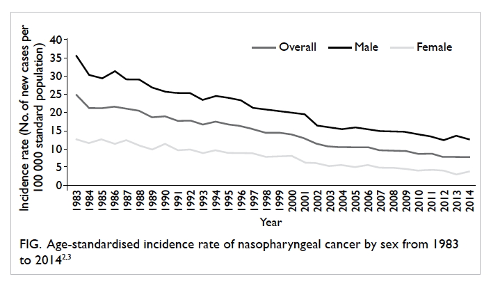
Nasopharyngeal cancer (NPC) was once considered endemic in the southern part of China. This type of cancer was so prevalent in Guangdong Province in southern China in the early 20th century that it was dubbed ‘Guangdong cancer’.1 Although the name is now less popular and the incidence of NPC has been decreasing since then (Fig),2 3 its occurrence in Hong Kong and Southeast Asia is still considerably higher than in other parts of the world: the age-standardised incidence rate was 7.9 per 100 000 population in 2014 in Hong Kong,3 compared with less than 1.0 per 100 000 population in North America and Europe.4 The global data from GLOBOCAN in 2012 showed that 38% of all new cases of NPC were registered in China.5
 

Figure. Age-standardised incidence rate of nasopharyngeal cancer by sex from 1983 to 20142 3
 
While Hong Kong is one of the regions that experience the most NPC,6 it has also become a centre for NPC research. Indeed, many important and landmark studies in NPC were performed in Hong Kong, where local experts have been responsible for developing practice guidelines with regard to the diagnosis, management, and follow-up of NPC.7 8 It is thus interesting to review the updated knowledge about the aetiology, risk factors, diagnosis, and treatment strategies of this ‘Guangdong cancer’.
 
Classification, aetiology, and risk factors
Classification and staging
Nasopharyngeal cancer can be categorised according to its histopathology: keratinising, non-keratinising (which can be further subdivided into differentiated and undifferentiated forms), and basaloid squamous cell carcinoma; all of which are to replace the old numerical classification system.9 In endemic regions such as Hong Kong, non-keratinising carcinoma predominates, whereas the keratinising type is more common in other parts of the world.10
 
Nasopharyngeal cancer is staged according to the tumour, node, metastasis system. To assist with the prognosis and guide treatment decisions, NPC can be further stratified into five different stages (stages I, II, III, IVA, and IVB), as suggested by the latest American Joint Committee on Cancer (8th edition) cancer staging manual.11
 
Viral factors
While it is widely believed that NPC is caused by the interaction of several factors, Epstein-Barr virus (EBV) infection is undoubtedly the most studied aetiological factor for NPC. This virus—as a primary aetiological agent of NPC, specifically the endemic non-keratinising type—has been supported by a large body of evidence12; a review in 2012 suggested that EBV accounted for more than 85% of NPC cases globally.13 Based on in-situ hybridisation techniques10 and the fact that EBV infects more than 90% of the population,14 EBV reactivation is considered necessary in the pathogenesis of NPC; inhibition of EBV reactivation is currently being investigated as a possible approach to preventing NPC relapse.15 What triggers the reactivation, however, is less well-defined, although cigarette smoking is among the possible reactivating factors.16 17
 
On the other hand, human papillomavirus (HPV), a common aetiological agent causing cervical cancer, is associated with the non-endemic, keratinising type of NPC, although evidence is limited due to its low prevalence.18 While EBV and HPV infections are nearly always mutually exclusive in the pathogenesis of NPC,9 studies have suggested that HPV-positive NPC is associated with poorer outcome when compared with EBV-positive NPC.19
 
Genetic factors
Genetic susceptibility has attracted intense interest since the development of various genomic techniques. A whole-exome sequencing study in 2014 revealed the genetic alterations that affect a number of cellular pathways, including chromatin modification, ErbB-phosphatidylinositol-3 kinase signalling, and autophagy machinery in NPC.20 Epigenetic alterations of various chromosomal regions, especially those regions with tumour-suppressor genes, were also found in NPC patients.21 Li et al22 recently identified genomic aberrations of multiple negative regulators of the nuclear factor-κB (NF-κB) pathway in 111 EBV-positive NPC samples in another whole-exome sequencing study, suggesting the pivotal role of activating the NF-κB signalling pathway in NPC and the potential therapeutic applications of NF-κB inhibitors.22 The researchers also revealed major histocompatibility complex class I gene aberrations in some of the samples, and the efficacy of immune checkpoint inhibitors (discussed below) may be affected in this subgroup of NPC patients.22 Although much in this field remains to be elucidated, it is expected that the genetic research will aid in the development of powerful biomarkers for the diagnosis, prognosis, and evaluation of the treatment for NPC.21
 
Environmental factors
An increased risk of NPC has been associated with a number of lifestyle factors, among which a history of salted fish consumption has the strongest association. Various studies have confirmed its association with NPC,23 24 and its relationship with the high prevalence of NPC in Hong Kong and neighbouring regions in the 20th century.6 N-nitrosamine found in the preserved salted fish is believed to be the carcinogen concerned.25 Other factors such as the use of Chinese medicinal herbs and high consumption of fermented food were also suggested, but the associations were often inconsistent among studies.24
 
Diagnosis
Nasendoscopy for a biopsy sample is essential for a definitive diagnosis of NPC. Detecting and diagnosing NPC at an early stage is of paramount importance: the disease stage is significantly correlated with the outcome in NPC, and early diagnosis may improve outcomes.12 Cell-free EBV DNA analysis was shown to have high sensitivity and specificity in detecting NPC, and has been further validated by various studies.26 A local study further showed that the analysis was useful in detecting early-stage NPC in asymptomatic individuals.27 An expanded phase II study involving over 20 000 participants to evaluate its feasibility as a screening tool (NCT02063399) has just been completed, showing excellent sensitivity and specificity (97.1% and 98.6%, respectively).28 Participants who were identified with NPC by this screening tool were detected significantly earlier and with better outcome when compared with those in a historical control.28
 
Other roles of Epstein-Barr virus DNA
With the substantial involvement of EBV in the pathogenesis of NPC, it is sensible to exploit EBV DNA as a biomarker in managing patients with NPC. One such application is the prediction of disease recurrence after treatment. Post-treatment EBV DNA level has been shown to be the most powerful predictor for disease recurrence and long-term survival in NPC patients of different ethnic origins, clinical stages, and treatment modalities.29 30 31 32 33 34 35 36 Recently Lee et al37 demonstrated that serial post–intensity modulated radiation therapy (IMRT) undetectable plasma EBV DNA was prognostic of all predefined survival end-points at 3 years in the modern IMRT era. Leung et al38 further showed that detectable plasma EBV DNA level at midcourse of radiotherapy (RT) or chemoradiotherapy (CRT) is adversely associated with worse overall survival (OS) and progression-free survival (PFS). This suggests the possibility of shifting prognostication from a post-therapy time-point to midcourse of therapy, and selecting high-risk patients for therapy intensification by measuring midcourse plasma EBV DNA level.38
 
Another notable application is the prediction of treatment outcome by measuring the clearance rate of plasma EBV DNA. Following the observation that EBV DNA was rapidly cleared from the circulation after surgical resection of NPC,39 subsequent studies demonstrated that patients with more rapid clearance of plasma EBV DNA responded better to chemotherapy or CRT compared with patients with a slower clearance.40 41 A prospective trial evaluating the response to chemotherapy by measuring plasma EBV DNA half-life together with tumour metabolic response (via fluorodeoxyglucose positron emission tomographic scan) is currently underway.
 
Treatment strategies
Radiotherapy
Radiotherapy has long been regarded as the mainstay of NPC treatment, due to the radiosensitive nature of the tumour, and the anatomical position of NPC that limits a surgical approach.10 Of note, IMRT is currently the preferred approach, with its improved OS and decreased toxicity,42 advantages in preserving parotid function and reducing severe xerostomia,43 and improved quality of life compared with conventional two-dimensional (2D) RT.44 It is currently used as a monotherapy for the early stage of NPC.
 
Since the pre-IMRT era, re-irradiation has been shown to be effective in non-metastatic, recurrent NPC (rNPC) patients after primary RT.45 46 47 With its introduction, IMRT has quickly emerged as the radiation modality of choice for rNPC as well, with or without the use of chemotherapy. Its efficacy has been established in various studies, with documented long-term OS rates ranging from 45% to 65%.48 49 50 51 52 53 54 55 56 Yet, most of the patients in those studies were treated with conventional 2D-RT in the pre-IMRT era. In a recent study conducted by Kong et al,56 77 patients received salvage IMRT for rNPC after a definitive course of primary IMRT. While the median OS and PFS were 37.0 and 20.5 months, respectively, of particular note is the re-irradiation toxicity. Of 34 patients, 18 died from treatment-induced severe adverse effects without evidence of disease progression during the study, including mucosal necrosis, temporal lobe necrosis, and cranial neuropathy,56 reflecting the limitations of salvage IMRT in the modern IMRT era. Other radiation modalities have been proposed, including particle therapy using proton and carbon ions,57 but long-term data are not yet available.
 
Chemotherapy
Chemotherapy is another important modality in managing NPC, and it is often combined with RT in the intermediate and advanced stages of NPC. The benefit of CRT was well-illustrated in a meta-analysis of seven trials, which showed significantly improved OS and 10-year PFS in the CRT group compared with the RT-alone group.58 A platinum-based regimen is often used as the chemotherapy of choice, in which cisplatin is most commonly used.10
 
While it is clear that chemotherapy is essential in the treatment of advanced NPC, its value as an add-on induction therapy (preceding CRT) and adjuvant therapy (following CRT) is less clear. Regarding induction therapy, a phase III trial recently showed that the addition of docetaxel, cisplatin, and fluorouracil prior to CRT was superior to CRT alone in terms of OS and PFS at 3 years,59 although another trial using cisplatin and fluorouracil as induction therapy failed to show significant differences in OS.60 The role of induction therapy requires further confirmation from other ongoing phase III trials.
 
Meanwhile, the use of adjuvant chemotherapy following CRT is debatable. A phase III trial with a median follow-up of 68.4 months failed to show significantly improved OS and PFS after adding cisplatin and fluorouracil as adjuvant therapy post-CRT in locally advanced NPC,61 but another study suggested adjuvant chemotherapy might be reserved for high-risk patients defined by post-treatment residual EBV DNA.62 63 It should be noted, however, that the benefit of more intensive therapy may be limited by the late toxicities of high cumulative doses of chemotherapy, most notably cisplatin, which are not reported in some of the studies.60 64
 
Platinum-containing doublet regimens remain the first-line systemic treatment for recurrent or metastatic NPC. Cisplatin and fluorouracil have been the conventional choices.10 A recent study by Zhang et al65 demonstrated that the combination of cisplatin plus gemcitabine was superior to the combination of cisplatin and fluorouracil, in terms of median PFS (7.0 vs 5.6 months; hazard ratio=0.55; 95% confidence interval, 0.44-0.68), although the cisplatin-gemcitabine group experienced more haematological toxicity, such as grade-3 or higher leukopenia, neutropenia, and thrombocytopenia.65 This randomised controlled trial has thus established the role of cisplatin and gemcitabine combination as the chemotherapy of choice in recurrent or metastatic NPC.
 
Surgery and targeted therapy
As mentioned above, surgery is usually not considered in the routine management of NPC; yet salvage therapy can be considered an option for selected patients with local recurrence in the neck.66 Molecular targeted therapy is considered hopeful for many other types of carcinoma, but its efficacy in treating NPC has been disappointing; studies of inhibitors of epidermal growth factor receptor (eg cetuximab) and vascular endothelial growth factor (eg sunitinib) failed to show superiority over standard treatments, and were largely limited to phase II trials.8 Lee et al8 attributed its failure to the scarcity of authentic NPC models that can be utilised in the preclinical studies of new drugs, and increased incidence of drug-related toxicities such as bleeding. The development of immunotherapy is therefore exciting as it presents a new hope for managing NPC.
 
Immunotherapy
The presence of EBV and the expression of viral antigens in almost all NPC cases make this disease an attractive target for the development of immunotherapy. For example, EBV nuclear antigen I (EBNA1) and latent membrane protein 2 (LMP2) are frequently expressed in EBV-associated NPC, and a recombinant virus–based vaccine that encodes an inactive fusion protein containing fragments of EBNA1 and LMP2 was shown to be effective in inducing T-cell response in a local phase I trial.67 The vaccine is currently being tested in a phase II clinical trial (NCT01094405).
 
As EBV that persists as a latent infection is controlled by cytotoxic T lymphocytes (CTL),68 it follows that the use of EBV-specific CTL for NPC appears logical as a treatment strategy. Adoptive immunotherapy that includes infusion of autologous CTL has been tested in a number of clinical trials, and the results have been promising. For example, a study in Singapore showed that chemotherapy followed by EBV-specific CTL achieved a response rate (full or partial) of 71.4% in 38 patients,69 and a phase III trial is currently underway to assess its efficacy (NCT02578641).
 
Among all the immunotherapies available, checkpoint inhibitors seem to be the most rapidly developing. Programmed death ligand–1 (PD-L1) was found to be expressed on antigen-presenting cells, and its interaction with the programmed death–1 (PD-1) receptor on T cells inhibits downstream signalling of T cell receptors.70 Tumour-associated PD-L1 was also found to mediate immune suppression by various other mechanisms, such as facilitating T cell apoptosis and inducing regulatory T cells.71 With PD-L1 expressed in many different carcinomas,72 blockade of PD-L1 and/or the PD-1 receptor has become the focus of new cancer drug development in the past 5 years.
 
While PD-L1 inhibitor has recently gained much attention in the treatment of non–small-cell lung cancer,73 its progress in the treatment of advanced NPC is exciting and much awaited. Pembrolizumab was shown to be well-tolerated with significant anti-tumour activity in NPC in a phase Ib trial,74 and is currently in a phase II trial to confirm the response rate and efficacy in terms of improvement in OS (NCT02611960). Nivolumab has just completed phase II trials; the preliminary results showed that it is active in heavily pre-treated recurrent or metastatic patients,75 76 and that PD-L1 expression may predict benefits from nivolumab.75
 
Conclusion
Once a nightmare in the eyes of many Hong Kong inhabitants, NPC has become less prevalent in southern China, but it still poses a threat to Hong Kong citizens as it was ranked as the 10th most common cancer in the city.3 With clearer understanding of its pathophysiology and advances in technology, it is expected that more refined treatment strategies and novel therapeutic agents will be available in the near future.
 
Acknowledgement
I would like to thank Prof Brigette Ma from Department of Clinical Oncology, The Chinese University of Hong Kong for her comments and advice.
 
References
1. Gibb AG, van Hasselt CA, editors. Nasopharyngeal carcinoma. 2nd ed. Hong Kong: The Chinese University Press; 1999.
2. Nasopharyngeal cancer. Available from: http://www.chp.gov.hk/en/content/9/25/54.html. Accessed 12 Apr 2017.
3. Hong Kong Cancer Registry, Hospital Authority. Available from: www3.ha.org.hk/cancereg/statistics.html. Accessed 12 Apr 2017.
4. 2014 Review of cancer medicines on the WHO list of essential medicines: Nasopharyngeal carcinoma. Geneva: World Health Organization; 2014.
5. Ferlay J, Soerjomataram I, Ervik M, et al. GLOBOCAN 2012: Estimated cancer incidence, mortality and prevalence worldwide in 2012 v1.0. Lyon, France: International Agency for Research on Cancer; 2013.
6. Tang LL, Chen WQ, Xue WQ, et al. Global trends in incidence and mortality of nasopharyngeal carcinoma. Cancer Lett 2016;374:22-30. Crossref
7. Chan AT, Grégoire V, Lefebvre JL, et al. Nasopharyngeal cancer: EHNS-ESMO-ESTRO clinical practice guidelines for diagnosis, treatment and follow-up. Ann Oncol 2012;23 Suppl 7:vii83-5. Crossref
8. Lee AW, Ma BB, Ng WT, Chan AT. Management of nasopharyngeal carcinoma: current practice and future perspective. J Clin Oncol 2015;33:3356-64. Crossref
9. Pathology and genetics of head and neck tumours. In: Barnes L, Eveson JW, Reichart P, Sidransky D, editors. World Health Organization classification of tumours. Lyon: IARC Press; 2005.
10. Chua ML, Wee JT, Hui EP, Chan AT. Nasopharyngeal carcinoma. Lancet 2016;387:1012-24. Crossref
11. Lydiatt WM, Patel SG, O’Sullivan B, et al. Head and neck cancers—major changes in the American Joint Committee on Cancer eighth edition cancer staging manual. CA Cancer J Clin 2017;67:122-37. Crossref
12. Raghupathy R, Hui EP, Chan AT. Epstein-Barr virus as a paradigm in nasopharyngeal cancer: from lab to clinic. Am Soc Clin Oncol Educ Book 2014: 149-53. Crossref
13. de Martel C, Ferlay J, Franceschi S, et al. Global burden of cancers attributable to infections in 2008: a review and synthetic analysis. Lancet Oncol 2012;13:607-15. Crossref
14. Raab-Traub N. Epstein-Barr virus in the pathogenesis of NPC. Semin Cancer Biol 2002;12:431-41. Crossref
15. Wu CC, Fang CY, Hsu HY, et al. EBV reactivation as a target of luteolin to repress NPC tumorigenesis. Oncotarget 2016;7:18999-9017. Crossref
16. Xu FH, Xiong D, Xu YF, et al. An epidemiological and molecular study of the relationship between smoking, risk of nasopharyngeal carcinoma, and Epstein-Barr virus activation. J Natl Cancer Inst 2012;104:1396-410. Crossref
17. Hsu WL, Chen JY, Chien YC, et al. Independent effect of EBV and cigarette smoking on nasopharyngeal carcinoma: a 20-year follow-up study on 9,622 males without family history in Taiwan. Cancer Epidemiol Biomarkers Prev 2009;18:1218-26. Crossref
18. Lo EJ, Bell D, Woo JS, et al. Human papillomavirus and WHO type I nasopharyngeal carcinoma. Laryngoscope 2010;120:1990-7. Crossref
19. Stenmark MH, McHugh JB, Schipper M, et al. Nonendemic HPV-positive nasopharyngeal carcinoma: association with poor prognosis. Int J Radiat Oncol Biol Phys 2014;88:580-8. Crossref
20. Lin DC, Meng X, Hazawa M, et al. The genomic landscape of nasopharyngeal carcinoma. Nat Genet 2014;46:866-71. Crossref
21. Dai W, Zheng H, Cheung AK, Lung ML. Genetic and epigenetic landscape of nasopharyngeal carcinoma. Chin Clin Oncol 2016;5:16. Crossref
22. Li YY, Chung GT, Lui VW, et al. Exome and genome sequencing of nasopharynx cancer identifies NF-κB pathway activating mutations. Nat Commun 2017;8:14121. Crossref
23. Ho JH. Nasopharyngeal carcinoma (NPC). Adv Cancer Res 1972;15:57-92. Crossref
24. Yu MC, Ho JH, Lai SH, Henderson BE. Cantonese-style salted fish as a cause of nasopharyngeal carcinoma: report of a case-control study in Hong Kong. Cancer Res 1986;46:956-61.
25. Guo X, Johnson RC, Deng H, et al. Evaluation of nonviral risk factors for nasopharyngeal carcinoma in a high-risk population of Southern China. Int J Cancer 2009;124:2942-7. Crossref
26. Fung SY, Lam JW, Chan KC. Clinical utility of circulating Epstein-Barr virus DNA analysis for the management of nasopharyngeal carcinoma. Chin Clin Oncol 2016;5:18. Crossref
27. Chan KC, Hung EC, Woo JK, et al. Early detection of nasopharyngeal carcinoma by plasma Epstein-Barr virus DNA analysis in a surveillance program. Cancer 2013;119:1838-44. Crossref
28. Chan KC, Woo JK, King A, et al. Analysis of Plasma Epstein–Barr Virus DNA to Screen for Nasopharyngeal Cancer. N Engl J Med 2017;377:513-22. Crossref
29. Chan AT, Lo YM, Zee B, et al. Plasma Epstein-Barr virus DNA and residual disease after radiotherapy for undifferentiated nasopharyngeal carcinoma. J Natl Cancer Inst 2002;94:1614-9. Crossref
30. Lin JC, Wang WY, Chen KY, et al. Quantification of plasma Epstein-Barr virus DNA in patients with advanced nasopharyngeal carcinoma. N Engl J Med 2004;350:2461-70. Crossref
31. Le QT, Jones CD, Yau TK, et al. A comparison study of different PCR assays in measuring circulating plasma Epstein-Barr virus DNA levels in patients with nasopharyngeal carcinoma. Clin Cancer Res 2005;11:5700-7. Crossref
32. Lin JC, Wang WY, Liang WM, et al. Long-term prognostic effects of plasma Epstein-Barr virus DNA by minor groove binder-probe real-time quantitative PCR on nasopharyngeal carcinoma patients receiving concurrent chemoradiotherapy. Int J Radiat Oncol Biol Phys 2007;68:1342-8. Crossref
33. An X, Wang FH, Ding PR, et al. Plasma Epstein-Barr virus DNA level strongly predicts survival in metastatic/recurrent nasopharyngeal carcinoma treated with palliative chemotherapy. Cancer 2011;117:3750-7. Crossref
34. Hou X, Zhao C, Guo Y, et al. Different clinical significance of pre- and post-treatment plasma Epstein-Barr virus DNA load in nasopharyngeal carcinoma treated with radiotherapy. Clin Oncol (R Coll Radiol) 2011;23:128-33. Crossref
35. Ferrari D, Codecà C, Bertuzzi C, et al. Role of plasma EBV DNA levels in predicting recurrence of nasopharyngeal carcinoma in a western population. BMC Cancer 2012;12:208. Crossref
36. Zhao FP, Liu X, Chen XM, et al. Levels of plasma Epstein-Barr virus DNA prior and subsequent to treatment predicts the prognosis of nasopharyngeal carcinoma. Oncol Lett 2015;10:2888-94. Crossref
37. Lee VH, Kwong DL, Leung TW, et al. Prognostication of serial post-intensity-modulated radiation therapy undetectable plasma EBV DNA for nasopharyngeal carcinoma. Oncotarget 2017;8:5292-308.
38. Leung SF, Chan KC, Ma BB, et al. Plasma Epstein-Barr viral DNA load at midpoint of radiotherapy course predicts outcome in advanced-stage nasopharyngeal carcinoma. Ann Oncol 2014;25:1204-8. Crossref
39. To EW, Chan KC, Leung SF, et al. Rapid clearance of plasma Epstein-Barr virus DNA after surgical treatment of nasopharyngeal carcinoma. Clin Cancer Res 2003;9:3254-9.
40. Hsu CL, Chang KP, Lin CY, et al. Plasma Epstein-Barr virus DNA concentration and clearance rate as novel prognostic factors for metastatic nasopharyngeal carcinoma. Head Neck 2012;34:1064-70. Crossref
41. Wang WY, Twu CW, Chen HH, et al. Plasma EBV DNA clearance rate as a novel prognostic marker for metastatic/recurrent nasopharyngeal carcinoma. Clin Cancer Res 2010;16:1016-24. Crossref
42. Peng G, Wang T, Yang KY, et al. A prospective, randomized study comparing outcomes and toxicities of intensity-modulated radiotherapy vs. conventional two-dimensional radiotherapy for the treatment of nasopharyngeal carcinoma. Radiother Oncol 2012;104:286-93. Crossref
43. Kam MK, Leung SF, Zee B, et al. Prospective randomized study of intensity-modulated radiotherapy on salivary gland function in early-stage nasopharyngeal carcinoma patients. J Clin Oncol 2007;25:4873-9. Crossref
44. Pow EH, Kwong DL, McMillan AS, et al. Xerostomia and quality of life after intensity-modulated radiotherapy vs. conventional radiotherapy for early-stage nasopharyngeal carcinoma: initial report on a randomized controlled clinical trial. Int J Radiat Oncol Biol Phys 2006;66:981-91. Crossref
45. Lee AW, Foo W, Law SC, et al. Reirradiation for recurrent nasopharyngeal carcinoma: factors affecting the therapeutic ratio and ways for improvement. Int J Radiat Oncol Biol Phys 1997;38:43-52. Crossref
46. Teo PM, Kwan WH, Chan AT, Lee WY, King WW, Mok CO. How successful is high-dose (>or = 60 Gy) reirradiation using mainly external beams in salvaging local failures of nasopharyngeal carcinoma? Int J Radiat Oncol Biol Phys 1998;40:897-913. Crossref
47. Chen C, Fee W, Chen J, et al. Salvage treatment for locally recurrent nasopharyngeal carcinoma (NPC). Am J Clin Oncol 2014;37:327-31. Crossref
48. Lu TX, Mai WY, Teh BS, et al. Initial experience using intensity-modulated radiotherapy for recurrent nasopharyngeal carcinoma. Int J Radiat Oncol Biol Phys 2004;58:682-7. Crossref
49. Chua DT, Sham JS, Leung LH, Au GK. Re-irradiation of nasopharyngeal carcinoma with intensity-modulated radiotherapy. Radiother Oncol 2005;77:290-4. Crossref
50. Qiu S, Lin S, Tham IW, Pan J, Lu J, Lu JJ. Intensity-modulated radiation therapy in the salvage of locally recurrent nasopharyngeal carcinoma. Int J Radiat Oncol Biol Phys 2012;83:676-83. Crossref
51. Hua YJ, Han F, Lu LX, et al. Long-term treatment outcome of recurrent nasopharyngeal carcinoma treated with salvage intensity modulated radiotherapy. Eur J Cancer 2012;48:3422-8. Crossref
52. Han F, Zhao C, Huang SM, et al. Long-term outcomes and prognostic factors of re-irradiation for locally recurrent nasopharyngeal carcinoma using intensity-modulated radiotherapy. Clin Oncol (R Coll Radiol) 2012;24:569-76. Crossref
53. Qiu S, Lu J, Zheng W, et al. Advantages of intensity modulated radiotherapy in recurrent T1-2 nasopharyngeal carcinoma: a retrospective study. BMC Cancer 2014;14:797. Crossref
54. Xiao W, Liu S, Tian Y, et al. Prognostic significance of tumor volume in locally recurrent nasopharyngeal carcinoma treated with salvage intensity-modulated radiotherapy. PLoS One 2015;10:e0125351. Crossref
55. Tian YM, Guan Y, Xiao WW, et al. Long-term survival and late complications in intensity-modulated radiotherapy of locally recurrent T1 to T2 nasopharyngeal carcinoma. Head Neck 2016;38:225-31. Crossref
56. Kong L, Wang L, Shen C, Hu C, Wang L, Lu JJ. Salvage intensity-modulated radiation therapy (IMRT) for locally recurrent nasopharyngeal cancer after definitive IMRT: a novel scenario of the modern era. Sci Rep 2016;6:32883. Crossref
57. Kong L, Hu J, Guan X, Gao J, Lu R, Lu JJ. Phase I/II trial evaluating carbon ion radiotherapy for salvaging treatment of locally recurrent nasopharyngeal carcinoma. J Cancer 2016;7:774-83. Crossref
58. Blanchard P, Lee A, Marguet S, et al. Chemotherapy and radiotherapy in nasopharyngeal carcinoma: an update of the MAC-NPC meta-analysis. Lancet Oncol 2015;16:645-55. Crossref
59. Sun Y, Li WF, Chen NY, et al. Induction chemotherapy plus concurrent chemoradiotherapy versus concurrent chemoradiotherapy alone in locoregionally advanced nasopharyngeal carcinoma: a phase 3, multicentre, randomised controlled trial. Lancet Oncol 2016;17:1509-20. Crossref
60. Cao SM, Yang Q, Guo L, et al. Neoadjuvant chemotherapy followed by concurrent chemoradiotherapy versus concurrent chemoradiotherapy alone in locoregionally advanced nasopharyngeal carcinoma: a phase III multicentre randomised controlled trial. Eur J Cancer 2017;75:14-23. Crossref
61. Chen L, Hu CS, Chen XZ, et al. Adjuvant chemotherapy in patients with locoregionally advanced nasopharyngeal carcinoma: Long-term results of a phase 3 multicentre randomised controlled trial. Eur J Cancer 2017;75:150-8. Crossref
62. Chan AT, Ngan RK, Hui EP, et al. A multicenter randomized controlled trial of adjuvant chemotherapy in nasopharyngeal carcinoma with residual plasma EBV DNA following primary radiotherapy or chemoradiotherapy. J Clin Oncol 2012;30(Suppl):abstract 5511.
63. Hui EP, Ma BB, Chan KC, et al. Clinical utility of plasma Epstein-Barr virus DNA and ERCC1 single nucleotide polymorphism in nasopharyngeal carcinoma. Cancer 2015;121:2720-9. Crossref
64. Lee AW, Tung SY, Chua DT, et al. Randomized trial of radiotherapy plus concurrent-adjuvant chemotherapy vs radiotherapy alone for regionally advanced nasopharyngeal carcinoma. J Natl Cancer Inst 2010;102:1188-98. Crossref
65. Zhang L, Huang Y, Hong S, et al. Gemcitabine plus cisplatin versus fluorouracil plus cisplatin in recurrent or metastatic nasopharyngeal carcinoma: a multicentre, randomised, open-label, phase 3 trial. Lancet 2016;388:1883-92. Crossref
66. Chan JY. Surgical salvage of recurrent nasopharyngeal carcinoma. Curr Oncol Rep 2015;17:433. Crossref
67. Hui EP, Taylor GS, Jia H, et al. Phase I trial of recombinant modified vaccinia ankara encoding Epstein-Barr viral tumor antigens in nasopharyngeal carcinoma patients. Cancer Res 2013;73:1676-88. Crossref
68. Khanna R, Burrows SR. Role of cytotoxic T lymphocytes in Epstein-Barr virus-associated diseases. Annu Rev Microbiol 2000;54:19-48. Crossref
69. Chia WK, Teo M, Wang WW, et al. Adoptive T-cell transfer and chemotherapy in the first-line treatment of metastatic and/or locally recurrent nasopharyngeal carcinoma. Mol Ther 2014;22:132-9. Crossref
70. Khalil DN, Smith EL, Brentjens RJ, Wolchok JD. The future of cancer treatment: immunomodulation, CARs and combination immunotherapy. Nat Rev Clin Oncol 2016;13:273-90. Crossref
71. Chen L, Han X. Anti–PD-1/PD-L1 therapy of human cancer: past, present, and future. J Clin Invest 2015;125:3384-91. Crossref
72. Chen DS, Mellman I. Oncology meets immunology: the cancer-immunity cycle. Immunity 2013;39:1-10. Crossref
73. Reck M, Rodríguez-Abreu D, Robinson A, et al. Pembrolizumab versus chemotherapy for PD-L1–positive non–small-cell lung cancer. N Engl J Med 2016;375:1823-33. Crossref
74. Hsu C, Lee SH, Ejadi S, et al. Antitumor activity and safety of pembrolizumab in patients with PD-L1-positive nasopharyngeal carcinoma: Interim results from a phase 1b study. Eur J Cancer 2015;51 Suppl 3:S558. Crossref
75. Ma BB, Goh BC, Lim WT, et al. Multicenter phase II study of nivolumab in previously treated patients with recurrent and metastatic non-keratinizing nasopharyngeal carcinoma [abstract]. Proceedings of the 107th Annual Meeting of the American Association for Cancer Research; 2017 Apr 1-5; Washington, DC: Philadelphia; 2017. Available from: http://www.abstractsonline.com/pp8/#!/4292/presentation/12342. Accessed Aug 2017.
76. Delord JP, Hollebecque A, De Boer JP, et al. An open-label, multicohort, phase I/II study to evaluate nivolumab in patients with virus-associated tumors (CheckMate 358): Efficacy and safety in recurrent or metastatic nasopharyngeal carcinoma. J Clin Oncol 2017;35(Suppl):abstract 6025.

Hypersensitivity to antipyretics: pathogenesis, diagnosis, and management

Hong Kong Med J 2017 Aug;23(4):395–403 | Epub 7 Jul 2017
DOI: 10.12809/hkmj166186
© Hong Kong Academy of Medicine. CC BY-NC-ND 4.0
 
REVIEW ARTICLE
Hypersensitivity to antipyretics: pathogenesis, diagnosis, and management
QU Lee, MB, ChB, FHKAM (Paediatrics)
Department of Paediatrics and Adolescent Medicine, Princess Margaret Hospital, Laichikok, Hong Kong
 
Corresponding author: Dr QU Lee (leequnui@gmail.com)
 
 Full paper in PDF
 
Abstract
Antipyretics are commonly prescribed drugs and hypersensitivity occurs at rates of 0.01% to 0.3%. Hypersensitivity can be due to immune mechanisms that include type I to IV hypersensitivity. Type I hypersensitivity results from specific immunoglobulin E production following sensitisation on first exposure. Subsequent exposures elicit degranulation of mast cells, culminating an immediate reaction. Non–type I hypersensitivity is a delayed reaction that involves various effector cells, resulting in maculopapular rash, fixed drug eruptions, drug reaction with eosinophilia and systemic symptoms, and Stevens-Johnson syndrome/toxic epidermal necrolysis. Antipyretics also cause non-immune hypersensitivity via cyclooxygenase inhibition. Apart from hypersensitivity to parent compounds, hypersensitivity to excipient has been reported. Clinical manifestations of antipyretic hypersensitivity involve the skin, mucosa, or multiple organs. Diagnosis of hypersensitivity requires a detailed history taking and knowledge of any underlying disorders. Differential diagnoses include infection, inflammatory conditions, and antipyretics acting as co-factors of other allergens. Investigations include specific immunoglobulin E assays, lymphocyte transformation test, basophil activation test, and skin prick test. Lack of standardisation and a scarcity of available commercial reagents, however, limit the utility of these tests. A drug provocation test under close supervision remains the gold standard of diagnosis. A trial of the culprit drug or other structurally different antipyretics can be considered. Patients with confirmed hypersensitivity to antipyretics should consider either avoidance or desensitisation. Other theoretical options include subthreshold or low-dose paracetamol, cyclooxygenase-2 inhibitors, pre-medication with antihistamines with or without a leukotriene receptor antagonist, co-administration of prostaglandin E2 analogue, traditional Chinese medicine, or desensitisation if antipyretics are deemed desirable. Safety and efficacy of unconventional treatments warrant future studies.


Introduction
Antipyretics (APs) are widely consumed drugs. In 2013, the National Institute for Health and Care Excellence advised that paracetamol and ibuprofen can be prescribed for febrile children in distress.1 In a national cross-sectional study in France, more than 80% of health care professionals resorted to AP to manage fever in children. Paracetamol was the first-choice AP among 88% of health care professionals while ibuprofen, a non-steroidal anti-inflammatory drug (NSAID), was preferred by 11%.2 Diclofenac sodium and mefenamic acid have also been advocated as APs for children.3 4 What makes use of APs truly ubiquitous is their non-prescription, over-the-counter availability. Widespread consumption often entails an increased chance of adverse drug reaction (ADR). Paracetamol and NSAIDs are two of the most common drugs to cause an allergic or pseudo-allergic reaction, secondary to general anaesthetic agents and beta-lactam antibiotics.5 Prevalence of NSAID hypersensitivity ranges from 0.1% to 0.3%.6 Hypersensitivity reactions to ibuprofen occur at 0.01%.7 The epidemiology of paracetamol hypersensitivity is unclear. This is understandable since prescription data for over-the-counter drugs are difficult to obtain. Nevertheless between 1982 and 1991, the Spanish Drug Monitoring System estimated the incidence of ADR to paracetamol to be less than 1 per 100 000 inhabitants below the age of 15 years. Among the reported ADRs, 30% were related to skin eruption, urticaria, or itchiness.8 The real incidence might have been higher, had unreported cases been included. This is a review of the pathogenesis, diagnosis, and management of hypersensitivity to APs.
 
Types of hypersensitivity reactions to antipyretics
Hypersensitivity reactions to APs are idiosyncratic responses of the body towards drugs given at a therapeutic dose. Around two thirds of patients with NSAID or paracetamol hypersensitivity are single reactors, while one third are cross-reactors.9 Reaction may either be to the active ingredient or to excipients. Hypersensitivity to APs can manifest as an immune-mediated reaction that stems from an immunoglobulin (Ig) E–mediated (immediate) reaction or a non–IgE-mediated (delayed) reaction. Unlike other drugs, hypersensitivity to APs can also be non–immune-mediated.
 
Immune-mediated hypersensitivity
Type I hypersensitivity
Type I hypersensitivity to APs, or an IgE-mediated reaction, is selective in nature. It presents with single NSAID-induced urticaria/angioedema or anaphylaxis (SNIUAA) or hypersensitivity to NSAIDs with structural similarity but tolerance to NSAIDs from different classes. Ibuprofen and paracetamol are two common causes of SNIUAA.10 Severity ranges from localised urticaria, mucosal swelling, and angioedema to anaphylaxis. Susceptible patients become sensitised to an AP on first exposure, with the production of drug-specific IgE. Specific IgE molecules become attached to high-affinity IgE receptors on mast cells or basophils. Re-exposure to the same AP or cross-reacting drugs leads to cross-linking of adjacent IgE receptors and subsequent degranulation of vasoactive inflammatory mediators like histamine and tryptase.11 Patients with SNIUAA against ibuprofen produce IgE against specific antigen determinants of the drug. Hence they may react to arylpropionic acids with similar chemical structure but tolerate NSAIDs from other groups, such as acetic acids.7 Similarly, patients with selective hypersensitivity to paracetamol confirmed by IgE tests or oral challenge can tolerate other NSAIDs.12
 
Non–type I hypersensitivity
Maculopapular eruptions
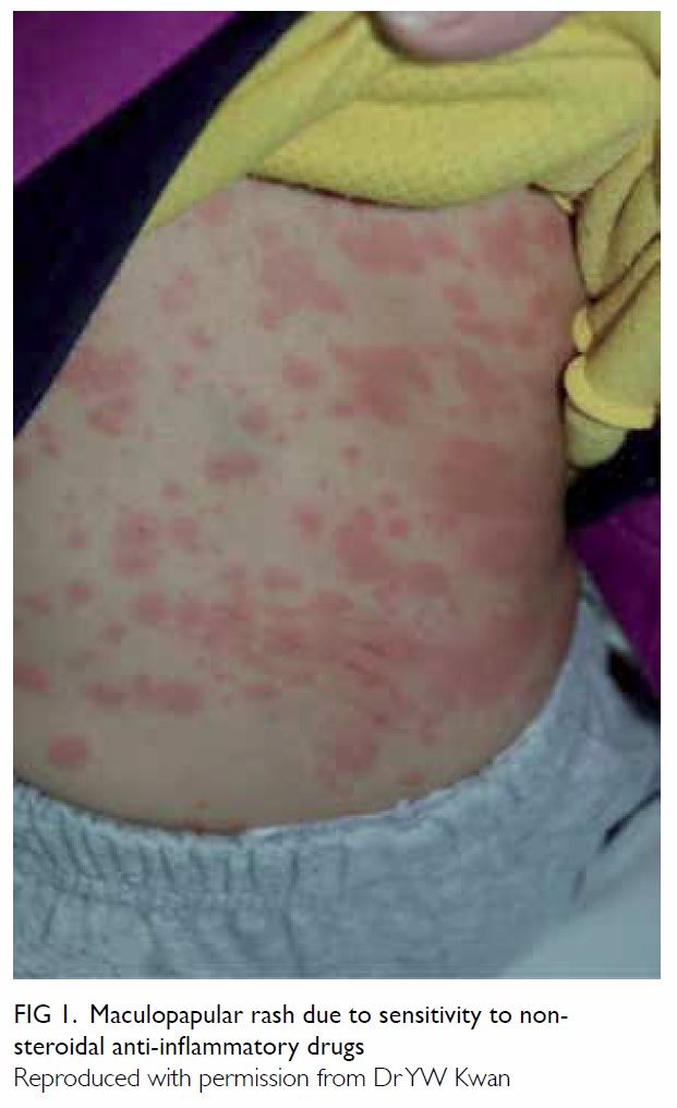
According to the revised Gell and Coombs classification, maculopapular eruption (MPE) is a type IV-c, T-cell–mediated delayed hypersensitivity reaction.13 It is said to be the most common delayed drug rash due to an AP. Implicated drugs include ibuprofen, diclofenac, and paracetamol.14 Such MPE manifests as a morbilliform or scarlatiniform rash that starts on the trunk with subsequent spread to the limbs. Onset of MPE ranges from within 7 to 14 days of first consumption of the drug, but may take only 2 to 3 days in patients with prior sensitisation. The reaction of MPE involves skin-homing T lymphocytes, drug-specific cells that express cutaneous lymphocyte antigen. Around two thirds of the T-cells are CD4+, while one third are CD8+. Having resided in the dermo-epidermal junction, these cells release perforin and granzyme B, two mediators of keratinocyte apoptosis, via their ability to induce pore formation in the cell membrane.15 Histological changes include intracellular, intercellular and dermal papilla oedema, dislodgment of epidermal basal cells, hydropic degeneration, spongiosis of the lower epidermis, and dyskeratosis and necrosis of keratinocytes. Inflammatory infiltration by T-cells is seen at the dermo-epidermal junction and eosinophils in the perivascular region.16
 
Fixed drug eruption
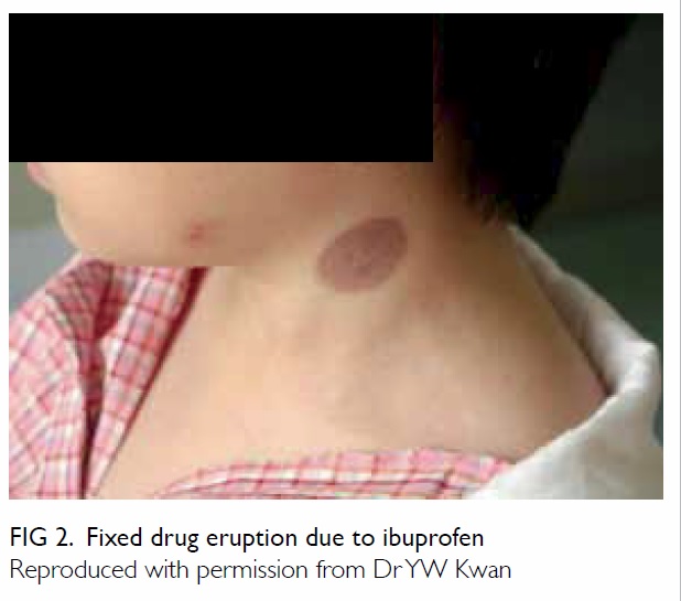
Fixed drug eruption (FDE) is a peculiar type of T-cell–mediated delayed drug hypersensitivity. It starts with solitary, well-circumscribed macules that erupt anywhere on the skin or mucosa, usually over the lips, palms, soles, groins, or glans penis. With time, the lesions evolve into plaques that recur at the same site on re-exposures to the same drug. The interval between drug intake and FDE is around 30 minutes to 8 hours. The eruption resolves spontaneously after cessation of the culprit, leaving hyperpigmentation at the affected site. Pathologically, migration and residence of drug-specific effector-memory CD8+ T-cells in the epidermal side of the dermo-epidermal junction of the affected area account for the recurrence of eruption at the same site. Upon drug re-exposure, quiescent CD8+ cells become activated and secrete interferon-γ and cytotoxic granules into the local microenvironment.17 Paracetamol is one of the most common causes of FDE, as are mefenamic acid, ibuprofen, and aspirin.18
 
Drug reaction with eosinophilia and systemic symptoms
Drug reaction with eosinophilia and systemic symptoms (DRESS) is classified as a type IV-b delayed hypersensitivity reaction with eosinophil involvement. It is characterised by fever, exfoliative dermatitis, lymphadenopathy, haematological abnormalities (hypereosinophilia, atypical lymphocytes), and organ dysfunction. The interval between drug consumption and onset of symptoms is quite prolonged, ranging from 3 weeks to 3 months. The pathophysiology of DRESS involves viral reactivation (eg human herpes type 6) and T-cell activation, two determining factors with a mutual causal relationship.19 FOXP3+ (forkhead box P3) regulatory T-cells are activated early in the course of DRESS, but are subsequently deactivated and become deficient, culminating in the emergence of autoimmune diseases commonly seen in the aftermath of DRESS. Ibuprofen and paracetamol have rarely been associated with DRESS.20 21
 
Stevens-Johnson syndrome/toxic epidermal necrolysis
Stevens-Johnson syndrome/toxic epidermal necrolysis (SJS/TEN) is a type IV-c delayed hypersensitivity reaction to infections or drugs including APs. The interval between intake of the culprit drug and SJS/TEN is shorter than that of DRESS, ranging from 1 to 21 days.22 Skin lesions in SJS/TEN are typically target-like with central necrosis, bullae formation, or purpuric lesions. In SJS, less than 10% of the body surface area is involved, whereas in TEN, more than 30% is involved. Gentle rubbing of ‘normal’ skin causes separation of the epidermis (Nikolsky sign). Mucosal and eye inflammation is present in 90% and 60% of cases, respectively. Severe cases culminate in corneal scarring, respiratory distress syndrome, pneumonia, and respiratory failure.23 A caveat in the diagnosis is that the prodromal phase of SJS/TEN may be mistaken as symptoms of a febrile illness, with consequent administration of APs. In the event that SJS/TEN occur secondary to other causes, subsequent appearance of skin and mucosal lesions may impart the wrong impression of AP as the causative agent. In SJS/TEN, CD4 T-cells accumulate in the dermis while CD8 T-cells predominate in the epidermis. T-cell infiltration causes massive apoptosis of the keratinocytes via the toxic action of perforin, granzyme, and Fas/Fas ligand interaction.24 Of note, SJS/TEN due to NSAIDs is exceedingly rare. The incidence for ibuprofen was 0.013 per 1 000 000 as opposed to 0.032 per 1 000 000 for oxicams.25 Compared with controls, the relative risk of paracetamol and ibuprofen for SJS/TEN in children ranges from 5 to 11.26 It is also noteworthy that APs are often prescribed together with antibiotics to treat infection, with the latter two factors (antibiotics and infection) potentially related to SJS/TEN.27
 
Acute generalised exanthematous pustulosis
Acute generalised exanthematous pustulosis (AGEP) is a rare type IV-d drug hypersensitivity with sterile subcorneal pustule formation. Onset of pustules occurs around 1 day after drug intake. Most patients present with fever. Non-follicular small pustules with an erythematous base start on the face or intertriginous area and subsequently become generalised. The pustules, which are itchy or burning, persist for 4 to 30 days before desquamation.28 Histological characteristics include papillary oedema, perivascular infiltration by neutrophils, and drug-specific T-cells and epidermal keratinocyte necrosis. Interleukin-8, a neutrophil chemoattractant, is expressed by drug-specific T-cells. Presence of human leukocyte antigens (HLAs)-DR within the inflammatory infiltrate suggests the role of a major histocompatibility complex in causing this peculiar type of drug eruption.29 Among NSAIDs, only the oxicams are significantly associated with AGEP, with a multivariate odds ratio of 8.4. Paracetamol is not considered at an increased risk of causing AGEP.30
 
Organ-specific delayed hypersensitivity
Of note, NSAIDs can cause an allergic inflammatory response in different organs. Cases of NSAID-induced hepatitis, pneumonitis, nephritis, and aseptic meningitis have been reported.6
 
Non–immune-mediated hypersensitivity: cyclooxygenase inhibition
Three types of non-immune drug hypersensitivity to NSAIDs have been described: NSAID-exacerbated respiratory disease (NERD), NSAID-exacerbated cutaneous disease (NECD), and NSAID-induced urticaria/angioedema (NIUA). In NERD, patients usually have asthma, rhinosinusitis, and/or nasal polyps. Aspirin or other NSAIDs may precipitate nasal congestion, rhinorrhoea, bronchial obstruction, or dyspnoea within 30 to 180 minutes of ingestion. Urticaria, angioedema, and flushing of the upper thorax may occur. Patients with NECD usually have underlying chronic spontaneous urticaria. Aspirin or NSAIDs may cause flare-up of urticaria and angioedema in 12% to 30% of patients with chronic spontaneous urticaria. On the other hand, NIUA occurs primarily in patients without underlying disease. Immediate reactions that occur less than 15 minutes following consumption and late reactions that occur after several hours have been described.10
 
Non-immune hypersensitivity to NSAIDs is the result of cyclooxygenase (COX) inhibition, a pharmacological property common to all NSAIDs that accounts for their propensity to cause cross-reactivity. Three COXs—COX-1, COX-2, and COX-3—have been identified, and NSAIDs like ibuprofen inhibit all three COXs. On the contrary, paracetamol is a weak inhibitor of COX-1 and COX-2, especially at a low dose, and preferentially inhibits COX-3.31 In susceptible patients, inhibition of COX leads to overproduction of pro-inflammatory cysteinyl leukotrienes by mast cells and eosinophils but depletion of the homeostatic and anti-inflammatory prostaglandin E2 (PGE2).31 Imbalance of leukotrienes and prostaglandins culminates in inflammation in the skin, nasal cavities, sinuses, and airway mucosa.32 Accumulation of leukotrienes in the skin results in urticaria and angioedema characterised by dermal oedema, and lymphatic dilation involving perivascular or interstitial cellular infiltration.33
 
Recent genetic studies have further elucidated the pathogenesis of NSAID hypersensitivity due to COX inhibition, explaining why it only occurs in some patients. Candidate genes are responsible for various enzymes, receptors, or mediators involved in dysregulation of arachidonic acid metabolism, initiation of immune response, dysfunction of epithelial cells, biochemical signalling, effector function in inflammatory cells, and aspirin metabolism.34 Studies revealed that HLAs are associated with NSAID hypersensitivity, for instance, subjects with HLA DPB1*0301 are at a higher risk of developing NERD.35 Aside from genes, methylation profiles of DNA have been associated with NERD, underscoring the role of epigenetics.36
 
Hypersensitivity to excipients
Discussion of hypersensitivity to APs is incomplete without mentioning the role of excipients that act as vehicles of drugs. It was thought that an excipient, being ostensibly inert, should not cause ADR. Recent reports of excipient hypersensitivity, however, have cast doubt on that.37 Common paracetamol preparations come in the form of tablets, syrup, and suppositories. As with other drugs, excipients in paracetamol contain preservatives, colouring, sugar, and ethanol. Parabens and benzoates, two potential allergens, are preservatives widely used in various paracetamol preparations.
 
Different excipients are added to produce different formulations. For instance, one type of paracetamol syrup contains propylene glycol, methyl hydroxybenzoate, propyl hydroxybenzoate, xanthan gum, sorbitol solution 70%, sucrose, mango flavouring, and purified water.38 There are currently more than 90 registered manufacturers of generic paracetamol in Hong Kong, producing a stunning inventory of more than 900 paracetamol-containing formulations in the drug registry of the Department of Health.39 Patients hypersensitive to the excipient of one product (eg paracetamol tablet) may tolerate another form (eg paracetamol syrup) or the same form of another brand. Unfortunately, pharmaceutical companies may not disclose excipient components of a drug in their entirety. This makes thorough comparison between different products difficult.
 
Diagnosis of hypersensitivity to antipyretics
History and clinical scoring system
Prudent management of hypersensitivity to APs starts with an attempt to confirm or exclude the diagnosis. As APs are usually prescribed for fever on an as-required basis, clinicians should concentrate on actual consumption rather than prescription. Reactions that appear within 1 to 2 hours of AP consumption constitute immediate hypersensitivity, while reactions that appear several hours or beyond are considered delayed hypersensitivity. Although symptoms usually subside within 24 to 48 hours, some may persist for up to 1 to 2 weeks.40
 
The number of previous exposures to an AP should be noted. The same drug tolerated on many occasions is unlikely to be the culprit. An AP tolerated only once before may trigger an IgE-mediated reaction the second time it is given to a susceptible patient. An AP given for the first time can still trigger a reaction via T-cell activation or COX inhibition. Previous exposure may not be apparent in case of poor recall or if the AP is given in the context of polypharmacy. With details of the past and present drug treatment, clinicians should estimate the probability of AP hypersensitivity before attaching the label. A validated scoring system can help classify patients as definite, probable, possible, or doubtful cases of ADR.41 The next step is to differentiate between single-reactors and cross-reactors by thorough history taking and collation of data from various sources, including written and electronic drug records.
 
Care is needed for proper drug identification, as APs may have many trade names. Clinicians can refer to the Drug Database of the Department of Health for a comprehensive list of registered drugs from different pharmaceutical companies.39 Over-the-counter drugs should be carefully studied in history taking. Patients should be encouraged to submit any remaining drugs to hand for identification. Clinicians should try to differentiate between hypersensitivity to the active ingredients versus excipients. Patients who react to different preparations of the same drug are likely hypersensitive to the active ingredient, while those who react only to some preparations may be suffering from hypersensitivity to excipient(s).
 
A clinical history is valuable in predicting hypersensitivity to APs: 17% of children with such hypersensitivity have a positive family history. Such children are more than 5 times likely to have NSAID hypersensitivity compared with controls.9 Emergence of an ADR within an hour of administration and a history of hypersensitivity to multiple NSAIDs are two other stronger predictors of challenge-proven NSAID hypersensitivity.42
 
Clinicians should then differentiate between various clinical manifestations. Urticarial rash and angioedema are found in type I hypersensitivity and reactions due to COX inhibition; whereas MPE is erythematous, non-itchy, and flat lesions that blanche on pressure (Fig 1). Isolated discoid lesions recurring at the same site are indicative of FDE (Fig 2). Presence of ‘red-flag signs’ signifies more sinister diseases. Mucosal inflammation and ulcerations associating with unremitting fever, intense skin pain, and Nikolsky sign should raise concern about possible development of SJS/TEN. Widespread MPE associating with persistent fever, peripheral eosinophilia, liver impairment but absence of mucosal inflammation is suggestive of DRESS. In NERD, patients typically have underlying chronic rhinosinusitis, nasal polyps, and asthma complicated by NSAID intolerance. Patients with NECD may have chronic spontaneous urticaria.10
 

Figure 1. Maculopapular rash due to sensitivity to non-steroidal anti-inflammatory drugs
 

Figure 2. Fixed drug eruption due to ibuprofen
 
Differential diagnoses of hypersensitivity to APs include hypersensitivity to concomitant drugs and diseases with skin or mucosal manifestations, eg viral infections, chronic urticaria, or Kawasaki disease. On the other hand, SJS is related to infection such as mycoplasma in 25% of affected children.27 As mentioned, AP may be given for fever control after the onset of other symptoms. The febrile illness that requires AP can also cause skin or mucosal symptoms. One should also consider the possibility that the AP is a co-factor of other allergens. A co-factor may not cause allergy per se, but may lower the threshold for allergic reaction to another allergen. Common co-factors include exercise, infection, menstruation, stress, alcohol, angiotensin-converting enzyme inhibitors, and NSAID. Possible mechanisms of co-factors include tight junction dysregulation, increased gastrointestinal absorption of allergens, and COX inhibition. The prevalence of co-factor–dependent anaphylaxis related to NSAID ranges from 1.2% to 4.7%.43
 
Workup for hypersensitivity to APs should be carried out 4 to 6 weeks after complete resolution of symptoms.44 A battery of in-vitro and in-vivo tests can confirm or exclude hypersensitivity to APs and ascertain safe alternative drugs.
 
In-vivo tests
Aside from diagnosis of allergy to an aeroallergen in patients with NERD, the skin prick test for AP is probably useful only in the context of IgE-mediated SNIUAA. A negative skin prick test, however, does not exclude hypersensitivity to APs as many reactions are non–IgE-mediated. Moreover, with the passage of time, even individuals with IgE-mediated hypersensitivity may lose skin test positivity. An intradermal test and atopic patch test may be helpful in diagnosing NSAID-induced delayed hypersensitivity. These tests are generally specific but not sensitive for diagnosis. Lack of standardisation and a scarcity of available commercial reagents limit their utility. Except for diagnosis of IgE-mediated hypersensitivity to APs, skin tests seem to have little diagnostic value.10
 
A drug provocation test (DPT), which works independently of the underlying mechanism, remains the gold standard for diagnosis of hypersensitivity to APs and establishment of cross-reactivity. As usual formulations are used, DPT is more feasible than skin tests for AP. In a Turkish paediatric study, only five (14%) of 36 children with a history of single NSAID hypersensitivity reacted positively to a DPT using the culprit drug. For 18 children with an alleged history of multiple NSAID hypersensitivity, DPT was positive in eight (44%). Among patients with NSAID hypersensitivity, 50% also reacted to paracetamol.9 Conversely, only 25% of patients with paracetamol hypersensitivity develop cross-intolerance to NSAID.12 The negative predictive value of DPT in children reaches 100% for NSAIDs, so patients who pass a DPT can be safely given the NSAID in future.45 A DPT is generally not recommended during pregnancy, intercurrent illness, or in patients with co-morbidities such as cardiac, hepatic or renal disease, or uncontrolled asthma. Contra-indications to DPT include a history of SJS/TEN, DRESS, AGEP, systemic vasculitis, drug-induced autoimmune diseases, and severe anaphylaxis.46
 
A typical protocol for DPT starts with 1/50 to 1/20 of a single maximum dose of an AP, followed by four to five incremental doses given at regular intervals (eg 60 minutes) until the single maximum dose is reached.9 Patients who pass a DPT on day 1 can be given a 2-day course on day 2 to ensure full tolerance to the test drug. In case symptoms or signs of ADR appear, DPT should be aborted and anti-allergic treatment immediately given. The threshold cumulative dose can then be determined. For paracetamol, this ranges from 75 mg to 325 mg.47 The same procedure can be repeated at least 1 week later, using another AP from a structurally unrelated class to determine cross-reactivity.48 For instance, patients who fail a DPT for ibuprofen, an arylpropionic acid, can undergo a subsequent DPT for diclofenac, an acetic acid. A DPT should be carried out in the hospital setting with resuscitation facilities available and supervised by clinicians experienced in managing drug hypersensitivity and anaphylactic reaction.
 
In-vitro tests
Most in-vitro tests to date have not been validated or standardised. Aside from research purposes they are not routinely recommended for clinical use.
 
Serum specific immunoglobulin E test
Demonstration of specific IgE (sIgE) against a NSAID in the serum theoretically aids diagnosis of SNIUAA. Serum sIgE against paracetamol has been demonstrated by some researchers.49 Compared with skin prick test, however, serum sIgE against NSAID is less useful. Sensitivity and specificity of sIgE are not known.14
 
Basophil activation test
Detection of CD63 signifies activation of basophils and forms the basis of the basophil activation test. As a diagnostic tool for NIUA, basophil activation test is relatively sensitive but not specific.50
 
Lymphocyte transformation test
As drug-specific T-lymphocytes are frequently involved in NSAID hypersensitivity, a lymphocyte transformation test (LTT) has been advocated as a diagnostic tool. The test is based on measurement of 3H-thymidine uptake by dividing T-cells. The NSAIDs considered suitable for LTT include diclofenac, mefenamic acid, and paracetamol. Sensitivity of the LTT ranges from 60% to 70% with specificity of approximately 85%. A positive LTT is useful for diagnosis, but a negative test does not exclude hypersensitivity. Involvement of a stringent protocol and need for expert interpretation means that LTT can be performed only by specialised laboratories.51
 
Management of hypersensitivity to antipyretics
Acute management
The offending AP should be stopped and antihistamine given. In case of anaphylactic reaction, emergent treatment and resuscitation should be performed. Oxygen, intramuscular adrenaline, and antihistamine should be given. A severe cutaneous adverse reaction should be managed in the intensive care unit. Standard treatment includes intravenous fluids, corticosteroid, intravenous Ig, and other immunosuppressants.23
 
Follow-up
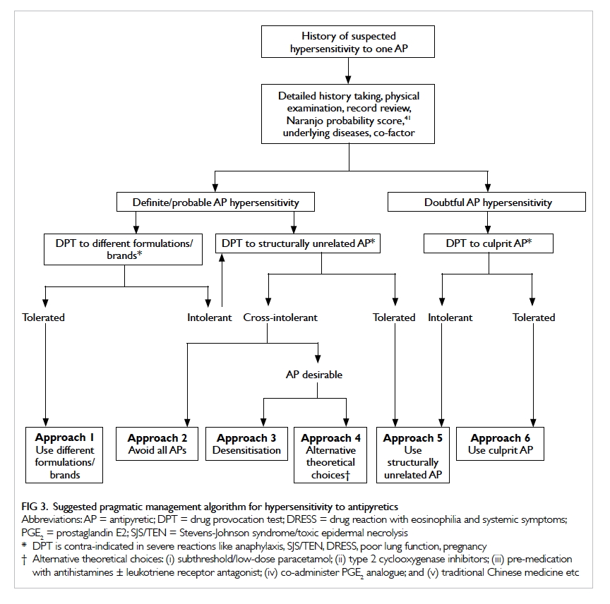
Management of suspected AP hypersensitivity starts with thorough discussion with patients or caretakers of the pros and cons of the AP as opposed to avoidance. The aims of investigation include confirmation of hypersensitivity and cross-reactivity, differentiation between hypersensitivity to the active ingredient versus excipients, and trial of safe alternatives. Detailed review of drug history is of paramount importance. Above all, DPT is pivotal to achieving the aims of investigation. A combination of drug history and DPT culminates in six alternative approaches to deal with hypersensitivity to APs (Fig 3).
 

Figure 3. Suggested pragmatic management algorithm for hypersensitivity to antipyretics
 
Patients allergic to excipients in one AP may tolerate a different brand or different formulation of the same drug (approach 1). Detailed comparison of constituents may reveal the excipient in question. In case of doubt, DPT can be performed on the alternative brand or formulation to confirm tolerance. In case the patient reacts to different formulations and brands of the same AP, a trial of AP with unrelated structure can be considered (approach 5). A common example is to try ibuprofen in patients with paracetamol hypersensitivity. As mentioned before, three quarters of patients with paracetamol hypersensitivity tolerate NSAIDs. Patients hypersensitive to ibuprofen, an arylpropionic acid, can consider DPT using paracetamol or an acetic acid such as diclofenac.
 
Patients with cross-intolerance to paracetamol and NSAIDs pose a management dilemma. Avoidance of all APs seems logical (approach 2), especially if the feverish patient is not ‘distressed’. Nonetheless whether a patient is in distress or not is a matter of subjective judgement. For cultural reasons, it is exceedingly difficult to persuade Hong Kong parents not to give APs to a child with a high fever. In case fever control is deemed desirable by either parents or physicians, viable solutions should be sought. Desensitisation (approach 3) is another viable option. A standard desensitisation protocol has been established for aspirin.52 Desensitisation is applicable to patients having NERD or NIUA.10 It is contra-indicated in patients with a history of severe, life-threatening drug reactions such as SJS/TENS or DRESS. Nonetheless desensitisation should only be carried out in medical facilities with resuscitation equipment and expertise in drug allergy. Alternative theoretical choices (approach 4) include subthreshold or low-dose paracetamol,47 53 COX-2 inhibitors,54 pre-medication with antihistamines with or without leukotriene receptor antagonist,55 co-administration of a PGE2 analogue,56 and traditional Chinese medicine.57 Future studies are needed to define the safety and efficacy of these unconventional treatments.
 
Patients with a mild or doubtful reaction to an AP can consider a DPT, the gold standard to diagnose or exclude hypersensitivity to the culprit drug. Patients who react to the culprit AP during DPT can either try a structurally unrelated AP (approach 5) or try a different brand/formulation (approach 1). Finally, patients who pass the DPT can be given the culprit drug in future (approach 6), as the test has a very high negative predictive value.10
 
Conclusion
It is arguable that APs may not be indicated in the first place and should be avoided in patients with hypersensitivity. Although APs should not be prescribed simply for the sake of ‘temperature control’, the need to mitigate patient discomfort should not be disregarded.58 Patients with illnesses such as heart failure, head injury, or sepsis present special problems. Their limited reserve to withstand the hypermetabolic state associated with febrile episodes puts them at particular risk.59 For these patients, APs seem beneficial. In case they have hypersensitivity to APs, viable options should be sought. Attempts to predict such hypersensitivity are daunting. Disappointingly, prediction of severe cutaneous adverse reactions to APs is virtually impossible. However, the presence of a positive family history, reaction within 1 hour of consumption, and history of multiple NSAID hypersensitivities may sound an alarm for the increased risk of genuine immediate hypersensitivity to APs. Clinicians need to strike a balance between ‘hypersensitivity phobia’ for the sake of drug safety and liberal use of APs to uphold patients’ rights. Knowledge of the pathogenesis of AP hypersensitivity and meticulous diagnostics are key to judicious management.
 
References
1. Davis T. NICE guideline: feverish illness in children—assessment and initial management in children younger than 5 years. Arch Dis Child Educ Pract Ed 2013;98:232-5. Crossref
2. Bertille N, Pons G, Khoshnood B, Fournier-Charrière E, Chalumeau M. Symptomatic management of fever in children: a national survey of healthcare professionals’ practices in France. PLoS One 2015;10:e0143230. Crossref
3. Polman HA, Huijbers WA, Augusteijn R. The use of diclofenac sodium (Voltaren) suppositories as an antipyretic in children with fever due to acute infections: a double-blind, between-patient, placebo-controlled study. J Int Med Res 1981;9:343-8. Crossref
4. Khubchandani RP, Ghatikar KN, Keny S, Usgaonkar NG. Choice of antipyretic in children. J Assoc Physicians India 1995;43:614-6.
5. Demoly P, Bousquet J. Epidemiology of drug allergy. Curr Opin Allergy Clin Immunol 2001;1:305-10. Crossref
6. Sánchez-Borges M. Clinical management of nonsteroidal anti-inflammatory drug hypersensitivity. World Allergy Organ J 2008;1:29-33. Crossref
7. Sánchez-Borges M, Capriles-Hulett A, Caballero-Fonseca F. Risk of skin reactions when using ibuprofen-based medicines. Expert Opin Drug Saf 2005;4:837-48. Crossref
8. Carvajal A, Prieto JR, Alvarez Requejo A, Martin Arias LH. Aspirin or acetaminophen? A comparison from data collected by the Spanish Drug Monitoring System. J Clin Epidemiol 1996;49:255-61. Crossref
9. Yilmaz O, Ertoy Karagol IH, Bakirtas A, et al. Challenge-proven nonsteroidal anti-inflammatory drug hypersensitivity in children. Allergy 2013;68:1555-61. Crossref
10. Kowalski ML, Asero R, Bavbek S, et al. Classification and practical approach to the diagnosis and management of hypersensitivity to nonsteroidal anti-inflammatory drugs. Allergy 2013;68:1219-32. Crossref
11. Schnyder B, Pichler WJ. Mechanisms of drug-induced allergy. Mayo Clin Proc 2009;84:268-72. Crossref
12. Rutkowski K, Nasser SM, Ewan PW. Paracetamol hypersensitivity: clinical features, mechanism and role of specific IgE. Int Arch Allergy Immunol 2012;159:60-4. Crossref
13. Pichler WJ. Drug hypersensitivity reactions: classification and relationship to T-cell activation. In: Pichler WJ, editor. Drug hypersensitivity. Basel: Karger; 2007: 168-89. Crossref
14. Kowalski ML, Makowska JS, Blanca M, et al. Hypersensitivity to nonsteroidal anti-inflammatory drugs (NSAIDs)—classification, diagnosis and management: review of the EAACI/ENDA(#) and GA2LEN/HANNA*. Allergy 2011;66:818-29. Crossref
15. Yawalkar N, Egli F, Hari Y, Nievergelt H, Braathen LR, Pichler WJ. Infiltration of cytotoxic T cells in drug-induced cutaneous eruptions. Clin Exp Allergy 2000;30:847-55. Crossref
16. Yawalkar N. Drug-induced exanthems. Toxicology 2005;209:131-4. Crossref
17. Shiohara T. Fixed drug eruption: pathogenesis and diagnostic tests. Curr Opin Allergy Clin Immunol 2009;9:316-21. Crossref
18. Savin JA. Current causes of fixed drug eruption in the UK. Br J Dermatol 2001;145:667-8. Crossref
19. Schrijvers R, Gilissen L, Chiriac AM, Demoly P. Pathogenesis and diagnosis of delayed-type drug hypersensitivity reactions, from bedside to bench and back. Clin Transl Allergy 2015;5:31. Crossref
20. Roales-Gómez V, Molero AI, Pérez-Amarilla I, et al. DRESS syndrome secondary to ibuprofen as a cause of hyperacute liver failure. Rev Esp Enferm Dig 2014;106:482-6.
21. Tank ND, Karelia BN, Bhansali NB. Paracetamol induced drug reaction with eosinophilia and systemic symptoms (Dress syndrome): a case report. Int J Pharm Sci Rev Res 2015;32:246-8.
22. Ward KE, Archambault R, Mersfelder TL. Severe adverse skin reactions to nonsteroidal antiinflammatory drugs: a review of the literature. Am J Health Syst Pharm 2010;67:206-13. Crossref
23. Borchers AT, Lee JL, Naguwa SM, Cheema GS, Gershwin ME. Stevens-Johnson syndrome and toxic epidermal necrolysis. Autoimmun Rev 2008;7:598-605. Crossref
24. Torres MJ, Mayorga C, Blanca M. Nonimmediate allergic reactions induced by drugs: pathogenesis and diagnostic tests. J Investig Allergol Clin Immunol 2009;19:80-90.
25. Mockenhaupt M, Kelly JP, Kaufman D, Stern RS; SCAR Study Group. The risk of Stevens-Johnson syndrome and toxic epidermal necrolysis associated with nonsteroidal antiinflammatory drugs: a multinational perspective. J Rheumatol 2003;30:2234-40.
26. Levi N, Bastuji-Garin S, Mockenhaupt M, et al. Medications as risk factors of Stevens-Johnson syndrome and toxic epidermal necrolysis in children: a pooled analysis. Pediatrics 2009;123:e297-304. Crossref
27. Ferrandiz-Pulido C, Garcia-Patos V. A review of causes of Stevens-Johnson syndrome and toxic epidermal necrolysis in children. Arch Dis Child 2013;98:998-1003. Crossref
28. Roujeau JC, Bioulac-Sage P, Bourseau C, et al. Acute generalized exanthematous pustulosis. Analysis of 63 cases. Arch Dermatol 1991;127:1333-8. Crossref
29. Britschgi M, Steiner UC, Schmid S, et al. T-cell involvement in drug-induced acute generalized exanthematous pustulosis. J Clin Invest 2001;107:1433-41. Crossref
30. Sidoroff A, Dunant A, Viboud C, et al. Risk factors for acute generalized exanthematous pustulosis (AGEP)—results of a multinational case-control study (EuroSCAR). Br J Dermatol 2007;157:989-96. Crossref
31. Szczeklik A, Sanak M. The broken balance in aspirin hypersensitivity. Eur J Pharmacol 2006;533:145-55. Crossref
32. Sánchez-Borges M. NSAID hypersensitivity (respiratory, cutaneous, and generalized anaphylactic symptoms). Med Clin North Am 2010;94:853-64, xiii. Crossref
33. Zembowicz A, Mastalerz L, Setkowicz M, Radziszewski W, Szczeklik A. Histological spectrum of cutaneous reactions to aspirin in chronic idiopathic urticaria. J Cutan Pathol 2004;31:323-9. Crossref
34. Kim SH, Sanak M, Park HS. Genetics of hypersensitivity to aspirin and nonsteroidal anti-inflammatory drugs. Immunol Allergy Clin North Am 2013;33:177-94. Crossref
35. Gómez F, Perkins JR, García-Martín E, Canto G, Cornejo-García JA. Genetic basis of hypersensitivity reactions to nonsteroidal anti-inflammatory drugs. Curr Opin Allergy Clin Immunol 2015;15:285-93. Crossref
36. Cheong HS, Park SM, Kim MO, et al. Genome-wide methylation profile of nasal polyps: relation to aspirin hypersensitivity in asthmatics. Allergy 2011;66:637-44. Crossref
37. Strauss J, Greeff O. Excipient-related adverse drug reactions: a clinical approach. Curr Allergy Clin Immunol 2015;28:24-7.
38. The electronic medicines compendium. Available from: https://www.medicines.org.uk/emc/medicine/10741. Accessed 23 May 2017.
39. Search Drug Database. Drug Office, Department of Health, The Government of the Hong Kong Special Administrative Region. Available from: https://www.drugoffice.gov.hk/eps/do/en/consumer/search_drug_database.html. Accessed 23 May 2017.
40. Knowles SR, Drucker AM, Weber EA, Shear NH. Management options for patients with aspirin and nonsteroidal antiinflammatory drug sensitivity. Ann Pharmacother 2007;41:1191-200. Crossref
41. Naranjo CA, Busto U, Sellers EM, et al. A method for estimating the probability of adverse drug reactions. Clin Pharmacol Ther 1981;30:239-45. Crossref
42. Topal E, Celiksoy MH, Catal F, Gamze Sayan Y, Sancak R. The value of the clinical history for the diagnosis of immediate nonsteroidal anti-inflammatory drug hypersensitivity and safe alternative drugs in children. Allergy Asthma Proc 2016;37:57-63. Crossref
43. Wölbing F, Fischer J, Köberle M, Kaesler S, Biedermann T. About the role and underlying mechanisms of cofactors in anaphylaxis. Allergy 2013;68:1085-92. Crossref
44. Demoly P, Adkinson NF, Brockow K, et al. International Consensus on drug allergy. Allergy 2014;69:420-37. Crossref
45. Misirlioglu ED, Toyran M, Capanoglu M, Kaya A, Civelek E, Kocabas CN. Negative predictive value of drug provocation tests in children. Pediatr Allergy Immunol 2014;25:685-90. Crossref
46. Aberer W, Bircher A, Romano A, et al. Drug provocation testing in the diagnosis of drug hypersensitivity reactions: general considerations. Allergy 2003;58:854-63. Crossref
47. Ho MH, Tung JY, Lee TL, Tsoi NS, Lau YL. Anaphylaxis to paracetamol. J Paediatr Child Health 2008;44:746-7.
48. Zambonino MA, Torres MJ, Muñoz C, et al. Drug provocation tests in the diagnosis of hypersensitivity reactions to non-steroidal anti-inflammatory drugs in children. Pediatr Allergy Immunol 2013;24:151-9. Crossref
49. de Paramo BJ, Gancedo SQ, Cuevas M, Camo IP, Martin JA, Cosmes EL. Paracetamol (acetaminophen) hypersensitivity. Ann Allergy Asthma Immunol 2000;85(6 Pt 1):508-11. Crossref
50. Ariza A, Fernandez TD, Doña I, et al. Basophil activation after nonsteroidal anti-inflammatory drugs stimulation in patients with immediate hypersensitivity reactions to these drugs. Cytometry A 2014;85:400-7. Crossref
51. Pichler WJ, Tilch J. The lymphocyte transformation test in the diagnosis of drug hypersensitivity. Allergy 2004;59:809-20. Crossref
52. Macy E, Bernstein JA, Castells MC, et al. Aspirin challenge and desensitization for aspirin-exacerbated respiratory disease: a practice paper. Ann Allergy Asthma Immunol 2007;98:172-4. Crossref
53. Kidon MI, Kang LW, Chin CW, et al. Early presentation with angioedema and urticaria in cross-reactive hypersensitivity to nonsteroidal antiinflammatory drugs among young, Asian, atopic children. Pediatrics 2005;116:e675-80. Crossref
54. Corzo JL, Zambonino MA, Muñoz C, et al. Tolerance to COX-2 inhibitors in children with hypersensitivity to nonsteroidal anti-inflammatory drugs. Br J Dermatol 2014;170:725-9. Crossref
55. Nosbaum A, Braire-Bourrel M, Dubost R, et al. Prevention of nonsteroidal inflammatory drug-induced urticaria and/or angioedema. Ann Allergy Asthma Immunol 2013;110:263-6. Crossref
56. Dobovišek A, Fajmut A, Brumen M. Strategy for NSAID administration to aspirin-intolerant asthmatics in combination with PGE2 analogue: a theoretical approach. Med Biol Eng Comput 2012;50:33-42. Crossref
57. Liew WK, Loh W, Chiang WC, Goh A, Chay OM, Iancovici Kidon M. Pilot study of the use of Yin Qiao San in children with conventional antipyretic hypersensitivity. Asia Pac Allergy 2015;5:222-9. Crossref
58. Section on Clinical Pharmacology and Therapeutics; Committee on Drugs, Sullivan JE, Farrar HC. Fever and antipyretic use in children. Pediatrics 2011;127:580-7. Crossref
59. Henker R. Evidence-based practice: fever-related interventions. Am J Crit Care 1999;8:481-7.

Current management of pregnancy-associated breast cancer

Hong Kong Med J 2017 Aug;23(4):387–94 | Epub 26 Jun 2017
DOI: 10.12809/hkmj166049
© Hong Kong Academy of Medicine. CC BY-NC-ND 4.0
 
REVIEW ARTICLE
Current management of pregnancy-associated breast cancer
Harry HY Yu, FCSHK, FHKAM (Surgery)1; Polly SY Cheung, FCSHK, FHKAM (Surgery)2; Roland CY Leung, MB, ChB, MRCP (UK)3; TN Leung, FHKCOG, FHKAM (Obstetrics and Gynaecology)4; WH Kwan, FHKCR, FHKAM (Radiology)5
1 Department of Surgery, Ruttonjee & Tang Shiu Kin Hospitals, Wanchai, Hong Kong
2 Breast Care Centre, Hong Kong Sanatorium & Hospital, Happy Valley, Hong Kong
3 Department of Medicine, Queen Mary Hospital, Pokfulam, Hong Kong
4 Obstetrics & Gynaecology Centre, Hong Kong Sanatorium & Hospital, Happy Valley, Hong Kong
5 Comprehensive Oncology Centre, Hong Kong Sanatorium & Hospital, Happy Valley, Hong Kong
 
Corresponding author: Dr Polly SY Cheung (pollyc@pca.hk)
 
 Full paper in PDF
 
Abstract
Pregnancy-associated breast cancer is the most common malignancy during pregnancy with an expected rise in incidence. The belief in the need for termination of pregnancy and that chemotherapy is contra-indicated during pregnancy is challenged by recent evidence. Patients can consider breast-conserving surgery and sentinel lymph node biopsy with acceptably low fetal risk from radiation exposure. A range of chemotherapeutics is possible in the second trimester in terms of drug class and frequency. Hormonal therapy and monoclonal antibody therapy are contra-indicated during pregnancy and lactation. Fetal outcome after in-utero exposure to chemotherapy appears similar to that in a non-pregnant population. Future pregnancy, in most situations, does not appear to be contra-indicated but a multidisciplinary and patient-centred approach is recommended. Fertility preservation techniques are also being developed with reported success and consequent pregnancies.


Introduction
Pregnancy-associated breast cancer (PABC) is defined as the diagnosis of breast cancer during pregnancy and those occurring within 1 year postpartum.1 It is the most common malignancy associated with pregnancy and the incidence is expected to rise in high-income countries.2 3 4 Historically, this disease entity was thought to have a worse prognosis when compared with patients diagnosed with cancer in the non-pregnant state and termination of pregnancy was common. Here, we reviewed recent publications and summarised the most recent information.
 
‘Dual’ effect of pregnancy
Generally it is thought that pregnancy is a protective factor that lowers the risk of breast cancer development. Nonetheless, studies have found that pregnancy increases the risk of breast cancer initially following delivery and has a protective effect after a period of time.3 The period of increased risk has been estimated to be between 10 and 15 years following a first pregnancy. The later the first pregnancy, the longer the duration of increased risk before the protective effect.5 In a Norwegian study, women who waited until aged over 35 years for their first child permanently increased their risk of breast cancer compared with nulliparous women.6
 
Risk of developing breast cancer decreases with multiple pregnancies, but the age at first birth remains the dominant influence on risk.5 BRCA1 and BRCA2 mutation carriers are not protected by early pregnancy from malignancy,2 but they do not have an increased risk of developing PABC compared with non-carrier women.5
 
Epidemiology
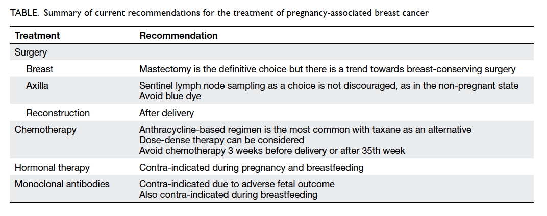
Breast cancer is the most common malignancy to occur during pregnancy, followed by cervical cancer, melanoma, and haematological malignancy.7 In the United Kingdom, 1.3 to 2.4 cases of breast cancer in women were diagnosed per 10 000 live births.1 It accounted for 3.3% to 10% of women diagnosed with premenopausal breast cancer.2 5
 
Meta-analysis has shown that risk of death is more than 40% in women with PABC compared with those with non-PABC (pooled hazard ratio=1.44; 95% confidence interval, 1.27-1.63), but other epidemiological studies have shown no direct relationship between pregnancy and mortality.8 Moreover, the prognosis of patients with breast cancer during pregnancy is similar to that of non-pregnant women of the same age and clinical stage at diagnosis.9 In fact, metastasis is a rare event during pregnancy, with only case series reported in the literature.10
 
Epidemiological data indicate that breast cancer diagnosed during lactation exhibits the most aggressive trait and elevation in cause-specific death.11 Compared with patients in the non-pregnant state or during pregnancy, patients diagnosed with breast cancer within 1 year of delivery have a shorter time to relapse12 and increased risk of metastasis and death.6 The lactating microenvironment is a strong driver of tumour progression. Lactating stromal adipose cells express higher levels of inflammatory cytokines that are highly angiogenic and growth promoting, causing the tumour to be more aggressive.11
 
Other factors proposed to be related to the poorer outcome include the change in breast tissue and the hormonal environment. There is a shorter phase of involution, an inflammatory-like process that has been suggested to have tumour-promoting properties by affecting the microenvironment and malignant potential of microtumours.8 A generally low index of suspicion of cancer in reproductive-aged women and, in particular, pregnant women has tended to delay diagnosis by approximately 1 to 2 months according to recent studies.8 13 A 1-month delay in the treatment of a primary tumour increases the risk of axillary metastases by 0.9% to 1.8%.14
 
Histopathology
As the group of affected women diagnosed with malignancy generally represents a younger population, PABC patients tend to have high-grade tumours and display lymphovascular invasion.2 5 9 They also have a lower incidence of positive hormone receptor status.3
 
Placental metastasis is rare but indicates poor maternal prognosis. Pathological assessment after delivery is recommended in all cases.2
 
Diagnosis
Of breast masses that present to a breast clinic, 80% are benign although any mass that persists for more than 2 to 4 weeks or that is associated with skin changes raises clinical suspicion.2 15 Nipple discharge and ‘milk rejection’ sign, ie refusal by the infant to nurse from a diseased breast, can be signs of underlying occult carcinoma, but are not frequent.13 14
 
Ultrasonography (USG) is the first imaging modality following clinical examination to assess a discrete lump.1 Mammography (MMG) can also supplement USG to assess extent of disease and examine the contralateral breast. Accuracy of MMG, however, is decreased in the pregnant state because of increased water content and change in the distribution of fat. Studies have shown a false-negative rate as high as 25%2 with sensitivity of 63% to 78%.14 Radiation exposure of the fetus from MMG is minimal (0.001-0.01 mGy). The International Commission on Radiological Protection (ICRP) concluded in 2007 that there was no detrimental effects of practical significance (threshold dose, 100 mGy).16 The use of magnetic resonance imaging is controversial during pregnancy, due to the challenges in discriminating malignant from physiological hypervascularisation that occurs during pregnancy.13 There are also no safety data regarding the use of gadolinium in pregnancy,15 and fetal abnormalities have been noted in rats exposed to gadolinium.2
 
To complete triple assessment, core needle or excision biopsy is the gold standard for tissue diagnosis. Histological grade, receptor status, and human epidermal growth factor receptor 2 (HER2) information should be obtained, just as for non-pregnant patients.1 2 Needle cytology shows good sensitivity but with a higher risk of false-positive results due to the presence of hyperproliferative cells in mammary tissue during pregnancy and lactation.2 The risk of developing a subsequent lactating fistula after biopsy is overestimated.15 Cabergoline can reduce the risk of fistula or abscess formation by suppressing lactation.2 Emptying the breast and the use of ice packs and binding before biopsy might also reduce the risk of fistula formation.13
 
Unless there is a strong clinical suspicion, metastatic work-up is not mandatory.1 Chest X-ray and USG of the liver are the main modalities used to look for metastases. Fetal exposure from a chest X-ray is about 0.1 mGy.13
 
Treatment
For those who develop breast cancer during pregnancy, any treatment intervention during pregnancy shows a trend towards improved overall survival compared with delaying evaluation and treatment until after delivery—78.6% overall survival for those who received treatment during pregnancy compared with 44.7% for those patients who did not.17 Management of postpartum breast cancer is similar to that for non-pregnant patients. Consideration of fetal well-being is a critical factor when making treatment decisions for the pregnant patient.
 
Surgery
Surgery is considered safe in all trimesters with negligible risk to the fetus. Studies, including a large review, concluded that surgery carries a similar probability of miscarriage to the background risk of spontaneous abortion, including first trimester.7 18
 
Mastectomy is the definitive procedure. There is a trend for favouring breast-conserving surgery as a suitable option for PABC patients, and this should be discussed with the patient whenever possible.19
 
As epidemiological data show a higher percentage of axillary metastases among PABC patients, axillary dissection is generally offered.2 When the tumour is diagnosed at an early stage, however, a considerable proportion of patients will have node-negative disease and therefore might benefit from sentinel lymph node biopsy (SLNB). Such procedure remains controversial18 but some studies have reported its success and safety.
 
The first issue to clarify in SLNB is fetal radiation exposure relating to lymphoscintigraphy. In Italy, Gentilini et al20 reviewed 26 young non-pregnant women scheduled for lymphoscintigraphy for SLNB. A single peritumoural injection of 99mTc-labelled human albumin colloid particles in a volume of 0.2 mL was administered prior to surgery. A thermoluminescent dosimeter was placed around the abdomen (epigastrium, umbilicus, and hypogastrium) to evaluate potential uptake by the fetus in different trimesters. In 23 of 26 patients, all absorbed dose measurements over the surface of the abdomen at the supposed level of a fetus were lower than the sensitivity of the dosimeter (<10 µGy). In the remaining three patients, the absorbed doses to the epigastrium, umbilicus, and hypogastrium were 40-320, 120-150, and 30-40 µGy, respectively. Another study estimated the maximal absorbed dose of radiation from SNLB by 99mTc sulfur colloid to be 4.3 mGy.18
 
Fetal exposure of >100 to 200 mGy is associated with central nervous system problems. A radiation dose that exceeds 100 mGy can result in reduced intelligence quotient. With a dose of 10 mGy, the risk of leukaemia and cancer is 1.4%, ie a 40% increase over the normal incidence. An absorbed dose of 20 µGy is comparable with 1 to 2 days of natural background radiation, 1/5000th of the threshold dose for malformation or other adverse effects.21 Of note, reports of the ICRP have shown that the most common procedures in diagnostic nuclear medicine rarely represent an indication for termination of pregnancy, and that pregnancy should not be a reason to avoid diagnostic nuclear medicine studies.22
 
Gentilini et al21 then reported the use of SLNB in pregnant patients from 2001 to 2007. During the period, 12 of 45 patients diagnosed with breast cancer during pregnancy were clinically node-negative and all underwent SLNB. Lymphatic mapping was performed by 99mTc-radiolabelled colloid lymphoscintigraphy alone with mean activity of 10 MBq (about 1 µGy/MBq) for the first eight patients and 3-4 MBq for the later four. Hot spots were identified in all patients (10 with 1 hot spot, and two with 2 hot spots) and the mean number of excised sentinel lymph nodes (SLNs) was two (range, 1-4). Ten patients had confirmed negative SLNs and were spared axillary dissection. One patient had micrometastasis in one of the four nodes and elected not to undergo any further axillary surgery. Another patient had a confirmed metastatic SLN intra-operatively and underwent axillary clearance at the same operation (3 of 24 positive nodes). No overt axillary lymph node reappeared for patients with negative SLN. Of the 12 pregnancies, 11 babies were born with a normal weight and no malformation after an uncomplicated pregnancy. One baby underwent surgery at 3 months of age for cardiac failure due to ventricular septal defect. This had been suspected at the 21st week of gestation prior to lymphoscintigraphy at the 26th week.
 
Two other studies also reported the use of SLNB with 99mTc colloid and/or blue dye (isosulfan or methylene) for pregnant patients.19 21 All 35 patients who underwent SLNB had successful mapping and surgery was performed without complications; 33 of 35 infants were healthy at delivery. One patient found herself pregnant on the day of her scheduled operation and decided to terminate her pregnancy in the first trimester after the breast surgery in order to start chemotherapy. Another child was born with a cleft palate to a mother who was a smoker with a history of methadone use.
 
Axillary staging provides important prognostic information and allows better local control but does not always influence the type of adjuvant therapy. With an acceptably low false-negative rate and the same capacity to detect nodal metastasis, SLNB has a lower morbidity compared with axillary lymph node dissection (lymphoedema, 5.3% vs 11.8%).21 23 The decrease in surgical morbidity might result in a shorter postoperative recovery, potentially allowing an earlier start for adjuvant chemotherapy. In young premenopausal patients, such treatment can be directed towards hormone insensitive tumours and might improve outcome.20 Proponents of SLNB also point out the possibility of performing breast-conserving surgery and SLNB under local anaesthesia to further reduce the risk of preterm labour and spontaneous miscarriage.20
 
There are, however, specific issues to be solved regarding SLNB use during pregnancy. Isosulfan blue and methylene blue are Food and Drug Administration pregnancy category C drugs, with unknown potential risks for teratogenicity.23 Anaphylaxis has been reported with the use of blue dye.18 24 25 Adverse outcomes associated with methylene blue dye include intestinal atresia and fetal demise.26
 
Fetal radiation exposure and successful mapping of lymphoscintigraphy depend on the administered activity and size of radiocolloid. To minimise unnecessary radiation exposure, pregnant patients should avoid contact with other patients receiving nuclear medicine therapy, eg by scheduling pregnant patients as the first procedure of the day, and keeping them in a single-bed room. Reducing the time between lymphoscintigraphy and surgery might further reduce the dose of administered radiocolloids.20
 
The latest European Society for Medical Oncology (ESMO) guidelines do not discourage SLNB in pregnant breast cancer patients in centres where SLNB is routinely practised in the non-pregnant setting, but discourages the use of blue dye.27 Despite weak evidence, however, the latest American Society of Clinical Oncology guidelines are still against performing SLNB during pregnancy.28
 
Immediate reconstruction is not recommended and should be delayed to avoid prolonged anaesthesia and to allow optimal symmetrical breast reconstruction after delivery.1
 
 
Systemic chemotherapy
Indications for chemotherapy are the same as for non-pregnant breast cancer patients. When the indication for chemotherapy is clear, it should not be delayed due to the potential detrimental effect on maternal outcome, with the exception of breast cancer diagnosed during the first trimester.29 Adjuvant chemotherapy should be started within 3 weeks of surgery in patients with hormone-negative tumours.2 29
 
Most chemotherapeutic agents are of low molecular weight, highly lipid soluble, and loosely protein bound. This facilitates transplacental transfer from the mother to the fetus.7 Teratogenicity is directly related to timing and dosage delivered. The most vulnerable period for fetal malformation and spontaneous abortion is 10 days to 8 weeks after conception, ie the period of organogenesis (17%).2 29 Throughout the first trimester, chemotherapy is still considered contra-indicated due to concerns about adverse events associated with ocular formation, genitals, the haematopoietic system, and the central nervous system before the 14th week.1
 
Chemotherapy is regarded as safe during the second trimester.1 However, administration during the second and third trimesters is associated with increased risk of intrauterine growth restriction and low birth rate, which may be related to both tumour burden and/or the aggressive nature of the tumour, as well as the toxicity of chemotherapy.29 Changes that occur during pregnancy, such as generation of a third space (fetal-placental amniotic fluid), increased volume distribution, and changes in the metabolism and elimination of drugs, may determine different toxicity patterns that may also indirectly affect the fetus.15 General chemotherapy risks include preterm delivery, low birth weight, transient tachypnoea of the newborn, and transient neonatal leukopenia.2 The reported fetal malformation risk following chemotherapy during the second and third trimesters is 3.8%, no higher than that in the general population.18 Incidence of preterm delivery for chemotherapy-exposed gestational breast cancer is 5% to 8%.29 Most children showed normal neurological development after exposure to chemotherapy in utero, although behavioural and emotional issues need further clarification and follow-up.29 30 For PABC patients, chemotherapy-induced gonadotoxicity may cause permanent amenorrhoea with complete loss of germ cells, transient amenorrhoea, menstrual irregularity, and subfertility, but this depends on the dose, agent, and patient age.1
 
The German Breast Group reported a multicentre study that included 197 patients who received chemotherapy during pregnancy (a total of 447 patients in 8 years).4 Overall, 50% of breast cancer patients delivered preterm, compared with 10% to 15% of the general population. Delivery before the 37th week of gestation is associated with a higher chance of side-effects, malformations, or newborn complications. Low birth weight is affected by chemotherapy exposure after adjustment for gestational age, but not by number of chemotherapy cycles (P=0.018). Adverse events were more common in those who received chemotherapy in utero than in those who were not exposed (31 [15%] of 203 vs 7 [4%] of 170 infants; P=0.00045). The proportion of malformations in the study was no different to that for the general population (approximately 9%). Two fetal deaths were reported and both were exposed to chemotherapy and delivered prematurely. Neither was thought to be related to treatment—one was related to diagnosis of trisomy 18 and the other died of necrotising enterocolitis after delivery at the 31st week with weight of 1895 g. The study concluded that although chemotherapy exposure in utero resulted in lower birth weight and more complications, the differences were not clinically significant and most likely related to premature delivery.
 
Optimum use of cytotoxic drugs in pregnant patients remains undefined, particularly regarding drug selection, dosing, and dose density.31 Assessment of treatment effectiveness in pregnant patients is complex. Calculation of chemotherapy dose is uncertain for the pregnant state. Physiological changes in pregnancy can also greatly affect drug disposition.
 
Anthracycline-based regimens (eg epirubicin and doxorubicin) are the most widely used chemotherapeutic agents as they have a favourable safety profile when administered during pregnancy,29 although no particular preference is given for one regimen over another.27 Assessment of maternal cardiac function by echocardiogram is recommended if an anthracycline-based regimen is to be administered.14 Studies are yet to show an increased risk of fetal cardiotoxicity secondary to in-utero exposure.27 Common side-effects include neutropenia, oral ulcers, anaphylaxis, constipation, tachycardia, and cellulitis. Fetal side-effects include low birth weight (7.6%) and birth defects (3.8%).18 Risk of congenital malformations is similar to that of patients not receiving chemotherapy. Long-term follow-up of children indicates that there are no sequelae associated with growth and maturation.9
 
Taxanes appear to be another alternative, but have not been as extensively studied as anthracyclines and most studies used small sample sizes.18 A meta-analysis showed that the addition of taxanes to anthracycline-based regimens resulted in a statistically significant reduction in the risk of relapse (relative reduction, 17%) and death (relative reduction, 15%) for high-risk early breast cancer patients. Disease-free survival benefit was independent of oestrogen-receptor expression, degree of nodal involvement, and type of taxane used.32 Taxanes are substrates of the P-glycoprotein that is highly expressed on the maternal compartment of the placenta. P-glycoprotein protects the fetus against xenobiotics and might therefore reduce transplacental transfer of taxanes.29 Data for baboon and human models showed that taxanes are hardly detectable in the fetus,4 and a recent overview of 50 breast cancer patients treated with taxanes showed that completely healthy neonate was born in a majority of cases.33 In a review of 40 pregnant women prescribed taxanes, there were no reports of intrauterine death or congenital malformations other than one infant with pyloric stenosis.34 Another retrospective cohort study of 12 patients with breast cancer and four with ovarian cancer who were exposed to taxane-based chemotherapy during pregnancy reported a mean gestational age at delivery of 36.9 weeks and mean birth weight of 2452 g (interquartile range, 2155-2619 g). One baby was diagnosed with hypertrophic pyloric stenosis at 4 weeks and underwent surgery at 6 weeks. One of a set of twins born in this study had hyperbilirubinaemia and jaundice, and was later diagnosed with Asperger’s syndrome and Tourette’s syndrome, while his twin exposed to the same chemotherapy is developmentally normal and excelled at school.32
 
Taxanes are metabolised by cytochrome P450 that increases by 50% to 100% during the third trimester, possibly resulting in a shorter half-life and higher clearance that could result in a reduced toxicity profile during pregnancy.29 Nonetheless this lowered serum concentration is a concern for drug efficacy during pregnancy.14 There are no data to analyse different taxanes for pharmacokinetics, toxicity profile, or efficacy due to small sample size. The latest ESMO guidelines also endorsed the use of taxanes during pregnancy in cases where “they are clinically indicated or the use of anthracyclines is contraindicated”.27
 
A dose-dense chemotherapy regimen for pregnant patients is another topic of heated debate. Chemotherapy cycles were administered every 1 to 2 weeks compared with 3-weekly cycles for conventional therapy. One study compared 10 patients who received doxorubicin and cyclophosphamide dose-dense therapy during gestation with 99 patients receiving conventional chemotherapy after the first trimester, and reported completion of chemotherapy for all patients (98% in conventional group). They had similar delivery outcomes, risk of congenital anomalies, incidence, and time to recurrence, and maternal overall survival at 3.5 years.35 Proponents suggest that dose-dense chemotherapy in pregnancy may allow faster completion of chemotherapy, sufficient time for maternal recovery for delivery,35 closer pregnancy monitoring, and better toxicity profile, and no need for high-dose steroid premedication or prophylactic use of granulocyte-colony stimulating factor.27
 
Chemotherapy is advised to be withheld 3 weeks before delivery or after 35 weeks of gestation to minimise the risk of sepsis and haemorrhage in the mother and newborn.1 2 It allows time for fetal drug excretion via the placenta, especially for preterm babies who have a limited ability to metabolise drugs through an immature liver and kidneys. Chemotherapy can resume after adequate recovery from delivery.29 Breastfeeding is contra-indicated during the treatment period but can resume 3 to 4 weeks after the last administered dose of chemotherapy.2
 
Radiotherapy
Radiotherapy is contra-indicated until after delivery unless it is used for life-saving issues or to preserve organ function, eg spinal cord compression.1 If radiotherapy is indicated during the pregnant state, fetal shielding should be considered or the option of elective early delivery discussed. Substituting whole-breast radiotherapy with partial-breast treatment is another alternative.2 Excess cancer risk to a fetus receiving radiation is 6.57 cases per 10 000 children per rad (0.01 Gy) per year. The typical external beam radiation dose to the breast ranges from 45 to 60 Gy, and may result in a fetal radiation exposure of 3.9 to 15 rad in the first trimester and up to 200 rad in the late third trimester.14 Other risks of radiotherapy include miscarriage, teratogenicity, microcephaly, fetal growth restriction, and induction of childhood malignancy and haematological disorders.
 
Adjuvant radiotherapy is not considered an urgent procedure and should be postponed until after delivery.27 Delaying treatment after 12 weeks, however, can increase the likelihood of axillary metastases by 0.028% to 0.057% per day2 and a delay over 6 months can increase the risk of local recurrence.27
 
Hormonal therapy
Tamoxifen is not used until after delivery.1 It is associated with oculo-auriculo-vertebral dysplasia (Goldenhar’s syndrome) and ambiguous genitalia.2 18 Because of unknown transmission of the drug in milk, it is also contra-indicated in breastfeeding.1 Long-term effects of the drug on female offspring are unknown.14
 
Monoclonal antibody
Trastuzumab is contra-indicated during pregnancy due to reported adverse fetal outcomes.1 Meta-analysis showed the main adverse event to be oligohydramnios (61.1%), with the incidence increasing with duration of treatment.18 Alteration of amniotic fluid volume is mostly attributed to the effect of trastuzumab on the fetal kidney, where the HER2 receptor is highly expressed.29 Nonetheless, if the fetus was exposed to the drug exclusively during the first trimester, all children were completely healthy at birth: transplacental transport of immunoglobulin G is very low early in pregnancy and increases gradually during the second trimester to reach concentrations similar to the mother by the end of gestation.29 Oligohydramnios is reversible if the drug is stopped, with good outcome observed in the majority of cases.
 
Another risk of trastuzumab for the fetus is renal failure,9 but there are no reports of serious fetal cardiac effects.14 Breastfeeding is also contra-indicated due to unknown transmission of the drug in milk. The drug is not associated with impaired fertility.1
 
Supportive agents
Serotonin antagonists and dexamethasone are the preferred antiemetics.1 Granulocyte-colony stimulating factor is recommended to minimise potential maternal and fetal problems associated with neutropenia1 and erythropoietin has been safely administered in pregnant patients.18
 
In-utero exposure to bisphosphonates has been shown to increase the risk of fetal skeletal anomalies and result in hypocalcaemia that may affect uterine contraction. It is suggested that these drugs be administered after delivery whenever possible.29
 
A summary of current recommendations regarding treatment of PABC is shown in the Table.
 

Table. Summary of current recommendations regarding treatment of pregnancy-associated breast cancer
 
 
Antenatal care
There is no evidence of a need for additional antenatal care, but it is standard practice to establish fetal well-being by USG before any treatment.2 15 Serial fetal growth assessment should be performed every 3 to 4 weeks, or prior to each chemotherapy cycle. Other forms of assessment of fetal well-being may be beneficial, such as umbilical artery Doppler to assess the status of the placenta, Doppler of the fetal middle cerebral artery to exclude fetal anaemia, and serial fetal echocardiograms (when potential cardiotoxic drugs like anthracyclines are being used). Assessment of amniotic fluid volume is also necessary because it can decrease reversibly with the use of some drugs.15
 
Unless there is a clear oncological or obstetric indication, delivery should be delayed until after the 37th week. Morbidity and mortality in newborns are directly related to gestational age at delivery. Infants born in late preterm (34th to 35th week) have increased morbidities including perinatal death, transient tachypnoea, respiratory distress, hypoglycaemia, pulmonary hypertension, as well as long-term cognitive and behavioural morbidities.7
 
Vaginal delivery is preferred because it is less likely to delay initiation of chemotherapy due to lower morbidity.29 Caesarean delivery should be reserved for the usual obstetric indications.2 Deep venous thrombosis prophylaxis should be considered, as pregnancy and malignancy are both risk factors for venous thromboembolism.
 
Termination of pregnancy
There is no evidence to suggest that termination of pregnancy improves prognosis.2 10 Once pregnancy has occurred, induction of abortion has no impact on maternal prognosis and is therefore strongly discouraged for such purposes.27 If maternal outcomes are not negatively impacted by the pregnancy itself, continuation of pregnancy seems not only reasonable, but recommended.7 Nevertheless, in case of advance disease stage (stage III or IV) or for high-grade or aggressive primary tumours diagnosed in the early first trimester, termination of pregnancy may be considered (teratogenic risk of chemotherapy during the first trimester).2 15
 
Future pregnancy
There is evidence that pregnancy after breast cancer does not lead to increased risk of recurrence and may even improve survival, although these findings could be due to the ‘healthy mother effect’.5 14 Large matched multicentre retrospective studies including more than 1000 patients confirmed that pregnancy after oestrogen receptor (ER)–positive breast cancer was not detrimental, at least during the first 5 years following pregnancy.27 The latest ESMO guidelines also “do not discourage pregnancy following breast cancer diagnosis irrespective of the ER status”.27
 
Nonetheless, the chance of subsequent pregnancy is nearly 70% lower when compared with the general population, probably secondary to frequent treatment with gonadotoxic chemotherapy, prolonged treatment periods with tamoxifen in patients with hormone sensitive disease, and also a general misconception that pregnancy could stimulate cancer recurrence given that it is a hormonally driven disease.27 The chance of recovery of menses is higher for patients under 40 years of age and the use of taxane-based chemotherapy.5
 
“Consult before conceive”—a multidisciplinary approach is recommended before planning a pregnancy. Anecdotal evidence suggests a 2-year wait after treatment and a 5-year wait for recurrent stage I and II disease.2 Patients with metastatic disease are advised against pregnancy due to their limited life expectancy and possible compromised treatment of disease.1 Interruption of full-course tamoxifen may have detrimental effects on breast cancer outcome. If, however, a woman is willing to accept the risk, interruption after 2 to 3 years of tamoxifen may be considered to allow pregnancy. Tamoxifen should be stopped for 3 months before trying to conceive. Latest ESMO guidelines “strongly encourage the resumption of tamoxifen following delivery”.1 27 It is also advised to continue active contraception up to 3 to 6 months following the last administered dose of anti-cancer therapy.27
 
Embryo or oocyte cryopreservation is the main method to preserve female fertility.36 Ovarian stimulation is carried out before commencing chemotherapy, but may result in relative delay in oncological treatment and increase serum oestradiol levels. This may be of concern in hormone-driven tumours like breast cancer. Laparoscopic ovarian tissue sampling and freezing before treatment are considered experimental. When needed, re-implantation of ovarian tissue in the pelvis after thawing may be a unique option for young girls with cancer. Over 60 pregnancies have been reported.37
 
Conclusion
The prognosis of PABC is similar to that of breast cancer in the non-pregnant state. Treatment should commence after diagnosis is established. Surgical treatment options are expanding and extensive data show that chemotherapy during pregnancy is safe and more options for treatment are now available. Whenever possible, the aim should be to carry the fetus to term. Future pregnancy is generally not contra-indicated.
 
References
1. RCOG Green-top guideline No. 12. Pregnancy and breast cancer. UK Royal College of Obstetricians and Gynaecologists; 2011.
2. Padmagirison R, Gajjar K, Spencer C. Management of breast cancer during pregnancy. Obstet Gynecol 2010;12:186-92. Crossref
3. Genin AS, Lesieur B, Gligorov J, Antoine M, Selleret L, Rouzier R. Pregnancy-associated breast cancers: do they differ from other breast cancers in young women? Breast 2012;21:550-5. Crossref
4. Lobiol S, Han SN, von Minchkitz G, et al. Treatment of breast cancer during pregnancy: an observational study. Lancet Oncol 2012;13:887-96. Crossref
5. Bell RJ, Fradkin P, Parathithasan N, Robinson PJ, Schwarz M, Davis SR. Pregnancy-associated breast cancer and pregnancy following treatment for breast cancer, in a cohort of women from Victoria, Australia, with a first diagnosis of invasive breast cancer. Breast 2013;22:980-5. Crossref
6. Borges VF, Schedin PJ. Pregnancy-associated breast cancer: an entity needing refinement of the definition. Cancer 2012;118:3226-8. Crossref
7. Walton JR, Prasad MR. Obstetric and neonatal outcomes of cancer treated during pregnancy. Clin Obstet Gynecol 2011;54:567-73. Crossref
8. Johansson AL, Andersson TM, Hsieh CC, et al. Stage at diagnosis and mortality in women with pregnancy-associated breast cancer (PABC). Breast Cancer Res Treat 2013;139:183-92. Crossref
9. Córdoba O, Llurba E, Saura C, et al. Multidisciplinary approach to breast cancer diagnosed during pregnancy: maternal and neonatal outcomes. Breast 2013;22:515-9. Crossref
10. Azim HA Jr, Peccatori FA. Treatment of metastatic breast cancer during pregnancy: we need to talk! Breast 2008;17:426-8. Crossref
11. McCready J, Arendt LM, Glover E, et al. Pregnancy-associated breast cancers are driven by differences in adipose stromal cells present during lactation. Breast Cancer Research 2014;16:R2. Crossref
12. Halaska MJ, Pentheroudakis G, Strnad P, et al. Presentation, management and outcome of 32 patients with pregnancy-associated breast cancer: a matched controlled study. Breast J 2009;15:461-7. Crossref
13. Rovera F, Frattini F, Coglitore A, et al. Breast cancer in pregnancy. Breast J 2010;16 Suppl 1:S22-5. Crossref
14. Viswanathan S, Ramaswamy B. Pregnancy-associated breast cancer. Clin Obstet Gynecol 2011;54:546-55. Crossref
15. Sánchez Martínez MC, Ruiz Simón A. Breast cancer during pregnancy. Breast Cancer Rest Treat 2010;123 Suppl 1:55-8. Crossref
16. International Commission on Radiological Protection. The 2007 recommendations of the International Commission on Radiological Protection. Ann ICRP 2007;37:1-332.
17. Beadle BM, Woodward WA, Middleton LP, et al. The impact of pregnancy on breast cancer outcomes in women ≤35 years. Cancer 2009;115:1174-84. Crossref
18. Krishna I, Lindsay M. Breast cancer in pregnancy. Obstet Gynecol Clin North Am 2013;40:559-71. Crossref
19. Khera SY, Kiluk JV, Hasson DM, et al. Pregnancy-associated breast cancer patients can safely undergo lymphatic mapping. Breast J 2008;14:250-4. Crossref
20. Gentilini O, Cremonesi M, Trifirò G, et al. Safety of sentinel node biopsy in pregnant patients with breast cancer. Ann Oncol 2004;15:1348-51. Crossref
21. Gentilini O, Cremonesi M, Toesca A, et al. Sentinel lymph node biopsy in pregnant patients with breast cancer. Eur J Nucl Med Mol Imaging 2010;37:78-93. Crossref
22. International Commission on Radiological Protection. Pregnancy and medical radiation. Ann ICRP 2000;30:iii-viii,1-43.
23. Gropper AB, Calvillo KZ, Dominici L, et al. Sentinel lymph node biopsy in pregnant women with breast cancer. Ann Surg Oncol 2014;21:2506-11. Crossref
24. Karam A. Update on breast cancer surgery approaches. Curr Opin Obstet Gynecol 2013;25:74-80. Crossref
25. te Velde EA, Sonke G, Rutgers EJ. Breast cancer and pregnancy: diagnosis and treatment options. Breast Cancer Online 2009;12:e10. Crossref
26. Pruthi S, Haakkenson C, Brost BC, et al. Pharmacokinetics of methylene blue dye for lymphatic mapping in breast cancer—implications for use in pregnancy. Am J Surg 2011;201:70-5. Crossref
27. Peccatori FA, Azim HA Jr, Orecchia R, et al. Cancer, pregnancy and fertility: ESMO clinical practice guidelines for diagnosis, treatment and follow-up. Ann Oncol 2013;24 Suppl 6:vi160-70. Crossref
28. Lyman GH, Temin S, Edge SB, et al. Sentinel lymph node biopsy for patients with early-stage breast cancer: American Society of Clinical Oncology clinical practice guideline update. J Clin Oncol 2014;32:1365-83. Crossref
29. Azim HA Jr, Del Mastro L, Scargone G, Peccatori FA. Treatment of breast cancer during pregnancy: regimen selection, pregnancy monitoring and more… Breast 2011;20:1-6. Crossref
30. Cardonick E. Treatment of maternal cancer and fetal development. Lancet Oncol 2012;13:218-20. Crossref
31. Cardonick E, Bhat A, Gilmandyar D, Somer R. Maternal and fetal outcomes of taxane chemotherapy in breast and ovarian cancer during pregnancy: case series and review of the literature. Ann Oncol 2012;23:3016-23. Crossref
32. Mir O, Berveiller P. Increased evidence for use of chemotherapy in pregnancy. Lancet Oncol 2012;13:852-4. Crossref
33. Zagouri F, Sergentanis TN, Chrysikos D, et al. Taxanes for breast cancer during pregnancy: a systematic review. Clin Breast Cancer 2013;13:16-23. Crossref
34. Mir O, Berveiller P, Goffinet F, et al. Taxanes for breast cancer during pregnancy: a systematic review. Ann Oncol 2010;21:425-6. Crossref
35. Cardonick E, Gilmandyar D, Somer RA. Maternal and neonatal outcomes of dose-dense chemotherapy for breast cancer in pregnancy. Obstet Gynecol 2012;120:1267-72. Crossref
36. Angarita AM, Johnson CA, Fader AN, Christianson MS. Fertility preservation: a key survivorship issue for young women with cancer. Front Oncol 2016;6:102. Crossref
37. Donnez J, Dolmans MM. Ovarian cortex transplantation: 60 reported live births brings the success and worldwide expansion of the technique towards routine clinical practice. J Assist Reprod Genet 2015;32:1167-70. Crossref

Common urological problems in children: inguinoscrotal pathologies

Hong Kong Med J 2017 Jun;23(3):272–81 | Epub 5 May 2017
DOI: 10.12809/hkmj165061
© Hong Kong Academy of Medicine. CC BY-NC-ND 4.0
 
REVIEW ARTICLE  CME
Common urological problems in children: inguinoscrotal pathologies
Ivy HY Chan, FRCSEd(Paed), FHKAM (Surgery); Kenneth KY Wong, PhD, FHKAM (Surgery)
Division of Paediatric Surgery, Department of Surgery, The University of Hong Kong, Queen Mary Hospital, Pokfulam, Hong Kong
 
Corresponding author: Dr Kenneth Wong (kkywong@hku.hk)
 
 Full paper in PDF
 
Abstract
Urological problems in children are often encountered in general clinical practice. This review forms the second paper of our series on common urological problems in children about inguinoscrotal pathologies. We aimed to provide concise information for doctors who are unfamiliar with this topic.


Introduction
Our previous review paper described and discussed disorders of the prepuce.1 In this paper, we focus on common inguinoscrotal pathologies in children. Inguinal hernia/hydrocoele, undescended testis, acute scrotum, and varicocoele will be discussed. These have a broad disease spectrum, but may have similar clinical presentation and may be discovered by parents, during routine paediatric assessment or incidentally in the clinic. Despite advances in medical technology, proper history taking, physical examination, and understanding of these conditions remain crucial for management and specialist referral.
 
Inguinal hernia/hydrocoele
These two conditions account for most of the pathologies in the inguinoscrotal region in children. For paediatric inguinal hernia, the cumulative incidence was reported to be 6.62% in boys up to 15 years old in a nationwide study in Taiwan: one in 15 boys would develop inguinal hernia before the age of 15 years.2
 
Both inguinal hernia and hydrocoele occur because of non-closure of the processus vaginalis (patent processus vaginalis). The processus vaginalis forms an extension of the peritoneum during the time of testicular descent into the scrotum in the fetus. It normally undergoes fusion or closure when testicular descent is complete. Failure to close may result in either inguinal hernia or hydrocoele, depending on the size of the defect. If it remains patent or unfused in girls, it becomes the canal of Nuck and may also result in inguinal hernia (Fig 1).
 

Figure 1. (a) Normal, (b) indirect inguinal hernia or communicating hydrocoele, both pathologies with patent processes vaginalis (PPV), (c) hydrocoele of cord with obliterated PPV and fluid collected in the cord, and (d) scrotal hydrocoele with obliterated PPV with excessive fluid around the testis
 
The incidence of inguinal hernia is highest in children under 1 year of age and thereafter decreases.2 In premature babies, the incidence is even higher with babies born less than 32 weeks of gestation having a reported incidence of 9.34%.3
 
Right-sided hernias are more common (ratios of right, left, and bilateral hernia are approximately 59%, 29%, and 12%, respectively).4 Boys are affected around 10 times more often than girls2; 99% of inguinal hernias in children are of the indirect type.4
 
Hydrocoele is an abnormal collection of fluid along the processus vaginalis, and can be communicating or non-communicating. In communicating hydrocoele, there is a patent processus vaginalis. Non-communicating hydrocoele includes scrotal hydrocoele and hydrocoele of cord, and here the processus vaginalis is obliterated with the collection of fluid in the tunica vaginalis. Hydrocoele can present in infants or older children and its management differs. For hydrocoele that presents before 1 year of age, there is a 62.7% to 89% chance of spontaneous complete resolution or significant improvement,5 6 7 with the mean time of resolution being about 6 months. Thus, it is worth allowing a period of time to observe a hydrocoele provided other conditions like inguinal hernia or testicular pathology have been excluded.
 
Clinical features
Parents usually notice the inguinoscrotal pathology in their child during bathing or changing nappies. A painless bulge over the inguinal region or even down to the scrotum may be seen in boys, or a bulge over the vulva in girls with inguinal hernia. The bulge will increase in size when the child cries and decrease when lying down. A proper clinical examination may not be easy in the clinic setting for children and the hernia may not be apparent. A well-taken clinical history is very important in making the correct diagnosis. An ‘intermittent inguinal swelling’ may indicate an inguinal hernia. A static painless scrotal swelling may indicate a scrotal/encysted hydrocoele.
 
For children who cooperate, the upper and lower extent of the inguinoscrotal mass should be carefully examined. The upper extent of an inguinal hernia should start at the internal ring, that is, the midpoint between the pubic tubercle and anterior superior iliac spine. For scrotal/encysted hydrocoele, clinicians should be able to get above the lesions. A transillumination test is a very helpful in adults but is not reliable in infants/small children. Because of the thin bowel wall the transillumination test can also be positive in infants/small children with inguinal hernia.
 
Pain and inconsolable crying in a child with a tender irreducible inguinal bulge may indicate an incarcerated inguinal hernia. A younger child is more likely to be affected. The mean age of hernia incarceration was shown to be 1.5 years in a previous study.4 The incidence of incarceration is 3 times higher in premature babies with inguinal hernia.
 
Very often, parents may not be able to describe the clinical features properly and no pathology can be demonstrated during a clinic visit. We suggest that parents take a clinical photo using mobile phones at home, which can be shown to the doctor during the subsequent clinic visit.
 
Investigations
Diagnosis of an inguinal hernia depends largely on clinical history and physical examination. Different imaging techniques can sometimes be helpful in making the diagnosis. Contrast herniography is only of historical interest because of its invasiveness. Ultrasonography (USG) of the inguinal canal to detect occult inguinal hernia has been described and its use varies in different countries. Studies show that the preoperative USG can decrease the future risk of developing metachronous inguinal hernia.8 A positive finding on USG strongly correlates with positive operative findings.9 10 Nonetheless as the accuracy of USG is largely operator-dependent, we feel that while a positive USG finding is strongly suggestive of a clinical hernia, a negative finding should be interpreted with care. Diagnosis of inguinal hernia will very much depend on clinical examination. Of note, USG of the inguinal canal is not a routine procedure when making a diagnosis of inguinal hernia. It may serve as an adjunct when there is doubt about incarcerated hernia versus hydrocoele of cord or concern about underlying testicular pathology for hydrocoele.
 
Indications and timing of surgery
Once the diagnosis of inguinal hernia is made, operation is indicated regardless of age due to the risk of incarceration, with a reported rate of approximately 4.19% to 8.2%.2 11 As young children and infants have a higher risk of incarceration, it may be wise to arrange earlier operation. Surgery, however, should still be arranged as early as possible in an otherwise healthy child.
 
For the management of premature babies with inguinal hernia, there are many factors that should be taken into consideration such as postoperative apnoea, respiratory distress, co-morbidities (eg chronic lung disease in premature babies), and risk of incarceration. There is always debate about the optimal timing of hernia repair in premature babies, that is, surgery just before discharge from neonatal intensive care unit or when the child is older. Although a long waiting time is associated with higher risk of incarceration in infants and premature babies, and emergency repair in patients with incarceration is associated with a higher likelihood of testicular atrophy,12 13 14 surgery is less technically demanding in older babies. Fewer perioperative morbidities are also observed when performing surgery later. Early inguinal hernia repair is associated with prolonged hospital stay and prolonged intubation.15 A study from Hong Kong reported one (1.3%) incarceration in 79 premature patients with a mean body weight at operation of 4360 g.16 This incarceration rate is relatively lower when compared with other studies—9% to 21% by Lautz et al13 from the United States, and 5.2% to 10.1% by Zamakhshary et al12 from Canada. It is likely to be due to the shorter travelling time between a patient’s home and hospital in Hong Kong. Based on our experience, it is reasonable in Hong Kong to wait for surgery until premature babies have achieved a reasonable body weight. Parents should be taught how to observe the symptoms and signs of incarceration before discharge. If the patient lives unreasonably far from the hospital and parents are not able to observe the symptoms and signs of incarceration, earlier surgery should be offered. From our perspective, we would offer repair when the patient’s body weight reaches 2.5 kg, provided there are no other indications for earlier or delayed repair.
 
As mentioned earlier, there is a high chance of spontaneous resolution of infantile hydrocoele during the first year of life. Patients should therefore be observed and monitored in the first 1 to 2 years of life.5 6 Parents should also be taught about the symptoms and signs of inguinal hernia during this observation period as an inguinal hernia may present as a communicating hydrocoele on first sight. Patients with persistent hydrocoele, giant or symptomatic hydrocoele, hydrocoele associated with inguinal hernia or other conditions (eg presence of ventriculoperitoneal shunt), or who require peritoneal dialysis should be offered surgery.
 
Open versus laparoscopic surgery
Open high ligation of patent processus vaginalis was the mainstay of treatment for both inguinal hernia and hydrocoele in children until the advent of laparoscopic surgery. Different laparoscopic techniques are largely categorised into intracorporeal or extracorporeal ligation of the patent processus vaginalis.17 18 19 Benefits of laparoscopic surgery include the possible visualisation of the contralateral deep ring and better cosmetic outcome. Yet it may incur a higher set-up cost or longer operating time.
 
Two recent systematic reviews/meta-analyses showed very similar results when comparing open and laparoscopic inguinal hernia repair in children.17 20 Both studies showed no significant difference in recurrence rate. Esposito et al17 reported a 1.4% recurrence rate following laparoscopic repair and 1.6% following open repair. For operating time, they noted a significantly shorter time for laparoscopic repair of bilateral hernia when compared with an open approach. There was no difference for unilateral hernia repair. Feng et al20 reported that a laparoscopic extraperitoneal method had a much shorter operating time in both unilateral and bilateral hernia repair. Lower pain scores were also reported in two randomised controlled trials for laparoscopic repair.21 22
 
Feng et al20 further showed more testicular complications in open repair. Another retrospective review showed similar results.23 Of note, a higher recurrence rate of up to 3.4% for laparoscopic repair was observed in the early era of laparoscopic surgery.18 There is no consensus, however, on whether a laparoscopic or open approach is superior.
 
In contrast to laparoscopic inguinal hernia repair, laparoscopic repair of hydrocoele has not received the same popularity, with open high ligation still being the mainstay of treatment. Indeed, only a handful of studies can be found in the literature. One reason may be the belief that the deep ring is already closed. A recent study noted that 97% of patients with a clinically non-communicating hydrocoele had a patent processus vaginalis during laparoscopy.24 Among the small number of reports, Saka et al19 showed no significant difference in terms of outcome between open and laparoscopic repair of hydrocoele.
 
Contralateral exploration
Metachronous contralateral inguinal hernia (MCH)—presence of contralateral inguinal hernia—may present in some patients after successful repair of inguinal hernia. An initial presentation with left-sided hernia,25 26 27 and prematurity are risk factors for MCH.
 
Before the era of laparoscopy, USG of the groin or routine contralateral exploration were suggested by some surgeons to reduce the risk of MCH and the need for second operation/anaesthesia. Miltenburg et al28 reviewed the use of laparoscopic evaluation and concluded that it could successfully decrease the incidence of MCH.
 
Today, routine examination during laparoscopic hernia repair has shown the rate of contralateral patent contralateral processus vaginalis (CPPV) to be 30% to 39.7%.25 29 Yet, a meta-analysis revealed that only 6% of all patients returned with MCH after unilateral inguinal hernia repair.26 Thus, the presence of CPPV does not necessarily equate to subsequent clinical inguinal hernia. Kokorowski et al29 concluded that patent processus vaginalis might be over-treated. Repair of the patent processus vaginalis will definitely decrease the risk of MCH, the costs, and the risk of future hernia incarceration. On the other hand, it also carries an operative risk of vas injury, haematoma, and infection of the contralateral side. In view of the uncertainties about the overall balance of risks and benefits of routine prophylactic repair of CPPV, parents should be counselled on the benefits of avoiding potential future development of clinical hernia and the risks of the procedure.
 
Undescended testis (cryptorchidism)
Cryptorchidism has an estimated incidence of 22.8 per 10 000 live births.30 It remains an important condition because of its potential sequelae of subfertility, testicular malignancy, and accompanying inguinal hernia.
 
Embryology and pathophysiology
Testicular descent is a complex process. Male differentiation starts at around 7 to 8 weeks of gestation under the influence of the SRY gene. Testicular descent involves two phases, the transabdominal phase and the inguinoscrotal phase. The gonad descends from the area near the urogenital ridge to the level of the deep ring in the transabdominal phase. This phase ends at around 15 weeks of gestation. The inguinoscrotal phase is a more complex process in which the peritoneal membrane bulges out and elongates to form the processus vaginalis through which the testes then descend. This phase usually starts at around 25 weeks and ends at 35 weeks of gestation. After descent, the testes will anchor to the connective tissue within the scrotum.
 
Both phases of testicular descent are controlled by hormones. Any derangement in the pathway can result in cryptorchidism. Conditions known to be associated with cryptorchidism include prematurity, Klinefelter syndrome, and abdominal wall defects like omphalocoele. There is no single cause that can be identified to account for all the scenarios of cryptorchidism.
 
Clinical evaluation
Cryptorchidism is mostly diagnosed by history and proper clinical examination (Fig 2). As most newborns will undergo a clinical examination before discharge from hospital, the position of the testes should be ascertained. During clinical examination, the infant should be placed supine with legs abducted in a relaxed and warm environment. A cold environment may induce a cremasteric reflex that may move the testes up. Bimanual examination (two-handed technique) should be performed. The doctor’s left hand should be placed at the deep ring and gently sweep along the inguinal canal down to the scrotum. The right hand should try to detect the testes at the scrotum or the lowest possible position. Size, mobility, and consistency of the testes should be assessed. If there are no palpable testes, a potential location of ectopic testes should be sought such as the femoral canal or perineum. The suprapubic region should be examined as well. Both sides should be examined carefully with the same technique. Other concurrent urological anomalies (eg hypospadias) or problems other than undescended testes (eg disorder of sexual differentiation) should be checked. These may warrant further in-depth investigations or indicate a need for urgent karyotyping in the newborn period.
 

Figure 2. Clinical pathway for unilateral empty scrotum
 
A retractile testis should be distinguished from genuine undescended testis. The clinician should be able to bring the retractile testis down to the scrotum and the testis should stay in the scrotum for a while. It may retract with the cremasteric reflex. Patients with retractile testes should have normal testicular volume and fertility. If there is doubt about the diagnosis, regular assessment or scrotal orchidopexy can be offered.
 
Ascending testis is a condition in which the testis was previously within the scrotum but later ascends. Presence is usually associated with a history of retractile testis. The postulation is that the spermatic cord does not elongate with age. These patients usually present late. A history of previously noted testicular position should be sought. It has been shown that ascending testis shares a similar histopathology to congenital undescended testis.31 Orchidopexy is also recommended.
 
Investigations
Surgical approach for the undescended testis differs depending on the location of the testis. The most important factor is whether or not the testis is palpable. Clinicians, however, may not be able to locate the testis during a clinical examination if the child struggles. If the testis is impalpable, conditions such as intra-abdominal testis, atrophic testis, and ectopic testis should also be considered. Ultrasonography can be used to detect the location and size of the testis.32 Nijs et al33 noted a high sensitivity of USG scan for inguinal testis but very low sensitivity for intra-abdominal testis. They suggested laparoscopic exploration when the testis cannot be detected on physical examination and USG.33 34
 
Use of gadolinium-enhanced magnetic resonance angiogram was described in 1998 as another investigation for undescended testes with high sensitivity and specificity.35 In another study published in 2000 that compared magnetic resonance imaging (MRI) with laparoscopy in non-palpable testes, the false-positive rate of MRI was 32% and true-positive rate for laparoscopy was 100%.36 As MRI is also limited by its costs, availability and potential need for general anaesthesia in a small child, many clinicians prefer laparoscopy as the method of choice for impalpable testis. Nonetheless there may still be a role for MRI in patients with suspected persistent Müllerian structures or a disorder of sexual differentiation.37
 
Nowadays, early operation on undescended testis is advocated to reduce the risk of malignancy and potentially preserve fertility. Early referral to a surgical specialist may be more important than investigations. The guideline from American Urological Association (AUA) published in 2014 advised that health care providers should not perform USG or other imaging modalities in the evaluation of boys with cryptorchidism prior to referral, as these studies rarely assist in decision making by the general practitioner.38
 
Treatment
As testicular descent will continue after birth, watchful waiting should be the initial management of undescended testes. However, it is advised that all patients with undescended testis discovered at birth be re-examined at 6 months of age and referral made to a specialist if the condition persists.38
 
A study performed in 1974, which compared the histology of those with undescended testes before and after 2 years of age,39 showed loss of spermatogonia in patients with undescended testis after 2 years of age. Another study also showed that the uncorrected position of undescended testes was associated with ongoing germ cell damage and later fertility problems and risk of malignancy.40 Thus, orchidopexy is advised before 18 months of age.
 
The surgical plan for undescended testes will be dictated by the position and size of the testis. For palpable testis in the inguinal canal, inguinal orchidopexy should be performed. If no palpable testis can be identified during clinical examination, adjunct imaging studies in an attempt to locate the testis can be ordered before surgery. Laparoscopy, however, remains the gold standard in diagnosing clinically impalpable testis. In most cases the testis is located along the line of normal testicular descent, intra-abdominally or just at the deep ring of the inguinal canal. Rarely, the testis can also be atrophic, ectopic, or even absent. Three approaches have been described for intra-abdominal testes: primary orchidopexy, one-stage Fowler-Stephens orchidopexy, or two-stage Fowler-Stephens orchidopexy. Short testicular vessels are postulated to be the reason why the testis cannot be brought to the scrotum. The Fowler-Stephens procedure involves the division of testicular vessels and repositioning of the testis to the scrotum, either immediately during the procedure (one-stage) or 6 months later (two-stage). The vascular supply will depend on the cremasteric vessels and collateral vessels to the vas after this procedure. Testicular atrophy and ascent (incidence of both approximately 8%) are potential complications of this procedure.41
 
On the subject of alternative therapy, the AUA guideline advises against using hormone therapy to induce testicular descent as evidence shows low response rates and a lack of evidence for long-term efficacy.38
 
Prognosis/long-term outcome
Patients should be counselled about the increased risk of testicular malignancy and subfertility.40 A histological study of the undescended testes in human fetuses, neonates, and infants showed that the absolute number of germ cells was decreased.42 Mengel et al39 noted a significant decrease in the content of spermatogonia and a lack of tubular growth at the beginning of the third year of life. Lee and Coughlin40 compared paternity rates, the semen and hormone profiles of patients with previously unilateral undescended testes, bilateral undescended testes and normal control subjects; the paternity rates of these three groups of subjects were 89.7%, 65.3% and 93.2%, respectively. Historical studies have indicated improved fertility in patients who had orchidopexy at an earlier age.43
 
Undescended testis is a known risk factor for testicular germ cell tumour. In a meta-analysis, Dieckmann and Pichlmeier44 showed a relative risk of 4.8 compared with the normal population. The age at which orchidopexy is performed is also important. Pettersson et al45 compared the risk of testicular malignancy between patients who received early or late orchidopexy and showed the relative risk of testicular cancer among those who underwent orchidopexy before reaching 13 years of age was 2.23 (95% confidence interval [CI], 1.58-3.06); for those treated at 13 years of age or older, the relative risk was 5.40 (95% CI, 3.20-8.53). It is postulated that the higher surrounding temperature in undescended testis could arrest germ cell maturation and become carcinoma in situ.46
 
Acute scrotum
Although different disease conditions can present clinically with acute scrotum, testicular torsion should be the top differential diagnosis for all patients, as this condition needs precise clinical appreciation and urgent management. The time from presentation to operation is critical and will determine if the affected testis can be salvaged. A high index of suspicion with immediate referral is crucial.
 
A nationwide epidemiological study in Korea showed the incidence of testicular torsion was 2.9 per 100 000 person-years of males younger than 25 years, and with a bimodal age distribution with peak incidence in infancy and adolescents.47 The salvage rates differ, ranging from 75.7% to 29%.48 49 The diagnosis of torsion depends mostly on clinical evaluation. Diffuse testicular tenderness, hydrocoele, high-lying testis with transverse lie, and absent cremasteric reflex are clinical signs of torsion. Scrotal exploration should be the management of choice in clinically suspect cases. As exploration may be negative, many clinicians have tried adjunctive methods such as the TWIST (Testicular Workup for Ischemia and Suspected Torsion) clinical scoring system,49 Doppler USG, nuclear scintigraphy, MRI, or evaluation of testicular oxygen saturation with near-infrared spectroscopy.50 For Doppler USG, accuracy depends on the skill of the radiologist. Normal intra-testicular perfusion does not exclude the possibility of torsion and there remains a false-negative rate in this modality.51 52 As a missed diagnosis of testicular torsion is one of the most common medicolegal claims in adolescent patients in the United States,53 we must stress again the prime importance of clinical evaluation and decision. Nuclear scintigraphy and MRI are not popular for the same reason. Near-infrared spectroscopy is a novel technology that measures testicular oxygen saturation but its accuracy has not been fully validated in a large population. Scrotal exploration involves open examination of the affected testes and a decision on whether to keep or remove the affected testes and also fix the contralateral side. A bell-clapper anomaly may be appreciated during examination of the testes (Fig 3).
 

Figure 3. Bell-clapper anomaly
A congenital anomaly in which the testicle lies horizontally within the tunica vaginalis in the scrotum and is likened to the clapper (gong) inside a bell (arrow). The testicle can move freely on its axis and thus predisposes to higher risk of torsion
 
Even after successful testicular salvage, testicular atrophy has been reported in almost 50% of patients. Those patients who presented with pain for more than 1 day were more likely to develop testicular atrophy. No testes survived in a patient who presented with pain for more than 3 days.54
 
Other causes of acute scrotum include torsion of the testicular appendage (hydatid of Morgagni), epididymo-orchitis, idiopathic scrotal oedema, incarcerated hernia, or Henoch-Schönlein purpura. Torsion of the testicular appendage presents in a very similar manner to torsion of the testes (Fig 4). The patient also complains of intense scrotal pain. A firm-to-hard pea-sized lesion may be palpable at the head of the epididymis. The classic ‘blue dot sign’ may also be appreciated. If testicular torsion can confidently be excluded, treatment can be by observation or simple excision of the testicular appendage. Epididymo-orchitis is a form of urinary tract infection. In small children or early adolescence, it may be a presentation of congenital anatomical urinary tract anomalies.55
 

Figure 4. Torsion and testicular appendage
Torsion of a testicular appendix (hydatid of Morgagni) is shown (arrow)
 
Overall we advise general practitioners to seek urgent referral to a specialist without any delay if a patient presents with acute scrotum. Operative exploration should not be delayed by investigations whenever there is a suspicion of testicular torsion.
 
Varicocoele
Varicocoele is an abnormal venous dilatation and/or tortuosity of the pampiniform plexus in the cord. It may present as ‘bags of worms’ clinically and is identified in 15% of healthy men and up to 35% of men with primary infertility.56 It appears in adolescent boys, with 7.8% in boys aged 11 to 14 years and 14.1% aged 15 to 19 years.57
 
Potential clinical problems
Patients may not have any symptoms and can present late with subfertility. Increased temperature around the testis is believed to affect spermatogenesis and endocrine function of the testis.
 
The World Health Organization stated that varicocoele was clearly associated with impaired testicular function and infertility.58 They investigated 34 subfertility centres with 9034 men and identified varicocoele in 25.4% of men with abnormal semen compared with 11.7% of men with normal semen.
 
Comparison of semen analysis in young men (17-19 years old) with and without varicocoele revealed significant differences in total and progressive sperm motility and vitality, which were lower in boys with varicocoele, and number of normal sperm forms.59 It was concluded that spermatogenesis could be affected even at this young age.
 
Clinical evaluation
Clinical (palpable) varicocoele is detected and graded based on physical examination: a grade 1 varicocoele is one that is only palpable during the Valsalva manoeuvre; a grade 2 varicocoele is easily palpable with or without Valsalva but is not visible; grade 3 refers to a large varicocoele that is easily palpable and detected on visual inspection of the scrotum. Size of both testes should be examined and measured with an orchidometer. Size discrepancy or testicular atrophy may indicate the need for further surgical management. Apart from examination of the genitalia, abdominal examination is also needed. Any abdominal mass/tumour that causes compression or obstruction of the renal veins or gonadal vessels can present with varicocoele. Ultrasonography of the abdomen and scrotum may be needed if an abdominal mass is suspected. Semen analysis may not be practical or necessary in these child or adolescent patients.
 
Indications for surgical management
Catch-up growth of the testis after varicocoele operation has been demonstrated.60 61 Nonetheless the same has also been demonstrated in varicocoele patients managed conservatively.62 Guidelines or recommendations have been suggested by different professional bodies including the AUA, American Society for Reproductive Medicine, European Association of Urology, and European Society of Paediatric Urology (ESPU). These guidelines are mainly directed to adult patients and there is no consensus on the indications for surgery. A systematic review tried to collaborate the opinions from all the four professional bodies.63 Some agreed indications include varicocoele with reduced ipsilateral testicular volume, bilateral varicocoeles, and varicocoele with pathological semen quality. Symptomatic varicocoele was suggested as an indication by ESPU but not the other three associations.
 
Surgical options and complications
Different open surgical methods have been described: subinguinal, inguinal (Palomo), and microsurgical subinguinal approach. All involve ligation of testicular vessels, with or without the testicular artery. Laparoscopic high ligation of the vessels has become popular in this era. Lymphatic or artery-sparing techniques are preferred by some as they decrease the postoperative hydrocoele rate.64 65 Radiological embolisation of the testicular vein is another option.
 
Recurrence rates vary for different surgical procedures but are reported to be ranging from 0% to 35%. The rate of recurrence following various surgical methods have been quoted as: laparoscopic (1.2%), non-magnified inguinal (1.3%), open retroperitoneal (9.3%), microsurgical subinguinal (0.9%-2.5%), retrograde sclerotherapy (3.6%-8.6%), and antegrade sclerotherapy (9%).66
 
Conclusion
Inguinoscrotal pathologies encompass a large disease spectrum in children. They may have very similar clinical presentations. Careful history taking and accurate clinical examination are crucial in achieving a correct diagnosis resulting in proper and timely management. There are some points to note:
  • A properly performed clinical examination is important in establishing the diagnosis of hydrocoele, inguinal hernia, and undescended testes.
  • Clinically evident paediatric inguinal hernia should be offered surgical repair when the patient’s general condition permits general anaesthesia, no matter the age.
  • Infantile hydrocoele should be managed with an expectant approach. Parents should be taught to observe for associated inguinal hernia.
  • Undescended testis is associated with clinical problems of subfertility, increased risk of testicular torsion, and increased risk of testicular malignancy. Early orchidopexy (<18 months of age) should be advised.
  • Testicular torsion is a surgical emergency. Urgent referral to a specialist should be made. Insistent investigations (USG) may confuse the clinical diagnosis and delay surgical management.
  • Varicocoele is associated with testicular atrophy and subfertility. Intra-abdominal mass should be excluded. Catch-up growth of the affected testis has been noted in some studies after operation.
 
References
1. Chan IH, Wong KK. Common urological problems in children: prepuce, phimosis, and buried penis. Hong Kong Med J 2016;22:263-9. Crossref
2. Chang SJ, Chen JY, Hsu CK, Chuang FC, Yang SS. The incidence of inguinal hernia and associated risk factors of incarceration in pediatric inguinal hernia: a nation-wide longitudinal population-based study. Hernia 2016;20:559-63. Crossref
3. Kumar VH, Clive J, Rosenkrantz TS, Bourque MD, Hussain N. Inguinal hernia in preterm infants (≤32-week gestation). Pediatr Surg Int 2002;18:147-52. Crossref
4. Ein SH, Njere I, Ein A. Six thousand three hundred sixty-one pediatric inguinal hernias: a 35-year review. J Pediatr Surg 2006;41:980-6. Crossref
5. Naji H, Ingolfsson I, Isacson D, Svensson JF. Decision making in the management of hydroceles in infants and children. Eur J Pediatr 2012;171:807-10. Crossref
6. Koski ME, Makari JH, Adams MC, et al. Infant communicating hydroceles—do they need immediate repair or might some clinically resolve? J Pediatr Surg 2010;45:590-3. Crossref
7. Christensen T, Cartwright PC, Devries C, Snow BW. New onset of hydroceles in boys over 1 year of age. Int J Urol 2006;13:1425-7. Crossref
8. Toki A, Watanabe Y, Sasaki K, et al. Ultrasonographic diagnosis for potential contralateral inguinal hernia in children. J Pediatr Surg 2003;38:224-6. Crossref
9. Chen KC, Chu CC, Chou TY, Wu CJ. Ultrasonography for inguinal hernias in boys. J Pediatr Surg 1998;33:1784-7. Crossref
10. Kaneda H, Furuya T, Sugito K, et al. Preoperative ultrasonographic evaluation of the contralateral patent processus vaginalis at the level of the internal inguinal ring is useful for predicting contralateral inguinal hernias in children: a prospective analysis. Hernia 2015;19:595-8. Crossref
11. Gholoum S, Baird R, Laberge JM, Puligandla PS. Incarceration rates in pediatric inguinal hernia: do not trust the coding. J Pediatr Surg 2010;45:1007-11. Crossref
12. Zamakhshary M, To T, Guan J, Langer JC. Risk of incarceration of inguinal hernia among infants and young children awaiting elective surgery. CMAJ 2008;179:1001-5. Crossref
13. Lautz TB, Raval MV, Reynolds M. Does timing matter? A national perspective on the risk of incarceration in premature neonates with inguinal hernia. J Pediatr 2011;158:573-7. Crossref
14. Pini Prato A, Rossi V, Mosconi M, et al. Inguinal hernia in neonates and ex-preterm: complications, timing and need for routine contralateral exploration. Pediatr Surg Int 2015;31:131-6. Crossref
15. Lee SL, Gleason JM, Sydorak RM. A critical review of premature infants with inguinal hernias: optimal timing of repair, incarceration risk, and postoperative apnea. J Pediatr Surg 2011;46:217-20. Crossref
16. Chan IH, Lau CT, Chung PH, et al. Laparoscopic inguinal hernia repair in premature neonates: is it safe? Pediatr Surg Int 2013;29:327-30. Crossref
17. Esposito C, St Peter SD, Escolino M, et al. Laparoscopic versus open inguinal hernia repair in pediatric patients: a systematic review. J Laparoendosc Adv Surg Tech A 2014;24:811-8. Crossref
18. Schier F, Montupet P, Esposito C. Laparoscopic inguinal herniorrhaphy in children: a three-center experience with 933 repairs. J Pediatr Surg 2002;37:395-7. Crossref
19. Saka R, Okuyama H, Sasaki T, Nose S, Yoneyama C, Tsukada R. Laparoscopic treatment of pediatric hydrocele and the evaluation of the internal inguinal ring. J Laparoendosc Adv Surg Tech A 2014;24:664-8. Crossref
20. Feng S, Zhao L, Liao Z, Chen X. Open versus laparoscopic inguinal herniotomy in children: a systematic review and meta-analysis focusing on postoperative complications. Surg Laparosc Endosc Percutan Tech 2015;25:275-80. Crossref
21. Chan KL, Hui WC, Tam PK. Prospective randomized single-center, single-blind comparison of laparoscopic vs open repair of pediatric inguinal hernia. Surg Endosc 2005;19:927-32. Crossref
22. Celebi S, Uysal AI, Inal FY, Yildiz A. A single-blinded, randomized comparison of laparoscopic versus open bilateral hernia repair in boys. J Laparoendosc Adv Surg Tech A 2014;24:117-21. Crossref
23. Steven M, Carson P, Bell S, Ward R, McHoney M. Simple purse string laparoscopic versus open hernia repair. J Laparoendosc Adv Surg Tech A 2016;26:144-7. Crossref
24. Wang Z, Xu L, Chen Z, Yao C, Su Z. Modified single-port minilaparoscopic extraperitoneal repair for pediatric hydrocele: a single-center experience with 279 surgeries. World J Urol 2014;32:1613-8. Crossref
25. Toufique Ehsan M, Ng AT, Chung PH, Chan KL, Wong KK, Tam PK. Laparoscopic hernioplasties in children: the implication on contralateral groin exploration for unilateral inguinal hernias. Pediatr Surg Int 2009;25:759-62. Crossref
26. Wenk K, Sick B, Sasse T, Moehrlen U, Meuli M, Vuille-dit-Bille RN. Incidence of metachronous contralateral inguinal hernias in children following unilateral repair—A meta-analysis of prospective studies. J Pediatr Surg 2015;50:2147-54. Crossref
27. Hoshino M, Sugito K, Kawashima H, et al. Prediction of contralateral inguinal hernias in children: a prospective study of 357 unilateral inguinal hernias. Hernia 2014;18:333-7. Crossref
28. Miltenburg DM, Nuchtern JG, Jaksic T, Kozinetiz C, Brandt ML. Laparoscopic evaluation of the pediatric inguinal hernia—a meta-analysis. J Pediatr Surg 1998;33:874-9. Crossref
29. Kokorowski PJ, Wang HH, Routh JC, Hubert KC, Nelson CP. Evaluation of the contralateral inguinal ring in clinically unilateral inguinal hernia: a systematic review and meta-analysis. Hernia 2014;18:311-24. Crossref
30. Agopian AJ, Langlois PH, Ramakrishnan A, Canfield MA. Epidemiologic features of male genital malformations and subtypes in Texas. Am J Med Genet A 2014;164A:943-9. Crossref
31. Rusnack SL, Wu HY, Huff DS, et al. The ascending testis and the testis undescended since birth share the same histopathology. J Urol 2002;168:2590-1. Crossref
32. Kullendorff CM, Hederström E, Forsberg L. Preoperative ultrasonography of the undescended testis. Scand J Urol Nephrol 1985;19:13-5. Crossref
33. Nijs SM, Eijsbouts SW, Madern GC, Leyman PM, Lequin MH, Hazebroek FW. Nonpalpable testes: is there a relationship between ultrasonographic and operative findings? Pediatr Radiol 2007;37:374-9. Crossref
34. Shoukry M, Pojak K, Choudhry MS. Cryptorchidism and the value of ultrasonography. Ann R Coll Surg Engl 2015;97:56-8. Crossref
35. Lam WW, Tam PK, Ai VH, et al. Gadolinium-infusion magnetic resonance angiogram: a new, noninvasive, and accurate method of preoperative localization of impalpable undescended testes. J Pediatr Surg 1998;33:123-6. Crossref
36. Siemer S, Humke U, Uder M, Hildebrandt U, Karadiakos N, Ziegler M. Diagnosis of nonpalpable testes in childhood: comparison of magnetic resonance imaging and laparoscopy in a prospective study. Eur J Pediatr Surg 2000;10:114-8. Crossref
37. Tasian GE, Copp HL, Baskin LS. Diagnostic imaging in cryptorchidism: utility, indications, and effectiveness. J Pediatr Surg 2011;46:2406-13. Crossref
38. Kolon TF, Herndon CD, Baker LA, et al. Evaluation and treatment of cryptorchidism: AUA guideline. J Urol 2014;192:337-45. Crossref
39. Mengel W, Hienz HA, Sippe WG 2nd, Hecker WC. Studies on cryptorchidism: a comparison of histological findings in the germinative epithelium before and after the second year of life. J Pediatr Surg 1974;9:445-50. Crossref
40. Lee PA, Coughlin MT. Fertility after bilateral cryptorchidism. Evaluation by paternity, hormone, and semen data. Horm Res 2001;55:28-32.
41. Alagaratnam S, Nathaniel C, Cuckow P, et al. Testicular outcome following laparoscopic second stage Fowler-Stephens orchidopexy. J Pediatr Urol 2014;10:186-92. Crossref
42. Cortes D, Thorup JM, Beck BL. Quantitative histology of germ cells in the undescended testes of human fetuses, neonates and infants. J Urol 1995;154:1188-92. Crossref
43. Hanerhoff BL, Welliver C. Does early orchidopexy improve fertility? Transl Androl Urol 2014;3:370-6.
44. Dieckmann KP, Pichlmeier U. Clinical epidemiology of testicular germ cell tumors. World J Urol 2004;22:2-14. Crossref
45. Pettersson A, Richiardi L, Nordenskjold A, Kaijser M, Akre O. Age at surgery for undescended testis and risk of testicular cancer. N Engl J Med 2007;356:1835-41. Crossref
46. Hutson JM, Li R, Southwell BR, Petersen BL, Thorup J, Cortes D. Germ cell development in the postnatal testis: the key to prevent malignancy in cryptorchidism? Front Endocrinol (Lausanne) 2013;3:176. Crossref
47. Lee SM, Huh JS, Baek M, et al. A nationwide epidemiological study of testicular torsion in Korea. J Korean Med Sci 2014;29:1684-7. Crossref
48. Yu Y, Zhang F, An Q, Wang L, Li C, Xu Z. Scrotal exploration for testicular torsion and testicular appendage torsion: emergency and reality. Iran J Pediatr 2015;25:e248. Crossref
49. Sheth KR, Keays M, Grimsby GM, et al. Diagnosing testicular torsion before urological consultation and imaging: validation of the TWIST score. J Urol 2016;195:1870-6. Crossref
50. Burgu B, Aydogdu O, Huang R, Soygur T, Yaman O, Baker L. Pilot feasibility study of transscrotal near infrared spectroscopy in the evaluation of adult acute scrotum. J Urol 2013;190:124-9. Crossref
51. Kalfa N, Veyrac C, Lopez M, et al. Multicenter assessment of ultrasound of the spermatic cord in children with acute scrotum. J Urol 2007;177:297-301. Crossref
52. Lam WW, Yap TL, Jacobsen AS, Teo HJ. Colour Doppler ultrasonography replacing surgical exploration for acute scrotum: myth or reality? Pediatr Radiol 2005;35:597-600. Crossref
53. Selbst SM, Friedman MJ, Singh SB. Epidemiology and etiology of malpractice lawsuits involving children in US emergency departments and urgent care centers. Pediatr Emerg Care 2005;21:165-9.
54. Lian BS, Ong CC, Chiang LW, Rai R, Nah SA. Factors predicting testicular atrophy after testicular salvage following torsion. Eur J Pediatr Surg 2016;26:17-21. Crossref
55. Redshaw JD, Tran TL, Wallis MC, deVries CR. Epididymitis: a 21-year retrospective review of presentations to an outpatient urology clinic. J Urol 2014;192:1203-7. Crossref
56. Alsaikhan B, Alrabeeah K, Delouya G, Zini A. Epidemiology of varicocele. Asian J Androl 2016;18:179-81. Crossref
57. Akbay E, Cayan S, Doruk E, Duce MN, Bozlu M. The prevalence of varicocele and varicocele-related testicular atrophy in Turkish children and adolescents. BJU Int 2000;86:490-3. Crossref
58. World Health Organization. The influence of varicocele on parameters of fertility in a large group of men presenting to infertility clinics. Fertil Steril 1992;57:1289-93. Crossref
59. Paduch DA, Niedzielski J. Semen analysis in young men with varicocele: preliminary study. J Urol 1996;156(2 Pt 2):788-90. Crossref
60. Paduch DA, Niedzielski J. Repair versus observation in adolescent varicocele: a prospective study. J Urol 1997;158(3 Pt 2):1128-32. Crossref
61. Kass EJ, Belman AB. Reversal of testicular growth failure by varicocele ligation. J Urol 1987;137:475-6.
62. Preston MA, Carnat T, Flood T, Gaboury I, Leonard MP. Conservative management of adolescent varicoceles: a retrospective review. Urology 2008;72:77-80. Crossref
63. Roque M, Esteves SC. A systematic review of clinical practice guidelines and best practice statements for the diagnosis and management of varicocele in children and adolescents. Asian J Androl 2016;18:262-8. Crossref
64. Esposito C, Valla JS, Najmaldin A, et al. Incidence and management of hydrocele following varicocele surgery in children. J Urol 2004;171:1271-3. Crossref
65. Chiarenza SF, Giurin I, Costa L, et al. Blue patent lymphography prevents hydrocele after laparoscopic varicocelectomy: 10 years of experience. J Laparoendosc Adv Surg Tech A 2012;22:930-3. Crossref
66. Rotker K, Sigman M. Recurrent varicocele. Asian J Androl 2016;18:229-33. Crossref

Clinical use of venoarterial extracorporeal membrane oxygenation

Hong Kong Med J 2017 Jun;23(3):282–90 | Epub 5 May 2017
DOI: 10.12809/hkmj166096
© Hong Kong Academy of Medicine. CC BY-NC-ND 4.0
 
REVIEW ARTICLE
Clinical use of venoarterial extracorporeal membrane oxygenation
George WY Ng, FCICM, FHKAM (Medicine); Henry J Yuen, FHKCA(Intensive Care), FHKAM (Anaesthesiology); KC Sin, FHKCP, FHKAM (Medicine); Anne KH Leung, FCICM, FHKAM (Anaesthesiology); KW Au Yeung, FCICM, FHKAM (Anaesthesiology); KY Lai, FRCP (Edin), FHKAM (Medicine)
Department of Intensive Care, Queen Elizabeth Hospital, Jordan, Hong Kong
 
Corresponding author: Dr George WY Ng (georgeng77@yahoo.com)
 
 Full paper in PDF
 
Abstract
With advances in mechanical circulation, venoarterial extracorporeal membrane oxygenation has become an established technique to provide cardiopulmonary support for patients with cardiovascular collapse. This article reviews the physiological principles of such extracorporeal technique and its interaction with the native heart. Practical aspects including equipment, patient selection, and common complications with their prevention and specific management are summarised. The strategy for weaning from venoarterial extracorporeal membrane oxygenation is also discussed.


Introduction
Venoarterial extracorporeal membrane oxygenation (VA-ECMO) is an extracorporeal life support system that can temporarily provide support to the body circulation while the pumping function of the heart is absent or very weak. It is used in different scenarios of severe cardiac failure to support the patient and serves as a bridge to recovery, to implantation of a ventricular assist device, or to transplantation.
 
Types of extracorporeal membrane oxygenation circuits
The basic ECMO circuit comprises a non-pulsatile pump for blood propulsion, and a membrane oxygenator for gas exchange. In general, an ECMO circuit can have two configurations: venovenous (VV) and venoarterial (VA). While VV-ECMO provides only pulmonary support, VA-ECMO can provide both pulmonary and cardiac support. Of note, VA-ECMO can be categorised further into central VA and peripheral VA.
 
Peripheral
The VA-ECMO technique can provide both respiratory and cardiac support. The circuit is connected in parallel to the heart and lungs. In peripheral VA-ECMO, the access cannula is usually placed in either the right internal jugular vein or the femoral vein. Deoxygenated blood is extracted from the right heart circulation by a motor pump that then drives the blood through an oxygenator. Oxygen diffuses across the oxygenator membrane into the blood that is returned to the arterial system via the return cannula placed either at the femoral or axillary artery (Fig 1a).
 

Figure 1. Two different configurations of venoarterial extracorporeal membrane oxygenation (VA-ECMO)
(a) Peripheral and (b) central VA-ECMO
 
Central
In central VA-ECMO, the access cannula is usually placed in the right atrium. This requires surgical placement with an open sternotomy (Fig 1b). The oxygenated blood returns to the arterial system via the return cannula placed in the ascending aorta.
 
Clinical indications and contra-indications for venoarterial extracorporeal membrane oxygenation
According to the Extracorporeal Life Support Organization (ELSO) Registry, 41% of all patients who underwent VA-ECMO survived to discharge or transfer.1 On the contrary, the intra-aortic balloon pump (IABP) was shown to have no survival benefit over conventional medical treatment alone.2 3 Therefore, VA-ECMO has emerged as the first-line treatment to provide rapid support to patients with cardiogenic shock, defined as a state of end-organ hypoperfusion due to cardiac failure. Nonetheless, ECMO therapy is not an ultimate treatment. It only helps to sustain life for bridging to a definitive plan. Careful selection of suitable candidates for VA-ECMO is vital for a favourable outcome. Table 1 illustrates the common indications for and contra-indications to VA-ECMO. 4 5
 

Table 1. Common indications for and contra-indications to VA-ECMO
 
Physiology of the artificial heart
Support to the native heart
Unlike cardiopulmonary bypass, VA-ECMO provides only approximately 80% of the predicted resting cardiac output in the ideal setting. The rest of the perfusion is still provided by the native heart.6 Of note, VA-ECMO allows the heart to rest by decreasing venous return and subsequent volume work, wall tension, and oxygen consumption of the heart.6 In addition, animal studies have shown that the decrease in preload decreases left ventricular end-diastolic volume and pressure, thus promoting a better coronary perfusion pressure due to a greater pressure gradient (coronary perfusion pressure = aortic diastolic pressure – left ventricular end-diastolic pressure).7 8 The return of oxygenated blood from the ECMO circuit to the arterial system, however, may negatively affect the left ventricle (LV) by increasing afterload and the pressure work of the myocardium.9 The overall effect of the decrease in volume work and the increase in pressure work depends on the degree of ECMO support as well as the native myocardial function and its response to these triggers.
 
After commencing peripheral VA-ECMO support, there is initially a decrease in cardiac performance due to an increase in LV afterload. The cardiac performance can usually return to baseline by 72 hours.10 Right and left ventricular stroke volumes are almost identical at baseline but vary inversely with increasing ECMO pump flow rates. The mean arterial pressure (MAP) is directly related to the total aortic flow, which is the summation of the native cardiac output and the ECMO pump flow. The MAP should be kept above 60 mm Hg for adequate organ perfusion:
MAP = (native CO + pump flow) x SVR
(where CO = cardiac output; SVR = systemic vascular resistance)
 
The blood flow created by ECMO is nonpulsatile. The patient with poor heart contractility will have a narrow pulse contour and a small pulse pressure. Absence of pulsatility in the arterial waveform means that there is no blood being ejected from the LV. At this point the aortic valve does not open due to poor heart contractility, and total bypass occurs (ECMO circuit takes over 100% of the cardiac output).
 
Oxygenation
With the same blood flow and extracorporeal circuit setting, VA-ECMO may theoretically provide better lung support than VV-ECMO. First, the artificially oxygenated blood returns directly to the arterial systemic circulation to perfuse end-organs. Second, in the presence of hypoxia, the pulmonary arteries constrict so that blood is directed to the alveoli with higher oxygen content. The extracorporeal circuit of VV-ECMO is connected in series with the native lungs. When blood with a high oxygen saturation from the return cannula reaches the pulmonary arteries, the shunt fraction of the native lung will increase due to the loss of hypoxic pulmonary vasoconstriction. In VA-ECMO, the extracorporeal circuit is in parallel with the native lungs. The return of oxygenated blood bypasses the venous and pulmonary circulation so there is no loss of oxygenation via the native lungs.11
 
Combined use of an intra-aortic balloon pump and extracorporeal membrane oxygenation
Peripheral VA-ECMO increases the afterload of the LV as it directs blood into the descending aorta in a retrograde direction. Intra-aortic balloon pump deflates in systole and inflates in diastole and helps reduce afterload and improve coronary perfusion, respectively. An IABP is sometimes used concomitantly with VA-ECMO, especially for those patients who are already on IABP but still have refractory shock. It is believed that the concomitant use of IABP can facilitate aortic valve opening and achieve better LV decompression, improve aortic diastolic pressure and thus coronary perfusion.12 A recent meta-analysis, however, found that the concomitant use of IABP and ECMO in adult patients with cardiogenic shock and cardiac arrest offered no survival benefit.2 3 13 14 Special attention should be paid to the arterial waveform when using IABP with VA-ECMO. The pressure waveform generated by the IABP and the arterial pulsation generated by the native heart may have similar morphology. We recommend examining the native arterial pulsation with the IABP set to 2:1 assist ratio, and checking for aortic valve opening by echocardiography with the IABP on standby (Fig 2).
 

Figure 2. Relationship of electrocardiogram and arterial waveform in a patient with concomitant use of intra-aortic balloon pump (IABP) and venoarterial extracorporeal membrane oxygenation (VA-ECMO)
The patient was concomitantly supported by IABP (2:1 ratio) and VA-ECMO. The electrical cardiac rhythm and arterial pulsation was in a 2:1 ratio. The arterial pulsation was generated by the IABP only. If IABP is switched off, only the mean arterial pressure is present and there is no pulse pressure seen
 
Procedure
Cannulation
In peripheral VA-ECMO, the access cannula is normally placed in the common femoral vein or the right internal jugular vein. The return cannula is usually placed in the common femoral artery with the tip located in the iliac artery or abdominal aorta. The cannulation procedure is accomplished at the bedside via a Seldinger technique and serial dilatation under ultrasound or fluoroscopic guidance. Ultrasound is commonly used for cannulation in the intensive care unit setting. The femoral artery is round in shape and pulsatile, has a thicker wall, is less easily compressed, and is lateral to the femoral vein. In small patients, ultrasound estimation of the vessel calibre is particularly useful to select an appropriately sized cannula and predict the need for distal limb perfusion. The guidewire of the return cannula should preferably be visualised by transoesophageal echocardiography or fluoroscopy to confirm placement in the lumen of the aorta before dilatation.
 
In central VA-ECMO, cannulation is done in the operating theatre by the cardiothoracic surgical team. The access cannula is placed in the right heart circulation (superior vena cava or right atrium) and the return cannula in the ascending aorta through an open sternum. This is usually performed when the patient fails to wean from cardiopulmonary bypass after open heart surgery, as direct access is already available.
 
Priming of circuit
The circuit, pump head, and oxygenator should be primed with 0.9% normal saline before use so that air inside the circuit is eliminated. The priming procedure is done passively by gravity drainage from the priming bag followed by active priming by the ECMO machine.
 
Reperfusion cannula
The femoral artery is commonly used for cannulation in peripheral VA-ECMO due to its ease of access. As the return cannula is placed in a retrograde direction in the femoral artery, perfusion of the ipsilateral lower limb may be compromised as the direction of blood flow from the return cannula is opposite to that from the native heart. The risk of distal limb ischaemia is even higher if large-size cannulas are used. Successful distal limb perfusion in VA-ECMO has been reported using various approaches, including antegrade cannulation of the superficial femoral artery, and retrograde cannulation of the dorsalis pedis or posterior tibial artery.15 16 17 Antegrade percutaneous cannulation of the superficial femoral artery is the most commonly used technique in our locality. The reperfusion cannula should be placed using the Seldinger technique under fluoroscopic or ultrasound guidance. If ultrasound is used, in-plane visualisation is helpful to differentiate the superficial femoral artery from the deep femoral artery, and to guide the angle and depth of the puncture needle (Fig 3a). The proximal end of the reperfusion cannula is connected to the return cannula of the ECMO circuit so that oxygenated blood can directly perfuse the distal part of the lower limb (Fig 3b). Surgical exposure for vascular access is an alternative in difficult cases. A blood flow of 100-150 mL/min in the superficial femoral artery is usually sufficient to perfuse the leg.15 17 An additional flowmeter can be used to monitor the blood flow of this limb of the circuit.
 

Figure 3. Reperfusion cannula to the venoarterial extracorporeal membrane oxygenation (VA-ECMO)
(a) Ultrasound is used to assess size of femoral artery and assist insertion of reperfusion cannula. In-plane visualisation method is used during reperfusion cannula insertion. The reperfusion cannula should be placed inside the superficial femoral artery. (b) The reperfusion cannula and return cannula in the femoral artery have different directions of blood flow. The proximal end of the reperfusion cannula is connected to the return cannula of the ECMO circuit so that oxygenated blood can directly perfuse the distal part of the lower limb
 
Possible complications related to venoarterial extracorporeal membrane oxygenation
The outcome of patients treated with VA-ECMO is dependent on two factors: careful selection of patients and avoidance of ECMO-related complications. Improved understanding and heightened awareness of known complications are good preventive measures. Early detection and timely treatment may prevent or mitigate harm to patients due to complications. Table 2 shows the common complications related to VA-ECMO.
 

Table 2. Common complications of VA-ECMO
 
General complications
Bleeding
Bleeding is one of the most frequent complications and may lead to catastrophic events. Bleeding can occur at the sternotomy wound in central VA-ECMO, the cannulation sites in peripheral VA-ECMO, and gastrointestinal, intra-abdominal, intrathoracic, or intracranial areas. Risk factors for severe bleeding include central VA-ECMO, use of dual antiplatelet agents, platelet dysfunction, and over anticoagulation.
 
Neurological complications
According to the ELSO Registry, approximately 15% of patients who received VA-ECMO developed neurological complications. Among the 4522 adult patients supported with VA-ECMO from 1992 to 2013, 358 (7.9%) had brain death, 161 (3.6%) had a cerebral infarction, 83 (1.8%) developed seizures, and 80 (1.8%) were found to have cerebral haemorrhage.18 Those who developed neurological complications had a significantly higher hospital mortality rate than those who did not. Neurological complications were more frequently observed in patients who required cardiopulmonary resuscitation before receiving VA-ECMO. Age,19 pre-ECMO cardiac arrest, use of inotropes while on ECMO, and post-ECMO hypoglycaemia were associated with the development of neurological complications.18
 
Cerebral hypoxia and hypoperfusion after cardiac arrest, post-resuscitation reperfusion injury, coagulopathy, and thromboembolism are usually the underlying causes of neurological complications. Presence of intracranial haemorrhage in patients supported with VA-ECMO had a mortality rate of higher than 90%. Kasirajan et al20 reported that female gender and thrombocytopaenia, especially with platelet counts of <50 000 cells/mm3, were predictors of intracranial haemorrhage.
 
Haemolysis
The new generation of centrifugal pumps is safer and causes less haemolysis related to blood stagnation, heating, and thrombosis. They are, however, still associated with some degree of haemolysis. The haemolysis is usually caused by cavitation rather than mechanical shearing or squeezing of red cells. When the pump speed is more than 3000 revolutions per minute, the negative pressure generated within the pump head can exceed –700 mm Hg and cavitation may occur.21 Plasma-free haemoglobin level will be elevated when haemolysis is significant. The increase in plasma-free haemoglobin is a risk factor for acute kidney injury during ECMO.22
 
Complications specific to venoarterial extracorporeal membrane oxygenation
Thrombus formation
In patients with severe LV dysfunction, stasis of blood can occur at sites of stagnant flow. The retrograde flow of blood from peripheral VA-ECMO can oppose the opening of the aortic valve if LV contractility is very poor. Lack of cardiac ejection causes stasis of blood inside the LV that can result in catastrophic intracardiac thrombus formation. Rarely, thrombus may form at the aortic root and ascending aorta when the aortic valve does not open.23 Systemic thromboembolism may occur when the aortic valve opens again later in the recovery phase of the heart. A higher anticoagulation intensity and target activated clotting time or activated partial thromboplastin time should be considered in patients with very poor LV contractility.
 
Acute pulmonary oedema
The configuration of peripheral VA-ECMO, unlike cardiopulmonary bypass, does not provide 100% bypass of the cardiopulmonary circulation. Some venous blood may still flow through the right ventricle and pulmonary circulation and finally into the LV. The left heart also receives blood through collaterals of the bronchial and pulmonary arterial circulations. In the presence of severe LV failure, the LV fails to eject blood and the aortic valve fails to open due to retrograde pressure from the peripheral VA-ECMO. Distension of LV occurs followed by hydrostatic pulmonary oedema. Daily echocardiographic assessment is necessary to assess spontaneous echo contrast or thrombus inside the LV chamber. An inotrope (eg adrenaline) can be used to increase LV contractility and promote the emptying of blood. Vasodilators, when used in a delicate balance with inotropes, can help to decrease the afterload and facilitate aortic valve opening. Percutaneous drainage of the left atrium or surgical drainage of the LV has to be considered in refractory cases.24 25 26
 
Differential hypoxia
Patients who have concomitant respiratory and cardiac failure, who are on peripheral VA-ECMO with the return cannula inside the femoral artery, are at risk of developing differential hypoxia. Patients with poor respiratory function eject poorly oxygenated blood antegrade from the LV into the ascending aorta where it mixes with the fully oxygenated blood flowing retrograde from the ECMO circuit. During the recovery phase of the heart, more poorly oxygenated blood is pumped from the LV while the retrograde flow from ECMO, which carries fully oxygenated blood, is relatively weaker. At this state, the upper body including the brain is perfused by poorly oxygenated blood while the lower part of the body is supplied by fully oxygenated blood. As the left and right coronary arteries originate from the root of the aorta, the myocardium is susceptible to hypoxia.27 Therefore, it is important to monitor the oxygen saturation in both hands for patients receiving peripheral VA-ECMO. If differential hypoxia occurs, ventilator settings are increased to maximise lung oxygenation. In severe cases, the VA-ECMO circuit may be converted into a venoarterial-venous (VAV) configuration with an additional return cannula placed in the right internal jugular vein. Oxygenated blood will then be ejected from the LV to supply the upper part of the body.28
 
Complications specific to cannulation
Vascular injury
Vessel wall injury can be catastrophic with massive blood loss. To minimise this risk, cannulation should be carried out by trained personnel under ultrasound or fluoroscopic guidance. The cannula introducer should be retracted once the cannula tip is inside the vessel before further cannula advancement.
 
Limb ischaemia
Limb ischaemia is commonly seen in peripheral VA-ECMO when the return cannula is placed in the femoral artery. Peripheral vascular disease, young age, and large-calibre cannula are underlying risk factors.29 30 If the cannulation procedure is difficult requiring multiple attempts at vessel puncture, any haematoma formation with its mass effect will compromise downstream perfusion of the leg. The leg may then develop ischaemia, compartment syndrome, or rhabdomyolysis if a reperfusion cannula is not placed in a timely manner.31
 
Role of echocardiography in extracorporeal membrane oxygenation
Echocardiography is essential during the course of VA-ECMO support. It helps select suitable patients, assist in safe cannulation, monitor response to ECMO support, detect complications, and assess recovery for weaning from ECMO.32
 
Selection of suitable patients
Patients who require VA-ECMO support usually have severe heart failure. Echocardiography can be used to exclude contra-indications to VA-ECMO such as cardiac tamponade, unrepaired aortic dissection, and severe aortic regurgitation.
 
Assist safe cannulation
Echocardiography also helps assess structural heart abnormalities that may hinder correct positioning of the cannula. The presence of Chiari network in the right atrium, and prominent interatrial septal aneurysm are well-known congenital defects that impede proper cannula position. This is particularly important in the setting of VA-ECMO when a second return cannula is placed inside the right internal jugular vein to form a VAV configuration. Ideally the tip of the access cannula, if placed inside the IVC, should be just proximal to the entry into the right atrium, whereas the tip of the return cannula should be at the mid-right atrium (Fig 4a). Transoesophageal echocardiography is especially useful to guide correct placement of the ECMO return cannula in the superior vena cava, whereas transthoracic echocardiography is useful to locate the position of the access cannula that is placed inside the inferior vena cava.
 

Figure 4. Use of echocardiography in venoarterial extracorporeal membrane oxygenation (VA-ECMO)
(a) Position of the access cannula inside the inferior vena cava (arrow)—the subcostal view of transthoracic echocardiography is often used to locate the position of the access cannula. Ideally the tip of the access cannula should be just beyond the junction of the right atrium and inferior vena cava
(b) Presence of thrombus inside the left ventricle (arrow)—this patient has a non–ST-segment elevation myocardial infarction and developed refractory cardiogenic shock during percutaneous coronary intervention (PCI). Peripheral VA-ECMO is set up to provide haemodynamic support. Day 1 after PCI, the left ventricular ejection fraction was approximately 10%. Spontaneous echo contrast and thrombus were seen inside the left ventricle
 
Monitoring response to extracorporeal membrane oxygenation support
Daily echocardiographic examination is necessary to assess aortic valve opening, LV chamber size, and ventricular contractility. Patients on peripheral VA-ECMO have a lower LV preload and higher LV afterload. The LV may become enlarged when the aortic valve does not open due to increased afterload. Stasis of blood inside the LV increases the risk of thrombus formation (Fig 4b). Inotropes may be used to increase myocardial contractility while vasodilators may be used to decrease afterload, both facilitating the opening of the aortic valve.
 
Detection of complications
Transoesophageal echocardiography is particularly useful to locate intracardiac thrombi. Thrombus can be present inside the LV chamber, inside the superior vena cava, above the aortic valve, or between the balloon of the IABP and the tip of the return cannula inside the descending aorta. Migration of the thrombus can cause pulmonary embolism or acute cerebral embolic infarction.
 
Weaning of extracorporeal membrane oxygenation
There are no specific echocardiographic protocols for ECMO weaning. A trial-off ECMO can be considered, however, when the left ventricular ejection fraction (LVEF) is >40%, left ventricular outflow tract velocity time integral (LVOT VTI) is >10 cm, and there is no LV dilatation or cardiac tamponade. During the weaning trial, ECMO flow is usually reduced by 0.5 L/min every 30-60 minutes while vital signs (blood pressure, pulse pressure, pulse rate) and echocardiographic parameters (LVEF, LV dimension, aortic valve opening, LVOT VTI) are closely monitored. Patients who are able to wean off VA-ECMO retain stable blood pressure, satisfactory contractility, and LVOT VTI during the weaning process.33
 
Weaning from venoarterial extracorporeal membrane oxygenation
The timing of weaning from VA-ECMO is important as premature withdrawal will expose the heart to the stress effects of high-dose inotropes, whereas unnecessary extension of VA-ECMO can increase the risks of ECMO-related complications. Different ECMO centres may have different weaning guidelines. In general, patients who are ready to be weaned from VA-ECMO have signs of native heart recovery that include improving aortic pulsatility (pulse pressure), improving myocardial contraction on echocardiography, reduction of ECMO blood flow with the same pump driving force, lessening of inotrope dependence, less demand for renal replacement therapy, decreasing pulmonary capillary wedge pressure and central venous pressure, and improving right ventricular function.34 Weaning from VA-ECMO is commonly done under echocardiographic assessment. An LVEF of >40%, LVOT VTI of >10 cm, and normal LV size all suggest a higher chance of successful weaning.35 36 37 Even when the trial-off ECMO is successful, some centres will consider leaving the cannulae temporarily (<24 hours) in place in case the patient deteriorates. Continuous infusion of heparinised saline to these cannulae is necessary to avoid thrombus formation.
 
Conclusion
Venoarterial ECMO is a proven strategy and is being increasingly used to support patients with cardiovascular collapse, as a bridge to recovery or more definitive therapies. Initiation should be carefully considered with regard to patient selection. Understanding the physiology of and interplay between the artificial circuit and the native heart, together with management strategies for specific patient care, stringent monitoring, and early detection of complications are essential for the success of VA-ECMO.
 
References
1. ECLS registry report. International summary. Ann Arbor, MI: The Extracorporeal Life Support Organization; Jul 2016.
2. Zeymer U, Hochadel M, Hauptmann KE, et al. Intra-aortic balloon pump in patients with acute myocardial infarction complicated by cardiogenic shock: results of the ALKK-PCI registry. Clin Res Cardiol 2013;102:223-7. Crossref
3. Thiele H, Zeymer U, Neumann FJ, et al. Intraaortic balloon support for myocardial infarction with cardiogenic shock. N Engl J Med 2012;367:1287-96. Crossref
4. Beckmann A, Benk C, Beyersdorf F, et al. Position article for the use of extracorporeal life support in adult patients. Eur J Cardiothorac Surg 2011;40:676-80. Crossref
5. ELSO adult cardiac failure supplement to the ELSO general guidelines. Version 1.3. Ann Arbor, MI: The Extracorporeal Life Support Organization; 2013.
6. Chung M, Shiloh AL, Carlese A. Monitoring of the adult patient on venoarterial extracorporeal membrane oxygenation. Scientific World Journal 2014;2014:393258. Crossref
7. Bělohlávek J, Mlček M, Huptych M, et al. Coronary versus carotid blood flow and coronary perfusion pressure in a pig model of prolonged cardiac arrest treated by different modes of venoarterial ECMO and intraaortic balloon counterpulsation. Crit Care 2012;16:R50. Crossref
8. Brehm C, Schubert S, Carney E, et al. Left anterior descending coronary artery blood flow and left ventricular unloading during extracorporeal membrane oxygenation support in a swine model of acute cardiogenic shock. Artif Organs 2015;39:171-6. Crossref
9. Ostadal P, Mlcek M, Kruger A, et al. Increasing venoarterial extracorporeal membrane oxygenation flow negatively affects left ventricular performance in a porcine model of cardiogenic shock. J Transl Med 2015;13:266. Crossref
10. Chauhan S, Subin S. Extracorporeal membrane oxygenation, an anesthesiologist’s perspective: physiology and principles. Part 1. Ann Card Anaesth 2011;14:218-29. Crossref
11. Gattinoni L, Carlesso E, Langer T. Clinical review: Extracorporeal membrane oxygenation. Crit Care 2011;15:243. Crossref
12. Petroni T, Harrois A, Amour J, et al. Intra-aortic balloon pump effects on macrocirculation and microcirculation in cardiogenic shock patients supported by venoarterial extracorporeal membrane oxygenation. Crit Care Med 2014;42:2075-82. Crossref
13. Romeo F, Acconcia MC, Sergi D, et al. Percutaneous assist devices in acute myocardial infarction with cardiogenic shock: Review, meta-analysis. World J Cardiol 2016;8:98-111. Crossref
14. Lin LY, Liao CW, Wang CH, et al. Effects of additional intra-aortic balloon counter-pulsation therapy to cardiogenic shock patients supported by extra-corporeal membranous oxygenation. Sci Rep 2016;6:23838. Crossref
15. Madershahian N, Nagib R, Wippermann J, Strauch J, Wahlers T. A simple technique of distal limb perfusion during prolonged femoro-femoral cannulation. J Card Surg 2006;21:168-9. Crossref
16. Kimura N, Kawahito K, Ito S, et al. Perfusion through the dorsalis pedis artery for acute limb ischemia secondary to an occlusive arterial cannula during percutaneous cardiopulmonary support. J Artif Organs 2005;8:206-9. Crossref
17. Spurlock DJ, Toomasian JM, Romano MA, Cooley E, Bartlett RH, Haft JW. A simple technique to prevent limb ischemia during veno-arterial ECMO using the femoral artery: the posterior tibial approach. Perfusion 2012;27:141-5. Crossref
18. Lorusso R, Barili F, Mauro MD, et al. In-hospital neurologic complications in adult patients undergoing venoarterial extracorporeal membrane oxygenation: results from the Extracorporeal Life Support Organization Registry. Crit Care Med 2016;44:e964-72. Crossref
19. Mateen FJ, Muralidharan R, Shinohara RT, Parisi JE, Schears GJ, Wijdicks EF. Neurological injury in adults treated with extracorporeal membrane oxygenation. Arch Neurol 2011;68:1543-9. Crossref
20. Kasirajan V, Smedira NG, McCarthy JF, Casselman F, Boparai N, McCarthy PM. Risk factors for intracranial hemorrhage in adults on extracorporeal membrane oxygenation. Eur J Cardiothorac Surg 1999;15:508-14. Crossref
21. Toomasian JM, Bartlett RH. Hemolysis and ECMO pumps in the 21st century. Perfusion 2011;26:5-6. Crossref
22. Lyu L, Long C, Hei F, et al. Plasma free hemoglobin is a predictor of acute renal failure during adult venous-arterial extracorporeal membrane oxygenation support. J Cardiothorac Vasc Anesth 2016;30:891-5. Crossref
23. Madershahian N, Weber C, Scherner M, Langebartels G, Slottosch I, Wahlers T. Thrombosis of the aortic root and ascending aorta during extracorporeal membrane oxygenation. Intensive Care Med 2014;40:432-3. Crossref
24. Rupprecht L, Flörchinger B, Schopka S, et al. Cardiac decompression on extracorporeal life support: a review and discussion of the literature. ASAIO J 2013;59:547-53. Crossref
25. Alkhouli M, Narins CR, Lehoux J, Knight PA, Waits B, Ling FS. Percutaneous decompression of the left ventricle in cardiogenic shock patients on venoarterial extracorporeal membrane oxygenation. J Card Surg 2016;31:177-82. Crossref
26. Soleimani B, Pae WE. Management of left ventricular distension during peripheral extracorporeal membrane oxygenation for cardiogenic shock. Perfusion 2012;27:326-31. Crossref
27. Cove ME. Disrupting differential hypoxia in peripheral veno-arterial extracorporeal membrane oxygenation. Crit Care 2015;19:280. Crossref
28. Biscotti M, Lee A, Basner RC, et al. Hybrid configurations via percutaneous access for extracorporeal membrane oxygenation: a single-center experience. ASAIO J 2014;60:635-42. Crossref
29. Foley PJ, Morris RJ, Woo EY, et al. Limb ischemia during femoral cannulation for cardiopulmonary support. J Vasc Surg 2010;52:850-3. Crossref
30. Bisdas T, Beutel G, Warnecke G, et al. Vascular complications in patients undergoing femoral cannulation for extracorporeal membrane oxygenation support. Ann Thorac Surg 2011;92:626-31. Crossref
31. Roussel A, Al-Attar N, Khaliel F, et al. Arterial vascular complications in peripheral extracorporeal membrane oxygenation support: a review of techniques and outcomes. Future Cardiol 2013;9:489-95. Crossref
32. Douflé G, Roscoe A, Billia F, Fan E. Echocardiography for adult patients supported with extracorporeal membrane oxygenation. Crit Care 2015;19:326. Crossref
33. Aissaoui N, Guerot E, Combes A, et al. Two-dimensional strain rate and Doppler tissue myocardial velocities: analysis by echocardiography of hemodynamic and functional changes of the failed left ventricle during different degrees of extracorporeal life support. J Am Soc Echocardiogr 2012;25:632-40. Crossref
34. Pappalardo F, Pieri M, Arnaez Corada B, et al. Timing and strategy for weaning from venoarterial ECMO are complex issues. J Cardiothorac Vasc Anesth 2015;29:906-11. Crossref
35. Platts DG, Sedgwick JF, Burstow DJ, Mullany DV, Fraser JF. The role of echocardiography in the management of patients supported by extracorporeal membrane oxygenation. J Am Soc Echocardiogr 2012;25:131-41. Crossref
36. Scherer M, Sirat AS, Moritz A, Martens S. Extracorporeal membrane oxygenation as perioperative right ventricular support in patients with biventricular failure undergoing left ventricular assist device implantation. Eur J Cardiothorac Surg 2011;39:939-44. Crossref
37. Santelices LC, Wang Y, Severyn D, et al. Development of a hybrid decision support model for optimal ventricular assist device weaning. Ann Thorac Surg 2010;90:713-20. Crossref

Genetically modified foods and allergy

Hong Kong Med J 2017 Jun;23(3):291–5 | Epub 5 May 2017
DOI: 10.12809/hkmj166189
© Hong Kong Academy of Medicine. CC BY-NC-ND 4.0
 
REVIEW ARTICLE
Genetically modified foods and allergy
TH Lee, ScD, FRCP1; HK Ho, MD, FRCPCH2; TF Leung, MD, FRCPCH3
1 Allergy Centre, Hong Kong Sanatorium and Hospital, Happy Valley, Hong Kong
2 Department of Paediatrics and Adolescent Medicine, Queen Mary Hospital, The University of Hong Kong, Pokfulam, Hong Kong
3 Department of Paediatrics, Prince of Wales Hospital, The Chinese University of Hong Kong, Shatin, Hong Kong
 
Corresponding author: Dr TH Lee (takhong.lee@hksh.com)
 
 Full paper in PDF
 
Abstract
2015 marked the 25th anniversary of the commercial use and availability of genetically modified crops. The area of planted biotech crops cultivated globally occupies a cumulative two billion hectares, equivalent to twice the land size of China or the United States. Foods derived from genetically modified plants are widely consumed in many countries and genetically modified soybean protein is extensively used in processed foods throughout the industrialised countries. Genetically modified food technology offers a possible solution to meet current and future challenges in food and medicine. Yet there is a strong undercurrent of anxiety that genetically modified foods are unsafe for human consumption, sometimes fuelled by criticisms based on little or no firm evidence. This has resulted in some countries turning away food destined for famine relief because of the perceived health risks of genetically modified foods. The major concerns include their possible allergenicity and toxicity despite the vigorous testing of genetically modified foods prior to marketing approval. It is imperative that scientists engage the public in a constructive evidence-based dialogue to address these concerns. At the same time, improved validated ways to test the safety of new foods should be developed. A post-launch strategy should be established routinely to allay concerns. Mandatory labelling of genetically modified ingredients should be adopted for the sake of transparency. Such ingredient listing and information facilitate tracing and recall if required.


Introduction
Genetically modified (GM) foods have had their DNA changed by genetic engineering to enhance resistance to pathogens and herbicides and/or to provide better nutritional value. New GM crops are now also being developed for the production of recombinant medicines and industrial products.1 2 3 The first GM food in the form of the Flavr Savr late-ripening tomato was marketed unsuccessfully about two decades ago.4 The research that produced the Flavr Savr tomato was a scientific success but it was a commercial failure. This demonstrated the difficulty of bringing GM products to market; how objections with little or no scientific evidence can influence public opinion and ultimately determine commercial success or failure.4
 
2015 marked the 25th anniversary of the commercialisation of GM crops. In the last two decades the area of biotech crops planted globally has increased at an astonishing rate. A cumulative two billion hectares, equivalent to twice the land size of China or the US, were successfully cultivated globally between 1996 and 2015.5 Most of the growth has focused on crops in high demand including potato, canola, maize, cotton, soybean, rice, and squash. Foods derived from GM plants are now widely consumed especially in the US but also in other countries, and GM soybean protein is extensively used in processed foods throughout the industrialised world.
 
Concerns about genetically modified foods
When a new gene is introduced into a plant’s genome, a new protein may result that could become an antigen when eaten if it is foreign to a person’s normal diet. In 2000, Grace Booth in the US developed anaphylaxis after eating corn tacos. Earlier that year it was discovered that some taco shells contained a pesticidal protein, Cry9C, derived from Bacillus thuringiensis. Cry9C was introduced into GM corn to kill several predatory insects and was only ever approved for animal feeding. It entered the human food chain because of cross-pollination when the GM crop was planted too close to normal crops. As other causes of Booth’s anaphylaxis could not be determined, Cry9C protein was presumed to be the culprit. The US Centers for Disease Control and Prevention never proved any direct link between Cry9C and development of allergies, but the episode perpetuated the spectre in the minds of the public and media that GM foods cause new allergies.6
 
Opponents of GM technology have suggested that GM foods contribute to the huge increase in food allergies in the US, especially in children.7 8 This ignores the fact that there are no GM versions of the many foods that commonly cause food allergies, namely eggs, dairy, shellfish, tree nuts, and peanut so the increasing prevalence of these most common food allergies cannot be attributed directly to GM technology.
 
Despite this logic, critics of the GM food revolution have made a substantial impact to the extent that some nations have rejected much-needed food aid to alleviate famine.9 In the developing world many millions of people are chronically undernourished and do not have access to sufficient food. Such GM food technology may be able to help solve some of these global challenges.
 
Safety of genetically modified foods
The World Health Organization stated that it is not possible to make generalisations about the safety of GM foods and this should be assessed on a case-by-case basis.10 Notwithstanding this statement, GM foods that are available for public consumption have passed detailed risk assessments, including tests for allergenicity. Foods derived from GM technology have been consumed by millions of people across the world without any consistent reports of ill effects. Furthermore, many conventional foods have been produced over centuries through genetic transfer achieved through artificial breeding. Technology has always played a central role in natural food production.11
 
A recent scientific advisory board of the National Academies of Sciences, Engineering, and Medicine found “no substantiated evidence of a difference in risk to human health between commercially available GM crops and conventionally bred crops”.12 The advisory board also discovered no persuasive evidence that GM crops had caused any adverse health effects.
 
Two major concerns about the safety of GM foods are whether they are allergenic or toxic. Allergenicity may have arisen in several ways. Genetic engineering may have resulted in a new protein, or a known allergen was introduced, or the inherent ability of a GM crop to cause allergies was enhanced.
 
Two widely reported cases of allergenicity in experiments on GM foods fuelled speculation that they may be responsible in part for the worldwide increase in allergies. The first, in 1966, involved transfer of a Brazil-nut protein into a soybean to enhance the soya bean’s nutritional value. An allergenic protein was also transferred and caused an allergic reaction in human volunteers.13 This food was never approved for the market. The second, in 2005, involved experiments on mice in which a bean engineered to resist pea weevil triggered an immune reaction in the lungs of the animals.14 These examples are often cited to support claims that GM technology is dangerous and unpredictable. An alternative interpretation is that safety testing was effective in both cases before either product was released onto the market.
 
Critics of GM food have also claimed that the rise in the number of soybean-allergic subjects in the UK was linked to the development of GM soybean destined for the US market but there was very little exposure to GM soybeans in the UK.15 More likely the rise in prevalence of soybean allergy in the UK was caused by the greater recent consumption of non-GM soybean.15
 
There is a complex interplay between a person’s immune system and a potential allergen. Proteins become allergens when they can bind immunoglobulin E. However, even proteins that can bind immunoglobulin E will only cause allergies if the person has a corresponding sensitivity. The more readily GM foods become available, the more people may be exposed to new proteins. Although there is potential for new sensitivities to develop, this is not a foregone conclusion. In addition, GM foods do not always contain a new protein, for example, when some genes are suppressed or a protein is removed. There is research, for example, into the identification and removal of an allergenic protein from soybean using recombinant DNA technology16 and similar work is ongoing for peanut.17
 
Although this review concerns the allergenic potential of GM foods, it should be highlighted that toxicity of a new gene product is another major concern. This can occur because the transgene encodes a toxin; or transgenesis may cause an unintended effect such as silencing of suppressor genes; or there is overexpression of inherent toxins of the host. Although the level of risk for a single product is readily evaluated by standard toxicological tests, complex admixtures of chemicals as in GM foods are more difficult to analyse. Despite these difficulties, there is very little documented peer-reviewed literature to show that GM crops are potentially toxic.18 One paper reported in 1999 that rats fed with GM potatoes expressing the gene for the lectin Galanthus nivalis agglutinin developed gut mucosal damage,19 but the data were subsequently discredited by the Royal Society.20 21
 
Regulation and safety testing
Definitive testing of new products for safety is complex and it is difficult to predict with complete certainty the potential for any protein to be a food allergen. Robust regulatory measures that include the use of validated scientific protocols for assessments should minimise the risk. Of note, GM crops are tightly regulated by the European Food Safety Authority, US Food and Drug Administration, the US Environmental Protection Agency, and the Animal and Plant Health Inspection Service under the US Department of Agriculture. Consequently, GM plants undergo extensive and detailed safety testing prior to commercialisation, but there is no international consensus on laboratory testing methods on GM foods.
 
The Codex Alimentarius Commission has adopted guidelines in an attempt to standardise pre-market risk assessment.22 A number of other guidelines have also been published to evaluate allergenic potential.23 24 25 For instance, there are some common features that many allergens share so new GM proteins can be checked against these characteristics on extensive databases. It should be possible, at least theoretically, to determine if a new GM protein is likely to be an allergen by comparing its amino acid sequence and structure with that of known allergens. For this bioinformatic strategy to be useful, there probably has to be a minimum cut-off of 35% homology over an 80-amino-acid window.26 Other approaches include examining whether the serum of allergic individuals reacts with GM foods; and the use of animal models to screen GM foods for allergenicity. The use of animal models is controversial and some scientists believe that although they provide mechanistic information, their use to predict food allergies has not been validated.26 Testing strategies are constantly evolving and each test when used alone has drawbacks. Nevertheless when used in combination, the current analytical tools offer a powerful screen for allergenic potential.
 
Safety assessment schemes generally follow the principles of substantial equivalence; if a new food is found to be substantially equivalent to an existing food, the new food is considered to be as safe as its conventional counterpart.24 27 28 29 30 31 Safety assessments for GM foods consider seven domains, namely composition; dietary intake; nutritional data; toxicology; allergenic properties; and characteristics of the donor and host organisms.30 31 32 To establish substantial equivalence, extensive comparative studies in both the GM and conventional food have to be conducted. If differences are discovered, further detailed analyses have to be performed. Studies of this type establish to a high degree of certainty that the level of safety of the new GM food is likely to be equivalent to that of non-GM foods. Such testing is not generally required for conventional foods, so there is a marked divergence in the regulatory control of these two different food groups.
 
Other measures have been used to improve the safety of GM crops in addition to the testing described above. They include measures to separate planting of GM crops from conventional crops.33 At the very least, planting of GM and unmodified crops is separated by a buffer zone with size proportional to the distance pollen can travel. This precaution, however, can only be relative because how far pollens are carried by bees or other pollinators cannot be estimated with any certainty. Other techniques for containment are expensive but have included growing the crops in greenhouses, or in areas where no weed or food crops are grown. Genetic containment has also been tried. This involved the use of technology to limit transfer of pollens or to interfere with fertility and seed formation.33
 
Post-launch monitoring of consumers for evidence of previously unidentified allergenicity may be critical. Finally, mandatory labelling of GM ingredients has been enforced by legislation in some countries for the sake of transparency. Such ingredient listing and information facilitate tracing and recall if required.
 
Situation in China and in Hong Kong
China has a fifth of the world’s population but only about 7% of its arable land. Food security is a national priority. In February 2016, state-owned ChemChina announced its bid to buy the pesticide- and seed-producing giant Syngenta, one of the biggest acquisitions in China’s history. Technology and especially GM crops are viewed by China to be central to a sustainable future. Nonetheless there are major public health concerns about food safety in China including the side-effects and toxicity of GM foods.
 
China issued its first licence to a GM crop in 1997, namely cotton, that is now widely used. Papaya that are GM was approved 6 years ago but China has since restricted the import of most GM foods34 and regulations demand their mandatory labelling.35 The Ministry of Agriculture has issued a list of GM foods that can be sold in China if clearly labelled and these include: soy products (soybean seeds, soybeans, soybean powder, soybean oil, and soybean meal); corn products (seed corn, corn, corn oil, and corn powder); rape products (planting seed of rape, rapeseed, rapeseed oil, and rapeseed meal); cotton seed; and tomato products (tomato seed, fresh tomatoes, and tomato paste).36 It is generally accepted that China’s slow adoption of GM rice and GM corn has had more to do with negative public pressures than scientific concerns. The formal policy address affirmed that the country will speed up innovative application of agricultural biotechnology breeding to develop new biological varieties that have important value for fostering a large and strong modern seed industry.37 38
 
Hong Kong has no commercial production of GM crops or livestock. Food products on shop shelves that contain GM food ingredients have been approved for human use by the authorities in their country of origin.
 
The Hong Kong SAR Government conducted a public consultation followed by an external regulatory impact assessment. This was completed in 2003, after which the Government issued guidelines for voluntary labelling of GM foods so consumers could make an informed choice. It is highly doubtful that a voluntary scheme for food labelling will provide the kind of reassurance the public demands. The Government also decided that it would be appropriate to consider introducing premarket safety assessments to ensure the safety of GM foods.39
 
Conclusion
Allergies to non-GM foods are common—for example, peanut, shrimp, fish, and soft fruits—as seen in the oral allergy syndrome, so foods produced by both conventional breeding and GM technology have the potential to be allergenic. There are no persuasive data that GM foods pose risks that are anywhere comparable with those encountered daily from consumption of naturally occurring food allergens that are not banned. The recent introduction of kiwifruit has resulted in the appearance of new allergies, but they have not been removed from the market place. Instead food labelling is used to help the consumer avoid exposure if required. There is a continuing need to develop improved validated tools to predict allergenic potential of new GM proteins. Only then can scientific evidence be separated from the realms of fevered speculation. Greater public engagement, post-launch monitoring, and mandatory labelling of GM foods will also go a long way to reassure the community about their safety.
 
References
1. Sticken M. Plant genetic engineering to improve biomass characteristics for biofuels. Curr Opin Biotechnol 2006;17:315-9. Crossref
2. Conrad U. Polymers from plants to develop biodegradable plastics. Trends Plant Sci 2005;10:511-2. Crossref
3. Ma JK, Drake PM, Christou P. The production of recombinant pharmaceutical proteins in plants. Nat Rev Genet 2003;4:794-805. Crossref
4. Bruening G, Lyons JM. The case of the FLAVR SAVR tomato. Calif Agric 2000;54:6-7. Crossref
5. International Service for the Acquisition of Agri-Biotech Applications. Brief 51-2015: Executive summary. Available from: http://isaaa.org/resources/publications/briefs/51/executivesummary/default.asp. Accessed 15 Dec 2016.
6. Xu C. Nothing to sneeze at: allergenicity of GMOs. Available from: http://sitn.hms.harvard.edu/flash/2015/allergies-and-gmos/. Accessed 15 Dec 2016.
7. Smith JM. Seeds of deception: exposing industry and government lies about the safety of the genetically engineered food you’re eating. Fairfield, IA: Yes! Books; 2003.
8. Smith JM. Genetic roulette: the documented health risks of genetically engineered foods. Fairfield, Iowa: Yes! Books; 2007.
9. Michael MT. Africa bites the bullet on genetically modified food aid. Available from: http://www.worldpress.org/Africa/737.cfm. Accessed 15 Dec 2016.
10. World Health Organization. Food safety. Available from: http://www.who.int/foodsafety/areas_work/food-technology/faq-genetically-modified-food/en/. Accessed 15 Dec 2016.
11. Bradford KJ, Van Deynze A, Gutterson N, Parrott W, Strauss SH. Regulating transgenic crops sensibly: lessons from plant breeding, biotechnology and genomics. Nat Biotechnol 2005;23:439-44. Crossref
12. National Academies of Sciences, Engineering, and Medicine. Genetically engineered crops: experiences and prospects. Washington, DC: The National Academies Press; 2016.
13. Nordlee JA, Taylor SL, Townsend JA, Thomas LA, Bush RK. Identification of a Brazil-nut allergen in transgenic soybeans. N Engl J Med 1996;334:688-92. Crossref
14. Prescott VE, Campbell PM, Moore A, et al. Transgenic expression of bean alpha-amylase inhibitor in peas results in altered structure and immunogenicity. J Agric Food Chem 2005;53:9023-30. Crossref
15. Herman EM. Genetically modified soybeans and food allergies. J Exp Bot 2003;54:1317-9. Crossref
16. Herman EM. Soybean allergenicity and suppression of the immunodominant allergen. Crop Sci 2005;45:462-7. Crossref
17. Plundrich NJ, White BL, Dean LL, Davis JP, Foegeding EA, Lila MA. Stability and immunogenicity of hypoallergenic peanut protein–polyphenol complexes during in vitro pepsin digestion. Food Funct 2015;6:2145-54. Crossref
18. Hollingworth RM, Bjeldanes LF, Bolger M, et al. The safety of genetically modified foods produced through biotechnology. Toxicol Sci 2003;71:2-8. Crossref
19. Ewen SW, Pusztai A. Effect of diets containing genetically modified potatoes expressing Galanthus nivalis lectin on rat small intestine. Lancet 1999;354:1353-4. Crossref
20. Burke D. GM food and crops: what went wrong in the UK? Many of the public’s concerns have little to do with science. EMBO Rep 2004;5:432-6. Crossref
21. Key S, Ma JK, Drake PM. Genetically modified plants and human health. J R Soc Med 2008;101:290-8. Crossref
22. Codex Alimentarius Commission. Appendix III, Guideline for the conduct of food safety assessment of foods derived from recombinant-DNA plants and Appendix IV, Annex on the assessment of possible allergenicity; 2003. Alinorm 03/34: Joint FAO/WHO Food Standard Programme, Codex Alimentarius Commission, Twenty-Fifth Session, Rome, Italy; 2003 Jun 30 to Jul 5: 47-60.
23. WHO/FAO. Strategies for assessing the safety of foods produced by biotechnology. Report of the Joint WHO/FAO Consultation. Geneva: FAO/WHO; 1991.
24. Policy statement: Foods derived from new plant varieties. US Food and Drug Administration: 1992 (57 FR 22984).
25. Organization for Economic Co-operation and Development (OECD). Food safety evaluation. Paris: OECD Documents; 1996.
26. Goodman RE, Vieths S, Sampson HA, et al. Allergenicity assessment of genetically modified crops—what makes sense. Nat Biotechnol 2008;26:73-81. Crossref
27. Kuiper HA, Kleter GA, Noteburn HP, Kok EJ. Assessment of the food safety issues related to genetically modified foods. Plant J 2001;27:503-28. Crossref
28. Maryanski JH. US Food and Drug Administration policy for foods developed by biotechnology. In Engel KH, Takeoka GR, Teranishi R, editors. Genetically modified foods: safety aspects. Washington, DC: American Chemical Society; 1995: 12-22. Crossref
29. Organization for Economic and Cooperation Development (OECD). Safety evaluation of foods derived by modern biotechnology: Concepts and principles. Paris: OECD; 1993.
30. Food and Agriculture Organization of the United Nations and World Health Organization. Safety aspects of genetically modified foods of plant origin. Report of a joint FAO/WHO expert consultation on foods derived from biotechnology. Geneva: WHO; 2000.
31. Food and Agriculture Organization of the United Nations and World Health Organization. Safety assessments of foods derived from genetically modified microorganisms. Report of a joint FAO/WHO expert consultation on foods derived from biotechnology. Geneva: WHO; 2001.
32. Food and Agriculture Organization of the United Nations and World Health Organization. Evaluation of allergenicity of genetically modified foods. Report of a joint FAO/WHO expert consultation on allergenicity of foods derived from biotechnology. Food and Agriculture Organization of the United Nations and World Health Organization. Rome, Italy: FAO; 2001.
33. Mascia PN, Flavell RB. Safe and acceptable strategies for producing foreign molecules in plants. Curr Opin Plant Biol 2004;7:189-95. Crossref
34. State Council of the People’s Republic of China. 農業轉基因生物安全管理條例 [Regulations on administration of agricultural genetically modified organisms safety]. Promulgated by State Council 2001 May 23, revised 2011 Jan 8. Available from: http://www.fas.usda.gov/gainfiles/200106/110681034.pdf. Accessed Apr 2017.
35. Ministry of Health of the People’s Republic of China. 新資源食品管理辦法 [Administrative measures for novel food]. Promulgated by MOH 2007 Jul 2, effective 2007 Dec 1, repealed 2013 May 31.
36. Ministry of Agriculture of the People’s Republic of China. Which genetically modified agricultural plants are permitted to import to be used as raw materials? Are they permitted to cultivate domestically? [in Chinese]. 2013 Apr 27. Available from: http://www.moa.gov.cn/ztzl/zjyqwgz/zswd/201304/t20130427_3446861.htm. Accessed Apr 2017.
37. Ministry of Agriculture of the People’s Republic of China. 中華人民共和國國民經濟和社會發展第十二個五年規劃綱要 [Excerpt of China’s 12th Five-Year Plan––agriculture part]. 2012 Apr 28. Available from: http://english.agri.gov.cn/hottopics/five/201301/t20130115_9545.htm. Accessed Apr 2017.
38. Central Government of the People’s Republic of China’s 13th Five-Year Plan [in Chinese]. 2016 Mar 17. Available from: http://news.xinhuanet.com/politics/2016lh/2016-03/17/c_1118366322.htm#. Accessed Apr 2017.
39. Centre for Food Safety, Hong Kong SAR Government. Genetically modified food: Situation in Hong Kong. Available from: http://www.cfs.gov.hk/english/programme/programme_gmf/programme_gmf_gi_info6.html. Accessed 15 Dec 2016.

Best practices to prevent transmission and control outbreaks of hand, foot, and mouth disease in childcare facilities: a systematic review

Hong Kong Med J 2017 Apr;23(2):177–90 | Epub 17 Mar 2017
DOI: 10.12809/hkmj166098
© Hong Kong Academy of Medicine. CC BY-NC-ND 4.0
 
REVIEW ARTICLE
Best practices to prevent transmission and control outbreaks of hand, foot, and mouth disease in childcare facilities: a systematic review
Joyce HY Chan, MPH (HK)1; CK Law, PhD1; Esther Hamblion, PhD2; H Fung, MHP (HK)1; James Rudge3
1 The Jockey Club School of Public Health and Primary Care, The Chinese University of Hong Kong, Shatin, Hong Kong
2 Independent Consultant Epidemiologist, London, United Kingdom
3 Communicable Diseases Policy Research Group, Department of Global Health and Development, London School of Hygiene and Tropical Medicine, Bangkok Office, Thailand
 
Corresponding author: Dr Joyce HY Chan (hychan@cuhk.edu.hk)
 
 Full paper in PDF
 
Abstract
Introduction: Hand, foot, and mouth disease continues to cause seasonal epidemics in the Asia-Pacific Region. Since the current Enterovirus 71 vaccines do not provide cross-protection for all Enterovirus species that cause hand, foot, and mouth disease, there is an urgent need to identify appropriate detection tools and best practice to prevent its transmission and to effectively control its outbreaks. This systematic review aimed to identify characteristics of outbreak and assess the impact and effectiveness of detection tools and public health preventive measures to interrupt transmission. The findings will be used to recommend policy on the most effective responses and interventions in Hong Kong to effectively minimise and contain the spread of the disease within childcare facilities.
 
Methods: We searched the following databases for primary studies written in Chinese or English: MEDLINE, EMBASE, Global Health, WHO Western Pacific Region Index Medicus database, China National Knowledge Infrastructure Databases, and Chinese Scientific Journals Database. Studies conducted during or retrospective to outbreaks of hand, foot, and mouth disease caused by Enterovirus 71 from 1980 to 2012 within childcare facilities and with a study population of 0 to 6 years old were included.
 
Results: Sixteen studies conducted on outbreaks in China showed that hand, foot, and mouth disease spread rapidly within the facility, with an outbreak length of 4 to 46 days, especially in those with delayed notification (after 24 hours) of clustered outbreak (with five or more cases discovered within the facility) to the local Center for Disease Control and Prevention and delayed implementation of a control response. The number of classes affected ranged from 1 to 13, and the attack rate for children ranged from 0.97% to 28.18%.
 
Conclusions: Communication between key stakeholders about outbreak confirmation, risk assessment, and surveillance should be improved. Effective communication facilitates timely notification (within 24 hours) of clustered outbreaks to a local Center for Disease Control and Prevention. Timely implementation of a control response is effective in minimising incidence and length of an outbreak in childcare facilities. The government should provide incentives for childcare facilities to train infection control specialists who can serve as the first contact, knowledge, and communication points, as well as facilitate exchange of information and provision of support across stakeholders during a communicable disease epidemic.
 
 
Introduction
Hand, foot, and mouth disease (HFMD) is an increasing burden in the Western Pacific Region including Australia, Brunei Darussalam, China, Japan, Malaysia, Mongolia, the Republic of Korea, Singapore, and Vietnam with 3 million cases and 400 deaths reported in 2014.1 It is a common infection in children aged between 1 and 5 years during the hot and humid season across East and South-East Asia.1 2 Transmission of HFMD is increased in crowded and closed environments, such as kindergartens or child daycare centres, where infectious droplets can easily spread via sharing of objects or surfaces (fomites).3 In 2014, China accounted for nearly a third of this burden with 2.6 million HFMD cases and 371 deaths.1 Hong Kong, a Special Administrative Region (HKSAR) of China, has also been affected by HFMD outbreaks. In 2015, there were around 700 institutional HFMD outbreaks affecting 4200 Hong Kong people; 60% of which occurred in childcare centres or kindergartens.4
 
Hand, foot, and mouth disease is caused by viruses belonging to the Enterovirus genus, such as Coxsackievirus A16 (CA16) and Enterovirus 71 (EV71). In the Western Pacific Region of the World Health Organization (WHOWPR), infections with high severity and complications are mainly associated with EV71.5 6 7 Outbreaks of EV71 in the region have been identified in 41% of HFMD cases, 81% of severe cases, and 93% of deaths, resulting in higher rates of complications, neurological disease, and fatalities compared with other causative agents such as CA16.6 8 9 10 11 12 It is therefore important to distinguish EV71 from other HFMD strains during the outbreak.13
 
In 2011, following the emergence of HFMD outbreaks throughout the Western Pacific Region, the HKSAR Government announced EV71 infection as one of the 47 statutorily notifiable communicable diseases under the Prevention and Control of Disease Ordinance (Central Notification Office).14 According to HFMD outbreak management practices issued by the WHO, the HKSAR Centre for Health Protection (CHP) issued guidelines and letters to all childcare facilities, specifically describing management plans in the event of a suspected EV71 outbreak, including steps and standard forms for notification of suspected outbreaks. They also recommended that public health control measures be implemented, namely promoting personal and hand hygiene, regular body checks, environmental disinfection, full or partial closure of the facility, and sentinel surveillance.6 14 Despite efforts of the HKSAR Government and health education through public media over the years, institutional outbreaks of HFMD have continued to occur within childcare facilities during the summer and winter periods with approximately 780, 350, and 700 outbreaks in 2013, 2014, and 2015, respectively.4 The outbreak size ranged from 2 to 56 persons (median, 4 persons).4 Among them, EV71 accounted for 19% of cases in 2014 but only 8% in 2015.4 No fatal case was reported in 2015, but 86% of EV71 cases required hospitalisation and 11% developed severe complications such as encephalitis, meningitis, and cerebellitis albeit with no long-term neurological consequences.4 The burden of HFMD remains prevalent among young children in institutional settings.15
 
Given the health and social impact in the Western Pacific Region, China, Taiwan, and Singapore started developing EV71 vaccines for children under 5 years old.16 Three vaccines that provided more than 90% protection against EV71-associated HFMD for children under 5 years old were granted a license from the China Food and Drug Administration in 2014.16 17 Nonetheless, despite their clinical efficacy, they could not provide cross-protection across other Enterovirus species such as CA16, one of the main agents responsible for HFMD outbreaks throughout the Western Pacific Region.16 It remains a challenge to control HFMD outbreaks at a community or institutional level that are often caused by multiple Enterovirus strains. Past studies suggested that early implementation of outbreak management practices can be effective in minimising HFMD spread.3 7 18 19 There has been no comprehensive systematic review of evidence from HFMD outbreaks, however, to elucidate the most important factors for outbreak control.
 
We performed a systematic review to identify, collate, and review the current evidence for the effectiveness of public health measures and diagnostic tools to control and interrupt HFMD transmission among young children in childcare facilities. In this study, childcare facilities included kindergartens, playgroups, nurseries, preschool facilities, and child daycare centres. The evidence will be used to inform practice and recommend policy on the most effective responses and interventions in the HKSAR to effectively minimise and contain the spread of HFMD within childcare facilities.
 
Methods
Data sources and search strategies
We conducted electronic searches of the following databases for primary studies written in Chinese or English: MEDLINE (1946 to present), EMBASE (1980 to 6 April 2012), and Global Health (1973 to March 2012). Databases specific to South-East Asia were also interrogated including the WHO Western Pacific Region Index Medicus database and country-specific databases within China—China National Knowledge Infrastructure Databases (CNKI; www.cnki.net) [1989 to 2012] and Chinese Scientific Journals Database (CSJD-VIP; www.cqvip.com) [1990 to 2012]. We searched the database with the medical subject headings or key words “hand foot and mouth disease”, “child care facilities”, “prevention and control”, “outbreak notification”, “molecular diagnostic technique”, and “morbidity and mortality”. Searches were supplemented by references identified from reference lists of all included papers. To ensure that no relevant studies were missed, the following websites were also browsed: Centre for Health Protection (www.chp.gov.hk/), World Health Organization (www.who.int/), and WHO Western Pacific Region (www.wpro.who.int/) for relevant reports and grey literature.
 
Study selection
We included studies conducted during or retrospective to HFMD outbreaks caused by EV71 from 1980 to 2012 within childcare facilities, with a study population within the age range of 0 to 6 years. Studies with a description of public health control measures implemented and diagnostic methods used to isolate EV71 from the HFMD outbreaks, with findings supported by empirical data in text or figures were included.
 
Identified studies were saved in endnote and duplicate studies removed. The titles and abstracts were screened for relevance and eligible full-text papers obtained and reviewed. Studies without the availability of full text were excluded as were those that did not fulfil the inclusion criteria after full-text review.
 
Data extraction
Two researchers extracted the data on outbreak characteristics, methods for detection and diagnosis of EV71, interventions applied, and recommendations for dealing with future outbreaks. Assessment of methodological quality was based on study design, population settings, outcome, interventions, strengths, weaknesses, and areas of potential bias using the method recommended by Critical Appraisal Skills Programme (CASP).20 Ratings of ‘good’, ‘satisfactory’, and ‘poor’ were assigned according to the number of criteria fulfilled. A modified quality assessment tool for systematic reviews of observational studies (QATSO) checklist21 was also developed for this review to assess external validity, reporting bias, and confounding. This was used to generate QATSO scores for each domain. The studies were appraised according to objectives to see if they provided sufficient evidence to measure the impact and effectiveness of detection tools and public health control measures during the HFMD outbreak. Recommendations for dealing with future outbreaks were extracted from the studies and examined for relevance to the childcare setting in Hong Kong.
 
Results
Search results
The Figure depicts the search and selection process which identified 16 studies that met the inclusion criteria. All studies were conducted on outbreaks in China—14 were case-series studies (located in Beijing City, Dafeng City, Dalian City, Jining City, Laiwu City, Langfang City, Qianjiang City, Shanghai City, Shenzhen City, Zhengzhou City), and two were mixed case-control and case-series studies (located in Shenzhen and Zhengzhou City). Of the 16 studies, two investigated clustered outbreaks involving multiple (7 and 61) kindergartens,22 23 and the other 14 studies each focused on an outbreak within a single kindergarten.24 25 26 27 28 29 30 31 32 33 34 35 36 37 Of the included articles, 15 were published by the Chinese Center for Disease Control and Prevention (China CDC)22 23 24 25 26 27 28 29 30 31 32 33 34 36 37 and one by a local hospital as an outbreak investigation report.35 All included articles were published in Chinese, with some of them having an English abstract.
 

Figure. Study flow diagram
 
Quality appraisal of included studies
The methodological quality of included studies was moderate to good based on the CASP assessment, with three, six, and seven studies scoring 2, 3, and 4 (out of 5), respectively. The most common weaknesses across studies were insufficient data and discussion of the role, impact, and effectiveness of detection tools and control measures in controlling HFMD outbreak (Table 122 23 24 25 26 27 28 29 30 31 32 33 34 35 36 37).
 

Table 1. Quality appraisal of included studies22 23 24 25 26 27 28 29 30 31 32 33 34 35 36 37
 
The 14 descriptive case-series studies included in this review can be classified as level IV evidence, which is the lowest tier of the evidence hierarchy proposed by the National Health and Medical Research Council.38 The two studies that involved a case-control design to identify risk factors of HFMD can be classed as level III-2 evidence.38 There was potential for selection bias as the participating kindergartens were not randomly selected. Moreover, there was likely detection bias as the majority of specimens were collected only from symptomatic children. Also, reporting bias is present due to different versions (2008, 2009, or 2012 versions) of the China CDC guidelines being applied and different outcome definitions being used across studies. All included studies scored good (QATSO score, 67%-100%) in the QATSO bias assessment. Most of the included studies were outbreak investigation reports and did not mention a sampling method. Laboratory tests were used to measure the study objective for the effectiveness of the detection tool. The response rate of study participants was not applicable to the epidemiological investigation (case series) reports. It was only applicable in two case-control studies. For HFMD cases, all studies used the anonymous or surname only to describe HFMD cases to protect their privacy. The sensitivity regarding HFMD-affected cases, and kindergarten anonymity was carefully considered and confidentiality maintained even in risk communication among the public in different regions of China.
 
Synthesis of evidence
Population
A majority of studies gave few or no details about the childcare facility. Only eight studies, covering a total of 14 outbreaks, reported whether they were public or private facilities (four and 10 outbreaks, respectively).22 23 24 27 28 29 30 35 Three outbreaks were reported in an urban area,28 29 32 and not specified for the other outbreaks. In China, a city or district may include counties, towns and villages, and encompass both urban and rural areas. Therefore it was difficult to distinguish the nature of the kindergarten when it was not specified.39 The study population for the individual outbreaks ranged from 102 to 889 children and for the clustered outbreaks in 7 and 61 kindergartens, the study sizes were 830 and 16 780 children, respectively (Table 222 23 24 25 26 27 28 29 30 31 32 33 34 35 36 37).
 

Table 2. Outbreak characteristics of included studies22 23 24 25 26 27 28 29 30 31 32 33 34 35 36 37
 
Characteristics of outbreaks
Of the included studies, 15 reported length of outbreak, with a mean and median of 15 days, and a range of 4 to 46 days. Minor variations were seen in the definition of length of outbreak applied. Of the 14 individual outbreaks, 11 (78.6%) were reported to occur between March and June (Table 222 23 24 25 26 27 28 29 30 31 32 33 34 35 36 37). The timing of the clusters of outbreaks that were examined in two articles also included these months. Different rates of HFMD were reported across studies, with a mean attack rate of 8.4% (range, 0.97%-28.18%), a mean severe case rate of 5.3% (range, 0%-50%), and a mean hospitalisation rate of 2.8% (range, 0%-33.86%). The majority of the sampled cases were symptomatic except for the report of an asymptomatic healthy carrier in a study by Wang et al.29 No deaths were reported in any of the included studies.
 
Attack rate of HFMD was significantly higher among children aged 2 to 3 years than those aged 4 to 6 years. Individual attack rates for those aged 2, 3, 4, 5, and 6 years were 2.4%, 2.95%, 1.15%, 0.72%, and 0.39%, respectively (Chi squared=143.58) at P<0.05 level.22 Li et al25 revealed that children who were admitted to kindergarten between 2 and 3 years (odds ratio [OR]=7.0; 95% confidence interval [CI], 1.2-46; P=0.01) and had contact with symptomatic cases (OR=6.75; 95% CI, 1.15-44; P=0.01) posed a 7-times higher risk for HFMD infection.
 
A majority of studies did not provide sufficient data for assessment of delay between index case, notification of CDC, and specimen collection (Table 322 23 24 25 26 27 28 29 30 31 32 33 34 35 36 37). Across the three studies in which data were available, the time between index case and reporting to CDC varied widely, from just a single day in one study to over 2 weeks in the other two studies.34 36 37 The “timely” or “delayed” description summarised in Table 3 is based on study text that reflected the China CDC guidelines.24 30 32 33 Across the five studies for which data were available, the mean time between index case and specimen collection was 12 days (range, 5-25 days).24 32 34 36 37
 

Table 3. Detection method of included studies22 23 24 25 26 27 28 29 30 31 32 33 34 35 36 37
 
Specimen collection
A total of 315 specimens of five different types were collected across the studies. Of these, 56.8% were stool samples, 14.0% blood samples, 12.7% anal swabs, 8.6% throat swabs, 0.6% oral rinse samples, and 7.3% unspecified. As shown in Table 3, one study collected samples from a selection of symptomatic and asymptomatic cases (method of selection not reported),29 three studies collected samples from all symptomatic cases,28 36 37 and other studies collected specimens from a sample of symptomatic cases (method of selection was not reported across these studies).22 23 24 25 26 27 30 31 32 33 34 35
 
Detection and diagnosis
All included studies reported the use of laboratory diagnostic tools to detect EV71 strains as the aetiological agent for HFMD (Table 3), although only seven studies specified the approach, with six using polymerase chain reaction (PCR)–based detection,24 25 26 27 29 37 and two using immunodiagnosis.30 34 The EV71 strain was detected in all studies, with a mean detection rate of 56.7% (range, 7.6%-100%), and CA16 strain was detected in two studies,22 28 with a mean detection rate of 1.9% (range, 0%-11.76%). The majority of the studies collected specimens and tested a selection of symptomatic cases only. Wang et al29 reported that 8.9% were symptomatic and 7.6% cases were asymptomatic after testing a selection of symptomatic and asymptomatic cases.
 
Control measures
All studies provided some description of the public health measures applied during the outbreak. These measures could be referenced to the “Management guide for clustered and epidemic hand, foot and mouth disease outbreak” shown in Table 422 23 24 25 26 27 28 29 30 31 32 33 34 35 36 37 and Appendix.40 The most frequently implemented public health control measures included environmental disinfection (all 16 studies) and facility closure (14 studies), with the latter usually last for 2 weeks (range, 6-30 days). Other commonly reported measures were promotion of personal or hand hygiene (9 studies), isolation of symptomatic cases (15 studies), body check (11 studies), and health education (14 studies). Various approaches are summarised in Table 4.
 

Appendix. Management guideline for clustered and epidemic outbreak of hand, foot, and mouth disease (2012 version)40
 

Table 4. Key control measures highlighted in included studies22 23 24 25 26 27 28 29 30 31 32 33 34 35 36 37
 
The impact of control interventions on outbreaks
Given the limited number of included studies with frequently missing information and the wide range of reported contexts and control measures, it is difficult to make any meaningful comparison of outbreak characteristics (eg attack rate and duration) and the use and timeliness of specific interventions across studies. It is, however, worth noting that the mean attack rate for the three studies with timely notification to the CDC was 4.0% (range, 3.1%-5.2%).30 32 37 The mean attack rate for the four studies with delayed outbreak report to the CDC was 15.1% (range, 3.8%-28.2%).24 33 34 36
 
In addition, the study by Jiang24 identified deliberate misreporting of patient names and kindergartens for symptomatic cases at hospitals or clinics during HFMD check-up. This was because isolation measures for symptomatic cases could stigmatise a child and their family within their community.
 
Discussion
Main findings of this study
The purpose of this review was to investigate the role, impact, and effectiveness of detection methods and public health control measures within childcare centres for children aged ≤6 years. All the included studies applied a list of management practices advised by the China CDC after epidemiological investigation and risk assessment. Of these studies, central to HFMD outbreak investigation was the effectiveness of identification, reporting, and response with the implementation of appropriate control measures to minimise its spread and incidence. In this review, similar outbreak progression and population characteristics were evident across studies.
 
According to the China CDC, a clustered outbreak is defined as two or more HFMD cases in the same class or five or more cases within the whole facility.40 Timely notification of a clustered outbreak within 24 hours to the China CDC was crucial to shortening length of outbreak and attack rate.40 Length of outbreak with timely notification (≤24 hours) could be shortened to 6 days with a lower attack rate and number of classroom units affected within the facility37 compared with late notification (>24 hours) of over 2 weeks.33 34 36
 
Effective communication, environmental disinfection, and sanitation instructed by the facility and control supervision from the local CDC may be another reason for the disparity in attack rates and length of outbreak between these facilities. Outbreak control response showed a similar trend to length of outbreak suggesting its influence on main outbreak characteristics (Tables 2 and 4). The China CDC 2012 prevention and control guideline40 advised a 24-hour outbreak report, epidemiological investigation, and prevention enforcement such as isolation of symptomatic cases (infected children must stay home until symptoms resolve), improved regular body checks, environmental disinfection, and facility closure following risk assessment.
 
Asymptomatic healthy carriers (7.62%) could serve as a source of a HFMD outbreak, according to laboratory results from both symptomatic and asymptomatic cases.29 Early isolation of symptomatic individuals was the most effective means of containing the spread of HFMD.6 These measures were employed in all studies immediately after identification of the first case. In two studies, affected children were only allowed to return following medical proof of recovery.24 26 Full or partial facility closure was recommended by the China CDC where there was failure to contain spread within affected classes using isolation and environmental disinfection measures.41 Compared with facility or class closure, the socio-economic impact of isolating symptomatic cases and environmental disinfection on parents and kindergartens is likely to be lower.
 
The majority of the existing research focuses on the virology and immunology of epidemic strains. It is known that EV71 and CA16 have prolonged survival on a contaminated object or environment surfaces for up to 3 days with 90% relative humidity.42 Environmental disinfection of classrooms, common areas, or commonly shared objects is therefore likely to play an important role in interrupting HFMD transmission. No studies recommended a specific cleaning method or reagent for EV71 inactivation. A non-enveloped virus like EV71 is resistant to common disinfectants, eg 70% alcohol and 1% quaternary ammonium compounds.43 Kadurugamuwa and Shaheen44 recommended the use of “sodium hypochlorite at a concentration of 3120 ppm for 5 minutes” for effective cleaning to reduce the viral load of EV71 or CA16 and minimise the risk of environmental spread during HFMD outbreaks.
 
Real-time PCR was widely used for EV71 or CA16 detection in past international outbreaks to provide a cheaper and rapid confirmation of the pathogen within 3 hours and with high accuracy,45 46 47 and is recommended by the WHOWPR Office.6 Nonetheless, a standardised evaluation system is required to compare their reliability to identify the pathogen in different outbreak settings.6
 
Applicability of the findings
This study reported results from the first systematic review of institution-based public health prevention of HFMD. To date, little is known about the impact of various health prevention practices. The findings from this review derive mainly from studies in China which has a loose cross-border relationship with HKSAR regarding climate pattern, culture, and ethos.48 A series of political events has increased the population mix in Hong Kong at a social, economic, and cultural level since Hong Kong was returned to China as a special administrative region in 1997.48 49 50 Under the ‘One Way Permit Scheme’, the influx of 150 immigrants daily from China to reside in Hong Kong has increased the population mix across the border.50 The number of cross-border newborns increased under the Basic Law Article 24 that states Chinese citizens born in Hong Kong enjoyed the right of abode regardless of their parents’ immigration status until the enforcement of the “Zero quota” policy for births to non-local pregnant women for delivery in Hong Kong on 1 January 2013.49 51 By 2016, these newborns had become cross-border students who accounted for 20% of the kindergarten population in Hong Kong, a rise of 30% since 2011.52 53
 
The findings from this review are likely to be of value and to inform policymakers to control HFMD in Hong Kong childcare institutions. In Hong Kong, one important difference is the private nature of childcare facilities compared with public-operated facilities.54 The kindergartens must bear the financial cost of preventive resources within an already-limited budget to operate the facility itself. Most cannot afford to hire a public health specialist to coordinate infection control measures from notification to implementation of interventions within the facility. On the contrary, most public kindergartens in China received extra support, for example, a school doctor on duty to communicate with the CDC and reinforce these guidelines during a HFMD outbreak.25 26 32
 
Limitations of this study
There were some limitations in this review. First, the aim and objective of some studies were more biological and aetiological in nature and less concerned with the focus on the review of effectiveness and impact of intervention.23 37 Different versions of the China CDC prevention and control guidelines were applied in different studies to define outcome measures. Included studies were heterogeneous and focused on different aspects of public health.40 55 56
 
Second, insufficient data were provided to assess the risk factors, effectiveness, and impact of multiple strategic measures, either as a combined or separate approach in outbreak detection and control. Only two included studies interpreted and analysed the OR for potential risk factors, where younger age, direct contact with HFMD cases, and travelling on the school bus would heighten the risk of HFMD infection.25 26 In contrast, other studies proposed risk factors but did not further analyse them or provide statistical evidence.22 23 24 27 28 29 30 31 32 33 34 35 36 37 57 58 For example, morning body check and health education were described in detail, but their effectiveness and impact were hardly measured which is inappropriate in the context of outbreak investigations (Table 4).22 23 24 25 26 27 28 29 30 31 32 33 34 35 36 37 Dates for implementation of control measures and notification of outbreaks were often not reported, meaning that delays between outbreak onset and response could not be estimated in most studies. Most studies did not quantify ‘effectiveness’ at different time points of the detection process, nor discuss further the value of these outbreak detection methods in managing a HFMD outbreak.
 
Third, all included studies concerned children in a kindergarten in various provinces of China. Apart from public and private kindergartens in China, there are few data about outbreak management at child daycare centres or other paediatric care facilities.3 59 Evidence available from other countries has derived mostly from community surveillance or outbreaks among wider population age-groups, without laboratory confirmation of EV71 as the causative strain, and were therefore excluded from this review.7 44 58 60 61 62 63 64 65
 
Recommendations for future outbreaks
Cost-effectiveness studies and mathematical modelling
Wang et al29 pointed out that 7.62% of asymptomatic cases acted as carriers for transmission within the kindergarten. Two studies mentioned the contribution of asymptomatic cases to HFMD transmission in a kindergarten, but no further tests were conducted to confirm their presence.28 33 It is possible that the attack rate across studies could be underestimated due to the presence of asymptomatic cases. To investigate the incidence of asymptomatic cases and their serological characteristics, a prospective cohort study of EV71 immunity status could be conducted using a neutralisation test against EV71 to measure the herd EV71 immunity level within the community before and at different time intervals after an outbreak.
 
A compartment disease transmission model suggested by Roy and Halder66 could be used to explore the relationship in the modelled population that may include EV71 susceptible cases, infective symptomatic cases, infective asymptomatic cases, and recovered cases at an initial time and the disease transmission rate at any given time. The model could reveal the impact of asymptomatic cases and the epidemic potential at different time points of the outbreak.
 
Furthermore, a cost-effectiveness study could be conducted to identify a cheap, quick, accurate, and user-friendly diagnostic tool that can be applied to a large population to actively search for asymptomatic cases, for example, a rapid antigen detection kit for EV71.
 
To compare the effectiveness of different public health measures, we could apply a spatial deterministic epidemic model to all the kindergartens located in each of the 18 districts in Hong Kong.67 It is possible to assess the effectiveness of basic (eg personal health measure, health education) and contingency public health control measures (isolation, facility closure) implemented during the epidemic.67 At the same time, the model can explore the expected impact of an isolation or facility closure policy depending on when it is implemented.67
 
To facilitate early warning and implementation of precautions, Li et al37 recommended the use of molecular epidemiological analysis of EV71 genotypes using the VP1 sequence database as described in their study. The geographical and evolution origin, transmission pattern, and current prevalence strains could be detected by comparing genetic variation between EV71 strains.37
 
Policy recommendations
At the moment, a standard guideline on prevention of communicable diseases within childcare facilities and two letters to kindergartens with a hotline number to warn about the disease is the only information issued by the CHP. The following measures should be incorporated into future HFMD prevention and control policy in Hong Kong.
(1) A clear definition of an outbreak should be developed for individual and clustered outbreaks. Currently, there is only a section for the definition of an outbreak of communicable diseases, and a brief definition provided in a letter to kindergartens.
(2) As the key government agency for infection control and public health practice, CHP should work with the Hospital Authority and other health care providers to obtain consent from parents to gain timely access to the relevant medical records and laboratory results. This would accelerate confirmation of the source and subsequent cases in an outbreak.
(3) Epidemiological investigation is important for risk assessment during a HFMD outbreak to inform subsequent preventive measures. A guideline should be developed on how and when it should be carried out. As with the China CDC, the CHP should initiate an investigation within 24 hours at an affected facility upon the report of a clustered outbreak.
(4) “Enhance health surveillance” is mentioned in the guideline for children without symptoms at the facilities.14 A more active surveillance plan that will enable identification of symptomatic and asymptomatic cases during an HFMD outbreak should be developed.
(5) The government should provide incentives to encourage childcare facilities to train extra staff as ‘infection control specialists’. These specialists will act as the first contact, knowledge, and communication points to exchange information and support stakeholders during a communicable disease epidemic. They should be invited to attend CHP’s regular seminar and training workshops about current communicable diseases and provide a forum to exchange ideas and discuss prevention methods.
(6) Although a response team is available for different communicable diseases, information is not immediately made public. To improve communication and transparency, an online interactive forum should be developed where parents, childcare facilities, and a CHP specialist team can exchange news of communicable diseases and the appropriate measures taken. The 24-hour hotline and notification forms are not interactive nor an effective means of communication across stakeholders.
 
Conclusions
Timely notification of a clustered outbreak within 24 hours and implementation of isolation measures according to the CDC guidelines are crucial to minimise attack rate of HFMD within childcare facilities. To achieve this, communication between stakeholders (childcare facilities, CHP, parents, and health care providers) about outbreak confirmation, risk assessment, and sentinel surveillance in the form of regular body checks should be enhanced by the provision of clear guidelines and an interactive platform. Findings from this review informed us of more comprehensive HFMD policy and practices to apply in Hong Kong that, similar to the China CDC framework, will improve disease communication. The government should provide incentives for childcare facilities to train public health specialists to coordinate infection control measures from notification to implementation of the intervention and enforce the guideline within the facility. In China, the health specialist facilitates communication between the CDC and kindergarten, enables accelerated outbreak notification, and effectively manages control efforts.25 26 32 In light of all the missing information in the included studies, standardised reporting and outbreak investigation guidelines could be established. It should be provided in both a paper and electronic format and shared across all childcare facilities in Hong Kong monitored by the CHP response team to generate more timely, comprehensive, and comparable evidence during outbreaks. Future research could generate more robust evidence for the effectiveness of different control practices using a spatial deterministic epidemic model. Applied across all kindergartens located in each of the 18 districts in Hong Kong, it could assess the effectiveness of basic (eg personal health measures, health education) and contingency public health control measures.
 
Declaration
All authors have disclosed no conflicts of interest.
 
References
1. Western Pacific Regional Office of World Health Organization. Hand, foot, and mouth disease situation update. Available from: http://www.wpro.who.int/emerging_diseases/hfmd_biweekly_29dec2014.pdf. Accessed 29 Mar 2015.
2. Hand, foot and mouth disease (HFMD). Centre for Health Protection, Department of Health of the Hong Kong SAR Government. Communicable Diseases Watch 2015;12(4):17.
3. Ruan F, Yang T, Ma H, et al. Risk factors for hand, foot, and mouth disease and herpangina and the preventive effect of hand-washing. Pediatrics 2011;127:e898-904. Crossref
4. Hand, foot and mouth disease (HFMD). Centre for Health Protection, Department of Health of the Hong Kong SAR Government. Communicable Diseases Watch 2016;13(4):17.
5. Lee MS, Lin TY, Chiang PS, et al. An investigation of epidemic enterovirus 71 infection in Taiwan, 2008: clinical, virologic, and serologic features. Pediatr Infect Dis J 2010;29:1030-4. Crossref
6. Western Pacific Regional Office of World Health Organization. A guide to clinical management and public health response for hand, foot and mouth disease. Available from: http://www.wpro.who.int/publications/docs/GuidancefortheclinicalmanagementofHFMD.pdf. Accessed 16 Mar 2013.
7. Ma E, Chan KC, Cheng P, Wong C, Chuang SK. The enterovirus 71 epidemic in 2008—public health implications for Hong Kong. Int J Infect Dis 2010;14:e775-80. Crossref
8. Tsao KC, Chan EC, Chang LY, et al. Responses of IgM for enterovirus 71 infection. J Med Virol 2002;68:574-80. Crossref
9. Cardosa MJ, Perera D, Brown BA, et al. Molecular epidemiology of human enterovirus 71 strains and recent outbreaks in the Asia-Pacific region: comparative analysis of the VP1 and VP4 genes. Emerg Infect Dis 2003;9:461-8. Crossref
10. McMinn P, Lindsay K, Perera D, Chan HM, Chan KP, Cardosa MJ. Phylogenetic analysis of enterovirus 71 strains isolated during linked epidemics in Malaysia, Singapore, and Western Australia. J Virol 2001;75:7732-8. Crossref
11. Bible JM, Pantelidis P, Chan PK, Tong CY. Genetic evolution of enterovirus 71: epidemiological and pathological implications. Rev Med Virol 2007;17:371-9. Crossref
12. Shimizu H, Utama A, Onnimala N, et al. Molecular epidemiology of enterovirus 71 infection in the Western Pacific Region. Pediatr Int 2004;46:231-5. Crossref
13. Zhang Y, Tan X, Cui A, et al. Complete genome analysis of the C4 subgenotype strains of enterovirus 71: predominant recombination C4 viruses persistently circulating in China for 14 years. PLoS One 2013;8:e56341. Crossref
14. Centre for Health Protection, Department of Health of the Hong Kong SAR Government. Guidelines on Prevention of Communicable Diseases in Schools/ Kindergartens/Kindergartens-cum-Child Care Centers/Child Care Centres 2011. Available from: http://www.chp.gov.hk/files/pdf/guidelines_on_prevention_of_communicable_ diseases_in_schools_kindergartens_kindergartens_cum_child_care-centres_child_are_centres.pdf. Accessed 20 Sep 2012.
15. Hand, foot and mouth disease (HFMD). Centre for Health Protection, Department of Health of the Hong Kong SAR Government. Communicable Diseases Watch 2017;14(2):6-7.
16. Reed Z, Cardosa MJ. Status of research and development of vaccines for enterovirus 71. Vaccine 2016;34:2967-70. Crossref
17. Lu S. EV71 vaccines: a milestone in the history of global vaccine development. Emerg Microbes Infect 2014;3:e27. Crossref
18. Kim KH. Enterovirus 71 infection: an experience in Korea, 2009. Korean J Pediatr 2010;53:616-22. Crossref
19. Chang LY, King CC, Hsu KH, et al. Risk factors of enterovirus 71 infection and associated hand, foot, and mouth disease/herpangina in children during an epidemic in Taiwan. Pediatrics 2002;109:e88. Crossref
20. Critical Appraisal Skills Programme: Making sense of evidence. Available from: http://www.casp-uk.net/. Accessed 12 Feb 2013.
21. Wong WC, Cheung CS, Hart GJ. Development of a quality assessment tool for systematic reviews of observational studies (QATSO) of HIV prevalence in men having sex with men and associated risk behaviours. Emerg Themes Epidemiol 2008;5:23. CrossRef
22. Duan SB, Ren LQ, Shi ZM. Analysis of clustered outbreaks of hand foot and mouth disease in Jining City in 2009 [in Chinese]. South China J Prev Med 2010;36(2):37-9,41.
23. Qu H. Analysis of emergency of hand foot and mouth disease in Zhongshan District in Dalian City in 2009 [in Chinese]. Prev Med Trib 2010;16(3):277-8.
24. Jiang QS. Outbreak investigation of an outbreak of hand foot and mouth disease in a kindergarten in Zhengzhou City [in Chinese]. China Mod Doct 2011;49(2):85,120.
25. Li MZ, Chen YZ, Li P. Epidemiological investigation of an outbreak of hand foot and mouth disease in a kindergarten in Zhengzhou City [in Chinese]. Henan J Prev Med 2010;21(6):461-2,473.
26. Li XY, Si XH, Li LF. Investigation of an outbreak of hand, foot and mouth disease in a kindergarten in Futian District of Shenzhen [in Chinese]. Pract Prev Med 2008;15(6):1787-8.
27. Lu GY, Liu GP, Yue JL. Report on an outbreak of the hand-foot-and-mouth disease epidemic situation [in Chinese]. J Public Health Prev Med 2008;19(5):55-6.
28. Tao T, Liu L, Song SP, Zheng JF, Hao YJ. Investigation of an outbreak of hand, foot and mouth disease in a kindergarten of army unit [in Chinese]. Occup Heal 2009;25(19):2087.
29. Wang G, Liu Y, Luo S, Wang T, Zhan J. Epidemiological investigation of an outbreak of hand foot and mouth disease in a kindergarten in Shenzhen, Guangdong [in Chinese]. China Heal Mon 2010;29:82-3.
30. Wu C. Epidemiological survey of an outbreak of HFMD in a kindergarten of Baoshan District [in Chinese]. Occup Heal 2011;27(20):2334-5.
31. Yu HZ, Xiang N, Cui LM, et al. An outbreak of hand-foot-mouth disease caused by EV71 virus in a kindergarten of Fangshan District in Beijing [in Chinese]. China J Infect Control 2009;8(2):113-4.
32. Zhang HJ, Qin ZK. Epidemiological investigation of an outbreak of hand foot and mouth disease in a kindergarten [in Chinese]. Jiangsu J Prev Med 2007;18(2):15.
33. Zhang J, Ren HY. Epidemiological investigation of an outbreak of hand foot and mouth disease in a kindergarten [in Chinese]. Occup Heal 2010;26(3):328-9.
34. Zhang A, Li Y, Zhang J, et al. Investigation of an outbreak of EV71 hand foot and mouth disease [in Chinese]. Shanghai J Prev Med 2001;13(12):587-8.
35. Ge GX, Lu L. Survey on outbreak of hand-foot-mouth disease at a kindergarten in Gangcheng District, Laiwu City, 2008 [in Chinese]. Lit Inf Prev Med 2010;16(9):764,847.
36. Chen H, Situ CM, Tian HW. Investigation of an outbreak of hand foot and mouth disease in children [in Chinese]. J Prev Med Inf 2007;23:364-5.
37. Li J, Jia L, Liu JR, et al. Epidemiological and etiological characteristics of hand foot and mouth disease cluster in a kindergarten in Beijing, 2010. Dis Surveill 2011;26:790-3.
38. Merlin T, Weston A, Tooher R. Extending an evidence hierarchy to include topics other than treatment: revising the Australian ‘levels of evidence’. BMC Med Res Methodol 2009;9:34. Crossref
39. China Internet Information Center. Administrative division system 2016. Available from: http://www.china.org.cn/english/feature/38436.htm. Accessed 21 Apr 2016.
40. Chinese Center for Disease Control and Prevention. Management guide for clustered and epidemic hand, foot and mouth disease outbreak (2012 version) [in Chinese]. Available from: http://www.chinacdc.cn/jkzt/crb/szkb/jszl_2275/201206/t20120629_63852.htm. Accessed 15 Jul 2012.
41. Chinese Center for Disease Control and Prevention. EV71 infection diagnostic and treatment guide (2008 version) [in Chinese]. Available from: http://www.chinacdc.cn/jkzt/crb/szkb/jszl_2275/200805/t20080506_24703.htm. Accessed 5 Jul 2012.
42. Wong SS, Yip CC, Lau SK, Yuen KY. Human enterovirus 71 and hand, foot and mouth disease. Epidemiol Infect 2010;138:1071-89. Crossref
43. Chan YF, Abu Bakar S. Virucidal activity of Virkon S on human enterovirus. Med J Malaysia 2005;60:246-8.
44. Kadurugamuwa JL, Shaheen E. Inactivation of human enterovirus 71 and coxsackie virus A16 and hand, foot, and mouth disease. Am J Infect Control 2011;39:788-9. Crossref
45. Tan EL, Yong LL, Quak SH, Yeo WC, Chow VT, Poh CL. Rapid detection of enterovirus 71 by real-time TaqMan RT-PCR. J Clin Virol 2008;42:203-6. Crossref
46. Fujimoto T, Yoshida S, Munemura T, et al. Detection and quantification of enterovirus 71 genome from cerebrospinal fluid of an encephalitis patient by PCR applications. Jpn J Infect Dis 2008;61:497-9.
47. Xiao XL, He YQ, Yu YG, et al. Simultaneous detection of human enterovirus 71 and coxsackievirus A16 in clinical specimens by multiplex real-time PCR with an internal amplification control. Arch Virol 2009;154:121-5. Crossref
48. Kennedy KJ. Immigration and Hong Kong: “New immigrants” and ethnic minorities. Hong Kong report prepared for the UNESCO-KEDI Regional Policy Seminar 2012—Education policy: Making in the age of migration in Asia and the Pacific; 2012 Jul 10-12: 8.
49. The Basic Law of the Hong Kong SAR Government. Chapter III: Fundamental rights and duties of the residents. Available from: http://www.basiclaw.gov.hk/en/basiclawtext/chapter_3.html. Accessed 10 Jan 2017.
50. One-way Permit Scheme [press release]. The Hong Kong SAR Government; 2014 Jan 22.
51. Government reaffirms its strict enforcement of the “zero quota” policy [press release]. The Hong Kong SAR Government; 2012 Dec 28.
52. Cross-border students up 118% in five years. Harbour Times. 2016 Jun 23.
53. Education Bureau of the Hong Kong SAR Government. Student enrolment statistics, 2015/16 (kindergarten, primary and secondary levels). Available from: http://www.edb.gov.hk/attachment/en/about-edb/publications-stat/figures/Enrol_2015.pdf. Accessed 10 Jan 2017.
54. Education Bureau of the Hong Kong SAR Government. Overview of kindergarten education in Hong Kong. Available from: http://www.edb.gov.hk/en/edu-system/preprimary-kindergarten/overview/index.html. Accessed 26 Aug 2015.
55. Chinese Center for Disease Control and Prevention. Hand, foot and mouth disease prevention and control guideline (2009 version) [in Chinese]. Available from: http://www.chinacdc.cn/jkzt/crb/szkb/jszl_2275/200906/t20090612_24707.htm. Accessed 10 Jul 2012.
56. Chinese Center for Disease Control and Prevention. Hand foot and mouth disease as notifiable disease notification by Health Bureau [in Chinese]. Available from: http://www.chinacdc.cn/jkzt/crb/szkb/jszl_2275/200805/t20080506_24699.htm. Accessed 3 Jul 2012.
57. Goh KT, Doraisingham S, Tan JL, Lim GN, Chew SE. An outbreak of hand, foot, and mouth disease in Singapore. Bull World Heal Organ 1982;60:965-9.
58. Chen KT, Chang HL, Wang ST, Cheng YT, Yang JY. Epidemiologic features of hand-foot-mouth disease and herpangina caused by enterovirus 71 in Taiwan, 1998-2005. Pediatrics 2007;120:e244-52. Crossref
59. Nguyen NT, Pham HV, Hoang CQ, et al. Epidemiological and clinical characteristics of children who died from hand, foot and mouth disease in Vietnam, 2011. BMC Infect Dis 2014;14:341. Crossref
60. Edmond M, Wong C, Chuang SK. Evaluation of sentinel surveillance system for monitoring hand, foot and mouth disease in Hong Kong. Public Health 2011;125:777-83. Crossref
61. Sarma N, Sarkar A, Mukherjee A, Ghosh A, Dhar S, Malakar R. Epidemic of hand, foot and mouth disease in West Bengal, India in August, 2007: a multicentric study. Indian J Dermatol 2009;54:26-30. Crossref
62. Ooi EE, Phoon MC, Ishak B, Chan SH. Seroepidemiology of human enterovirus 71, Singapore. Emerg Infect Dis 2002;8:995-7. Crossref
63. Chakraborty R, Iturriza-Gómara M, Musoke R, Palakudy T, D’Agostino A, Gray J. An epidemic of enterovirus 71 infection among HIV-1-infected orphans in Nairobi. AIDS 2004;18:1968-70. Crossref
64. Shimizu H, Okuyama K, Hirai Y. Epidemic of hand, foot and mouth disease in Kawasaki City, Japan. Jpn J Infect Dis 2005;58:330-1.
65. Wu PC, Huang LM, Kao CL, Fan TY, Cheng AL, Chang LY. An outbreak of coxsackievirus A16 infection: comparison with other enteroviruses in a preschool in Taipei. J Microbiol Immunol Infect 2010;43:271-7. Crossref
66. Roy N, Halder N. Compartmental modeling of hand, foot and mouth infectious disease (HFMD). Res J Appl Sci 2010;5:177-82. Crossref
67. Chowell G, Hyman JM. Mathematical and statistical modeling for emerging and re-emerging infectious diseases. US: Springer International Publishing Switzerland; 2016. Crossref

Clinical use of venovenous extracorporeal membrane oxygenation

Hong Kong Med J 2017 Apr;23(2):168–76 | Epub 17 Mar 2017
DOI: 10.12809/hkmj166070
© Hong Kong Academy of Medicine. CC BY-NC-ND 4.0
 
REVIEW ARTICLE
Clinical use of venovenous extracorporeal membrane oxygenation
George WY Ng, FCICM, FHKAM (Medicine); Henry J Yuen, FHKCA(Intensive Care), FHKAM (Anaesthesiology); KC Sin, FHKCP, FHKAM (Medicine); Anne KH Leung, FCICM, FHKAM (Anaesthesiology); KW Au Yeung, FCICM, FHKAM (Anaesthesiology); KY Lai, FRCP (Edin), FHKAM (Medicine)
Department of Intensive Care, Queen Elizabeth Hospital, Jordan, Hong Kong
 
Corresponding author: Dr George WY Ng (georgeng77@yahoo.com)
 
 Full paper in PDF
 
Abstract
Extracorporeal membrane oxygenation has been used clinically for more than 40 years. The technique provides respiratory and/or circulatory support via venovenous and veno-arterial configurations, respectively. We review the basic physiological principles of extracorporeal membrane oxygenation systems in venovenous extracorporeal membrane oxygenation. Clinical aspects including patient selection, equipment, setup, and specific patient management are outlined. Pros and cons of the use of extracorporeal membrane oxygenation in respiratory failure are discussed.
 
 
Introduction
Extracorporeal membrane oxygenation (ECMO) provides temporary extracorporeal support of the respiratory and/or cardiac system for patients who fail to respond to conventional therapies, buying time for recovery from the underlying condition, and for specific treatment to take effect. The concept and technology of ECMO originated from cardiopulmonary bypass that was first used in 1953.1 The first case of successful ECMO support in an adult patient was published by Hill et al in 1972.2 3 Currently, ECMO use is not confined to the operating theatre, but has been extended to the intensive care unit and during inter-hospital transfer.
 
According to the Extracorporeal Life Support Organization (ELSO) Registry report, 86 287 patients received extracorporeal life support (ECLS) globally (up to January 2017).4 The majority of patients were neonates (44.8%), while 24.1% were children and 31.1% were adults. The types of ECLS included respiratory support (58.4%), cardiac support (31.9%), and extracorporeal cardiopulmonary resuscitation (9.7%).4
 
Types of extracorporeal membrane oxygenation
There are two main types of ECMO configuration: venovenous (VV) and veno-arterial (VA)—VV-ECMO provides solely lung support, while VA-ECMO provides both heart and lung support. Depending on the clinical indications and disease progress, the ECMO circuit can be interchanged between VV and VA, from VV to high-flow VV or from VV to veno-arterial-venous ECMO.
 
Configuration of extracorporeal membrane oxygenation
The ECMO circuit consists of an access cannula that drains deoxygenated venous blood from the patient, a pump, an artificial lung (oxygenator) that provides oxygenation and carbon dioxide (CO2) removal, a heat exchanger, and a return cannula that returns oxygenated blood via a central vein (ie VV-ECMO) or major artery (ie VA-ECMO).
 
Venovenous extracorporeal membrane oxygenation
The technique VV-ECMO provides only lung support (Fig 1a). The circuit is connected in series with the right heart and the lungs with the access cannula commonly inserted at the femoral vein. Deoxygenated blood is drained from the venous circulation by a motor pump. The deoxygenated blood then passes through an oxygenator where oxygen diffuses across the oxygenator membrane into the blood. The oxygenated blood then returns to the right heart via a return cannula, placed at the femoral vein or the right internal jugular vein.
 

Figure 1. Venovenous extracorporeal membrane oxygenation (ECMO) configuration
(a) Venovenous and (b) veno-arterial-venous ECMO circuits
 
High-flow venovenous extracorporeal membrane oxygenation
The maximal blood flow that can be achieved in an ECMO system is determined by elements of the Hagen-Poiseuille equation, of which the key factor is the cannula size. In situations where maximal blood flow is unable to meet the metabolic demand of the patient, such as sepsis with high cardiac output or large body size, an additional access cannula is inserted.
 
Veno-arterial-venous extracorporeal membrane oxygenation
Patients with acute severe respiratory failure sometimes develop refractory shock due to severe sepsis and/or myocardial ischaemia. In this case, veno-arterial-venous ECMO can provide both respiratory and circulatory support (Fig 1b). This is achieved by the insertion of an additional return cannula into a major artery in the existing VV-ECMO system, such that a proportion of the oxygenated blood is diverted to the arterial circulation to support organ perfusion.
 
Physiology of the extracorporeal membrane oxygenation system
In the native lungs, gaseous exchange occurs by diffusion at the blood gas interface in the alveoli. In the ECMO oxygenator, the same principle applies. The efficiency of gaseous exchange depends on the driving force of the gas and the diffusion efficiency. The driving force of a gas is related to the partial pressure difference across the blood gas interface for the individual gas. The efficiency of diffusion depends on the area for diffusion, distance over which gas has to pass, and the diffusion coefficient for the individual gas. The amount of oxygen delivered by VV-ECMO is influenced by the oxygenator membrane surface area, membrane thickness, haemoglobin level, and ECMO blood flow.
 
The venous partial pressure of oxygen (PaO2) at the blood interface of the oxygenator is around 5.3 kPa, while dry 100% oxygen (O2) runs through the gas interface equivalent to a partial pressure of 101 kPa. This provides a partial pressure gradient for oxygen to diffuse across the semipermeable membrane of the oxygenator from the gas interface to the blood interface.5 An example is shown in the Box.
 

Box. Oxygen delivery by ECMO as derived from the oxygen flux equation
 
In reality, the maximal oxygen transfer capability is also determined by the gas exchange surface area and the amount of disruption to laminar flow as blood passes through the oxygenator.
 
The O2 consumption (VO2) is approximately 250 mL/min in a VV-ECMO adult patient.6 Thus from the above example, the DO2/VO2 ratio is greater than 2:1 and within the safe range to prevent anaerobic metabolism (DO2 = oxygen delivery). If the ECMO blood flow is 4 L/min and the cardiac output is 5 L/min, 1 L of blood that returns to the right heart theoretically passes through the pulmonary circulation and the native sick lungs achieving limited gaseous exchange. Therefore, the final O2 saturation will not be 100% due to this admixture.
 
Active sepsis results in both increased VO2 and increased cardiac output. In most situations, the native sick lungs can still partially serve the function of gaseous exchange and contribute to DO2. Nonetheless when DO2/VO2 ratio falls below 2:1, anaerobic respiration occurs with consequent accumulation of lactate.
 
Similar to O2, CO2 can pass through the oxygenator membrane from the blood to the gas interface by diffusion; CO2 has a higher diffusion coefficient than O2 because of its greater blood solubility. As a result, CO2 transfer is 6 times faster than O2 for the same membrane thickness and pressure gradient. Unlike O2, CO2 transfer is independent of blood flow. The most important limiting factor for CO2 transfer is the relative concentration of CO2 on either side of the membrane. The partial pressure of CO2 in the blood interface is about 6 kPa, but there is negligible CO2 in the gas interface of the membrane. It is possible to remove 250 mL/min of CO2 with an ECMO blood flow of <1 L/min by flushing the membrane lung with fresh gas flow (Sweep gas) to maintain the high CO2 gradient across the membrane.
 
Venovenous extracorporeal membrane oxygenation: patient selection and clinical evidence
Mechanical ventilation is the mainstay treatment for respiratory failure in patients with severe acute respiratory distress syndrome (ARDS). The use of positive pressure ventilation, however, can induce lung injury by over-distension of the alveoli or from cyclical collapsing and re-opening of the alveoli. Moreover, ventilation-induced lung injury can trigger a systemic inflammatory response and result in multi-organ failure.7
 
A lung protective ventilation strategy is proven to reduce ventilator-associated lung injury and mortality in ARDS patients.7 Yet in clinical practice, it is often difficult to adhere to these ventilator parameters without causing significant hypercarbia and respiratory acidosis. Of note, ECMO can overcome this challenge as it provides an additional source of blood oxygenation and CO2 removal through the extracorporeal circuit and oxygenator, and allows lung rest. Table 18 lists the common indications for and contra-indications to VV-ECMO.
 

Table 1. Indications for and contra-indications to venovenous extracorporeal membrane oxygenation8
 
Careful patient selection for VV-ECMO is crucial to achieve a favourable outcome, balancing the perceived benefit of improving oxygenation with the risk of complications during ECMO initiation and maintenance. Alternatives to ECMO including prone ventilation and inhaled nitric oxide are also considered in this risk-benefit assessment. Indeed, VV-ECMO for severe ARDS is currently established as a sound rescue therapy rather than a standard of care. The landmark VV-ECMO trial (CESAR: Conventional ventilatory support versus Extracorporeal membrane oxygenation for Severe Adult Respiratory failure) was the first to report improved survival in patients with severe ARDS transferred to an ECMO referral centre compared with those who received conventional treatment.9 This—together with good outcomes from Australasian,10 French,11 and Italian12 experiences in patients with H1N1-associated ARDS—has contributed to the heightened interest in VV-ECMO and its increased use during and after the H1N1 pandemic.
 
The results of the CESAR trial could be interpreted as a recommendation for transfer to an ECMO-capable centre, where the improved outcomes may have been from the total care at the ECMO centre, and not from ECMO therapy alone.9 Nonetheless no randomised controlled trial has proven that VV-ECMO should be the standard of care for severe ARDS in non-influenza patients. The ongoing ECMO for severe ARDS (EOLIA) trial is a multicentre, international, randomised controlled trial evaluating early ECMO (within 3-6 hours) after the diagnosis of very severe ARDS (PaO2/FiO2 <80 mm Hg, where FiO2 = fraction of inspired oxygen) in patients not responding favourably to optimal ventilation. A positive outcome from this trial may further support the use of VV-ECMO for respiratory failure, and prompt a review of our territory-wide referral network for coordinated transfers to improve patient care.13
 
An ELSO Centre of Excellence recently reported the long-term outcome for patients with respiratory failure or sepsis treated with ECMO from 1995 to 2013 with 64% survival to discharge and better long-term survival for patients treated for infection.14 This suggests that the reversibility of the disease process is an important consideration when selecting appropriate patients for ECMO.
 
Different ECMO centres have proposed their own selection criteria for ECMO treatment.15 16 A pretreatment predicting a mortality of higher than 80% is widely accepted as a cut-off for screening a potential ECMO candidate. In addition, ELSO suggests that this 80% mortality risk broadly corresponds to a PaO2/FiO2 of <100 mm Hg on FiO2 >90% and/or Murray score of 3-4 despite optimal care for ≥6 hours.8 In addition, the longer the duration of mechanical ventilation before initiation of ECMO, the worse the outcome.17 It was also reported that ECMO patients had a better survival rate when they were managed in an experienced quaternary referral ECMO centre.9
 
Murray score
In 1988, Murray et al18 proposed an extended definition of ARDS, taking into account various pathophysiological features of the clinical syndrome. The Murray scoring system includes four components for stratification of the severity of ARDS. Each component is scored from 0 to 4 according to severity (Table 218). The final mean score is obtained by dividing the collective score by four, which is the number of components used. A score of 0 indicates no lung injury, 1-2.5 indicates mild-to-moderate lung injury, and >2.5 indicates the presence of ARDS.
 

Table 2. Murray score18
 
Equipment, setup, and specific patient management
Equipment
As an ECLS system, there are three key components of an ECMO circuit—blood pump, oxygenator, and cannula.
 
The function of the blood pump is to provide the required blood flow to the patient. This can be achieved by means of mechanical (roller pump) or centrifugal force (centrifugal pump). With advances in technology, a centrifugal pump has replaced the roller pump in ECMO as it is more durable, lighter, and results in less haemolysis, and less platelet and complement activation. A centrifugal pump is preload sensitive and afterload dependent. ‘Preload sensitive’ means the blood flow is sensitive to volume at the pump inlet. In the presence of inadequate blood drainage such as in hypovolaemia, the flow reduces automatically. ‘Afterload dependent’ means the blood flow is also sensitive to resistance at the pump outlet. Flow reduction occurs when post-pump resistance increases. Examples include the presence of thrombus in the oxygenator, kinking of the return cannula, and excessive systemic vascular resistance or mean arterial pressure.
 
The oxygenator was traditionally made of silicone membrane because of their high biocompatibility. It was later replaced by microporous polypropylene hollow fibres with an advantage of lower pressure drop, need for lower priming volume, and better gas exchange efficiency. Direct contact between the gas interface and blood through the microporous fibres, however, results in plasma leakage, which is a serious complication. The newest-generation oxygenator contains multiple hollow fibres that are coated with polymethylpentene and avoids direct contact between the gas interface and blood. Gas transfer is by diffusion. This new oxygenator is more efficient (with lower volume), more effective in gas exchange, causes less platelet and plasma protein loss, and has a thrombo-resistant coating.19
 
An ideal ECMO cannula provides the desired flow with minimal pressure drop, and minimal damage to blood cells or activation of inflammatory response. According to the Hagen-Poiseuille equation, a cannula with the shortest length and the largest diameter offers the greatest flow. Blood in contact with the inner layer of a cannula activates the coagulation and complement cascades, the kallikrein-kinin system, and other inflammatory cells. Currently available cannulas have a special surface coating to minimise the risk of thrombus formation. An example of such coating is a heparin-coated surface although there remains a risk of developing heparin-induced thrombocytopenia. Newer technology employs a hydrophilic layer with negatively charged groups to repel negatively charged inflammatory proteins and platelets.20
 
Setup
Role prioritisation and good communication are the keys for successful ECMO initiation and setup. Informed consent should be obtained from the patient’s family. The patient should be well-sedated, and coagulopathy (if any) should be corrected before cannulation.
 
The cannulation procedure is usually performed at the bedside under aseptic conditions. The Seldinger technique is employed with serial dilatation under ultrasound or fluoroscopic guidance. For VV-ECMO, one common approach is a two-cannula technique with either femoral-jugular or femoral-femoral cannulation. Some centres use a single cannula technique and insert a dual lumen catheter (Avalon Laboratories, Rancho Dominguez [CA], United States) into the right internal jugular vein. If a femoral-jugular approach for the two-cannula technique is used, the patient should be placed in the Trendelenburg position. The left internal jugular vein is a less preferred site due to its smaller size and higher risk of vascular injury at the internal jugular-brachiocephalic vein junction. Ultrasound is commonly used during the cannulation procedure to locate the vessel, assess vessel size, and to confirm correct guidewire placement and final position of the cannula. Systemic anticoagulation should be started immediately after cannulation provided there is no absolute contra-indication.
 
Specific patient management
Ventilation strategies
Patients with severe ARDS often have both hypoxaemia and hypercarbia that make the setting of ventilator parameters difficult. Inappropriate setting of mechanical ventilation can lead to further lung damage including excessive transpulmonary pressure (barotrauma), excessive lung volume inside the alveoli (volutrauma), and shearing stress during repetitive opening and closing of the alveoli (atelectrauma).21 Several large randomised controlled trials have proven that a low tidal volume strategy (≤6 mL/kg of ideal body weight) achieves better outcomes than a traditional ventilatory strategy (10-12 mL/kg of ideal body weight ).7 22 23 In addition, maintaining a plateau pressure of <30 cm H2O minimises the risk of barotrauma. It is hard to achieve these targets in a sick lung using mechanical ventilation alone.
 
The use of ECMO overcomes the problem as CO2 removal and blood oxygenation take place in an extracorporeal system. This permits complete lung rest with the application of low tidal volume and plateau pressure (ie lung protective ventilation strategy).24 One of the ventilation strategies recommended by ELSO guidelines is as follows8: pressure-controlled ventilation at positive end-expiratory pressure (PEEP) 15 cm H2O and pressure control above PEEP at 10 cm H2O, rate 5/min, inspiratory-to-expiratory time ratio 2:1, and FiO2 50%.
 
The optimal ventilator strategy for patients on VV-ECMO, especially the level of PEEP, is still controversial. Randomised controlled trials show that in patients where a higher PEEP (14-15 cm H2O) was used, there were fewer mechanical ventilator days and number of organ failure compared with patients using a lower PEEP (9-10 cm H2O). There was, however, no mortality benefit. A criticism of the randomised controlled trials is that they recruited a large proportion of patients in whom PEEP was inappropriate and who consequently did not respond to its application.25 26 27 More importantly, all these studies were performed in non-ECMO settings.
 
A conservative fluid strategy is commonly adopted in patients on VV-ECMO for ARDS and may be associated with lower mortality, better lung function, and shorter duration of mechanical ventilation.28 Nonetheless, volume deficit can lead to ECMO blood flow reduction as the centrifugal pump is preload sensitive. Line shaking can occur if volume depletion has become significant with pump flow reduction.
 
Renal replacement therapy
Previous studies showed that 70% of ECMO patients who developed acute kidney injury had a poorer outcome.29 30 31 Continuous renal replacement therapy (CRRT) is commonly used in ECMO patients for renal support and fluid management. There are various methods for delivering CRRT in ECMO patients: by inserting a new venous cannula for CRRT independent of the ECMO circuit, integrating the haemofiltration filter into the ECMO circuit, or by connecting a CRRT machine to the ECMO circuit (Fig 2). To minimise the risk of air embolism, the CRRT circuit should be connected after the ECMO motor pump.32 33 This combination enables a longer filter life and more accurate fluid management.34
 

Figure 2. Integration of CRRT circuit into the ECMO circuit
 
Sedation
There are special concerns regarding the choice of sedative agents in ECMO patients. The ECMO circuit tubing absorbs lipophilic drugs. As such, lipophilic sedative agents such as propofol, midazolam, and fentanyl are rapidly removed from the systemic circulation and larger doses are required to achieve the target effect.35 36 Medications that are formulated as lipid suspensions, such as propofol, can also corrode the oxygenator membrane, especially when there is a high infusion rate.37 These agents should be avoided.
 
Anticoagulation
Haemorrhagic and thromboembolic events are common and are the main causes of morbidity and mortality in ECMO patients. Anticoagulation is necessary during ECMO because of the high thrombogenicity associated with blood contact with a non-biological surface.38 Close monitoring is nonetheless essential to avoid overanticoagulation. Activated clotting time is frequently used as a point-of-care monitoring of heparin in ECMO patients,39 while activated partial thromboplastin time is a conventional method for testing the intrinsic and common coagulation pathways. Unfractionated heparin is widely used as the anticoagulant for ECMO patients although potential complications including bleeding, heparin-induced thrombocytopenia, and tachyphylaxis, may arise.40 Bivalirudin, a direct thrombin inhibitor, has been tested in ECMO patients as an alternative to heparin41 and been shown to cause less bleeding, be more cost-effective, and results in the need for fewer allogenic transfusions.42
 
Possible complications related to venovenous extracorporeal membrane oxygenation
Extracorporeal membrane oxygenation is a complex and high-risk procedure. Medical crises occur rarely but can be life-threatening (Table 3).4 Making the correct decision hinges on a rapid understanding the situation and the primary cause of the complication.
 

Table 3. Possible major complications of venovenous extracorporeal membrane oxygenation4
 
Weaning from extracorporeal membrane oxygenation
As the native lung improves, the demand on the extracorporeal system decreases. In general, ECMO weaning can be considered if the patient has substantial signs of native lung recovery, and can tolerate low ECMO support (ie ECMO blood flow <2.0 L/min and lower sweep gas FiO2 to keep peripheral oxygen saturation >95%).8 Decreasing the ECMO blood flow to <2.0 L/min, however, risks thrombus formation. As an alternative, a weaning trial can be done by either switching off the sweep gas flow or decreasing the FiO2 to 0.21. The setting of the mechanical ventilator is adjusted accordingly. Decannulation can be considered if the patient is stable for >1 hour following the adjustment.43
 
The ECMO cannulae are removed after heparin infusion has been stopped for more than 60 minutes, and there is no major coagulopathy. Cannulae that were inserted by a cutdown procedure should be removed in the operating theatre as vessel repair is necessary. Those that were inserted percutaneously can be removed at the bedside.
 
Conclusion
Venovenous ECMO is considered one of the rescue treatments in patients who fail to respond to conventional therapies. With the accumulation of experience and advances in technology, the application of VV-ECMO has been extended from viral or bacterial pneumonia to other causes of respiratory failure. The number of adult patients receiving this therapy substantially increased following the CESAR trial. One key principle of patient selection is the underlying disease reversibility. The VV-ECMO procedure should be regarded as temporary lung support that serves as a bridge to disease recovery or transplantation. Individual ECMO centres may develop their own inclusion and exclusion criteria based on availability of expertise, resources, and supporting services. Of note, ECMO is not risk free. Understanding the equipment physiology, configuration, and potential complications is essential for its safe application. Meticulous monitoring combined with appropriate patient management is crucial to the success of VV-ECMO in ARDS patients.
 
References
1. Gibbon JH Jr, Hill JD. Part I. The development of the first successful heart-lung machine. Ann Thorac Surg 1982;34:337-41. Crossref
2. Hill JD, O’Brien TG, Murray JJ, et al. Prolonged extracorporeal oxygenation for acute post-traumatic respiratory failure (shock-lung syndrome). Use of the Bramson membrane lung. N Engl J Med 1972;286:629-34. Crossref
3. Hill JD, De Leval MR, Fallat RJ, et al. Acute respiratory insufficiency. Treatment with prolonged extracorporeal oxygenation. J Thorac Cardiovasc Surg 1972;64:551-62.
4. Extracorporeal Life Support Organization. ECLS registry report, international summary 2017. Available from: https://www.elso.org/Registry/Statistics/InternationalSummary.aspx. Accessed Jan 2017.
5. Chauhan S, Subin S. Extracorporeal membrane oxygenation, an anesthesiologist’s perspective: Physiology and principles. Part 1. Ann Card Anaesth 2011;14:218-29. Crossref
6. Holzgraefe B, Broomé M, Kalzén H, Konrad D, Palmér K, Frenckner B. Extracorporeal membrane oxygenation for pandemic H1N1 2009 respiratory failure. Minerva Anestesiol 2010;76:1043-51.
7. Ventilation with lower tidal volumes as compared with traditional tidal volumes for acute lung injury and the acute respiratory distress syndrome. The Acute Respiratory Distress Syndrome Network. N Engl J Med 2000;342:1301-8. Crossref
8. ELSO adult respiratory failure supplement to the ELSO general guidelines. Version 1.3. Ann Arbor, MI: Extracorporeal Life Support Organization; 2013.
9. Peek GJ, Mugford M, Tiruvoipati R, et al. Efficacy and economic assessment of conventional ventilatory support versus extracorporeal membrane oxygenation for severe adult respiratory failure (CESAR): a multicentre randomised controlled trial. Lancet 2009;374:1351-63. Crossref
10. Australia and New Zealand Extracorporeal Membrane Oxygenation (ANZ ECMO) Influenza Investigators, Davies A, Jones D, et al. Extracorporeal membrane oxygenation for 2009 influenza A(H1N1) acute respiratory distress syndrome. JAMA 2009;302:1888-95. Crossref
11. Pham T, Combes A, Rozé H, et al. Extracorporeal membrane oxygenation for pandemic influenza A(H1N1)–induced acute respiratory distress syndrome: a cohort study and propensity-matched analysis. Am J Respir Crit Care Med 2013;187:276-85. Crossref
12. Patroniti N, Zangrillo A, Pappalardo F, et al. The Italian ECMO network experience during the 2009 influenza A(H1N1) pandemic: preparation for severe respiratory emergency outbreaks. Intensive Care Med 2011;37:1447-57. Crossref
13. Extracorporeal membrane oxygenation for severe acute respiratory distress syndrome (EOLIA). Available from: https://clinicaltrials.gov/ct2/show/NCT01470703. Accessed Dec 2016.
14. von Bahr V, Hultman J, Eksborg S, Frenckner B, Kalzén H. Long-term survival in adults treated with extracorporeal membrane oxygenation for respiratory failure and sepsis. Crit Care Med 2017;45:164-70. Crossref
15. Lewandowski K. Extracorporeal membrane oxygenation for severe acute respiratory failure. Crit Care 2000;4:156-68. Crossref
16. Vasilyev S, Schaap RN, Mortensen JD. Hospital survival rates of patients with acute respiratory failure in modern respiratory intensive care units. An international, multicenter, prospective survey. Chest 1995;107:1083-8. Crossref
17. Pranikoff T, Hirschl RB, Steimle CN, Anderson HL 3rd, Bartlett RH. Mortality is directly related to the duration of mechanical ventilation before the initiation of extracorporeal life support for severe respiratory failure. Crit Care Med 1997;25:28-32. Crossref
18. Murray JF, Matthay MA, Luce JM, Flick MR. An expanded definition of the adult respiratory distress syndrome. Am Rev Respir Dis 1988;138:720-3. Crossref
19. Formica F, Avalli L, Martino A, et al. Extracorporeal membrane oxygenation with a poly-methylpentene oxygenator (Quadrox D). The experience of a single Italian centre in adult patients with refractory cardiogenic shock. ASAIO J 2008;54:89-94. Crossref
20. Kohler K, Valchanov K, Nias G, Vuylsteke A. ECMO cannula review. Perfusion 2013;28:114-24. Crossref
21. Tremblay LN, Slutsky AS. Ventilator-induced lung injury: from the bench to the bedside. Intensive Care Med 2006;32:24-33. Crossref
22. Hickling KG, Walsh J, Henderson S, Jackson R. Low mortality rate in adult respiratory distress syndrome using low-volume, pressure-limited ventilation with permissive hypercapnia: a prospective study. Crit Care Med 1994;22:1568-78. Crossref
23. Amato MB, Barbas CS, Medeiros DM, et al. Effect of a protective-ventilation strategy on mortality in the acute respiratory distress syndrome. N Engl J Med 1998;338:347-54. Crossref
24. Ng GW, Leung AK, Sin KC, et al. Three-year experience of using venovenous extracorporeal membrane oxygenation for patients with severe respiratory failure. Hong Kong Med J 2014;20:407-12.
25. Meade MO, Cook DJ, Guyatt GH, et al. Ventilation strategy using low tidal volumes, recruitment maneuvers, and high positive end-expiratory pressure for acute lung injury and acute respiratory distress syndrome: a randomized controlled trial. JAMA 2008;299:637-45. Crossref
26. Mercat A, Richard JC, Vielle B, et al. Positive end-expiratory pressure setting in adults with acute lung injury and acute respiratory distress syndrome: a randomized controlled trial. JAMA 2008;299:646-55. Crossref
27. Gattinoni L, Caironi P. Refining ventilatory treatment for acute lung injury and acute respiratory distress syndrome. JAMA 2008;299:691-3. Crossref
28. National Heart, Lung, and Blood Institute Acute Respiratory Distress Syndrome (ARDS) Clinical Trials Network, Wiedemann HP, Wheeler AP, et al. Comparison of two fluid-management strategies in acute lung injury. N Engl J Med 2006;354:2564-75. Crossref
29. Lin CY, Chen YC, Tsai FC, et al. RIFLE classification is predictive of short-term prognosis in critically ill patients with acute renal failure supported by extracorporeal membrane oxygenation. Nephrol Dial Transplant 2006;21:2867-73. Crossref
30. Smith AH, Hardison DC, Worden CR, Fleming GM, Taylor MB. Acute renal failure during extracorporeal support in the pediatric cardiac patient. ASAIO J 2009;55:412-6. Crossref
31. Yan X, Jia S, Meng X, et al. Acute kidney injury in adult postcardiotomy patients with extracorporeal membrane oxygenation: evaluation of the RIFLE classification and the Acute Kidney Injury Network criteria. Eur J Cardiothorac Surg 2010;37:334-8.
32. Chen H, Yu RG, Yin NN, Zhou JX. Combination of extracorporeal membrane oxygenation and continuous renal replacement therapy in critically ill patients: a systematic review. Crit Care 2014;18:675. Crossref
33. Santiago MJ, Sánchez A, López-Herce J, et al. The use of continuous renal replacement therapy in series with extracorporeal membrane oxygenation. Kidney Int 2009;76:1289-92. Crossref
34. Symons JM, McMahon MW, Karamlou T, Parrish AR, McMullan DM. Continuous renal replacement therapy with an automated monitor is superior to a free-flow system during extracorporeal life support. Pediatr Crit Care Med 2013;14:404-8. Crossref
35. Harthan AA, Buckley KW, Heger ML, Fortuna RS, Mays K. Medication adsorption into contemporary extracorporeal membrane oxygenator circuits. J Pediatr Pharmacol Ther 2014;19:288-95.
36. Lemaitre F, Hasni N, Leprince P, et al. Propofol, midazolam, vancomycin and cyclosporine therapeutic drug monitoring in extracorporeal membrane oxygenation circuits primed with whole human blood. Crit Care 2015;19:40. Crossref
37. Nader-Djalal N, Khadra WZ, Spaulding W, Panos AL. Does propofol alter the gas exchange in membrane oxygenators? Ann Thorac Surg 1998;66:298-9. Crossref
38. Oliver WC. Anticoagulation and coagulation management for ECMO. Semin Cardiothorac Vasc Anesth 2009;13:154-75. Crossref
39. ELSO anticoagulation guidelines 2014. Ann Arbor, MI: Extracorporeal Life Support Organization; 2014.
40. Pollak U, Yacobobich J, Tamary H, Dagan O, Manor-Shulman O. Heparin-induced thrombocytopenia and extracorporeal membrane oxygenation: a case report and review of the literature. J Extra Corpor Technol 2011;43:5-12.
41. Sanfilippo F, Asmussen S, Maybauer DM, et al. Bivalirudin for alternative anticoagulation in extracorporeal membrane oxygenation: a systematic review. J Intensive Care Med 2016 Jun 29. Epub ahead of print. Crossref
42. Ranucci M, Ballotta A, Kandil H, et al. Bivalirudin-based versus conventional heparin anticoagulation for postcardiotomy extracorporeal membrane oxygenation. Crit Care 2011;15:R275. Crossref
43. ELSO general guidelines for all ECLS cases. Version 1.3. Ann Arbor, MI: Extracorporeal Life Support Organization; 2013.

Review and update of the Hong Kong Epilepsy Guideline on status epilepticus

Hong Kong Med J 2017 Feb;23(1):67–73 | Epub 6 Jan 2017
DOI: 10.12809/hkmj166025
© Hong Kong Academy of Medicine. CC BY-NC-ND 4.0
 
REVIEW ARTICLE  CME
Review and update of the Hong Kong Epilepsy Guideline on status epilepticus
Eva LW Fung, MB, ChB, FHKAM (Paediatrics)1; Ben BH Fung, MB, BS, FHKAM (Medicine)2; Subcommittee on the Consensus Statement of The Hong Kong Epilepsy Society
1 Department of Paediatrics, Prince of Wales Hospital, The Chinese University of Hong Kong, Shatin, Hong Kong
2 Department of Medicine and Therapeutics, Prince of Wales Hospital, The Chinese University of Hong Kong, Shatin, Hong Kong
 
Corresponding author: Dr Ben BH Fung (drbhfung@yahoo.com.hk)
 
 Full paper in PDF
 
Abstract
Convulsive status epilepticus is the most extreme form of seizure. It is a medical and neurological emergency that requires prompt and appropriate treatment. Treatment of convulsive status epilepticus is usually divided into stages/steps. The International League Against Epilepsy has released a new definition of status epilepticus that may help to unify the definition in future studies. Over the last few years new information has become available regarding its management. The Rapid Anticonvulsant Medication Prior to Arrival Trial demonstrated non-inferiority of intramuscular midazolam in early status epilepticus compared with intravenous lorazepam. Valproate and levetiracetam have also emerged as possible alternatives to phenytoin in established status epilepticus. The potential role of lacosamide in this stage of status epilepticus remains to be defined. The ongoing Established Status Epilepticus Treatment Trial may help to determine the most effective treatment for benzodiazepine-resistant status epilepticus. Management of refractory status epilepticus and super-refractory status epilepticus remains mostly non–evidence-based. Increasing recognition of a possible autoimmune aetiology has led to the use of immune-modulation in super-refractory status epilepticus. Ketamine is also increasingly used in this challenging condition. There are also reports of potential use of a ketogenic diet and magnesium.
 
 
Introduction
Status epilepticus (SE) is the most extreme form of a seizure. It is a medical and neurological emergency that requires prompt and appropriate treatment. Treatment of SE is divided into stages/steps.1 Traditionally, SE has been defined as a “seizure that is sufficiently prolonged or repeated at sufficiently brief intervals so as to produce an unvarying and enduring epileptic condition.”2 Recently the International League Against Epilepsy (ILAE) Task Force on Classification of Status Epilepticus proposed a new conceptual definition of SE based on the best-known (and yet incomplete) pathophysiology and neurobiology of SE. The Task Force proposed that SE be defined as “a condition resulting either from the failure of mechanisms responsible for seizure termination or from the initiation of mechanisms, which lead to abnormally prolonged seizures (after time point t1) and can have long-term consequences (after time point t2), including neuronal death, neuronal injury, and alteration of neuronal networks, depending on the type of seizures.” The definition for t1 and t2 was largely based on animal experiments and clinical research. For convulsive (tonic-clonic) seizures, 5 minutes and 30 minutes have been proposed for t1 and t2, respectively.3
 
When SE continues despite treatment with benzodiazepines and one other antiepileptic drug (AED), it becomes ‘refractory’ (ie refractory status epilepticus, RSE) and intensive care with anaesthetic agents should be seriously considered. Among 10% to 15% of patients with RSE, seizures may continue despite use of anaesthetic agents or may recur soon after weaning of anaesthetic agents. These patients are considered having super-refractory status epilepticus (SRSE).4
 
Since the publication of the Hong Kong Epilepsy Guideline of 2009,1 there have been advances in the management of convulsive status epilepticus (CSE). In this study, we review the latest evidence/clinical experience and update the management algorithm of CSE accordingly. Here we focus on management of CSE only, which is the target of most studies. Although non-CSE is common and can also have long-term consequences, the variable and subtle clinical features make diagnosis and treatment challenging. The treatment therefore remains diverse and controversial5 and detailed discussion is beyond the scope of this article.
 
Early status epilepticus
Early treatment of SE by paramedics reduces the number of patients with a persistent seizure state on arrival at the emergency department and admission to the intensive care unit.6 In the past decade, several studies have evaluated the efficacy of different benzodiazepines in early SE. The RAMPART (Rapid Anticonvulsant Medication Prior to Arrival Trial) was a randomised, double-blind, phase 3, non-inferiority clinical trial. It involved 4314 paramedics in 33 emergency medical service agencies and 79 hospitals across the United States. In this trial, a total of 893 subjects were randomised and treated. They were children with an estimated body weight of ≥13 kg and adults with >5 minutes of convulsions who were still seizing upon arrival of paramedics. A fixed dose of midazolam administered by intramuscular (IM) autoinjector was found to be at least as safe and effective as intravenous (IV) lorazepam. Patients treated with IM midazolam were more likely to have stopped seizing on arrival at the emergency department, and less likely to be hospitalised or admitted to an intensive care unit.7
 
Previous studies have shown that lorazepam is superior to diazepam in early SE,8 but this may not apply to paediatric patients. In a double-blind, randomised clinical trial in 273 paediatric patients, lorazepam was reported to be only comparable with diazepam in terms of efficacy and safety in early SE.9
 
Other trials have also attempted to study the role of non-benzodiazepines in early SE. In a recent trial, addition of levetiracetam (LEV) to clonazepam had no advantage over clonazepam alone in the control of generalised CSE before admission to hospital.10 In another randomised, open-label study comparing IV LEV 20 mg/kg over 15 minutes with lorazepam 0.1 mg/kg over 2 to 4 minutes in patients with early SE, both regimens were equally effective. Lorazepam, however, was associated with a significantly higher need for artificial ventilation and insignificantly higher frequency of hypotension. Nonetheless, this was an open-label pilot study that involved only 79 patients.11 The role of LEV in early SE remains to be defined.
 
In summary, benzodiazepines are used in the initial management of SE. As well as IV lorazepam and diazepam, IM midazolam is an effective and safe parenteral alternative. Buccal/intranasal midazolam or rectal diazepam can be considered as well, although the former is still not registered in Hong Kong. It should be stressed that initial therapy should be given at an adequate single full dose rather than broken into multiple smaller doses. Repeated doses of benzodiazepines are associated with a higher risk of respiratory depression but less efficacious than first dose.12 In-vitro studies have demonstrated that SE results in a decrease in the phosphorylation of γ-aminobutyric acid (GABA) type A receptor β 3 subunits by protein kinase C. This leads to reduced synaptic inhibition, that is, increased excitatory drive and a lower seizure threshold as well as decreased benzodiazepine sensitivity.13
 
Established status epilepticus
In most guidelines or protocols for management of SE, after failure of benzodiazepines (in one or two adequate doses), a second AED will be employed. Phenytoin or phenobarbital have been widely used in the territory, largely based on the recommendations of the Veterans Affairs Status Epilepticus Cooperative Study.14 Subsequently further properly controlled trials to support other treatment options for established SE and RSE have not been carried out.
 
Over the past decades, there have been many reports on the use of IV valproate (VPA) in treating SE following benzodiazepine failure. There are also case series that report the use of newer AEDs with IV formulations, eg LEV.15 16 17 18 19 20 21 22
 
Despite the lack of good randomised controlled trials, in a recent meta-analysis, the uses of IV lacosamide, LEV, VPA, phenytoin, and phenobarbital were compared. It found that LEV, VPA, and phenobarbital were comparable in terms of efficacy in stopping benzodiazepine-resistant SE.23 Yet the evidence does not support the use of phenytoin despite its continued and common use. There is insufficient evidence to support the routine use of lacosamide and it was excluded from the meta-analysis. The conclusion highlighted the urgent need for randomised controlled trials in this scenario.23
 
Valproate
With an IV formulation available since 1993, VPA is a broad-spectrum AED effective against all seizure types.24 25 The exact mechanism of anticonvulsant effect has not been fully established. It may involve potentiation of GABAergic functions, N-methyl-D-aspartate (NMDA) inhibition, reduction of γ-hydroxybutyrate release, and blockade of voltage-dependent sodium currents.26 Of note, IV VPA does not require organic solvents for dissolution, thus minimising the risk of injection site reactions such as ‘purple glove’. It can be injected at a physiological pH and is not incompatible with other commonly used IV solutions. It is a broad-spectrum AED that can be used effectively in virtually all types of seizure, and safely in patients whose seizure type is poorly characterised.25 The mean efficacy of VPA has been reported to be 70% to 76% in more than 800 patients with SE.23 25 The response rate was better in children than in adults and did not differ between the SE types. The effective loading doses vary across different studies. A dose of 40 mg/kg (maximum, 3000 mg) can be given over 5 to 10 minutes.27
 
Safety studies have reported a <10% overall incidence of adverse events. The reported side-effects were mainly dizziness, thrombocytopenia and mild hypotension, independent of infusion rate. It was well tolerated even at high doses and fast infusion rates up to 30 mg/kg at 10 mg/kg/min with little cardiorespiratory side-effects. High doses of IV VPA, however, are more likely to cause hyperammonaemia that may be dangerous in susceptible patients, eg young children with undiagnosed underlying metabolic diseases. There is also a risk of hepatic and pancreatic toxicity, and VPA encephalopathy.23 25
 
Levetiracetam
The drug LEV is currently licensed as monotherapy for the treatment of partial-onset seizures with and without secondary generalisation and as add-on therapy for myoclonic and primary generalised tonic-clonic seizures. Intravenous LEV is currently approved by the United States Food and Drug Administration as an adjunctive treatment in patients aged ≥16 years as an AED when oral therapy is not tolerated.28 29
 
Of note is that LEV is characterised by a linear pharmacokinetic profile with less than 10% protein binding and no hepatic metabolism. With twice daily infusion, a steady state can be achieved within 48 hours. Approximately 60% of the drug is excreted unchanged by the kidneys, and its plasma half-life is reported to be between 6 and 8 hours. It has a low risk of systemic side-effects and drug-drug interactions.29 Acutely applied LEV may preferentially modulate neuronal activity by inhibition of intracellular Ca2+ increase, delayed rectifier K+ currents, Cl-/HCO3- exchanger, and AMPA-receptors.30
 
In a recent meta-analysis, the mean efficacy was 68.5% in 234 patients with SE (95% confidence interval, 56.2%-78.7%).23 A dose of LEV up to 60 mg/kg (maximum, 4500 mg) could be given over 10 minutes in established SE.27
 
From the available evidence and clinical experience, IV VPA or LEV can both be considered an alternative to phenytoin in benzodiazepine-resistant SE, ie established SE. Input from neurologists will be helpful in subsequent management.
 
The ongoing Established Status Epilepticus Treatment Trial (NCT 01960075) is an international, multicentre study that is designed to find out the most effective and/or least effective treatment of established SE among patients older than 2 years by comparing three treatment arms: fosphenytoin, LEV, and valproic acid. It is hoped that the results can provide better guidance for our management.
 
Refractory status epilepticus
When seizures continue despite benzodiazepine and one other AED, it becomes refractory, regardless of the elapsed time. Patients with RSE are typically comatose or have a decreased consciousness level, and have cardiopulmonary compromise. They are best managed in an intensive care facility. The most commonly used continuous infusions are midazolam, propofol, and thiopentone. Currently there is insufficient evidence to recommend any one particular agent.31 Intensive care support is preferable at this stage of management.
 
Midazolam has a rapid onset of action and is easily titrated. Its main disadvantage is the development of tachyphylaxis with prolonged infusion.32 A recent report suggested that use of a higher infusion rate is associated with a reduction in seizure recurrence within 48 hours of discontinuation.33
 
Propofol has an onset of action within 3 to 5 minutes, and its half-life is only 5 to 10 minutes. The main concern with its use is development of propofol-related infusion syndrome, a rare but potentially lethal complication with dysrhythmia, heart failure, hyperkalaemia, hypertriglyceridaemia, metabolic acidosis, and rhabdomyolysis. Propofol infusion with dose exceeding 5 mg/kg/h is not recommended for >48 hours, especially in children.34 Propofol is associated with less tachyphylaxis and less hypotension than midazolam and thiopentone, respectively.
 
Thiopentone infusion has been used in RSE for over 50 years and is associated with a lower frequency of treatment failure. Its side-effects, however, include significant hypotension requiring pressors, prolonged mechanical ventilation, allergic reaction, long half-life (15-50 hours), and immunosuppression.
 
Electroencephalogram (EEG) is essential when monitoring the effects of anaesthetics in the treatment of RSE. Although the primary goal of treatment is seizure control, the optimal extent of EEG suppression remains controversial: seizure suppression, burst suppression, or flat recording. There is no evidence from either prospective or retrospective studies to suggest an ‘adequate’ effect.35 Critical care continuous EEG monitoring is recommended to monitor the efficacy of continuous infusions of midazolam, propofol, or thiopentone. It can confirm seizure cessation and absence of seizure recurrence. Concurrent video recording is also strongly recommended as a supplement to clinical examination. If available, the EEG recording should be initiated as soon as possible after treatment of RSE, be continued until seizures are controlled for at least 24 hours, and preferably be continued for another 24 hours after the agent is withdrawn. Non-convulsive seizures are common after treatment of convulsive seizures, both in adults and children.36 37 Seizures may also recur despite EEG-confirmed burst suppression or complete suppression of background seizures, such that intermittent recording may not be adequate.31 Ideally the recording should also be reviewed periodically, at least twice daily. Nonetheless, such intensive EEG monitoring is currently not feasible in most settings and we should consider offering the most pragmatic level of care that resources allow.38 There are no studies to suggest the optimal duration and weaning of anaesthetic infusion although most experts would agree that continuing the infusion for approximately 24 hours after controlling the seizures and weaning over 6 to 12 hours is a reasonable regimen.39
 
Recently there have been reports to suggest that use of anaesthetic coma in patients might be associated with prolonged hospital stay, higher infection rates, and increased mortality. Both Sutter et al40 and Marchi et al41 reported similar findings and there was at best class III evidence that patients treated with therapeutic coma had a poorer outcome, independent of possible confounders. Nonetheless both studies were retrospective and were performed at a single tertiary centre where subjects were non-randomised and managed according to current guidelines. There remain many unanswered questions regarding use of anaesthetic agents in SE.42 Meanwhile we should carefully and individually balance the risks and benefits of using anaesthetic agents to treat patients with RSE.
 
Super-refractory status epilepticus
The term SRSE was first used in the Third London-Innsbruck Colloquium on Status Epilepticus held in Oxford on 7-9 April 2011.4 It is an uncommon but challenging condition to manage. Current management is based mainly on case series and expert opinion.39 Treatment aims in SRSE include: (1) establishing and treating underlying causes where feasible, (2) controlling the seizures, (3) neuroprotection, and (4) avoiding or treating systemic complications associated with prolonged anaesthesia.
 
The underlying cause is by far the most significant factor in determining the outcome of SRSE. Besides basic investigations, other uncommon causes may need to be considered, especially because some may have therapeutic implications. Such causes include: (1) immune-mediated diseases, (2) mitochondrial diseases, (3) uncommon infective disorders, (4) genetic disorders, and (5) drugs and toxins. Lists of these conditions are found in other reviews.43 44 A brief summary of important investigations is shown in the Table.45 Regarding the maintenance AEDs, Shorvon and Ferlisi39 recommended: (1) polytherapy with two AEDs at high doses, (2) avoiding frequent switching of drugs, (3) preferably choosing drugs with low interaction potential and no renal/hepatic toxicity, and (4) avoiding GABAergic AEDs.
 

Table. Investigations in status epilepticus45
 
The importance of recognising the underlying aetiology in overall management cannot be overemphasised. The prototype example is the increasing reports of autoimmune causes in de-novo–onset SRSE.45 There is increasing recognition of potential pathogenicity of various cell surface antigen autoantibodies in patients with acute onset of epilepsy. Thus, a high index of suspicion is essential for diagnosis. Many of these antibody tests are not available locally and results take weeks to return. Empirical therapy with immune-modulatory treatment (methylprednisolone, IV immunoglobulin, and/or plasma exchange) may be considered, especially if the patient has evidence of central nervous system inflammation on cerebrospinal fluid (ie elevated protein, pleocytosis and/or oligoclonal bands) or on magnetic resonance brain scan (ie mesial temporal or parenchymal fluid-attenuated inversion recovery/T2-weighted hyperintensity). Supportive clinical features include viral prodrome, antecedent psychiatric symptoms, and a history of systemic autoimmunity.46
 
Treatment of SRSE remains a challenge for clinicians. Various options have been tried in case reports/case series to minimise prolonged use of anaesthetic agents. Ketamine, a non-competitive antagonist of the NMDA glutamate receptor, has demonstrated its antiepileptic properties in clinical studies and it is not associated with significant cardiovascular depression. Clinical experience in case series also supports its efficacy. Response to ketamine was shown to be better when used as a third- or fourth-line agent rather than late in the course of SRSE and when the maintenance dose was greater than 0.9 mg/kg/h.47
 
Intravenous magnesium has also been used in SRSE, even in the absence of evidence of hypomagnesaemia. Its antiepileptic effect, however, has not been consistently supported in an experimental setting. A recent review found reports of use in 28 non-eclamptic patients in whom magnesium stopped SE in half. Adverse events of limb weakness and heart block were documented in three patients. Its possible role in SRSE, therefore, remains to be defined.48
 
A ketogenic diet has also been used in SRSE. It has been used in paediatric patients for many years. Its use in adult SE also appears promising. In a multicentre retrospective study of 10 adult patients, 90% of patients achieved ketosis, and SE ceased in a median of 3 days in all patients who achieved ketosis.49 Prospective trials are warranted to examine the efficacy of a ketogenic diet in adults with RSE.49 Other management modalities that include hypothermia, electroconvulsive therapy, and epilepsy surgery are still under investigation with variable success rate.50 51 52
 
An updated algorithm for the overall management of SE is summarised in the Figure.27 46 49 53
 

Figure. Updated algorithm for management of convulsive status epilepticus27 46 49 53
 
Conclusion
Convulsive SE—from established SE, RSE, to SRSE—remains a challenging condition to manage. The heterogeneous aetiology, presentation, natural history, and outcome increase the difficulty in performing randomised clinical trials that satisfy contemporary standards. The RAMPART study demonstrated non-inferiority of IM midazolam in early SE. In addition, VPA and LEV have also emerged as possible alternatives for phenytoin in established SE. The potential role of lacosamide in this stage of SE remains to be defined. Management of RSE and SRSE remains mostly non–evidence-based. Increasing international collaboration is important to better understand the condition. The new definition of SE from ILAE may also help to limit the methodological heterogeneity by unifying definitions and stages.
 
Acknowledgement
This project was supported in part by an unrestricted grant of The Hong Kong Epilepsy Society.
 
Disclaimer
This review is designed to assist clinicians by providing an analytical framework for evaluating and treating patients with convulsive status epilepticus. It is not intended to replace a clinician’s medical judgement, or establish a protocol for all patients. The clinical conditions contemplated by this algorithm will not apply to nor work with all patients.
 
References
1. Guideline Development Group, Hong Kong Epilepsy Society. The Hong Kong epilepsy guideline 2009. Hong Kong Med J 2009;15 Suppl 5:6S-28S.
2. Gastaut H. Clinical and electroencephalographical classification of epileptic seizures. Epilepsia 1970;11:102-13. Crossref
3. Trinka E, Cock H, Hesdorffer D, et al. A definition and classification of status epilepticus—Report of the ILAE Task Force on Classification of Status Epilepticus. Epilepsia 2015;56:1515-23. Crossref
4. Shorvon S, Trinka E. Status epilepticus—making progress. Epilepsia 2011;52 Suppl 8:1-2. Crossref
5. Sutter R, Semmlack S, Kaplan PW. Nonconvulsive status epilepticus in adults—insights into the invisible. Nat Rev Neurol 2016;12:281-93. Crossref
6. Alldredge BK, Gelb AM, Isaacs SM, et al. A comparison of lorazepam, diazepam, and placebo for the treatment of out-of-hospital status epilepticus. N Engl J Med 2001;345:631-7. Crossref
7. Silbergleit R, Durkalski V, Lowenstein D, et al. Intramuscular versus intravenous therapy for prehospital status epilepticus. N Engl J Med 2012;366:591-600. Crossref
8. Prasad K, Al-Roomi K, Krishnan PR, Sequeira R. Anticonvulsant therapy for status epilepticus. Cochrane Database Syst Rev 2005;(4):CD003723. Crossref
9. Chamberlain JM, Okada P, Holsti M, et al. Lorazepam vs diazepam for pediatric status epilepticus: a randomized clinical trial. JAMA 2014;311:1652-60. Crossref
10. Navarro V, Dagron C, Elie C, et al. Prehospital treatment with levetiracetam plus clonazepam or placebo plus clonazepam in status epilepticus (SAMUKeppra): a randomised, double-blind, phase 3 trial. Lancet Neurol 2016;15:47-55. Crossref
11. Misra UK, Kalita J, Maurya PK. Levetiracetam versus lorazepam in status epilepticus: a randomized, open labeled pilot study. J Neurol 2012;259:645-8. Crossref
12. Chin RF, Neville BG, Peckham C, Wade A, Bedford H, Scott RC. Treatment of community-onset, childhood convulsive status epilepticus: a prospective, population-based study. Lancet Neurol 2008;7:696-703. Crossref
13. Jacob TC, Moss SJ, Jurd R. GABAA receptor trafficking and its role in the dynamic modulation of neuronal inhibition. Nat Rev Neurosci 2008;9:331-43. Crossref
14. Treiman DM, Meyers PD, Walton NY, et al. A comparison of four treatments for generalized convulsive status epilepticus. Veterans Affairs Status Epilepticus Cooperative Study Group. N Engl J Med 1998;339:792-8. Crossref
15. Olsen KB, Taubøll E, Gjerstad L. Valproate is an effective, well-tolerated drug for treatment of status epilepticus/serial attacks in adults. Acta Neurol Scand Suppl 2007;187:51-4. Crossref
16. Timakao S, Sawanyawisuth K, Chancharoen A. The efficacy of intravenous sodium valproate and phenytoin as the first-line treatment in status epilepticus: a comparison study. BMC Neurol 2013;13:98. Crossref
17. Peters CN, Pohlmann-Eden B. Intravenous valproate as an innovative therapy in seizure emergency situations including status epilepticus—experience in 102 adult patients. Seizure 2005;14:164-9. Crossref
18. Agarwal P, Kumar N, Chandra R, Gupta G, Antony AR, Garg N. Randomized study of intravenous valproate and phenytoin in status epilepticus. Seizure 2007;16:527-32. Crossref
19. Lee YJ, Yum MS, Kim EH, Ko TS. Intravenous levetiracetam versus phenobarbital in children with status epilepticus or acute repetitive seizures. Korean J Pediatr 2016;59:35-9. Crossref
20. Rossetti AO, Bromfield EB. Levetiracetam in the treatment of status epilepticus in adults: a study of 13 episodes. Eur Neurol 2005;54:34-8. Crossref
21. Atmaca MM, Orhan EK, Bebek N, Gurses C. Intravenous levetiracetam treatment in status epilepticus: a prospective study. Epilepsy Res 2015;114:13-22. Crossref
22. Mundlamuri RC, Sinha S, Subbakrishna DK, et al. Management of generalised convulsive status epilepticus (SE): a prospective randomised controlled study of combined treatment with intravenous lorazepam with either phenytoin, sodium valproate or levetiracetam—Pilot study. Epilepsy Res 2015;114:52-8. Crossref
23. Yasiry Z, Shorvon SD. The relative effectiveness of five antiepileptic drugs in treatment of benzodiazepine-resistant convulsive status epilepticus: a meta-analysis of published studies. Seizure 2014;23:167-74. Crossref
24. Perucca E. Pharmacological and therapeutic properties of valproate: a summary after 35 years of clinical experience. CNS Drugs 2002;16:695-714. Crossref
25. Trinka E, Höfler J, Zerbs A, Brigo F. Efficacy and safety of intravenous valproate for status epilepticus: a systematic review. CNS Drugs 2014;28:623-39. Crossref
26. Löscher W. Basic pharmacology of valproate: a review after 35 years of clinical use for the treatment of epilepsy. CNS Drugs 2002;16:669-94. Crossref
27. Glauser T, Shinnar S, Gloss D, et al. Evidence-based guideline: treatment of convulsive status epilepticus in children and adults: report of the Guideline Committee of the American Epilepsy Society. Epilepsy Curr 2016;16:48-61. Crossref
28. Lang N, Esser W, Evers S, et al. Intravenous levetiracetam in clinical practice—Results from an independent registry. Seizure 2015;29:109-13. Crossref
29. Wright C, Downing J, Mungall D, et al. Clinical pharmacology and pharmacokinetics of levetiracetam. Front Neurol 2013;4:192. Crossref
30. Surges R, Volynski KE, Walker MC. Is levetiracetam different from other antiepileptic drugs? Levetiracetam and its cellular mechanism of action in epilepsy revisited. Ther Adv Neurol Disord 2008;1:13-24. Crossref
31. Claassen J, Hirsch LJ, Emerson RG, Mayer SA. Treatment of refractory status epilepticus with pentobarbital, propofol, or midazolam: a systematic review. Epilepsia 2002;43:146-53. Crossref
32. Shafer A. Complications of sedation with midazolam in the intensive care unit and a comparison with other sedative regimens. Crit Care Med 1998;26:947-56. Crossref
33. Fernandez A, Lantigua H, Lesch C, et al. High-dose midazolam infusion for refractory status epilepticus. Neurology 2014;82:359-65. Crossref
34. Vasile B, Rasulo F, Candiani A, Latronico N. The pathophysiology of propofol infusion syndrome: a simple name for a complex syndrome. Intensive Care Med 2003;29:1417-25. Crossref
35. Rossetti AO, Lowenstein DH. Management of refractory status epilepticus in adults: still more questions than answers. Lancet Neurol 2011;10:922-30. Crossref
36. Claassen J, Mayer SA, Kowalski RG, Emerson RG, Hirsch LJ. Detection of electrographic seizures with continuous EEG monitoring in critically ill patients. Neurology 2004;62:1743-8. Crossref
37. Abend NS, Wusthoff CJ, Goldberg EM, Dlugos DJ. Electrographic seizures and status epilepticus in critically ill children and neonates with encephalopathy. Lancet Neurol 2013;12:1170-9. Crossref
38. Herman ST, Abend NS, Bleck TP, et al. Consensus statement on continuous EEG in critically ill adults and children, part I: indications. J Clin Neurophysiol 2015;32:87-95. Crossref
39. Shorvon S, Ferlisi M. The treatment of super-refractory status epilepticus: a critical review of available therapies and a clinical treatment protocol. Brain 2011;134(Pt 10):2802-18. Crossref
40. Sutter R, Marsch S, Fuhr P, Kaplan PW, Rüegg S. Anesthetic drugs in status epilepticus: risk or rescue? A 6-year cohort study. Neurology 2014;82:656-64. Crossref
41. Marchi NA, Novy J, Faouzi M, Stähli C, Burnand B, Rossetti AO. Status epilepticus: impact of therapeutic coma on outcome. Crit Care Med 2015;43:1003-9. Crossref
42. Sutter R. Kaplan PW. Can anesthetic treatment worsen outcome in status epilepticus? Epilepsy Behav 2015;49:294-7. Crossref
43. Tan RY, Neligan A, Shorvon SD. The uncommon causes of status epilepticus: a systematic review. Epilepsy Res 2010;91:111-22. Crossref
44. Trinka E, Hofler J, Zerbs A. Causes of status epilepticus. Epilepsia 2012;53 Suppl 4:127-38. Crossref
45. Davis R, Dalmau J. Autoimmunity, seizures, and status epilepticus. Epilepsia 2013;54 Suppl 6:46-9. Crossref
46. Toledano M, Pittock SJ. Autoimmune epilepsy. Semin Neurol 2015;35:245-58. Crossref
47. Gaspard N, Foreman B, Judd LM, et al. Intravenous ketamine for the treatment of refractory status epilepticus: a retrospective multicenter study. Epilepsia 2013;54:1498-503. Crossref
48. Zeiler FA, Matuszczak M, Teitelbaum J, Gillman LM, Kazina CJ. Magnesium sulfate for non-eclamptic status epilepticus. Seizure 2015;32:100-8. Crossref
49. Thakur KT, Probasco JC, Hocker SE, et al. Ketogenic diet for adults in super-refractory status epilepticus. Neurology 2014;82;665-70. Crossref
50. Zeiler FA, Matuszczak M, Teitelbaum J, Gillman LM, Kazina CJ. Electroconvulsive therapy for refractory status epilepticus: a systematic review. Seizure 2016;35:23-32. Crossref
51. Zeiler FA, Zeiler KJ, Teitelbaum J, Gillman LM, West M. Therapeutic hypothermia for refractory status epilepticus. Can J Neurol Sci 2015;42:221-9. Crossref
52. Winkler PA. Surgical treatment of status epilepticus: a palliative approach. Epilepsia 2013;54 Suppl 6:68-71. Crossref
53. Trinka E, Hofler J, Leitinger M, Brigo F. Pharmacotherapy for status epilepticus. Drugs 2015;75:1499-521. Crossref

A review of the clinical approach to persistent pain following total hip replacement

Hong Kong Med J 2016 Dec;22(6):600–7 | Epub 31 Oct 2016
DOI: 10.12809/hkmj164969
© Hong Kong Academy of Medicine. CC BY-NC-ND 4.0
 
REVIEW ARTICLE
A review of the clinical approach to persistent pain following total hip replacement
YF Lam, MB, ChB, MRCS (HKIBSC)1; PK Chan, FHKCOS, FHKAM (Orthopaedic Surgery)2; Henry Fu, FHKCOS, FHKAM (Orthopaedic Surgery)2; CH Yan, FHKCOS, FHKAM (Orthopaedic Surgery)2; KY Chiu, FHKCOS, FHKAM (Orthopaedic Surgery)2
1 Department of Orthopaedics and Traumatology, Princess Margaret Hospital, Laichikok, Hong Kong
2 Department of Orthopaedics and Traumatology, Queen Mary Hospital, Pokfulam, Hong Kong
 
Corresponding authors: Dr YF Lam (pmjraphael@gmail.com), Dr PK Chan (cpk464@yahoo.com)
 
 Full paper in PDF
 
Abstract
Total hip arthroplasty is effective in reducing pain and improving functional outcome for a variety of hip pathologies. Approximately 27% patients, however, complain of pain at 6 months’ follow-up following surgery. The pain may worsen over time and can become severe and chronic in around 4% of patients who ultimately require revision surgery. Therefore, it is important for clinicians to comprehensively assess patients undergoing total hip arthroplasty in order to identify the underlying pathology of a painful hip and then offer prompt treatment. Causes of hip pain after total hip arthroplasty are analysed in this article, as well as the systematic approach to evaluation and appropriate diagnostic investigations.
 
 
Introduction
Total hip arthroplasty (THA) is an effective means of relieving pain and improving functional outcome1 2 3 in a variety of hip pathologies. In Hong Kong, the most common reasons for THA are osteonecrosis, osteoarthritis, and post-traumatic arthritis of the hip.4 Although surgical techniques and implant quality of THA have evolved over the last two decades, approximately 27% patients complain of pain at the first 6-month follow-up after THA.5 6 The pain may worsen with time: up to 4% of patients develop severe chronic pain and require revision surgery.5 Therefore, it is important for clinicians to comprehensively assess patients undergoing THA to determine the pathology of a painful hip and offer prompt treatment.
 
In this article, we analyse the causes of hip pain following THA, the systematic approach to evaluation and the appropriate diagnostic investigations. Several patients with similar complaints of painful hip but different pathologies will be presented.
 
Causes
Traditionally, the causes of hip pain following THA are classified as intrinsic or extrinsic. Intrinsic causes include pathologies arising from the hip region, and can be further classified as intra-capsular or extra-capsular. Intra-capsular causes relate to components of the implant and include infection, loosening, instability, and implant failure. Extra-capsular causes include pathologies from the surrounding soft tissue such as iliopsoas tendon and trochanteric bursa, as well as heterotrophic ossification. Extrinsic causes include pathologies arising outside the hip region. A very common example is lumbar spine pathology such as lumbar stenosis, disc herniation, or spondylosis. Common intrinsic and extrinsic causes are summarised in the Table.
 

Table. Common intrinsic and extrinsic causes of hip pain after total hip arthroplasty
 
History
A comprehensive history can undoubtedly provide most of the important clues to diagnosis. Pain should be explored from different aspects including temporal onset, its nature, location, exacerbating factors, and severity. Whether the patient has a pain-free period following THA is important. If there has been an initial pain-free period followed by later onset of hip pain, events that occurred before the onset of pain should be carefully explored. For example, if the patient has recently fallen or sustained a trauma to the hip then pain may be due to a periprosthetic fracture. A recent dental procedure or infection elsewhere could lead to haematogenous spread of bacteria and subsequent prosthetic joint infection. Other causes of pain that appears after a pain-free period include aseptic loosening, instability, osteolysis, and soft tissue irritation. The nature of pain when it is persistent should then be clarified. If it is similar to that which was present before surgery, then the initial indications for THA should be reviewed. If they were misdiagnosed, then there may be untreated hip pathology. Persistent infection of the joint could also be a cause.
 
Different types of pain can indicate different pathologies. Mechanical pain may reflect aseptic loosening, stress fracture, or instability of implants. Constant, nocturnal, and rest pain may be a sign of infection or, rarely, malignancy. Burning pain at the right hip, associated with numbness or radiation from the back could be referred pain of lumbar spine pathology. Sharp pain occurs following periprosthetic fracture or soft tissue irritation. Deep and dull pain may indicate intrinsic causes such as infection or osteolysis.
 
Exacerbating factors should be sought when assessing hip pain. Pain that increases with initiation of movement or during weight bearing, and is relieved by rest could indicate loosening of components. Pain that begins after a certain level of exacerbation or activity suggests vascular or neurogenic claudication. Pain aggravated by climbing stairs or rising from a seated position may be due to iliopsoas tendinitis.
 
The location of pain may provide a clue as to the location of the pathology or defective components. Groin pain may indicate a failing acetabulum component. Other intrinsic causes of groin pain include iliopsoas impingement or tendinitis. Extrinsic causes include local neurological or vascular pathology, inguinal hernia, spinal pathology or radiculopathy or, rarely, malignancy. Thigh pain may suggest involvement of the femoral component and relate to stem loosening, subsidence and instability, modulus mismatch, or impingement on bone cortex. Nerve injury, for example to the lateral femoral cutaneous nerve, may present as thigh pain. Buttock and leg pain could be secondary to spinal stenosis or radiculopathy.
 
Perioperative details such as the model and size of implant used, urinary catheterisation, wound, and other systemic infections during the recovery period could be important.
 
In addition to details about the pain and surgical history, a routine general medical history should not be ignored. In patients with a history of immunosuppression, inflammatory arthritis, obesity or diabetes, there may be a higher rate of prosthetic joint infection. Patients who are depressed or overemotional may be more prone to chronic pain or complex regional pain syndrome. Those prescribed long-term immunosuppressants, biologics, or steroids are at high risk of infection and hence adjustment of these drugs before operation is necessary.
 
Physical examination
A complete physical examination of the hip should include the painful as well as the contralateral side, the spine and knees as well as a neurological examination of the lower limbs—all critical to making the right diagnosis. The conventional approach to hip examination is to ‘look, feel, and move’. For inspection, we examine the surgical incision that will indicate the approach of the previous THA and quality of postoperative wound healing. Hyperplasia of the surgical scar may indicate previous wound infection. Signs of infection such as erythema, pain, swelling, and increased warmth should be noted if present. The presence of sinus tracts indeed is pathognomonic for prosthetic joint infection. Muscle wasting may be due to deconditioning or nerve injury. Gait analysis is also important as it may reflect abductor insufficiency if the patient walks with a Trendelenburg gait. Short limb gait may indicate leg length discrepancy. When assessing leg length, patients should be asked if they have noted any progressive change in leg length discrepancy, as it may suggest subsidence of the femoral stem. For palpation, sites of local tenderness should be sought as these may pinpoint the exact location of hip pathology. Any swelling at the groin must be carefully examined. Characteristics such as nature, margin, tenderness, fluctuance, compressibility, emptiability, pulsatility, and positive Tinel’s signs should be noted. A reducible mass in the groin with or without cough impulse could be an inguinal, femoral, or obturator hernia. A pulsatile mass could be a true or pseudo-aneurysm. A vague, deeply seated tender swelling could be an ‘aseptic lymphocyte-dominated vasculitis-associated lesion’ if a metal-on-metal bearing or a modular metal-on-metal head-neck articulation has been used. During assessment of patient movement, the range and any tenderness triggered by a specific movement or manoeuvre should be noted. Reproducible pain upon extreme range of movement may indicate instability or impingement by implants. Pain elicited during active movement may indicate instability or loosening while that which appears during passive movement could be due to infection. Any pain triggered by active or resisted hip flexion may be due to acetabulum component loosening, iliopsoas tendinitis, or impingement.
 
Laboratory tests
Serological, microbiological, and cytological investigations are important in the assessment of patients with painful THA. Common serum inflammatory markers that could indicate prosthetic joint infection are white blood cell count (WBC), erythrocyte sedimentation rate (ESR), and C-reactive protein (CRP). Spangehl et al7 reported the sensitivity and specificity of WBC of >11.0 x 10⁹ /L as 0.2 and 0.96, that of ESR as 0.82 and 0.85, and that of CRP as 0.96 and 0.92, respectively. It has been suggested that interpretation of ESR and CRP together improves sensitivity and specificity. In the presence of abnormal serum parameters and/or clinical suspicion of infected THA, aspiration should be performed to obtain synovial fluid for microbiological and cytological examination.8 Gram smear and bacterial culture are routinely requested, while fungal and tuberculosis culture are ordered selectively if indicated. Nonetheless, the sensitivity of Gram staining is low, ranging from 10% to 67%.9 10 Hip aspirate culture is reported to have a variable sensitivity of 50% to 86%.11 12 13 To increase the yield of bacterial culture, all aspirated specimens should be processed immediately.14 Font-Vizcarra et al15 advocated transport of aspirated synovial fluid in a blood culture bottle to achieve higher sensitivity and specificity. Cell count with neutrophil differential also provides an important clue for prosthetic joint infection. Different cut-off values of cell count and neutrophil percentage have been suggested. An international consensus on periprosthetic joint infection in 2013 proposed 3000 cells/µL and neutrophil differential of >80% as being indicative of active infection.16 Parvizi et al17 suggested use of leukocyte esterase reagent strips as a rapid, inexpensive, highly sensitive and specific test to detect periprosthetic joint infection.
 
Radiological investigations
Plain radiographs are always the first-line investigation for a painful hip following THA. The anteroposterior view of the pelvis, and the anteroposterior and lateral views of the affected hip, including the tip of the stem area, are standard. These often provide clues about pre-existing hip disease, fixation method of the prosthesis, and design features of the prosthesis and articulations—all of which are important in determining the cause of hip pain. For example, cementless femoral stems, especially extensively porous-coated long stems, could cause mid-thigh pain due to modulus mismatch and stress shielding. Osteolysis is not uncommonly present in metal-on-polyethylene articulation. Details of the procedure should also be evaluated. The abduction angle, horizontal and vertical positions, and version for the socket, as well as the coronal alignment, grades of cement mantle for cemented stem and canal filling for cementless stem are important and should be reviewed. Malalignment of the socket and/or stem can result in instability and increase the risk of early loosening, polyethylene wear, and dislocation. Quality of the cement mantle can be assessed by the Barrack classification that grades according to the percentage of radiolucency present in the medullary canal.18 A poor grade of cementation may lead to loosening and early failure of the implant. To assess loosening of a cemented femoral stem, Harris criteria described three categories: definite, probable and possible, depending on the size of radiolucent zone at the cement-bone interface, subsidence, and presence of fractured cement mantle or stem.19 DeLee and Charnley20 divided the cement-bone area around the cemented socket into three types—radiolucent lines at the lateral one third as type I, involvement at the middle one third as type II, and complete involvement of the cement-bone interface as type III. If one zone is involved, the rate of loosening of the cup is 7%. The risk significantly increases to 71% and 94% in type II and III, respectively.21 For cementless stems, as described by Engh’s classification,22 presence of spot welding and parallel demarcation lines indicates stable bone ingrowth and fibrous fixation, respectively. Subsidence, calcar hypertrophy, and pedestal at the tip of the stem are signs of unstable stem fixation. For cementless cups, signs of loosening include change in abduction angle of >8°, migration of ≥3 mm, implant failure, halo around screws, and shredding of porous coating.23 Endosteal scalloping and periosteal reaction are classic signs of infection. Presence of osteolysis on plain radiographs may indicate particle disease.
 
Computed tomography (CT) can be useful in evaluating the complications of THA, provided proper parameter modifications are adopted to reduce artefact from the prosthesis.24 25 Accurate measurement of the acetabulum cup version can be achieved with CT because of the ability to measure in multiple orthogonal planes.26 27 28 29 Other potential uses of CT include preoperative assessment of bone loss for acetabulum and femur, evaluation of bone density for stress shielding, and detection of osteolysis, liner wear, and metallosis. Magnetic resonance imaging (MRI) is excellent for evaluation of the periprosthetic soft tissue and hence detection of THA complications. Nonetheless, its use, as with CT, is limited by the occurrence of artefact from the prosthesis. To improve the diagnostic value of MRI in the evaluation of THA complications, metal artefact reduction sequence (MARS)–MRI has been developed and achieves better visualisation of the periprosthetic soft tissue structure that is obscured by signal void in conventional MRI sequences.30 31 The imaging, MARS-MRI, has a high sensitivity to detect particle diseases that can result in proliferative synovitis, pseudotumours, loosening, and osteolysis.32 33 34 Involvement of superficial and deep soft tissue surrounding the prosthesis can also be assessed by MRI.
 
A nuclear medicine scan such as technetium-99 is often advocated when there is no obvious diagnosis despite extensive investigations. It has a high sensitivity to detect a wide variety of complications including infection, loosening, instability, and stress fractures. Nonetheless, the specificity is rather low and increased uptake can occur for 2 years in uncomplicated THA.35 If a technetium-99 scan is positive, indium-111 white cell scan may be used to differentiate between an infective or non-infective pathology.36
 
Local anaesthetic test
To differentiate between the intrinsic or extrinsic source of pain, a local anaesthetic agent such as marcaine 0.5% can be injected with an 18-Gauge spinal needle under fluoroscopic guidance to the tender spots. Immediate pain relief following injection will confirm the exact site of pathology. Crawford et al37 reported sensitivity of up to 96% for this technique that offered a rapid, reliable diagnostic test with low morbidity.
 
Illustrative cases
Case 1
A 66-year-old woman prescribed a long-term steroid for systemic lupus erythematous underwent Austin-Moore arthroplasty in 1978 for avascular necrosis of bilateral femoral heads. She underwent multiple revision surgeries on both hips due to infective loosening. The latest operation in 2011 was revision of the loosened right acetabulum cup due to infection. The femoral stem was retained at that time as it was well fixed. She enjoyed a pain-free period and could walk with a stick. Serial radiographs showed no loosening of components. She complained of right hip pain during follow-up in 2014, however, and radiographs of the right hip showed endosteal scalloping over the THA (Fig 1a, 1b). Blood tests revealed an elevated ESR and CRP. Hip aspiration was performed and 2 mL of turbid synovial fluid was aspirated. Bacterial culture was negative but cell count was 33 400 cells/µL. The provisional diagnosis was an infected right THA and a two-stage revision was proposed. While waiting for revision, she was admitted for worsening right hip pain for 2 weeks. Radiographs showed a radiolucent line across all Gruen zones and lucent lines were present at zones I and II around the acetabulum cup. Periosteal reaction and endosteal scalloping were also noted. Serum inflammatory markers were all elevated. Extended trochanteric osteotomy, removal of implant, and placement of antibiotic-loaded cement spacer was performed (Fig 1c). Multiple specimens were taken for culture. Erysipelothrix rhusiopathiae was cultured from the anterior capsule granulation tissue. Postoperatively she was given intravenous ampicillin for 4 weeks and switched to oral ampicillin for a further 8 weeks. Levels of ESR and CRP returned to normal. Repeated right hip aspiration, after antibiotics had been stopped for 2 weeks, were negative on bacterial culture. Cell count was 325 cells/µL with neutrophils of 27%. Second-stage revision with cementless acetabulum cup and extensive porous-coated long stem prosthesis was performed and was uneventful (Fig 1d). After 3 months, she had no hip pain and could walk with a stick for more than 30 minutes. Radiographs showed no interval change in alignment nor loosening.
 

Figure 1. Case 1: right hip in anteroposterior view
(a) Early postoperative film in 2011 after acetabulum cup revision. (b) Film taken on admission in 2014 when the patient presented with right hip pain. (c) Removal of infected implants with placement of antibiotic-loaded cement spacer. (d) Second-stage revision with cementless cup and screws on acetabulum and an extensively porous-coated long stem on femur
 
Case 2
A 65-year-old woman had a medical history of tuberculosis of the right hip with auto-fusion, followed by conversion to THA in 1995. She underwent acetabulum cup revision in 2004 due to aseptic loosening. The procedure was uneventful and she was asymptomatic afterwards. Twelve years later she complained of right hip pain for 2 weeks with no history of trauma. She had been febrile for several days with chills and rigor. She denied any respiratory, abdominal, or urinary symptoms. She walked with a limping gait after onset of pain. Examination upon admission revealed a high fever with stable vital signs. Palpation of her right groin revealed a vague, tender swelling that was neither compressible, reducible, nor pulsatile. Active and passive range of movement of the right hip was significantly limited by pain. Pain was aggravated by internal rotation of the affected hip. Neurovascular status appeared intact. Radiographs of the right hip showed no loosening or migration of THA components. No periosteal reaction or endosteal scalloping was noted. Serum WBC, ESR, and CRP were all elevated (WBC, 14 x 109 /L; ESR, 104 mm/h; CRP, 9.26 mg/L). In view of her febrile state and tender groin swelling, CT right hip with contrast was arranged. No abnormal increase in periprosthetic hypodensities was noted and loosening was unlikely but a rim-enhancing lesion of 3.3 x 6.8 x 11 cm in size at the right iliopsoas was noted and psoas abscess was diagnosed. Then CT-guided drainage was performed by radiologists and 30 mL of blood-stained purulent fluid was aspirated. The aspirate was sent immediately for bacterial culture and revealed Parabacteroides merdae sensitive to rifampicin. She was treated with antibiotics according to the sensitivity tests. Colonoscopy was arranged as the cultured bacteria is usually of gastrointestinal origin. After aspiration of the psoas abscess and administration of antibiotics, she improved clinically. Hip pain resolved, fever subsided, and she was able to walk unaided without pain. Blood tests showed reducing ESR and CRP. Serial CT abdomen and pelvis showed regression of psoas abscess (Fig 2). Despite her clinical improvement and reassuring radiological and serological tests, it remained uncertain whether the right THA was infected. Hip aspiration posed a risk of introducing the bacteria into the hip joint because of the close proximity of the psoas abscess, causing ‘iatrogenic’ prosthetic joint infection. She is being closely monitored and surgical drainage can be offered if she deteriorates in future.
 

Figure 2. Case 2: axial views of computed tomographic abdomen and pelvis on admission
(a) A rim-enhancing lesion at right psoas muscle (arrows), compatible with abscess formation and (b) psoas abscess showing regression of signs after administration of antibiotics for 2 weeks
 
Case 3
A 75-year-old woman underwent dynamic hip screw for fixation in 1991 due to intertrochanteric fracture of the right proximal femur. In 1992 she underwent cementless THA for the cut-through dynamic hip screw. Unfortunately during follow-up she was noted to have subsidence of the femoral stem with impingement at the lateral cortex. Infection was excluded and revision THA was offered but refused by the patient who could walk with a stick for 30 minutes and had no hip pain. In 2010, she was diagnosed with carcinoma of the transverse colon and right hemicolectomy was performed. Intra-operative specimens showed clear margins and there was no evidence of local or distant metastasis. She defaulted from surgical follow-up, however. In 2014, 22 years after the THA, she complained of insidious onset of right groin and thigh pain for several months. She experienced nocturnal pain at the right hip and an intermittent low-grade fever. Unexplained weight loss over 1 month was noted. She could only walk with a stick for 5 minutes since the onset of thigh pain. Examination showed shortening of the right lower limb by 2 cm and tenderness at the right femur shaft. Serum WBC was slightly elevated (12.5 x 10⁹ /L), and both ESR and CRP were markedly increased (ESR, 111 mm/h; CRP, 13.3 mg/L). Serum tumour marker levels were normal and carcinoembryonic antigen level was static. Radiographs showed extensive osteolytic lesions at the anterior and posterior aspects of the acetabulum cup. Migration of the cup position was noted (Fig 3). In view of the history of malignancy of the transverse colon and abnormal radiographs of the right hip, CT pelvis and right hip with contrast was performed and revealed a large soft tissue mass in the right pelvis with extensive bony erosion of the acetabulum, ilium, ischium, and superior pubic ramus with loosening of the implant (Fig 4). The diagnosis was bone metastasis to the pelvis with erosion. Exploration was performed and a large friable soft tissue mass with extensive destruction of the acetabulum was noted. Intra-operative specimens were revealed on frozen section to be metastatic adenocarcinoma. In view of the massive bone loss over the acetabular side, her advanced age and underlying medical condition, excision arthroplasty was performed in the same operation. Further histopathological tests of intra-operative specimens confirmed metastatic adenocarcinoma that was likely of colorectal origin. All other specimens for microbiological culture, including tuberculosis culture, were negative. She was referred to oncologists and underwent radiotherapy for local control of disease. Her right groin and thigh pain was much relieved after operation. She tolerated sitting well and could ambulate in a wheelchair. The patient was referred to a hospice and finally succumbed 4 months later due to a chest infection.
 

Figure 3. Case 3: (a) radiograph of right hip before onset of right groin and thigh pain. (b) Film repeated after admission for right hip pain showing extensive osteolytic lesion over the anterior and posterior column of acetabulum with migration of cup (arrows)
 

Figure 4. Case 3: axial view of computed tomographic abdomen and pelvis showing a large soft tissue mass at the right ileum with extensive bone destruction (arrows)
 
Case 4
A 50-year-old woman with osteoarthritis of the left hip secondary to untreated developmental hip dysplasia underwent total hip replacement in the private sector in January 2012. She gained satisfactory pain relief at her left hip until March 2012 when she presented with increased left hip pain that was aggravated by active flexion, stair walking, getting out of bed, and getting on public transport. Examination showed her left lower limb to be lengthened by 1 cm. No local tender spot at the left hip was noted. Active range of flexion was 0° to 110°. Severe pain was noted during active flexion of hip. Blood tests were all unremarkable. Ultrasound-guided aspiration of the left hip showed no growth. Computed tomography of the left hip showed protrusion of the anterior rim of the acetabular cup (Fig 5). After excluding infection, a working diagnosis of iliopsoas tendon impingement due to severe pain triggered by active hip flexion was proposed. Ultrasound-guided injection of local anaesthetic to the left iliopsoas tendon insertion to the lesser trochanter was performed to relieve the pain although it returned 1 week later. Arthroscopic release of the left iliopsoas tendon was performed and was uneventful. Upon follow-up at 6 weeks after operation, her left hip pain was much improved and no pain was noted on walking upstairs or active flexion of left hip.
 

Figure 5. Case 4: axial view of computed tomography showing protrusion of anterior rim of acetabular cup
 
Conclusion
Any pain that appears after THA should not be overlooked. Making an accurate diagnosis of the pain requires a detailed history, thorough clinical examination, and appropriate investigations. Large-scale reviews in the literature report instability, mechanical loosening, and infection as the three main causes of implant failure necessitating revision surgery38 39 and all should be considered during evaluation. Patients who have undergone THA but have postoperative hip pain should be reviewed by the operating surgeon for further management after an initial assessment. The ultimate goal is to unearth the underlying cause and offer timely treatment, hence preventing unnecessary revision surgery and facilitating the patient’s return to normal activity.
 
Acknowledgement
We would like to thank Dr HC Cheng, Chief of Service of the Department of Orthopaedics and Traumatology, United Christian Hospital, Hong Kong for providing an illustrative case of iliopsoas tendon impingement after total hip replacement in this article.
 
Declaration
All authors have disclosed no conflicts of interest.
 
References
1. Visuri T, Koskenvuo M, Honkanen R. The influence of total hip replacement on hip pain and the use of analgesics. Pain 1985;23:19-26. Crossref
2. Laupacis A, Bourne R, Rorabeck C, et al. The effect of elective total hip replacement on health related quality of life. J Bone Joint Surg Am 1993;75:1619-26.
3. Alberta Hip Improvement Project, MacKenzie JR, O’Connor GJ, et al. Functional outcomes for 2 years comparing hip resurfacing and total hip arthroplasty. J Arthroplasty 2012;27:750-7.e2. Crossref
4. Chan VW, Chan PK, Chiu KY, Yan CH, Ng FY. Why do Hong Kong patients need total hip arthroplasty? An analysis of 512 hips from 1998 to 2010. Hong Kong Med J 2016;22:11-5. Crossref
5. Wylde V, Hewlett S, Learmonth ID, Dieppe P. Persistent pain after joint replacement: prevalence, sensory qualities, and postoperative determinants. Pain 2011;152:566-72. Crossref
6. Britton AR, Murray DW, Bulstrode CJ, McPherson K, Denham RA. Pain levels after total hip replacement: their use as endpoints for survival analysis. J Bone Joint Surg Br 1997;79:93-8. Crossref
7. Spangehl MJ, Masri BA, O’Connel JX, Duncan CP. Prospective analysis of preoperative and intraoperative investigations for the diagnosis of infection at the sites of two hundred and two revision total hip arthroplasties. J Bone Joint Surg Am 1999;81:672-83.
8. O’Neill DA, Harris WH. Failed total hip replacement: assessment by plain radiographs, arthrograms, and aspiration of the hip joint. J Bone Joint Surg Am 1984;66:540-6.
9. Barrack RL, Harris WH. The value of aspiration of the hip joint before revision total hip arthroplasty. J Bone Joint Surg Am 1993;75:66-76.
10. Virolainen P, Lahteenmaki H, Hiltunen A, Sipola E, Meurman O, Nelimarkka O. The reliability of diagnosis of infection during revision arthroplasties. Scand J Surg 2002;91:178-81.
11. Fehring TK, Cohen B. Aspiration as a guide to sepsis in revision total hip arthroplasty. J Arthroplasty 1996;11:543-7. Crossref
12. Ali F, Wilkinson JM, Cooper JR, et al. Accuracy of joint aspiration for the preoperative diagnosis of infection in total hip arthroplasty. J Arthroplasty 2006;21:221-6. Crossref
13. Lachiewicz PF, Rogers GD, Thomason HC. Aspiration of the hip joint before revision total hip arthroplasty. Clinical and laboratory factors influencing attainment of a positive culture. J Bone Joint Surg Am 1996;78:749-54.
14. Wilson ML, Winn W. Laboratory diagnosis of bone, joint, soft-tissue, and skin infections. Clin Infect Dis 2008;46:453-7. Crossref
15. Font-Vizcarra L, Garcia S, Martinez-Pastor JC, Sierra JM, Soriano A. Blood culture flasks for culturing synovial fluid in prosthetic joint infections. Clin Orthop Relat Res 2010;468:2238-43. Crossref
16. Parvizi J, Gehrke T, Chen AF. Proceedings of the International Consensus on Periprosthetic Joint Infection. Bone Joint J 2013;95-B:1450-2. Crossref
17. Parvizi J, Jacovides C, Antoci V, Ghanem E. Diagnosis of periprosthetic joint infection: the utility of a simple yet unappreciated enzyme. J Bone Joint Surg Am 2011;93:2242-8. Crossref
18. Barrack RL, Mulroy RD Jr, Harris WH. Improved cementing techniques and femoral component loosening in young patients with hip arthroplasty. A 12-year radiographic review. J Bone Joint Surg Br 1992;74:385-9.
19. Harris WH, McCarthy JC Jr, O’Neill DA. Femoral component loosening using contemporary techniques of femoral cement fixation. J Bone Joint Surg Am 1982;64:1063-7.
20. DeLee JG, Charnley J. Radiological demarcation of cemented sockets in total hip replacement. Clin Orthop Relat Res 1976;(121):20-32. Crossref
21. Hodgkinson JP, Shelley P, Wroblewski BM. The correlation between the roentgenographic appearance and operative findings at the bone-cement junction of the socket in Charnley low friction arthroplasties. Clin Orthop Relat Res 1988;(228):105-9. Crossref
22. Engh CA, Glassman AH, Suthers KE. The case for porous coated hip implants. The femoral side. Clin Orthop Relat Res 1990;(261):63-81.
23. Massin P, Schmidt L, Engh CA. Evaluation of cementless acetabular component migration. An experimental study. J Arthroplasty 1989;4:245-51. Crossref
24. Buckwalter KA, Parr JA, Choplin RH, Capello WN. Multichannel CT imaging of orthopedic hardware and implants. Semin Musculoskelet Radiol 2006;10:86-97. Crossref
25. Mahnken AH, Raupach R, Wildberger JE, et al. A new algorithm for metal artifact reduction in computed tomography: in vitro and in vivo evaluation after total hip replacement. Invest Radiol 2003;38:769-75. Crossref
26. Marx A, von Knoch M, Pförtner J, Wiese M, Saxler G. Misinterpretation of cup anteversion in total hip arthroplasty using planar radiography. Arch Orthop Trauma Surg 2006;126:487-92. Crossref
27. Kalteis T, Handel M, Herold T, Perlick L, Paetzel C, Grifka J. Position of the acetabular cup—accuracy of radiographic calculation compared to CT-based measurement. Eur J Radiol 2006;58:294-300. Crossref
28. Blendea S, Eckman K, Jaramaz B, Levison TJ, Digioia AM 3rd. Measurements of acetabular cup position and pelvic spatial orientation after total hip arthroplasty using computed tomography/radiography matching. Comput Aided Surg 2005;10:37-43. Crossref
29. Wines AP, McNicol D. Computed tomography measurement of the accuracy of component version in total hip arthroplasty. J Arthroplasty 2006;21:696-701. Crossref
30. Toms AP, Smith-Bateman C, Malcolm PN, Cahir J, Graves M. Optimization of metal artefact reduction (MAR) sequences for MRI of total hip prostheses. Clin Radiol 2010;65:447-52. Crossref
31. Potter HG, Foo LF, Nestor BJ. What is the role of magnetic resonance imaging in the evaluation of total hip arthroplasty? HSS J 2005;1:89-93. Crossref
32. Chen Z, Pandit H, Taylor A, Gill H, Murray D, Ostlere S. Metal-on-metal hip resurfacings—a radiological perspective. Eur Radiol 2011;21:485-91. Crossref
33. Cooper HJ, Ranawat AS, Potter HG, Foo LF, Koob TW, Ranawat CS. Early reactive synovitis and osteolysis after total hip arthroplasty. Clin Orthop Relat Res 2010;468:3278-85. Crossref
34. Fabbri N, Rustemi E, Masetti C, et al. Severe osteolysis and soft tissue mass around total hip arthroplasty: description of four cases and review of the literature with respect to clinico-radiographic and pathologic differential diagnosis. Eur J Radiol 2011;77:43-50. Crossref
35. Utz JA, Lull RJ, Galvin EG. Asymptomatic total hip prosthesis: natural history determined using Tc-99m MDP bone scans. Radiology 1986;161:509-12. Crossref
36. Merkel KD, Brown ML, Dewanjee MK, Fitzgerald RH Jr. Comparison of indium-labeled-leukocyte imaging with sequential technetium-gallium scanning in the diagnosis of low-grade musculoskeletal sepsis. A prospective study. J Bone Joint Surg Am 1985;67:465-76.
37. Crawford RW, Gie GA, Ling RS, Murray DW. Diagnostic value of intra-articular anaesthetic in primary osteoarthritis of the hip. J Bone Joint Surg Br 1998;80:279-81. Crossref
38. Bozic KJ, Kurtz SM, Lau E, Ong K, Vail TP, Berry DJ. The epidemiology of revision total hip arthroplasty in the United States. J Bone Joint Surg Am 2009;91:128-33. Crossref
39. Ulrich SD, Seyler TM, Bennett D, et al. Total hip arthroplasties: what are the reasons for revision? Int Orthop 2008;32:597-604. Crossref

Pages