Systematic review and meta-analysis of ketamine-associated uropathy

Hong Kong Med J 2022;28(6):466–74 | Epub 5 Dec 2022
© Hong Kong Academy of Medicine. CC BY-NC-ND 4.0
 
REVIEW ARTICLE  CME
Systematic review and meta-analysis of ketamine-associated uropathy
Erica OT Chan, MRCS, MB, ChB1; Vinson WS Chan, MB, ChB2,3,4; Tori ST Tang1; Vanessa Cheung1; Martin CS Wong, FHKCFP, FHKAM (Family Medicine)5; CH Yee, FRCSEd (Urol), FHKAM (Surgery)1; CF Ng, FRCSEd (Urol), FHKAM (Surgery)1; Jeremy YC Teoh, FRCSEd (Urol), FHKAM (Surgery)1
1 Department of Surgery, The Chinese University of Hong Kong, Hong Kong
2 Division of Surgery and Interventional Sciences, University College London, London, United Kingdom
3 Royal Derby Hospital, University Hospitals of Derby and Burton NHS Foundation Trust, Derby, United Kingdom
4 Leeds Institute of Medical Research, University of Leeds, Leeds, United Kingdom
5 The Jockey Club School of Public Health and Primary Care, The Chinese University of Hong Kong, Hong Kong
 
Corresponding author: Prof Jeremy YC Teoh (jeremyteoh@surgery.cuhk.edu.hk)
 
 Full paper in PDF
 
Abstract
Introduction: This systematic review and meta-analysis focused on the literature regarding ketamine-associated uropathy to summarise its clinical manifestations, the results of urological assessments, and current management.
 
Methods: A literature search was conducted using keywords and MeSH terms related to ketamine abuse, urinary tracts, and urological examinations. Databases including Embase, MEDLINE, and the Cochrane Central Register of Controlled Trials were searched up to 26 June 2020.
 
Results: In total, 1365 articles were retrieved; 45 articles (4921 patients) were included in the analysis of patient demographics, clinical manifestations, examination results, and treatments. Frequency was the most common manifestation (pooled prevalence 77.1%, 95% confidence interval [CI]=56.9%-92.2%), followed by urgency (69.9%, 95% CI=48.8%-87.3%) and suprapubic pain (60.4%, 95% CI=35.3%-82.9%). Upper urinary tract involvement was less common; the pooled prevalence of hydronephrosis was 30.2% (95% CI=22.0%-39.2%). Further workup revealed a pooled functional bladder capacity of 95.23 mL (95% CI=63.57-126.88 mL), pooled voided volume of 113.31 mL (95% CI=59.44-167.19 mL), and pooled maximum urine flow rate of 8.69 mL/s (95% CI=5.54-11.83 mL/s). Cystoscopic examinations and bladder biopsy revealed frequent urothelial denudation, inflammatory changes, and inflammatory cell infiltration. Treatments included oral medications for symptomatic relief, intravesical therapy, and surgery (eg, hydrodistension and bladder reconstruction), but ketamine abstinence was necessary for improvement.
 
Conclusion: Ketamine-associated uropathy frequently involves frequency, urgency, and suprapubic pain; upper urinary tract involvement is less common. Affected patients showed reductions in bladder capacity and urine flow rate. Endoscopic and histological analyses often revealed cystitis. Despite variations in treatment, ketamine abstinence is important for all patients with ketamine-associated uropathy.


Introduction
First synthesised in the 1960s as an antagonist of the N-methyl-D-aspartate receptor, ketamine is a short-acting anaesthetic agent which can also serve as an analgesic for chronic pain management.1 Ketamine was used recreationally beginning in the 1970s and subsequently became popular among young people.2 In Hong Kong, ketamine was the most commonly abused psychotropic substance from 2009 to 2014; for example, 5280 individuals reportedly abused ketamine in 2009.3 Data from the Central Registry of Drug Abuse indicate that ketamine abuse persists in Hong Kong, although its rate of abuse has decreased in the past several years. For example, 405 individuals reportedly abused ketamine in 2019, which comprised 7.3% of all drug users in Hong Kong that year.3
 
Ketamine abuse is known to affect the urinary system, and lower urinary tract symptoms (LUTS) are the most common manifestations. In 2007, the clinical feature of ketamine-associated cystitis was proposed by Shahani et al4 in their report of nine patients with a history of ketamine abuse who had LUTS; those patients also presented with bladder wall thickening, reduced bladder capacity, and cystoscopic findings indicative of cystitis. In 2008, Chu et al5 described 59 patients with a history of ketamine abuse who had LUTS. Their findings indicated that although ketamine abuse consistently affected the lower urinary tract, it also affected the upper urinary tract in some patients, as indicated by the presence of hydronephrosis, renal papillary necrosis, and an elevated level of creatinine. Because ketamine-associated uropathy (KAU) is a relatively new clinical entity, and its incidence is influenced by the time- and location-dependent popularity of ketamine, relevant literature remains limited. Thus, it is challenging to optimise management for young patients who present with uropathy and a history of ketamine abuse.
 
Here, we performed a systematic review and meta-analysis of KAU to summarise its key clinical manifestations, the results of urological assessments, and current management approaches. This review is expected to provide insights concerning the optimal management of KAU.
 
Methods
This systematic review and meta-analysis of published literature regarding KAU was conducted in accordance with the Preferred Reporting Items for Systematic Reviews and Meta-Analysis (PRISMA) statement.6
 
Literature search
A literature search was conducted using combinations of keywords and Medical Subject Headings (MeSH) terms related to ‘ketamine use’, ‘ketamine addict’, ‘urinary tracts’, ‘urinary organs’, and ‘urological examinations’. Databases included in the search were Embase, MEDLINE, and the Cochrane Central Register of Controlled Trials. The search was limited to human studies published in English up to 26 June 2020, excluding conference abstracts. The reference lists of the included articles were searched to identify additional relevant literature. The search strategy is shown in the online Appendix 1.
 
Screening process and selection criteria
All articles retrieved from the literature search were independently screened by at least two independent reviewers. Case series, case-cohort studies, non-randomised studies, and randomised controlled trials related to ketamine abuse and uropathy were included. Articles were excluded if they met any of the following criteria: they were editorials, commentaries, reviews, or case reports; they described non-human studies; the full text was not available in English; no full text was available; and/or they described the use of ketamine for anaesthesia. Disagreements during the screening process were resolved by a third senior reviewer.
 
Data extraction
The following data were extracted from eligible studies using a standardised form: patient demographics, clinical features, examination results (eg, urodynamics, radiological workup, and endoscopic workup), and the treatment administered. Patient demographics included age, sex, and cumulative exposure to ketamine. Clinical features included presenting symptoms and the results of urinalysis and renal function tests. Urodynamic results included the voided volume, maximum urine flow rate (Qmax), and detrusor overactivity. Radiological features included hydronephrosis, ureteral stricture, and descriptive findings (eg, bladder wall thickening). Endoscopic workup included cystoscopic findings and bladder biopsy results. Questionnaire scores were also recorded. Finally, treatments and the corresponding responses were recorded.
 
Data analysis and statistical analysis
Quantitative and qualitative assessments were conducted in this systematic review and meta-analysis. Quantitative assessments included prevalences of urological symptoms, scores of questionnaires that reflected urological symptoms, and results of urodynamics workups. Pooled data were analysed by OpenMeta[Analyst] when results were available for more than two studies with >100 patients. The studies included in each pooled analysis were individually reviewed to ensure that there was no patient overlap among the studies. Random effects models and double arcsine transformation were used to pool the results. Cochran’s Q test was used to identify heterogeneity, and P<0.10 was considered indicative of significant heterogeneity. I2 statistics were used to measure variations across studies, and I2>50% was considered indicative of significant heterogeneity. Qualitative assessments included narrative descriptions of cystoscopic findings, bladder biopsy results, and treatments administered.
 
Results
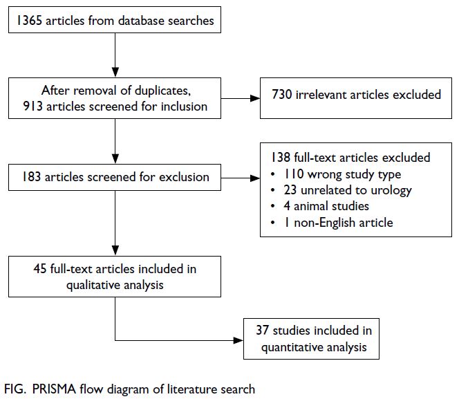
In total, 1365 articles were identified by database searches, whereas no articles were identified by manual searches. After the removal of duplicates, 913 articles remained; 730 articles were removed during the initial screening, and 138 articles were removed during full-text screening. Forty-five articles were included in the qualitative analysis, and 37 articles were included in the quantitative analysis. The PRISMA flow diagram is shown in the Figure.
 

Figure. PRISMA flow diagram of literature search
 
Demographics
Our analysis included 4890 patients (3518 men and 1372 women) with a mean age of 25.46 ± 6.03 years and mean ketamine exposure duration of 54.99 ± 43.54 months.
 
Clinical manifestations
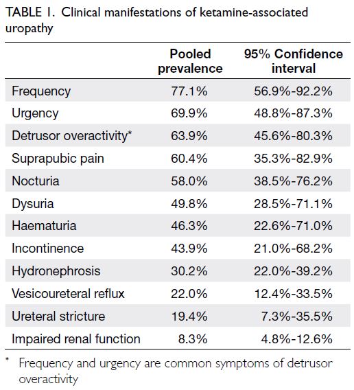
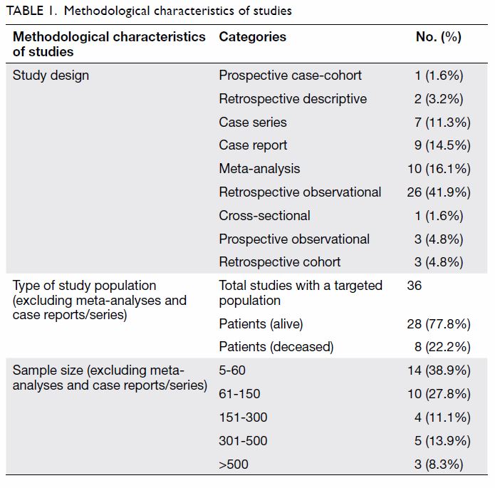
Our meta-analysis concerning clinical manifestations of KAU included 37 studies with 4314 patients. All reported manifestations are summarised in Table 1 and online Appendix 2. Frequency was the most common manifestation (pooled prevalence 77.1%, 95% confidence interval [CI]=56.9%-92.2%), followed by urgency (69.9%, 95% CI=48.8%-87.3%), bladder or suprapubic pain (60.4%, 95% CI=35.3%-82.9%), and nocturia (58.0%, 95% CI=38.5%-76.2%). Additionally, substantial numbers of patients presented with dysuria, haematuria, and/or incontinence. Upper urinary tract involvement was less common. The pooled prevalence of hydronephrosis was 30.2% (95% CI=22.0%-39.2%), whereas approximately 20% of patients presented with ureteral strictures and 10% of patients presented with impaired renal function.
 

Table 1. Clinical manifestations of ketamine-associated uropathy
 
Symptom scores
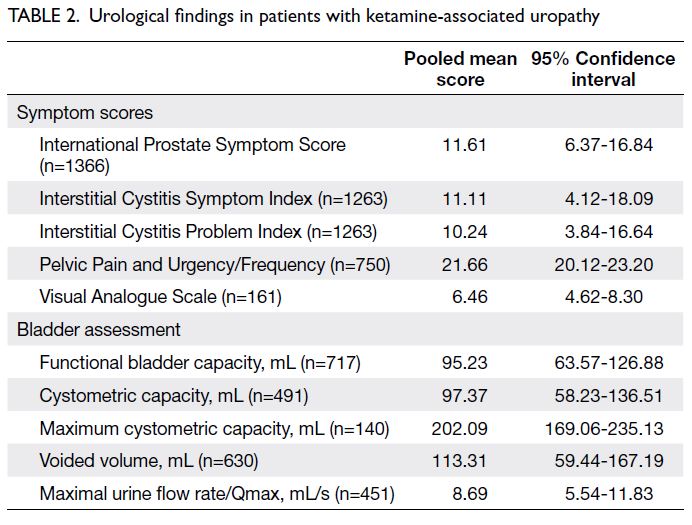
Various questionnaires and scoring systems were used among studies to assess the severities of patient symptoms. The International Prostate Symptom Score (IPSS), Interstitial Cystitis Symptom Index (ICSI), Interstitial Cystitis Problem Index (ICPI), Pelvic Pain and Urgency/Frequency (PUF) total score, and Visual Analogue Scale (VAS) were most commonly used. Our meta-analysis of symptom scores included 13 studies (2135 patients); the results are summarised in Table 2 and online Appendix 3. Five studies with 1366 patients were focused on IPSS; the pooled score was 11.61 (95% CI=6.37-16.84). Five studies (1263 patients) were focused on ICSI and ICPI; the pooled ICSI score was 11.11 (95% CI=4.12-18.09) and the pooled ICPI score was 10.24 (95% CI=3.84-16.64). The pooled PUF score was 21.66 (95% CI=20.12-23.20), and the pooled VAS score was 6.46 (95% CI=4.62-8.30).
 
Bladder assessments
Bladder assessments in the reviewed articles included cystoscopy, urodynamic studies, videocystometry, bladder imaging, and bladder biopsy. The results are shown in Table 2 and online Appendix 4. Fifteen studies (1221 patients) included data regarding bladder capacity, including functional bladder capacity, cystometric capacity, or maximum cystometric capacity. The pooled functional bladder capacity was 95.23 mL (95% CI=63.57-126.88 mL). The pooled and maximum cystometric capacities were 97.37 mL (95% CI=58.23-136.51 mL) and 202.09 mL (95% CI=169.06-235.13 mL), respectively.
 
Urodynamic studies revealed a pooled mean voided volume of 113.31 mL (95% CI=59.44-167.19 mL) and a pooled mean Qmax of 8.69 mL/s (95% CI=5.54-11.83 mL/s). The pooled prevalence of detrusor overactivity was 63.9% (95% CI=45.6%-80.3%). Videocystometry or bladder imaging (eg, ultrasound or computed tomography) showed vesicoureteral reflux with a pooled prevalence of 22.0% (95% CI=12.4%-33.5%).
 

Table 2. Urological findings in patients with ketamine-associated uropathy
 
In total, 14 studies included cystoscopy results and bladder biopsy findings. Common findings on cystoscopy included epithelial inflammation in the bladder mucosa, erythematous patches, neovascularisation, and ulcerations. Petechial haemorrhage was observed in patients with severe disease. Bladder biopsy results were consistent with the above inflammatory changes. Urothelial denudation and focal reactive changes were evident. The lamina propria exhibited granulation tissue, vascular congestion, and oedema, along with infiltrating inflammatory cells including lymphocytes, eosinophils, and mast cells. Perivesical fibrosis was present in some patients. Chu et al5 found such inflammatory changes in 71% of their patients.
 
Treatments
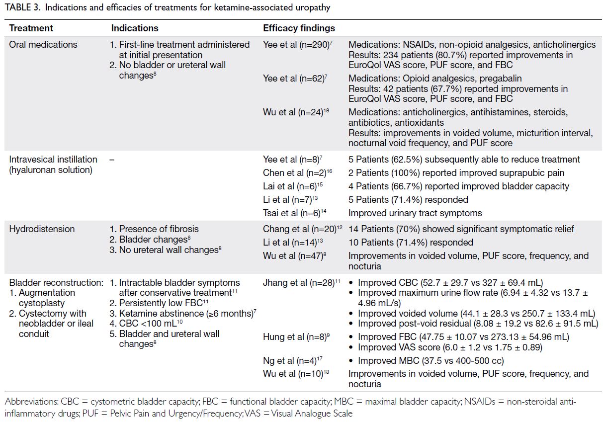
Treatments for KAU considerably varied among studies, and there were no trends consistent among all studies. In total, 19 articles described the treatment of KAU, which included oral, intravesical, and surgical treatments. Yee et al7 and Wu et al8 adopted a tiered treatment approach, which began with the least invasive oral treatment and was escalated to the most invasive surgical treatment if other treatments failed. However, ketamine abstinence was regarded as a key aspect of patient management in 11 of the 19 articles. Various treatments with their potential indications and efficacies are summarised in Table 3.7 8 9 10 11 12 13 14 15 16 17 18
 

Table 3. Indications and efficacies of treatments for ketamine-associated uropathy
 
Oral treatment
Oral treatment approaches involved non-steroidal anti-inflammatory drugs, opioid or non-opioid analgesics, anticholinergics, pregabalin, steroids, antibiotics, antihistamines, antioxidants, and/or pentosane polysulphate sodium. Yee et al7 and Wu et al18 reported that oral treatments led to positive outcomes; most patients showed improvements in symptoms, functional bladder capacity, voided volume, PUF scores, and EuroQol VAS scores. In a case series of nine patients, Shahani et al4 observed that oral pentosane polysulphate sodium produced symptomatic relief when combined with ketamine abstinence. However, 11 patients in another study exhibited poor responses to treatment involving non-steroidal anti-inflammatory drugs, anticholinergics, and antibiotics.14
 
Intravesical treatment
In total, seven studies reviewed intravesical instillation of hyaluronan solutions (eg, sodium hyaluronate and hyaluronic acid). In six of these studies (34 patients), 30 patients showed improvements in symptoms, including suprapubic pain (2 patients) and bladder capacity (4 patients).7 13 14 15 16 19 In the remaining study (2 patients), intravesical treatment did not lead to symptomatic improvement.12
 
Surgical treatment
Surgical treatments for patients with KAU included hydrodistension and bladder reconstruction. In most studies, surgical treatments were performed after patients had not responded to oral treatment. Three studies described the efficacy of hydrodistension, which involves the use of fluid to stretch the bladder in an anaesthetised patient. Wu et al8 reported that hydrodistension led to improvements in voided volume, PUF score, frequency, and nocturia in 35 of 47 patients (74.5%). Chang et al12 and Li et al13 reported that 70.0% (14/20) and 71.4% (10/14) of patients, respectively, exhibited symptomatic relief after hydrodistension. In a few studies, botulinum toxin type A injection was conducted along with hydrodistension. Whereas Zeng et al20 reported that symptoms were generally improved with such treatment, Sihra et al21 found that only four of 29 patients experienced subjective symptomatic relief.
 
Another surgical approach comprises bladder reconstruction, which can be achieved by augmentation cystoplasty or by cystectomy and subsequent construction of a neobladder or an ileal conduit.21 Augmentation cystoplasty increases the bladder capacity by anastomosing a segment of elastic gastrointestinal tract to the diseased fibrotic bladder wall. In the cystectomy method, retention of the bladder neck is combined with the construction of a neobladder to store urine or an ileal conduit to divert urine. Among five studies concerning these reconstruction methods, all demonstrated efficacy in the treatment of advanced ketamine cystitis (KC; a key feature of KAU).9 10 11 17 22 Lee et al10 reported that 26 of 26 patients showed postoperative improvements in bladder capacity, voided volume, and residual volume; similarly, Hung et al9 reported that all eight patients showed postoperative improvements in bladder capacity, renal function, and pain perception. Furthermore, concomitant ureteral reimplantation can be performed in patients with vesicoureteral reflux severity of grade ≥3.10 Importantly, surgical treatments were only effective when patients avoided further ketamine abuse.10 11 17 22
 
Discussion
Clinical presentation of ketamine-associated uropathy
Ketamine-associated uropathy is a clinical entity that has emerged with the increasing popularity of ketamine as an illicit drug among young people. In 37 studies with >4000 patients, the most common manifestations—observed in approximately 60% of patients diagnosed with KAU—were frequency, nocturia, urgency, and bladder pain. Other common urinary symptoms, with prevalences of 40% to 50%, included dysuria, haematuria, and incontinence. Upper urinary tract involvement (eg, hydronephrosis and/or ureteral stricture) was observed in fewer than one-third of patients; renal function was impaired in <10% of patients. Further examinations revealed functional and cystometric bladder capacities of <100 mL, as well as voiding dysfunction. The bladder assessment findings were consistent with the clinical manifestations among affected patients. Symptom severity may be associated with the chronicity and dose of ketamine abuse.23 In the United Kingdom, a survey of 3806 individuals—51.1% with a long-term history of ketamine abuse and 33.8% with a history of ketamine abuse in the previous 1 year—revealed that the dose and frequency of ketamine abuse were associated with the extent of urinary symptoms.23 In Taiwan, Tsai et al14 observed that urinary symptoms began 1 month after the initiation of ketamine abuse and were considerably worse after 1 year of abuse.
 
Potential pathophysiology of ketamine-associated uropathy
Despite the establishment of an association between ketamine abuse and urinary tract damage, the underlying pathophysiology remains unclear. Multiple hypotheses have been proposed to explain the mechanisms that underlie the onset of urinary tract damage. Chu et al5 described four potential mechanisms: direct toxin damage to the urinary tract interstitium, microvascular injury to the urinary tract, autoimmune reaction to ketamine exposure in the urothelial epithelium and submucosa, and (less likely) unrecognised bacteriuria. The initial mechanism ultimately induces chronic inflammatory changes in the bladder, which lead to fibrosis, poor compliance, and contracture. The extent of bladder fibrosis may serve as an indicator of disease severity.24 The occurrence of fibrotic changes is consistent with the common manifestations of frequency, nocturia, and pain associated with reduced bladder capacity; the occurrence of such changes is also consistent with the reduced bladder capacity observed during further urological workup. Wu et al8 attributed the onset of LUTS and reduced bladder capacity among individuals abusing ketamine to a dysfunctional bladder epithelium and a defective glycosaminoglycan layer. The results of our review indicated that glomerulation, erythematous congestion, neovascularisation, and ulceration were common findings in cystoscopic examination of the urothelial epithelium. Bladder biopsy examinations revealed that urothelial denudation was common; infiltrating eosinophils and mast cells were observed in the lamina propria. Accordingly, KAU may involve hypersensitivity or an allergic reaction; this hypothesis is supported by the work of Jhang et al,25 who found that the level of serum immunoglobulin E was higher in patients with KAU than in the control group. Although the exact pathophysiological mechanism remains unclear, the urothelial changes induced by ketamine and its metabolites suggest the onset of an inflammatory process that results in cystitis.8
 
Clinical diagnosis of ketamine-associated uropathy
Ketamine cystitis, a key feature of KAU, is closely associated with LUTS. Notably, KC is similar to interstitial cystitis (IC) in terms of clinical and histological findings. Despite these similarities, it is not difficult to distinguish KC and IC. The presence of cystitis in a patient with a history of ketamine abuse is strongly suggestive of KC. However, it is challenging to determine whether a patient is engaged in ketamine abuse because many patients do not voluntarily provide such information. The onset of cystitis in a patient aged ≤30 years is a potential clue because IC / bladder pain syndrome usually occurs in patients aged ≥40 years. Because of differences in pathophysiology, Yek et al26 suggested that the course of disease progression differed between KC and IC. For example, KC causes bladder contraction that leads to progressive obstructive uropathy and the onset of hydronephrosis / hydroureter. This process explains the potential for upper urinary tract involvement in patients with KAU. In contrast, IC is a chronic bladder condition; its severity generally is not equivalent to the severity of KC.24 In a comparison of clinical characteristics between 16 patients with IC and 13 patients with KC, Wu et al18 found that patients with KC had more severe bladder dysfunction, along with reduced bladder capacity, greater bladder wall thickness, and increased bladder mass volume. Patients with KC also reported greater subjective discomfort (ie, a lower VAS score), compared with patients who had IC.
 
The similarities between KC and IC should be considered during the workup of suspected KAU. The main consideration involves the use of questionnaires in assessing symptoms and disease severity. Among the articles included in this review, the IPSS, ICSI, ICPI, and PUF were commonly used to assess IC; they generally helped to identify patients who required further examination.27 Clemons et al28 suggested that the ICSI and ICPI were suitable for the assessment of LUTS in patients with IC, but these questionnaires may not be appropriate for use during initial diagnosis. Accordingly, Clemons et al28 proposed that 7 of 15 and 5 of 14 should be regarded as the respective scores for the ICSI and ICPI. In our review, the pooled ICSI and ICPI scores were 11.11 and 10.24, respectively. In 2012, the PUF score was validated by Ng et al29 for the assessment of LUTS associated with the use of street ketamine—the score was correlated with disease severity, as indicated by patient symptoms and the results of other examinations. In that study, Ng et al29 proposed using a PUF score of 17 as a threshold to indicate serious urological consequences. In our review, the pooled PUF score was 21.66. The use of questionnaires during the workup of suspected KAU is a subjective assessment of patient symptoms that may provide insights concerning disease severity and quality of life. Although there is no consensus or standardised questionnaire specific to KAU, these non-invasive questionnaires are useful for initial patient assessment and can help guide subsequent management.
 
In addition to the use of subjective questionnaires, the workup of suspected KAU commonly consists of radiological, urodynamic, and endoscopic assessments. Despite the absence of standard diagnostic criteria, suspicion of KAU may be based on the presence of urinary tract symptoms combined with a history of ketamine abuse. Multiple aspects should be considered during the workup of suspected KAU. The main goal is the exclusion of other causes that explain the symptoms, including urinary tract infections and urolithiasis. Therefore, standard initial examinations comprise urinalysis, urine culture, and X-ray imaging. Anatomical involvement can be assessed by computed tomography urography to identify upper urinary tract involvement (eg, ureteral stricture, vesicoureteral reflux, and hydronephrosis) and confirm lower urinary tract involvement (eg, diffuse bladder wall thickening, mucosal enhancement, and inflammatory changes in perivesical tissue).30 Urinary system functionality can be evaluated by urodynamics studies that investigate voided volume, Qmax, detrusor compliance, and detrusor overactivity. Cystoscopy and biopsy enable detailed bladder assessments that clarify cystitis severity and should be considered during a later stage of workup if indicated. However, caution is needed when considering the use of cystoscopy. Routine cystoscopy is not recommended17 because it may discourage young patients from undergoing further medical examinations, with long-term impacts on their health.
 
Management of ketamine-associated uropathy
Although there is no specific regimen or algorithm for the treatment of KAU, ketamine abstinence is consistently effective, regardless of KAU severity. The effects of ketamine abstinence on alleviating and controlling symptoms of KAU are well known.31 32 In a study of 66 individuals with a history of ketamine abuse, Mak et al33 observed significantly improved maximal voided volume after ≥1 year of abstinence (387 mL), compared with patients who had <3 months of abstinence (243 mL). Yee et al24 found that hydronephrosis spontaneously resolved in patients who abstained from ketamine abuse. At initial presentation, medications such as analgesics, anti-inflammatory drugs, and antihistamines were commonly used for symptomatic control.26 Further management options involve intravesical instillation of pentosan polysulphate and hyaluronic acid to protect the bladder lining and repair the damaged glycosaminoglycan layer in the urothelium.19 There is considerable variation in patient responses to the above oral and intravesical treatments. Surgery is indicated when conservative treatments fail. Bladder reconstruction surgery generally leads to improvements in symptoms and bladder functionality,32 but it carries more risks than other treatment options. The available treatment options comprise a spectrum of effectiveness and invasiveness. The most invasive bladder reconstruction surgery is associated with greater effectiveness. Yee et al7 proposed a standardised treatment protocol that comprised the following four-tier approach, where patients receive increasingly invasive treatment if less invasive treatments are ineffective: (1) anti-inflammatory or anticholinergic drugs, (2) opioid or pregabalin, (3) intravesical hyaluronic acid, and (4) hydrodistension and bladder reconstruction surgery (including augmentation cystoplasty and cystectomy with an ileal conduit or neobladder). Wu et al8 adopted a similar tiered approach that involved assigning patients to three clinical staging groups according to their radiological and urodynamic findings. Stage I patients received lifestyle modification guidance and appropriate medications, stage II patients underwent hydrodistension, and stage III patients underwent bladder reconstruction surgery if other treatments were ineffective for 3 months. This tiered approach favourably balances the invasiveness and effectiveness among the available treatment modalities.
 
Limitations
There were some limitations in this systematic review and meta-analysis. First, all included studies were retrospective; there is a need for prospective studies of patients with KAU. Second, there was heterogeneity among the included studies, primarily because of variations in workup and treatment; standardisation would be beneficial for future management of patients with KAU. Third, treatment efficacy could not be fully assessed because baseline characteristics were not described in a consistent manner in the included studies. Fourth, because the incidence of KAU depends on the locality-dependent popularity of ketamine, most studies were from Asian countries/regions (eg, Taiwan and Hong Kong). Nevertheless, this comprehensive review provides an important summary concerning the limited available information about KAU.
 
Conclusion
Patients with KAU most commonly present with frequency, urgency, suprapubic pain, and nocturia; the upper urinary tract is occasionally involved. The occurrence of urinary symptoms in young patients with a history of ketamine abuse should lead to suspicion of KAU. Validated symptom scores are useful in patient-based subjective assessment of symptom severity and treatment progress, whereas radiological and urodynamic examinations objectively define the extent of urinary tract involvement and the functional impairment that results from KAU. In terms of management, ketamine abstinence is essential and a tiered treatment approach is preferred, beginning with the least invasive medications and progressing to surgery if conservative treatments are ineffective.
 
Author contributions
Concept or design: EOT Chan, VWS Chan, MCS Wong, JYC Teoh.
Acquisition of data: EOT Chan, VWS Chan, TST Tang, V Cheung.
Analysis or interpretation of data: EOT Chan, VWS Chan, JYC Teoh.
Drafting of the manuscript: EOT Chan.
Critical revision of the manuscript for important intellectual content: MCS Wong, CH Yee, CF Ng, JYC Teoh.
 
All authors had full access to the data, contributed to the study, approved the final version for publication, and take responsibility for its accuracy and integrity.
 
Conflicts of interest
As editors of the journal, MCS Wong, CF Ng, and JYC Teoh were not involved in the peer review process. Other authors have disclosed no conflicts of interest.
 
Funding/support
This research received no specific grant from any funding agency in the public, commercial, or not-for-profit sectors.
 
Ethics approval
Ethics approval is not required for this study which is a review on published research and not involving patient data collection and retrieval of patient data.
 
References
1. Noppers I, Niesters M, Aarts L, Smith T, Sarton E, Dahan A. Ketamine for the treatment of chronic non-cancer pain. Expert Opin Pharmacother 2010;11:2417-29. Crossref
2. Morgan CJ, Curran HV, Independent Scientific Committee on Drugs. Ketamine use: a review. Addiction 2012;107:27- 38. Crossref
3. Narcotics Division, Security Bureau, Hong Kong SAR Government. Central Registry of Drug Abuse Sixty-ninth Report. 2019. Available from: https://www.nd.gov.hk/en/crda_69th_report.html. Accessed 11 Nov 2020.
4. Shahani R, Streutker C, Dickson B, Stewart RJ. Ketamine-associated ulcerative cystitis: a new clinical entity. Urology 2007;69:810-2. Crossref
5. Chu PS, Ma WK, Wong SC, et al. The destruction of the lower urinary tract by ketamine abuse: a new syndrome? BJU Int 2008;102:1616-22. Crossref
6. Moher D, Liberati A, Tetzlaff J, Altman DG, PRISMA Group. Preferred reporting items for systematic reviews and meta-analyses: the PRISMA statement. Int J Surg 2010;8:336-41. Crossref
7. Yee CH, Lai PT, Lee WM, Tam YH, Ng CF. Clinical outcome of a prospective case series of patients with ketamine cystitis who underwent standardized treatment protocol. Urology 2015;86:236-43. Crossref
8. Wu P, Wang Q, Huang Z, Wang J, Wu Q, Lin T. Clinical staging of ketamine-associated urinary dysfunction: a strategy for assessment and treatment. World J Urol 2016;34:1329-36. Crossref
9. Hung CH, Hsieh SW, Chen SK, Lin CM. Augmentation enterocystoplasty for patients with ketamine-induced cystitis: an 8-year experience and a review of series. Urol Sci 2019;30:232-7. Crossref
10. Lee YK, Jhang JF, Kuo HC. Clinical outcome of augmentation enterocystoplasty for patients with ketamine-induced cystitis. Pain Physician 2017;20:E431-6. Crossref
11. Jhang JF, Birder LA, Chancellor MB, Kuo HC. Patient characteristics for different therapeutic strategies in the management ketamine cystitis. Neurourol Urodyn 2017;36:687-91. Crossref
12. Chang T, Lin CC, Lin AT, Fan YH, Chen KK. Ketamine-induced uropathy: a new clinical entity causing lower urinary tract symptoms. Low Urin Tract Symptoms 2012;4:19-24. Crossref
13. Li CC, Wu ST, Cha TL, Sun GH, Yu DS, Meng E. A survey for ketamine abuse and its relation to the lower urinary tract symptoms in Taiwan. Sci Rep 2019;9:7240. Crossref
14. Tsai TH, Cha TL, Lin CM, et al. Ketamine-associated bladder dysfunction. Int J Urol 2009;16:826-9. Crossref
15. Lai Y, Wu S, Ni L, et al. Ketamine-associated urinary tract dysfunction: an underrecognized clinical entity. Urol Int 2012;89:93-6. Crossref
16. Chen CH, Lee MH, Chen YC, Lin MF. Ketamine-snorting associated cystitis. J Formos Med Assoc 2011;110:787-91. Crossref
17. Ng CF, Chiu PK, Li ML, et al. Clinical outcomes of augmentation cystoplasty in patients suffering from ketamine-related bladder contractures. Int Urol Nephrol 2013;45:1245-51. Crossref
18. Wu SY, Jhang JF, Jiang YH, Kuo HC. Increased bladder wall thickness is associated with severe symptoms and reduced bladder capacity in patients with bladder pain syndrome. Urol Sci 2016;27:263-8. Crossref
19. Meng E, Tsao CW, Tang SH, et al. Intravesical hyaluronic acid treatment for ketamine-associated cystitis: preliminary results. Urol Sci 2015;26:176-9. Crossref
20. Zeng J, Lai H, Zheng D, et al. Effective treatment of ketamine-associated cystitis with botulinum toxin type a injection combined with bladder hydrodistention. J Int Med Res 2017;45:792-7. Crossref
21. Sihra N, Ockrim J, Wood D. The effects of recreational ketamine cystitis on urinary tract reconstruction—a surgical challenge. BJU Int 2018;121:458-65. Crossref
22. Chu PS, Kwok SC, Lam KM, et al. ‘Street ketamine’–associated bladder dysfunction: a report of ten cases. Hong Kong Med J 2007;13:311-3.
23. Winstock AR, Mitcheson L, Gillatt DA, Cottrell AM. The prevalence and natural history of urinary symptoms among recreational ketamine users. BJU Int 2012;110:1762-6. Crossref
24. Yee CH, Teoh JY, Lai PT, et al. The risk of upper urinary tract involvement in patients with ketamine-associated uropathy. Int Neurourol J 2017;21:128-32. Crossref
25. Jhang JF, Hsu YH, Jiang YH, Kuo HC. Elevated serum IgE may be associated with development of ketamine cystitis. J Urol 2014;192:1249-56. Crossref
26. Yek J, Sundaram P, Aydin H, Kuo T, Ng LG. The clinical presentation and diagnosis of ketamine-associated urinary tract dysfunction in Singapore. Singapore Med J 2015;56:660-4. Crossref
27. Kushner L, Moldwin RM. Efficiency of questionnaires used to screen for interstitial cystitis. J Urol 2006;176:587-92. Crossref
28. Clemons JL, Arya LA, Myers DL. Diagnosing interstitial cystitis in women with chronic pelvic pain. Obstet Gynecol 2002;100:337-41. Crossref
29. Ng CM, Ma WK, To KC, Yiu MK. The Chinese version of the pelvic pain and urgency/frequency symptom scale: a useful assessment tool for street-ketamine abusers with lower urinary tract symptoms. Hong Kong Med J 2012;18:123-30.
30. Mason K, Cottrell AM, Corrigan AG, Gillatt DA, Mitchelmore AE. Ketamine-associated lower urinary tract destruction: a new radiological challenge. Clin Radiol 2010;65:795-800. Crossref
31. Wood D, Cottrell A, Baker SC, et al. Recreational ketamine: from pleasure to pain. BJU Int 2011;107:1881-4. Crossref
32. Chung SD, Wang CC, Kuo HC. Augmentation enterocystoplasty is effective in relieving refractory ketamine-related bladder pain. Neurourol Urodyn 2014;33:1207-11. Crossref
33. Mak SK, Chan MT, Bower WF, et al. Lower urinary tract changes in young adults using ketamine. J Urol 2011;186:610-4. Crossref

Cardiovascular complications of COVID-19

Hong Kong Med J 2022 Jun;28(3):249–56  |  Epub 31 May 2022
© Hong Kong Academy of Medicine. CC BY-NC-ND 4.0
 
REVIEW ARTICLE
Cardiovascular complications of COVID-19
YS Archie Lo, MD (UChicago), FACC1; C Jok, BA2; HF Tse, MD, FRCP3,4
1 Faculty of Medicine School of Public Health and Primary Care, The Chinese University of Hong Kong, Hong Kong
2 St Louis University School of Medicine, United States
3 Cardiology Division, Department of Medicine, School of Clinical Medicine, LKS Faculty of Medicine, The University of Hong Kong, Hong Kong
4 Cardiac and Vascular Center, The University of Hong Kong-Shenzhen Hospital, Shenzhen, PR China
 
Corresponding author: Dr YS Archie Lo (olasydm@gmail.com)
 
 Full paper in PDF
 
Abstract
Cardiac injury associated with coronavirus disease 2019 (COVID-19) is associated with high fatality rates. We reviewed the literature on COVID-19-related cardiovascular complications to elucidate the putative causes, diagnosis, and management of cardiovascular complications of COVID-19. Putative causes of these cardiovascular complications include cytokine storm, myocarditis, coronary plaque rupture, hypercoagulability, stress cardiomyopathy or combinations thereof. Cardiac troponin, D-dimer, and N-terminal pro B-type natriuretic peptide levels all provide prognostic information on COVID-19-related cardiovascular complications: elevated levels correlate with poorer prognosis. Coronary thrombosis due to COVID-19 may be associated with a higher thrombus burden than that from other causes. Hypercoagulability can be extremely challenging to treat, and in the absence of contra-indications, thromboprophylaxis is generally indicated in intensive care unit patients. With the exception of percutaneous coronary intervention for acute myocardial infarction, there are no specific treatments for COVID-19-related cardiovascular complications and management is primarily supportive. Whether antiviral therapies, coupled with monoclonal antibodies administered early in the course of COVID-19 illness will prevent severe cardiovascular complications remains to be seen.


Introduction
Coronavirus disease 2019 (COVID-19), caused by severe acute respiratory syndrome coronavirus 2 (SARS-CoV-2), is a contagious respiratory illness which can cause serious complications including stroke, kidney failure, and cardiovascular complications.1 Cardiovascular complications are a major risk factor for COVID-19 mortality.2 3 4 The aim of this paper was to review the literature, published by December 2020, in order to elucidate the risk factors, putative causes, diagnosis, and management of cardiovascular complications of COVID-19.
 
Incidence, risk factors and mortality in patients with COVID-19
Incidence
Two early studies on COVID-19 reported that 20% to 28% of patients with COVID-19 had cardiac injury associated with cardiac dysfunction and arrhythmias.2 3 In a cohort of 416 patients hospitalised with confirmed COVID-19, cardiac injury was reported to occur in 19.7%, and was associated with an unexpectedly high risk of mortality during hospitalisation. Symptoms of COVID-19 were more severe when accompanied by cardiac injury; the mortality rate was higher among patients with cardiac injury than among those without (51.2% vs 4.5%).2
 
Risk factors: age, sex, and co-morbidities
Independently, in another cohort of 187 patients, those with cardiac injury were more likely to be male, older and to have more co-morbidities including diabetes, hypertension, coronary artery disease, chronic kidney disease, chronic lung disease, etc. Severe COVID-19 infections were also potentially associated with cardiac arrhythmias and the need for mechanical ventilation. The mortality during hospitalisation was 7.62% for patients without underlying cardiovascular disease and normal cardiac troponin (c-TN) levels, but as high as 69.44% for those with underlying cardiovascular disease and elevated c-TN.3
 
In another report of 72 314 cases (44 672 confirmed) of COVID-19, the crude mortality rate was 2.3%.4 For octogenarians, the case fatality rate was 14.8%. A history of coronary artery disease was present in 4.2% of all cases, but in 22.7% of fatal cases. Case fatality rates were 10.5% for coronary artery disease, 7.3% for diabetes, and 6% for hypertension. Risk of COVID-19 death is highest among the oldest and lowest among the youngest populations. Compared with those aged 18 to 29 years, people aged 75 to 84 years and those aged ≥85 years have 200-times and 630-times, respectively, higher average death rates.5
 
In a retrospective study of 393 patients, the prevalence of obesity and male sex also appears to be higher in patients with COVID-19 who developed severe symptoms compared with those who did not.6
 
Putative causes of cardiovascular complications in patients with COVID-19
Cardiovascular complications of COVID-19 are generally associated with poor prognosis. Therefore, prevention and treatment of COVID-19 should be considered a priority. To that end, an understanding of the possible pathogenetic mechanisms resulting in myocardial injury would be helpful. Putative causes of cardiovascular complications in patients with COVID-19 include: cytokine storm, myocarditis, extreme physical and emotional stress, ischaemic injury caused by cardiac microangiopathy or macrovascular coronary artery disease, hypercoagulopathy, right heart strain, and cor pulmonale associated with adult respiratory distress syndrome.
 
Cytokine storm
In addition to direct viral damage, uncontrolled inflammation or ‘cytokine storm’—indicated by high levels of inflammatory markers including C-reactive protein (CRP), ferritin, and D-dimer, and increased levels of inflammatory cytokines and chemokines—has been reported in patients with COVID-19.7 8 However, the exact pathogenetic relevance of cytokine storm has yet to be confirmed.9
 
Myocarditis
Myocardial inflammation (myocarditis) is evidenced by elevated c-TN level in some patients10 and autopsy data show mononuclear infiltrate in the myocardium, with related cardiomyocyte necrosis.11 Although there have been case reports of myocarditis in patients with COVID-19, it is unclear whether myocarditis is caused by direct viral invasion or an uncontrolled inflammatory response.10 12
 
In a cohort study of 39 autopsy cases of COVID-19, cardiac infection with COVID-19 was frequently found; however, overt myocarditis was not observed in the acute phase.13 In contrast, another study reported on the detection of SARS-CoV-2 genomes in endomyocardial biopsies.14
 
A cardiac magnetic resonance (MR) imaging study of 100 patients recently recovered from COVID-19 reported cardiac involvement in 78% of them, with evidence of ongoing myocardial inflammation in 60% of them. Such involvement appeared independent of pre-existing conditions, severity, overall course of the acute illness, and the time from diagnosis.13 Of 26 competitive athletes, four (15.4%) had cardiac MR findings suggestive of myocarditis and eight additional athletes (30.8%) exhibited late gadolinium enhancement without T2 elevation suggestive of prior myocardial injury.15
 
In a study of 145 student athletes with COVID-19 who were either asymptomatic or had mild to moderate symptoms during acute infection, cardiac MR findings (at a median of 15 days after a positive test result for COVID-19) were consistent with myocarditis in only two patients (1.4%), based on updated Lake Louise criteria.16
 
In contrast, preliminary data based on a small autopsy study of 40 patients showed that cardiac injury results more from clotting than from inflammation; microthrombi were frequent, whereas none of the patients had myocarditis.17 While this observation has implications for thromboprophylaxis, whether COVID-19 can cause a viral myocarditis is yet to be confirmed.
 
Physical and emotional stress
Cases of typical stress cardiomyopathy have also been reported,18 suggesting that both physical and emotional stress may be in part contributory to some cases of cardiovascular complications of COVID-19.
 
Ischaemic injury
In some patients, ST-segment elevation myocardial infarction (STEMI) may be the first clinical manifestation of COVID-19.19 However, patients with c-TN elevations may not have epicardial coronary artery obstruction at angiography. In a case series of 18 patients with COVID-19 with STEMI, nine patients underwent coronary angiography; six of them (67%) had obstructive disease. A total of 13 patients died in the hospital (4 due to fatal myocardial infarction and 9 due to noncoronary myocardial injury).20 In contrast, patients with COVID-19 with STEMI had more thrombus burden and required more anticoagulation than patients with no COVID-19 infection.21 Very-late stent thrombosis has also been reported with patients with COVID-19 and can be one of the presenting features of COVID-19 in those with a history of coronary stenting.22
 
Hypercoagulopathy
Coronavirus disease 2019 is associated with a hypercoagulable state.23 Although the pathogenesis is not completely understood, the following may be observed: elevated fibrinogen and D-dimer; prolongation of both the prothrombin time and activated partial thromboplastin time; and mild thrombocytosis or thrombocytopenia. Major adverse cardiovascular events, and symptomatic thromboembolism, occur frequently in patients with COVID-19, especially among those in the intensive care unit (ICU), even after thromboprophylaxis.24
 
Stroke
Unchecked vascular thrombosis may result in neurological complications. In a case series of 214 patients with COVID-19, neurological symptoms were seen in 36.4% of patients and were more common in patients with severe infection.25 A retrospective study of 214 patients reported six patients with acute stroke, of which five were ischaemic stroke.26 Stroke has also been reported in younger patients (aged 33-49 years) with COVID-19.27
 
Thromboembolism
Post-mortem studies of 12 patients have reported pulmonary embolism as the direct cause of death in four patients (33%) and deep venous thrombosis in seven patients (58%).28 The risk for venous thromboembolism is markedly elevated with prevalence up to 32%,24 29 30 highest with patients in the ICU.30 In a large study involving 3334 consecutive hospitalised patients with COVID-19, among 829 patients in the ICU, 29.4% had a thrombotic event (13.6% venous and 18.6% arterial).30 Although low-dose anticoagulation has been used for thromboprophylaxis, in a series of 184 critically ill patients with COVID-19, 31% suffered clinically significant thrombotic complications despite low-dose nadroparin.31
 
Thrombocytopenia
A meta-analysis demonstrated thrombocytopenia in patients with severe disease is associated with increased risk of COVID-19 mortality.32 How thrombocytopenia should be factored into the decision to prescribe anticoagulant therapy has yet to be studied.
 
Cor pulmonale, right heart strain, pulmonary hypertension
An echocardiographic study of 110 COVID-19 cases noted right ventricular dilation in 31% of patients.33 Another study demonstrated that when compared with those in the lowest quartile, patients with the highest right ventricular longitudinal strain quartile had an increased risk of elevated D-dimer and CRP levels, acute cardiac injury, acute respiratory distress syndrome, deep vein thrombosis as well as mortality.34 Acute cor pulmonale, right heart strain, and/or pulmonary hypertension should always be considered in critically ill patients with COVID-19.35
 
Other significant cardiac issues in COVID-19
Arrhythmias
Early data suggested an incidence of 16.7% arrhythmias among hospitalised patients with COVID-19 and 44.4% of ICU admissions.36 A multicentre study of 192 patients with COVID-19 reported a prevalence of 12.5% for atrial fibrillation among hospitalised patients with COVID-19.37 Another study evaluating 115 patients with COVID-19 reported atrial tachyarrhythmia in 16.5% of patients, with atrial fibrillation being the most common (63%).38 Those with atrial tachyarrhythmia had higher CRP and D-dimer levels compared with those without atrial tachyarrhythmia. Among 393 patients with COVID-19, atrial arrhythmias were more common among patients on ventilators (18.5% vs 1.9%).6 In another study of 700 patients with COVID-19, nine patients experienced cardiac arrest. All cardiac arrests occurred in patients in the ICU. No patients experienced sustained monomorphic ventricular tachycardia, ventricular fibrillation, or complete heart block. Twenty-five patients had atrial fibrillation, nine had significant bradyarrhythmia, and 10 had non-sustained ventricular tachycardia.39 Among 187 patients with COVID-19, when compared with patients with normal c-TN levels, those with elevated c-TN levels developed more frequent malignant arrhythmias (17.3% vs 1.5%), including ventricular tachycardia/ventricular fibrillation.3
 
Heart failure
Patients with cardiovascular disease and heart failure are more susceptible to COVID-19 and have a more severe clinical course once infected.40 41 In two studies of patients with COVID-19 hospitalised in Wuhan, heart failure was identified as a complication in about 50% of the fatalities.42 In a retrospective multicentred study, among 8383 patients with heart failure who were hospitalised with COVID-19, nearly one in four died during hospitalisation.43 Evidently, heart failure in patients with COVID-19 may be triggered or aggravated by the acute infection in patients with pre-existing cardiovascular disease or incident acute myocardial insult.
 
Cardiac arrest
Malignant tachyarrhythmias resulting in cardiac arrest present a dilemma for caregivers. The outcomes of out-of-hospital cardiac arrest were worse during the first weeks of the COVID-19 pandemic in the United States, and this was observed not only in areas with high case-fatality rates but also ones with lower rates.44 In a retrospective study of 136 patients with COVID-19, 119 (87.5%) had a respiratory cause for their cardiac arrest, and the initial rhythm was asystole in 89.7%, pulseless electrical activity in 4.4%, and shockable in 5.9%. The return-of-spontaneous-circulation rate was 13.2% and 30-day survival rate was only 2.9%.45 In another study of 54 patients with COVID-19, the mortality rate following cardiopulmonary resuscitation was even worse (100%). The initial rhythm was non-shockable for 52 patients (96.3%), with pulseless electrical activity being the most common (81.5%). Although the return-of-spontaneous-circulation rate was achieved in 29 patients (53.7%), none survived to be discharged home.46
 
Prognostic laboratory parameters for cardiovascular complications in patients with COVID-19
Prognostic parameters for cardiovascular complications in patients with COVID-19 include c-TN level, D-dimer level, and N-terminal pro B-type natriuretic peptide (NT-proBNP) level.
 
Cardiac troponin level
Increases in c-TN level indicative of myocardial injury is common in patients with COVID-19 and is associated with adverse outcomes such as arrhythmias and death. The risk of cardiac injury, as diagnosed by increased c-TN levels (>99th percentile), was found in up to 22% of patients in the ICU, and in 59% of those that died.36 In another study of 2736 patients with COVID-19, c-TN elevation was observed in 36%, and c-TN elevation (>0.09 ng/dL) appears to triple the mortality risk.47 Other studies of patients with COVID-19 have also demonstrated a poorer prognosis, including mortality, in patients with c-TN elevation.41 48 Both c-TN and NT-proBNP levels were documented to be elevated significantly during the course of hospitalisation among those who eventually died, but no dynamic changes were observed among the survivors.3 Moreover, patients with COVID-19 with myocardial injury who also have transthoracic echocardiography abnormalities had a higher mortality risk.49
 
D-dimer level
Elevated D-dimer levels were higher among patients with COVID-19 and was correlated with a poorer prognosis. Multivariate analysis showed increasing odds of in-hospital death associated with D-dimer value above 1 μg/mL.50 In a study of 343 patients with COVID-19, D-dimer levels ≥2.0 μg/mL had a higher incidence of mortality compared with those with D-dimer levels <2.0 μg/mL (12/67 vs 1/267, P<0.001).51 A markedly elevated D-dimer (>6 times the upper limit of normal) is a consistent predictor of thrombotic events and poor overall prognosis.52 Indeed, the International Society on Thrombosis and Haemostasis has advised that for patients who have markedly raised D-dimers (arbitrarily defined as three- to four-fold increase), admission to hospital should be considered even in the absence of other severe symptoms.53 The importance of D-dimer is emphasised in several other international guidelines.52 53 54 55
 
N-terminal pro B-type natriuretic peptide level
As a biomarker of heart failure, NT-proBNP levels are commonly elevated in hospitalised patients with COVID-19, particularly in those with elevated c-TN levels. The report by Shi et al2 showed that NT-proBNP levels were significantly higher in patients with elevated c-TN levels than in those without c-TN elevation (1689 vs 139 pg/mL). A study of 3219 hospitalised patients with COVID-19, elevated c-TN was detected in 6.5%, and an elevated NT-proBNP level in 12.9%.56 The adjusted hazard ratio for 28-day mortality for c-TN was 7.12 and for NT-proBNP 5.11, confirming that elevated NT-proBNP levels also carry prognostic information. Although NT-proBNP provides corroborating laboratory information on heart failure, the caveat is that NT-proBNP levels increase with age and with various other conditions including renal failure, thus compromising its utility in older patients with confounding variables.
 
Management of cardiovascular complications of COVID-19
Coronary thrombosis
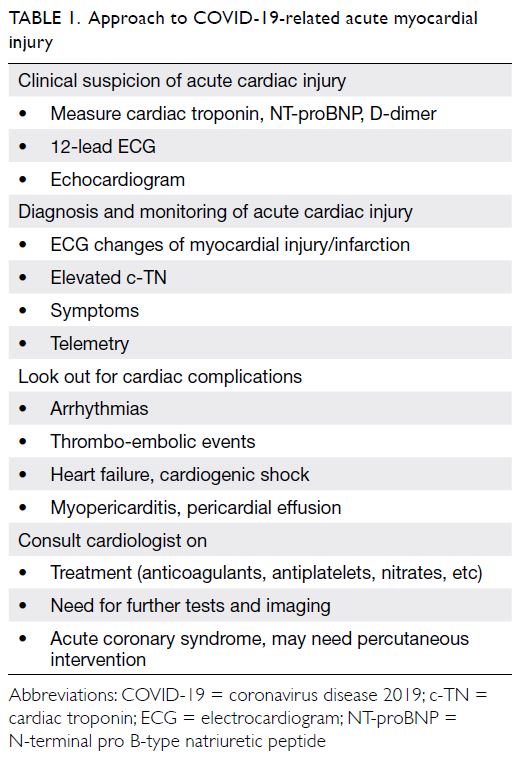
The approach to the diagnosis and management of STEMI in patients with COVID-19 is similar to that for patients without (Table 1). The approaches endorsed by the American College of Cardiology are recommended57: their emphasis is on patient selection for the cardiac catheterisation laboratory, resource allocation, and protection of the interventional team and other healthcare workers involved in caring for the COVID-19 patient.
 

Table 1. Approach to COVID-19-related acute myocardial injury
 
On occasion, it is reasonable to liberalise the use of intravenous thrombolytic therapy relative to primary percutaneous coronary intervention. Intravenous thrombolytic therapy can be considered for a relatively stable patient with STEMI and COVID-19. Obviously, in those STEMI patients who are critically ill with COVID-19, the decision to reperfuse with either primary percutaneous coronary intervention or intravenous thrombolytic therapy should be individualised, and contingent upon hospital resources. In this regard, the consensus statement from the Taiwan Society of Cardiology is both pragmatic and reasonable.58
 
In the event that primary percutaneous coronary intervention is to be performed, maximum personal protective equipment is essential. Intubation, suction, and cardiopulmonary resuscitation all result in aerosolisation of respiratory secretions and increase the risks to the hospital staff. Patients already intubated pose less of an infectious risk. Hence patients with COVID-19 or suspected COVID-19 requiring intubation should be intubated prior to arrival to the catheterisation suite.
 
In the treatment of STEMI patients, an early Hong Kong study reported that both the “symptom onset to first medical contact” and the “door-to-device” times pertaining to primary percutaneous coronary intervention were reported to be substantially prolonged.59
 
Studies from both England60 and the United States61 have confirmed that hospital admissions for acute coronary syndrome declined by 40% to 48% in the early days of COVID-19. It is likely that patients with acute coronary syndromes avoided attending hospital during this period.
 
Heart failure
Standard indications for use of various agents for treatment of heart failure apply to patients with COVID-19. The coexistence of heart failure and COVID-19 complicates diagnosis and management because of overlapping chest findings; however, there are notable differences in chest computed tomography between heart failure and COVID-19 pneumonia, such as lesion distribution/morphology, and pulmonary vein engorgement, which can all help to differentiate between the two.62
 
Cardiopulmonary resuscitation Cardiopulmonary resuscitation poses
Cardiopulmonary resuscitation poses a very high risk for viral spread, and full personal protective equipment should be provided. Immediate intubation should be prioritised in order to minimise the duration of any aerosolisation. While awaiting intubation, bag/mask ventilation with filter is advised.
 
Hypercoagulopathy
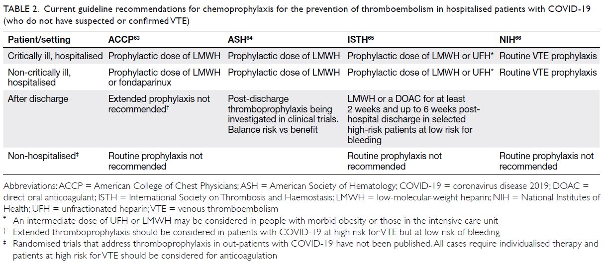
Several international guidelines have issued recommendations advocating chemoprophylaxis in all hospitalised patients with COVID-19,63 64 65 66 in the absence of both contra-indications and bleeding complications (Table 2). In the event thromboprophylaxis is deemed indicated, low-molecular-weight heparin is preferred, but unfractionated heparin can be used if low-molecular-weight heparin is unavailable or if kidney function is severely impaired. Low-molecular-weight heparin may be preferred over unfractionated heparin for staff safety reasons.
 

Table 2. Current guideline recommendations for chemoprophylaxis for the prevention of thromboembolism in hospitalised patients with COVID-19 (who do not have suspected or confirmed VTE)
 
Athletes recovering from COVID-19
As for athletes who have recovered from COVID-19 infections, a recent expert consensus article recommended 2-week convalescence followed by no diagnostic cardiac testing if asymptomatic, and an electrocardiogram and transthoracic echocardiogram in mildly symptomatic athletes with COVID-19 to return to participate in competitive sports.67
 
Summary
Cardiovascular complications of COVID-19 are associated with higher fatality rates. Putative causes of cardiac injury include cytokine storm, myocarditis, extreme physical and emotional stress, ischaemic injury, hypercoagulopathy, right heart strain, and cor pulmonale, or combinations thereof. Echocardiography and c-TN, D-dimer, and NT-proBNP levels all provide prognostic information. Aside from percutaneous coronary intervention for STEMI, there is no specific treatment for COVID-19-associated cardiac injury, and management is primarily supportive. Whether antiviral therapies administered early in the course of disease will prevent severe disease and cardiovascular complications associated with COVID-19 remain to be seen.
 
Author contributions
Concept or design: YSA Lo.
Acquisition of data: YSA Lo, C Jok.
Analysis or interpretation of data: YSA Lo.
Drafting of the manuscript: YSA Lo.
Critical revision of the manuscript for important intellectual content: YSA Lo, HF Tse.
 
All authors had full access to the data, contributed to the study, approved the final version for publication, and take responsibility for its accuracy and integrity.
 
Conflicts of interest
The authors have no conflicts of interest to disclose.
 
Funding/support
This research received no specific grant from any funding agency in the public, commercial, or not-for-profit sectors.
 
References
1. Drake TM, Riad AM, Fairfield CJ, et al. Characterisation of in-hospital complications associated with COVID-19 using the ISARIC WHO Clinical Characterisation Protocol UK: a prospective, multicentre cohort study. Lancet 2021;398:223-7. Crossref
2. Shi S, Qin M, Shen B, et al. Association of cardiac injury with mortality in hospitalized patients with COVID-19 in Wuhan, China. JAMA Cardiol 2020;5:802-10. Crossref
3. Guo T, Fan Y, Chen M, et al. Cardiovascular implications of fatal outcomes of patients with coronavirus disease 2019 (COVID-19). JAMA Cardiol 2020;5:811-8. Crossref
4. Wu Z, McGoogan JM. Characteristics of and important lessons from the coronavirus disease 2019 (COVID-19) outbreak in China: summary of a report of 72 314 cases from the Chinese Center for Disease Control and Prevention. JAMA 2020;323:1239-42. Crossref
5. Centers for Disease Control and Prevention, US Department of Health & Human Services. COVID-19 hospitalization and death by age. 11 February 2020 (updated 18 August 2020). Available from: https://www.cdc. gov/coronavirus/2019-ncov/covid-data/investigations-discovery/hospitalization-death-by-age.html. Accessed 2 Dec 2020.
6. Goyal P, Choi JJ, Pinheiro LC, et al. Clinical characteristics of Covid-19 in New York city. N Engl J Med 2020;382:2372-4. Crossref
7. Mehta P, McAuley DF, Brown M, et al. COVID-19: consider cytokine storm syndromes and immunosuppression. Lancet 2020;395:1033-4. Crossref
8. Zhang X, Tan Y, Ling Y, et al. Viral and host factors related to the clinical outcome of COVID-19. Nature 2020;583;437-40. Crossref
9. Sinha P, Matthay MA, Calfee CS. Is a “Cytokine Storm” relevant to COVID-19? JAMA Intern Med 2020;180:1152-4. Crossref
10. Zeng J, Liu Y, Yuan J, et al. First case of COVID-19 complicated with fulminant myocarditis: a case report and insights. Infection 2020;48:773-7. Crossref
11. Yao XH, Li TY, He ZC, et al. A pathological report of three COVID-19 cases by minimally invasive autopsies [in Chinese]. Zhonghua Bing Li Xue Za Zhi 2020;49:411-7.
12. Lindner D, Fitzek A, Bräuninger H, et al. Association of cardiac infection with SARS-CoV-2 in confirmed COVID-19 autopsy cases. JAMA Cardiol 2020;5:1281-5. Crossref
13. Puntmann VO, Carerj ML, Wieters I, et al. Outcomes of cardiovascular magnetic resonance imaging in patients recently recovered from coronavirus disease 2019 (COVID-19). JAMA Cardiol 2020;5:1265-73. Crossref
14. Escher F, Pietsch H, Aleshcheva G, et al. Detection of viral SARS-CoV-2 genomes and histopathological changes in endomyocardial biopsies. ESC Heart Fail 2020;7:2440-7. Crossref
15. Rajpal S, Tong MS; Borchers J, et al. Cardiovascular magnetic resonance findings in competitive athletes recovering from COVID-19 Infection. JAMA Cardiol 2021;6:116-8. Crossref
16. Starekova J, Bluemke DA, Bradham WS, et al. Evaluation for myocarditis in competitive student athletes recovering from coronavirus disease 2019 with cardiac magnetic resonance imaging. JAMA Cardiol 2021;6:945-50. Crossref
17. Phend C. COVID heart autopsies point more to clot damage than myocarditis. Available from: https://www.medpagetoday.com/meetingcoverage/tct/89143. Accessed 7 Dec 2020.
18. Jabri A, Kalra A, Kumar A, et al. Incidence of stress cardiomyopathy during the coronavirus disease 2019 pandemic. JAMA Netw Open 2020;3:e2014780. Crossref
19. Stefanini GG, Montorfano M, Trabattoni D, et al. ST-elevation myocardial infarction in patients with COVID-19: clinical and angiographic outcomes. Circulation 2020;141:2113-6. Crossref
20. Bangalore S, Sharma A, Slotwiner A, et al. ST-segment elevation in patients with Covid-19—a case series. N Engl J Med 2020;382:2478-80. Crossref
21. Choudry FA, Hamshere SM, Rathod KS, et al. High thrombus burden in patients with COVID-19 presenting with ST-segment elevation myocardial infarction. J Am Coll Cardiol 2020;76:1168-76. Crossref
22. Prieto-Lobato A, Ramos-Martínez R, Vallejo-Calcerrada N, Corbí-Pascual M, Córdoba-Soriano JG. A case series of stent thrombosis during the COVID-19 pandemic. JACC Case Rep 2020;2:1291-6. Crossref
23. Maier CL, Truong AD, Auld SC, Polly DM, Tanksley CL, Duncan A. COVID-19-associated hyperviscosity: a link between inflammation and thrombophilia? Lancet 2020;395:1758-9. Crossref
24. Cui S, Chen S, Li X, Liu S, Wang F. Prevalence of venous thromboembolism in patients with severe novel coronavirus pneumonia. J Thromb Haemost 2020;18:1421-4. Crossref
25. Mao L, Jin H, Wang M, et al. Neurologic manifestations of hospitalized patients with coronavirus disease 2019 in Wuhan, China. JAMA Neurol 2020;77:683-90. Crossref
26. Li Y, Li M, Wang M, et al. Acute cerebrovascular disease following COVID-19: a single center, retrospective, observational study. Stroke Vasc Neurol 2020;5:279-84. Crossref
27. Oxley TJ, Mocco J, Majidi S, et al. Large-vessel stroke as a presenting feature of Covid-19 in the young. N Engl J Med 2020;382:e60. Crossref
28. Wichmann D, Sperhake JP, Lütgehetmann M, et al. Autopsy findings and venous thromboembolism in patients with COVID-19: a prospective cohort study. Ann Intern Med 2020;173:268-77. Crossref
29. Artifoni M, Danic G, Gautier G, et al. Systematic assessment of venous thromboembolism in COVID-19 patients receiving thromboprophylaxis: incidence and role of D-dimer as predictive factors. J Thromb Thrombolysis 2020;50:211-6. Crossref
30. Bilaloglu S, Aphinyanaphongs Y, Jones S, Iturrate E, Hochman J, Berger JS. Thrombosis in hospitalized patients with COVID-19 in a New York City Health System. JAMA 2020;324:799-801. Crossref
31. Klok FA, Kruip MJ, van der Meer NJ, et al. Incidence of thrombotic complications in critically ill ICU patients with COVID-19. Thromb Res 2020;191:153-5. Crossref
32. Lippi G, Plebani M, Henry BM. Thrombocytopenia is associated with severe coronavirus disease 2019 (COVID-19) infections: a meta-analysis. Clin Chim Acta 2020;506:145-8. Crossref
33. Argulian E, Sud K, Vogel B, et al. Right ventricular dilation in hospitalized patients with COVID-19 infection. JACC Cardiovasc Imaging 2020;13:2459-61. Crossref
34. Li Y, Li H, Zhu S, et al. Prognostic value of right ventricular longitudinal strain in patients with COVID-19. JACC Cardiovasc Imaging 2020;13:2287-99. Crossref
35. Creel-Bulos C, Hockstein M, Amin N, Melhem S, Truong A, Sharifpour M. Acute cor pulmonale in critically Ill patients with Covid-19. N Engl J Med 2020;382:e70. Crossref
36. Wang D, Hu B, Hu C, et al. Clinical characteristics of 138 hospitalized patients with 2019 novel coronavirus–infected pneumonia in Wuhan, China. JAMA 2020;323:1061-9. Crossref
37. Russo M, Di Maio M, Attena E, et al. Clinical impact of pre-admission antithrombotic therapy in hospitalized patients with COVID-19: a multicenter observational study. Pharmacol Res 2020;159:104965. Crossref
38. Colon CM, Barrios JG, Chiles JW, et al. Atrial arrhythmias in COVID-19 patients. JACC Clin Electrophysiol 2020;6:1189-90. Crossref
39. Bhatla A, Mayer MM, Adusumalli S, et al. COVID-19 and cardiac arrhythmias. Heart Rhythm 2020;17:1439-44. Crossref
40. Tomasoni D, Italia L, Adamo M, et al. COVID-19 and heart failure: from infection to inflammation and angiotensin II stimulation. Searching for evidence from a new disease. Eur J Heart Fail 2020;22:957-66. Crossref
41. Du RH, Liang LR, Yang CQ, et al. Predictors of mortality for patients with COVID-19 pneumonia caused by SARS-CoV-2: a prospective cohort study. Eur Respir J 2020;55:2000524. Crossref
42. Chen T, Wu D, Chen H, et al. Clinical characteristics of 113 deceased patients with coronavirus disease 2019: retrospective study. BMJ 2020;368:m1091. Crossref
43. Bhatt AS, Jering KS, Vaduganathan M, et al. Clinical outcomes in patients with heart failure hospitalized with COVID-19. JACC Heart Fail 2021;9:65-73. Crossref
44. Chan PS, Girotra S, Tang Y, et al. Outcomes for out-of-hospital cardiac arrest in the United States during the coronavirus disease 2019 pandemic. JAMA Cardiol 2021;6:296-303. Crossref
45. Shao F, Xu S, Ma X, et al. In-hospital cardiac arrest outcomes among patients with COVID-19 pneumonia in Wuhan, China. Resuscitation 2020;151:18-23. Crossref
46. Thapa SB, Kakar TS, Mayer C, Khanal D. Clinical outcomes of in-hospital cardiac arrest in COVID-19. JAMA Intern Med 2021;181:279-81. Crossref
47. Lala A, Johnson KW, Januzzi JL, et al. Prevalence and impact of myocardial injury in patients hospitalized with COVID-19 infection. J Am Coll Cardiol 2020;76:533-46. Crossref
48. Lippi G, Lavie CJ, Sanchis-Gomar F. Cardiac troponin I in patients with coronavirus disease 2019 (COVID-19): evidence from a meta-analysis. Prog Cardiovasc Dis 2020;63:390-1. Crossref
49. Giustino G, Croft LB, Stefanini GG, et al. Characterization of myocardial injury in patients with COVID-19. J Am Coll Cardiol 2020;76:2043-55. Crossref
50. Zhou F, Yu T, Du R, et al. Clinical course and risk factors for mortality of adult inpatients with COVID-19 in Wuhan, China: a retrospective cohort study. Lancet 2020;395:1054-62. Crossref
51. Zhang L, Yan X, Fan Q, et al. D-dimer levels on admission to predict in-hospital mortality in patients with Covid-19. J Thromb Haemost 2020;18:1324-9. Crossref
52. Spyropoulos AC, Levy JH, Ageno W, et al. Scientific and Standardization Committee communication: clinical guidance on the diagnosis, prevention and treatment of venous thromboembolism in hospitalized patients with COVID-19. J Thromb Haemost 2020;18:1859-65. Crossref
53. Thachil J, Tang N, Gando S, et al. ISTH interim guidance on recognition and management of coagulopathy in COVID- 19. J Thromb Haemost 2020;18:1023-6. Crossref
54. American Society of Hematology. COVID-19 and VTE/ anticoagulation: frequently asked questions. Available from: https://www.hematology.org/covid-19/covid-19-and-vte-anticoagulation. Accessed 23 Jun 2020.
55. Bikdeli B, Madhavan MV, Jimenez D, et al. COVID-19 and thrombotic or thromboembolic disease: implications for prevention, antithrombotic therapy, and follow-up: JACC state-of-the-art review. J Am Coll Cardiol 2020;75:2950-73. Crossref
56. Qin JJ, Cheng X, Zhou F, et al. Redefining cardiac biomarkers in predicting mortality of inpatients with COVID-19. Hypertension 2020;76:1104-12. Crossref
57. Mahmud E, Dauerman HL, Welt FG, et al. Management of acute myocardial infarction during the COVID-19 pandemic: a position statement from the Society for Cardiovascular Angiography and Interventions (SCAI), the American College of Cardiology (ACC), and the American College of Emergency Physicians (ACEP). J Am Coll Cardiol 2020;76:1375-84. Crossref
58. Li YH, Wang MT, Huang WC, Hwang JJ. Management of acute coronary syndrome in patients with suspected or confirmed coronavirus disease 2019: Consensus from Taiwan Society of Cardiology. J Formos Med Assoc 2021;120:78-82. Crossref
59. Tam CC, Cheung KS, Lam S, et al. Impact of coronavirus disease 2019 (COVID-19) outbreak on ST-segment–elevation myocardial infarction care in Hong Kong, China. Circ Cardiovasc Qual Outcomes 2020;13:e006631. Crossref
60. Mafham MM, Spata E, Goldacre R, et al. COVID-19 pandemic and admission rates for and management of acute coronary syndromes in England. Lancet 2020;396:381-9. Crossref
61. Solomon MD, McNulty EJ, Rana JS, et al. The covid-19 pandemic and the incidence of acute myocardial infarction. N Engl J Med 2020;383:691-3. Crossref
62. Zhu ZW, Tang JJ, Chai XP, et al. Comparison of heart failure and COVID-19 in chest CT features and clinical characteristics. Zhonghua Xin Xue Guan Bing Za Zhi 2020;48:467-71.
63. Moores LK, Tritschler T, Brosnahan S, et al. Prevention, diagnosis, and treatment of VTE in patients with coronavirus disease 2019 CHEST guideline and expert panel report. Chest 2020;158:1143-63. Crossref
64. American Society of Hematology. ASH Guidelines on Use of Anticoagulation in Patients with COVID-19. Available from: https://www.hematology.org/education/clinicians/guidelines-and-quality-care/clinical-practice-guidelines/venous-thromboembolism-guidelines/ash-guidelines-on-use-of-anticoagulation-in-patients-with-covid-19. Accessed 7 Dec 2020.
65. Thachil J, Juffermans NP, Ranucci M, et al. ISTH DIC subcommittee communication on anticoagulation in COVID-19. J Thromb Haemost 2020;18:2138-44. Crossref
66. National Institutes of Health. Antithrombotic therapy in patients with COVID-19. Last updated: 12 May 2020. Available from: https://www.covid19treatmentguidelines. nih.gov/adjunctive-therapy/antithrombotic-therapy/. Accessed 7 Dec 2020.
67. Phelan D, Kim JH, Chung EH. A game plan for the resumption of sport and exercise after coronavirus disease 2019 (COVID-19) infection. JAMA Cardiol 2020;5:1085-6. Crossref

Utility of cardiac magnetic resonance imaging in troponin-positive chest pain with non-obstructive coronary arteries: literature review

© Hong Kong Academy of Medicine. CC BY-NC-ND 4.0
 
REVIEW ARTICLE  CME
Utility of cardiac magnetic resonance imaging in troponin-positive chest pain with non-obstructive coronary arteries: literature review
Jonan CY Lee, FRCR, FHKAM (Radiology)1; Jeanie B Chiang, FRCR, FHKAM (Radiology)1; PP Ng, MB, ChB1; Boris CK Chow, MB, BS, FRCR1; YW Cheng, MRCP (UK), FHKAM (Medicine)2; CY Wong, MRCP (UK), FHKAM (Medicine)2
1 Department of Radiology and Imaging, Queen Elizabeth Hospital, Hong Kong
2 Department of Medicine, Queen Elizabeth Hospital, Hong Kong
 
Corresponding author: Dr Jonan CY Lee (jonanleecy@yahoo.com)
 
 Full paper in PDF
 
Abstract
There is no general consensus on the investigation and subsequent management of patients presenting with acute chest pain and elevated cardiac troponin levels, but with non-obstructive coronary arteries on angiography. Recent technological advances in cardiac magnetic resonance imaging have aided in the understanding of the underlying pathophysiology, allowing accurate diagnosis, prognostic information, and guidance for management in these patients. This article reviews the evidence supporting the usefulness of cardiac magnetic resonance imaging in patients with acute chest pain and elevated cardiac troponin levels, but with non-obstructive coronary arteries, and offers insights into the role and future development of this imaging modality in this disease.


Introduction
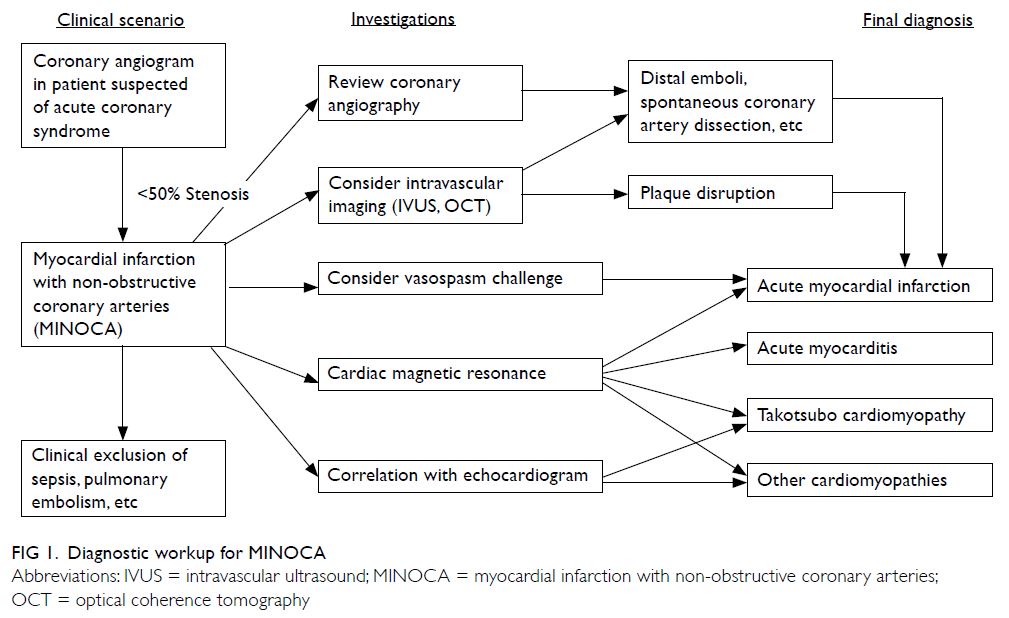
Patients presenting with acute coronary syndrome require immediate management with coronary angiography to identify the culprit coronary stenosis.1 2 A small subset of patients with suspected acute coronary syndrome may have angiographically non-obstructive coronary arteries, termed myocardial infarction with non-obstructive coronary arteries (MINOCA).3 Myocardial infarction with non-obstructive coronary arteries is indistinguishable in its clinical presentation from myocardial infarction with coronary artery disease. The normal coronary angiography results pose a dilemma to the managing physician because the underlying aetiology is not immediately apparent. Arriving at a diagnosis is challenging, with significant implications regarding patients’ prognosis, management, and subsequent follow-up.
 
Myocardial infarction with non-obstructive coronary arteries is a distinct clinical entity with a prevalence of 6% (95% confidence interval [CI], 5%-7%)3 that deserves further meticulous investigation. Despite having non-obstructive coronary arteries, patients with MINOCA have an increased risk of experiencing major cardiovascular events (MACE) including death. Pasupathy et al3 reported 4.7% annual mortality in this group of patients, which is lower than for myocardial infarction with coronary artery disease (6.7%) but much higher than in patients with stable chest pain (0.2% annual mortality).
 
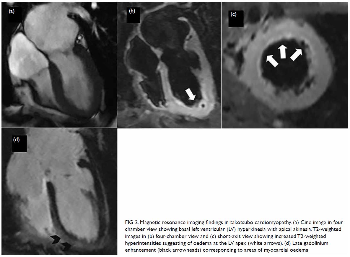
Causes of MINOCA include acute myocardial infarction (AMI) with spontaneous recanalisation, coronary vasospasm, acute myocarditis, takotsubo cardiomyopathy, and other cardiomyopathies.4 Distinguishing between ischaemic and non-ischaemic aetiologies is crucial in patients presenting with MINOCA, in order to tailor treatments accordingly, such as dual antiplatelet therapy and other secondary preventive medications for myocardial infarction,4 or heart failure medications for myocarditis or cardiomyopathies.
 
Cardiac magnetic resonance (CMR) imaging has been increasingly recognised as a first-line imaging modality in the management of patient presenting with MINOCA, to detect the aetiology in a timely manner. High-resolution cardiac images are acquired with tissue characterisation using different MR sequences.
 
The referral rate of CMR imaging for MINOCA has been low, with only 3% of all eligible patients undergoing further testing by CMR imaging in a retrospective study between 2000 and 2016.5 This is expected to change with the widespread availability and improved image quality of CMR imaging.
 
This review aims to summarise the current evidence regarding the use of CMR imaging in patients presenting with MINOCA, to demonstrate its use in various clinical scenarios, and to identify areas for future research. In particular, we review the optimal timing of CMR imaging. We also examine how CMR imaging may change or confirm the aetiology, offers prognostic information, and change management strategy.
 
We reviewed the medical literature in the PubMed database and Google Scholar, using the key terms ‘MINOCA’, ‘myocardial infarction with non-obstructive coronary arteries’, ‘troponin-positive acute chest pain’, ‘non-obstructive coronary arteries’, ‘cardiac magnetic resonance’, ‘myocarditis’, ‘acute myocardial infarction’ and ‘takotsubo cardiomyopathy’, for studies published up to April 2020. There was no language restriction. Abstracts were reviewed to determine their relevance to the aim of our review. Case reports and papers with unclear or inappropriate statistical methods were excluded. The discussion is based on, but not limited to, the search terms.
 
Definition
According to the European Society of Cardiology working group positional paper6 and the scientific statement from American Heart Association,7 MINOCA is a distinct clinical syndrome characterised by evidence of AMI,8 but with no evidence of obstructive coronary artery disease on angiography (stenosis <50% diameter in a major epicardial vessel). The term MINOCA refers to ischaemic-related coronary disorders, namely plaque rupture, coronary vasospasm, microvascular dysfunction, distal embolisation, and coronary artery dissection. The American Heart Association states that it is imperative to exclude (a) clinically overt causes for elevated troponin (eg, sepsis, pulmonary embolism), (b) clinically over-looked obstructive disease, and (c) non-ischaemic disease that can mimic myocardial infarction (eg, myocarditis). In clinical practice, however, exclusion of non-ischaemic mechanism is often not straightforward. Elevated cardiac troponin levels signify myocardial injury, but the marker is non-specific for the underlying pathophysiological mechanism. For instance, acute myocarditis and takotsubo cardiomyopathy may present as MINOCA and may sometimes be even more frequent than ischaemic causes.9 Other non-cardiac causes such as pulmonary embolism or tumour infiltration may also present as MINOCA.6
 
Recently, the term troponin-positive chest pain with non-obstructive coronary arteries (TpNOCA) has been proposed to encompass all patients with ischaemic causes as well as non-ischaemic myocardial disorders and non-cardiac diseases.10 The Dutch ACS working group suggested that the term MINOCA can be understood as either myocardial infarction or myocardial injury with non-obstructive coronary arteries.11 Given the numerous underlying possibilities, a detailed diagnostic workup is required for patient presenting with a working diagnosis of MINOCA (Fig 1).
 

Figure 1. Diagnostic workup for MINOCA
 
Cardiac magnetic resonance imaging protocol
A targeted CMR imaging protocol tailored to the investigation of MINOCA should require no more than 30 to 40 minutes to perform and is feasible in most patients except the most critically ill. The goal of CMR imaging is to assess cardiac motion and characterise myocardial tissue with full left ventricular coverage, to detect myocardial oedema and necrosis for the diagnoses of various disorders, in particular myocarditis and myocardial infarction. Commonly performed CMR imaging sequences are detailed in the online supplementary Appendix.
 
Myocardial perfusion assessment with pharmacological stress (eg, adenosine) to evaluate reversible perfusion defects is seldom required in patients with MINOCA except for specific indications such as evaluation of ischaemic extent, and may be contra-indicated in patients with AMI.12 Therefore, this evaluation is not recommended as part of the routine assessment.
 
Timing of cardiac magnetic resonance imaging
Ideally, CMR imaging should be performed as soon as possible to identify oedema and acute wall motion abnormalities. Although CMR imaging is typically performed after 1 to 4 weeks, the diagnostic value for patients with MINOCA improves significantly when performed within 2 weeks of acute presentation. Studies with longer times to CMR imaging generally show lower sensitivity for demonstrating pathology. One study showed that performing CMR imaging within 2 weeks allowed an underlying cause to be identified in a higher percentage of the study population than if CMR imaging was performed after 2 weeks (82% vs 54%, respectively).13 While the Dutch ACS working group recommends CMR imaging within 4 weeks of presentation,11 a stricter timeframe of performing CMR imaging within 1 week has been suggested by Ferreira et al.4 The local practice may depend on availability of imaging resources.
 
Differential diagnoses shown in cardiac magnetic resonance in patients presenting with myocardial infarction with non-obstructive coronary arteries
In a recent study by Dastidar et al,9 the predominant underlying causes of TpNOCA on CMR imaging in 388 consecutive patients were AMI (25%), myocarditis (25%), and cardiomyopathy (25%), although the median time from clinical presentation to CMR imaging was 37 days. In a recent study by Bhatia et al5 involving 215 patients, myocarditis (32%) was the most common cause, followed by AMI (22%), cardiomyopathy (20%) and takotsubo cardiomyopathy (9%). The strength of the study was the short time interval from clinical presentation to CMR imaging (median: 3.6 days), which could explain the higher proportion of CMR imaging studies resulting in positive diagnosis and the higher incidence of acute myocarditis and takotsubo cardiomyopathy, in which CMR imaging findings may be transient.14 Overall, CMR imaging can provide a diagnosis in 30% to 90% of patients, as shown in several studies.5 9 15 16 17 18 19 20 21 22 The main reasons for the wide range in diagnostic rates among different studies are likely related to the timing of the CMR imaging, heterogeneity of the patient population, different referral patterns and imaging sequences and non-standardised diagnostic criteria. Studies have also shown that CMR imaging results in a change in clinical diagnosis in more than half of patients,20 23 and a change in management in 32% to 42%23 24 of patients.
 
Takotsubo cardiomyopathy
Takotsubo cardiomyopathy (Fig 2), also known as stress-induced cardiomyopathy or apical ballooning syndrome, is a reversible cardiomyopathy induced by extreme physical or emotional stress.25 26 27 However, in some patients, no stressful trigger is identified. The exact pathophysiology of takotsubo cardiomyopathy is unclear; however, some postulate that the underlying pathophysiology is related to microvascular vasoreactivity25 or hormonal disturbances.26 Previously considered a benign condition, an arrhythmogenic risk and increased cardiac mortality are increasingly recognised in patients with takotsubo cardiomyopathy.27 In one study, the prevalence of takotsubo cardiomyopathy in patients undergoing CMR imaging was as high as 27%.20 Takotsubo cardiomyopathy is diagnosed according to the proposed Mayo Clinic criteria.28 Because these criteria do not focus on the role of CMR imaging, an update in 2016 by the Heart Failure Association of the European Society of Cardiology endorsed the use of CMR imaging for its excellent depiction of right and left ventricular regional wall motion abnormalities and myocardial oedema.29


Figure 2. Magnetic resonance imaging findings in takotsubo cardiomyopathy. (a) Cine image in fourchamber view showing basal left ventricular (LV) hyperkinesia with apical akinesia. T2-weighted images in (b) four-chamber view and (c) short-axis view showing increased T2-weighted hyperintensities suggesting of oedema at the LV apex (white arrows). (d) Late gadolinium enhancement (black arrowheads) corresponding to areas of myocardial oedema
 
On CMR cine imaging, takotsubo cardiomyopathy has a typical appearance of mid-cavity to apical akinesia with sparing of basal segments. Although these findings can also be seen in echocardiography and left ventricular angiography, the ability of CMR imaging to assess areas of myocardial oedema and late gadolinium enhancement (LGE), as well as to exclude alternative diagnoses (eg, AMI), makes this an important modality when assessing takotsubo cardiomyopathy. Myocardial oedema (as evidenced using short tau inversion recovery or T2 mapping techniques) on CMR images correlates with acute myocardial inflammation30 and electrographic pattern/repolarisation indices31 in takotsubo cardiomyopathy. The presence of LGE is believed to be transient rather than irreversible.27 32 33 Another study showed that LGE in the acute phase was associated with acute cardiogenic shock, higher peak creatine kinase levels, and delayed recovery.34 Neil et al35 found that the extent of the increase in T2-weighted signal intensity correlated with myocardial strain and the release of both catecholamines and N-terminal pro-B-type natriuretic peptide.
 
Although no specific treatment is currently available, and spontaneous and complete recovery is often expected, Dastidar et al9 showed that mortality in patients with takotsubo cardiomyopathy can be as high as 15% over 3 years, rejecting the notion that this is an entirely benign condition. More studies exploring the underlying mechanism and management strategy for takotsubo cardiomyopathy are required.
 
Acute myocarditis
Acute myocarditis (Fig 3) accounts for 15% to 81% of CMR imaging diagnoses in multiple studies. There are myriad causes of acute myocarditis, including viral infections, autoimmune disease, and toxins.36 Patients’ clinical courses vary and range from complete recovery to progression to chronic myocarditis and dilated cardiomyopathy. Endomyocardial biopsy remains the gold standard for diagnosing acute myocarditis, although its use is declining because of its invasiveness and the possibility of sampling error.37 A previous study has validated CMR imaging results compared with endomyocardial biopsy38; CMR-guided endomyocardial biopsy can improve the diagnostic rate.39 40


Figure 3. Magnetic resonance imaging findings in acute myocarditis. T2-weighted imaging in (a) short axis view and (b) four-chamber view showing evidence of myocardial oedema (white arrows) in the lateral left ventricle. (c) Four-chamber early gadolinium enhancement images showing evidence of myocardial hyperaemia (black arrow). (d) Late gadolinium enhancement images showing epicardial enhancement (black arrowheads) compatible with acute myocarditis
 
The CMR diagnosis of acute myocarditis has been made according to the original Lake Louise criteria, which were established in 2009.41 These criteria are based on the presence of at least two of three CMR imaging findings: myocardial oedema on T2-weighted images, hyperaemia and capillary leak on EGE, and fibrosis/necrosis on LGE. These criteria have a diagnostic accuracy of 78% for acute myocarditis. However, co-existing skeletal inflammation may lead to false-negative results in T2 short tau inversion recovery/early gadolinium enhancement images.42 With the development of parametric mapping, T1 mapping can establish the diagnosis of myocarditis, even without contrast injection for LGE.43 T1 mapping as an individual parameter was found to have superior diagnostic performance for detecting myocarditis compared with T2-weighted oedema imaging.42 More recently, a Journal of the American College of Cardiology scientific expert panel updated the use of CMR imaging in myocarditis to include parametric mapping based on at least one T2-based criterion (global or regional increase in myocardial T2 relaxation time or an increased signal intensity in T2-weighted CMR images), with at least one T1-based criterion (increased myocardial T1, extracellular volume or LGE).44 The inclusion of global or regional T1 or T2 myocardial values is expected to improve the diagnostic accuracy of CMR imaging compared with the original Lake Louise criteria. Extracellular volume measurements can also be obtained after contrast administration, adjusting for individual variation in the haematocrit value that may affect the result. The presence of both T2- and T1-based criteria is diagnostic of acute myocardial inflammation, while having only one criterion may still support the diagnosis in an appropriate clinical scenario, albeit with less specificity. The updated Lake Louise criteria have been validated by Luetkens et al45 to have better sensitivity than the original Lake Louise criteria (88% vs 73%, P=0.031), with a similar high specificity of 96%.
 
In addition to diagnosing acute myocarditis, CMR imaging findings have prognostic implications and can help guide patient management. Grun et al46 indicated that LGE was the best independent predictor of all-cause mortality and of cardiac mortality in 222 consecutive patients with biopsy-proven viral myocarditis. A recent systematic review and meta-analysis by Yang et al47 showed that LGE in patients with myocarditis or suspected myocarditis was significantly associated with MACE (pooled odds ratio=4.57, 95% confidence interval [CI]=2.18-9.59; P<0.001), regardless of the left ventricular ejection fraction. A study by Grani et al48 showed that both the pattern and extent of LGE were significantly associated with MACE. Aquaro et al49 showed that the prognostic value of CMR imaging extends beyond the acute phase, with the presence of LGE with oedema at 6 months being an independent predictor of adverse cardiac events and associated with worse prognosis, especially mid-wall septal patterns in LGE.
 
More studies are required to determine whether CMR imaging can help differentiate the subtypes of myocarditis (viral, eosinophilic, autoimmune and giant cell myocarditis).44
 
Acute myocardial infarction
Acute myocardial infarction (Fig 4) was either the most common or second most common aetiology detected by CMR imaging in previous studies, ranging from 11% to 26%.5 9 The underlying pathophysiological mechanisms included plaque disruption with spontaneous recanalisation, distal embolisation, coronary vasospasm, dissection, or distal small branch disease. On CMR imaging, a classic subendocardial or transmural LGE pattern corresponding to the coronary artery territory, with or without microvascular obstruction, is diagnostic of myocardial infarction. If an infarct is seen, it is essential to review the coronary angiographic images for subtle missed obstructive lesions or coronary artery dissection, and to rule out vasospasm or distal embolisation. Further investigations may depend on clinical suspicion and local practice, and may include intravascular imaging such as optical coherence tomography and intravascular ultrasonography for plaque assessment, provocative tests for coronary vasospasm, echocardiography to identify an embolic source (eg, patent foramen ovale) and thrombophilia screening for hypercoagulable disorders. In Asian populations, vasospastic angina is particularly common and should be carefully managed.50 Drugs such as cocaine are well-documented causes of coronary vasospasm and careful elucidation of history is required.


Figure 4. Magnetic resonance imaging findings in acute myocardial infarction. (a) Cine image in four-chamber view showing hypokinesis of the mid-septum (black arrow). (b) The corresponding region on T2-weighted image showing myocardia oedema (white arrow). (c) Late gadolinium enhancement (LGE) image in short axis view showing transmural infarction in the mid-septum (black arrowhead), suggestive of left anterior descending territory infarct. (d) Another patient’s LGE image demonstrating a hypointense core of microvascular obstruction (curved arrow), which is associated with a worse prognosis
 
In addition to providing a diagnosis, CMR imaging can also assess myocardial oedema and myocardium at risk in the acute phase to calculate the salvageable area, as well as to assess complications of AMI, such as pseudoaneurysms or intra-cardiac thrombus. Both the presence of a scar and the quantifiable extent of the infarct on LGE have been shown to carry prognostic significance in AMI for predicting morbidity and mortality.51 52 The presence of microvascular obstruction is also associated with a worse prognosis.53 T1 mapping and extracellular volume measurement may be able to differentiate between acute and chronic myocardial infarction.54
 
Non-ischaemic cardiomyopathies
Hypertrophic cardiomyopathy and dilated cardiomyopathy are the most common forms of non-ischaemic cardiomyopathy presenting with MINOCA (Fig 5). These cardiomyopathies can be diagnosed using CMR imaging according to their morphology and LGE patterns.55 The prevalence of non-ischaemic cardiomyopathies in MINOCA varies widely in the literature, and it is unclear whether affected patients were excluded in some studies. Bhatia et al5 showed the highest prevalence of cardiomyopathy among studies that included affected patients, with a prevalence of 20%, making cardiomyopathy the third most common aetiology in MINOCA. A recent study by Dastidar et al9 showed that cardiomyopathy had the worst prognosis among all diagnoses.


Figure 5. Magnetic resonance imaging findings in hypertrophic cardiomyopathy. (a, b) Cine images showing septal wall thickening (white arrows) that is markedly asymmetrical compared with the inferior or lateral wall. (c, d) Late gadolinium enhancement images showing patchy mid-wall and subepicardial enhancing areas in the septum (black arrows). Focal enhancement is seen at the posterior right ventricular insertion point (black arrowhead). Full-thickness enhancement is seen in areas of burnt-out myocardium with myocardial thinning (double arrowhead). These late enhancement patterns are suggestive of hypertrophic cardiomyopathy
 
A systematic review by Kuruvilla et al56 showed that patients with non-ischaemic cardiomyopathy with LGE had greater all-cause mortality compared with patients without LGE (odds ratio=3.27; 95% CI=1.94-5.51; P<0.00001).56 In hypertrophic cardiomyopathy, a meta-analysis57 showed that the presence of LGE was associated with an increased risk of sudden cardiac death, heart failure, and cardiovascular mortality and that the extent of LGE was also strongly associated with the risk of sudden cardiac death, suggesting that quantifying LGE is an important tool for risk stratification.
 
The growing use of parametric mapping will no doubt further enhance the diagnostic capability of CMR imaging in non-ischaemic cardiomyopathies.54
 
Normal/inconclusive cardiac magnetic resonance
Cardiac magnetic resonance may sometimes not reveal a specific diagnosis, the proportion of which depends on the timing of CMR imaging as well as patients’ demographics. Patients with negative CMR imaging findings typically have a lower troponin level.20 Occasionally, an infarct may be too small to be visualised by conventional LGE sequences.58 Negative CMR imaging findings do not exclude MINOCA. Regardless of whether the underlying cause is identified, the absence of positive CMR imaging findings is associated with a better prognosis.9
 
Managing patients with myocardial infarction with non-obstructive coronary arteries
Limited guidelines exist regarding the current recommended management of patients with MINOCA, and the management algorithm differs in different centres. Treatment obviously depends on the underlying diagnosis, if identified. In patients without an apparent cause, even by CMR imaging, evidence-based therapies are lacking. Recently, aspirin, statins and calcium channel blockers have been proposed as routine medical treatment in patients with no clear aetiology for elevated troponin on CMR images, to potentially treat underlying thromboembolism, coronary plaque disruption and coronary artery vasospasm.6 The evidence for the use of beta-blocker is conflicting.59 60 The confirmation of the benefits of these therapies would require a multicentre randomised controlled trial.
 
Questions to be addressed
There is a distinct lack of published studies evaluating patients of Asian descent with MINOCA, for whom the local disease spectrum with CMR imaging and the prognostic significance may differ from studies evaluating patients from Western countries, because of differences in the underlying risk factors. It is still unclear in current studies whether performing CMR imaging improves patient outcomes regarding shortening hospital stay, preventing re-admission and lowering MACE and mortality rates. This hypothesis requires validation in further studies in a large patient cohort, with longer follow-up of clinical outcomes. Further studies are also needed to evaluate the relationship between troponin and the extent of LGE, the optimal management pathway and secondary prevention, as well as the role of long-term imaging surveillance to guide management in patients with MINOCA.
 
Future directions
With the emergence of novel parametric mapping techniques, namely T1/T2 mapping and extracellular volume measurement, the sensitivity of CMR imaging is expected to improve, as most previous studies did not use T1 and T2 mapping. The optimal mapping techniques and post-processing methods are still being determined,61 after which the capability of CMR imaging for diagnosis and prognostication can be further enhanced, providing a better understanding of the underlying pathophysiology in MINOCA. A gadolinium-free or LGE-free protocol combining T2-based CMR imaging with T1 mapping holds significant promise, especially for patients contra-indicated for gadolinium, but further studies are required before this approach can be routinely implemented. Further developments in CMR imaging techniques, such as three-dimensional free-breathing high-resolution LGE,58 can lead to a higher rate of definitive myocardial LGE evaluation, thereby reducing the false-negative rate in MINOCA diagnosis. Dedicated rapid CMR imaging protocols or compressed sensing cine can shorten scanning times and permit acquiring diagnostic CMR imaging information even in critically ill patients.
 
In conclusion, troponin-positive chest pain with non-obstructive coronary arteries should be recognised as a distinct clinical entity that deserves an active search for the underlying cause and a detailed management plan. The absence of obstructive disease on angiography does not necessarily exclude AMI. When performed early in the disease course, CMR imaging is the ideal non-invasive adjunct to conventional cardiac investigations in patients presenting as MINOCA. Cardiac magnetic resonance should be routinely used in these patients for diagnosis and risk stratification to guide further therapy.
 
Author contributions
Concept or design: JCY Lee.
Acquisition of data: All authors.
Analysis or interpretation of data: JCY Lee, JB Chiang, PP Ng, BCK Chow.
Drafting of the manuscript: JCY Lee, JB Chiang, YW Cheng, CY Wong.
Critical revision of the manuscript for important intellectual content: JCY Lee, YW Cheng, CY Wong.
 
All authors had full access to the data, contributed to the study, approved the final version for publication, and take responsibility for its accuracy and integrity.
 
Conflicts of interest
The authors have disclosed no conflicts of interest.
 
Funding/support
This research received no specific grant from any funding agency in the public, commercial, or not-for-profit sectors.
 
Ethics approval
The patients were treated in accordance with the Declaration of Helsinki. The patients provided written informed consent for all procedures.
 
References
1. Roffi M, Patrono C, Collet JP, et al. 2015 ESC Guidelines for the management of acute coronary syndromes in patients presenting without persistent ST-segment elevation: Task Force for the management of acute coronary syndromes in patients presenting without persistent ST-segment elevation of the European Society of Cardiology (ESC). Eur Heart J 2016;37:267-315. Crossref
2. Ibanez B, James S, Agewall S, et al. 2017 ESC Guidelines for the management of acute myocardial infarction in patients presenting with ST-segment elevation: The Task Force for the management of acute myocardial infarction in patients presenting with ST-segment elevation of the European Society of Cardiology (ESC). Eur Heart J 2018;39:119-77. Crossref
3. Pasupathy S, Air T, Dreyer RP, Tavella R, Beltrame JF. Systematic review of patients presenting with suspected myocardial infarction and nonobstructive coronary arteries. Circulation 2015;131:861-70. Crossref
4. Ferreira VM. CMR should be a mandatory test in the contemporary evaluation of ‘MINOCA’. JACC Cardiovasc Imaging 2019;12:1983-6. Crossref
5. Bhatia S, Anstine C, Jaffe AS, et al. Cardiac magnetic resonance in patients with elevated troponin and normal coronary angiography. Heart 2019;105:1231-6. Crossref
6. Agewall S, Beltrame JF, Reynolds HR, et al. ESC working group position paper on myocardial infarction with non-obstructive coronary arteries. Eur Heart J 2017;38:143-53.
7. Tamis-Holland JE, Jneid H, Reynolds HR, et al. Contemporary diagnosis and management of patients with myocardial infarction in the absence of obstructive coronary artery disease: a scientific statement from the American Heart Association. Circulation. 2019;139:e891-e908. Crossref
8. Thygesen K, Alpert JS, Jaffe AS, et al. Fourth universal definition of myocardial infarction (2018). Circulation 2018;138:e618-e651. Crossref
9. Dastidar AG, Baritussio A, De Garate E, et al. Prognostic role of CMR and conventional risk factors in myocardial infarction with nonobstructed coronary arteries. JACC Cardiovasc Imaging 2019;12:1973-82. Crossref
10. Pasupathy S, Tavella R, Beltrame JF. Myocardial infarction with nonobstructive coronary arteries (MINOCA): the past, present, and future management. Circulation 2017;135:1490-3. Crossref
11. Pustjens TF, Appelman Y, Damman P, et al. Guidelines for the management of myocardial infarction/injury with non-obstructive coronary arteries (MINOCA): a position paper from the Dutch ACS working group. Neth Heart J 2020;28:116-30. Crossref
12. Jo Y, Kim J, Park CH, et al. Guideline for cardiovascular magnetic resonance imaging from the Korean Society of Cardiovascular Imaging—part 1: standardized protocol. Korean J Radiol 2019;20:1313-33. Crossref
13. Dastidar AG, Singhal P, Rodrigues JC, et al. Improved diagnostic role of CMR in acute coronary syndromes and unobstructed coronary arteries: the importance of time-to-CMR. J Cardiovasc Magn Reson 2015;17(Suppl 1):O87. Crossref
14. Sechtem U, Seitz A, Ong P. MINOCA: unravelling the enigma. Heart 2019;105:1219-20. Crossref
15. Assomull RG, Lyne JC, Keenan N, et al. The role of cardiovascular magnetic resonance in patients presenting with chest pain, raised troponin, and unobstructed coronary arteries. Eur Heart J 2007;28:1242-9. Crossref
16. Monney PA, Sekhri N, Burchell T, et al. Acute myocarditis presenting as acute coronary syndrome: role of early cardiac magnetic resonance in its diagnosis. Heart 2011;97:1312-8. Crossref
17. Leurent G, Langella B, Fougerou C, et al. Diagnostic contributions of cardiac magnetic resonance imaging in patients presenting with elevated troponin, acute chest pain syndrome and unobstructed coronary arteries. Arch Cardiovasc Dis 2011;104:161-70. Crossref
18. Mahmoudi M, Harden S, Abid N, et al. Troponin-positive chest pain with unobstructed coronary arteries: definitive differential diagnosis using cardiac MRI. Br J Radiol 2012;85:e461-6. Crossref
19. Collste O, Sörensson P, Frick M, et al. Myocardial infarction with normal coronary arteries is common and associated with normal findings on cardiovascular magnetic resonance imaging: results from the Stockholm Myocardial Infarction with Normal Coronaries study. J Intern Med 2013;273:189-96. Crossref
20. Pathik B, Raman B, Mohd Amin NH, et al. Troponin-positive chest pain with unobstructed coronary arteries: incremental diagnostic value of cardiovascular magnetic resonance imaging. Eur Heart J Cardiovasc Imaging 2016;17:1146-52. Crossref
21. Chopard R, Jehl J, Dutheil J, et al. Evolution of acute coronary syndrome with normal coronary arteries and normal cardiac magnetic resonance imaging. Arch Cardiovasc Dis 2011;104:509-17. Crossref
22. Laraudogoitia Zaldumbide E, Pérez-David E, Larena JA, et al. The value of cardiac magnetic resonance in patients with acute coronary syndrome and normal coronary arteries [in Spanish]. Rev Esp Cardiol 2009;62:976-83. Crossref
23. Dastidar AG, Rodrigues JC, Johnson TW, et al. Myocardial infarction with nonobstructed coronary arteries: impact of CMR early after presentation. JACC Cardiovasc Imaging 2017;10(10 Pt A):1204-6. Crossref
24. Gerbaud E, Harcaut E, Coste P, et al. Cardiac magnetic resonance imaging for the diagnosis of patients presenting with chest pain, raised troponin, and unobstructed coronary arteries. Int J Cardiovasc Imaging 2012;28:783-94. Crossref
25. Galiuto L, De Caterina AR, Porfidia A, et al. Reversible coronary microvascular dysfunction: a common pathogenetic mechanism in apical ballooning or Tako-Tsubo syndrome. Eur Heart J 2010;31:1319-27. Crossref
26. Pizzino G, Bitto A, Crea P, et al. Takotsubo syndrome and estrogen receptor genes: partners in crime? J Cardiovasc Med (Hagerstown) 2017;18:268-76. Crossref
27. Dastidar AG, Frontera A, Palazzuoli A, Bucciarelli-Ducci C. Takotsubo cardiomyopathy: unravelling the malignant consequences of a benign disease with cardiac magnetic resonance. Heart Fail Rev 2015;20:415-21. Crossref
28. Madhavan M, Prasad A. Proposed Mayo Clinic criteria for the diagnosis of Tako-Tsubo cardiomyopathy and long-term prognosis. Herz 2010;35:240-3. Crossref
29. Lyon AR, Bossone E, Schneider B, et al. Current state of knowledge on takotsubo syndrome: a position statement from the Taskforce on Takotsubo Syndrome of the Heart Failure Association of the European Society of Cardiology. Eur J Heart Fail 2016;18:8-27. Crossref
30. Iacucci I, Carbone I, Cannavale G, et al. Myocardial oedema as the sole marker of acute injury in takotsubo cardiomyopathy: a cardiovascular magnetic resonance (CMR) study. Radiol Med 2013;118:1309-23. Crossref
31. Perazzolo Marra M, Zorzi A, Corbetti F, et al. Apicobasal gradient of left ventricular myocardial edema underlies transient T-wave inversion and QT interval prolongation (Wellens’ ECG pattern) in Tako-Tsubo cardiomyopathy. Heart Rhythm 2013;10:70-7. Crossref
32. Prasad A, Lerman A, Rihal CS. Apical ballooning syndrome (Tako-Tsubo or stress cardiomyopathy): a mimic of acute myocardial infarction. Am Heart J 2008;155:408-17. Crossref
33. Rolf A, Nef HM, Möllmann H, et al. Immunohistological basis of the late gadolinium enhancement phenomenon in tako-tsubo cardiomyopathy. Eur Heart J 2009;30:1635-42. Crossref
34. Naruse Y, Sato A, Kasahara K, et al. The clinical impact of late gadolinium enhancement in takotsubo cardiomyopathy: serial analysis of cardiovascular magnetic resonance images. J Cardiovasc Magn Reson 2011;13:67. Crossref
35. Neil C, Nguyen TH, Kucia A, et al. Slowly resolving global myocardial inflammation/oedema in Tako-Tsubo cardiomyopathy: evidence from T2-weighted cardiac MRI. Heart 2012;98:1278-84. Crossref
36. Kindermann I, Barth C, Mahfoud F, et al. Update on myocarditis. J Am Coll Cardiol 2012;59:779-92. Crossref
37. Leone O, Veinot JP, Angelini A, et al. 2011 Consensus statement on endomyocardial biopsy from the Association for European Cardiovascular Pathology and the Society for Cardiovascular Pathology. Cardiovasc Pathol 2012;21:245-74. Crossref
38. Lurz P, Eitel I, Adam J, et al. Diagnostic performance of CMR imaging compared with EMB in patients with suspected myocarditis. JACC Cardiovasc Imaging 2012;5:513-24. Crossref
39. Mahrholdt H, Goedecke C, Wagner A, et al. Cardiovascular magnetic resonance assessment of human myocarditis: a comparison to histology and molecular pathology. Circulation 2004;109:1250-8. Crossref
40. Baccouche H, Mahrholdt H, Meinhardt G, et al. Diagnostic synergy of non-invasive cardiovascular magnetic resonance and invasive endomyocardial biopsy in troponin-positive patients without coronary artery disease. Eur Heart J 2009;30:2869-79. Crossref
41. Friedrich MG, Sechtem U, Schulz-Menger J, et al. Cardiovascular magnetic resonance in myocarditis: A JACC White Paper. J Am Coll Cardiol 2009;53:1475-87. Crossref
42. Ferreira VM, Piechnik SK, Dall’Armellina E, et al. T1 mapping for the diagnosis of acute myocarditis using CMR: comparison to T2-weighted and late gadolinium enhanced imaging. JACC Cardiovasc Imaging 2013;6:1048-58. Crossref
43. Ferreira VM, Piechnik SK, Dall’Armellina E, et al. Native T1-mapping detects the location, extent and patterns of acute myocarditis without the need for gadolinium contrast agents. J Cardiovasc Magn Reson 2014;16:36. Crossref
44. Ferreira VM, Schulz-Menger J, Holmvang G, et al. Cardiovascular magnetic resonance in nonischemic myocardial inflammation: expert recommendations. J Am Coll Cardiol 2018;72:3158-76. Crossref
45. Luetkens JA, Faron A, Isaak A, et al. Comparison of original and 2018 Lake Louise criteria for diagnosis of acute myocarditis: results of a validation cohort. Radiol Cardiothorac Imaging 2019;1:e190010. Crossref
46. Grün S, Schumm J, Greulich S, et al. Long-term follow-up of biopsy-proven viral myocarditis: predictors of mortality and incomplete recovery. J Am Coll Cardiol 2012;59:1604-15. Crossref
47. Yang F, Wang J, Li W, et al. The prognostic value of late gadolinium enhancement in myocarditis and clinically suspected myocarditis: systematic review and meta-analysis. Eur Radiol 2020;30:2616-26. Crossref
48. Gräni C, Eichhorn C, Bière L, et al. Prognostic value of cardiac magnetic resonance tissue characterization in risk stratifying patients with suspected myocarditis. J Am Coll Cardiol 2017;70:1964-76. Crossref
49. Aquaro GD, Ghebru Habtemicael Y, Camastra G, et al. Prognostic value of repeating cardiac magnetic resonance in patients with acute myocarditis. J Am Coll Cardiol 2019;74:2439-48. Crossref
50. Beltrame JF, Crea F, Kaski JC, et al. The who, what, why, when, how and where of vasospastic angina. Circ J 2016;80:289-98. Crossref
51. Larose E, Rodés-Cabau J, Pibarot P, et al. Predicting late myocardial recovery and outcomes in the early hours of ST-segment elevation myocardial infarction traditional measures compared with microvascular obstruction, salvaged myocardium, and necrosis characteristics by cardiovascular magnetic resonance. J Am Coll Cardiol 2010;55:2459-69. Crossref
52. Yokota H, Heidary S, Katikireddy CK, et al. Quantitative characterization of myocardial infarction by cardiovascular magnetic resonance predicts future cardiovascular events in patients with ischemic cardiomyopathy. J Cardiovasc Magn Reson 2008;10:17. Crossref
53. Taylor AJ, Al-Saadi N, Abdel-Aty H, Schulz-Menger J, Messroghli DR, Friedrich MG. Detection of acutely impaired microvascular reperfusion after infarct angioplasty with magnetic resonance imaging. Circulation 2004;109:2080-5. Crossref
54. Haaf P, Garg P, Messroghli DR, Broadbent DA, Greenwood JP, Plein S. Cardiac T1 mapping and extracellular volume (ECV) in clinical practice: a comprehensive review. J Cardiovasc Magn Reson 2016;18:89. Crossref
55. Karamitsos TD, Francis JM, Myerson S, Selvanayagam JB, Neubauer S. The role of cardiovascular magnetic resonance imaging in heart failure. J Am Coll Cardiol 2009;54:1407-24. Crossref
56. Kuruvilla S, Adenaw N, Katwal AB, Lipinski MJ, Kramer CM, Salerno M. Late gadolinium enhancement on cardiac magnetic resonance predicts adverse cardiovascular outcomes in nonischemic cardiomyopathy: a systematic review and meta-analysis. Circ Cardiovasc Imaging 2014;7:250-8. Crossref
57. Weng Z, Yao J, Chan RH, et al. Prognostic value of LGE-CMR in HCM: a meta-analysis. JACC Cardiovasc Imaging 2016;9:1392-402. Crossref
58. Lintingre PF, Nivet H, Clément-Guinaudeau S, et al. High-resolution late gadolinium enhancement magnetic resonance for the diagnosis of myocardial infarction with nonobstructed coronary arteries. JACC Cardiovasc Imaging 2020;13:1135-48. Crossref
59. Lindahl B, Baron T, Erlinge D, et al. Medical therapy for secondary prevention and long-term outcome in patients with myocardial infarction with nonobstructive coronary artery disease. Circulation 2017;135:1481-9. Crossref
60. Pelliccia F, Pasceri V, Niccoli G, et al. Predictors of mortality in myocardial infarction and nonobstructed coronary arteries: a systematic review and meta-regression. Am J Med 2020;133:73-83.e4. Crossref
61. Peker E, Gülpınar B, Elhan AH, Erden MI. Diagnostic accuracy of mapping techniques and postprocessing methods for acute myocarditis. AJR Am J Roentgenol 2020;215:105-15. Crossref

Incidence, patterns, risk factors, and histopathological findings of liver injury in coronavirus disease 2019 (COVID-19): a scoping review

Hong Kong Med J 2021 Jun;27(3):198–209  |  Epub 30 Oct 2020
Hong Kong Academy of Medicine. CC BY-NC-ND 4.0
 
REVIEW ARTICLE
Incidence, patterns, risk factors, and histopathological findings of liver injury in coronavirus disease 2019 (COVID-19): a scoping review
Taha Bin Arif, MB, BS; Saad Khalid, MB, BS; Mishal S Siddiqui, MB, BS; Harmla Hussain, MB, BS; Hassan Sohail, MB, BS
Department of Internal Medicine, Dow University of Health Sciences, Karachi, Pakistan
 
Corresponding author: Dr Taha Bin Arif (tahaarif20@yahoo.com)
 
 Full paper in PDF
 
Abstract
Background: Coronavirus disease 2019 (COVID-19) exhibits many extrapulmonary manifestations, including liver injury. This scoping review aimed to provide insight into the incidence, patterns, risk factors, histopathological findings, and relationship with disease severity of COVID-19-associated liver injury. Furthermore, we identified existing gaps in the research on the hepatic manifestations of COVID-19 and highlighted areas for future investigations.
 
Methods: A scoping review was conducted following the methodological framework suggested by Arksey and O’Mallay. Five online databases, along with grey literature, were searched for articles published until 22 May 2020, and we included 62 articles in the review. The research domains, methodological characteristics, and key conclusions were included in the analysis.
 
Results: Retrospective observational studies comprised more than one-third (41.9%) of the included publications, and 77.8% were conducted on living patients. The incidence of liver injury varied widely across the studies (4.8%-78%), and liver injury was frequently associated with severe COVID-19. We identified the following risk factors for liver injury: male sex, lymphopoenia, gastrointestinal involvement, old age, increased neutrophil count, and the use of hepatotoxic drugs. Histopathological findings indicate that COVID-19 has direct cytopathic effects and causes liver function test derangements secondary to inflammation, hypoxia, and vascular insult.
 
Conclusions: Liver injury following COVID-19 infection is common and primarily hepatocellular, with a greater elevation of aspartate aminotransferase tahn of alanine aminotransferase. However, the evidence regarding hepatic failure secondary to COVID-19 is insufficient. Standardised criteria to diagnose liver injury need to be devised. Current use of hepatotoxic drugs necessitates close monitoring of liver function.


Introduction
The coronavirus disease 2019 (COVID-19) pandemic has spread to 213 countries and territories, posing a severe threat to public healthcare systems worldwide. As of 29 May 2020, the total number of confirmed cases has surged to 5 701 337, with 357 688 deaths recorded worldwide.1 Although multiple pharmacological agents are being evaluated, no beneficial, targeted drug or vaccine has been discovered to date, and the number of cases is rising daily. The causative agent of COVID-19 is severe acute respiratory syndrome coronavirus 2 (SARS-CoV-2), which is believed to be transmitted through respiratory droplets and person-to-person contact. However, evidence of viral RNA in the faeces of COVID-19 patients also suggests the possibility of faecal-oral transmission.2 3 The disease typically presents with viral pneumonia-like symptoms of fever, dry cough, shortness of breath, and fatigue. Nevertheless, gastrointestinal symptoms like diarrhoea, vomiting, and abdominal pain have also been reported.4
 
Although coronavirus mainly targets the respiratory system, it also exhibits many extrapulmonary manifestations. Sepsis, acute cardiac injury, multiple organ failure, and alkalosis are some of the critical complications that have been observed in patients who die of COVID-19.5 Several studies have acknowledged the presence of liver injury in patients with COVID-19, mainly indicated by abnormal liver function tests (LFTs).6 7 The exact pathophysiology behind the LFT derangements is unknown. It has been suggested that SARS-CoV-2 causes liver injury either via direct viral insult or through an inflammatory cytokine storm.8 Other potential mechanisms, such as drug-induced hepatotoxicity and hypoxic injury, have also been implicated.
 
Given the rampant nature of SARS-CoV-2 and its repercussions on human health, the research community has responded expeditiously to the new virus, and studies regarding its systemic involvement are continuously being published. We have conducted a scoping review to summarise all articles published regarding hepatic damage in this setting. In this review, we aim to provide evidence of the incidence, patterns, risk factors, and histopathological findings of liver injury in COVID-19 and its association with the severity of disease. Furthermore, we highlight hepatotoxicity in patients with COVID-19 who are treated with antiviral (lopinavir/ritonavir) or antimalarial (hydroxychloroquine) drugs. We identify the existing gaps in current knowledge regarding the topic and provide recommendations for further research. This will help healthcare providers to identify hepatic complications during the pandemic.
 
Methods
Study design
A scoping review was conducted following the methodological framework of Arksey and O’Malley9 by taking the following steps: (a) identification of a definite research objective and search strategy; (b) identification and screening of research articles; (c) selection of research articles according to pre-defined eligibility criteria; (d) extraction and charting of data, and (e) reporting, summarising, and discussing the results.
 
Literature search strategies
The reviewed literature was identified by searching five online databases (PubMed, Google Scholar, Scopus, Wiley, and ScienceDirect) without any language restriction from 1 January 2020 to 22 May 2020. Grey literature was also searched in medRxiv and bioRxiv. Moreover, the reference lists of all identified articles were searched for additional sources. A variety of keywords were employed, according to the following search string: “liver injury” OR “hepatic damage” OR “liver functional abnormality” OR “cirrhosis” OR “decompensated liver disease” OR “acute liver failure” OR “chronic liver failure” OR “acute on chronic liver failure” AND “COVID-19” OR “SARS-CoV-2” OR “coronavirus disease”. The full electronic search strategy is provided in the online supplementary Appendix.
 
Identification, screening, and selection of relevant studies
We aimed to summarise all of the scientific literature demonstrating liver dysfunction in COVID-19 and to identify the gaps in knowledge regarding hepatic damage in SARS-CoV-2 infection for further research. Three researchers (TBA, SK, and MSS) independently searched through the literature, and all sets of literature were then compared. Disagreements on the inclusion or exclusion of literature were resolved through discussion or, if necessary, by including a fourth researcher (HH) to make the final decision. Articles were screened according to pre-defined eligibility criteria. The inclusion criteria were as follows: (1) study design: retrospective observational study, retrospective cohort study, retrospective descriptive study, prospective observational study, prospective case-cohort, cross-sectional, case report, case series, or meta-analysis; (2) language: studies published in English only; (3) publication status: preprints and published articles; (4) dates considered: studies published from 1 January 2020 to 22 May 2020; and (5) all relevant papers describing functional abnormalities of the liver in COVID-19. The exclusion criteria were as follows: (1) language: articles published in any language other than English; (2) study design: review article, editorial, letter to the editor, correspondence, or commentary; and (3) studies conducted on patients who had undergone organ transplants. Duplicate articles were excluded. Ultimately, 62 articles were included in this review conducted in accordance with the Preferred Reporting Items for Systematic Reviews and Meta-Analyses extension for scoping reviews (Fig 1).10
 

Figure 1. Preferred Reporting Items for Systematic Reviews and Meta-Analyses (PRISMA) flow diagram of the scoping review process
 
Data extraction and charting from included studies
After article selection, data were extracted and recorded on a pre-designed datasheet. The extracted data included the article’s title, study design, study setting, study population, sample size, research domain, and key conclusions.
 
Summarising the studies
The articles that assessed liver injury in patients with COVID-19 belong to two categories: (a) studies that employed pre-defined clinical criteria for liver injury in COVID-19, and (b) studies that did not employ any pre-defined criteria and only reported LFT derangements in COVID-19. Based on the primary research objectives, each article was classified into one of the following main research domains: incidence of liver injury, patterns of liver injury, and risk factors for liver injury in COVID-19, associations of liver injury or underlying liver disease (eg, chronic liver disease) with the severity of COVID-19, drug-induced liver injury in COVID-19, and histopathological findings of liver injury in COVID-19. The methodological characteristics (study design, study setting, type of population, and sample size) of all studies were also analysed.
 
Results and discussion
Characteristics of studies
A total of 62 articles were included in this scoping review, among which 10 were preprints, and 52 were published in peer-reviewed journals, including The Lancet and Journal of the American Medical Association. About 23 studies (16 retrospective observational, 2 retrospective cohort, 1 retrospective descriptive, 1 prospective observational, 1 prospective case-cohort, 1 cross-sectional, and 1 meta-analysis) documented the incidence of liver injury in COVID-19.11 12 13 14 15 16 17 18 19 20 21 22 23 24 25 26 27 28 29 30 31 32 33 Around half of the eligible studies (n=29, 46.8%) showed an association between the severity of COVID-19 and the degree of liver injury.11 12 13 14 15 16 17 19 20 21 22 23 28 29 34 35 36 37 38 39 40 41 42 43 44 45 46 47 48
 
Out of the 27 studies assessing liver injury in COVID-19, 44.4% (n=12) had pre-defined clinical criteria for liver injury, whereas 55.6% (n=15) did not have any specific pre-defined criteria. The details of these studies are given below. The studies predominantly depicted significant elevation of aspartate aminotransferase (AST) than of alanine aminotransferase (ALT) in case of liver injury, which was found to be proportional to the severity of COVID-19.
 
Fewer studies (n=6) mentioned any histopathological findings of liver injury in patients with COVID-19, but the most common findings mentioned were mild sinusoidal dilatation, microvesicular steatosis, and minimal lymphocytic infiltration.36 49 50 51 52 53 Eight studies assessed the impact of drugs on potential liver damage. Half of those studies (n=4, 50%) concluded that the use of lopinavir/ritonavir increases the odds of liver injury. Other drugs described as having the potential to cause hepatotoxicity in COVID-19 included hydroxychloroquine (n=1, 12.5%), tocilizumab (n=2, 25%), and remdesivir (n=1, 12.5%).12 18 54 55 56 57 58 59
 
The methodological characteristics of the finalised studies were also analysed. The largest number of the studies were retrospective observational studies (n=26, 41.9%), followed by meta-analyses (n=10, 16.1%), case reports (n=9, 14.5%), case series (n=7, 11.3%), prospective observational studies (n=3, 4.8%), and others (Table 1). All studies except for meta-analyses, case reports, and series included a targeted population. Among the 36 articles with a targeted population, more than three-quarters (n=28, 77.8%) were conducted only on living patients with COVID-19, whereas the remainder (n=8, 22.2%) included patients who died. Of the finalised studies, 38.9% (n=14) had sample sizes of 5 to 60. The included studies’ methodological characteristics are given in Table 1.
 

Table 1. Methodological characteristics of studies
 
Incidence of liver injury in patients with COVID-19
Several observational studies documenting the clinical characteristics of patients with COVID-19 have reported liver injury.11 12 13 14 15 16 17 18 19 20 21 22 23 24 25 26 27 28 29 30 31 32 33 They have mentioned liver enzyme elevation without commenting on the clinical signs of hepatic dysfunction, which include hepatomegaly, ascites, and jaundice.11 12 13 14 15 16 17 18 19 20 21 22 23 24 25 26 27 28 29 30 32 The incidence of liver injury has varied widely across studies, from 4.8% to a striking 78%.11 12 13 14 15 16 17 18 19 20 21 22 23 24 25 26 27 28 29 30 31 32 33 However, the term ‘liver injury’ has not been defined uniformly. The definitions used in various studies have ranged from slight transaminasaemia to enzyme elevation more than 3 times higher than the upper limit of normal (Table 2 11 12 16 17 18 20 22 23 24 31 32 33). Additionally, several studies did not establish any clinical or laboratory criteria to define liver injury (Table 3 13 14 15 19 21 25 26 27 28 29 34 35 37 46 60). Many studies failed to mention the date on which LFTs were performed, thus creating non-uniformity in their reported values. Case reports have identified the presence of liver injury across the entire age spectrum, ranging from 55 days to 65 years.50 57 58 61 62
 

Table 2. Summary of COVID-19 studies with pre-defined criteria for liver injury/hepatic dysfunction
 

Table 3. Summary of studies without pre-defined criteria for liver injury/hepatic dysfunction in COVID-19
 
Pathogenesis of liver dysfunction in COVID-19
The pathogenesis of liver involvement in COVID-19 infection is assumed to be multifactorial. However, none of the available hypotheses provide a complete explanation, and further investigation is required not only to understand the mechanism but also to formulate appropriate management plans. Figure 2 illustrates the possible mechanisms of hepatic dysfunction in COVID-19.
 

Figure 2. Pathogenesis of COVID-19-associated liver dysfunction
 
Direct viral invasion
The proposed receptor for the virus, angiotensinconverting enzyme 2 receptor (ACE2R), has been found only sparsely in hepatocytes. Chai et al63 demonstrated ACE2 expression in 2.6% of hepatocytes, whereas up to 59.7% of cholangiocytes expressed ACE2R. Seow et al64 also revealed the presence of ACE2 in liver progenitor cells, especially those destined to become cholangiocytes. These findings imply direct invasion of cholangiocytes and progenitor cells, thus resulting in necrosis and impaired regeneration of cholangiocytes. The tight junctions between cholangiocytes also seem to be altered during COVID-19 infection, which may be responsible for the observed cholestasis in patients.65 Significant necrosis of and rapid viral replication within cholangiocytes has also been observed by Zhao et al65 in a human liver ductal organoid model.
 
However, Zhou et al66 argued against this proposed mechanism by highlighting that ACE2Rs on cholangiocytes are confined to the apical surface, from where viral invasion is unlikely. Furthermore, the hepatic pattern of LFT elevation fails to explain this possible ductal pathology. Hepatocytes also express the protein furin, which may play a role in liver damage upon entry of the virus into the cells.
 
Hypoxia
Decreased oxygen saturation has been a feature of COVID-19 pneumonia, and this may result in hypoxic injury to multiple organs, including the liver.67
 
High positive end expiratory pressure
High values of positive end expiratory pressure used during mechanical ventilation in severe patients may result in hepatic congestion by increasing the pressure on the right atrium and thereby impeding venous return. However, the presence of comparable liver functional abnormalities in patients without ventilation renders that assumption inconclusive.68
 
Systemic inflammation and cytokine storm
An inflammatory response to the virus may lead to persistent leukocytic activation and the release of many mediators responsible for cellular injury. The involvement of a cytokine storm in liver damage has been supported by patients’ elevated levels of interleukins 2, 6, and 10, interferon-gamma, serum ferritin, and C-reactive protein.17
 
Endothelial dysfunction
Liver dysfunction may occur secondary to vascular pathology, resulting in endotheliitis, coagulopathy, thrombus formation, and ischaemic parenchymal necrosis. Angiotensin-converting enzyme 2 receptors are expressed on the endothelium, making it susceptible to viral invasion, which leads to the recruitment of inflammatory cells and elaboration of inflammatory cytokines. The immune response may also exacerbate the damage.69
 
Drug-induced hepatotoxicity
A number of drugs currently in use have hepatotoxic potential, which might be further exaggerated in the setting of chronic liver disease (CLD).12 56 58 The mechanisms for individual drugs are not clearly defined.
 
Patterns of liver injury in COVID-19
The patterns of liver injury in COVID-19 patients include both reversible dysfunction and irreversible injury as a component of multiorgan failure in terminally ill patients.40 57 58 59 61 62 However, hepatic dysfunction in COVID-19 cases is usually mild, and deranged LFTs tend to recover within a few days after discharge.48 Predominant elevation of ALT and AST indicates hepatocellular injury.12 The abnormalities in AST have been more severe compared with those of ALT.20 22 23 34 37 44 45 This finding is intriguing, as ALT, being more liver-specific, is the enzyme that is generally expected to be significantly elevated in case of hepatocellular injury. However, a few studies have hypothesised that AST elevation could be secondary to viral-mediated direct liver damage.20 21 The mechanism behind predominant AST elevation in the presence of viral aetiology remains unclear.
 
Elevation of the ductal enzymes gamma-glutamyl transferase and alkaline phosphatase (ALP) has been reported in some studies.22 23 70 Elevated ALP was also reported alongside elevated AST and ALT in a case of acute hepatitis following COVID-19 infection.61 Cardoso et al71 studied the temporal patterns of liver enzyme levels in critically ill patients and observed that a cholestatic pattern emerged later in the course of illness. Most studies have not mentioned the presence of any liver enzyme abnormalities at the time of liver injury. Hence, the extent of liver damage and the pattern of injury could not be accurately assessed.
 
Histopathological findings of liver injury in COVID-19
Histopathological findings of autopsied liver samples have provided evidence of direct viral invasion and changes secondary to hypoxia, sepsis, and pro-inflammatory and pro-coagulant states. Wang et al11 revealed the presence of hepatic apoptosis, occasional bi- or multi-nucleated hepatocytes, mitochondrial swelling, and decreased glycogen granules. These findings strongly suggest direct cytopathic effects of COVID-19 on the liver. Electron microscopy also showed the presence of viral particles. Other non-specific findings have included varying degrees of steatosis,11 12 49 50 51 52 mild portal lymphocytic infiltration,11 36 50 51 mild sinusoidal dilation,36,51,53 and inflamed cells within the sinusoids.12 However, samples obtained via needle biopsy did not facilitate effective determination of the histology of the ductal epithelium, which carries a higher density of ACE2Rs.51 Ductal pathology was highlighted by Lax et al,52 indicating the presence of canalicular cholestasis and mild nuclear pleomorphism of cholangiocytes. Patterns of both massive and focal patchy necrosis were reported in the periportal and centrilobular areas.51 52 The authors suggested that sepsis and systemic inflammation might be responsible for acute hepatic necrosis. Furthermore, reverse transcription-polymerase chain reaction of one liver sample was positive for COVID-19.51 In that case, an ultrasound-guided autopsy observed centrilobular congestion (which was likely attributable to shock), ischaemic necrosis, portal tract inflammation, and Kupffer cell activation.51
 
The watery degeneration of some hepatocytes observed by Cai et al12 was likely due to ischaemia and hypoxia. The presence of thrombi within the liver, among other organs, also demonstrates the possibility of COVID-associated coagulopathy.52 Liver involvement with COVID-19 infection may further elaborate the inflammatory cascade and alter the secretion of coagulation factors, thus playing a role in causing widespread thrombosis.52 Endotheliitis, acute and chronic vascular changes, and sinusoidal arterialisation due to pressure elevation observed in the liver further support the involvement of underlying endothelial pathology in causing coagulative derangements.53 69
 
Liver injury as a marker of the severity of COVID-19
Studies have consistently shown liver injury to be associated with severe COVID-19.11 12 13 14 15 16 34 42 43 44 Deranged LFTs have also been linked to prolonged hospital stays18 and worse clinical outcomes.19 38 40 45 Disease severity is most likely linked with the elevation of AST rather than ALT.20 21 45 Additionally, hypoproteinaemia and cholestasis in early-stage disease have been shown to increase the risk of death.15 However, the impact of AST on mortality has been controversial.22 46
 
These findings cannot reliably establish that elevated LFT levels were solely caused by COVID-19 infection, as many studies did not exclude patients with CLD, nor did they consider other possible reasons for liver enzyme elevation. Furthermore, there is still not enough evidence to suggest that mild derangement has a high likelihood of progressing into fulminant liver failure. Yet, patients with deranged LFT patterns of the hepatocellular or mixed types at the time of admission or during hospitalisation were more likely to progress to severe disease,12 47 thus necessitating adequate monitoring.38 44 45 48 Additionally, there is no evidence that liver dysfunction can directly cause mortality in patients with COVID-19.
 
Risk factors for liver injury in COVID-19
Studies have reported associations between multiple risk factors and liver injury in the setting of SARS-CoV-2 infection:
 
1. Abnormal white blood cell parameters, including elevated neutrophils and decreased lymphocytes, have been associated with elevated risk of liver injury.14 15 17 22 The loss of lymphocytes responsible for suppression of the immune response during viral infection may have contributed to the damage.14 Similarly, high levels of C-reactive protein and procalcitonin were associated with increased risk of liver damage.14 17 18 The cytokine storm and systemic inflammation might be implicated, as they result in leukocyte activation and the release of a large quantity of inflammatory mediators that directly or indirectly damage cells.14
2. The use of hepatotoxic drugs, including antivirals, hydroxychloroquine, tocilizumab (discussed below),12 16 18 58 and antifungals for superimposed infections has been established as a risk factor for liver dysfunction.22 Systemic corticosteroids were also associated with an increased risk of AST elevation,22 perhaps due to drug-induced lymphopoenia and alteration of the immune response.
3. A correlation between the severity of lung involvement and the incidence of liver injury has also been noticed.17 As severe lung lesions indicate a robust inflammatory state, the liver might be affected for the same reason (ie, a hyperinflammatory state).17 That study did not indicate the role of hypoxia, which is also a possible contributing factor to hepatic damage.
4. Non-modifiable risk factors such as male sex (odds ratio=1.60; P<0.001) and old age (odds ratio=1.01; P=0.031) have been linked to a higher risk of liver damage.22 23
5. Patients with elevated ALT levels were more likely to have a history of drinking (P=0.032).14 However, that study did not comment on elevation of AST, the dominant enzyme involved in both alcoholic liver disease and COVID-19. Furthermore, that study’s very small sample size necessitates further investigation of this risk factor.
6. Patients with gastrointestinal symptoms were more likely to have liver injury than those without such symptoms (P=0.035).24
7. Diabetes mellitus was a risk factor for cholestasis in patients with COVID-19 (P=0.044),15 which is predicted to be a possible mechanism of liver injury in the setting of viral infection.65
8. Invasive mechanical ventilation increased the risk of LFT elevation42 and acute liver injury.33 Such injury may be caused by hepatic congestion that results from elevated right atrial pressure, which in turn is caused by high levels of positive end expiratory pressure.68
 
Drug-induced hepatotoxicity in COVID-19
The drugs currently used to manage COVID-19 infection also carry hepatotoxic potential. Muhović et al56 reported a 40-fold rise in transaminases following two doses of tocilizumab, an interleukin-6 receptor antagonist, which regressed 10 days later. Morena et al59 also reported elevated liver enzymes in 29% of patients who were receiving tocilizumab. In addition, Falcão et al58 reported a 10-fold elevation in transaminases following two doses of hydroxychloroquine. Upon withdrawal, the enzyme levels dropped to near normal after 5 days.
 
Antivirals have also been demonstrated to cause liver toxicity. In one study, people receiving lopinavir/ritonavir had a higher incidence of liver dysfunction compared with those in whom these drugs were not administered (51.8% vs 31.3%, respectively).18 Similarly, Young et al55 reported abnormal LFTs in three out of five patients receiving lopinavir/ritonavir. According to Cai et al,12 the use of lopinavir/ritonavir increased the likelihood of liver injury 4-fold. Durante-Mangoni et al54 reported that remdesivir caused elevation of liver enzymes in three out of four patients, and Weber et al57 suggested that drugs may play a role in precipitating acute liver failure. Administration of lopinavir/ritonavir and interferon was followed by progressive worsening of LFTs. This effect may have been attenuated by the use of Ramipril for arterial hypertension.57 Additionally, Lei et al22 showed that elevated AST and ALP levels were associated with the use of antifungal medications. The above findings are from case reports, retrospective studies, and very small-scale prospective studies. Further large-scale prospective studies, including randomised controlled trials, need to be conducted to establish their efficacy and safety in patients with COVID-19.
 
Chronic liver disease and COVID-19
The effects of underlying liver disease on the severity of COVID-19 are controversial. Zhou et al35 suggested the presence of CLD as a risk factor for severe COVID-19. However, that study included only eight known cases of CLD. Similarly, Qi et al60 indicated that decompensated liver cirrhosis might be a risk factor for poor outcomes of COVID-19. In contrast, a meta-analysis by Wang et al72 that included five studies concluded that prior liver disease does not impact the severity of COVID-19. Likewise, the presence of pre-existing cirrhosis had no direct prognostic association in the setting of COVID-19.73 Some studies in our review included patients with CLD, which may account for some of the LFT derangements observed in patients. A meta-analysis by Mantovani et al41 estimated that the baseline prevalence of CLD was 3%. This figure is much lower than the proportion of people with liver dysfunction. Hence, the role of CLD in worsening the prognosis of COVID-19 infection seems to be minor, if there is any.
 
Nevertheless, acute-on-chronic liver failure (ACLF) following COVID-19 infection has been reported. One case was a female patient with decompensated alcoholic cirrhosis (ACLF Grade 2) who developed a mixed hepatic and cholestatic pattern of liver dysfunction following COVID-19 infection. However, her prognosis was good.74 Another patient was an older man with ACLF Grade 1 non-alcoholic cirrhosis. He developed hepatorenal syndrome-type acute kidney injury following COVID-19 infection. His liver failure subsequently progressed to Grade 2 after catheter-associated urinary tract infections and complicated paracentesis.75
 
Oro-faecal transmission and liver injury
Cui et al61 revealed that anal swabs of an infant with liver injury remained positive for COVID-19 even after throat swabs returned to a negative state. However, polymerase chain reaction of stool samples was not performed in most of the articles included in our review. The association of liver dysfunction with the risk of oro-faecal transmission remains to be investigated. If transmission via this route is possible, existing isolation and discharge protocols may need to be revised.
 
Limitations and recommendations
Our review is subject to certain limitations. First, the majority of the included studies did not have any specific pre-defined clinical criteria for diagnosing liver injury in COVID-19. Moreover, the included studies did not distinguish between a history of liver disease (eg, CLD) and liver injury secondary to COVID-19. Hence, our results need to be interpreted cautiously, as they do not accurately describe the level of incidence of liver injury that is caused by COVID-19. Second, we did not include studies published in any language other than English, which might have provided additional insight. Third, the inclusion of a large number of studies prevented us from critically appraising the individual studies’ sources of evidence. There is a need for a comprehensive systematic review or meta-analysis to summarise the statistics and provide a clearer picture of liver injury in SARS-CoV-2 infection. Transplant recipients, a group that is vulnerable to liver injury, were also not reviewed.
 
Many studies have defined ‘liver injury’ as non-specific elevation of LFTs above the upper limit of normal. Further, many of the investigated studies did not assess the bilirubin levels or coagulation profiles of patients with COVID-19, both of which are important indicators of liver function. Moreover, there have been no reports of liver failure or hepatic cell death secondary to COVID-19 to date. Hence, we recommend the use of scientifically relevant terms ‘liver dysfunction’ or ‘liver enzyme derangement’ to explain non-specific LFT abnormalities until an appropriate definition for liver injury is devised. Furthermore, we propose that pre-defined criteria for liver injury should be set and that mild, non-specific derangements of liver function should not be labelled as liver injury. Given the existing controversy in the literature, we recommend a thorough investigation into the pathogenesis of liver injury, especially the mode of direct viral invasion.
 
Additional studies are required to investigate whether mild derangement of liver function can cause hepatic failure in COVID-19. The reason for the hepatocellular pattern with predominant AST elevation also needs to be elucidated. Finally, the safety and efficacy of hepatotoxic drugs in COVID-19 should also be established via randomised controlled trials.
 
Conclusion
Liver injury is a common extrapulmonary feature of COVID-19. However, the absence of standardised clinical criteria for liver injury in this setting needs to be addressed. Derangements of LFT levels are markers of the severity of COVID-19 infection, but the association between LFT derangements and disease progression requires further investigation because to date, liver dysfunction has not been shown to directly cause mortality in patients with COVID-19. The pattern of injury is predominantly hepatocellular, accompanied by greater elevation of AST than of ALT. Possible pathogenetic mechanisms include direct viral invasion, hypoxia, systemic inflammation, endothelial dysfunction, and the use of mechanical ventilation. Histopathological findings in the liver support viral-induced pathology in addition to non-specific changes. Nevertheless, these studies are sparse, and more research is required. Potentially hepatotoxic drugs have also been observed to cause liver injury in patients with COVID-19, and thus, the administration of these drugs necessitates careful monitoring. Large-scale studies are needed to establish their role in the management of COVID-19.
 
Author contributions
Concept or design: T Bin Arif.
Acquisition of data: T Bin Arif, S Khalid, MS Siddiqui, H Hussain.
Analysis or interpretation of data: H Sohail.
Drafting of the manuscript: S Khalid, MS Siddiqui, H Hussain.
\ Critical revision of the manuscript for important intellectual content: T Bin Arif, H Sohail.
 
All authors had full access to the data, contributed to the study, approved the final version for publication, and take responsibility for its accuracy and integrity.
 
Conflicts of interest
All authors have disclosed no conflicts of interest.
 
Funding/support
This research received no specific grant from any funding agency in the public, commercial, or not-for-profit sectors.
 
References
1. World Health Organization. Coronavirus disease (COVID-2019) situation reports–124. Available from: https://www.who.int/docs/default-source/coronaviruse/situation-reports/20200523-covid-19-sitrep-124.pdf?sfvrsn=9626d639_2. Accessed 29 May 2020.
2. Wang W, Xu Y, Gao R, et al. Detection of SARS-CoV-2 in different types of clinical specimens. JAMA 2020;323:1843- 4. Crossref
3. Chen J, Zhu H, Wang D, et al. Clinical features of stool SARS-CoV-2 RNA positive in 137 COVID-19 patients in Taizhou, China. SSRN Electronic J 2020 Mar 23. Available from: https://papers.ssrn.com/sol3/papers.cfm?abstract_id=3551383. Accessed 28 May 2020. Crossref
4. Pan L, Mu M, Yang P, et al. Clinical characteristics of COVID-19 patients with digestive symptoms in Hubei, China: a descriptive, cross-sectional, multicenter study. Am J Gastroenterol 2020;115:766-73. Crossref
5. Chen T, Wu D, Chen H, et al. Clinical characteristics of 113 deceased patients with coronavirus disease 2019: retrospective study. BMJ 2020;368:m1091. Crossref
6. Chen N, Zhou M, Dong X, et al. Epidemiological and clinical characteristics of 99 cases of 2019 novel coronavirus pneumonia in Wuhan, China: a descriptive study. Lancet 2020;395:507-13. Crossref
7. Zhang C, Shi L, Wang FS. Liver injury in COVID-19: management and challenges. Lancet Gastroenterol Hepatol 2020;5:428-30. Crossref
8. Mehta P, McAuley DF, Brown M, et al. COVID-19: consider cytokine storm syndromes and immunosuppression. Lancet 2020;395:1033-4. Crossref
9. Arksey H, O’Malley L. Scoping studies: towards a methodological framework. Int J Soc Res Methodol 2005;8:19-32. Crossref
10. Tricco AC, Lillie E, Zarin W, et al. PRISMA extension for scoping reviews (PRISMA-ScR): checklist and explanation. Ann Inter Med 2018;169:467-73. Crossref
11. Wang Y, Liu S, Liu H, et al. SARS-CoV-2 infection of the liver directly contributes to hepatic impairment in patients with COVID-19. J Hepatol 2020;73:807-16. Crossref
12. Cai Q, Huang D, Yu H, et al. COVID-19: Abnormal liver function tests. J Hepatol 2020;73:566-74. Crossref
13. Ding Q, Lu P, Fan Y, Xia Y, Liu M. The clinical characteristics of pneumonia patients coinfected with 2019 novel coronavirus and influenza virus in Wuhan, China. J Med Virol 2020 Mar 20. Epub ahead of print. Crossref
14. Li L, Li S, Xu M, et al. Risk factors related to hepatic injury in patients with corona virus disease 2019. medRxiv. 2020 Mar 10. Available from: https://www.medrxiv.org/content/10.1101/2020.02.28.20028514v2. Accessed 27 May 2020. Crossref
15. Fu L, Fei J, Xu S, et al. Acute liver injury and its association with death risk of patients with COVID-19: a hospital-based prospective case-cohort study. medRxiv. 2020 Apr 6. Available from: https://www.medrxiv.org/content/10.1101/ 2020.04.02.20050997v1. Accessed 27 May 2020. Crossref
16. Cai Q, Huang D, Ou P, et al. COVID-19 in a designated infectious diseases hospital outside Hubei Province, China. Allergy 2020;75:1742-52. Crossref
17. Xie H, Zhao J, Lian N, Lin S, Xie Q, Zhuo H. Clinical characteristics of non-ICU hospitalized patients with coronavirus disease 2019 and liver injury: a retrospective study. Liver Int 2020;40:1321-6. Crossref
18. Fan Z, Chen L, Li J, et al. Clinical features of COVID-19-related liver functional abnormality. Clin Gastroenterol Hepatol 2020;18:1561-6. Crossref
19. Yang F, Shi S, Zhu J, Shi J, Dai K, Chen X. Analysis of 92 deceased patients with COVID-19. J Med Virol 2020 Apr 15. Epub ahead of print. Crossref
20. Bloom PP, Meyerowitz EA, Reinus Z, et al. Liver biochemistries in hospitalized patients with COVID-19. Hepatology 2020 May 16. Epub ahead of print. Crossref
21. Cholankeril G, Podboy A, Aivaliotis VI, et al. High prevalence of concurrent gastrointestinal manifestations in patients with SARS-CoV-2: early experience from California. Gastroenterology 2020;159:775-7. Crossref
22. Lei F, Liu YM, Zhou F, et al. Longitudinal association between markers of liver injury and mortality in COVID19 in China. Hepatology 2020;72:389-98. Crossref
23. Hajifathalian K, Krisko T, Mehta A, et al. Gastrointestinal and hepatic manifestations of 2019 novel coronavirus disease in a large cohort of infected patients from New York: clinical implications. Gastroenterology 2020;159:1137-40. e2. Crossref
24. Jin X, Lian JS, Hu JH, et al. Epidemiological, clinical and virological characteristics of 74 cases of coronavirus-infected disease 2019 (COVID-19) with gastrointestinal symptoms. Gut 2020;69:1002-9. Crossref
25. Du Y, Tu L, Zhu P, et al. Clinical features of 85 fatal cases of COVID-19 from Wuhan: a retrospective observational study. Am J Respir Crit Care Med 2020;201:1372-9. Crossref
26. Li X, Wang L, Yan S, et al. Clinical characteristics of 25 death cases with COVID-19: a retrospective review of medical records in a single medical center, Wuhan, China. Int J Infect Dis 2020;94:128-32. Crossref
27. Lian J, Jin X, Hao S, et al. Epidemiological, clinical, and virological characteristics of 465 hospitalized cases of coronavirus disease 2019 (COVID-19) from Zhejiang province in China. Influenza Other Respir Viruses 2020;14:564-74. Crossref
28. Yang X, Yu Y, Xu J, et al. Clinical course and outcomes of critically ill patients with SARS-CoV-2 pneumonia in Wuhan, China: a single-centered, retrospective, observational study. Lancet Respir Med 2020;8:475-81. Crossref
29. Zhang B, Zhou X, Qiu Y, et al. Clinical characteristics of 82 death cases with COVID-19. medRxiv. 2020 Feb 27. Available from: https://www.medrxiv.org/content/10.1101/2020.02.26.20028191v1. Accessed 28 May 2020. Crossref
30. Zhu J, Ji P, Pang J, et al. Clinical characteristics of 3062 COVID-19 patients: a meta-analysis. J Med Virol 2020 Apr 15. Epub ahead of print. Crossref
31. Chen G, Wu D, Guo W, et al. Clinical and immunological features of severe and moderate coronavirus disease 2019. J Clin Invest 2020;130:2620-9. Crossref
32. Qi X, Liu C, Jiang Z, et al. Multicenter analysis of clinical characteristics and outcome in patients with COVID-19 who develop liver injury. J Hepatol 2020;73:455-8. Crossref
33. Zheng Y, Sun L, Xu M, et al. Clinical characteristics of 34 COVID-19 patients admitted to ICU in Hangzhou, China. medRxiv. 2020 Apr 15. Available from: https://www.medrxiv.org/content/10.1101/2020.04.12.20062604v1.Accessed 28 May 2020. Crossref
34. Wan S, Xiang Y, Fang W, et al. Clinical features and treatment of COVID-19 patients in Northeast Chongqing. J Med Virol 2020;92:797-806. Crossref
35. Zhou Y, He Y, Yang H, et al. Development and validation a nomogram for predicting the risk of severe COVID-19: a multi-center study in Sichuan, China. PLoS One 2020;15:e0233328. Crossref
36. Zhang Y, Zheng L, Liu L, Zhao M, Xiao J, Zhao Q. Liver impairment in COVID-19 patients: a retrospective analysis of 115 cases from a single centre in Wuhan City, China. Liver Int 2020;40:2095-103. Crossref
37. Wang L, He W, Yu X, et al. Coronavirus disease 2019 in elderly patients: characteristics and prognostic factors based on 4-week follow-up. J Infect 2020;80:639-45. Crossref
38. Parohan M, Yaghoubi S, Seraj A. Liver injury is associated with severe coronavirus disease 2019 (COVID-19) infection: a systematic review and meta-analysis of retrospective studies. Hepatol Res 2020;50:924-35. Crossref
39. Mao R, Qiu Y, He JS, et al. Manifestations and prognosis of gastrointestinal and liver involvement in patients with COVID-19: a systematic review and meta-analysis. Lancet Gastroenterol Hepatol 2020;5:667-78. Crossref
40. Henry BM, de Oliveira MH, Benoit S, Plebani M, Lippi G. Hematologic, biochemical and immune biomarker abnormalities associated with severe illness and mortality in coronavirus disease 2019 (COVID-19): a meta-analysis. Clin Chem Lab Med 2020;58:1021-8. Crossref
41. Mantovani A, Beatrice G, Dalbeni A. Coronavirus disease 2019 and prevalence of chronic liver disease: a meta-analysis. Liver Int 2020;40:1316-20. Crossref
42. Israelsen SB, Kristiansen KT, Hindsberger B, et al. Characteristics of patients with COVID-19 pneumonia at Hvidovre Hospital, March-April 2020. Dan Med J 2020;67:A05200313.
43. Zhao X, Lei Z. The impact of coronavirus disease 2019 (COVID-19) on liver injury in China: a systematic review and meta-analysis. medRxiv. 2020 May 8. Available from: https://www.medrxiv.org/content/10.1101/2020.05.03.20089557v1. Accessed 28 May 2020. Crossref
44. Dong X, Zeng DY, Cai YY, et al. Liver chemistries in patients with severe or non-severe COVID-19: a metaanalysis. medRxiv. 2020 Apr 29. Available from: https:// www.medrxiv.org/content/10.1101/2020.04.24.20074179v1. Accessed 29 May 2020. Crossref
45. Xing QQ, Dong X, Ren YD, et al. Liver chemistries in COVID-19 patients with survival or death: a metaanalysis. medRxiv. 2020 May 1. Available from: https://www.medrxiv.org/content/10.1101/2020.04.26.20080580v1. Accessed 28 May 2020. Crossref
46. Wu C, Chen X, Cai Y, et al. Risk factors associated with acute respiratory distress syndrome and death in patients with coronavirus disease 2019 pneumonia in Wuhan, China. JAMA Intern Med 2020;180:1-11. Crossref
47. Xu L, Mao Y, Chen G. Risk factors for severe corona virus disease 2019 (COVID-19) patients: a systematic review and meta analysis. medRxiv. 2020 Apr 1. Available from: https://www.medrxiv.org/content/10.1101/2020.03.30.20047415v1. Accessed 28 May 2020. Crossref
48. Chen C, Jiang J, Xu X, Hu Y, Hu Y, Zhao Y. Dynamic liver function indexes monitoring and clinical characteristics in three types of COVID-19 patients. medRxiv. 2020 May 20. Available from: https://www.medrxiv.org/content/10.1101/2020.05.13.20099614v2. Accessed 28 May 2020. Crossref
49. Nunes Duarte-Neto A, de Almeida Monteiro RA, da Silva LF, et al. Pulmonary and systemic involvement of COVID-19 assessed by ultrasound-guided minimally invasive autopsy. Histopathology 2020 May 22. Epub ahead of print. Crossref
50. Xu Z, Shi L, Wang Y, et al. Pathological findings of COVID-19 associated with acute respiratory distress syndrome. Lancet Respir Med 2020;8:420-2. Crossref
51. Tian S, Xiong Y, Liu H, et al. Pathological study of the 2019 novel coronavirus disease (COVID-19) through postmortem core biopsies. Modern Pathol 2020;33:1007-14. Crossref
52. Lax SF, Skok K, Zechner P, et al. Pulmonary arterial thrombosis in COVID-19 with fatal outcome: results from a prospective, single-center, clinicopathologic case series. Ann Intern Med 2020 May 14. Epub ahead of print. Crossref
53. Sonzogni A. Liver histopathology in COVID 19 infection is suggestive of vascular alteration. 2020 May 11. Available from: https://www.medrxiv.org/content/10.1101/2020.05. 06.20092718v1. Accessed 28 May 2020. Crossref
54. Durante-Mangoni E, Andini R, Bertolino L, et al. Early experience with remdesivir in SARS-CoV-2 pneumonia. Infection 2020;48:779-82. Crossref
55. Young BE, Ong SW, Kalimuddin S, et al. Epidemiologic features and clinical course of patients infected with SARS-CoV- 2 in Singapore. JAMA 2020;323:1488-94. Crossref
56. Muhović D, Bojović J, Bulatović A, et al. First case of drug-induced liver injury associated with the use of tocilizumab in a patient with COVID-19. Liver Int 2020 May 17. Epub ahead of print. Crossref
57. Weber S, Mayerle J, Irlbeck M, Gerbes AL. Severe liver failure during SARS-CoV-2 infection. Gut 2020;69:1365-7. Crossref
58. Falcão MB, Pamplona de Góes Cavalcanti L, Filgueiras Filho NM, Antunes de Brito CA. Case report: hepatotoxicity associated with the use of hydroxychloroquine in a patient with COVID-19. Am J Trop Med Hyg 2020;102:1214-6. Crossref
59. Morena V, Milazzo L, Oreni L, et al. Off-label use of tocilizumab for the treatment of SARS-CoV-2 pneumonia in Milan, Italy. Eur J Intern Med 2020;76:36-42. Crossref
60. Qi X, Wang J, Li X, et al. Clinical course of COVID-19 in patients with pre-existing decompensated cirrhosis: initial report from China. Hepatol Int 2020;14:478-82. Crossref
61. Wander P, Epstein M, Bernstein D. COVID-19 presenting as acute hepatitis. Am J Gastroenterol 2020;115:941-2. Crossref
62. Cui Y, Tian M, Huang D, et al. A 55-day-old female infant infected with 2019 novel coronavirus disease: presenting with pneumonia, liver injury, and heart damage. J Infect Dis 2020;221:1775-81. Crossref
63. Chai X, Hu L, Zhang Y, et al. Specific ACE2 expression in cholangiocytes may cause liver damage after 2019-nCoV infection. bioRxiv. 2020 Feb 4. Available from: https://www.biorxiv.org/content/10.1101/2020.02.03.931766v1. Accessed 28 May 2020. Crossref
64. Seow JJ, Pai R, Mishra A, et al. scRNA-seq reveals ACE2 and TMPRSS2 expression in TROP2+ liver progenitor cells: implications in COVID-19 associated liver dysfunction. bioRxiv. 2020 Mar 25. Available from: https://www.biorxiv.org/content/10.1101/2020.03.23.002832v1. Accessed 28 May 2020.
65. Zhao B, Ni C, Gao R, et al. Recapitulation of SARS-CoV-2 infection and cholangiocyte damage with human liver ductal organoids. Protein Cell 2020;11:771-5. Crossref
66. Zhou L, Niu Z, Jiang X, et al. Systemic analysis of tissue cells potentially vulnerable to SARS-CoV-2 infection by the protein-proofed single-cell RNA profiling of ACE2, TMPRSS2 and Furin proteases. 2020 Apr 18. Available from: https://www.biorxiv.org/content/10.1101/2020.04.06.028522v2. Accessed 28 May 2020. Crossref
67. Waseem N, Chen PH. Hypoxic hepatitis: a review and clinical update. J Clin Transl Hepatol 2016;4:263-8.
68. Kukla M, Skonieczna-Żydecka K, Kotfis K, et al. COVID-19, MERS and SARS with concomitant liver injury—systematic review of the existing literature. J Clin Med 2020;9:1420. Crossref
69. Varga Z, Flammer AJ, Steiger P, et al. Endothelial cell infection and endotheliitis in COVID-19. Lancet 2020;395:1417-8. Crossref
70. Tian S, Zhu X, Sun X, et al. Longitudinal analysis of laboratory findings during the process of recovery for patients with COVID-19. medRxiv. 2020 Apr 7. Available from: https://www.medrxiv.org/content/10.1101/2020.04.04.20053280v1. Accessed 28 May 2020. Crossref
71. Cardoso FS, Pereira R, Germano N. Liver injury in critically ill patients with COVID-19: a case series. Crit Care 2020;24:190. Crossref
72. Wang B, Li R, Lu Z, Huang Y. Does comorbidity increase the risk of patients with COVID-19: evidence from meta-analysis. Aging (Albany NY) 2020;12:6049-57. Crossref
73. Qi X, Liu Y, Wang J, et al. Clinical course and risk factors for mortality of COVID-19 patients with pre-existing cirrhosis: a multicentre cohort study. Gut 2020 May 20. Epub ahead of print. Crossref
74. Qiu H, Wander P, Bernstein D, Satapathy SK. Acute on chronic liver failure from novel severe acute respiratory syndrome coronavirus 2 (SARS-CoV-2). Liver Int 2020;40:1590-3. Crossref
75. Große K, Kramer M, Trautwein C, Bruns T. SARS-CoV-2 as an extrahepatic precipitator of acute-on-chronic liver failure. Liver Int 2020;40:1792-3. Crossref

Potential effects of COVID-19 on reproductive systems and fertility; assisted reproductive technology guidelines and considerations: a review

Hong Kong Med J 2021 Apr;27(2):118–26  |  Epub 15 Apr 2021
© Hong Kong Academy of Medicine. CC BY-NC-ND 4.0
 
REVIEW ARTICLE
Potential effects of COVID-19 on reproductive systems and fertility; assisted reproductive technology guidelines and considerations: a review
WY Lee, MB, ChB1; Alex Mok, MB, ChB1; Jacqueline PW Chung, FHKCOG, FHKAM (Obstetrics and Gynaecology)2
1 Faculty of Medicine, The Chinese University of Hong Kong, Hong Kong
2 Department of Obstetrics and Gynaecology, The Chinese University of Hong Kong, Hong Kong
 
Corresponding author: Prof Jacqueline PW Chung (jacquelinechung@cuhk.edu.hk)
 
 Full paper in PDF
 
Abstract
Severe acute respiratory syndrome coronavirus 2 (SARS-CoV-2) employs the angiotensin-converting enzyme 2 (ACE2) receptor in the renin-angiotensin system for viral entry. The ACE2 receptor is present in both female and male reproductive systems, and reports of multi-organ involvement have led to uncertainty regarding its effects on the reproductive system and fertility. We review the existing literature regarding the function of ACE2 and the renin-angiotensin system in the female and male reproductive systems to postulate the possible implications of SARS-CoV-2 regarding fertility. Because of the presence of ACE2 in the ovaries, SARS-CoV-2 infection may disrupt ovarian function and hence oocyte quality. Higher expression of ACE2 in the endometrium with age and during the secretory phase raises concern about increased susceptibility to infection during periods of high ACE2 expression. The possibility of vertical transmission and the presence of ACE2 in the placenta and during pregnancy are also discussed. The presence of SARS-CoV-2 RNA in semen is controversial, but impaired semen quality has been found in men with moderate coronavirus disease 2019 infection. Evidence of orchitis and hormonal changes seen in male coronavirus disease 2019 infection may lead to infertility. The implications of these effects on assisted reproductive technology (ART) outcomes are also explored. The ART guidelines from different fertility societies for the management of patients treated with ART are provided. The importance of prioritising ‘time-sensitive’ patients for ART, counselling patients about the uncertainty and risks of ART, and pregnancy during the pandemic is discussed. Recommendations are also provided for infection control and safe regulation of ART centres and laboratories.


Introduction
Coronavirus disease 2019 (COVID-19) is a serious respiratory disease caused by severe acute respiratory syndrome coronavirus 2 (SARS-CoV-2) infection. The SARS-CoV-2 employs the angiotensin-converting enzyme 2 (ACE2) receptor in the renin-angiotensin system (RAS) for viral entry.1 The ACE2 receptor is present in the reproductive system, and reports of multi-organ involvement have led to uncertainty regarding COVID-19’s effects on the reproductive system and fertility.2 We reviewed the existing literature regarding the function of ACE2 and RAS in the reproductive system. Our aim was to postulate and understand the effects of SARS-CoV-2 infection on fertility and assisted reproductive technology (ART) outcomes through RAS, so as to prompt further quantitative research. We also discuss guidelines on the management of patients treated with ART and safe regulation of ART centres/laboratories to improve infection control during the pandemic.
 
Relationships between severe acute respiratory syndrome coronavirus, angiotensin-converting enzyme 2 receptor, and renin-angiotensin system
The SARS-CoV-2 virus enters the body via binding to ACE2 expressed on target host cells.1 The spike protein of SARS-CoV-2 attaches to this receptor, similarly to SARS-CoV-1, to facilitate endocytosis and cellular infection.1 The Figure illustrates the role of ACE2 in RAS.1 3
 

Figure. Role of angiotensin-converting enzyme 2 in the renin angiotensin system1 3
 
The angiotensin II (Ang II) and Ang-(1-7) hormones have opposing effects. Whereas Ang II is pro-inflammatory, pro-fibrotic, and pro-apoptotic with tissue remodelling properties, Ang-(1-7) is anti-inflammatory and anti-fibrotic.3 In other words, ACE1 and ACE2 counteract each other, and their roles are essential in balancing RAS.
 
Infection with SARS-CoV-2 causes reduced ACE2 activity and downregulation. This increases circulating Ang II in patients with SARS-CoV-2 infection.1 4 5 This explains the inflammatory and fibrotic effects seen in COVID-19 lung injury.4 5
 
There are increasing reports of multi-organ involvement, with SARS-CoV-2 found in blood, stools, urine, and saliva.2 3 6 This suggests that ACE2 in different organ systems may contribute to the pathophysiology of COVID-19 dissemination through viraemia.1 2 3 As ACE2 is also present in the testes and female reproductive system, it is speculated that the reproductive system may also be affected by SARS-CoV-2.7 8 We analysed the local RAS and its function in the reproductive system to postulate how COVID-19 may affect female and male steroidogenesis, germ cells, and reproductive health.8 9
 
Influence of coronavirus disease 2019 on the female reproductive system
Ovarian and follicular development
Although ACE2 is ubiquitous in the female reproductive system, it is found mostly in the ovaries.8 9 10 All other components of RAS are also found in the ovaries, making them potential targets for damage by SARS-CoV-2.8 9 10
 
Angiotensin II, found predominantly in granulosa cells, regulates follicular development, oocyte maturation, and ovulation.8 9 It is involved in sex hormone secretion, follicular atresia, and ovarian and corpus luteum angiogenesis.8 10 Angiotensin-(1-7), presenting in theca-interstitial cells, is involved in steroidogenesis, oocyte meiosis resumption, follicular development, atresia, and enhancing ovulation.8 9 The ACE2, Ang-(1-7), and Mas-receptor are found in all stages of follicular development, and studies in rats demonstrated that their expression was altered by gonadotropin, implicating the pathway’s potential role in fertility.8 9 Furthermore, Ang-(1-7) levels in follicular fluid collected during ovarian stimulation for in vitro fertilisation positively correlate with the proportion of mature oocytes.11 Combined with animal studies proving a causal relationship between Ang-(1-7) and oocyte maturation, the evidence indicates that Ang-(1-7) may be a human oocyte maturation factor.11
 
The downregulation of ACE2 by SARS-CoV-2 may cause alterations in normal ovarian physiology, such as follicular development and oocyte maturation, impacting oocyte quality and fertility. Oxidative stress is also increased by Ang II as it exerts pro-inflammatory effects.9 This may be detrimental to reproductive ability. Further investigations ought to be done to demonstrate whether increased Ang II/Ang II type 1 receptor (AT1R) signalling in SARS-CoV-2 cases affects ovarian physiology and fertility.
 
Uterus and fallopian tubes
The RAS is present in the uterus, mostly confined to the epithelial and stromal cells of the endometrium.8 10 12 Thus, if COVID-19 damages endometrial epithelial cells, it may affect early embryo implantation.13 Little research has been done to analyse RAS function in the uterus, but the expression of RAS components fluctuates during the cycle.10 The ACE2, Ang-(1-7), and Mas receptor expression are higher in the secretory than the proliferative phase, whereas Ang II and AT1R expression are higher in the proliferative than the secretory phase.8 10 12 This raises concern about whether the endometrium is more susceptible to SARS-CoV-2 during the secretory phase. Nevertheless, a study by Henarejos-Castillo et al14 revealed low overall endometrial susceptibility to SARS-CoV-2. They also reported a positive correlation between age and SARS-CoV-2–related gene expression (ACE2), suggesting increased susceptibility to endometrial infection in older women.14 15
 
A normal level of Ang II expression is crucial to maintaining regular menstrual cycles and endometrial activity, as it facilitates regeneration of blood vessels and the endometrium and initiates menstruation.8 Endometrial and myometrial activities including endometrial regeneration, proliferation, fibrosis, and stromal proliferation are regulated by the intricate balance of Ang II and Ang-(1-7) in the uterus, ie, stimulated by Ang II and inhibited by Ang-(1-7).8 Infection of the uterus with SARS-CoV-2 may severely disrupt such balance. Disruption of Ang II levels has been found to be related to dysfunctional uterine bleeding associated with hyperplastic endometrium.8 Whether this has any correlation to altered blood flow and increased risk of miscarriage is unknown and requires further quantitative research.
 
In the fallopian tubes, Ang II has been found in the endothelium and stroma.10 Both AT1R and AT2R are present in the epithelium.8 10 12 Similarly to Ang II/AT1R expression fluctuation in the uterus, AT1R expression also changes throughout the cycle (ie, higher in the proliferative and lower in the secretory phase).12 The function of Ang II remains unclear, but one study reported that it stimulates the ciliary beat frequency in epithelial cells.8 10 12
 
Placenta and pregnancy
Studies regarding vertical transmission are controversial, and there is insufficient evidence to confirm transplacental COVID-19 infection. One study detected SARS-CoV-2 in the placental and fetal membranes, but the infants tested negative in the first 5 days of life.16 Possible contamination sources include maternal blood, vaginal secretions, and amniotic fluid.16 Nevertheless, the risk of placental/amniotic sac COVID-19 infection still cannot be ruled out, warranting further research.
 
Expression of ACE2 is higher in the placenta than in the lungs,8 further substantiating the risk of placental SARS-CoV-2 infection. Low placental ACE2 and Ang-(1-7) have been reported to be associated with intrauterine growth restriction, an outcome that has also been seen in pregnant patients with COVID-19.8 This signifies that placental COVID-19 infection may have severe implications for pregnancy outcomes.
 
Local RAS expression has been identified in the placenta and cell lines as early as 6 weeks of gestation, but its function remains ambiguous.10 One study reported possible RAS involvement in trophoblast invasion and angiogenesis and suggested that local RAS alteration may contribute to abnormal uteroplacental perfusion, leading to pre-eclampsia.10
 
The maternal decidua and pericytes of endometrial spiral arteries also contain Ang II. Angiotensin II type 1 receptor is found in maternal decidua, cytotrophoblasts, syncytiotrophoblasts, and fetal capillaries,10 and Ang-(1-7) and ACE2 are localised in syncytiotrophoblasts, cytotrophoblasts, and the endothelium and vascular smooth muscle of primary and secondary villi.8 10 Angiotensin-converting enzyme 2 is also localised in invading and intravascular trophoblasts and in decidual cells of maternal stroma.8 In the umbilical cord, ACE2 is localised in smooth muscles and the vascular endothelium.8 All of these serve as potential SARS-CoV-2 entry points to the placenta.
 
In addition, RAS expression fluctuates throughout pregnancy.10 Whereas AT1R expression increases during gestation and peaks at the end, ACE2 peaks early in gestation.8 10 Whether this causes increased susceptibility to placental SARS-CoV-2 infection during early gestation is unknown. The expression of ACE2 also differs in location throughout pregnancy: it appears in the primary and secondary decidual zones, the luminal zone, and the glandular epithelium during early gestation and in the labyrinth placenta and the epithelium of the amniotic and yolk sac during late gestation.8
 
Implications on outcomes of assisted reproductive technology
Whereas COVID-19 has not yet been reported to damage female fertility, its potential detrimental effects cannot be ignored. If patients who recover from COVID-19 undergo ART, it is unknown whether their oocyte quality, quantity, and other parameters will be affected, nor is the duration of abnormality. Future research should be conducted to assess these parameters.
 
Influence of coronavirus disease 2019 on the male reproductive system
Angiotensin-converting enzyme 2 receptor in the male reproductive system
Some parts of the testis have been found to contain ACE2 (the spermatogonia, Leydig cells, and Sertoli cells), rendering them potential SARS-CoV-2 targets.9 17 The Leydig cells, Sertoli cells, and seminiferous tubules also contain Ang-(1-7) and Mas receptor.9 17
 
Infertile men with severely impaired spermatogenesis have lower ACE2, Ang-(1-7), and Mas receptor levels compared with fertile men.9 Men with non-obstructive azoospermia were found to have absence of Ang-(1-7) and Mas receptor in the seminiferous tubules.17 As Leydig cells are responsible for steroidogenesis and secretion, particularly testosterone, ACE2, Ang-(1-7) and Mas expression in Leydig cells strongly suggests their potential roles in the regulation of steroidogenesis and secretion, spermatogenesis, and hence their influence on male fertility.9 17 Therefore, ACE2 downregulation in COVID-19 may impair spermatogenesis and male fertility. Nevertheless, ACE2 knockout mice demonstrated no reduction in fertility, suggesting the possibility of other rescue mechanisms that may compensate for ACE2 loss.9 17
 
Angiotensin II in the testes inhibits Leydig cells and testosterone production and regulates anion and fluid secretion from the epididymis.18 The increase of Ang II induced by COVID-19 may hypothetically affect these functions.
 
Positive severe acute respiratory syndrome coronavirus 2 in semen
Presence of SARS-CoV-2 RNA in semen has been controversial among studies. A cross-sectional observational study by Pan et al19 was unable to identify SARS-CoV-2 in semen samples among 34 confirmed cases 1 month after diagnosis. Another study by Li et al20 revealed six cases that were positive for SARS-CoV-2 in semen: four during the acute infection and two during the recovery phase. This raises concern about sexual transmission during the acute and particularly the recovery phase of infection. This may have negative implications on fertility, assisted reproduction, vertical transmission, and fetal development. Abstinence and condoms should be used to reduce the potential risk of sexual transmission until more evidence is available.20
 
Orchitis in coronavirus infection
Multiple studies have reported a high risk of male patients with COVID-19 developing orchitis-like symptoms, suggesting viral orchitis.17 A histological study of 12 testes of deceased patients with COVID-19 revealed characteristics of viral orchitis, lymphocytic infiltration, seminiferous tubular injury, reduced numbers of Leydig cells, vascular congestion, and extensive germ cell destruction.21 As COVID-19 is associated with coagulopathy, the orchitis could have resulted from vasculitis.22
 
The possibility of orchitis leading to infertility as a complication of infection with coronaviruses such as SARS-CoV-1 is widely accepted.23 24 Similar to the case in COVID-19, pathology revealed focal testicular atrophy, germ cell destruction with decreased number of spermatozoa, and inflammatory cell infiltrates.17 23 24 Interestingly, SARS-CoV-1 was not identified in the testis; instead, high immunoglobulin G precipitation was detected in the seminiferous epithelium, suggesting that an immune-mediated response was causing the testicular damage, rather than direct testicular infection.17 23 24 Male patients with COVID-19 and high immunoglobulin G titre might also have adverse reproductive effects, possibly caused by anti-sperm antibodies such as antiphospholipid antibodies, which interfere with fertilisation.25
 
Inflammatory infiltration may disrupt spermatogenesis, impede steroidogenesis, and destroy cells in seminiferous tubules.26 Moreover, SAR-CoV-2 induces oxidative stress via inflammatory responses, which might disrupt the process of spermiogenesis and lead to spermatozoa having poorly remodelled chromatin.27 Cytokine release activates a secondary autoimmune response and production of antibodies within the seminiferous tubules, leading to autoimmune orchitis and the presence of antibodies in semen.25 26 The cytokine response may also suppress the hypothalamic-pituitary-gonadal axis, leading to reduction of testosterone and sperm production.25 This is consistent with studies that have revealed reduced serum testosterone in patients with COVID-19.25 Semen analysis and follow-up of patients with orchitis during COVID-19 infection should be conducted to evaluate their reproductive functioning.
 
Hormonal changes in patients with coronavirus disease 2019: signs of hypogonadism
Multiple studies have revealed significant increases in serum luteinising hormone (LH) and prolactin levels among male patients with COVID-19.28 29 A significant decrease in testosterone to LH ratio and follicle-stimulating hormone to LH ratio were also reported.29 It is postulated that the LH increase in COVID-19 resulted from the early stage of impaired testosterone production and was caused by reduction of Leydig cells. This could have caused negative feedback that stimulated Leydig cells to temporarily increase testosterone production.28 There may be a risk of clinical hypogonadism as the disease progresses.28 It is therefore important to perform follow-up with post-recovery patients for at least 3 to 6 months, with serum LH and testosterone-to-LH ratio serving as clinical indicators of primary hypogonadism.29
 
Implications on the outcomes of assisted reproductive technology
Infection and viral-mediated immune response to SARS-CoV-2 may disrupt steroidogenesis and spermatogenesis and destroy cells of the seminiferous tubules.17 26 A systematic review on semen analysis by Khalili et al30 revealed significantly impaired semen quality in patients with moderate active COVID-19 infection compared with mild active infection and a control group. Semen samples of patients with moderate SARS-CoV-2 infection were shown to have significantly lower sperm concentration (P<0.05), lower total number of sperm per ejaculation, lower total number of motile sperm, and lower total number of progressively motile sperm than normal patients.30 In combination with the risk of sexual transmission, the consideration of deferring conception in recovered patients until more evidence is available should be taken seriously. Sperm donation/cryopreservation of active/recovered COVID-19 patients should be avoided, as many viruses remain viable and infectious when cryopreserved.31
 
Assisted reproductive technology recommendations for patients with coronavirus disease 2019 and the general public during the pandemic
Coronavirus disease 2019 infection and possible outcomes of assisted reproductive technology
In consideration of the lack of legitimate evidence and the fact that the available data are mostly derived from studies with small sample sizes, the risk of serious implications of COVID-19 on fertility cannot be ruled out. Furthermore, fever is common in SARS-CoV-2 infection. In female patients who are undergoing ovarian stimulation for in vitro fertilisation, fever negatively affects follicular development and ovarian oestradiol production.32 In male patients, fever transiently impairs spermatogenesis and sperm parameters (count, motility, and DNA integrity) for 50 to 70 days.17 33 Male patients with SAR-CoV-2 may also develop cytokine storm syndrome, which may disrupt testicular function.21 Therefore, patients treated with ART, gamete donors, and gestation carriers with acute/recovered SARS-CoV-2 infection should avoid participation in any fertility programmes until more research is conducted.34
 
Guidelines on assisted reproductive technology procedures
Infertility is a time-sensitive disease: the longer it is left untreated, the lower the patient’s chances of becoming a biological parent. Previously, fertility societies recommended cessation of all reproductive care except urgent cases.35 However, as countries around the world begin to successfully mitigate the spread of COVID-19, a new joint statement was released on 29 May 2020 by the American Society for Reproductive Medicine, the European Society of Human Reproduction and Embryology (ESHRE), and the International Federation of Fertility Societies. The statement sanctioned gradual resumption of full reproductive care in areas where COVID-19 has been well controlled.35 Recognition of the importance of fertility care provides relief for infertile patients whose reproductive time is running out.
 
Risk assessment should take place within ART centres to access practices before restarting services.36 Staff should closely monitor the local COVID-19 situation for updated epidemiological data and changes to governmental regulations.36 The ESHRE/local triage questionnaires, and if feasible, COVID-19 testing should be done in all patients and partners before starting ART.37 In Hong Kong, testing is also performed before ovarian retrieval and embryo transfer, as test results are only valid for 72 hours under local guidelines.
 
Specific protocols should be enforced regarding screening and management of patients treated with ART during the pandemic.38 Table 1 provides such guidelines by different fertility societies.38 39 40 41 42 43 The ESHRE guideline is used as a reference point. Other societies’ recommendations that are the same as those of ESHRE are omitted, ie, only extra information is added for other societies. Because of the ever-changing nature of this pandemic and the variability of cases between countries, there may be future changes to ART regulations. The most updated country-specific regulations should be followed.
 

Table 1. Guidelines for screening and management of patients treated with assisted reproductive technology
 
There is an increased risk of lung and kidney complications if patients with COVID-19 develop ovarian hyperstimulation syndrome during ovarian stimulation.44 An individualised approach should be adopted. Anti-Müllerian hormone and antral follicle count should be used to assess ovarian reserve and guide the dosage of gonadotrophins. The gonadotrophin-releasing hormone (GnRH) antagonist protocol (with GnRH agonist triggering oocyte maturation and elective cryopreservation of embryos) is extremely effective at minimising the risk of ovarian hyperstimulation syndrome.43 Moreover, the risk of coagulopathy in COVID-19 may augment the risk of thromboembolic complications during ovarian stimulation.45 Other than using GnRH agonist in high responders/patients with COVID-19, suggested solutions to reduce thromboembolic risk include segmenting the in vitro fertilisation cycle and administering prophylactic low-molecular-weight heparin.45
 
Infection control in assisted reproductive technology centres and laboratories
Table 2 lists recommendations for infection control in ART centres and laboratories to help reduce the spread of COVID-19.36 38 43 44 46 47 48 49
 

Table 2. Infection control recommendation for assisted reproductive technology centres and laboratories
 
As SARS-CoV-2 can be present in semen, strict protective protocols should be implemented in specimen handling to avoid spillage/exposure.50 If the operator becomes infected, cryopreserved semen samples handled by the operator should be tested via polymerase chain reaction.48 The viral titre of COVID-19-positive semen should be kept at the lowest possible level.51 For gametes/embryos, repeated washing should be done to dilute out any viral contaminants.51
 
Identification of ‘time-sensitive’ patients for assisted reproductive technology
With the gradual resumption of reproductive services, it is crucial to identify and prioritise patients who have a low prognosis of ART success and whose fertility potential deteriorates rapidly.43 47 Stratifying patients according to Poseidon groups, patients in Poseidon groups 2 and 4 (advanced maternal age with normal/reduced ovarian reserve) should be prioritised, followed by group 3 (age <35 years but with reduced ovarian reserve).47 In male patients who undergo medical treatment to improve sperm quality and quantity, their ‘fertility window’ is short and transient. Sperm analysis and banking should be done as soon as possible to increase their prospects of biological parenthood.
 
Regarding fertility preservation, patients with cancer and inflammatory and autoimmune diseases should be given priority, as their treatments are gonadotoxic.43 47 48 Fertility preservation can only be done during the ‘remission window’, which is achieved after temporary discontinuation of therapy for 3 to 4 months.47 48 If those patients’ remission window coincided with the pandemic, they would have to either forego this ART opportunity and start gonadotoxic drugs again, meaning reduced ART success in future attempts as they age, or go without drugs for an extended period of time in the hopes of resuming fertility care. This would cause them to bear the risk of their medical conditions flaring up.48 Furthermore, the COVID-19 pandemic should be a novel indication for fertility preservation, especially in Poseidon groups 2 and 4.52
 
Considerations for members of the general public who wish to undergo in vitro fertilisation
Because of the lack of data and knowledge about SARS-CoV-2, it is imperative to discuss the uncertainties of COVID-19’s effects on fertility and ART with patients. Well-documented informed consent should be signed before commencing ART treatment. Patients should understand all the risks involved, including the risk of exposure at the ART clinic during treatment. In addition, it is important to counsel patients about the available options, from postponing to resuming treatment. Balancing should be done between the risks of deferring treatment in patients with low ART prognosis and those of undergoing treatment on fertility and pregnancy.47
 
The unknown effects of COVID-19 on pregnancy outcomes must also be discussed. Although there is no clear evidence of vertical transmission, it still cannot be ruled out.2 16 43 46 53 54 Immunosuppression and hormonal fluctuation during pregnancy leave women more vulnerable to respiratory pathogens and severe pneumonia.46 Nevertheless, a study revealed no higher susceptibility to COVID-19 in pregnant women than non-pregnant women.36 Further, pregnant women with COVID-19 do not have more severe symptoms than non-pregnant women.46 53 55
 
Despite the unclear pathogenesis of COVID-19 in pregnancy, it is associated with more maternal and fetal complications.46 These include preterm birth (most common), fetal distress, intrauterine growth restriction, and increase in Caesarean sections.46 54 56 57 Miscarriages and neonatal and maternal deaths have been reported, but no evidence has suggested that they are caused directly by COVID-19.46 54 56 57 Pre-existing co-morbidity in pregnant women with COVID-19 is associated with increased severity, higher intensive care unit admission, invasive ventilation, and neonatal unit admission of their newborns.58 These data are from women infected during the third trimester, and COVID-19’s effects during the first trimester are unknown.46 53 59
 
Patients with infertility face a high amount of stress, from fear of ART failure to uncertainty about the pandemic.43 Clinical and psychological support should be provided to advocate for patients’ well-being and to reduce treatment dropout.
 
Conclusion
Coronavirus disease 2019 has affected every part of the world, and it is likely to persist in the coming years. The potential risk of SARS-CoV-2 infection in the reproductive system and its effects on reproductive parameters and fertility cannot be ignored and warrant further quantitative research.
 
Shared decisions between doctors and patients should be made regarding fertility care. Patients’ autonomy allows them to decide whether to resume or postpone treatment, but it is their doctors’ responsibility to counsel them on all the risks and benefits involved. Individualisation of patients’ ART treatment is the key to safe practice during this ongoing pandemic.
 
Author contributions
Concept or design: All authors.
Acquisition of data: WY Lee, A Mok.
Analysis or interpretation of data: All authors.
Drafting of the manuscript: All authors.
Critical revision of the manuscript for important intellectual content: JPW Chung.
 
All authors had full access to the data, contributed to the study, approved the final version for publication, and take responsibility for its accuracy and integrity.
 
Conflicts of interest
As an editor of the journal, JPW Chung was not involved in the peer review process. Other authors have disclosed no conflicts of interest.
 
Funding/support
This research received no specific grant from any funding agency in the public, commercial, or not-for-profit sectors.
 
References
1. Sriram K, Insel PA. A hypothesis for pathobiology and treatment of COVID-19: The centrality of ACE1/ACE2 imbalance. Br J Pharmacol 2020;177:4825-4. Crossref
2. Li Y, Zhou W, Yang L, You R. Physiological and pathological regulation of ACE2, the SARS-CoV-2 receptor. Pharmacol Res 2020;157:104833. Crossref
3. Bourgonje AR, Abdulle AE, Timens W, et al. Angiotensin-converting enzyme 2 (ACE2), SARS-CoV-2 and the pathophysiology of coronavirus disease 2019 (COVID-19). J Pathol 2020;251:228-48. Crossref
4. Verdecchia P, Cavallini C, Spanevello A, Angeli F. The pivotal link between ACE2 deficiency and SARS-CoV-2 infection. Eur J Intern Med 2020;76:14-20. Crossref
5. Kuba K, Imai Y, Rao S, et al. A crucial role of angiotensin converting enzyme 2 (ACE2) in SARS coronavirus–induced lung injury. Nat Med 2005;11:875-9. Crossref
6. Wang W, Xu Y, Gao R, et al. Detection of SARS-CoV-2 in different types of clinical specimens. JAMA 2020;323:1843-4. Crossref
7. Fu J, Zhou B, Zhang L, et al. Expressions and significances of the angiotensin-converting enzyme 2 gene, the receptor of SARS-CoV-2 for COVID-19. Mol Biol Rep 2020;47:4383-92. Crossref
8. Yan J, Li RQ, Wang HR, et al. Potential influence of COVID-19/ACE2 on the female reproductive system. Mol Hum Reprod 2020;26:367-73. Crossref
9. Pan PP, Zhan QT, Le F, Zheng YM, Jin F. Angiotensinconverting enzymes play a dominant role in fertility. Int J Mol Sci 2013;14:21071-86. Crossref
10. Herr D, Bekes I, Wulff C. Local renin-angiotensin system in the reproductive system. Front Endocrinol (Lausanne) 2013;4:150. Crossref
11. Cavallo IK, Dela Cruz C, Oliveira ML, et al. Angiotensin-(1-7) in human follicular fluid correlates with oocyte maturation. Hum Reprod 2017;32:1318-24. Crossref
12. Vinson GP, Saridogan E, Puddefoot JR, Djahanbakhch O. Tissue renin-angiotensin systems and reproduction. Hum Reprod 1997;12:651-62. Crossref
13. Li R, Yin T, Fang F, et al. Potential risks of SARS-CoV-2 infection on reproductive health. Reprod Biomed Online 2020;41:89-95. Crossref
14. Henarejos-Castillo I, Sebastian-Leon P, Devesa-Peiro A, Pellicer A, Diaz-Gimeno P. SARS-CoV-2 infection risk assessment in the endometrium: viral infection-related gene expression across the menstrual cycle. Fertil Steril 2020;114:223-32. Crossref
15. Abhari S, Kawwass JF. Endometrial susceptibility to SARS CoV-2: explained by gene expression across the menstrual cycle? Fertil Steril 2020;114:255-6. Crossref
16. Penfield CA, Brubaker SG, Limaye MA, et al. Detection of severe acute respiratory syndrome coronavirus in placental and fetal membrane samples. Am J Obstet Gynecol MFM 2020;2:100133. Crossref
17. Younis JS, Abassi Z, Skorecki K. Is there an impact of the COVID-19 pandemic on male fertility? The ACE2 connection. Am J Physiol Endocrinol Metab 2020;318:E878-80. Crossref
18. Leung PS, Sernia C. The renin-angiotensin system and male reproduction: new functions for old hormones. J Mol Endocrinol 2003;30:263-70. Crossref
19. Pan F, Xiao X, Guo J, et al. No evidence of severe acute respiratory syndrome-coronavirus 2 in semen of males recovering from coronavirus disease 2019. Fertil Steril 2020;113:1135-9. Crossref
20. Li D, Jin M, Bao P, Zhao W, Zhang S. Clinical characteristics and results of semen tests among men with coronavirus disease 2019. JAMA Netw Open 2020;3:e208292. Crossref
21. Singh B, Gornet M, Sims H, Kisanga E, Knight Z, Segars J. Severe acute respiratory syndrome coronavirus 2 (SARS-CoV-2) and its effect on gametogenesis and early pregnancy. Am J Reprod Immunol 2020;84:e13351. Crossref
22. Corona G, Baldi E, Isidori AM, et al. SARS-CoV-2 infection, male fertility and sperm cryopreservation: a position statement of the Italian Society of Andrology and Sexual Medicine (SIAMS) (Società Italiana di Andrologia e Medicina della Sessualità). J Endocrinol Invest 2020;43:1153-7. Crossref
23. Segars J, Katler Q, McQueen DB, et al. Prior and novel coronaviruses, Coronavirus Disease 2019 (COVID-19), and human reproduction: what is known? Fertil Steril 2020;113:1140-9. Crossref
24. Xu J, Qi L, Chi X, et al. Orchitis: a complication of severe acute respiratory syndrome (SARS). Biol Reprod 2006;74:410-6. Crossref
25. Huang C, Ji X, Zhou W, et al. Coronavirus: a possible cause of reduced male fertility. Andrology 2021;9:80-7. Crossref
26. Paoli D, Pallotti F, Colangelo S, et al. Study of SARS-CoV-2 in semen and urine samples of a volunteer with positive naso-pharyngeal swab. J Endocrinol Invest 2020;43:1819-22. Crossref
27. Sengupta P, Dutta S. Does SARS-CoV-2 infection cause sperm DNA fragmentation? Possible link with oxidative stress. Eur J Contracept Reprod Health Care 2020;25:405-6. Crossref
28. Illiano E, Trama F, Costantini E. Could COVID-19 have an impact on male fertility? Andrologia 2020;52:e13654. Crossref
29. Verma S, Saksena S, Sadri-Ardekani H. ACE2 receptor expression in testes: implications in coronavirus disease 2019 pathogenesis. Biol Reprod 2020;103:449-51. Crossref
30. Khalili MA, Leisegang K, Majzoub A, et al. Male fertility and the COVID-19 pandemic: systematic review of the literature. World J Mens Health 2020;38:506-20. Crossref
31. Yakass MB, Woodward B. COVID-19: should we continue to cryopreserve sperm during the pandemic? Reprod Biomed Online 2020;40:905. Crossref
32. Awwad J, Ghazeeri G, Toth T, Hannoun A, Abdallah MA, Farra C. Fever in women may interfere with follicular development during controlled ovarian stimulation. Int J Hyperthermia 2012;28:742-6. Crossref
33. Sergerie M, Mieusset R, Croute F, Daudin M, Bujan L. High risk of temporary alteration of semen parameters after recent acute febrile illness. Fertil Steril 2007;88:970.e1-7. Crossref
34. Cardona Maya WD, Du Plessis SS, Velilla PA. SARS-CoV-2 and the testis: similarity with other viruses and routes of infection. Reprod Biomed Online 2020;40:763-4. Crossref
35. Veiga A, Gianaroli L, Ory S, Horton M, Feinberg E, Penzias A. Assisted reproduction and COVID-19: a joint statement of ASRM, ESHRE and IFFS. Hum Reprod Open 2020;2020:hoaa033. Crossref
36. American Society for Reproductive Medicine. Patient management and clinical recommendations during the coronavirus (COVID-19) pandemic. Update #3 (April 24, 2020 through May 11, 2020). Available from: https://www.asrm.org/globalassets/asrm/asrm-content/news-and-publications/covid-19/covidtaskforceupdate3.pdf. Accessed 11 May 2020.
37. European Society of Human Reproduction and Embryology. Safe ART services during the third phase of the COVID-19 pandemic. Available from: https://www.eshre.eu/Home/COVID19WG. Accessed 14 Oct 2020.
38. European Society of Human Reproduction and Embryology. ESHRE guidance on recommencing ART treatments. Available from: https://www.eshre.eu/-/media/sitecore-files/Guidelines/COVID19/ESHRE-Guidance-on-Recommencing-ART-treatments_update-04052020.pdf. Accessed 3 Dec 2020.
39. American Society for Reproductive Medicine. Patient management and clinical recommendations during the Coronavirus (COVID-19) pandemic. Update #4 (May 11, 2020 through June 8, 2020). Available from: https://www.asrm.org/globalassets/asrm/asrm-content/news-and-publications/covid-19/covidtaskforceupdate4.pdf. Accessed 3 Dec 2020.
40. The Association of Reproductive and Clinical Scientists, British Fertility Society. Best practice guidelines for reintroduction of routine fertility treatments during the COVID-19 pandemic. Available from: https://www.britishfertilitysociety.org.uk/wp-content/uploads/2020/05/ARCS-BFS-COVID-19-guideline-v1.1-1.pdf. Accessed 3 Dec 2020.
41. The Fertility Society of Australia. Statement of the COVID-19 FSA Response Committee (19 March 2020). Available from: https://www.fertilitysociety.com.au/wp-content/uploads/20200319-COVID-19-Statement-FSA-Response-Committee.pdf. Accessed 3 Dec 2020.
42. The Fertility Society of Australia. Updated Statement of the FSA COVID-19 Response Committee. Available from: https://www.fertilitysociety.com.au/home/fsa-statement-covid-19/. Accessed 3 Dec 2020.
43. Vaiarelli A, Bulletti C, Cimadomo D, et al. COVID-19 and ART: the view of the Italian Society of Fertility and Sterility and Reproductive Medicine. Reprod Biomed Online 2020;40:755-9. Crossref
44. La Marca A, Niederberger C, Pellicer A, Nelson SM. COVID-19: lessons from the Italian reproductive medical experience. Fertil Steril 2020;113:920-2. Crossref
45. Fabregues F, Peñarrubia J. Assisted reproduction and thromboembolic risk in the COVID-19 pandemic. Reprod Biomed Online 2020;41:361-4. Crossref
46. Monteleone PA, Nakano M, Lazar V, Gomes AP, de H Martin, Bonetti TC. A review of initial data on pregnancy during the COVID-19 outbreak: implications for assisted reproductive treatments. JBRA Assist Reprod 2020;24:219-25. Crossref
47. Alviggi C, Esteves SC, Orvieto R, et al. COVID-19 and assisted reproductive technology services: repercussions for patients and proposal for individualized clinical management. Reprod Biol Endocrinol 2020;18:45.Crossref
48. Esteves SC, Lombardo F, Garrido N, et al. SARS-CoV-2 pandemic and repercussions for male infertility patients: a proposal for the individualized provision of andrological services. Andrology 2021;9:10-8. Crossref
49. Arav A. A recommendation for IVF lab practice in light of the current COVID-19 pandemic. J Assist Reprod Genet 2020;37:1543. Crossref
50. Kashi AH. COVID-19 and semen: an unanswered area of research. Urol J 2020;17:328.
51. Pascolo L, Zito G, Zupin L, et al. Renin angiotensin system, COVID-19 and male fertility: any risk for conceiving? Microorganisms 2020;8:1492. Crossref
52. Geber S, Prates N, Sampaio M, Valle M, Meseguer M. COVID-19 should be a novel indication for fertility preservation. JBRA Assist Reprod 2020;24:233-4. Crossref
53. Souza M, Nakagawa H, Taitson PF, Cordts EB, Antunes RA. Management of ART and COVID-19: infertility in times of pandemic. What now? JBRA Assist Reprod 2020;24:231-2. Crossref
54. Juan J, Gil MM, Rong Z, Zhang Y, Yang H, Poon LC. Effects of coronavirus disease 2019 (COVID-19) on maternal, perinatal and neonatal outcomes: a systematic review. Ultrasound Obstet Gynecol 2020;56:15-27. Crossref
55. Elshafeey F, Magdi R, Hindi N, et al. A systematic scoping review of COVID-19 during pregnancy and childbirth. Int J Gynaecol Obstet 2020;150:47-52. Crossref
56. Dashraath P, Wong JL, Lim MX, et al. Coronavirus disease 2019 (COVID-19) pandemic and pregnancy. Am J Obstet Gynecol 2020;222:521-31. Crossref
57. Yang Z, Wang M, Zhu Z, Liu Y. Coronavirus disease 2019 (COVID-19) and pregnancy: a systematic review. J Matern Fetal Neonatal Med 2020 Apr 30. Epub ahead of print. Crossref
58. Allotey J, Stallings E, Bonet M, et al. Clinical manifestations, risk factors, and maternal and perinatal outcomes of coronavirus disease 2019 in pregnancy: living systematic review and meta-analysis. BMJ 2020;370:m3320. Crossref
59. Rodriguez-Wallberg KA, Wikander I. A global recommendation for restrictive provision of fertility treatments during the COVID-19 pandemic. Acta Obstet Gynecol Scand 2020;99:569-70. Crossref

Clinical manifestations and outcomes of COVID-19 in the paediatric population: a systematic review

Hong Kong Med J 2021 Feb;27(1):35–45  |  Epub 30 Sep 2020
© Hong Kong Academy of Medicine. CC BY-NC-ND 4.0
 
REVIEW ARTICLE
Clinical manifestations and outcomes of COVID-19 in the paediatric population: a systematic review
Maha Jahangir, Marrium Nawaz, Deedar Nanjiani, Mishal S Siddiqui
Dow Medical College, Dow University of Health Sciences, Karachi, Pakistan
 
Corresponding author: Ms M Jahangir (jahangirmaha@yahoo.com)
 
 Full paper in PDF
 
Abstract
Objective: Coronavirus disease 2019 (COVID-19), the respiratory illness caused by severe acute respiratory syndrome coronavirus 2, has affected hundreds of thousands of people. We aim to report the distribution of cases, prevalence, and clinical, radiological, and laboratory signs and outcomes of COVID-19 in paediatric patients. Moreover, we intend to evaluate neonatal clinical outcomes. Hence, our age range of interest is 0 to 19 years.
 
Methods: A systematic literature review was conducted using the Medline database to identify papers published between 1 December 2019 and 9 April 2020 on COVID-19.
 
Results: The search identified 27 relevant scientific papers and letters. The review showed that the prevalence of COVID-19 in the paediatric population accounts for a small percentage of patients, whose clinical signs and symptoms are often milder than those of adults. Despite better prognosis and low mortality in children, the disease can progress to severe pneumonia in some cases, especially in the presence of co-morbidities. Children are likely to become a hidden source of infection because of their atypical presentation, and they may play a role in community transmission, leading to unfavourable outcomes. There is little evidence about intrauterine vertical transmission. As no vaccine or specific antiviral is currently available, management plans include supportive treatment.
 
Conclusion: As compared with that in adults, the presentation of COVID-19 in children is mild and has a better prognosis. Sufficient evidence regarding the probability of intrauterine vertical transmission could not be found, and further studies need to be conducted to establish this relationship.


Introduction
Severe acute respiratory system coronavirus-2 (SARS-CoV-2) outbreak emerged as a series of idiopathic cases of severe pneumonia in early December 2019, with the first report made to the regional office of the World Health Organization on 30 December 2019.1 The epidemiological curve of affected individuals rose steeply worldwide, thus leading to the outbreak being declared as a public health emergency of international concern on 30 January 20201 and subsequently a pandemic on 11 March 2020.2 As of 30 April 2020, 3 090 445 cases have been reported worldwide,3 with a mortality rate of 3.4%,4 culminating in the deaths of 217 769 individuals.3
 
Like other members of the Coronaviridae family, SARS-CoV-2, which causes coronavirus disease 2019 (COVID-19), is a positive-sense single-stranded RNA virus with an icosahedral capsid that primarily affects the respiratory tract.5 Other members of the family have caused pandemics with similar clinical presentations, such as Middle East respiratory syndrome–related coronavirus (MERS-CoV) and SARS-CoV. However, COVID-19 outnumbers both of the others in terms of cases and deaths, despite its lower mortality rate compared with SARS-CoV and MERS-CoV.6
 
The COVID-19 outbreak primarily affects adults, and the severity of disease increases in an age-dependent fashion. Additional risk factors include male sex and co-morbidities including diabetes, hypertension, and previous respiratory impairments.7 Children and adolescents comprise a relatively minor proportion of patients who have tested positive for the virus. Moreover, the paediatric body responds to the disease differently from the adult body. This leads to heterogeneous clinical presentation, disease severity, and mortality rates across the age spectrum.
 
review of 72 314 cases by the Chinese Centre for Disease Control and Prevention reported that the age-groups of 10 to 19 years and under 10 years contributed 1% each to the total disease burden.8 Children aged under 18 years have been reported to comprise 1.2% of the total cases in Italy,9 whereas in the US and Madrid, Spain, the contribution of this age-group has been reported as 1.7%10 and 0.8%11, respectively. Statistics from Pakistan reveal that children aged 10 to 19 years comprise 7.25% of the total number of cases, and this age-group’s mortality has been approximated as 0.52%.12 Although the outbreak appears to be stabilising or declining in certain areas of the world, many regions are still witnessing an upward trend or even a resurgence.
 
Increased incidence of asymptomatic carriage and milder symptoms may lead to a decreased need for testing in the paediatric population, especially in already burdened healthcare systems. Hence, this age-group may remain as a source of continued transmission, the magnitude of which remains unexplored. Thus, we aimed to review the characteristics and presentation of COVID-19 among children and describe any subtle characteristics that may strengthen the clinical suspicion of infection and prompt further testing. This may help to break the chain of transmission and effectively decrease the global burden of the pandemic.
 
Methods
Considering the date of the earliest confirmed report of COVID-19, we searched Medline for studies published from 1 December 2019 until 9 April 2020, with no language restriction and with a combination of the key search terms “coronavirus” OR “COVID-19” or “2019-nCoV” OR “SARS-CoV-2” AND “baby” OR “babies” OR “pediatric” OR “paediatric” OR “newborn” OR “neonate” OR “adolescent” OR “child” OR “children” OR “infant” OR “boy” OR “girl” OR “teenage”. We used a comprehensive search strategy to identify the relevant studies. The screening process was conducted by two independent reviewers (MJ and MN), and a third reviewer (DN) was consulted in the event of discrepancies. The articles were screened on the basis of title and abstract to assess their relevance to the aims of our study, followed by full-text screening. For each retrieved full-text article, we hand-searched and examined the citation chain for additional studies. The PubMed search identified 325 articles. After excluding 229 irrelevant articles, 96 full-text articles were reviewed, of which only 27 were included in this review.10 11 13 14 15 16 17 18 19 20 21 22 23 24 25 26 27 28 29 30 31 32 33 34 35 36 37
 
This systematic review was conducted in accordance with the PRISMA statement.38 An inclusive approach to eligibility assessment was taken. Studies were deemed eligible if they provided clinical data on COVID-19 in the paediatric population (ie, aged 0-19 years). Therefore, a few letters that provided original data were also included in the study. We excluded letters, case reports, editorials, opinion pieces, or studies from which no data from paediatric patients could be extracted, focused on other coronaviruses than COVID-19, epidemiological studies that provided no clinical findings, and those in other languages with no English translation. The Figure shows PRISMA chart outlining the search strategy.
 

Figure. PRISMA flowchart outlining the search strategy
 
Results and Discussion
The clinical manifestations and outcomes of COVID-19 in the paediatric population are summarised in Table 1.10 11 14 15 16 18 19 20 21 22 23 24 25 26 27 28 29 30 33 39 40 41
 

Table 1. Summary of clinical manifestations and outcomes of COVID-19 in the paediatric population
 
Age
Korean data suggested that a 10-year-old female child was the first paediatric case, and the youngest paediatric case of COVID-19 as of 2 March 2020 was a 45-day-old male baby.42 To date, the youngest reported paediatric case was a 36-hour-old baby in China.13
 
The median age of the paediatric patients with COVID-19 from Madrid, Spain was 1 year (range, 0-15 years). The largest dataset of 2143 paediatric patients from China reported the median age at diagnosis as 7 years.14 However, in the largest-to-date American dataset of 2572 COVID-19 cases in children aged <18 years, the median age was reported as 11 years.10
 
Comparing the two large studies,10 14 we believe that there is variation in the median age between different regions of the world. However, paediatric data from other parts of the world to confirm this are still lacking. We deduce that children of all ages can be infected, including neonates, infants, and young children.
 
Sex
An observational cohort study of 36 patients identified that 64% of the patients infected with SARS-CoV-2 were male.15 This is consistent with findings from other small studies.16 17 However, a large study by Dong et al14 did not find any statistically significant trend in the sex of paediatric patients, with boys comprising 1213 cases (56.6%). This is in line with another large study from the US describing 2490 paediatric cases: 57% of the patients were male.10
 
Incubation period
The incubation period of COVID-19 ranged from less than 1 day to as long as 16 days.19 The median incubation period in children reported by Han et al18 and Shen et al19 was 5 days and 7.5 days, respectively.
 
Symptoms
Infected children were either asymptomatic or reported symptoms like fever and dry cough as the most common symptoms.13 15 19 20 21 22 23 24 25 26 Data reported by the US Centers for Disease Control and Prevention resonate with these findings: fever and cough were reported in 56% and 54% of the paediatric patients, respectively, whereas in another study, 68% of the paediatric cases had no obvious symptoms.10 A case series comprising of six patients reported fever and cough in all of them and vomiting in four of them.27 These results were confirmed by Xia et al,16 who found cough almost equally frequently as fever (65% and 60%, respectively). However, polypnoea has been reported as the most common symptom in severely affected patients, followed by fever and cough.28 A high proportion of patients presented initially with gastrointestinal symptoms such as nausea, vomiting, abdominal pain, constipation, and diarrhoea.10 15 16 18 19 21 22 27 28 29 Other reported symptoms were fatigue, myalgia, headache,10 16 18 22 28 and upper respiratory tract symptoms such as sore throat, nasal discharge, tachypnoea, and expectoration.10 13 15 16 17 19 21 22 23 24 25 28 29
 
Presentation of COVID-19 in paediatric patients is much milder than that in adults. Therefore, children may be a hidden source of infection.22 Fever, cough, and shortness of breath were more commonly reported among adult patients (93%) than paediatric patients (73%).10 Another study showed a statistically significant difference between symptomatic presentation in children and adults (P<0.0001): fever (36% and 86%, respectively), cough (19% and 62%), pneumonia (53% and 95%), and severe disease type (0% and 23%).15 In contrast, gastrointestinal symptoms were more common in children.10 18 Another difference was observed between the median duration of fever in children (1 day; range, 0-3 days) and adults (4 days; range, 1-10 days).18
 
Dong et al14 reported that only 34.1% of cases were laboratory-confirmed, whereas the remainder had clinically suspected disease on the basis of their symptoms. However, this does not rule out their chance of other respiratory infections, so we cannot fully rely on clinical suspicion. Children with SARS-CoV, SARS-CoV-2, and H1N1 influenza presented with somewhat similar symptoms, with the most common being fever. However, cough and pharyngeal congestion are not as common in SARS-CoV-2 (7% and 3%, respectively) as in SARS-CoV (64% and 14%) and H1N1 influenza (83% and 95%). These findings indicate that SARS-CoV-2 has little effect on the upper respiratory tract of children.15 24 25
 
There are many plausible explanations for why the disease’s manifestations are milder in the paediatric population as compared with adults. First, it is very rare for children to have co-morbidities like diabetes, cardiovascular disease, and hypertension. Adults have a higher prevalence of C-reactive protein and longer duration of fever, suggesting a stronger immunological response compared with that in children.15 18 Further, children tend to remain at home and have fewer opportunities for exposure to pathogens or patients. Angiotensin-converting enzyme II is the receptor speculated to be affected by SARS-CoV-2.43 44 Children are less sensitive to COVID-19 because of their lower maturity and binding ability to a different distribution of angiotensin-converting enzyme II receptors.45 Furthermore, children experience respiratory infections in winter more often than adults do, which may result in higher levels of antibodies against other respiratory viruses, providing cross-protection against SARS-CoV-2. Additionally, children are mostly infected by the second or third generation of viral infections; hence, the viruses with which they are infected have weak virulence. Further, children’s immune response is still under development and may show different responses to pathogens than that of adults.23 24 A different yet possible explanation is that like SARS and MERS-CoV infection in children, SARS-CoV-2 infection may follow a much milder and shorter course.21
 
Radiological findings
Among the studies that focused on radiographic findings in paediatric patients (n=224), 147 (65.6%) patients showed positive findings suggestive of pneumonia. Most of the patients presented with abnormalities, such as multiple bilateral, peripheral ground-glass opacities (GGO), and consolidation.15 18 19 20 23 24 27 29 30 In comparison with adults, pulmonary inflammatory changes have been reported to be milder,15 23 24 and nodular changes (75%), revealed as halo sign and air bronchogram sign on computed tomography (CT), are more common in paediatric patients.16 22 23 Therefore, these should be considered as typical signs in paediatric patients.
 
Chest CT images demonstrated bilateral lung involvement in about 70% of children aged <3 years, and unilateral lesions and normal lungs were reported more frequently in children aged ≥6 years.21 Lesions were mainly distributed in the middle and outer bands of the lungs near the pleura. However, 53% had no obvious abnormalities.22 When another group of 24 asymptomatic carriers was reviewed, 29.2% had normal CT images.30 Feng et al24 examined nine of the 15 confirmed paediatric patients, of whom 54% had no clear symptoms on admission. However, their CT results were typical of SARS-CoV-2 infection. Chest CT scans revealed improvement in children after 3 to 5 days of treatment.17 20 24 29 However, lesions were sometimes still visible on chest CT despite two consecutive negative nucleic acid tests.16
 
The severity of the lesions was limited in the early stage, but they increased in density as the disease advanced, involving multiple lobes bilaterally.16 22 Earlier in the course of disease, CT showed consolidation with surrounding halo sign, GGO, fine mesh shadow, and tiny nodules in 50%, 60%, 20%, and 15% of cases, respectively.16 In severely ill patients, bilateral multiple patch-like shadows, GGO, ‘white lung’ change accompanied by air bronchogram sign, and pleural thickening was the characteristic picture on CT.16 21 28 In neonates, chest X-ray commonly showed GGO and blurred lung margins followed by bilateral pneumothorax and signs of neonatal respiratory distress syndrome.17
 
Computed tomography features could play an important role in screening suspected cases radiographically while awaiting confirmation by real-time reverse transcription–polymerase chain reaction (PCR), the results of which could be used to decide the subsequent plan of action. Computed tomography can also be useful in cases that yield multiple false-negative real-time reverse transcription–PCR tests despite being clinically symptomatic.
 
Laboratory findings
Analysis of laboratory tests has revealed different laboratory parameters in children compared with adults. In children, the peripheral white blood cell count and absolute lymphocyte count are usually normal or slightly reduced.11 15 19 20 22 24 27 28 29 31 In contrast, laboratory analyses of adults have shown low leukocyte counts and significant reductions in absolute peripheral blood lymphocyte counts. One study revealed leukopenia as a common finding among adults (20%), whereas leukocytosis was more frequent in children (28.6%; P=0.014).18 One study found an elevated lymphocyte count on the initial routine blood test in 66% of paediatric subjects.23 Although the association is not clear, this altered immune response and lack of significant lymphopenia might help to cause the milder presentation in children. Some features differed significantly according to disease severity. Sun et al28 observed normal or mildly raised levels of leukocytes, neutrophils, and lymphocytes in severely ill patients, whereas low counts were observed in critically ill patients with serious complications. Abnormalities in the cytokine spectrum, characterised by increased plasma concentrations of inflammatory cytokines, were seen more frequently in critically ill than severe patients.28
 
Inflammatory markers like C-reactive protein and erythrocyte sedimentation rate were normal or transiently elevated.19 22 23 38 Raised levels of procalcitonin (PCT) were linked with severe disease in children.11 16 22 The erythrocyte sedimentation rate was raised significantly in adults as compared with children (P=0.047), whereas PCT was elevated in 42.9% of children but no adult patients (P=0.007).18 Elevated PCT in children may indicate bacterial co-infection, and timely administration of antibiotics might prove beneficial.
 
Another characteristic feature of COVID-19 is that it affects vital organs like the lungs, liver, and heart, indicated by increased levels of myocardial enzymes, aspartate aminotransferase, alanine aminotransferase, and D-dimer. Myocardial zymography revealed a higher frequency of elevated levels of isoenzyme in children than in adults.11 16 18 20 28 The level of creatine kinase, an indicator of myocardial injury, is significantly higher in severely ill patients.28 Moreover, brain natriuretic peptide has been found in a few paediatric patients.28 The presence of creatine kinase and brain natriuretic peptide indicates that SARS-CoV-2 has the potential to cause heart injury. Therefore, attention should be paid to those laboratory results.
 
In summary, the laboratory findings reported in children with SARS-CoV-2 are inconsistent with those observed in adult cases. Disease progression is characterised by amplified inflammatory response or cytokine storm. Monitoring of laboratory parameters is suggested to identify patients who might show improvement with anti-inflammatory treatments.
 
Treatment
The principles of early identification, early isolation, early diagnosis, and early treatment should be stressed. Our review did not identify any treatment protocols or trials specific to the paediatric population. Although most children with mild disease may not have indications for hospitalisation, supervision must be ensured to contain and prevent transmission. As no vaccine is currently available, management plans include bed rest and supportive treatments like maintenance of water electrolyte balance and homeostasis, administration of antipyretics, and administration of broad-spectrum antibiotics because of the probability of co-infecti on.10 16 19 20 21 22 27 29 30 31 32 33 34 38
 
Given by spray or nebulisation in the early phase of disease, interferons, alone or in combination with other antivirals, have been shown to improve symptoms.10 11 20 22 23 29 30 33 38 Oral lopinavir/ritonavir or ribavirin have been used; however, their efficacy and safety remain to be determined.10 11 19 22 23 27 29 30 33 34 38 Corticosteroids and intravenous immunoglobulin have been used in severe cases only.10 18 19 22 27 33 38 Use of steroids for treatment of SARS-CoV and MERS-CoV resulted in increased rates of secondary bacterial and fungal infections and longer duration of hospital stay. Thus, in addition to suppressing the inflammatory response, steroids also delay viral clearance.46 Because of the lack of evidence regarding efficacy, the World Health Organization’s interim guidance advised against the use of steroids for treatment of novel coronavirus, unless indicated.47 For such cases, it is recommended to use steroids only in the short term, and only as a part of a clinical trial, to efficiently weigh their harms and benefits.48
 
Patients with COVID-19 should be closely monitored for signs of clinical deterioration, such as rapidly progressive respiratory failure, central cyanosis, coma, convulsion, and sepsis. If respiratory distress develops despite the use of a nasal catheter or mask oxygenation, a heated humidified high-flow nasal cannula and non-invasive ventilation should be used to target SpO2 ≥94%.9 11 17 18 19 27 32 42 Mechanical ventilation with endotracheal intubation should be adopted when no improvement is seen.10 11 38 Increased levels of pro-inflammatory factors have been seen in children.11 16 19 20 22 23 24 27 28 29 31 38 Thus, targeted anti-inflammatory therapies that might help with early control of disease progression are warranted in the future. Monitoring patients’ conditions closely and the application of timely and effective therapeutic protocols through multidisciplinary approaches could serve as the cornerstones of COVID-19 treatment.
 
Co-morbidities
Compared with adults, children rarely had co-morbidities.15 18 Zheng et al21 reported two patients with congenital heart disease, one of whom also had malnutrition and metabolic diseases. Among 345 paediatric cases with information on underlying conditions, 23% had at least one underlying condition, with chronic lung disease being the most common, followed by cardiovascular and immunosuppressive diseases.10 Tagarro et al11 reported that 27% of patients had underlying disease. Furthermore, Xia et al16 reported that 35% patients had a history of underlying diseases, which may indicate that such patients are more susceptible to SARS-CoV-2.
 
Outcomes
Although paediatric patients are susceptible to COVID-19, the case fatality rate of severe paediatric patients is much lower than that of adults (49.0%),15 which indicates that they have favourable outcomes compared with adults.21 Paediatric patients mostly recover in 1 to 3 weeks and are generally discharged after consecutive negative nucleic acid tests.15 16 18 19 20 21 22 24 27 29 30 31 33 Tagarro et al11 reported that 60% of paediatric patients were hospitalised, with only 10% admitted to paediatric intensive unit care. Sun et al28 also reported that most severely ill patients recovered and were discharged. Another two children who received paediatric intensive unit care recovered without any adverse outcomes reported.21
 
Disease duration is relative to the severity of the disease: the duration is over 10 days across all patients and over 20 days in critically ill patients.28 Qiu et al15 concluded that patients with the moderate clinical type spent more days in hospital compared with those with mild clinical type (P=0.017). The length of hospital stay has varied between different studies: the minimum and maximum averages reported have been 8.327 and 15.3 days,19 respectively. Because this parameter is multifactorial and influenced by isolation policy and the availability of health facilities and laboratory tests in different hospitals, the length of hospital stay does not necessarily predict the prognosis, and detailed analysis is expected on its significance.
 
Overall, the prognosis in neonates is also good. Most of them have been discharged after consecutive negative nucleic acid test results.13 A few have been kept under observation despite stable condition and negative clinical and radiological findings because of positive COVID-19 pharyngeal swab nucleic acid test results.13 16 17
 
Complications
Almost all studies have reported recovery without any complications. In critically ill patients, septic shock and multiple organ dysfunction syndrome were the most common complications, and intussusception, toxic encephalopathy, status epilepticus, disseminated intravascular coagulation (DIC), hydronephrosis, cardiac insufficiency, coagulopathy, hypoglobulinaemia, and gastroenteritis were also reported.28 One study reported two critical cases with abnormal renal function and coagulapathy.21
 
Deaths
In China, the deaths of a 14-year-old boy14 and a 10-month-old baby with intussusception who developed multi-organ failure 4 weeks post-admission were reported.39 Three paediatric deaths were reported in the US.10 None had been reported in Italy or Spain as of 15 March and 8 April 2020, respectively.9 11
 
Window of stool polymerase chain reaction detection
The ability of SARS-CoV-2 to infect the gastrointestinal tract is supported by detection of its nucleic acids in stool samples from adults and children.15 20 26 29 32 In spite of negative nucleic acid results from throat swab specimens, children’s stools were still nucleic acid–positive after 10 days of recovery.29 Other studies have reported re-admission of discharged children with positive stool specimens but negative respiratory specimens. Although their prognosis is better than that of adults; the period of PCR positivity is longer in children.15 20 26 Poorer hand hygiene practices causing faecal-oral transmission might be a reason for the delayed clearance of viral RNA in children’s stools. Although positive results cannot confirm that live virus is present in the stool, this still increases the infection risk to the public, so follow-up of specimen collection should be considered. The isolation period for children should be reviewed because of the transmission risk.
 
Transmission patterns
The virus is mainly transmitted through respiratory droplets or contact.28 Transmission among the paediatric population mostly occurs by close contact with family members,14 18 19 20 21 22 23 29 a history of exposure to the epidemic area, or both.15 21 According to Wang et al,22 90% of cases were clustered in families. Another study reported that 62.5% cases were associated with familial clustering.28 Neonates and infants are more likely than adults to be infected via close contact with COVID-19-positive family members.16 25
 
Potential of intrauterine vertical transmission
To date, nine studies have reported on neonates born to COVID-19 positive mothers (n=115); only seven of those neonates were SARS-CoV-2-positive.13 16 32 Xia et al,16 Yu et al,13 and Zeng et al32 reported that 3/20 (15%), 1/7 (14%), and 3/33 (9%) neonates were positive, respectively. Despite the placenta and cord blood being negative for SARS-CoV-2, one neonate was diagnosed as positive 36 hours after birth.13 Other studies have also found that the placenta, cord blood, and breast milk were negative for SARS-CoV-2.17 31 34 37 Because of the limited evidence regarding vertical transmission, we speculate that close contact could explain the positive results. We are still not confident about the probability of vertical maternal-fetal transmission, and further studies need to be performed on this subject. Neonates born to infected mothers should be separated immediately after birth and undergo an isolation period.
 
Impact of COVID-19 on fetal outcomes
Although the majority of the studies showed that neonates were negative for SARS-COV-2 and that most neonates had excellent outcomes,36 a few studies revealed that neonates born to COVID-19-positive mothers could develop other complications that could lead to poor neonatal outcomes. The rate of premature birth among newborns born to mothers with confirmed COVID-19 pneumonia (23.5%) was significantly higher than the 2020 and 2019 control rates (5.8% and 5.0%, respectively). Low birth weight was also more frequent in infants of infected mothers (17.6%) than in the control group (2.5%).34 Some incidence of premature birth, fetal distress, premature rupture of membranes, small size for gestational age, and large size for gestational age was observed in neonates born to COVID-19-positive mothers.17 35 Two COVID-19-positive neonates developed DIC, of whom one died on the 9th day secondary to refractory shock, multiple organ dysfunction syndrome, and DIC.17 The neonatal outcomes are summarised in Table 2.13 16 17 31 32 34 35 36 37
 

Table 2. Outcomes of neonates born to COVID-19–positive mothers
 
Conclusion
Paediatric patients make up a small fraction of COVID-19 cases, and they have a better prognosis than adult patients have. The differences in the mechanisms behind COVID-19’s clinical manifestations between children and adults need to be verified by large, well-designed studies. Children are likely to become a hidden source of infection, which may delay the diagnosis of COVID-19, leading to unfavourable outcomes and causing community transmission. The probability of intrauterine vertical transmission in neonates is low, and close contact is the only plausible explanation for the observed positive results in neonates.
 
Author contributions
M Jahangir designed the study. All authors contributed to the acquisition and analysis of data, and wrote the manuscript. M Jahangir had critical revision of the manuscript for important intellectual content. All authors had full access to the data, contributed to the study, approved the final version for publication, and take responsibility for its accuracy and integrity.
 
Conflicts of interest
The authors have disclosed no conflicts of interest.
 
Funding/support
This research received no specific grant from any funding agency in the public, commercial, or not-for-profit sectors.
 
References
1. World Health Organization. Coronavirus disease (COVID-19)–events as they happen. Rolling updates on coronavirus disease (COVID-19). Available from: https://www.who.int/emergencies/diseases/novel-coronavirus-2019/events-as-they-happen. Accessed 30 Apr 2020.
2. World Health Organization. Coronavirus disease 2019 (COVID-19) situation report–51. Available from: https://www.who.int/docs/default-source/coronaviruse/situation-reports/20200311-sitrep-51-covid-19.pdf?sfvrsn=1ba62e57_10. Accessed 30 Apr 2020.
3. World Health Organization. Coronavirus disease 2019 (COVID-19) situation report–101. Available from: https://www.who.int/docs/default-source/coronaviruse/situation-reports/20200430-sitrep-101-covid-19.pdf?sfvrsn=2ba4e093_2. Accessed 30 Apr 2020.
4. World Health Organization. WHO Director-General’s opening remarks at the media briefing on COVID-19–3 March 2020. Available from: https://www.who.int/dg/speeches/detail/who-director-general-s-opening-remarks-at-the-media-briefing-on-covid-19---3-march-2020. Accessed 30 Apr 2020.
5. Cascella M, Rajnik M, Cuomo A, Dulebohn SC, Di Napoli R. Features, evaluation and treatment coronavirus (COVID-19). StatPearls Publishing; 2020 Jan. Available from: https://www.statpearls.com/as/infectious/52171/. Accessed 30 Apr 2020.
6. Mahase E. Coronavirus: COVID-19 has killed more people than SARS and MERS combined, despite lower case fatality rate. BMJ 2020;368:m641. Crossref
7. Worldometer. Age, sex, existing conditions of COVID-19 cases and deaths. Available from: https://www.worldometers.info/coronavirus/coronavirus-age-sex-demographics/. Accessed 30 Apr 2020.
8. Wu Z, McGoogan JM. Characteristics of and important lessons from the coronavirus disease 2019 (COVID-19) outbreak in China: summary of a report of 72 314 cases from the Chinese Center for Disease Control and Prevention. JAMA 2020;323:1239-42. Crossref
9. Livingston E, Bucher K. Coronavirus disease 2019 (COVID-19) in Italy. JAMA 2020;323:1335. Crossref
10. CDC COVID-19 Response Team. Coronavirus disease 2019 in children–United States, February 12–April 2, 2020. MMWR Morb Mortal Wkly Rep 2020;69:422-6. Crossref
11. Tagarro A, Epalza C, Santos M, et al. Screening and severity of coronavirus disease 2019 (COVID-19) in children in Madrid, Spain. JAMA Pediatr 2020;Apr 8. Epub ahead of print. Crossref
12. Health Advisory Platform by Ministry of National Health Services Regulations and Coordination, Government of Pakistan. COVID-19. Available from: http://covid.gov.pk/stats/pakistan. Accessed 5 May 2020.
13. Yu N, Li W, Kang Q, et al. Clinical features and obstetric and neonatal outcomes of pregnant patients with COVID-19 in Wuhan, China: a retrospective, single-centre, descriptive study. Lancet Infect Dis 2020;20:559-64. Crossref
14. Dong Y, Mo X, Hu Y, et al. Epidemiology of COVID-19 among children in China. Pediatrics 2020;145:e20200702. Crossref
15. Qiu H, Wu J, Hong L, Luo Y, Song Q, Chen D. Clinical and epidemiological features of 36 children with coronavirus disease 2019 (COVID-19) in Zhejiang, China: an observational cohort study. Lancet Infect Dis 2020;20:689- 96. Crossref
16. Xia W, Shao J, Guo Y, Peng X, Li Z, Hu D. Clinical and CT features in pediatric patients with COVID-19 infection: different points from adults. Pediatr Pulmonol 2020;55:1169-74. Crossref
17. Zhu H, Wang L, Fang C, et al. Clinical analysis of 10 neonates born to mothers with 2019-nCoV pneumonia. Transl Pediatr 2020;9:51-60. Crossref
18. Han YN, Feng ZW, Sun LN, et al. A comparative-descriptive analysis of clinical characteristics in 2019-coronavirusinfected children and adults. J Med Virol 2020;Apr 6. Epub ahead of print. Crossref
19. Shen Q, Guo W, Guo T, et al. Novel coronavirus infection in children outside of Wuhan, China. Pediatr Pulmonol 2020;55:1424-9. Crossref
20. Su L, Ma X, Yu H, et al. The different clinical characteristics of corona virus disease cases between children and their families in China—the character of children with COVID-19. Emerg Microbes Infect 2020;9:707-13. Crossref
21. Zheng F, Liao C, Fan QH, et al. Clinical characteristics of children with coronavirus disease 2019 in Hubei, China. Curr Med Sci 2020;40:275-80. Crossref
22. Wang D, Ju XL, Xie F, et al. Clinical analysis of 31 cases of 2019 novel coronavirus infection in children from six provinces (autonomous region) of northern China [in Chinese]. Zhonghua Er Ke Za Zhi 2020;58:269-74.
23. Zhou Y, Yang GD, Feng K, et al. Clinical features and chest CT findings of coronavirus disease 2019 in infants and young children [in Chinese]. Zhongguo Dang Dai Er Ke Za Zhi 2020;22:215-20.
24. Feng K, Yun YX, Wang XF, et al. Analysis of CT features of 15 children with 2019 novel coronavirus infection [in Chinese]. Zhonghua Er Ke Za Zhi 2020;58:E007.
25. Wei M, Yuan J, Liu Y, Fu T, Yu X, Zhang ZJ. Novel coronavirus infection in hospitalized infants under 1 year of age in China. JAMA 2020;323:1313-4. Crossref
26. Ma X, Su L, Zhang Y, Zhang X, Gai Z, Zhang Z. Do children need a longer time to shed SARS-CoV-2 in stool than adults? J Microbiol Immunol Infect 2020;53:373-6. Crossref
27. Liu W, Zhang Q, Chen J, et al. Detection of COVID-19 in children in early January 2020 Wuhan, China. N Engl J Med 2020;382:1370-1. Crossref
28. Sun D, Li H, Lu X, et al. Clinical features of severe pediatric patients with coronavirus disease 2019 in Wuhan: a single center’s observational study. World J Pediatr 2020;16:251-9. Crossref
29. Zhang T, Cui X, Zhao X, et al. Detectable SARS-CoV-2 viral RNA in feces of three children during recovery period of COVID-19 pneumonia. J Med Virol 2020;92:909-14. Crossref
30. Hu Z, Song C, Xu C, et al. Clinical characteristics of 24 asymptomatic infections with COVID-19 screened among close contacts in Nanjing, China. Sci China Life Sci 2020;63:706-11. Crossref
31. Chen H, Guo J, Wang C, et al. Clinical characteristics and intrauterine vertical transmission potential of COVID-19 infection in nine pregnant women: a retrospective review of medical records. Lancet 2020;395:809-15. Crossref
32. Zeng L, Xia S, Yuan W, et al. Neonatal early-onset infection with SARS-CoV-2 in 33 neonates born to mothers with COVID-19 in Wuhan, China. JAMA Pediatr 2020;174:722-5. Crossref
33. Li W, Cui H, Li K, Fang Y, Li S. Chest computed tomography in children with COVID-19 respiratory infection. Pediatr Radiol 2020;50:796-9. Crossref
34. Li N, Han L, Peng M, et al. Maternal and neonatal outcomes of pregnant women with COVID-19 pneumonia: a case-control study. Clin Infect Dis 2020;Mar 30. Epub ahead of print. Crossref
35. Zhang L, Jiang Y, Wei M, et al. Analysis of the pregnancy outcomes in pregnant women with COVID-19 in Hubei Province [in Chinese]. Zhonghua Fu Chan Ke Za Zhi 2020;55:166-71.
36. Chen S, Liao E, Cao D, Gao Y, Sun G, Shao Y. Clinical analysis of pregnant women with 2019 novel coronavirus pneumonia. J Med Virol 2020;Mar 28. Epub ahead of print. Crossref
37. Fan C, Lei D, Fang C, et al. Perinatal transmission of COVID-19 associated SARS-CoV-2: Should we worry? Clin Infect Dis 2020;Mar 17. Epub ahead of print.
38. Moher D, Liberati A, Tetzlaff J, Altman DG, PRISMA Group. Preferred reporting items for systematic reviews and meta-analyses: The PRISMA Statement. PLoS Med 2009;6:e1000097. Crossref
39. Lu X, Zhang L, Du H, et al. SARS-CoV-2 infection in children. N Engl J Med 2020;382:1663-5. Crossref
40. Recommendations for the diagnosis, prevention and control of the 2019 novel coronavirus infection in children (first interim edition) [in Chinese]. Zhonghua Er Ke Za Zhi 2020;58(0):E004.
41. National Health Commission of the People’s Republic of China. Diagnosis and treatment scheme for new coronavirus infected pneumonia (fifth interim edition, revised). Available from: http://www.gov.cn/zhengce/zhengceku/2020-02/09/5476407/files/765d1e65b7d1443081053c29ad37fb07.pdf. Accessed 30 Apr 2020.
42. Park JY, Han MS, Park KU, Kim JY, Choi EH. First pediatric case of coronavirus disease 2019 in Korea. J Korean Med Sci 2020;35:e124. Crossref
43. Zhou P, Yang XL, Wang XG, et al. A pneumonia outbreak associated with a new coronavirus of probable bat origin. Nature 2020;579:270-3. Crossref
44. Wrapp D, Wang N, Corbett KS, et al. Cryo-EM structure of the 2019-nCoV spike in the prefusion conformation. Science 2020;367:1260-3. Crossref
45. Fang F, Luo XP. Facing the pandemic of 2019 novel coronavirus infections: the pediatric perspectives [in Chinese]. Zhonghua Er Ke Za Zhi 2020;58:81-5.
46. Russell CD, Millar JE, Baillie JK. Clinical evidence does not support corticosteroid treatment for 2019-nCoV lung injury. Lancet 2020;395:473-5. Crossref
47. World Health Organization. Clinical management of severe acute respiratory infection (SARI) when COVID-19 disease is suspected: interim guidance. Available from: https://www.who.int/publications/i/item/clinical-management-of-covid-19. Accessed 29 May 2020.
48. Shen K, Yang Y, Wang T, et al. Diagnosis, treatment, and prevention of 2019 novel coronavirus infection in children: experts’ consensus statement. World J Pediatr 2020;16:223-31. Crossref

Medication-related problems in older people: how to optimise medication management

Hong Kong Med J 2020 Dec;26(6):510–9  |  Epub 16 Dec 2020
© Hong Kong Academy of Medicine. CC BY-NC-ND 4.0
 
REVIEW ARTICLE
Medication-related problems in older people: how to optimise medication management
CW Wong, FHKAM (Medicine), FHKCP
Department of Medicine and Geriatrics, Caritas Medical Centre, Kowloon, Hong Kong
 
Corresponding author: Dr CW Wong (wong.chitwai@gmail.com)
 
 Full paper in PDF
 
Abstract
Older patients are at risk of medication-related problems because of age-related physiological changes and multiple medications taken for multiple co-morbidities. The resultant polypharmacy is frequently associated with inappropriate medication use, which in turn contributes to a range of adverse consequences, including geriatric syndromes (eg, falls, cognitive decline, urinary incontinence) and hospitalisation. In addition, medication non-adherence or discrepancies between the medications prescribed and those actually taken by patients, either intentional or unintentional, are prevalent and can lead to treatment failure. A large proportion of adverse drug events are preventable, and medication errors occur most commonly at the stages of prescribing and subsequent monitoring. There are a number of strategies to address these issues with the aim of ensuring safe prescribing. Furthermore, deprescribing with withdrawal of medications that are inappropriate or of minimal value for patients is increasingly emphasised for optimising medication management. In general, optimisation of medication management should be patient-centred, considering individual circumstances and preferences to determine the treatment goals or priorities for individual patients, and a multidisciplinary approach is recommended.


Introduction
With advances in medicine, new drugs are developed and approved every year for particular diseases and conditions. In addition, evidence-based clinical practice guidelines are being developed to assist clinicians’ decisions about the best care for patients with specific diseases or clinical conditions for which medications are frequently recommended. The urge for increased prescribing gives rise to medication-related problems, which are particularly problematic in elderly patients because of their tendency to have multiple co-morbidities and age-related changes in pharmacokinetics and pharmacodynamics. The resultant polypharmacy seems to be unavoidable nowadays, and it is often associated with potentially inappropriate medications (PIM) and adverse drug events. Furthermore, medication adherence is insufficient in elderly patients. All of these factors predispose patients to adverse health outcomes, which deviate from the original intention of therapeutic benefit but produce morbidity and mortality with hospitalisation1 2 and increase the social health cost.
 
Appropriate medication management with reduction of polypharmacy, reduction of use of inappropriate medications, and improvement of medication adherence is relevant to optimisation of older patients’ care. Because older people are highly heterogeneous in terms of co-morbidities, functional state, and psychosocial aspects, clinical and medication management should be individualised. The principle of prescribing to older people should weigh the clinical benefits against the risks of medication therapies or overprescribing to address the patients’ individual care goals, current functioning level, social support, life expectancy, values, and preference.
 
This article starts with case scenarios in each section to illustrate and discuss the medication-related problems of polypharmacy/inappropriate medications, adverse drug events and prescribing cascade, medication non-adherence, and post-discharge medication discrepancy. These are followed by patient-centred measures that recommend prioritisation of treatment, deprescribing, and use of a multidisciplinary approach. Table 1 3 4 5 6 7 8 9 10 summarises the terms mentioned in this article with definitions.
 

Table 1. Terminology and definitions
 
Polypharmacy and potentially inappropriate medications
Case scenario 1
An 80-year-old woman had hypertension, diabetes mellitus, and ischaemic heart disease. She had 17 medications on her prescription list: aspirin, famotidine, amlodipine, methyldopa, enalapril, gliclazide, metformin, simvastatin, enalapril, isosorbide, frusemide, potassium chloride, and as-needed drugs (betahistine, chlorphenamine, cocillana, benadryl expectorant, and zopiclone). She strongly refused medications tapering during follow-up at an out-patient clinic because of fear of worsening blood pressure control, feet oedema, dizziness, coughing, and insomnia. She was recently admitted because of dizziness and fall, found to have low blood pressure of 108/50 mm Hg and a heart rate of 90 beats per minute, mild dehydration with hyperkalaemia (urea 7.8 mmol/L, creatinine 60 μmol/L, sodium 144 mmol/L, potassium 4.8 mmol/L) and vitamin B12 deficiency (100 pmol/L).
 
Case scenario 2
A 75-year-old man had benign prostatic hyperplasia and was taking terazosin. He developed herpes zoster with postherpetic neuralgia, and amitriptyline was prescribed. He then developed retention of urine.
 
Multiple co-morbidities are often prevalent among older people. In Hong Kong, older patients attending specialist out-patient clinics had an average of four to five co-morbidities.11 With new drugs and clinical guidelines being developed for each disease, the number of medications taken by individual patients is increasing. The mean number of medications prescribed per patient were six and nine in out-patient and in-patient settings, respectively.11 12 13 Using the concurrent use of five or more daily medications to define polypharmacy, as many as 77.8% of older patients in Hong Kong had the burden of polypharmacy, which was more than double the corresponding proportion of 32% from two decades ago,12 14 and 11% took 10 or more medications.11 Polypharmacy is positively associated with PIM and subsequent adverse drug events, drug–drug interactions, and drug–disease interactions.11 14 15 16 Potentially inappropriate medications are not solely attributable to inappropriate prescribing but are also related to changes in the benefits and risks of a medication with time: medications can become inappropriate for a given patient even if they were previously appropriate. Approximately 30% to 59% of patients in Hong Kong have been found to be taking at least one inappropriate medication.11 12 13 The most common causes of inappropriateness were medications without clear indications, lack of effectiveness, incorrect dosage, difficult instructions, unacceptable duration of therapy, and high cost in comparison to alternatives of equal efficacy and safety.13 Very often, it is difficult to measure the direct influence of inappropriate medications on the patient and healthcare system. The non-specific symptoms and geriatric syndromes caused by inappropriate medications include fatigue, dizziness, falls, urinary incontinence, and functional or cognitive decline. These might be disregarded as being related to ageing, making adverse drug events difficult to recognise.15 16 Nonetheless, inappropriate medications would potentially increase adverse drug events or reactions with increased morbidity, hospitalisation, and healthcare cost.17 18 19
 
Screening for potentially inappropriate medications
Since polypharmacy are difficult to avoid nowadays, the practical way to deal with is to enhance appropriate use of medications with reduction of PIM. Criteria and evaluation processes have been developed to screen for PIM in older people. Most are explicit criteria based on trial evidence, systematic reviews, expert panel suggestions, and consensus evaluation with the aims of improving medication appropriateness and minimising adverse drug events and subsequent hospitalisation. The most widely used explicit prescribing criteria are the Beers criteria and the Screening Tool of Older Person potentially inappropriate Prescriptions (STOPP)/Screening Tool to Alert doctors to the Right Treatment (START) criteria.
 
The Beers criteria were designed for use in people aged 65 years or older in ambulatory, acute, and institutionalised settings but not for those receiving hospice or palliative care.20 The Beers criteria have undergone review and been updated regularly since first being published in 1991. The most updated version, the Beers 2019 criteria,21 lists PIMs to be avoided in five categories: (1) Drugs and drug classes to avoid; (2) Drugs and drug classes to avoid in certain diseases or syndromes; (3) Drugs to be used with caution; (4) Drug–drug interactions that should be avoided; and (5) Drugs to be avoided or dosage reduced in cases with kidney disease. Each criterion stated is supplemented by the rationale for the recommendation, level of evidence, and strength of the recommendation.
 
The STOPP/START criteria provide prescribing guidance tailored for older people aged 65 years or older.22 Like the Beers criteria, the STOPP criteria comprise a list of PIMs. In addition, a list of potential prescribing omissions (START criteria) alert clinicians about appropriate and indicated prescribing. Instead of listing the offending drugs, the STOPP/START criteria outline clinical circumstances with each criterion to address the drug or drug class that is deemed to be inappropriate or drugs that should be considered, so they are considered to be more relevant in clinical practice. The STOPP/START criteria have also been updated to maintain their clinical relevance: the latest edition (version 2) was published in 2015.23
 
These explicit criteria provide only evidence-based references but do not address individual patients’ circumstances, such as co-morbidities and preference. Therefore, they cannot replace clinical judgement about patient-centred decisions but can alert clinicians to potential instances of inappropriate medication use.
 
Implicit evaluation, in contrast, is judgement-based and focused on patients rather than drugs or diseases. The Medication Appropriateness Index is one set of implicit tool and is frequently used in research.24 It includes 10 items to determine the appropriateness of a given medication: indication, effectiveness, correct dosage, practical direction, drug–drug interactions, drug–disease interactions, duplication, acceptable duration, and expense. “Yes” or “No” is applied for each item in which “Yes” scores 0 whereas “No” scores from 1 to 3 depending on its importance in the assessment of the appropriateness of a given drug with indication and effectiveness are given most weigh. A total score is then generated with higher scores indicating more inappropriate medications. Although these evaluation processes are patient-centred and address the entire medication regimen, their applicability is limited by the fact that they are time-consuming and dependent on the clinician’s knowledge and experience.
 
Remarks
Scenario 1 illustrated polypharmacy with the adverse outcomes of dizziness and fall. The inappropriate medications prescribed were those with no clear indications or questionable effectiveness, or for the treatment of a drug adverse reaction, such as frusemide (STOPP criteria) for feet oedema, which was possibly related to the adverse effects of amlodipine; benadryl and cocillana for cough, which were possibly related to the adverse effects of enalapril; and betahistine for dizziness, which was possibly related to methyldopa (STOPP and Beers criteria) and vitamin B12 deficiency as a result of long-term metformin intake. Medications were adjusted by replacing enalapril with losartan, replacing methyldopa and amlodipine with metoprolol for blood pressure control and ischaemic heart disease (START criteria), and adding a vitamin B12 supplement. The patient was tapered off frusemide, potassium chloride, and all as-needed medications.
 
In scenario 2, prescribing amitriptyline (which has strong anticholinergic properties) to an elderly man with benign prostatic hypertrophy is considered to be inappropriate (Beers and STOPP criteria), as it could precipitate urinary retention, and there are alternatives with fewer anticholinergic effects. For example, gabapentin can be chosen for postherpetic neuralgia.
 
Adverse drug events and prescribing cascade
Case scenario 3
A 70-year-old man had behavioural psychological symptoms of dementia and was prescribed memantine, sertraline, quetiapine, and lorazepam. Because of fall with back pain, paracetamol and tramadol were prescribed. He developed nausea and vomiting, and then metoclopramide was given. Later, he developed fever, restlessness, and limb rigidity. Serotonin syndrome as a result of concomitant use of sertraline, tramadol, and metoclopramide was suspected.
 
Adverse drug events are common in clinical practice. Large-scale studies have found overall rates of adverse drug events of 50.1 per 1000 person-years in ambulatory older people and 1.89 per 100 person-months in institutionalised elderly residents.25 26 The most commonly implicated agents were cardiovascular drugs in the ambulatory setting and antipsychotics in the institutional setting, which might be related to the frequent use of these drugs in those corresponding settings. Accordingly, the most common types of adverse events were electrolyte/renal, gastrointestinal tract, and haemorrhage events in ambulatory patients, whereas neuropsychiatric events (oversedation, confusion, hallucinations, delirium) predominated in the institutional setting. Up to 51% of the observed adverse drug events were preventable, and serious, life-threatening and fatal events were more likely to be preventable than were less severe events.25 26 The errors associated with those preventable events most commonly occurred at the stages of prescribing and monitoring. Prescribing errors included wrong dosage, significant drug interactions, and wrong choice of drugs (eg, using drugs with significant anticholinergic effects instead of safer alternatives). Monitoring errors refer to inadequate laboratory monitoring, delayed response or failure to respond to signs and symptoms, and/or laboratory evidence of toxicity.
 
Adverse drug events or reactions may precipitate a prescribing cascade. Prescribing cascade occurs when adverse drug events are mistaken as a new medical condition and leads to addition of new drugs for treatment.10 It places patients at risk of developing additional adverse drug events because of the potentially unnecessary treatment. Adverse consequences of prescribing cascade include polypharmacy and its associated adverse event as in Case 1 (see Case 1 remarks), and exacerbation of the harmful effects of adverse drug reactions as in Case 3, serotonin syndrome resulted from the use of serotonin reuptake inhibitors together with tramadol and metoclopramide. In addition to the drugs involved in Cases 1 and 3 (amlodipine, angiotensin-converting enzyme inhibitor, serotonin reuptake inhibitor, and tramadol), other common drugs implicated in prescribing cascade are cholinesterase inhibitors which cause urinary incontinence with subsequent oxybutynin added, non-steroidal anti-inflammatory drugs which cause or exacerbate hypertension with antihypertensive agent added, and antipsychotic agents which cause extrapyramidal sign with levodopa or anticholinergic added.27 They are largely preventable provided that clinicians are aware of this during the prescribing process.
 
Preventing adverse drug events and prescribing cascade
Provided that a significant proportion of adverse drug events and common sources of error are preventable, adverse drug events are amenable to prevention strategies. Computerised order entry is widely used nowadays to alert prescribers about the drug dosage, need for dose adjustment according to renal function, potential drug interactions, or allergic reactions, and this significantly reduces medication errors.28 However, this cannot replace clinical judgements about relevant indications, correct drug choices, and simplification of medication regimens. Regular review to obtain an updated medication list is good practice, especially when a new prescription or change in prescription is instituted. Whenever a new symptom occurs, assessment to rule out adverse effects from currently taken medications to prevent a ‘prescribing cascade’, or adding drugs to treat other drugs’ adverse effects, should be considered. Non-pharmacological therapies could be effective alternatives to replace some psychoactive medications.29 The development of a systematic approach based on patient-centred care to facilitate decision-making about prescribing certain medications to frail, elderly patients (eg, anticoagulants) and subsequent monitoring is anticipated. Furthermore, enhancement of surveillance and reporting systems for adverse drug events with subsequent analysis and correction of the underlying systematic faults could achieve significant error reduction.30
 
Remarks
In scenario 3, the series of adverse drug events and the serious medical consequence of serotonin syndrome was preventable if the prescribing cascade had been broken by minimising medications for the behavioural psychological symptoms of dementia, such as stopping quetiapine or lorazepam for fall risk at the initial stage or stopping tramadol to treat the adverse drug reaction of nausea and vomiting instead of giving metoclopramide at a later stage.
 
Medication adherence
Case scenario 4
A 76-year-old man had paroxysmal supraventricular tachycardia, for which he had been prescribed sotalol, and iatrogenic Cushing syndrome, for which he was on hydrocortisone replacement. He was admitted for a supraventricular tachycardia attack. He admitted that he did not take sotalol because of fatigue and low heart rate and took hydrocortisone only occasionally because of facial puffiness.
 
Case scenario 5
A 79-year-old woman lived alone and was referred to a community nurse for medication management. Many drug stocks were found in her home. She was suspected to have cognitive impairment on initial assessment.
 
Poor medication adherence is common in clinical practice, with a 50% adherence rate observed among patients with chronic conditions.3 Poor medication adherence is even more problematic in elderly people because they may have decreased functionality and cognitive impairment. Because of the differences in measurement methods used and the settings of the studied populations, the prevalence of medication non-adherence among elderly patients has varied widely in local studies (9%-54%).31 32 33 Medication non-adherence leads to drug waste and treatment failure, with resultant hospitalisation and increased healthcare cost. It may also prompt inappropriate increases in drug dosage, addition of or changes to more potent drugs, and increased risk of adverse drug events if it is unrecognised and regarded as poor response to treatment.
 
There are many ways to assess medication adherence, such as measuring medication or their metabolite levels in blood, using questionnaires or self-reports, and pill counting. Nonetheless, the simplest and most direct method is asking the patient nonjudgmentally about how often they miss doses and encouraging them to talk about their difficulties with medication management.4 Barriers to poor medication adherence are multifactorial and can generally be categorised into prescriber-related (eg, poor communication or relationship with patients, lack of time for patient education), patient-related (eg, poor knowledge, fear of adverse reactions, depression, diminished physical or mental capacity), medication-related (eg, polypharmacy, complexity of medication regimen), and poor social support.3 34 35 Common factors related to poor medication adherence in studies have included adverse drug events,32 33 35 complicated drug regimen,32 33 35 36 recent changes in medication regimen,37 and multiple morbidities or self-perceived poor health.31 35 36 Because both cognitive impairment and depression are prevalent among elderly patients, and these conditions can impede functionality and thus medication management, these two conditions should be looked for in older patients with poor medication adherence.31 35 36
 
Improving medication adherence
As poor medication adherence is often multifactorial, a multidimensional approach is required. In general, such an approach includes patient education, enhancing clinician–patient communication, improving medication regimens, and facilitating social support. Providing education to the patient and their family members or caregivers on medication-and disease-related information, indications and adverse reactions of the medications prescribed, and how to handle the regimen is effective for improving adherence.38 Besides, enhancing communication by listening to the patient or caregiver’s concerns and formulating a compromised treatment plan can encourage adherence.4 Furthermore, encouraging the patient to participate in disease management, such as self-monitoring of blood pressure or blood glucose, can also enhance adherence.4 Simplification and regular review of the medication regimen should be emphasised.31 38 Older patients with suspicion of cognitive impairment or depression should be assessed for management. Family members of patients with cognitive impairment, mood disorders, or decreased functionality are encouraged to assist in medication management.31 32 For those with poor family support, social support from, eg, a community nursing service to assist in packing medications with use of medication boxes or charts can also be helpful.32 38 Reinforcement of the above strategies and assessment of adherence should be performed continuously to maintain adherence.
 
Remarks
In scenario 4, adverse drug reactions resulted in poor medication adherence. After education and explanation about his medical condition and medication indications, and after his concerns were addressed, the patient agreed to take hydrocortisone and resumed low-dose sotalol with regular review at follow-up clinic.
 
In scenario 5, cognitive impairment impeded proper drug management. Patients with poor social support, poor disease control, and many drug stocks at home should have a high index of suspicion. The drug regimen was simplified to once/day, and the patient was referred for cognitive assessment, management, and social support.
 
Post-hospital discharge medication discrepancies
Case scenario 6
An 80-year-old woman complained of dizzy spells, and she had a blood pressure of 87/39 mm Hg and a pulse of 50 beats per minute at follow-up clinic. She had recently been hospitalised for congestive heart failure, and her medications were adjusted (atenolol was replaced by carvedilol, and ramipril and frusemide were added). However, she did not notice that the medications had been adjusted and took all of the previous medications together with the newly prescribed medications.
 
Sometimes, it is not the patient’s intention not to follow the medication instructions, but they may not notice or may misunderstand the changes in their medication regimen. Such errors often occur at the time of hospital discharge, as hospital admission typically results in adjustments to medication regimens. Discrepancies between the medications listed on discharge instructions and the medications actually taken by patients were found in up to half of patients following hospital discharge.39 40 Older patients are particularly at risk, with fewer than 10% of community-dwelling older patients adhering completely to their discharge medication lists in one study.41 Medication discrepancies include addition/duplication or omission of medications and changes to dosing or frequency. This might endanger patients’ health because of adverse drug events or suboptimal disease control. One study found that 14% of older patients with medication discrepancies were re-hospitalised at 30 days after hospital discharge.42
 
The discharge process is often criticised for its contribution to medication discrepancy. Poor clinician–patient communication, the lack of or incomplete review of medication regimen, or failure to inform the patient about medication changes upon discharge are often blamed as causes.43 In addition, inaccurate discharge medication instructions, such as duplication, omission, incorrect dosage or frequency of medications, and unclear prescribing instructions are commonly encountered.40 42 44 Discharge medication lists that are not integrated with medications the patient took from other specialties before admission may cause confusion as to whether the patient should continue to take those prior medications or adjust their dosages after discharge. Patients at high risk of post-discharge medication discrepancy include those with depression,39 impaired cognitive function or low medication literacy (ie, difficulty understanding medication-related information),39 40 and those who receive multiple medications or complicated regimens.41 42
 
Minimising post-discharge medication discrepancy
Improvement of the hospital discharge process is the first and most important step to improve medication adherence and reduce preventable post-hospitalisation complications. Medications reconciliation to construct an integrated discharge medication list, which combines medications adjusted during hospitalisation and those from other specialties, is recommended.45 In addition, reminders to the involved clinicians from other departments about the changes to medications previously prescribed by them are suggested. Counselling and education for patients and their families or caregivers about the new regimen of medications with focus on medication changes from pre-admission and high-risk medications (eg, warfarin and insulin) could promote adherence and medication safety after discharge.46 Post-discharge surveillance by phone contact with patients to address post-discharge problems or answer questions has been found to be beneficial.45 Arrangements for early follow-up can help with monitoring post-hospitalisation medication adherence, adverse effects, and prescribing mistakes.
 
Remarks
Medication incidents similar to those in scenario 6 could be avoided with better communication highlighting the adjusted medications upon discharge.
 
Prioritisation of disease management
Case scenario 7
A 60-year-old man had metastatic lung carcinoma and opted for palliative care. Other medical history included diabetes mellitus, hypertension, and cerebrovascular disease. He had cachexia, pain, dyspnoea, and poor oral intake. Medications included morphine, metoclopramide, senokot, enalapril, metformin, gliclazide, simvastatin, aspirin, and pantoprazole.
 
Patient-centred care has increasingly emphasised addressing the needs of individual patients to maintain their quality of life and functioning.45 The goal of care should be individualised, taking into account the disease’s impact on both the patient’s short- and long-term health, the patient’s circumstances, and their preference. Patient-centred care identifies the patient’s clinically dominant condition to determine the priority of care. This can guide medication use towards the area most important to the patient and reduce or stop medications that are less meaningful to their health status. In scenario 6, a patient with multiple co-morbidities had been diagnosed with metastatic carcinoma and had a limited life expectancy. Both the patient and their family opted for palliative care (goal of care), and their main concerns were pain and shortness of breath (clinical dominant condition). Proper control of these distressing symptoms should be the priority. In addition to non-pharmacological therapy, medications should be adjusted for symptomatic relief. Medications for other chronic conditions, such as diabetes mellitus and cerebrovascular disease, should be minimised, as the potential benefits would likely not be observed, but there would be increased drug interactions and adverse events (eg, hypoglycaemia, poor appetite, bleeding).
 
Deprescribing
Case scenario 8
A 90-year-old woman with right middle cerebral artery infarction was discharged to a residential home. She was bedbound, non-communicable, and totally dependent. Her medications included warfarin, amlodipine, simvastatin, lansoprazole, donepezil, haloperidol, and quetiapine.
 
Deprescribing is increasingly considered as a part of good clinical practice. Through the process of medication review to withdraw medications that are no longer appropriate or to taper medications to the minimum effective dosage, the benefits and risks of medications can be balanced according to the patient’s current health status. Deprescribing is particularly relevant to patients with the burden of polypharmacy or changing clinical conditions. The evidentiary base for deprescribing in older people is growing. Systematic reviews have demonstrated that carefully selecting patients to undergo planned medications withdrawal had no detrimental effects in a substantial proportion of older people.47 48 49 Common drug classes studied in medication withdrawal trials are antihypertensive agents, benzodiazepines, and psychotropic agents. The benefits of discontinuation of these medications is not limited to polypharmacy reduction but also include reduced fall risk and improved cognition and psychomotor function.47 48 49 Although deprescribing is generally regarded as feasible and safe, fear of rebound or return of symptoms and exacerbation of underlying conditions are the major barriers to prescribing. There are practical guides and algorithms to assist with the deprescribing process.5 50 51 All highlight that it is a supervised process. Table 2 shows the key steps to deprescribing.
 

Table 2. Key steps to deprescribing
 
Besides the general approach to deprescribing, there is medication class-based guidance on how to taper particular medications. There are evidence-based guidelines that provide the reasons for and benefits of deprescribing an identified medication, recommendations on how to taper or stop, the period and symptoms to monitor, non-medication approaches for management of symptoms, and instructions if symptoms relapse.52 53 The medication classes considered as high priority for deprescribing in older patients are those with high prevalence of use or overtreatment, significant adverse effects, other effective treatment options available, or those that are easy to stop. These medication classes include antipsychotics, statins, benzodiazepines, proton pump inhibitors, nonsteroidal anti-inflammatory drugs, furosemide, antihypertensive agents, antihyperglycaemic agents, cholinesterase inhibitors, and memantine.51 52 53 54
 
Patients or caregivers who are reluctant or disagree with medications cessation are common. Such attitudes negatively influence the success of deprescribing. The reasons against medications cessation include patients’ perceptions that the medications are necessary or beneficial and their fears of worsening clinical conditions or withdrawal effects, especially if they had previous bad experiences with medication cessation.55 Another barrier is the limited consultation time and lack of support from clinicians.55 A stepwise approach with time given for shared decision-making about deprescribing is reasonable.51 Such an approach starts with patient or caregiver education on the purpose of deprescribing, including exploration of and addressing their concerns. Then, options should be provided with respect to medication tapering, non-pharmacological interventions, and monitoring for adverse withdrawal effects, and reassurance should be given that the discontinued medications can be restarted if needed. A multidisciplinary approach with the clinician being supported by other healthcare professionals can relieve time pressure during consultations. Patients with depression and anxiety disorders may need treatment for their psychiatric conditions before they can participate in deprescribing.
 
Remarks
The patient in scenario 8 was non-communicable and totally dependent. Anticoagulants and simvastatin, which no longer provided cardiovascular/cerebrovascular benefits, and donepezil, which no longer provided cognitive benefits, were tapered off. Haldol and quetiapine were also tapered off because of the patient’s lack of disturbing behaviour.
 
Multidisciplinary team approach
A multidisciplinary team approach is recommended to optimise medication management and treatment decisions, minimise adverse drug effects, enhance medication safety, and promote medication adherence.45 56
 
Clinician
The clinician plays a central role in prescribing and the subsequent medication-related problems. The following points are recommended to improve clinicians’ medication management for older patients:
 
  • Think about the indications, the potential benefits and adverse effects, and the patient’s capability to follow instructions before prescribing. It is often easier to start medications than withdraw them.
  • Avoid prescribing cascades. Be alert to whether the patient’s medical complaints could be related to drugs’ adverse effects. Instead of adding additional drugs to treat adverse drug effects, potentially offending medications should be withdrawn.
  • Provide information to the patient whenever starting a medication.
  • Simplify the patient’s drug regimen.
  • Regularly review the patient’s medication regimen, especially when the patient’s condition changes.
  • Improve communication with patients. Liberally ask them about any problems with the drugs taken, adherence, and adverse effects and adjust the medication regimen accordingly.
  •  
    Pharmacist
    The role of the pharmacist in the healthcare system is expanding from dispensing service to direct patient care. Pharmacists can help with medication-related problems in different settings.13 57 58 59 Polypharmacy and inappropriate medications: the pharmacist can check the appropriateness of medications to make recommendations for clinicians; Non-adherence: the pharmacist can check the patient’s medication adherence or discrepancy, provide counselling, identify the patient’s difficulties, and communicate with clinicians to modify the medication regimen. Pharmacists provide both in-patient and out-patient service, the latter in the form of pharmacist-led drug compliance clinics, and their services further extend to the community through public-private partnerships.
     
    Although the results of studies on the efficacy of clinical pharmacy service are equivocal,58 pharmacists’ positive impact cannot be denied, as those equivocal results are probably related to the complexity of pharmacists’ interventions and the lack of an evaluation standard in studies. Nonetheless, a recent local study on a pharmacist-led medication review programme for hospitalised elderly patients that included medication reconciliation, medication review, and medication counselling showed significantly reduced numbers of inappropriate medications and unplanned hospital admissions.13 Another local study also demonstrated a positive impact of pharmacists on identifying, resolving, and preventing medication-related problems.59
     
    Nurse
    Besides administering medications, nurses are also involved in medication care for older patients in the following ways:
     
  • Nurses record any relevant information about the patient to make suggestions in treatment decision-making.
  • Nurses provide support in the community through home visits or phone calls to those who lack of social support and during the post-discharge period and arrange further support if needed.
  • Nurses observe, monitor, and document parameters, symptoms, or adverse drug reaction, and then they communicate with clinicians for management.
  • Nurses carry out medication management by checking the medication list and medication adherence and tidying up the drug stock and storage. They use pill boxes and drug calendars to remind patients about drug intake.
  • Nurses provide education and empower patients in medication management.
  •  
    Conclusion
    Numerous studies have shown that medication-related problems (eg, polypharmacy, inappropriate medication, adverse drug events, medication non-adherence, and medication discrepancy) are common in older patients. Strategies or interventions such as screening tools for inappropriate medications, deprescribing, and multidisciplinary approaches have been introduced to optimise medication management. However, helping patients to take medications safely and effectively is still challenging. Very often, we are aware of the problem, but it is difficult to alter or deal with, as there are multiple barriers (eg, pressure from patients/caregivers, short consultation time). Thus, in addition to continuous education, reminding clinicians about appropriate prescribing, regular review, and medication regimen adjustment, public education to promote the rational use of medications is important. Continuing to review and address the effects of deficiencies in the healthcare system on medication safety could lead to a reduction in medication-related problems with time.
     
    Author contributions
    The author contributed to the concept or design of the study, acquisition of the data, analysis or interpretation of the data, drafting of the manuscript, and critical revision of the manuscript for important intellectual content. The author had full access to the data, contributed to the study, approved the final version for publication, and takes responsibility for its accuracy and integrity.
     
    Conflicts of interest
    The author has disclosed no conflicts of interest.
     
    Funding/support
    This research received no specific grant from any funding agency in the public, commercial, or not-for-profit sectors.
     
    References
    1. Lazarou J, Pomeranz BH, Coret RN. Incidence of adverse drug reactions in hospitalized patients: a meta-analysis of prospective studies. JAMA 1998;279:1200-5. Crossref
    2. Davies EC, Green CF, Taylor S, Williamson PR, Mottram DR, Pirmohamed M. Adverse drug reactions in hospital in-patients: prospective analysis of 3695 patient-episodes. PLoS One 2009;4:e4439. Crossref
    3. World Health Organization. Adherence to long term therapies: evidence for action. 2003. Available from: https://apps.who.int/iris/bitstream/handle/10665/42682/9241545992.pdf. Accessed 23 Nov 2020.
    4. Osterberg L, Blaschke T. Adherence to medication. N Engl J Med 2005;353:487-97. Crossref
    5. Scott IA, Hilmer SN, Reeve E, et al. Reducing inappropriate polypharmacy: the process of deprescribing. JAMA Intern Med 2015;175:827-34. Crossref
    6. VA Centre for Medication Safety, VHA Pharmacy Benefits Management Strategic Healthcare Group, Medical Advisory Panel. Adverse drug events, adverse drug reactions, medications error. Frequently asked questions. November 2006. Available from: https://www.pbm.va.gov/PBM/vacenterformedicationsafety/tools/AdverseDrugReaction.pdf. Accessed 23 Nov 2020.
    7. The Joint Commission. National patient safety goals effective July 2020 for the ambulatory health care program. Available from: https://www.jointcommission.org/-/media/tjc/documents/standards/national-patient-safety-goals/2020/npsg_chapter_ahc_jul2020.pdf. Accessed 23 Nov 2020.
    8. Masnoon N, Shakib S, Kalish-Ellett L, Caughey GE. What is polypharmacy? A systematic review of definitions. BMC Geriatr 2017;17:230. Crossref
    9. Corsonello A, Pranno L, Garasto S, Fabietti P, Bustacchini S, Lattanzio F. Potentially inappropriate medications in elderly hospitalized patients. Drugs Aging 2009;26 Suppl 1:31-9. Crossref
    10. Rochon PA, Gurwitz JH. Optimising drug treatment for elderly people. The prescribing cascade. BMJ 1997;315:1096-9. Crossref
    11. Lam DP, Mak CF, Chan SM, Yao RW, Leung SS, You JH. Polypharmacy and inappropriate prescribing in elderly Hong Kong Chinese patients. J Am Geriatr Soc 2010;58:203-5. Crossref
    12. Lam MP, Cheung BM, Wong IC. Prevalence of potentially inappropriate prescribing among Hong Kong older adults: a comparison of the Beers 2003, Beers 2012, and screening tool of older person’s prescriptions and screening tool to alert doctors to right treatment criteria. J Am Geriatric Soc 2015;63:1471-2. Crossref
    13. Chiu P, Lee A, See T, Chan F. Outcomes of a pharmacist-led medication review programme for hospitalised elderly patients. Hong Kong Med J 2018;24:98-106.
    14. Ko CF, Ko PS, Tsang ML. A survey on the polypharmacy and use of inappropriate medication in a geriatric outpatient clinic. J HK Geriatr Soc 1996;7:28-31.
    15. Maher RL, Hanlon J, Hajjar ER. Clinical consequence of polypharmacy in elderly. Expert Opin Drug Saf 2014;13:57- 65. Crossref
    16. Hajjar ER, Cafiero AC, Hanlon JT. Polypharmacy in elderly patients. Am J Geriatr Pharmacother 2007;5:345-51. Crossref
    17. Onda M, Imai H, Takada Y, Fujii S, Shono T, Nanaumi Y. Identification and prevalence of adverse drugs events caused by potentially inappropriate medication in homebound elderly patients: a retrospective study using a nationwide survey in Japan. BMJ Open 2015;5:e007581. Crossref
    18. Fu Az, Liu GG, Christensen DB. Inappropriate medication use and health outcomes in the elderly. J Am Geriatr Soc 2002;52:1934-9. Crossref
    19. Laroche ML, Charmes JP, Nouaille Y, Picard N, Merle L. Is inappropriate medication use a major cause of adverse drug reactions in the elderly? Br J Clin Pharmacol 2007;63:177-86. Crossref
    20. American Geriatrics Society 2015 Beers Criteria Update Expert Panel. American Geriatrics Society 2015 updated Beers Criteria for potentially inappropriate medication use in older adults. J Am Geriatr Soc 2015;63:2227-46. Crossref
    21. American Geriatrics Society 2019 Beers Criteria Update Expert Panel. American Geriatrics Society 2019 updated AGS Beers Criteria for potentially inappropriate medication use in older adults. J Am Geriatr Soc 2019;67:674-94. Crossref
    22. Gallagher P, Ryan C, Byrne S, Kennedy J, O’Mahony D. STOPP (Screening Tool of Older Person’s Prescriptions) and START (Screening Tool to Alert doctors to Right Treatment). Consensus validation. Int J Clin Pharmacol Ther 2008;46:72-83. Crossref
    23. O’Mahony D, O’Sullivan D, Byne S, O’Connor MN, Ryan C, Gallagher P. STOPP/START criteria for potentially inappropriate prescribing in older people: version 2. Age Ageing 2015;44:213-8. Crossref
    24. Hanlon JT, Schmader KE, Samsa GP, et al. A method for assessing drug therapy appropriateness. J Clin Epidemiol 1992;45:1045-51. Crossref
    25. Gurwitz JH, Field TS, Harrold LR, et al. Incidence and preventability of adverse drug events among older persons in the ambulatory setting. JAMA 2003;289:1107-16. Crossref
    26. Gurwitz JH, Field TS, Avorn J, et al. Incidence and preventability of adverse drug events in nursing homes. Am J Med 2000;109:87-94. Crossref
    27. Kalisch LM, Caughey GE, Roughead EE, Gilbert AL. The prescribing cascade. Aust Prescr 2011;34:162-6. Crossref
    28. Bates DW, Leape LL, Cullen DJ, et al. Effect of computerised physician order entry and a team intervention on prevention of serious medication error. JAMA 1998;280:1311-6. Crossref
    29. de Oliverira AM, Radanovic M, de Mello PC, et al. Nonpharmacological interventions to reduce behavioural and psychological symptoms of dementia: a systematic review. Biomed Res Int 2015;2015:218980. Crossref
    30. Leape LL. Error in medicine. JAMA 1994;272:1851-7. Crossref
    31. Leung DY, Bai X, Leung AY, Lou BC, Chi I. Prevalence of medication adherence and its associated factors among community-dwelling Chinese older adults in Hong Kong. Geriari Gerontol Int 2015;15:789-96. Crossref
    32. Lam PW, Lum CM, Leung MF. Drug non-adherence and associated risk factors among Chinese geriatric patients in Hong Kong. Hong Kong Med J 2007;13:284-92.
    33. Chong CK, Chan JC, Chang S, Yuen YH, Lee SC, Critchley JA. A patient compliance survey in a general medical clinic. J Clin Pharm Ther 1997;22:323-6. Crossref
    34. Hale LS, Calder DR. Managing medication nonadherence. In: Muma RD, Lyons BA, editors. Patient Education: a Practical Approach. 2nd ed. Boston, MA: Jones & Barlett Learning; 2012: 41-7.
    35. Yap AF, Thirumoorthy T, Kwan YH. Systematic review of the barriers affecting medication adherence in older adults. Geriatric Gerontol Int 2016;16:1093-101. Crossref
    36. Smaje A, Weston-Clark M, Raj R, Orlu M, Davis D, Rawle M. Factors associated with medication adherence in older patients: a systematic review. Aging Med (Milton) 2018;1:245-66. Crossref
    37. Barber N, Parsons J, Clifford S, Darracott R, Horne R. Patients’ problems with new medication for chronic conditions. Qual Saf Health Care 2004;13:172-5. Crossref
    38. Wilhelmsen NC, Eriksson T. Medication adherence intervention and outcomes: an overview of systematic reviews. Eur J Hosp Pharm 2019;26:187-92. Crossref
    39. Mixon AS, Myers AP, Leak CL, et al. Characteristics associated with post-discharge medications errors. Mayo Clin Proc 2014;89:1041-51. Crossref
    40. Lindquist LA, Go L, Fleisher J, Jain N, Friesema E, Baker DW. Relationship of health literacy to intentional and unintentional non-adherence of hospital discharge medications. J Gen Intern Med 2012;27:173-8. Crossref
    41. Mulhem E, Lick D, Varugese J, Barton E, Ripley T, Haveman J. Adherence to medications after hospital discharge in the elderly. Int J Family Med 2013;2013:901845. Crossref
    42. Coleman EA, Smith JD, Raha D, Min SJ. Posthospital medication discrepancies: prevalence and contributing factors. Arch Intern Med 2005;165:1842-7. Crossref
    43. Forster AJ, Murff HJ, Peterson JF, Gandhi TK, Bates DW. The incidence and severity of adverse events affecting patient after discharge from the hospital. Ann Intern Med 2003;138:161-7. Crossref
    44. Caleres G, Modig S, Midlöv P, Chalmers J, Bondesson A. Medication discrepancies in discharge summaries and associated risk factors for elderly patients with many drugs. Drugs Real World Outcomes 2020;7:53-62. Crossref
    45. National Institute for Health and Care Excellence, UK Government. Medicines optimisation: the safe and effective use of medicine to enable the best possible outcomes. NICE guideline [NG5]. 2015. Available from: https://www.nice.org.uk/guidance/ng5/chapter/1-Recommendations#medicines-related-models-of-organisational-and-cross-sector-working. Accessed 23 Nov 2020.
    46. Kripalani S, Roumie CL, Dala AK, et al. Effect of a pharmacist intervention on clinically important medication errors after hospital discharge: a randomized controlled trial. Ann Intern Med 2012;157:1-10. Crossref
    47. Iyer S, Naganathan V, McLachlan AJ, Le Couteur DG. Medication withdrawal trials in people aged 65 years and older: a systematic review. Drugs Aging 2008;25:1021-31. Crossref
    48. van der Cammen TJ, Rajkumar C, Onder G, Sterke CS, Petrovic M. Drug cessation in complex older adults: time for action. Age Ageing 2014;43:20-5. Crossref
    49. Declercq T, Petrovic M, Azermai M, et al. Withdrawal versus continuation of chronic antipsychotic drugs for behavioural and psychological symptoms in older people with dementia. Cochrane Database Syst Rev 2013;(3):CD007726. Crossref
    50. Page AT, Potter K, Clifford R, Etherton-Beer C. Deprescribing in older people. Maturitas 2016;91:115-34. Crossref
    51. NHS Derby and Derbyshire. Clinical Commissioning Group. Deprescribing: a practical guide. NHS 2019. Available from: http://www.derbyshiremedicinesmanagement.nhs.uk/assets/Clinical_Guidelines/clinical_guidelines_front_page/Deprescribing.pdf. Accessed 23 Nov 2020.
    52. McGrath K, Hajjar ER, Kumar C, Hwang C, Salzman B. Deprescribing: a simple method for reducing polypharmacy. J Fam Pract 2017;66:436-45.
    53. Deprescribing.org. Deprescribing guidelines and algorithms. Available from: https://deprescribing.org/resources/deprescribing-guidelines-algorithms/. Accessed 23 Nov 2020.
    54. Farrell B, Tsang C, Raman-Wilms L, Irving H, Conklin J, Pottie K. What are priorities for deprescribing for elderly patients? Capturing the voice of practitioners: a modified delphi process. PLoS One 2015;10:e0122246. Crossref
    55. Reeve E, To J, Hendrix I, Shakib S, Roberts MS, Wiese MD. Patient barriers to and enablers of deprescribing: a systematic review. Drug Aging 2013;30:793-807. Crossref
    56. Topinková E, Baeyens JP, Michel JP, Lang PO. Evidence-based strategies for the optimization of pharmacotherapy in older people. Drugs Aging 2012;29:477-94. Crossref
    57. Bonetti AF, Reis WC, Mendes AM, et al. Impact of pharmacist-led discharge counselling on hospital readmission and emergency department visits: a systematic review and meta-analysis. J Hosp Med 2020;15:52-9. Crossref
    58. Renaudin P, Boyer L, Esteve MA, Bertault-Peres P, Auquier P, Honore S. Do pharmacist-led medication reviews in hospitals help reduce hospital readmission? A systematic review and meta-analysis. Br J Clin Pharmacol 2016;82:1660-73. Crossref
    59. Chung AY, Anand S, Wong I, et al. Improving medication safety and diabetes management in Hog Kong: a multidisciplinary approach. Hong Kong Med J 2017;23:158-67. Crossref

    Labour analgesia: update and literature review

    Hong Kong Med J 2020 Oct;26(5):413–20  |  Epub 17 Sep 2020
    Hong Kong Academy of Medicine. CC BY-NC-ND 4.0
     
    REVIEW ARTICLE  CME
    Labour analgesia: update and literature review
    KK Lam, MB, ChB, FHKAM (Anaesthesiology); May KM Leung, MB, BS, FHKAM (Anaesthesiology); Michael G Irwin, MB, ChB, FHKAM (Anaesthesiology)
    Department of Anaesthesiology, The University of Hong Kong, Hong Kong
     
    Corresponding author: Dr KK Lam (dr.patricklam.hk@gmail.com)
     
     Full paper in PDF
     
    Abstract
    Pain relief is an important component of modern obstetric care and can be produced by neuraxial, systemic, or inhalational analgesia or various physical techniques. We review the most recent evidence on the efficacy and safety of these techniques. Over the past decade, the availability of safer local anaesthetics, ultra-short acting opioids, combined spinal-epidural needles, patient-controlled analgesic devices, and ultrasound have revolutionised obstetric regional analgesia. Recent meta-analyses have supported epidural analgesia as the most efficacious technique, as it leads to higher maternal satisfaction and good maternal and fetal safety profiles. We examine the controversies and myths concerning the initiation, maintenance, and discontinuation of epidural analgesia. Recent evidence will also be reviewed to address concerns about the effects of epidural analgesia on the rates of instrumental and operative delivery, lower back pain, and breastfeeding. New developments in labour analgesia are also discussed.


    Introduction
    Labour pain is so notoriously painful that opium and its derivatives have been used in childbirth for several thousand years, along with numerous folk medicines and remedies. Nulliparous women suffer greater sensory pain during the early stage of labour compared with multiparous women, for whom the second stage is more intense.1 Labour pain has both visceral and somatic components.2 The first stage of labour pain is caused by contraction of the uterus and gradual dilatation of the cervix. The visceral pain is carried by small unmyelinated C-fibres through sympathetic nerves to the T10 to L1 segments of the dorsal horn of the spinal cord. The pain is often referred to as located in the front and back of the lower abdomen and sacrum. Stretching of the vaginal wall, perineum, and vaginal surface of the cervix in the later stage of labour causes ischaemic pain, which is conducted through thick myelinated A fibres in the pudendal and perineal branches of the posterior cutaneous nerve in the thigh to the S2 to S4 nerve roots, Thus, women who are giving birth feel sharp somatic pain in the perineum.
     
    As well as being unpleasant, labour pain may have harmful effects on the mother and baby,1 3 as pain stimulates catecholamine release, which constricts the uterine blood vessels. Pain also causes maternal hyperventilation, resulting in hypocapnia, which further constricts the uterine vessels and decreases the mother’s ventilatory drive between contractions, thereby causing the left shift of the maternal oxygen dissociation curve. These factors compromise oxygen supply to the fetus and can lead to fetal hypoxaemia and fetal metabolic acidosis (Fig 1). Premature ‘bearing down’ can also lead to birth canal trauma and birth injury. Parenteral opioids can exacerbate maternal respiratory depression, whereas regional analgesia can reduce the adverse effects of labour pain on respiration and the sympathetic nervous system. Therefore, good labour analgesia should aim not only to relieve the pain and suffering of the mother but also to decrease fetal acidosis and make the delivery process safer for both the mother and baby. Traditionally, pain relief methods are classified into non-pharmacological, pharmacological, and regional techniques. In this article, we examine the most recent evidence on the efficacy and safety of the commonly available methods.
     

    Fig 1. Effects of labour pain on mother and fetus
     
    Non-pharmacological techniques
    Mild labour pain may be reduced by massage, psychological relaxation techniques, transcutaneous electrical nerve stimulation, aromatherapy, hypnosis, sterile water injection, acupuncture, deep breathing, and hydrotherapy. However, most of the evidence on non-drug interventions is based on anecdotal reports from a small number of studies. A Cochrane systematic review reported that immersion and relaxation produced good satisfaction, and both relaxation and acupuncture decreased the use of forceps and ventouse, with acupuncture also decreasing the number of Caesarean sections.4 There was insufficient evidence to judge whether or not hypnosis, biofeedback, sterile water injection, aromatherapy, and transcutaneous electrical nerve stimulation are effective.4
     
    Pharmacological techniques
    Entonox is a mix of 50% nitrous oxide in oxygen that has been in use for a long time. It has some analgesic efficacy, but many women who used it felt drowsy, nauseous, or were sick.4 Nitrous oxide has detrimental effects on vitamin B12 metabolism, and there are valid concerns about occupational exposure to healthcare professionals in the delivery suite, although the use of a proper scavenging system can help. It has the advantage of being easy to use by self-administration, but around 30% to 40% of patients found pain relief inadequate with Entonox alone.5
     
    Sub-anaesthetic doses (0.8% in oxygen) of sevoflurane have been evaluated as an alternative to Entonox.6 7 In those studies, despite its lack of analgesic effects and increased level of sedation, most women preferred it to Entonox. It also caused less nausea and vomiting than Entonox. However, there are valid concerns about loss of consciousness, fetal toxicity, and air pollution; therefore, it is not popular.
     
    Intramuscular pethidine is widely prescribed. Pethidine is a potent opioid, making the side-effects of somnolence, nausea, vomiting, and respiratory depression common. It is less effective than epidural analgesia4 and cannot be given near the end of the first stage or during the second stage of labour because of its respiratory depressant effects on the baby. It also has a neuroexcitatory metabolite, norpethidine.
     
    Remifentanil, an ultra-short acting opioid with a half-life of about 3 minutes irrespective of the duration of infusion, is usually given intravenously using a patient-controlled analgesic pump. In 2001, we found that the time to first request for rescue analgesia and maternal satisfaction were higher with patient-controlled analgesic remifentanil compared with intramuscular pethidine. There was no sedation, apnoea, or oxygen desaturation in either group, and Apgar scores of the groups were similar.8 In 2018, the RESPITE trial showed that remifentanil halved the proportion of epidural conversions compared with intramuscular pethidine.9 The pooled risk ratio for rescue analgesia of remifentanil relative to pethidine was 0.54. The study also reported that remifentanil posed no excessive risk of respiratory depression to the mothers or babies, thus challenging pethidine’s routine use as a first-line opioid in the management of labour pain. Although its analgesia is not superior to an epidural, remifentanil is an efficacious alternative for patients who have contra-indications to epidural administration, including back problems, coagulopathy, and fixed cardiac output diseases. Many local and overseas centres have incorporated this option into their labour pain management programmes. The RemiPCA SAFE Network has been established to set standards and monitor maternal and fetal outcomes when remifentanil is used for labour analgesia.10
     
    Neuraxial analgesic techniques
    Epidural analgesia, introduced in the 1960s, is still the most effective method of labour pain relief.11 It involves placing a very fine catheter into the epidural space for repeat boluses or continuous infusion of local anaesthetics. This allows for continuous pain relief throughout labour and ‘top-up’ boluses, if required, for operative deliveries. New drugs and technological advancements have improved safety, and our understanding of its effects on obstetric outcomes has been revised (Table 1). Levobupivacaine and ropivacaine are the newest amide local anaesthetics, and they are less cardiotoxic than bupivacaine. Traditionally, a high concentration of local anaesthetic (eg, 0.2%-0.25% bupivacaine) has been used to maintain labour epidural analgesia. Over the years, the adoption of a lower concentration of local anaesthetic (0.0625%-0.1%) and lipophilic opioids (fentanyl or sufentanil) has lessened side-effects such as motor blockage and hypotension.12 These drugs have made it possible for women to walk or move around more easily in bed and retain a mild sensation of uterine contraction and urgency of bearing down, thereby facilitating pushing the baby out in the second stage of labour. In the Comparative Obstetric Mobile Epidural Trial study, the use of low-dose infusion significantly reduced the incidence of assisted vaginal delivery.13 Meta-analysis showed that a lower concentration of local anaesthetic reduces the incidence of assisted vaginal delivery and urinary retention and shortens the second stage compared with a higher concentration.14 A 2018 Cochrane review stated that this type of epidural analgesia has no adverse impact on the proportions of Caesarean section, long-term backache, or neonatal outcomes.11
     

    Table 1. Advanced techniques for regional labour analgesia
     
    Combined spinal-epidural technique
    In the ‘needle-through-needle’ combined spinal-epidural (CSE) technique, a 25- or 27-G pencil point spinal needle with a locking device is inserted through the epidural needle that allows the deposition of a small dose of local anaesthetic, with or without opioids, into the cerebrospinal fluid in the intrathecal space. The onset of analgesia is rapid. An epidural catheter is then threaded through the epidural needle after withdrawing the spinal needle. A review of the complications has concluded that CSE is equally safe to a conventional epidural.15 The use of CSE has increased relative to that of the conventional epidural technique, as it has a quicker onset of analgesia in mothers with severe pain, those in the advanced stage of labour, and those who are multiparous. The technique also improves the success of correct functioning epidural catheter placement by prior verification of placement in the subarachnoid space with the spinal needle.16 Despite the increasingly widespread use of this technique and numerous published investigations, the optimal intrathecal drug regimen has not yet been determined. The disadvantage of CSE is immediate uncertainty about whether the epidural is working because of the initial effects of spinal analgesia. However, a 2016 study refuted this and favoured CSE earlier detection of failed epidural analgesia.17 The use of a 27-G spinal needle is preferred, as its small size is associated with a lower risk of post-dural puncture headache.18 Although there is faster onset of analgesia, the effects on maternal satisfaction are controversial. A systematic review found no differences in maternal satisfaction, mode of delivery, or ambulatory ability between CSE and the conventional epidural technique.19 Subsequently, the choice between conventional epidural and CSE has often been dictated by the clinical situation, institutional protocols, available equipment, and practitioner preference/experience.
     
    Continuous intrathecal technique
    In continuous intrathecal labour analgesia, local anaesthetic with or without opioids is directly deposited into the intrathecal space using a 23- to 28-G microcatheter. This technique can provide rapid analgesia or anaesthesia and higher maternal satisfaction with less use of local anaesthetic, but it is also associated with more technical difficulties and catheter failure compared with epidural analgesia. It is theoretically advantageous in the management of morbidly obese patients, patients with significant co-morbidities who cannot tolerate haemodynamic instability, and patients with potentially difficult airways who undergo Caesarean section, as it allows gradual titration and slower onset of subarachnoid blockage.20 This technique is still uncommonly used because of various concerns including post-dural puncture headache and neuraxial infection. Further studies are required to assess whether it can assist in the management of patients with conditions that make neuraxial labour analgesia challenging.
     
    Maintenance of neuraxial analgesia
    Once an epidural catheter is placed, analgesia can be maintained by intermittent top-ups, continuous infusion, patient-controlled analgesia, or programmed intermittent epidural boluses (PIEB). Continuous infusion technique became popular in the early 1980s. This delivery method reduced the variability of analgesia during labour, especially when high concentrations of local anaesthetics were replaced by low concentrations with the addition of a lipophilic opioid. Unfortunately, this modality does not suit all patients despite many combinations of infusion rate, local anaesthetic concentration, and additives having been investigated. Many patients still require clinician-initiated top-ups or experience unacceptable motor blockage.
     
    Patient-controlled epidural analgesia
    Patient-controlled epidural analgesia (PCEA) was first described in 1988.21 Boluses of 4 to 8 mL of epidural mixture are delivered on patient demand with a lockout interval of 10 to 20 minutes. As labour pain has highly variable intensity, and the character of the pain often changes as it progresses, it makes sense that patients may be the best managers of their own pain relief. There is recent evidence that genetic polymorphism may also affect the patient’s labour progress and response to labour analgesia. One example is the Mu opioid receptor gene single-nucleotide polymorphism (OPRM1, A118G), which is believed to be present in 30% of women in labour and may affect the response to neuraxial opioids.22 23 Administration of PCEA allows for some self-titration. Over the past 20 years, PCEA has been widely studied and the technique refined. High-volume, dilute local anaesthetic solutions with a continuous background infusion appear to be the best PCEA regimen.24 The American Society of Anesthesiologists practice guidelines for obstetric anaesthesia advise that basal infusion improves analgesia when provided as part of a PCEA regimen.25 Studies have also shown that PCEA requires less anaesthesia intervention, lower doses of local anaesthetic, and produces less motor blockage than continuous epidural infusion.26 27 Although PCEA delivery devices tend to be more expensive than continuous infusion pumps, the technique may have important benefits. The optimum method of administration requires communication with both the midwife and the patient.
     
    Computer-integrated patient-controlled epidural analgesia
    An alternative approach to determining the background infusion rate during PCEA is the use of a computer programme to automatically adjust the background infusion rate according to the amount of local anaesthetic used in the previous hour. A laptop computer is connected to a PCEA pump. In theory, a system that responds to a patient’s analgesic requirements should improve efficacy while minimising the amount of local anaesthetic used for background infusions. Initial studies with this system have been encouraging. In a study comparing demand-only PCEA with computer-integrated background infusion PCEA (CIPCEA), the CIPCEA group had similar local anaesthetic consumption but increased maternal satisfaction.28 Another study found that CIPCEA reduced the incidence of breakthrough pain without increasing drug consumption compared with continuous epidural infusion.29 When CIPCEA was compared with PCEA using fixed-rate continuous infusion, the CIPCEA group had higher maternal satisfaction, whereas local anaesthetic consumption, visual analogue pain scores, and incidence of breakthrough pain were similar between the two groups.30 Therefore, an adjustable background infusion appears to increase maternal satisfaction and may further reduce the incidence of breakthrough pain without increasing local anaesthetic consumption.
     
    Programmed intermittent epidural boluses
    Programmed intermittent epidural boluses is a novel technology in which boluses of epidural mixture are delivered at predetermined intervals. Improved analgesia may be offered by PIEB, as the local anaesthetic is administered in boluses under high driving pressure, which can disperse the solution more widely than continuous infusion31 with multi-orifice catheters.32 A system has been developed in which a computer delivers both automated and manual boluses. The authors demonstrated that this ‘programmed intermittent mandatory epidural bolus’ with a PCEA regimen provided advantages over a PCEA plus background infusion regimen: the former used less local anaesthetic dose, but resulted in a higher maternal satisfaction and a longer duration of analgesia. However, there was no difference in the incidence of breakthrough pain between the two groups.33 34 In 2012 and 2014, respectively, Health Canada and the United States Food and Drug Administration approved PIEB combined with PCEA (CADD Solis Epidural Pump, Smiths Medical, St Paul [MN], United States) for clinical use.35 A 2013 systematic review investigating PIEB for maintenance of labour analgesia that included nine randomised controlled trials with 694 patients36 showed that the vast majority of studies associated PIEB with decreased local anaesthetic consumption, improved maternal satisfaction scores, decreased instrumental delivery, and lessened need for anaesthesia intervention. A recent trial confirmed that reduced motor blockage was associated with PIEB,37 although that study could not identify other significant outcomes.
     
    Ultrasound
    Although ultrasound is widely used in the placement of central venous catheters and peripheral nerve blockage, it is less commonly used in neuraxial analgesia for obstetric patients. It can be used either before the procedure to study the site of needle entry and the depth of the epidural space or for real-time needle guidance (Fig 2). Although the preprocedural use of ultrasound in normal pregnant mothers seems to have limited efficacy among both experienced clinicians38 and trainees,39 some study findings have suggested that it is a useful tool40 to consider in obese patients41 or those with lumbar spine problems. In 2008, the United Kingdom’s National Institute for Health and Care Excellence determined that sufficient evidence had been published to support the routine use of ‘ultrasound to facilitate the catheterisation of the epidural space’.42 In March 2016, the American Society of Regional Anesthesia and Pain Medicine43 published the second evidence-based medicine assessment of ultrasoundguided regional anaesthesia to ‘enable practitioners to make an informed evaluation regarding the role of ultrasound-guided regional anaesthesia in their practice’. A high-quality review article by Arzola outlined the controversies, advantages, and practical applications of preprocedural ultrasound in obstetric patients.44
     

    Figure 2. Lumbar spine. (a) Transverse interlaminar view; (b) anatomical section (virtual slice extraction from visiblehuman.epfl. ch); and (c) ultrasound probe orientation. Image courtesy of Chin KJ. Ultrasound-guided lumbar central neuraxial block. BJA Education 2016;16:213-20.
     
    Intralipid infusion
    Neuraxial analgesia is now also safer with the availability of intralipid as an antidote for local anaesthetic toxicity.45 46 Intralipid binds with amide local anaesthetic molecules in the plasma, thereby decreasing the free fraction available to bind with cardiac muscle. It has become widely adopted as part of the resuscitation protocol for local anaesthetic-caused systemic toxicity and should be readily available in all delivery units where neuraxial analgesia is practised. It is given intravenously by boluses followed by continuous infusion according to body weight (Table 2).
     

    Table 2. Management of local anaesthetic systemic toxicity
     
    When should an epidural catheter be sited?
    Previous concerns that early epidural initiation (when cervical dilatation <4 cm) would increase the rate of instrumental delivery and Caesarean section have been alleviated by more recent research. Wong et al47 found that neuraxial analgesia in early labour did not increase the rate of Caesarean delivery but provided better analgesia and resulted in a shorter duration of labour than systemic analgesia. The latest Cochrane review indicated that there is abundant high-quality evidence that early and late epidural initiation have similar effects on all measured outcomes.48 The American College of Obstetricians and Gynaecologists and the American Society of Anesthesiologists49 have also jointly emphasised that there is no need to wait until cervical dilation has reached 4 to 5 cm and stated that ‘maternal request is a sufficient indication for pain relief in labour’.50 When delivery is imminent, the decision to offer regional anaesthesia should be individualised and depends on various factors including a woman’s parity, fetal condition, and whether a prolonged second stage is expected, such as malposition of the fetus or macrosomia. The Royal College of Anaesthetists recommends that the time from epidural request to the anaesthetist attending should not exceed 30 minutes, after which a second anaesthetist should be available.51
     
    When should epidural analgesia be terminated?
    There is insufficient evidence to support the discontinuation of epidural analgesia late in labour as a means to reduce adverse delivery outcomes.52 Doing so also increases the rate of inadequate pain relief in the second stage of labour. A meta-analysis of high-quality studies did not show significant differences in outcomes with immediate and delayed pushing in the second stage of labour.53
     
    Other effects
    The effects of neuraxial analgesia on successful breastfeeding have been evaluated in several studies with controversial results. A recent large, randomised, double-blind, controlled trial showed that epidural solutions containing fentanyl concentrations as high as 2 μg/mL did not affect breastfeeding rates at 6 weeks postpartum.54 The results correlated with those of another study investigating women with previous breastfeeding experience, as both studies showed no difference in the breastfeeding rates at 6 weeks postpartum between groups of women who did and did not receive epidural analgesia.55 Therefore, factors other than epidural and fentanyl administration can affect the successful breastfeeding rate.
     
    The association of maternal fever with epidural analgesia has remained an area of clinical and research interest.56 A 2016 expert panel defined maternal fever as maternal temperature of ≥38°C measured orally for two readings 30 minutes apart.57 Up to one third of mothers may be affected, and the aetiology and prophylactic prevention are still not well understood, although the local anaesthetic used for epidural analgesia is a likely culprit. Sterile inflammation and activation of inflammasomes probably play a role,58 and this is an area of ongoing research.59
     
    Conclusions
    Epidural analgesia remains the best method of relieving pain during labour. Advances in technology have made it even safer than before. In the absence of any medical contra-indications, maternal request is a sufficient indication to initiate epidural analgesia, and if it is properly conducted, it can be considered at any stage of labour without affecting the rate of instrumental or Caesarean delivery. Future improvements may lie in preventing breakthrough pain via interaction with various closed-loop feedback drug delivery systems. Remifentanil-based opioid techniques are becoming a popular alternative if epidural is contra-indicated.
     
    Author contributions
    All authors contributed to the concept of study, drafting of the manuscript, and critical revision of the manuscript for important intellectual content. All authors had full access to the data, contributed to the study, approved the final version for publication, and take responsibility for its accuracy and integrity.
     
    Conflicts of interest
    As an editor of the journal, MG Irwin was not involved in the peer review process of the article. The other authors have no conflicts of interest to disclose.
     
    Acknowledgement
    The authors thank Prof Ki-jinn Chin, Associate Professor, Department of Anesthesia, Toronto Western Hospital, University of Toronto for permission to use the image in Figure 2.
     
    Funding/support
    This research received no specific grant from any funding agency in the public, commercial, or not-for-profit sectors.
     
    References
    1. Labor S, Maguire S. The pain of labour. Rev Pain 2008;2:15- 9. Crossref
    2. Rowlands S, Permezel M. Physiology of pain in labour. Bailliere Clin Obstet Gynaecol 1998;12:347-62. Crossref
    3. Wong CA. Advances in labor analgesia. Int J Womens Health 2009;1:139-54. Crossref
    4. Jones L, Othman M, Dowswell T, et al. Pain management for women in labour: an overview of systematic reviews. Cochrane Database Syst Rev 2012;(3):CD009234. Crossref
    5. Carstoniu J, Levytam S, Norman P, Daley D, Katz J, Sandler AN. Nitrous oxide in early labor safety and analgesic efficacy assessed by a double-blind, placebo-controlled study. Anesthesiology 1994;80:30-5. Crossref
    6. Yeo ST, Holdcroft A, Yentis SM, Stewart A. Analgesia with sevoflurane during labour: i. Determination of the optimum concentration. Br J Anaesth 2007;98:105-9. Crossref
    7. Yeo ST, Holdcroft A, Yentis SM, Stewart A, Bassett P. Analgesia with sevoflurane during labour: ii. Sevoflurane compared with Entonox for labour analgesia. Br J Anaesth 2007;98:110-5. Crossref
    8. Ng TK, Cheng BC, Chan WS, Lam KK, Chan MT. A double-blind randomised comparison of intravenous patient-controlled remifentanil with intramuscular pethidine for labour analgesia. Anaesthesia 2011;66:796-801. Crossref
    9. Wilson MJ, MacArthur C, Hewitt CA, et al. Intravenous remifentanil patient-controlled analgesia versus intramuscular pethidine for pain relief in labour (RESPITE): an open-label, multicentre, randomised controlled trial. Lancet 2018;392:662-72. Crossref
    10. Melber AA, Jelting Y, Huber M, et al. Remifentanil patient-controlled analgesia in labour: six-year audit of outcome data of the RemiPCA SAFE Network (2010-2015). Int J Obstet Anesth 2019;39:12-21.Crossref
    11. Anim-Somuah M, Smyth RM, Cyna AM, Cuthbert A. Epidural versus non-epidural or no analgesia for pain management in labour. Cochrane Database Syst Rev 2018;(5):CD000331. Crossref
    12. Sharma RM, Setlur R, Bhargava AK, Vardhan S. Walking epidural: an effective method of labour pain relief. Med J Armed Forces India 2007;63:44-6. Crossref
    13. Comparative Obstetric Mobile Epidural Trial (COMET) Study Group UK. Effect of low-dose mobile versus traditional epidural techniques on mode of delivery: a randomised controlled trial. Lancet 2001;358:19-23. Crossref
    14. Sultan P, Murphy C, Halpern S, Carvalho B. The effect of low concentrations versus high concentrations of local anesthetics for labour analgesia on obstetric and anesthetic outcomes: a meta-analysis. Can J Anaesth 2013;60:840-54. Crossref
    15. Skupski DW, Abramovitz S, Samuels J, Pressimone V, Kjaer K. Adverse effects of combined spinal-epidural versus traditional epidural analgesia during labor. Int J Gynecol Obstet 2009;106:242-5. Crossref
    16. Norris MC, Fogel ST, Conway-Long C. Combined spinalepidural versus epidural labor analgesia. Anesthesiology 2001;95:913-20. Crossref
    17. Booth JM, Pan JC, Ross VH, Russell GB, Harris LC, Pan PH. Combined spinal epidural technique for labor analgesia does not delay recognition of epidural catheter failures: a single-center retrospective cohort survival analysis. Anesthesiology 2016;125:516-24. Crossref
    18. Landau R, Ciliberto CF, Goodman SR, Kim-Lo SH, Smiley RM. Complications with 25-gauge and 27-gauge Whitacre needles during combined spinal-epidural analgesia in labor. Int J Obstet Anesth 2001;10:168-71. Crossref
    19. Simmons SW, Taghizadeh N, Dennis AT, Hughes D, Cyna AM. Combined spinal-epidural versus epidural analgesia in labour. Cochrane Database Syst Rev 2012;(10):CD003401. Crossref
    20. Drasner K, Smiley R. Continuous spinal analgesia for labor and delivery: a born-again technique? Anesthesiology 2008;108:184-6. Crossref
    21. Gambling DR, Yu P, Cole C, McMorland GH, Palmer L. A comparative study of patient controlled epidural analgesia (PCEA) and continuous infusion epidural analgesia (CIEA) during labour. Can J Anaesth 1988;35:249-54. Crossref
    22. Landau R, Cahana A, Smiley RM, Antonarakis SE, Blouin JL. Genetic variability of μ-opioid receptor in an obstetric population. Anesthesiology 2004;100:1030-3. Crossref
    23. Sia AT, Lim Y, Lim EC, et al. A118G single nucleotide polymorphism of human μ-opioid receptor gene influences pain perception and patient-controlled intravenous morphine consumption after intrathecal morphine for postcesarean analgesia. Anesthesiology 2008;109:520-6. Crossref
    24. Halpern SH, Carvalho B. Patient-controlled epidural analgesia for labor. Anesth Analg 2009;108:921-8. Crossref
    25. American Society of Anesthesiologists Task Force on Obstetric Anesthesia. Practice guidelines for obstetric anesthesia: an updated report by the American Society of Anesthesiologists Task Force on Obstetric Anesthesia. Anesthesiology 2007;106:843-63.
    26. van der Vyver M, Halpern S, Joseph G. Patient-controlled epidural analgesia versus continuous infusion for labour analgesia: a meta-analysis. Br J Anaesth 2002;89:459-65. Crossref
    27. D’Angelo R. New techniques for labor analgesia: PCEA and CSE. Clin Obstet Gynecol 2003;46:623-32. Crossref
    28. Lim Y, Sia AT, Ocampo CE. Comparison of computer integrated patient controlled epidural analgesia vs. conventional patient controlled epidural analgesia for pain relief in labour. Anaesthesia 2006;61:339-44. Crossref
    29. Sia AT, Lim Y, Ocampo CE. Computer-integrated patientcontrolled epidural analgesia: a preliminary study on a novel approach of providing pain relief in labour. Singapore Med J 2006;47:951-6.
    30. Sng BL, Sia AT, Lim Y, Woo D, Ocampo C. Comparison of computer-integrated patient-controlled epidural analgesia and patient-controlled epidural analgesia with a basal infusion for labour and delivery. Anaesth Intensive Care 2009;37:46-53. Crossref
    31. Hogan Q. Distribution of solution in the epidural space: examination by cryomicrotome section. Reg Anesth Pain Med 2002;27:150-6. Crossref
    32. Duncan LA, Fried MJ, Lee A, Wildsmith JA. Comparison of continuous and intermittent administration of extradural bupivacaine for analgesia after lower abdominal surgery. Br J Anaesth 1998;80:7-10. Crossref
    33. Sia AT, Lim Y, Ocampo C. A comparison of a basal infusion with automated mandatory boluses in parturient-controlled epidural analgesia during labor. Anesth Analg 2007;104:673-8. Crossref
    34. Leo S, Sia AT. Maintaining labour epidural analgesia: what is the best option? Curr Opin Anaesthesiol 2008;21:263-9. Crossref
    35. Carvalho B, George RB, Cobb B, McKenzie C, Riley ET. Implementation of programmed intermittent epidural bolus for the maintenance of labor analgesia. Anesth Analg 2016;123:965-71. Crossref
    36. George RB, Allen TK, Habib AS. Intermittent epidural bolus compared with continuous epidural infusions for labor analgesia: a systematic review and meta-analysis. Anesth Analg 2013;116:133-44. Crossref
    37. Ojo OA, Mehdiratta JE, Gamez BH, Hunting J, Habib AS. Comparison of programmed intermittent epidural boluses with continuous epidural infusion for the maintenance of labor analgesia: a randomized, controlled, double-blind study. Anesth Analg 2020;130:426-35. Crossref
    38. Ansari T, Yousef A, El Gamassy A, Fayez M. Ultrasound-guided spinal anaesthesia in obstetrics: is there an advantage over the landmark technique in patients with easily palpable spines? Int J Obstet Anesth 2014;23:213-6. Crossref
    39. Arzola C, Mikhael R, Margarido C, Carvalho JC. Spinal ultrasound versus palpation for epidural catheter insertion in labour: a randomised controlled trial. Eur J Anaesthesiol 2015;32:499-505. Crossref
    40. Perlas A, Chaparro LE, Chin KJ. Lumbar neuraxial ultrasound for spinal and epidural anesthesia: a systematic review and meta-analysis. Reg Anesth Pain Med 2016;41:251-60. Crossref
    41. Creaney M, Mullane D, Casby C, Tan T. Ultrasound to identify the lumbar space in women with impalpable bony landmarks presenting for elective Caesarean delivery under spinal anaesthesia: a randomised trial. Int J Obstet Anesth 2016;28:12-6. Crossref
    42. National Institute for Health and Care Excellence (NICE). Interventional procedures guidance (IPG249): Ultrasound-guided catheterisation of the epidural space. 23 Jan 2008.
    43. Neal JM, Brull R, Horn JL, et al. The Second American Society of Regional Anesthesia and Pain Medicine evidence-based medicine assessment of ultrasound-guided regional anesthesia: executive summary. Reg Anesth Pain Med 2016;41:181-94. Crossref
    44. Arzola C. Preprocedure ultrasonography before initiating a neuraxial anesthetic procedure. Anesth Analg 2017;124:712-3. Crossref
    45. Kosh MC, Miller AD, Michels JE. Intravenous lipid emulsion for treatment of local anesthetic toxicity. Ther Clin Risk Manag 2010;6:449-51. Crossref
    46. Weinberg GL. Lipid emulsion infusion: resuscitation for local anesthetic and other drug overdose. Anesthesiology 2012;117:180-7. Crossref
    47. Wong CA, Scavone BM, Peaceman AM, et al. The risk of Cesarean delivery with neuraxial analgesia given early versus late in labor. N Engl J Med 2005;352:655-65. Crossref
    48. Sng BL, Leong WL, Zeng Y, et al. Early versus late initiation of epidural analgesia for labour. Cochrane Database Syst Rev 2014;(10):CD007238. Crossref
    49. Lim G, Levine MD, Mascha EJ, Wasan AD. Labor pain, analgesia, and postpartum depression: are we asking the right questions? Anesth Analg 2020;130:610-4. Crossref
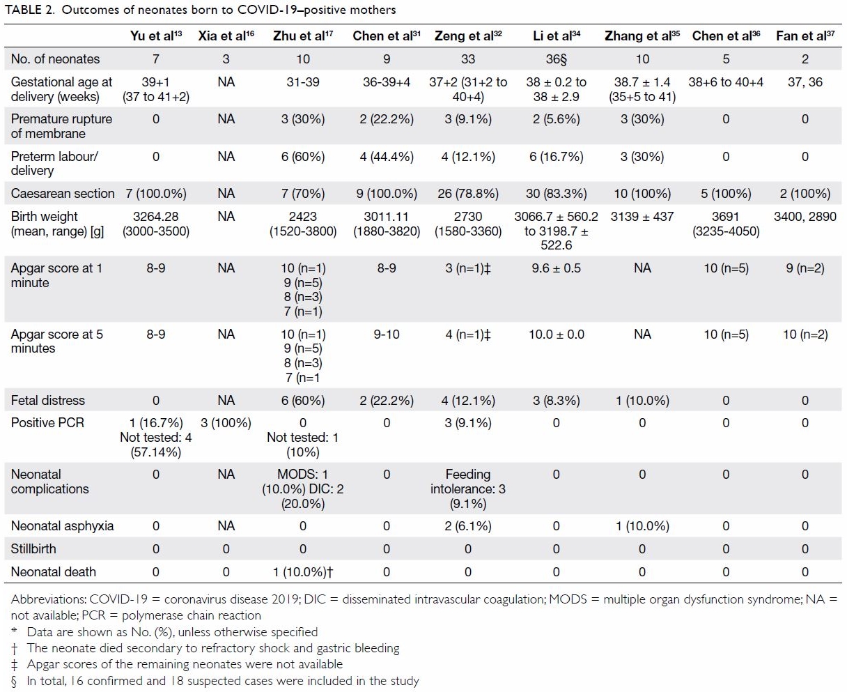
    50. Rendon KL, Wheeler V. Epidural analgesia and risk of Cesarean delivery. Am Fam Physician 2018 Nov 1;98:Online.
    51. Chapter 9: Guidelines for the provision of anaesthesia services (GPAS). Guidelines for the Provision of Anaesthesia Services for an Obstetric Population. Royal College of Anaesthetists; 2020: 9.
    52. Torvaldsen S, Roberts CL, Bell JC, Raynes-Greenow CH. Discontinuation of epidural analgesia late in labour for reducing the adverse delivery outcomes associated with epidural analgesia. Cochrane Database Syst Review 2004;(4):CD004457. Crossref
    53. Tuuli MG, Frey HA, Odibo AO, Macones GA, Cahill AG. Immediate compared with delayed pushing in the second stage of labor: a systematic review and meta-analysis. Obstet Gynecol 2012;120:660-8. Crossref
    54. Lee AI, McCarthy RJ, Toledo P, Jones MJ, White N, Wong CA. Epidural labor analgesia–fentanyl dose and breastfeeding success: a randomized clinical trial. Anesthesiology 2017;127:614-24. Crossref
    55. Orbach-Zinger S, Landau R, Davis A, et al. The effect of labor epidural analgesia on breastfeeding outcomes: a prospective observational cohort study in a mixed-parity cohort. Anesth Analg 2019;129:784-91. Crossref
    56. Chan JJ, Dabas R, Han RN, Sng BL. Fever during labour epidural analgesia. Trends in Anaesthesia and Crit Care 2018;20:21-5. Crossref
    57. Higgins RD, Saade G, Polin RA, et al. Evaluation and management of women and newborns with a maternal diagnosis of chorioamnionitis: summary of a workshop. Obstet Gynecol 2016;127:426-36. Crossref
    58. Sultan P, David AL, Fernando R, Ackland GL. Inflammation and epidural-related maternal fever: proposed mechanisms. Anesth Analg 2016;122:1546-53. Crossref
    59. Lim G, Facco FL, Nathan N, Waters JH, Wong CA, Eltzschig HK. A review of the impact of obstetric anesthesia on maternal and neonatal outcomes. Anesthesiology 2018;129:192-215. Crossref

    Congenital infections in Hong Kong: beyond TORCH

    Hong Kong Med J 2020 Aug;26(4):323–30  |  Epub 24 Jul 2020
    © Hong Kong Academy of Medicine. CC BY-NC-ND 4.0
     
    REVIEW ARTICLE
    Congenital infections in Hong Kong: beyond TORCH
    KL Hon, MB, BS, MD1; Karen KY Leung, MB, BS, MRCPCH1; Alexander KC Leung, FRCP (UK), FRCPCH2; Elim Man, MB, BS, MRCPCH1; Patrick Ip, FRCPCH, MPH3
    1 Department of Paediatrics and Adolescent Medicine, The Hong Kong Children’s Hospital, Hong Kong
    2 Department of Pediatrics, The University of Calgary, and The Alberta Children’s Hospital, Calgary, Alberta, Canada
    3 Department of Paediatrics and Adolescent Medicine, The University of Hong Kong, Hong Kong
     
    Corresponding author: Dr KL Hon (hon@cuhk.edu.hk)
     
     Full paper in PDF
     
    Abstract
    Congenital infections refer to a group of perinatal infections that are caused by pathogens transmitted from mother to child during pregnancy (transplacentally) or delivery (peripartum) which may have similar clinical presentations, including rash and ocular findings. TORCH is the acronym that covers these infections (toxoplasmosis, other [syphilis], rubella, cytomegalovirus, herpes simplex virus). Other important causes of intrauterine/perinatal infection include human immunodeficiency virus, varicella-zoster virus, Treponema pallidum, Zika virus, and parvovirus B19. This overview aims to describe various congenital infections beyond TORCH with a Hong Kong perspective. Intrauterine and perinatal infections are a major cause of in utero death and neonatal mortality, and an important contributor to childhood morbidity. A high index of suspicion for congenital infections and awareness of the prominent features of the most common congenital infections can help to facilitate early diagnosis, tailor appropriate diagnostic evaluation, and initiate appropriate early treatment. Intrauterine infections should be suspected in newborns with clinical features including microcephaly, seizures, cataract, hearing loss, congenital heart disease, hepatosplenomegaly, small for gestational age, and/or rash. Primary prevention of maternal infections during pregnancy is key to the prevention of congenital infection, and resources (if available) should focus on public health promotion and pre-marital counselling.


    Introduction
    Congenital infections are those that can cross the placenta and damage the fetus in utero or transmit to the infant during delivery in the peripartum period, resulting in neonatal infection.1 Congenital infections also account for 2% to 3% of all congenital anomalies apart from miscarriage, stillbirth, and neonatal deaths and are a significant cause of childhood morbidity.2 3 4 Immunologist Andres Nahmias first used the acronym ToRCH in 1971 to describe perinatal infections associated with toxoplasma (To), rubella (R), cytomegalovirus (C), and herpes simplex virus (H).5 Subsequently, the ‘O’ in TORCH has been broadened and now stands for ‘Others’, including the following pathogens: syphilis, parvovirus B19, coxsackievirus, listeriosis, hepatitis virus, varicella-zoster virus (VZV), Trypanosoma cruzi, enterovirus, human immunodeficiency virus (HIV), and the latest addition, Zika virus (ZIKV).1 4
     
    Congenital infections have remained a major global health issue, those in developing countries are particularly vulnerable. Congenital infections can lead to significant consequences, such as severe disabilities or even the death of the fetus. We recently overviewed “TORCH” in detail,6 and this overview focuses on the following congenital infections beyond TORCH, namely, HIV, VZV, ZIKV, and parvovirus B19. References were searched using key terms (“congenital infection”) and (“Hong Kong”) or (“Zika”) and (“Hong Kong”) in PubMed, limited to ‘human’, with no filters on article type or publication time. Discussion is based on but not limited to the search results.
     
    Congenital human immunodeficiency virus infection
    The HIV strains HIV-1 and HIV-2 are cytopathic lentiviruses belonging to the family Retroviridae.7 Paediatric HIV infection remains a significant global health issue: the Joint United Nations Programme on HIV/AIDS reported that an estimated 3.1 million children were living with HIV globally.8 Congenital infection with HIV can occur via the transplacental route to the developing fetus. Over 90% of HIV infections in children worldwide are caused by MTCT, which is one of the three general modes of HIV transmission.9
     
    The first case of HIV infection in Hong Kong was reported in 1984. The Department of Health has had a voluntary and anonymous HIV/AIDS reporting system since 1984, and according to its data, as of June 2019, there had been 35 reported perinatal HIV cases in Hong Kong. The incidence of HIV in Hong Kong is relatively low compared with that of other countries: the HIV seroprevalence in Hong Kong is <0.01% in the antenatal population.10
     
    Without any intervention or treatment, approximately 15% to 30% of infants born to HIV-positive women become infected with HIV via the transplacental route or during delivery, and a further 5% to 15% become infected through breastfeeding.8 In almost all newborns of HIV-positive mothers, HIV-specific antibodies are positive at birth. Thus, antibody assays are not informative for the diagnosis of infection in children younger than 24 months unless the assay results are negative.7 Serologic reversion has been reported to occur in a considerable number of children by age 18 months, and it has been reported up to 24 months of age.7 Diagnosis of congenital HIV infection can be confirmed by a positive polymerase chain reaction (PCR) test for HIV DNA or RNA. In this regard, HIV RNA PCR testing may be better than HIV DNA PCR testing for detection of HIV infection at birth.11 Cord blood should not be used for testing because of the possibility of contamination with maternal blood. Negative PCR tests at birth, 1 month, and 4 months of age are sufficient to exclude congenital HIV infection.
     
    Clinical manifestations of congenital HIV infection are diverse and often nonspecific and include lymphadenopathy, hepatosplenomegaly, microcephaly, oral candidiasis, and invasive bacterial infections.12 13 Recurrent pneumonia (notably Pneumocystis jirovecii pneumonia and lymphoid interstitial pneumonia), chronic diarrhoea, small size for date, failure to thrive, developmental delay, cardiomyopathy, encephalopathy, and nephropathy can also be seen.12 13 Recurrent VZV and herpes simplex virus infections, severe molluscum contagiosum, chronic fungal infections, and atopic dermatitis are common skin manifestations.14 15 Preconception counselling should be provided to women with HIV of childbearing age to discuss their reproductive desires. Promotion of safe sex and information about effective and appropriate contraceptive methods should be offered to avoid unplanned pregnancy. Alternatively, if pregnancy is desired, facilitating planned pregnancy is equally important, as HIV-positive couples could benefit from assisted reproduction.9
     
    To effectively prevent MTCT of HIV during pregnancy, the mother should receive antiretroviral (ARV) therapy during the antepartum, intrapartum, and postnatal periods. The treatment should be individualised with the goal of virologic suppression to undetectable levels.7 Intrapartum zidovudine is recommended if the pregnant woman with HIV presents late and has received no prior ARV therapy or if the HIV viral load is high (>50 copies/mL) close to delivery.16 Caesarean section is indicated if the mother’s viral load is >1000 copies/mL. For mothers with HIV infection who proceed to vaginal delivery, prolonged rupture of membranes, invasive fetal monitoring, and instrumental delivery should be avoided to reduce the risk of MTCT.16
     
    A paediatrician who is experienced in HIV management should be involved in the infant’s antenatal and post-delivery care. The exposed infant should begin with ARV prophylaxis soon after birth, preferably within 12 hours. If the mother has received ARV therapy during pregnancy, neonatal prophylaxis consists of zidovudine for 6 weeks after birth until HIV-negative status is confirmed. If the mother did not receive any ARV regimen or her HIV RNA level is high (≥1000 copies/mL) or unknown, neonatal prophylaxis to lower the rate of MTCT should consist of a two- or three-drug ARV regimen.7 Breastfeeding should be avoided if possible, as HIV transmission by breastfeeding accounts for one third to half of MTCT of HIV worldwide.7 17 Studies in the pre-ARV therapy era reported the postnatal transmission rate to be as high as 32% at 6 months.18 A Universal Antenatal HIV Testing Programme with an opt-out approach has been implemented in Hong Kong since 2001. The programme is widely accepted by antenatal women, and the opt-out rate has been <3%.9 However, despite the high coverage of ≥98%, five infants whose mothers were screened as negative by the Universal Antenatal HIV Testing Programme during the early antenatal period were found to be HIV-positive from 2009 to 2015. Hence, it was suspected the MTCT in those cases occurred in late pregnancy. Unprotected sex during pregnancy was the common risk factor among those cases, which reinforces the importance of advising protected sex during pregnancy.10 16 A second HIV test is recommended in the third trimester for patients at risk of HIV infection or if there are symptoms and signs compatible with acute HIV infection.16 If the mother’s HIV status is unknown at delivery, a highly sensitive and specific rapid HIV test should be performed.19 Prophylactic interventions against MTCT should be implemented if the result is positive.16 With the implementation of universal prenatal screening, effective ARV therapy for all pregnant women with HIV, scheduled Caesarean delivery for cases with high viral load, ARV for infants, and avoidance of breastfeeding, the rate of MTCT is <1%.20 21 This highlights the importance of universal screening and antenatal care that vertical transmission of HIV is potentially preventable when these interventions are implemented. Management of HIV in mothers and newborns has improved outcomes over the past 20 years.
     
    Congenital varicella syndrome
    Varicella-zoster virus is a member of the herpesvirus family. Pregnant women are several times more likely to develop fatal VZV than non-pregnant ones. Although rare, the fetus is at high risk of congenital varicella syndrome (CVS) if the mother is infected, and the neonate is at high risk of a severe or fatal form of VZV.22
     
    Varicella is the most frequently reported notifiable disease in Hong Kong.23 More than 95% of the population not vaccinated against VZV has been infected, mostly before 20 years of age.24 Approximately 95% of pregnant women were found to be seropositive for VZV in a 2009 study.25 With the vast majority of the population immune to VZV at childbearing age, the risk of primary infection during pregnancy (and thus the risk of congenital VZV infection) is relatively low. A worldwide systemic review of the literature from 1947 to 2013 indicated that there were only 130 reported cases of congenital VZV infection in that period.26 Only two cases of neonatal VZV infection were reported in Hong Kong from 2008 to 2010.27 Transmission of VZV can occur via multiple routes, including infected respiratory tract secretions, direct contact with infectious vesicular fluid from moist lesions, and occasionally through airborne spread, entering the host through the conjunctiva or the mucosal surface of the upper respiratory tract.7 28 Varicella is highly contagious from 1 to 2 days before the onset of the rash, until all lesions are crusted.7 28 Transplacental passage of VZV from maternal infection can result in congenital or neonatal VZV infection.7 28 The risk of vertical transmission reaches 25% if maternal primary infection occurs during the first or second trimesters, with around 12% risk of congenital anomalies among newborns of mothers who were infected during that period.29 The rate of vertical transmission reaches up to 50% when maternal VZV infection occurs within 1 to 4 weeks before delivery, with 23% developing neonatal VZV infection.30 31
     
    Fetal VZV infection in utero can result in a range of adverse sequelae. Low birth weight is universal, and intrauterine growth restriction is observed in around 23% of cases.30 Further, approximately 20% of infants with in utero VZV infection develop neonatal or infantile herpes zoster.30 Congenital anomalies resulting from congenital VZV infection are known collectively as CVS, which is expected in approximately 12% of infected fetuses.29 Maternal infection within the first 20 weeks of gestation is responsible for most cases of CVS, with newborns of mothers infected between the 7th and 20th weeks having the highest risk.28 No cases of CVS have been reported when maternal varicella infection occurs after the 28th week of gestation.32 The clinical manifestations of CVS include cutaneous scars in a dermatomal distribution, limb hypoplasia, microcephaly, cortical atrophy, hydrocephaly, mental retardation, microphthalmia, chorioretinitis, cataracts, muscle hypoplasia, developmental delay, and anomalies of the cardiovascular system, gastrointestinal tract, and genitourinary tract.7 30 33 34 During the first few months of life, CVS has a 30% mortality rate.30 Neonatal VZV infection can result from transplacental passage of VZV and ascending or postnatal infections. If VZV infection occurs during the first 10 to 12 days of life, it is often the result of intrauterine transmission, whereas VZV with later onset is usually acquired postnatally.27 30 Appropriate treatment with varicella-zoster immunoglobulin (VZIG) and antivirals has reduced the mortality rate of neonatal VZV to 7%.31
     
    Prenatal diagnosis of CVS is often based on detection of VZV DNA in the amniotic fluid or fetal blood by PCR. In addition, ultrasonography findings consistent with CVS, such as microcephaly, limb deformities, polyhydramnios, soft tissue calcification, and intrauterine growth restriction can be diagnostic when there is a history of maternal VZV infection.19 30 An ultrasound examination should be performed at least 5 weeks after the onset of rash in the mother.30
     
    Congenital varicella syndrome is diagnosed postnatally when there is history of maternal VZV during pregnancy, in the presence of skin lesions distributed along dermatome(s), and in the presence of other clinical manifestations consistent with CVS or neonatal seizures.35 The presence of VZV DNA in the newborn, detection of immunoglobulin M antibodies against VZV in the cord blood or fetal blood, immunoglobulin G antibodies against VZV persisting longer than the first 7 months of life, or development of herpes zoster during early infancy indicate intrauterine VZV infection regardless of CVS development.30
     
    Oral acyclovir with or without VZIG is recommended for pregnant women infected with varicella, as it can shorten the duration of fever and reduce the symptoms of varicella when antiviral therapy is initiated within 24 hours after the onset of rash.28 36 Acyclovir is effective at lowering the morbidity and mortality rates of both the fetus and the mother when administered within 24 and up to 72 hours of the onset of rash, and VZIG has been reported to lower the incidence and severity of varicella.30 A 10-day course of intravenous acyclovir is effective in neonates affected by varicella and complications of CVS.7 Prophylactic use of VZIG is also possible in susceptible pregnant mothers exposed to VZV within 72 and up to 96 hours of exposure but is not effective after clinical signs have appeared.37 Administration of VZIG in neonates should occur within 10 days of initial exposure for prophylaxis if the mother has perinatal varicella rash and absence of antibodies against VZV in the mother or the neonate.7 In women of childbearing age who are not immune to VZV, vaccination after exposure can effectively prevent infection, especially when administered within 3 days of exposure.38
     
    Vaccination against VZV was incorporated into the Hong Kong Childhood Immunisation Programme (HKCIP) in 2014.39 For eligible children born on or after 1 January 2013, the first vaccination is given at age 12 months, and the second dose (at around age 6-7 years) in primary one, given as part of the measles mumps, rubella, and varicella vaccine. Before being incorporated into the HKCIP, a varicella vaccine was available on the private market. A local study conducted in 2012 found that the parent-reported VZV vaccination uptake was 65% in kindergarten students, and the rate was 69% of preschool and schoolchildren in another study conducted in 2013.40 41 The vaccination rate is expected to rise after incorporation of the varicella vaccine into the HKCIP, as coverage of all other vaccines in the HKCIP has been maintained at over 95%. Because the incidence of varicella in younger children is likely to drop because of increased immunisation, the effects of the vaccination on incidence during pregnancy remain to be elucidated.
     
    Congenital Zika virus infection
    Zika virus is a neurotropic flavivirus that particularly infects neural progenitor cells. Approximately 80% of ZIKV infection is asymptomatic.42 Symptomatic infections can be categorised as either Zika fever or congenital Zika syndrome. Zika fever is a relatively mild disease that presents with low-grade fever, generalised maculopapular rash, non-purulent conjunctivitis, myalgia, and arthralgia of the small joints of the hands and feet, although more severe complications, including Guillain-Barre syndrome, seem to be associated as well.42 The disease is usually self-limiting and resolves within 3 to 7 days.42 Zika virus infection during pregnancy is associated with a higher rate of fetal loss, including stillbirths. In addition, serious sequelae can be observed in infants with congenital Zika syndrome, making it an important public health concern in some areas.
     
    Congenital Zika syndrome raised international public health concern after the increase in cases of congenital microcephaly in neonates in October 2015 following an outbreak of ZIKV infection in Brazil. Fortunately, since ZIKV infection became a notifiable disease in February 2016, the number of cases in Hong Kong has remained low, with only two cases in 2016 and one case in 2017.23 Therefore, the risk of congenital Zika syndrome is extremely low in Hong Kong.
     
    Transmission of ZIKV primarily occurs via infected Aedes mosquitoes. It may also be transmitted through sexual contact, blood transfusion, and organ transplantation.43 44 Vertical transmission through the placenta is a major concern because of the resulting congenital Zika syndrome. A report by the United States Zika Pregnancy Registry found congenital defects in 10% of 250 cases with laboratory-confirmed ZIKV infection during pregnancy.45 46
     
    Neurological findings seen in congenital Zika syndrome include microcephaly, cutis gyrata, ventriculomegaly, subcortical calcifications, polymalformative syndrome (including craniofacial disproportion and craniosynostosis), brainstem dysfunction, developmental delay, sensorineural hearing loss, seizures, marked hypertonia, hyperreflexia, and dysphagia.45 47 Ophthalmological findings include cataracts, asymmetrical ocular size, intraocular calcifications, macular abnormalities (pigmentary retinal mottling, chorioretinal atrophy/scarring), optic nerve abnormalities (optic nerve hypoplasia, increased cup-to-disk ratio), subretinal haemorrhage, coloboma, microcornea, microphthalmia, cataracts, glaucoma, and lens subluxation.48 49 50 Congenital heart disease occurs in 10% to 15% of cases.51 Although the full spectrum of the syndrome has not been completely delineated, a review study identified some characteristic features of congenital Zika syndrome, including severe microcephaly with partially collapsed skull, thin cerebral cortices with subcortical calcifications, macular scanning, focal pigmentary retinal mottling, congenital limb contractures, and marked early hypertonia with signs of extrapyramidal involvement.52 In one study of 117 liveborn infants born to pregnant women with confirmed ZIKV infection, the overall rate of functional and structural abnormalities was 42%.53
     
    Definitive diagnosis of congenital ZIKV infection can only be established within the first few days of life by detecting ZIKV RNA in the serum, urine, or cerebrospinal fluid via a real-time reverse transcription PCR nucleic acid test.54 Detection of immunoglobulin M antibodies against ZIKV in the serum or CSF by enzyme-linked immunosorbent assay can support the diagnosis.54 Because there is currently no effective antiviral treatment for ZIKV infection, supportive treatment is mainly offered to manage the complications of congenital Zika syndrome. Continuous healthcare support and referral to developmental specialists to monitor the condition are recommended.54
     
    As no vaccine against ZIKV is currently available, avoidance of ZIKV infection by personal protection against mosquito bites and vector control remains the most effective means to prevent congenital Zika syndrome. Pregnant mothers should avoid travelling to places with known ZIKV transmission by mosquitoes and maintain mosquito prevention practices, such as removing mosquitoes’ breeding grounds, wearing long-sleeved clothing, and using mosquito repellents. Protective measures against possible sexual transmission should also be adopted to minimise the risk of ZIKV infection during pregnancy.
     
    Parvovirus B19 infection
    Parvovirus B19 is the only pathogenic human parvovirus. In Hong Kong, the prevalence of antibodies to the B19 virus has been reported as 20%, which is much lower than the corresponding value for the Western population, where >60% of adults have been reported to be seropositive.55 56 The epidemiology of parvovirus B19 infection during pregnancy in Hong Kong is unknown, but it is likely to have low prevalence in light of the low population prevalence rate.57 Only three local case reports of hydrops fetalis due to parvovirus B19 were found in the literature.57 58 59
     
    Parvovirus B19 infection in adults is usually asymptomatic.60 Parvovirus infection in pregnant women is associated with hydrops fetalis because it causes severe fetal anaemia, sometimes leading to miscarriage or stillbirth.60 61 62 This is caused by a combination of haemolysis of red blood cells and the virus directly negatively affecting red blood cell precursors in the bone marrow. The risk of fetal loss is about 10% if infection occurs before week 20 of pregnancy (especially between weeks 14 and 20) but minimal after then. In contrast, parvovirus B19 infection during the second trimester is more commonly associated with hydrops fetalis or fetal loss. If the fetus does not develop any of these acute complications, or if intrauterine blood transfusion is successful in saving the fetus, the risk to the fetus presented by chronic parvovirus B19 infection is negligible.
     
    Knowledge of the mother’s parvovirus B19 immune status could allow her to avoid contact with individuals suspected or known to have ongoing parvovirus B19 infection. However, antenatal immunity testing is not currently recommended, as there is no effective means to prevent infection, no specific therapy, and no vaccines available. Thus, testing may increase maternal anxiety and fear without proven benefit.
     
    The best approach would be to recommend that all pregnant women avoid contact with children who currently have symptoms of parvovirus B19 infection. However, if a pregnant woman is exposed to parvovirus B19, serological testing should be performed as soon as possible to determine whether she should be monitored for seroconversion.63 If the results are suggestive of an acute parvovirus B19 infection, the fetus should be monitored by regular ultrasound assessment for signs of fetal hydrops and fetal anaemia. The peak systolic velocity of the fetal middle cerebral artery is an accurate predictor of fetal anaemia,63 and intrauterine blood transfusions can be considered in severe cases. Although some case reports have suggested that intrauterine parvovirus B19 infection caused developmental abnormalities in childhood, epidemiologic studies have not supported this association.64 65 The bulk of the available data suggest that parvovirus B19 is not teratogenic.19 61 No human vaccine against parvovirus B19 has been approved to date.66 67
     
    Conclusion
    Although Hong Kong still has relatively low rates of congenital infections among countries in more developed regions despite being densely populated, it is important to remain vigilant against any possible infections during pregnancy, which may lead to severe morbidities and mortality of the fetus or infant.
     
    The antenatal screening programme in Hong Kong is tailored to the local epidemiology of infectious and hereditary diseases, aiming to detect significant diseases that are potentially damaging to the fetus as early as possible. At present, the congenital infections for which the programme screens include rubella, hepatitis B, syphilis, and HIV, and the hereditary diseases screened for are thalassemia and Down syndrome.
     
    Health authorities (eg, the Centre for Health Protection), obstetricians, and paediatricians should collaborate to establish a central congenital infection disease registry. This registry could be used for disease surveillance and monitoring of the outcomes of congenital infections. These results would be useful for detection of disease clusters and determination of their prevalence, morbidity, and mortality. Such a registry would help to estimate the burden of these congenital infections on the healthcare system and guide resource allocation.
     
    The data for such a registry could be captured from the notifiable disease database and laboratory surveillance of antenatal blood samples. Congenital rubella syndrome and Zika virus are notifiable diseases in Hong Kong, as their risk of transmission is high, outbreaks of these diseases can impose a significant risk to the community, and public health measures can be implemented if an outbreak is detected early. In terms of other congenital infections, the universal antenatal screening programme in Hong Kong should be able to identify most cases of congenital syphilis, rubella, and HIV. If recourses are available, health authorities can consider including congenital HIV and congenital syphilis as notifiable diseases. At present, the Hong Kong Department of Health has a surveillance programme intended to detect most congenital HIV and syphilis infections.68 Mothers with herpes simplex virus who are symptomatic should present during an antenatal visit. Toxoplasmosis and cytomegalovirus are not part of the antenatal screening programme, but serological tests can be performed if there is suspicion of congenital infection.
     
    A congenital infection registry could act as a platform to provide information for disease surveillance. Public health promotion for primary prevention of maternal infections during pregnancy is the key to avoiding congenital infections.
     
    Author contributions
    Concept or design: KL Hon.
    Acquisition of data: KL Hon, KKY Leung.
    Analysis or interpretation of data: KL Hon, KKY Leung.
    Drafting of the manuscript: KL Hon, KKY Leung.
    Critical revision of the manuscript for important intellectual content: All authors.
     
    All authors had full access to the data, contributed to the study, approved the final version for publication, and take responsibility for its accuracy and integrity.
     
    Conflicts of interest
    As an editor of the journal, KL Hon was not involved in the peer review process. Other authors have disclosed no conflicts of interest.
     
    Funding/support
    This research received no specific grant from any funding agency in the public, commercial, or not-for-profit sectors.
     
    References
    1. DeVore NE, Jackson VM, Piening SL. Torch infections. Am J Nurs 1983;83:1660-5. Crossref
    2. Neu N, Duchon J, Zachariah P. TORCH infections. Clin Perinatol 2015;42:77-103, viii. Crossref
    3. Stegmann BJ, Carey JC. TORCH infections. Toxoplasmosis, other (syphilis, varicella-zoster, parvovirus B19), rubella, cytomegalovirus (CMV), and herpes infections. Curr Womens Health Rep 2002;2:253-8.
    4. Schwartz DA. The origins and emergence of Zika virus, the newest TORCH infection: what’s old is new again. Arch Pathol Lab Med 2017;141:18-25. Crossref
    5. Nahmias AJ, Walls KW, Stewart JA, Herrmann KL, Flynt WJ. The ToRCH complex-perinatal infections associated with toxoplasma and rubella, cytomegol- and herpes simplex viruses. Pediatr Res 1971;5:405-6. Crossref
    6. Leung KK, Hon KL, Yeung A, Leung AK, Man E. Congenital infections in Hong Kong: an overview of TORCH. Hong Kong Med J 2020;26:127-38. Crossref
    7. Kimberlin DW, Brady MT, Jackson Mam Long SS, editors. Red Book 2018: Report of the Committee on Infectious Diseases. Itasca: American Academy of Pediatrics; 2018.
    8. World Health Organization. Global guidance on criteria and processes for validation: elimination of mother-to- child transmission of HIV and syphilis. 2nd ed. 2017. Available from: https://www.who.int/reproductivehealth/ publications/emtct-hiv-syphilis/en/. Accessed 3 Oct 2019.
    9. Wong KH, Chan KC, Bognar FA. An update on HIV and pregnancy. Hong Kong J Gynaecol Obstet Midwifery 2007;7:48-52.
    10.Red Ribbon Centre, Centre for Health Protection, Department of Health, Hong Kong SAR Government. Wong CK, Lin WC, Wong KH. Closing the gap of perinatal HIV infection in Hong Kong. Available from: https://www. rrc.gov.hk/research/ab201701.pdf. Accessed 4 Oct 2019.
    11. Lilian RR, Kalk E, Bhowan K, et al. Early diagnosis of in utero and intrapartum HIV infection in infants prior to 6 weeks of age. J Clin Microbiol 2012;50:2373-7. Crossref
    12. Hegde HR, Leung AK, Robson WL. Anergy and AIDS. J Acquir Immune Defic Syndr 1988;4:1004.
    13. Hon KL, Leung TF, Yau YS. Pneumonitis and extreme failure to thrive. Hong Kong Med J 2014;20:556.e1-3. Crossref
    14. Leung AK, Barankin B, Hon KL. Molluscum contagiosum: an update. Recent Pat Inflamm Allergy Drug Discov 2017;11:22-31. Crossref
    15. Leung AK, Hon KL, Robson WL. Atopic dermatitis. Adv Pediatr 2007;54:241-73. Crossref
    16.Virtual AIDS Office of Hong Kong, Department of Health, Hong Kong SAR Government. Recommended clinical guidelines on the prevention of perinatal HIV transmission. 2018. Available from: https://www.aids.gov.hk/ice/ice201812.pdf. Accessed 4 Oct 2019.
    17. Leung AK, Sauve RS. Breast is best for babies. J Natl Med Assoc 2005;97:1010-9.
    18. Bispo S, Chikhungu L, Rollins N, Siegfried N, Newell ML. Postnatal HIV transmission in breastfed infants of HIV-infected women on ART: a systematic review and meta-analysis. J Int AIDS Soc 2017;20:21251. Crossref
    19. Mendelson E, Aboudy Y, Smetana Z, Tepperberg M, Grossman Z. Laboratory assessment and diagnosis of congenital viral infections: rubella, cytomegalovirus (CMV), varicella-zoster virus (VZV), herpes simplex virus (HSV), parvovirus B19 and human immunodeficiency virus (HIV). Reprod Toxicol 2006;21:350-82. Crossref
    20. Nesheim SR, Wiener J, Fitz Harris LF, Lampe MA, Weidle PJ. Brief report: estimated incidence of perinatally acquired HIV infection in the United States, 1978-2013. J Acquir Immune Defic Syndr 2017;76:461-4. Crossref
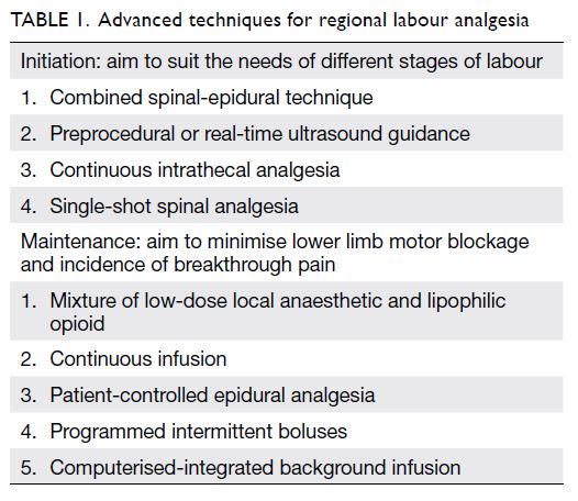
    21. Peters H, Francis K, Sconza R, et al. UK mother-to-child HIV transmission rates continue to decline: 2012-2014. Clin Infect Dis 2017;64:527-8. Crossref
    22. Narkeviciute I, Bernnatoniene J. Varicella zoster virus infection in pregnancy. In: Magel GD, Tyring SK, editors. Herpesviridae: a look into this unique family of viruses. 2012. Available from: https://www.intechopen.com/books/ herpesviridae-a-look-into-this-unique-family-of-viruses/ varicella-zoster-virus-infection-in-pregnancy. Accessed 4 Oct 2019. Crossref
    23. Centre for Health Protection, Department of Health, Hong Kong SAR Government. Number of notifiable infectious diseases by month. Available from: https://www.chp.gov. hk/en/static/24012.html. Accessed 3 Oct 2019.
    24. Banz K, Wagenpfeil S, Neiss A, et al. The cost-effectiveness of routine childhood varicella vaccination in Germany. Vaccine 2003;21:1256-67. Crossref
    25. Fung LW, Lao TT, Suen SS, et al. Seroprevalence of varicella zoster virus among pregnant women in Hong Kong: comparison with self-reported history. Vaccine 2011;29:8186-8. Crossref
    26. Ahn KH, Park YJ, Hong SC, et al. Congenital varicella syndrome: a systematic review. J Obstet Gynaecol 2016;36:563-6. Crossref
    27. Hon KL, Leung TF, Cheung HM, Chan PK. Neonatal herpes: what lessons to learn. Hong Kong Med J 2012;18:60- 2.
    28. Wilson CB, Nizet V, Maldonado Y, Remington JS, Klein JO. Remington and Klein’s infectious diseases of the fetus and newborn infant. 8th ed. Philadelphia: Elsevier; 2016: 513,520-5,675,677-9,685-90,693-4,729,732.
    29. Prober CG, Gershon AA, Grose C, McCracken GH Jr, Nelson JD. Consensus: varicella-zoster infections in pregnancy and the perinatal period. Pediatr Infect Dis J 1990;9:865-9. Crossref
    30. Lamont RF, Sobel JD, Carrington D, et al. Varicella-zoster virus (chickenpox) infection in pregnancy. BJOG 2011;118:1155-62. Crossref
    31. Miller E, Cradock-Watson JE, Ridehalgh MK. Outcome in newborn babies given anti-varicella-zoster immunoglobulin after perinatal maternal infection with varicella-zoster virus. Lancet 1989;2:371-3. Crossref
    32. Tan MP, Koren G. Chickenpox in pregnancy: revisited. Reprod Toxicol 2006;21:410-20. Crossref
    33. Sauve RS, Leung AK. Congenital varicella syndrome with colonic atresias. Clin Pediatr (Phila) 2003;42:451-3. Crossref
    34. Leung AK, Sauve RS. Colonic atresia and associated anomalies. J Paediatr Child Health 2003;39:239. Crossref
    35. Mustonen K, Mustakangas P, Valanne L, Professor MH, Koskiniemi M. Congenital varicella-zoster virus infection after maternal subclinical infection: clinical and neuropathological findings. J Perinatol 2001;21:141-6. Crossref
    36. Wallace MR, Bowler WA, Murray NB, Brodine SK, Oldfield EC 3rd. Treatment of adult varicella with oral acyclovir. A randomized, placebo-controlled trial. Ann Intern Med 1992;117:358-63. Crossref
    37. Ogilvie MM. Antiviral prophylaxis and treatment in chickenpox. A review prepared for the UK Advisory Group on Chickenpox on behalf of the British Society for the Study of Infection. J Infect 1998;36 Suppl 1:31-8. Crossref
    38. Watson B, Seward J, Yang A, et al. Postexposure effectiveness of varicella vaccine. Pediatrics 2000;105:84-8. Crossref
    39. Centre for Health Protection, Department of Health, Hong Kong SAR Government. Recommendations on the use of varicella (chickenpox) vaccine in childhood immunisation programme. Available from: https://www.chp.gov.hk/files/pdf/recommendations_on_the_use_of_varicella_vaccine_in_cip_r.pdf. Accessed 4 Oct 2019.
    40. Chan JY, Leung KM, Tam WW, Lee A. Varicella vaccine uptake and associated factors in children in Hong Kong. Epidemiol Infect 2014;142:994-1001. Crossref
    41. Tam WW, Chan J, Lo KK, et al. Parental attitudes and factors associated with varicella vaccination in preschool and schoolchildren in Hong Kong: a cross-sectional study. Med (Baltimore) 2015;94:e1519. Crossref
    42. Duffy MR, Chen TH, Hancock WT, et al. Zika virus outbreak on Yap Island, Federated States of Micronesia. N Engl J Med 2009;360:2536-43. Crossref
    43. Musso D, Nhan T, Robin E, et al. Potential for Zika virus transmission through blood transfusion demonstrated during an outbreak in French Polynesia, November 2013 to February 2014. Euro Surveill 2014;19:1-3. Crossref
    44. Prisant N, Bujan L, Benichou H, et al. Zika virus in the female genital tract. Lancet Infect Dis 2016;16:1000-1. Crossref
    45. Reynolds MR, Jones AM, Petersen EE, et al. Vital signs: update on Zika virus–associated birth defects and evaluation of all U.S. infants with congenital Zika virus exposure—U.S. Zika pregnancy registry, 2016. MMWR Morb Mortal Wkly Rep 2017;66:366-73. Crossref
    46. Chan JF, Choi GK, Yip CC, Cheng VC, Yuen KY. Zika fever and congenital Zika syndrome: an unexpected emerging arboviral disease. J Infect 2016;72:507-24. Crossref
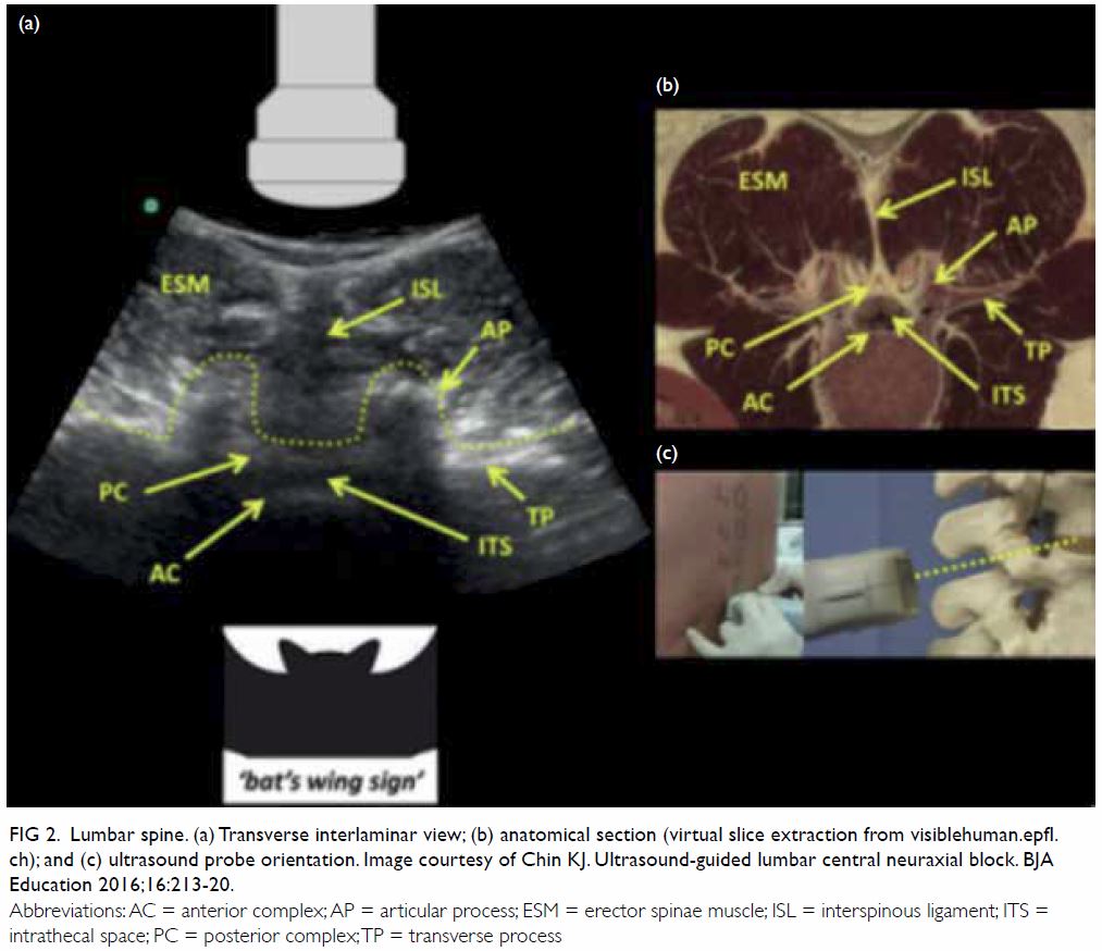
    47. Pan American Health Organization, World Health Organization. Neurological syndrome, congenital malformations, and Zika virus infection. Implications for public health in the Americas. 2015: 1-8. Available from: https://www.paho.org/hq/dmdocuments/2015/2015-dec- 1-cha-epi-alert-zika-neuro-syndrome.pdf. Accessed 5 Oct 2019.
    48. Ventura CV, Maia M, Bravo-Filho V, Góis AL, Belfort R Jr. Zika virus in Brazil and macular atrophy in a child with microcephaly. 2016;387:228. Crossref
    49. Ventura CV, Maia M, Ventura BV, et al. Ophthalmological findings in infants with microcephaly and presumable intra-uterus Zika virus infection. Arq Bras Oftalmol 2016;79:1-3. Crossref
    50. de Paula Freitas B, de Oliveira Dias JR, Prazeres J, et al. Ocular findings in infants with microcephaly associated with presumed Zika virus congenital infection in Salvador, Brazil. JAMA Ophthalmol 2016;134:529-35. Crossref
    51. Orofino DH, Passos SR, de Oliveira RV, et al. Cardiac findings in infants with in utero exposure to Zika 2018;12:e0006362. Crossref
    52. Moore CA, Staples JE, Dobyns WB, et al. Characterizing the pattern of anomalies in congenital Zika syndrome for pediatric clinicians. JAMA Pediatr 2017;171:288-95. Crossref
    53. Brasil P, Pereira JP Jr, Moreira ME, et al. Zika virus infection in pregnant women in Rio de Janeiro. N Engl J Med 2016;375:2321-34. Crossref
    54. Adebanjo T, Godfred-Cato S, Viens L, et al. Update: interim guidance for the diagnosis, evaluation, and management of infants with possible congenital Zika virus infection-United States, October 2017. MMWR Mork Mortal Wkly Rep 2017;66:1089-99. Crossref
    55. Kuo SH, Lin LI, Chang CJ, Liu YR, Lin KS, Cheng AL. Increased risk of parvovirus B19 infection in young adult cancer patients receiving multiple courses of chemotherapy. J Clin Microbiol 2002;40:3909-12. Crossref
    56. Lim WL, Wong KF, Lau CS. Parvovirus B19 infection in Hong Kong. J Infect 1997;35:247-9. Crossref
    57. Chan PK, To KF, Yip SK, Tam JS, Cheng AF. Parvovirus B19-associated hydrops fetalis: the first confirmed case in Hong Kong. Hong Kong Med J 1998;4:321-3.
    58. Ng PS, Leung TY, Leung TN, Lau TK. In utero treatment of hydrops fetalis due to parvovirus B19 infection: a case report. Hong Kong J Gynaecol Obstet Midwifery 2000;1:67-70.
    59. Leung TN, Leung TY, Chan PK, Fung TY, Lau TK. Fetal parvovirus B19 inferction in a twin pregnancy with 1 twin presenting with hydrops fetalis and the other asymptomatic: a case report. J Reprod Med 2007;52:419-21.
    60. Servey JT, Reamy BV, Hodge J. Clinical presentations of parvovirus B19 infection. Am Fam Physician 2007;75:373-6.
    61. Ergaz Z, Ornoy A Parvovirus B19 in pregnancy. Reprod Toxicol 2006;21:421-35. Crossref
    62. Ornoy A, Ergaz Z. Parvovirus B19 infection during pregnancy and risks to the fetus. Birth Defects Res 2017;109:311-23. Crossref
    63. American College of Obstetricians and Gynaecologists. Practice Bulletin No. 151: Cytomegalovirus, parvovirus B19, varicella zoster, and toxoplasmosis in pregnancy [editorial]. Obstet Gynecol 2016;127:405. Crossref
    64. Nagel HT, de Haan TR, Vandenbussche FP, Oepkes D, Walther FJ. Long-term outcome after fetal transfusion for hydrops associated with parvovirus B19 infection. Obstet Gynecol 2007;109:42-7. Crossref
    65. Kinney JS, Anderson LJ, Farrar J, et al. Risk of adverse outcomes of pregnancy after human parvovirus B19 infection. J Infect Dis 1988;154:663-7. Crossref
    66. Chandramouli S, Medina-Selby A, Coit D, et al. Generation of a parvovirus B19 vaccine candidate. Vaccine 2013;31:3872-8. Crossref
    67. Hon KL, Leung KK. Health advice for counselling: would parvovirus infection in puppies affect children and women? N Z Med J 2020;133:129-30.
    68. Centre for Health Protection, Department of Health, Hong Kong SAR Government. HIV/AIDS Surveillance Report 2015. 2016. Available from: https://www.chp.gov.hk/files/ pdf/aids15.pdf. Accessed 8 Oct 2019.

    Preservation of fertility in premenopausal patients with breast cancer

    Hong Kong Med J 2020 Jun;26(3):216–26  |  Epub 28 May 2020
    © Hong Kong Academy of Medicine. CC BY-NC-ND 4.0
     
    REVIEW ARTICLE
    Preservation of fertility in premenopausal patients with breast cancer
    Samuel SY Wang, Bmed MD1; Herbert Loong, MB, BS, MRCP2; Jacqueline PW Chung, MB, ChB, MRCOG3; Winnie Yeo, MB, BS, FRCP (Lond)4
    1 Prince of Wales Clinical School, Faculty of Medicine, University of New South Wales, Sydney, Australia
    2 Faculty of Medicine, Department of Clinical Oncology, Prince of Wales Hospital, The Chinese University of Hong Kong, Hong Kong
    3 Assisted Reproductive Technology Unit, Department of Obstetrics and Gynaecology, Prince of Wales Hospital, The Chinese University of Hong Kong, Hong Kong
    4 Department of Clinical Oncology, State Key Laboratory of Translational Oncology, Faculty of Medicine, The Chinese University of Hong Kong, Hong Kong
     
    Corresponding author: Dr Samuel SY Wang (z3424197@unsw.edu.au)
     
     Full paper in PDF
     
    Abstract
    Introduction: Cancer survivorship is increasingly important with advances in cancer therapeutics. Minimisation of treatment-related morbidity is an area that requires attention. This situation is most pressing in premenopausal patients with breast cancer, in whom advances in hormonal and targeted therapies have improved mortality rates. However, treatment-related infertility is still poorly addressed, and in East Asia, there is limited discussion regarding management of treatment-related infertility.
     
    Methods: A search of the literature was conducted using PubMed, Google Scholar, and Science Direct using the terms “breast cancer”, “fertility preservation”, “oocyte and embryonic cryopreservation”, “GnRH-a co-administration”, “ovarian tissue cryopreservation and transplantation”, “Japan”, “China”, “Korea”, and ‘Singapore”. Only studies published in English from 1980-2019 were included. The focus of the review was on identifying the current fertility preservation methods available to premenopausal women with breast cancer and the barriers that impede access.
     
    Results: Fertility preservation options include GnRH-a co-administration to minimise treatment-associated infertility, oocyte and embryonic cryopreservation, and emerging treatments such as ovarian tissue cryopreservation and transplantation. In East Asia, the uptake of fertility preservation options has been limited despite it being a major patient concern. A lack of awareness of fertility preservation treatments hinders discussion between patients and clinicians about fertility preservation.
     
    Conclusion: Despite progress in fertility preservation technologies, their impact for patients will be minimal if there is a lack of awareness/use of the technology. This review aims to raise awareness of such technologies among clinicians, enabling discussion between patients and clinicians about fertility preservation options.


    Introduction
    Recently, survival among patients with breast cancer has significantly improved. With better understanding of the disease’s diverse biology and increased availability of treatments, the 5-year survival rate for women diagnosed with breast cancer has increased from 75.2% between 1975 and 1977 to 88.2% between 2001 and 2008, leading to a substantial increase in breast cancer survivors.1 More strikingly, over 10% of breast cancer cases occur in women under age 45 years. Among these premenopausal survivors, 50% or more will live 20 years or longer following diagnosis. Thus, there is a need to address survivorship issues pertaining to long-term toxicity associated with breast cancer treatment.1 A study showed that 20 038 premenopausal women are diagnosed with breast cancer annually in the United States, with an estimated 96% (19 416) of these premenopausal patients at risk of infertility because of chemotherapy or hormonal therapy.1 Following chemotherapy, the reported incidence of amenorrhea varies between 40% and 68%.2 The agents most responsible for inducing amenorrhoea and premature ovarian failure (POF) are alkylating agents such as cyclophosphamide, whereas antimetabolites have a lesser effect.2 A brief summary of the association between infertility risk and type of chemotherapy can be seen in Table 1.3 4
     

    Table 1. List of common chemotherapeutic agents used in breast cancer and the risk of amenorrhoea3 4
     
    Apart from chemotherapy, hormonal modulation with tamoxifen is beneficial for hormone receptor–positive disease, which accounts for 70% of breast cancers. A 5-year course of tamoxifen reduces recurrence by 47% and mortality by 26%.5 However, tamoxifen is teratogenic, and pregnancy is contra-indicated during treatment; hence, pregnancy is often postponed. Although tamoxifen may not directly damage the ovaries, several studies have reported its association with higher rates of treatment-related amenorrhea, particularly after age 40 years.5 It is likely that tamoxifen’s length of therapy may indirectly contribute to infertility alongside age-related fertility decline.5 Apart from difficulties with conception, premenopausal patients with breast cancer also experience high rates of spontaneous abortion (29%) and premature deliveries with low birth weight (40%).2
     
    Minimising cancer treatment–associated infertility is especially important for premenopausal women who have not yet established a family. Female infertility in premenopausal women can cause these women great distress by preventing them from achieving the important life and social goal of motherhood. A recent study ranks this among the top five concerns among patients with premenopausal breast cancer.6 Moreover, in addition to impacting patients’ mental health, the fear of infertility also impacts treatment compliance.6 Of all patients, 29% do not comply with treatment because of treatment-associated infertility fears, which impacts patients’ prognosis and life expectancy.7 8 9
     
    While these fears are increasingly recognised by doctors, recent literature suggests that 32% of patients do not recall discussing fertility with their doctors. This lack of communication could be caused by a lack of awareness or confidence discussing fertility management in the context of breast cancer. This phenomenon was highlighted by a finding that 37% of oncologists feared delaying chemotherapy for fertility preservation, and 49% of oncologists were concerned about pregnancy safety after breast cancer treatment.8 10 A Hong Kong study showed that only 45.6% of clinicians from diverse fields such as clinical oncology, haematology, obstetrics and gynaecology, paediatrics, and surgery were familiar with fertility preservation.11 Unfortunately, this lack of clinicians’ awareness is detrimental to premenopausal patients with breast cancer. Modern reproductive medicine can preserve female fertility in these patients.12 This review aims to summarise the existing fertility preservation options for patients with breast cancer to enable clinicians to have informed discussions with their patients.
     
    Methods
    A literature search was conducted using PubMed, Google Scholar, and Science Direct using the terms “breast cancer”, “fertility preservation”, “oocyte and embryonic cryopreservation”, “GnRH-a co-administration”, and “ovarian tissue cryopreservation and transplantation”. Only studies published in English from 1980 to 2019 were included. The focus of the literature review was on identifying the current fertility preservation methods available to premenopausal women with breast cancer and the barriers that impede access.
     
    Co-administration of gonadotropin-releasing hormone agonist during chemotherapy
    Chemotherapy accelerates follicular destruction, which reduces synthesis of inhibins and oestrogens. The use of adjuvant gonadotropin-releasing hormone agonist (GnRH-a) to limit chemotherapy-induced ovarian toxicity has been proposed by Glode et al.13 Gonadotropin-releasing hormone agonist has been shown to be protective against ovarian follicular depletion in mice.14 15 A current postulate on the protective mechanism of GnRH-a is the creation of a hypogonadotropic state. Reduced oestrogen and inhibin levels increase follicle-stimulating hormone (FSH) secretion via negative feedback. Consequently, supraphysiological FSH levels accelerate preantral follicle maturation and recruitment, which is vulnerable to chemotherapy. Gonadotropin-releasing hormone agonist induces pituitary desensitisation, preventing any increase in FSH concentration and hence minimising chemotherapy-induced follicle destruction.16 17
     
    The evidence that GnRH-a reduces chemotherapy-associated POF and improves pregnancy rates is increasing.18 The use of GnRH-a to preserve ovarian function and fertility has recently been recommended as a reliable strategy for at least breast cancer.19 A pilot study undertaken by Recchia et al20 reported that patients <40 years with breast cancer who received chemotherapy with GnRH-a co-treatment of 3.6 mg goserelin every 28 days for 1 year resumed cyclic ovarian function. Following a median follow-up of 79 months, Recchia et al20 observed amenorrhea in none of the patients aged <40 years and 49% of patients aged >40 years. Four pregnancies were observed, three ended at term, and one was voluntarily terminated. Additionally, such a procedure did not affect the clinical outcomes of patients with breast cancer: after a median follow-up of 55 months, the disease-free survival and overall survival were 84% and 94%, respectively.20 The limitation of that study was the absence of a parallel control group. However, the observed excellent survival rates lessen the theoretical risk of hormonal manipulation in oestrogen-sensitive cancers. Other studies have also shown reduced onset of POF and no significant disruption of cyclical ovarian function.21 22
     
    Subsequent trials such as PROMISE-GIM6 have built upon the work by Del Mastro et al.23 In the PROMISE-GIM6 trial, GnRH-a triptorelin 3.75 mg was administered intramuscularly at least 1 week prior to chemotherapy and subsequently every 4 weeks for the duration of chemotherapy. Patients were premenopausal women with stage I to III breast cancer who were offered adjuvant or neoadjuvant chemotherapy.23 The early menopause rate was 25.9% in the control group and 8.9% in the triptorelin group: an absolute difference of -17% (95% confidence interval [95% CI]=-26% to -7.9%; P<0.001) was observed.23 Another trial showed that in premenopausal women with either hormone-receptor-positive or hormone-receptor-negative breast cancer, concurrent administration of triptorelin and chemotherapy was associated with a higher long-term probability of recovery of ovarian function compared with chemotherapy alone, without a statistically significant difference in pregnancy rate.24 Another recent prospective, randomised, parallel group study using GnRH-a goserelin administered prior and throughout chemotherapy for patients with stage I-IIIB breast cancer showed a reduction in the rate of POF for women aged <40 years.7 Goserelin reduced the prevalence of amenorrhoea between 12 and 24 months from 38% in the control group to 22% in the treated group. The prevalence of POF was also reduced to 18.5% from 34.8% in the control group.7 Finally, a meta-analysis of randomised control trials involving GnRH-a during chemotherapy in patients with premenopausal breast cancer showed a reduced rate of POF and increased pregnancy rate without negative prognostic consequences.24 The meta-analysis included 12 trials involving 1231 breast cancer patients, and the data showed that GnRH-a was associated with a significantly reduced risk of POF (odds ratio=0.36, 95% CI=0.23-0.57; P<0.001).25 Among the five trials that reported pregnancies, more patients treated with GnRH-a achieved pregnancy (33 vs 19 women; odds ratio=1.83, 95% CI=1.02-3.28; P=0.041). In the three studies that reported disease-free survival, no between-group difference was observed (hazard ratio=1.00, 95% CI=0.49-2.04; P=0.939).25 These results suggest that GnRH-a provides improved fertility preservation for female patients with breast cancer without affecting cancer progression and survival rates.24 However, data regarding live births (which are the primary goal of fertility preservation) following GnRH-a administration have been relatively scant. Currently, it seems that administration of GnRH-a to patients with breast cancer is a potentially beneficial fertility preservation strategy. The ease of GnRH-a co-administration also has the potential to benefit patients outside tertiary and university medical centres.
     
    Embryo and oocyte cryopreservation
    Embryo cryopreservation is the most established fertility preservation technique and has entered routine clinical practice. Following oocyte harvesting, oocytes can be fertilised in vitro by donor or partner sperm and the embryos cryopreserved. The benefit of this technique is that embryos tend to survive cryopreservation better than oocytes. The improvements in vitrification technology have led to an even higher embryo survival rate.
     
    An alternative to embryo cryopreservation is mature oocyte cryopreservation. After embryo cryopreservation, this is the second-most established technique, and it has entered clinical practice. An advantage to this technique over embryo cryopreservation is that it does not require sperm from a donor or partner, which is more suitable for single women.
     
    Vitrification has enabled mature oocyte cryopreservation by improving oocyte survival rates and clinical outcomes.26 A prospective, randomised study conducted with healthy young oocyte donors showed that a 97% survival rate was obtained through this technology.26 Traditional cryopreservation exposes cells to low temperatures for prolonged periods causing, cytoplasmic ice crystal formation, which compromises cell survival upon thawing.27 Vitrification is solidification that occurs without ice crystallisation but through extreme elevation in viscosity. This phenomenon is achieved using high cooling rates from -15 000 to -30 000°C/min, minimising ice crystal formation. Vitrification has been optimised to reduce cryoprotectant concentration and subsequent cytotoxicity.28
     
    A study on mature oocyte cryopreservation via vitrification for non-oncological patients yielded 693 oocytes, of which 666 (96.1%) survived.26 A total of 487 (73.1%) were then successfully fertilised, leading to 117 embryos transferred to 57 recipients. The pregnancy rate and implantation rate per transfer were 63.2% and 38.5%, respectively, resulting in 28 healthy babies born.26 An overview of the work of several research groups shows that oocyte survival rates using this method have ranged between 91% and 99%, fertilisation rates were between 87% and 91%, and pregnancy rates were between 33% and 57%.29 30 31 Subsequent work was done in cancer patients: 357 cancer patients had their oocytes cryopreserved, and 11 patients returned post-treatment for in vitro fertilisation (IVF).32 The oocyte survival rate was 92.3%, the fertilisation rate was 76.6%, and average number of embryos transferred was 1.8 ± 0.7. Four live births at term were achieved with no malformations.32 Table 2 summarises some outcomes of the recent studies on oocyte cryopreservation for fertility preservation in cancer.32 33
     

    Table 2. Compilation of recent studies summarising outcomes of oocyte cryopreservation for fertility preservation in cancer32 33
     
    Immature oocyte cryopreservation is another extension of oocyte cryopreservation technology. This technique is far less established, but it enables immature eggs to be removed before chemotherapy and have the eggs matured in vitro. This technique is suitable for single female patients who have oncological emergencies and cannot delay chemotherapy for ovarian stimulation. The benefit of immature oocytes over mature oocytes is their survivability: immature oocytes are relatively cryoresistant because of their smaller size and lack of meiotic spindle. The greatest challenge for clinical translation of immature oocyte cryopreservation technology is the difficulties encountered during in vitro maturation of immature oocytes. Currently, only a single live birth has been reported following the slow freezing of immature oocytes.34
     
    Protocols of ovarian stimulation
    Embryonic or oocyte cryopreservation is a proven approach in fertility preservation, especially in chemotherapy.35 For both types of cryopreservation, a period of 8 to 12 days is needed for ovarian stimulation and subsequent oocyte harvesting. Traditionally but now uncommonly performed, the ovarian stimulation protocol begins with GnRH-a administration during the preceding cycle’s luteal phase to promote ovarian quiescence followed by daily gonadotropin injections. Serial measurements of oestradiol levels and follicular diameter are taken for monitoring. When there are more than three dominant follicles present, human chorionic gonadotropin is administered to trigger ovulation and oocytes collected.36 This protocol has many disadvantages, chiefly delay of the commencement of chemotherapy and high oestrogen exposure (which leads to increased risk of breast cancer progression and recurrence, particularly in hormone-sensitive breast cancer). Therefore, various new stimulation protocols have developed to enable either a shorter stimulation period or a lower oestrogen level during stimulation.
     
    Random start protocols have been proposed to minimise the time for oocyte collection by decreasing total duration of the IVF cycle and have been shown to be equally effective as conventional start protocols in terms of the total number of mature oocytes retrieved, oocyte maturity rate, and fertilisation rate.37 38 39 Other novel stimulation protocols with tamoxifen or aromatase inhibitors have been developed to increase the safety margin of ovarian stimulation in patients with oestrogen-sensitive tumours. Tamoxifen is a selective oestrogen receptor modulator. In addition to its anti-oestrogenic action in breast tissue, it acts as an antagonist in the central nervous system and interferes with the negative feedback exerted by oestrogen on the hypothalamic/pituitary axis, leading to an increase in GnRH secretion from the hypothalamus. A few studies have explored tamoxifen use for ovarian stimulation in patients with breast cancer.40 41 42 A higher number of mature oocytes and subsequent embryos were obtained from the tamoxifen group compared with natural cycle, with at least one embryo generated per tamoxifen patient. Two patients conceived, one miscarried at 8 weeks of pregnancy, but her risk of spontaneous abortion was high at age 42 years. The other patient delivered a healthy set of twins.40 In a more recent study, co-administration of tamoxifen with ovarian stimulation for fertility preservation did not interfere with IVF results. Comparisons were made between those who did and did not receive concurrent tamoxifen. The mean percentages of oocytes retrieved were 12.65% and 10.2%, respectively, and the numbers of embryos cryopreserved were 8.5 and 6.4, respectively.42 Patients co-treated with tamoxifen had marginally higher oestradiol levels, but the difference was not statistically significant. However, co-treatment with tamoxifen was considered to be safe, as the long-term recurrence risk at up to 10 years was not increased.42
     
    Aromatase inhibitors (eg, letrozole) significantly suppress plasma oestrogen levels by competitively inhibiting aromatase enzyme activity. Centrally, it releases the hypothalamic/pituitary axis from oestrogenic negative feedback, increasing secretion of FSH by the pituitary gland while increasing the FSH sensitivity of the ovarian granulosa cells. Combined letrozole-FSH protocols have resulted in oestradiol levels lower than those seen in natural cycles alongside fertility outcomes similar to standard IVF protocols.43 44 In a recent trial, 131 women with stage ≤3 breast cancer underwent ovarian stimulation with concurrent daily letrozole 5 mg prior to cryopreserving embryos and subsequent adjuvant chemotherapy. The overall live birth rate per embryo transfer was similar to the United States national mean among infertile women of a similar age without cancer who underwent IVF–embryo transfer (45.0 vs 38.2; P=0.2).45 Another trial highlighted the safety, feasibility, and utility of two consecutive ovarian stimulation cycles with the use of letrozole-gonadotropin protocol for fertility preservation in patients with breast cancer.46 The mean total number of oocytes harvested (16.1 ± 13.2 vs 9.1 ± 5.2) and embryos generated (6.4 ± 2.9 vs 3.7 ± 3.1) were significantly higher in patients who underwent two cycles compared with those who underwent one cycle.46 There was no significant delay in time interval from surgery to chemotherapy between the two-cycle and single-cycle groups (63.7 ± 7.7 vs 58.0 ± 12.1 days, respectively). The recurrence rate was similar between two-cycle (0 of 17) and single-cycle (2 of 49) patients.46
     
    Ovarian tissue cryopreservation and transplantation
    Cryopreservation and autotransplantation of ovarian tissue are emerging technologies, and women considering such treatments should do so judiciously under specialised expertise in the setting of clinical trials. These techniques rely upon slow freezing technology used to cryopreserve oocytes and embryos, but it is more difficult to optimise the procedure because ovaries contain many different cell types. Upon freezing and thawing, problems occur with fertilisation of maturing oocytes.47 The best follicular survival rate is approximately 70% to 80%, with light microscopy revealing normal follicles. However, electron microscopy has detected ultrastructural changes.48 The benefits of autotransplantation of ovarian tissue include restoration of endocrine and reproductive function; however, greater clinical evaluation of this technology is needed. Autotransplantation of ovarian tissue can be orthotopic or heterotopic. Orthotopic refers to transplantation of ovarian tissue to the original ovary site, while heterotopic refers to transplanting the ovarian tissue to a foreign site. Currently, the most effective site for graft longevity is still unknown. Orthotopic ovarian transplantation allows for natural conception, while heterotopic transplantation in accessible sites minimises repeated invasive procedures and improves ease of oocyte recovery.49
     
    Oktay et al50 reported restoration of hormonal function, follicular growth, and oocyte retrieval after heterotopic transplantation in a patient with breast cancer. They retrieved 20 oocytes from subcutaneously implanted ovarian tissue 6 years after chemotherapy resulting in one fertilisation, but no pregnancy ensued.50 There have since been case reports of spontaneous pregnancy and live births after autotransplantation of cryopreserved human ovarian tissue in patients with cancer.51 52 An early case report described a patient with triple negative medullary breast cancer undergoing ovarian tissue cryopreservation. Following cancer treatment, she underwent an ovarian tissue transplant, and menses occurred 63 days after transplantation. Sixteen mature oocytes were harvested following four sessions of ovarian stimulation. All vitrified oocytes survived thawing, and 77.7% were fertilised. Two day 3 embryos were implanted, and two healthy boys were born at 34 weeks.53
     
    As of 2017, there have been an estimated 86 successful births and nine ongoing pregnancies using cryopreserved ovarian tissue internationally.54 The majority of the patient population that has undergone this procedure has a cancer diagnosis, of which many are haematological malignancies, which require urgent chemotherapy. Furthermore, of the singleton pregnancies, the mean gestational age was 39 weeks, and the mean birth weight was 3168 g, which are within normal standards.54 These early results suggest that ovarian tissue preservation and subsequent transplantation might become a suitable fertility preservation therapy in premenopausal women. A summary of a large case series of live births from ovarian tissue transplantation is shown in Table 3. It focuses on the perinatal outcomes of 40 live births from 32 women.54 The reference also briefly compiles the 86 live births and nine ongoing pregnancies after transplantation of frozen-thawed ovarian cortex.54
     

    Table 3. Summary of ovarian tissue cryopreservation live births published in peer-reviewed papers 54
     
    The advantage of ovarian cryopreservation and transplantation is that it does not require an ovarian stimulation protocol, which delays cancer treatment. Additionally, this procedure is especially suitable for prepubescent cancer patients, in whom ovarian stimulation is contra-indicated. Moreover, ovarian cryopreservation and transplantation not only restores fertility but also restores gonadal/endocrine function. Finally, because hundreds of immature oocytes can be harvested at once, a huge ovarian reserve can be preserved. The disadvantage of ovarian tissue cryopreservation and transplantation is that it requires at least two surgical operations: one for removal and another for future re-implantation. Following implantation, there is the challenge of minimising ischaemia, which could lead to follicle loss or initiate maturation of the immature oocytes. To minimise follicle loss post-transplant, the entire ovarian cortex is often cryopreserved. Another potential concern of autotransplantation is the risk of cancer cell transmission, which has the highest probability in cases of haematological cancers. Shaw et al55 reported lymphoma in mice with fresh or cryobanked grafted ovarian tissue from donor mice with lymphoma. Clinically, however, ovarian metastases are uncommon in cancers affecting young people.56 Kim et al57 reported in a mouse model that ovarian tissue from patients with high-grade lymphoma appears safe for autotransplantation, as none of the grafted mice tested positive for lymphoma. Further research is required to assess the optimal site for transplantation, improve methods of detecting residual disease in harvested tissue, ascertain the optimal size of ovarian grafts, optimise freezing/thawing techniques, and promote re-vascularisation of the transplanted tissue.
     
    Oncofertility in East Asia
    In the context of Asia, the Asian Society for Fertility Preservation (ASFP) was established to promote the science and clinical application of fertility preservation techniques. The members of the ASFP include China, Hong Kong, Taiwan, Singapore, Korea, Japan, Vietnam, India, Thailand, Indonesia, the Philippines, and Pakistan. This review will focus broadly on fertility preservation care services in East Asia (ie, China, Singapore, Korea, and Japan), as seen in Table 4.
     

    Table 4. Broad overview of oncofertility care services across East Asia58
     
    In Japan, oocyte, embryo, and ovarian tissue cryopreservation are available. However, despite the availability of the technology, uptake of fertility preservation techniques has been limited, as the majority of cancer hospitals do not provide fertility preservation services.58 This problem is further compounded by the lack of robust coordinated care networks to assist in delivering oncofertility care.58 To overcome this problem, the Japanese have been aggressively experimenting with various referral service models: (1) a reproductive care centre-led model wherein reproductive care centres reach out to cancer centres, and (2) a cancer centre model in which the cancer centre serves as the basis of the referral network.59 To facilitate the running of the referral network, the Japanese have focused on harnessing allied health to drive a psychosocial-based care delivery system.59 They aim to train oncofertility navigators that are able to provide psychosocial care whilst also guiding patients on the technical aspects of their fertility preservation journey.59
     
    The efforts of the Korean Society for Fertility Preservation (KSFP) have produced a well-established referral network that even covers regional healthcare centres, rendering high-quality fertility preservation treatments accessible at various institutions.58 60 Fertility preservation treatments have a multidisciplinary focus incorporating physicians, nurses, mental health professionals, office staff, and laboratory personnel.58 60 To facilitate patient communication regarding fertility preservation under the time pressure of cancer treatment, print material and web resources are distributed to patients during the fertility preservation consultation.58 60 However, despite this concerted effort, uptake of fertility preservation treatments has been limited.60 The main issue raised by the KSFP was access to care: the oncologists noted that there was a lack of discussion of fertility preservation options and referrals to fertility specialists.60 This may be for several reasons: exclusive focus on cancer treatment and its perceived urgency, the perception of limited options for fertility preservation, the perception that fertility is unimportant to patients, and not knowing the referral pathway for patients interested in fertility preservation.60
     
    Like many East Asian countries, China has the technology to perform fertility preservation techniques. However, similarly, the limiting factor for uptake of these technologies is barriers to access. Like in Korea, there is a lack of knowledge and awareness of fertility preservation techniques among healthcare professionals: among obstetrics and gynaecology specialists, despite knowing about cancer therapy being gonadotoxic, about 20% of them were not familiar with fertility preservation techniques.61 Only 50% of obstetrics and gynaecology specialists who were familiar with fertility preservation techniques had been consulted by oncologists about managing a patient’s infertility risk.61 Despite this, 96.6% of obstetrics and gynaecology specialists reported being keen on collaborating with oncologists to preserve the fertility of female patients with cancer.61 This suggests that despite a willingness to collaborate on fertility preservation, there has been limited communication between oncologists and obstetrics and gynaecology specialists.
     
    Finally, Singapore, being a small city-state, has limited resources for fertility preservation care delivery services: in fact, it does not have a fertility preservation society.58 All fertility preservation services are concentrated in one large tertiary hospital in Singapore.58 Reproductive preservation techniques exist in Singapore; however, access to them is again limited by both the patient’s and oncologist’s knowledge and awareness of the technology.58
     
    Discussion
    Fertility preservation is a major concern for premenopausal patients. Hence, at the outset of chemotherapy, its gonadotoxic effects must be discussed by a multidisciplinary team. When discussing fertility preservation options, their nature, success rate, risk, cost, and potential ethical implications should be discussed with the patient.62 Additionally, it is important to set realistic expectations about the fertility preservation treatment with the patient: factors such as patient age, ovarian reserve, presence of a partner, presence of previous live births, financial status, and religious background should be considered.62 Finally, medical factors that may influence the feasibility of fertility preservation, such as severity of the gonadotoxic chemotherapy, time available before commencing chemotherapy, and available expertise and facilities, should also be explained to the patient. A sample decision algorithm for deciding about the suitability of fertility preservation options in both emerging and routine clinical practice is shown in the Figure.
     

    Figure. Sample decision algorithm for deciding suitable fertility preservation options
     
    Another common patient concern is cancer recurrence or disease progression due to future pregnancy. This fear may cause patients to delay or abandon future pregnancies. A meta-analysis showed that women who become pregnant following breast cancer treatment have an improved prognosis, reflected by significantly increased overall survival compared with those who did not become pregnant (pooled hazard ratio=0.63, 95% CI=0.51-0.79).63 The meta-analysis also revealed a non-significant increase in disease-free survival for pregnant women.63 The meta-analysis findings, however, may be due to a selection bias termed the “healthy mother” effect, where healthier women are more likely to conceive than those who have relapsed hence skewing the true effect.64 Nonetheless a subsequent multicentre case-control study has supported the conclusion of the meta-analysis regarding pregnancy safety following breast cancer treatment.65 At 7.2 years after pregnancy, no difference in disease-free survival was observed between pregnant and nonpregnant patients.65 Although there was no difference in overall survival for oestrogen receptor–positive pregnant patients, there was an increased overall survival for oestrogen receptor–negative patients.65 Therefore, pregnancy is being considered safe following previous breast cancer diagnosis and maybe associated with an improved prognosis for oestrogen receptor–negative patients.
     
    All of the patient’s fears and concerns regarding fertility preservation should be discussed with a dedicated oncofertility unit. This allows for informed discussion between patient and healthcare practitioner, which can help to allay patients’ fears. Such communication can be facilitated via printouts, brochures, and decision aids, which can help to avoid miscommunication and allow patients to be fully informed of their potential choices in fertility preservation. Research has shown that a dedicated oncofertility unit can improve the frequency and thoroughness of fertility preservation discussions.66 This research is particularly relevant, as oncofertility units allow focus on young cancer patients, including those with breast cancer, who will benefit the most from fertility preservation options.
     
    Conclusions
    Fertility preservation is still a major issue for premenopausal patients with breast cancer. Several treatment modalities can now be considered and combined.67 A potential fertility preservation protocol for premenopausal patients with breast cancer could involve an initial oocyte and ovarian tissue harvest and subsequent cryopreservation of oocytes, embryos, and ovarian tissue. Gonadotropin-releasing hormone agonist can be co-administrated alongside chemotherapy to minimise POF.
     
    This review aimed to summarise and evaluate the current clinical status of fertility preservation techniques available to premenopausal patients with breast cancer, thereby raising awareness of fertility preservation techniques among oncologists, fertility specialists, surgeons, nurses, and psychologists who care for premenopausal patients with breast cancer. Hopefully, a multidisciplinary and holistic approach to fertility preservation treatments for premenopausal patients with breast cancer will be possible in East Asia.
     
    Author contributions
    Concept or design: SSY Wang.
    Acquisition of data: SSY Wang.
    Analysis or interpretation of data: All authors.
    Drafting of the manuscript: All authors.
    Critical revision of the manuscript for important intellectual content: All authors.
     
    All authors had full access to the data, contributed to the study, approved the final version for publication, and take responsibility for its accuracy and integrity.
     
    Conflicts of interest
    As editors of the journal, H Loong and JPW Chung were not involved in the peer review process. Other authors have no conflicts of interest to declare.
     
    Funding/support
    This research received no specific grant from any funding agency in the public, commercial, or not-for-profit sectors.
     
    References
    1. Trivers KF, Fink AK, Partridge AH, et al. Estimates of young breast cancer survivors at risk for infertility in the U.S. Oncologist 2014;19:814-22. Crossref
    2. Kasum M, Beketić-Orešković L, Peddi PF, Orešković S, Johnson RH. Fertility after breast cancer treatment. Eur J Obstet Gynecol Reprod Biol 2014;173:13-8. Crossref
    3. Levine J. Gonadotoxicity of cancer therapies in pediatric and reproductive-age females. In: Gracia C, Woodruff TK, editors. Oncofertility Medical Practice. New York: Springer; 2012: 3-14. Crossref
    4. Yuksel A, Bildik G, Senbabaoglu F, et al. The magnitude of gonadotoxicity of chemotherapy drugs on ovarian follicles and granulosa cells varies depending upon the category of the drugs and the type of granulosa cells. Human Reprod 2015;30:2926-35. Crossref
    5. Llarena NC, Estevez SL, Tucker SL, Jeruss JS. Impact of fertility concerns on tamoxifen initiation and persistence. J Natl Cancer Inst 2015;107(10):djv202. Crossref
    6. Howard-Anderson J, Ganz PA, Bower JE, Stanton AL. Quality of life, fertility concerns, and behavioral health outcomes in younger breast cancer survivors: a systematic review. J Natl Cancer Inst 2012;104:386-405. Crossref
    7. Paluch-Shimon S, Pagani O, Partridge AH, et al. ESO-ESMO 3rd international consensus guidelines for breast cancer in young women (BCY3). Breast 2017;35:203-17. Crossref
    8. Ruddy KJ, Gelber SI, Tamimi RM, et al. Prospective study of fertility concerns and preservation strategies in young women with breast cancer. J Clin Oncol 2014;32:1151-6. Crossref
    9. Partridge AH, Gelber S, Peppercorn J, et al. Web-based survey of fertility issues in young women with breast cancer. J Clin Oncol 2004;22:4174-83. Crossref
    10. Biglia N, Torrisi R, D’Alonzo M, Codacci Pisanelli G, Rota S, Peccatori FA. Attitudes on fertility issues in breast cancer patients: an Italian survey. Gynecol Endocrinol 2015;31:458-64. Crossref
    11. Chung JP, Lao T, Li TC. Evaluation of the awareness of, attitude to, and knowledge about fertility preservation in cancer patients among clinical practitioners in Hong Kong. Hong Kong Med J 2017;23:556-61. Crossref
    12. Doyle JO, Richter KS, Lim J, Stillman RJ, Graham JR, Tucker MJ. Successful elective and medically indicated oocyte vitrification and warming for autologous in vitro fertilization, with predicted birth probabilities for fertility preservation according to number of cryopreserved oocytes and age at retrieval. Fertil Steril 2016;105:459-66. e2. Crossref
    13. Glode LM, Robinson J, Gould SF. Protection from cyclophosphamide-induced testicular damage with an analogue of gonadotropin-releasing hormone. Lancet 1981;317:1132-4. Crossref
    14. Bohlmann MK, von Wolff M, Strowitzki T. Comment on the symposium article “Fertility after treatment for Hodgkin’s disease”, by Z. Blumenfeld, E. Dann, I. Avivi et al. (Ann Oncol 2002;13 Suppl 1:138-147). Ann Oncol 2003;14:499. Crossref
    15. Oktay K, Sönmezer M, Öktem Ö, Fox K, Emons G, Bang H. Absence of conclusive evidence for the safety and efficacy of gonadotropin-releasing hormone analogue treatment in protecting against chemotherapy-induced gonadal injury. Oncologist 2007;12:1055-66. Crossref
    16. Blumenfeld Z. How to preserve fertility in young women exposed to chemotherapy? The role of GnRH agonist cotreatment in addition to cryopreservation of embrya, oocytes, or ovaries. Oncologist 2007;12:1044-54. Crossref
    17. Lobo RA. Potential options for preservation of fertility in women. N Engl J Med 2005;353:64-73. Crossref
    18. Blumenfeld Z, von Wolff M. GnRH-analogues and oral contraceptives for fertility preservation in women during chemotherapy. Hum Reprod Update 2008;14:543-52. Crossref
    19. Lambertini M, Del Mastro L, Pescio MC, et al. Cancer and fertility preservation: international recommendations from an expert meeting. BMC Med 2016;14:1. Crossref
    20. Recchia F, Sica G, De Filippis S, Saggio G, Rosselli M, Rea S. Goserelin as ovarian protection in the adjuvant treatment of premenopausal breast cancer: a phase II pilot study. Anticancer Drugs 2002;13:417-24. Crossref
    21. Huser M, Crha I, Ventruba P, et al. Prevention of ovarian function damage by a GnRH analogue during chemotherapy in Hodgkin lymphoma patients. Hum Reprod 2008;23:863-8. Crossref
    22. Blumenfeld Z, Avivi I, Eckman A, Epelbaum R, Rowe JM, Dann EJ. Gonadotropin-releasing hormone agonist decreases chemotherapy-induced gonadotoxicity and premature ovarian failure in young female patients with Hodgkin lymphoma. Fertil Steril 2008;89:166-73. Crossref
    23. Del Mastro L, Boni L, Michelotti A, et al. Effect of the gonadotropin-releasing hormone analogue triptorelin on the occurrence of chemotherapy-induced early menopause in premenopausal women with breast cancer: A randomized trial. JAMA 2011;306:269-76. Crossref
    24. Lambertini M, Boni L, Michelotti A, et al. Ovarian suppression with triptorelin during adjuvant breast cancer chemotherapy and long-term ovarian function, pregnancies, and disease-free survival: A randomized clinical trial. JAMA 2015;314:2632-40. Crossref
    25. Lambertini M, Ceppi M, Poggio F, et al. Ovarian suppression using luteinizing hormone-releasing hormone agonists during chemotherapy to preserve ovarian function and fertility of breast cancer patients: a meta-analysis of randomized studies. Ann Oncol 2015;26:2408-19. Crossref
    26. Cobo A, Domingo J, Pérez S, Crespo J, Remohí J, Pellicer A. Vitrification: an effective new approach to oocyte banking and preserving fertility in cancer patients. Clin Transl Oncol 2008;10:268-73. Crossref
    27. Edgar DH, Gook DA. A critical appraisal of cryopreservation (slow cooling versus vitrification) of human oocytes and embryos. Hum Reprod Update 2012;18:536-54. Crossref
    28. Kuwayama M. Highly efficient vitrification for cryopreservation of human oocytes and embryos: The Cryotop method. Theriogenology 2007;67:73-80. Crossref
    29. Kuwayama M, Vajta G, Kato O, Leibo SP. Highly efficient vitrification method for cryopreservation of human oocytes. Reprod Biomed Online 2005;11:300-8. Crossref
    30. Lucena E, Bernal DP, Lucena C, Rojas A, Moran A, Lucena A. Successful ongoing pregnancies after vitrification of oocytes. Fertil Steril 2006;85:108-11. Crossref
    31. Antinori M, Licata E, Dani G, Cerusico F, Versaci C, Antinori S. Cryotop vitrification of human oocytes results in high survival rate and healthy deliveries. Reprod Biomed Online 2007;14:72-9. Crossref
    32. Martinez M, Rabadan S, Domingo J, Cobo A, Pellicer A, Garcia-Velasco JA. Obstetric outcome after oocyte vitrification and warming for fertility preservation in women with cancer. Reprod Biomed Online 2014;29:722-8. Crossref
    33. Cobo A, García-Velasco J, Domingo J, Pellicer A, Remohí J. Elective and onco-fertility preservation: factors related to IVF outcomes. Hum Reprod 2018;33:2222-31. Crossref
    34. Tucker MJ, Wright G, Morton PC, Massey JB. Birth after cryopreservation of immature oocytes with subsequent in vitro maturation. Fertil Steril 1998;70:578-9. Crossref
    35. Kim SS. Fertility preservation in female cancer patients: current developments and future directions. Fertil Steril 2006;85:1-11. Crossref
    36. Hacker NF, Gambone JC, Hobel CJ. Hacker & Moore’s essentials of obstetrics and gynecology. 6th ed. Philadelphia: Elsevier Health Sciences; 2015.
    37. Cakmak H, Rosen MP. Random-start ovarian stimulation in patients with cancer. Curr Opin Obstet Gynecol 2015;27:215-21. Crossref
    38. Cakmak H, Katz A, Cedars MI, Rosen MP. Effective method for emergency fertility preservation: random-start controlled ovarian stimulation. Fertil Steril 2013;100:1673- 80. Crossref
    39. Kuang Y, Hong Q, Chen Q, et al. Luteal-phase ovarian stimulation is feasible for producing competent oocytes in women undergoing in vitro fertilization/intracytoplasmic sperm injection treatment, with optimal pregnancy outcomes in frozen-thawed embryo transfer cycles. Fertil Steril 2014;101:105-11. Crossref
    40. Oktay K, Buyuk E, Davis O, Yermakova I, Veeck L, Rosenwaks Z. Fertility preservation in breast cancer patients: IVF and embryo cryopreservation after ovarian stimulation with tamoxifen. Hum Reprod 2003;18:90-5. Crossref
    41. Oktay K, Buyuk E, Libertella N, Akar M, Rosenwaks Z. Fertility preservation in breast cancer patients: a prospective controlled comparison of ovarian stimulation with tamoxifen and letrozole for embryo cryopreservation. J Clin Oncol 2005;23:4347-53. Crossref
    42. Meirow D, Raanani H, Maman E, et al. Tamoxifen co-administration during controlled ovarian hyperstimulation for in vitro fertilization in breast cancer patients increases the safety of fertility-preservation treatment strategies. Fertil Steril 2014;102:488-95.e3. Crossref
    43. Oktay K, Hourvitz A, Sahin G, et al. Letrozole reduces estrogen and gonadotropin exposure in women with breast cancer undergoing ovarian stimulation before chemotherapy. J Clin Endocrinol Metab 2006;91:3885-90. Crossref
    44. Azim AA, Costantini-Ferrando M, Oktay K. Safety of fertility preservation by ovarian stimulation with letrozole and gonadotropins in patients with breast cancer: a prospective controlled study. J Clin Oncol 2008;26:2630-5. Crossref
    45. Oktay K, Turan V, Bedoschi G, Pacheco FS, Moy F. Fertility preservation success subsequent to concurrent aromatase inhibitor treatment and ovarian stimulation in women with breast cancer. J Clin Oncol 2015;33:2424-9. Crossref
    46. Turan V, Bedoschi G, Moy F, Oktay K. Safety and feasibility of performing two consecutive ovarian stimulation cycles with the use of letrozole-gonadotropin protocol for fertility preservation in breast cancer patients. Fertil Steril 2013;100:1681-5.e1.Crossref
    47. Oktay K, Karlikaya G. Ovarian function after transplantation of frozen, banked autologous ovarian tissue. N Engl J Med 2000;342:1919. Crossref
    48. Kim SS, Battaglia DE, Soules MR. The future of human ovarian cryopreservation and transplantation: fertility and beyond. Fertil Steril 2001;75:1049-56. Crossref
    49. Donnez J, Martinez-Madrid B, Jadoul P, Van Langendonckt A, Demylle D, Dolmans MM. Ovarian tissue cryopreservation and transplantation: a review. Hum Reprod Update 2006;12:519-35. Crossref
    50. Oktay K, Buyuk E, Veeck L, et al. Embryo development after heterotopic transplantation of cryopreserved ovarian tissue. Lancet 2004;363:837-40. Crossref
    51. Demeestere I, Simon P, Emiliani S, Delbaere A, Englert Y. Fertility preservation: successful transplantation of cryopreserved ovarian tissue in a young patient previously treated for Hodgkin’s disease. Oncologist 2007;12:1437-42. Crossref
    52. Donnez J, Dolmans MM, Demylle D, et al. Livebirth after orthotopic transplantation of cryopreserved ovarian tissue. Lancet 2004;364:1405-10. Crossref
    53. Sánchez-Serrano M, Crespo J, Mirabet V, et al. Twins born after transplantation of ovarian cortical tissue and oocyte vitrification. Fertil Steril 2010;93:268.e11-3. Crossref
    54. Jensen AK, Macklon KT, Fedder J, Ernst E, Humaidan P, Andersen CY. 86 successful births and 9 ongoing pregnancies worldwide in women transplanted with frozen-thawed ovarian tissue: focus on birth and perinatal outcome in 40 of these children. J Assist Reprod Genet 2017;34:325-36. Crossref
    55. Shaw J, Bowles J, Koopman P, Wood E, Trounson A. Fresh and cryopreserved ovarian tissue samples from donors with lymphoma transmit the cancer to graft recipients. Hum Reprod 1996;11:1668-73. Crossref
    56. Lee SJ, Bae JH, Lee AW, Tong SY, Park YG, Park JS. Clinical characteristics of metastatic tumors to the ovaries. J Korean Med Sci 2009;24:114-9. Crossref
    57. Kim SS, Radford J, Harris M, et al. Ovarian tissue harvested from lymphoma patients to preserve fertility may be safe for autotransplantation. Hum Reprod 2001;16:2056-60. Crossref
    58. Harzif AK, Santawi VPA, Maidarti M, Wiweko B. Investigation of each society for fertility preservation in Asia. Front Endocrinol (Lausanne) 2019;10:151. Crossref
    59. Takai Y. Recent advances in oncofertility care worldwide and in Japan. Reprod Med Biol 2018;17:356-68. Crossref
    60. Kim J, Kim SK, Hwang KJ, Kim SH. Fertility preservation during cancer treatment: The Korean Society for Fertility Preservation clinical guidelines. Clin Exp Reprod Med 2017;44:171-4. Crossref
    61. Ju K, Kopp M, Wang Y, et al. A survey study of attitude and knowledge regarding female fertility preservation among reproductive health professionals in Fujian, China. J Adolesc Young Adult Oncol 2018;8:67-73. Crossref
    62. Tomasi-Cont N, Lambertini M, Hulsbosch S, Peccatori AF, Amant F. Strategies for fertility preservation in young early breast cancer patients. Breast 2014;23:503-10. Crossref
    63. Hartman EK, Eslick GD. The prognosis of women diagnosed with breast cancer before, during and after pregnancy: a meta-analysis. Breast Cancer Res Treat 2016;160:347-60. Crossref
    64. Sankila R, Heinävaara S, Hakulinen T. Survival of breast cancer patients after subsequent term pregnancy: “healthy mother effect”. Am J Obstet Gynecol 1994;170:818-23. Crossref
    65. Lambertini M, Kroman N, Ameye L, et al. Long-term safety of pregnancy following breast cancer according to estrogen receptor status. J Natl Cancer Inst 2018;110:426- 9. Crossref
    66. Lewin J, Ma JMZ, Mitchell L, et al. The positive effect of a dedicated adolescent and young adult fertility program on the rates of documentation of therapy-associated infertility risk and fertility preservation options. Support Care Cancer 2017;25:1915-22. Crossref
    67. Blumenfeld Z, Katz G, Evron A. ‘An ounce of prevention is worth a pound of cure’: the case for and against GnRHagonist for fertility preservation. Ann Oncol 2014;25:1719- 28. Crossref

    Pages