Percutaneous mechanical thrombectomy in the treatment of acute iliofemoral deep vein thrombosis: a systematic review

Hong Kong Med J 2019 Feb;25(1):48–57  |  Epub 14 Jan 2019
© Hong Kong Academy of Medicine. CC BY-NC-ND 4.0
 
REVIEW ARTICLE
Percutaneous mechanical thrombectomy in the treatment of acute iliofemoral deep vein thrombosis: a systematic review
PC Wong, MB, BS; YC Chan, MB, BS, MD; Y Law, MB, BS; Stephen WK Cheng, MB, BS, MS
Department of Surgery, Queen Mary Hospital, Pokfulam, Hong Kong
 
Corresponding author: Dr YC Chan (ycchan88@hkucc.hku.hk)
 
 Full paper in PDF
 
Abstract
Background: Conventional treatment of deep vein thrombosis (DVT) of the lower extremities by anticoagulation alone has been proven to be insufficient to prevent recurrence and post-thrombotic syndrome (PTS). Early restoration of venous patency and preservation of valvular function by endovascular surgery has been advocated. The aim of this study was to review the efficacy and safety of percutaneous mechanical thrombectomy (PMT) against catheter-directed thrombolysis (CDT) in the treatment of acute iliofemoral DVT.
 
Methods: Three hundred sixty-nine articles were identified through screening of the PubMed, EMBASE, and Cochrane databases from January 2006 to December 2016.
 
Results: Fifteen retrospective studies and one prospective registry, totalling 1170 patients, were recruited for qualitative synthesis. The venous patency rate ranged from 75% to 100% with mean follow-up of 12.3 months. The rates of PTS and recurrent DVT were less than 17% and 15%, respectively. The overall mortality rate was 0.26%. Compared with CDT, PMT was shown to reduce PTS at 1 year (Villalta score: 2.1 ± 3.0 in the PMT group and 5.1 ± 4.1 in the CDT group, P=0.03) and bleeding complications (packed cells transfused: 0.2 ± 0.3 units in the pharmacomechanical thrombectomy group and 1.2 ± 0.7 units in the CDT group, P<0.05).
 
Conclusion: Percutaneous mechanical thrombectomy is a safe and effective treatment for acute iliofemoral DVT in terms of restoration of venous patency, prevention of DVT recurrence, and PTS. Compared with CDT alone, PMT offers a lower risk of PTS and bleeding complications.


Introduction
Deep vein thrombosis (DVT) is a major cause of morbidity and mortality, as it can lead to post-thrombotic syndrome (PTS) and pulmonary embolism. According to the American College of Chest Physicians treatment guidelines, DVT has conventionally been treated with low-molecular-weight heparin, unfractionated heparin, or fondaparinux followed by vitamin K antagonists for at least 3 months.1 This recommended regimen is adequate for prevention of thrombus extension, but its effect on clot lysis is minimal. The reported 6-month venous patency rate in patients treated with anticoagulation alone was only 47.4%. Eventually, up to 55.6% of patients with iliofemoral DVT developed PTS as a result of valvular dysfunction.2 Up to 5% to 10% of patients had severe PTS in the form of venous ulceration, which caused significant morbidity and socio-economic cost.3
 
In view of the suboptimal treatment outcomes of anticoagulation, aggressive means have been developed to achieve early restoration of venous patency and thus preservation of valvular function. A Cochrane review suggested that early thrombus removal by means of systemic thrombolysis can prevent venous dysfunction and PTS. However, its use was limited by its significantly increased risk of bleeding.4
 
Endovascular modalities including catheter-directed thrombolysis (CDT) and percutaneous mechanical thrombectomy (PMT) were developed to achieve accelerated thrombolysis with less bleeding risk. Catheter-directed thrombolysis was shown to be superior to anticoagulation alone in terms of higher thrombolysis rate and lower rates of recurrence and PTS.2 It features loco-regional delivery of thrombolytic agent over the DVT site via a transluminal catheter. The dosage of the thrombolytic agent can be reduced compared with that of systemic thrombolysis, and thus, a reduction in bleeding complications can be achieved. Its benefits have been validated by a number of randomised controlled trials and meta-analyses, but its application rate remains low because of its substantial bleeding risk and cost.5
 
Percutaneous mechanical thrombectomy is another form of endovascular treatment, in which thrombectomy devices are passed to the site of DVT and blood clots are removed by different mechanical means. It can also be used as an adjunctive device to CDT or pharmacomechanical thrombectomy. When these two devices are used in combination, the dosage of thrombolytic agents can be lowered further and the duration of procedure can be shortened.6 According to the American College of Chest Physicians guidelines, PMT provides the greatest benefits for young and functionally active patients with acute presentation (<14 days, or presence of phlegmasia cerulea dolens) of extensive proximal DVT.1 Percutaneous mechanical thrombectomy has provided promising results in various studies, while high-level evidence to guide its implementation is still lacking. Against this background, this article aimed to review the evidence about PMT regarding its procedural outcomes and safety profile in the treatment of DVT.
 
Methods
Literature search
A systematic review was conducted in accordance with the PRISMA (Preferred Reporting Items for Systematic reviews and Meta-Analysis) statement (http://www.prisma-statement.org/). An electronic search was performed using the PubMed, EMBASE, and Cochrane Databases from January 2006 to December 2016. The medical subject heading (MeSH) terms used were “mechanical thrombectomy” and “venous thrombosis” or “deep vein thrombosis”.
 
Study selection
The inclusion criteria were as follows: DVT of the lower extremities; human study; study population aged ≥18 years; and articles published in English. Reviews and case reports were excluded. All studies of interest were obtained as full-text articles and assessed by two authors independently. Final decisions on inclusion in the study were made by the entire research team.
 
Data extraction and outcome measurement
Relevant data were extracted with the following items recorded: author, title, year of publication, number and age of patients, co-morbidities, duration of follow-up, onset of symptoms, location of DVT, type of thrombectomy device, and adjunctive modalities. Efficacy was measured in terms of rates of venous patency, recurrence, and PTS. Complications including bleeding, pulmonary embolism, and mortality were recorded. Secondary outcomes included dosage of thrombolytic agents, cost, and duration of procedure.
 
Data analysis
Statistical meta-analysis was not performed because of the heterogeneity of the original data. Therefore, descriptive data were summarised and presented in tables to provide a comprehensive overview of different clinical aspects of the studies.
 
Results
Our initial search yielded a total of 369 articles, including 260 articles from PubMed, 98 articles from EMBASE, and 11 studies from the Cochrane Library. Thirty-one duplicated records and 283 irrelevant studies were excluded upon screening of titles and abstracts, leaving 55 potentially eligible studies. A further 39 articles were excluded after full-text articles were assessed: 28 review articles; one study on the patients with inferior vena cava (IVC) filter; one study on the effect of clot age; seven studies without full text; and two non-English studies. Fifteen retrospective studies and one prospective registry were included into our analysis, in which seven articles reported comparative evidence of PMT versus CDT. There were no published randomised trials available.
 
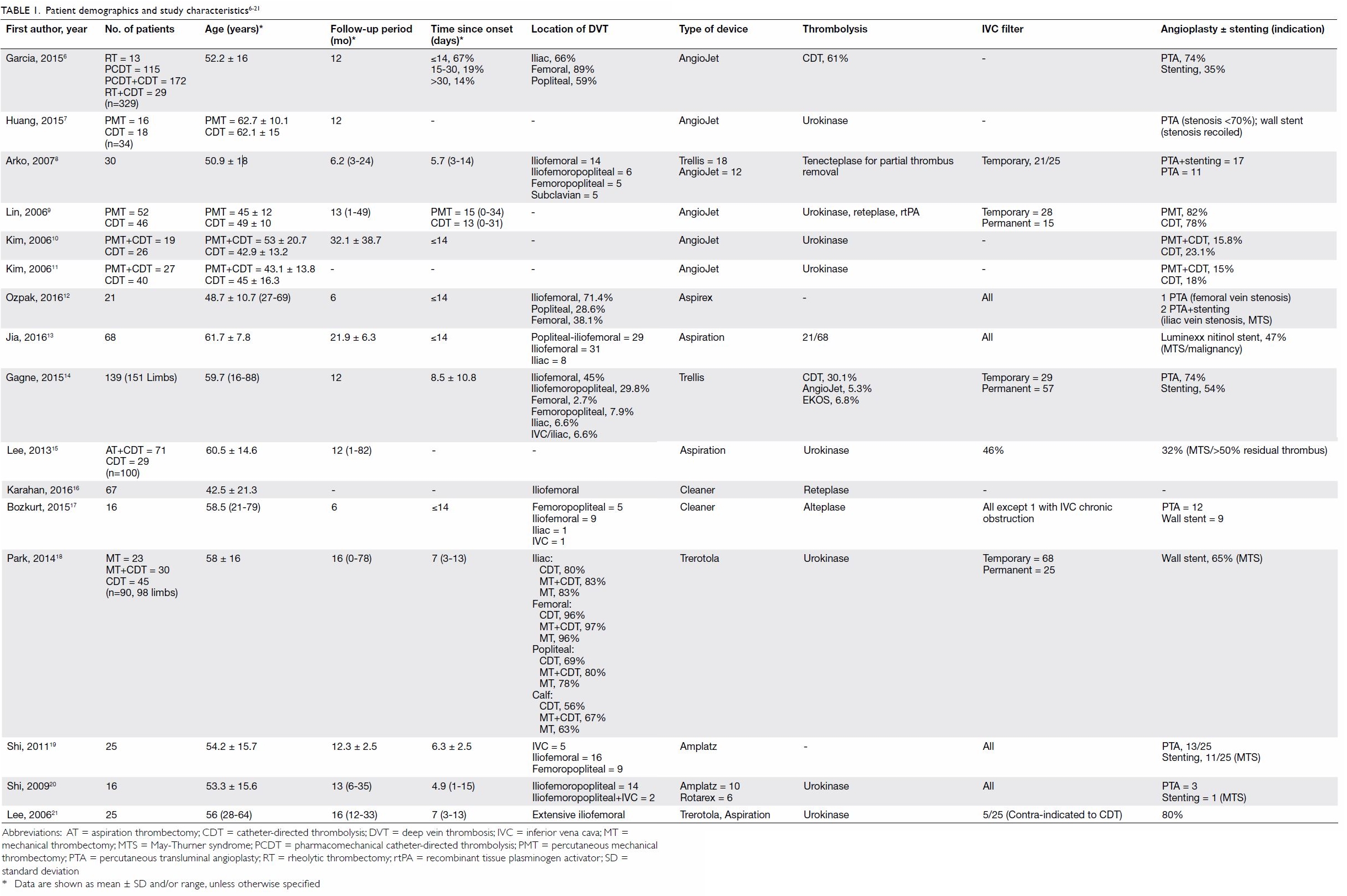
Baseline patient demographics and characteristics of the studies are summarised in Table 1.6 7 8 9 10 11 12 13 14 15 16 17 18 19 20 21 A total of 1170 patients were included (range, 16-329 patients) with a mean age of 53.5 years (range, 16 to 88 years). The mean follow-up time was 12.3 months (range, 1-82 months).
 

Table 1. Patient demographics and study characteristics6 7 8 9 10 11 12 13 14 15 16 17 18 19 20 21
 
Four different categories of thrombectomy devices were used among the included studies: rheolytic devices,6 7 8 9 10 11 aspiration devices,12 13 14 15 16 rotational devices,16 17 18 19 20 21 and ultrasound-enhanced thrombolysis devices.14 They aimed to achieve transcatheter removal of thrombi via different mechanical means.
 
The AngioJet system (Possis Medical, Minneapolis [MN], US) is a rheolytic device that generates high-velocity saline jets at the side of catheter, which create a localised low-pressure zone and thus result in maceration and aspiration of the thrombus.
 
During aspiration thrombectomy, the thrombus was aspirated out through the percutaneous catheter as the catheter was gradually pulled out. The process was repeated until complete removal of the thrombus or at least 90% disappearance of thrombi. Two of the aspiration systems were Aspirex (Straub Medical, Wangs, Switzerland) and the Trellis infusion system (Covidien, Mansfield [MA], US), which is a sophisticated system that contains an oscillation drive unit that mixes the thrombus with thrombolytic agents between two occlusion balloons.
 
Rotational devices feature high-frequency revolution of a helix that is controlled by a foot pedal. At least four different types of rotational devices were included, including the Amplatz thrombectomy device (Microvena, White Bear Lake [MN], US), Rotarex (Straub Medical, Wangs, Switzerland), Trerotola (Arrow International, Redding [PA], US), and Cleaner (Rex Medical, Fort Worth [TX], US and Argon Medical Devices, Inc, Plano [TX], US). They all consisted of a motor-driven fragmentation helix or basket that was rotated in the thrombosed vein. The thrombus was then aspirated out via the catheter.
 
An ultrasound-enhanced thrombolysis device (EKOS Corporation, Bothell [WA], US) was selectively used in one study14 for patients with inadequate thrombus removal despite the use of a rotational device. A high-frequency ultrasound wave was emitted through transducers inside the catheter to achieve maceration of the thrombus and mix it with thrombolytic agents.
 
Efficacy
Nine non-comparative and seven comparative studies were included in our analysis. Efficacy in terms of rates of venous patency, PTS, and recurrent thrombosis is shown in Table 2.6 7 8 9 10 11 12 13 14 15 16 17 18 19 20 21 Venous patency was measured most frequently by Duplex ultrasound (n=9) followed by computed tomographic (CT) venography (n=4) and contrast venography (n=2). Imaging modalities were not mentioned in three studies. Venous patency was further quantified according to a 3-tier system in five studies: Grade I (<50% clot lysis), Grade II (50%-99% clot lysis), and Grade III (100% clot lysis).22 Venous patency was measured at 6 months in four studies and at 1 year in the other 12 studies. Venous patency rates ranged from 75% to 100%.
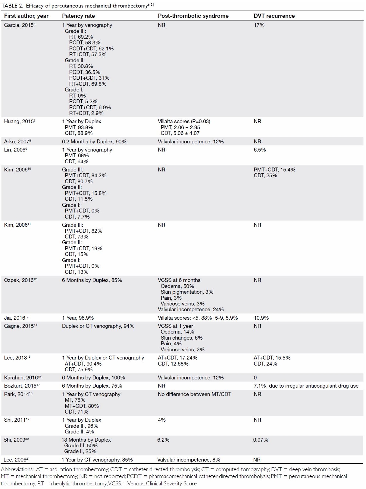
 

Table 2. Efficacy of percutaneous mechanical thrombectomy6 7 8 9 10 11 12 13 14 15 16 17 18 19 20 21
 
Rates of PTS were reported in terms of Villalta score (n=2)23 or Venous Clinical Severity Score (VCSS) [n=2].14 Four studies reported the rates of valvular incompetence from 8% to 24%. The rates of DVT recurrence, reported in eight studies, ranged from 0% to 17%.
 
Complications
The major complications of thrombectomy are shown in Table 3.6 7 8 9 10 11 12 13 14 15 16 17 18 19 20 21 Six studies reported rates of pulmonary embolism ranging from 0.3% to 17%, but none of them was clinically significant. No patient had pulmonary embolism in the remaining 11 studies. Garcia et al6 reported major bleeding complications in 3.6% of patients, including intracranial bleeding, gastrointestinal bleeding secondary to gastritis or gastric cancer, retroperitoneal bleeding, and haemolytic anaemia requiring transfusion. Minor bleeding complications were reported at frequencies of up to 28%, most of which were access site bleeding. Blood transfusion and surgical intervention were seldom required. No operative mortality was reported in 12 studies, while only two cases of fatal intracranial haemorrhage were noted in two separate studies. Another mortality was reported by Garcia et al,6 with an unknown cause of death. The overall mortality in this series was 0.26%.
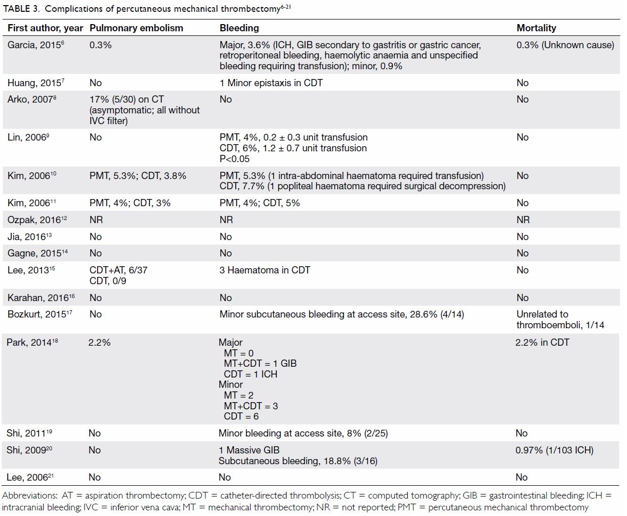
 

Table 3. Complications of percutaneous mechanical thrombectomy6 7 8 9 10 11 12 13 14 15 16 17 18 19 20 21
 
Secondary outcomes
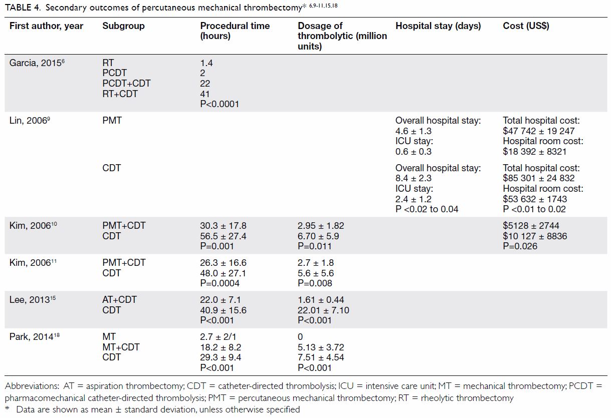
Seven studies reported comparative evidence about PMT versus CDT (Table 46 9 10 11 15 18). Huang et al7 showed that PMT significantly reduced PTS at 1 year, with lower Villalta scores in the PMT group (2.1±3.0) than in the CDT group (5.1±4.1; Wilcoxon rank-sum test, P=0.03). However, no statistical difference was shown in Villalta scores in another retrospective study conducted by Park et al.18
 

Table 4. Secondary outcomes of percutaneous mechanical thrombectomy6 9 10 11 15 18
 
Lin et al9 compared bleeding complications between the two groups in terms of the number of units of packed red blood cells transfused. There was a significant reduction of blood transfusion from 1.2±0.7 units in the CDT group to 0.2±0.3 units in the PMT group (Pearson Chi squared, P≤0.05).9
 
The dosage of thrombolytic infusion and average procedural time were significantly reduced in the CDT with adjunctive PMT group compared with the CDT alone group, as reported in at least four different retrospective studies.10 11 15 18
 
From the economic perspective, two retrospective studies performed cost analysis, and PMT was found to be associated with 44% to 49% reduction in total hospital costs.9 10 It was also consistent with shorter hospital and intensive care unit (ICU) stays in the PMT group (4.6±1.3 days of hospital stay and 0.6±0.3 days of ICU stay in the PMT group vs 8.4±2.3 days of hospital stay and 2.4±1.2 days of ICU stay in the CDT group; Pearson Chi squared, P<0.02 to 0.04).
 
No statistically significant differences in venous patency or symptom improvement between the two groups were reported in this series of comparative studies.
 
Comparison between types of thrombectomy devices
Three studies compared outcomes of different thrombectomy devices. Garcia et al6 created the first prospective multi-centre (PEARL) registry to document the use of the AngioJet rheolytic device. A total of 329 patients were stratified into four treatment subgroups: (1) rheolytic thrombectomy (RT) alone; (2) RT plus CDT; (3) pharmacomechanical CDT (PCDT), and (4) PCDT combined with CDT. Each of these subgroups differed in terms of the presence, timing, and delivery means of thrombolytic agents. Rheolytic thrombectomy was given before or after CDT in subgroup 2, and PCDT was defined as delivery of lytic agent through an AngioJet catheter. This registry demonstrated no statistical difference in venous patency rate between the subgroups, while a significant reduction in procedural time in non-CDT group was observed (Table 4). The investigators concluded that RT was effective and safe, and therefore, the needs for concomitant CDT and intensive care could potentially be reduced.
 
Shi et al20 and Arko et al8 compared the outcomes of Amplatz versus Rotarex and Trellis versus AngioJet devices, which again showed no significant differences in clinical outcomes between the two groups.
 
Use of adjunctive treatments
Inferior vena cava filter placement, angioplasty, and stenting were the most commonly performed adjunctive treatments in addition to thrombectomy. Inferior vena cava filters were used in 46% to 100% of patients among 11 studies. The majority of the filters were removed shortly after the procedure without major complications. Lee et al15 reported that 6 of 37 patients had thrombus entrapment in prophylactic IVC filters in the thrombectomy group compared with 0 of 9 patients in the CDT alone group. Arko et al8 reported that 17% of patients showed asymptomatic pulmonary embolism on CT after thrombectomy, in which all patients did not receive IVC filters. This showed that prophylactic IVC filtration could be a useful measure for prevention of pulmonary embolism, especially in patients who undergo aggressive thrombectomy.
 
Angioplasty with or without stenting was performed in 15 studies, ranging from 14% to 80% of patients. The two main indications were iliac vein compression syndrome (May-Thurner syndrome) and residual thrombus after thrombectomy. One study reported a significantly improved iliac vein patency rate in the group with stents (28.95%) than without stents (11.29%; log rank test, P=0.026).15
 
Discussion
Catheter-directed thrombolysis and PMT are both emerging techniques for treatment of acute DVT of the lower extremities that have the advantage of early restoration of venous patency and thus reduction of post-thrombotic complications. A 2015 meta-analysis compared the efficacy of CDT plus anticoagulation versus that of anticoagulation alone in the treatment of proximal DVT. It showed that additional CDT was associated with significantly improved 6-month venous patency and PTS rates. However, there was a two-fold increase in bleeding complications in the CDT group, and concomitant close monitoring under intensive care setting has had a substantial economic burden.5 These two main reasons have precluded the incorporation of CDT into the standard treatment recommendation despite encouraging procedural outcomes.
 
As compared with CDT, PMT is another endovascular option that has provided promising clinical outcomes with better controlled bleeding risk. This review has served as a comprehensive overview of clinical and safety outcomes across different categories of thrombectomy devices. It demonstrated well that the procedural outcomes of both PMT alone and that with pharmacomechanical devices were non-inferior to that of CDT in treatment of acute DVT in the lower extremities. The rates of PTS, bleeding complications, and hospital costs of PMT were all shown to be favourable to those of CDT alone. In addition, the mortality risk of PMT was minimal and comparable to that of patients treated with anticoagulation alone: 0.4% recurrent fatal venous thromboembolism and 0.2% fatal major bleeding events.24 As illustrated in this review, the balanced risks and benefits of PMT provide a basis for the future initiation of randomised controlled trials on its use.
 
In addition, PMT is potentially superior to CDT especially in patients in whom thrombolysis therapy is contra-indicated. According to the Society of Interventional Radiology recommendations, CDT is absolutely contra-indicated in patients with recent cerebrovascular events, neurosurgery or intracranial trauma, active internal bleeding, and those with absolute contra-indications to anticoagulation. Other strong relative contra-indications are listed in the Standard of Practice25: recent major surgery, obstetrical delivery or major trauma within 10 days, etc. These patients are prone to the development of DVT, and they have been conventionally treated with anticoagulation or IVC filters. Percutaneous mechanical thrombectomy is another option in this clinically challenging situation. Further studies on this particular group of high-risk patients are necessary to investigate the efficacy and safety of this novel technique.
 
Nevertheless, this review has several limitations. The studies were heterogeneous in terms of outcome measurements and the use of thrombectomy devices. Post-thrombotic syndrome was measured in terms of Villalta score, VCSS, or valvular incompetence rate. Although these systems were well-defined objective scales for monitoring and documentation of PTS, it was difficult to compare efficacy across studies. Similarly, the important index of venous patency rate was variously measured by Duplex ultrasound, CT venogram, or venography. Inaccuracies during direct comparison between studies were unavoidable.
 
Other adjunctive modalities in addition to the principal thrombectomy devices including iliac vein angioplasty, stenting, and prophylactic IVC filter were used in a major proportion of the studies. No standardised criteria were outlined for the usage of these devices, and they created a confounding factor during data analysis. With inadequate information on sub-categorisation of the study populations, analysis specific to each type of adjunctive devices was not feasible. Most of the studies were retrospective, and no randomised trials were available for quantitative analysis.
 
Conclusion
Percutaneous mechanical thrombectomy is a safe and effective treatment for acute iliofemoral DVT in terms of restoration of venous patency, prevention of DVT recurrence, PTS, and pulmonary embolism. The overall clinical outcomes of PMT are superior to those with anticoagulation alone. Compared with CDT alone, adjunctive PMT has a lower risk of PTS and bleeding complications. Randomised studies to demonstrate the efficacy of PMT versus anticoagulation and CDT and compare the efficacy of different types of PMT devices would be most beneficial to guide future strategies for treatment of acute proximal DVT.
 
Author contributions
All authors had full access to the data, contributed to the study, approved the final version for publication, and take responsibility for its accuracy and integrity.
 
Concept or design: All authors.
Acquisition of data: All authors.
Analysis or interpretation of data: PC Wong, YC Chan, Y Law.
Drafting of the manuscript: PC Wong, YC Chan, Y Law.
Critical revision: All authors.
 
Conflicts of interest
The authors declare no conflicts of interest.
 
Declaration
The paper was presented as an abstract in the 21st Asian Congress of Surgery by the Asian Surgical Association, 22-23 November 2017, Tokyo, Japan.
 
Funding/support
This research received no specific grant from any funding agency in the public, commercial, or not-for-profit sectors.
 
References
1. Kearon C, Kahn SR, Agnelli G, Goldhaber S, Raskob GE, Comerota AJ. Antithrombotic therapy for venous thromboembolic disease: American College of Chest Physicians Evidence-Based Clinical Practice Guidelines (8th edition). Chest 2008;133(6 Suppl):454S-545S. Crossref
2. Enden T, Haig Y, Kløw NE, et al. Long-term outcome after additional catheter-directed thrombolysis versus standard treatment for acute iliofemoral deep vein thrombosis (the CaVenT study): a randomised controlled trial. Lancet 2012;379:31-8. Crossref
3. Baldwin MJ, Moore HM, Rudarakanchana N, Gohel M, Davies AH. Post-thrombotic syndrome: a clinical review. J Thromb Haemost 2013;11:795-805. Crossref
4. Watson LI, Armon MP. Thrombolysis for acute deep vein thrombosis. Cochrane Database Syst Rev 2004;(4):CD002783. Crossref
5. Du GC, Zhang MC, Zhao JC. Catheter-directed thrombolysis plus anticoagulation versus anticoagulation alone in the treatment of proximal deep vein thrombosis—a meta-analysis. Vasa 2015;44:195-202. Crossref
6. Garcia MJ, Lookstein R, Malhotra R, et al. Endovascular management of deep vein thrombosis with rheolytic thrombectomy: final report of the prospective multicenter PEARL (Peripheral Use of AngioJet Rheolytic Thrombectomy with a Variety of Catheter Lengths) registry. J Vasc Interv Radiol 2015;26:777-85. Crossref
7. Huang CY, Hsu HL, Kuo TT, Lee CY, Hsu CP. Percutaneous pharmacomechanical thrombectomy offers lower risk of post-thrombotic syndrome than catheter-directed thrombolysis in patients with acute deep vein thrombosis of the lower limb. Ann Vasc Surg 2015;29:995-1002. Crossref
8. Arko FR, Davis CM 3rd, Murphy EH, et al. Aggressive percutaneous mechanical thrombectomy of deep venous thrombosis: early clinical results. Arch Surg 2007;142:513-9. Crossref
9. Lin PH, Zhou W, Dardik A, et al. Catheter-direct thrombolysis versus pharmacomechanical thrombectomy for treatment of symptomatic lower extremity deep venous thrombosis. Am J Surg 2006;192:782-8. Crossref
10. Kim HS, Patra A, Paxton BE, Khan J, Streiff MB. Catheter-directed thrombolysis with percutaneous rheolytic thrombectomy versus thrombolysis alone in upper and lower extremity deep vein thrombosis. Cardiovasc Intervent Radiol 2006;29:1003-7. Crossref
11. Kim HS, Patra A, Paxton BE, Khan J, Streiff MB. Adjunctive percutaneous mechanical thrombectomy for lower-extremity deep vein thrombosis: clinical and economic outcomes. J Vasc Interv Radiol 2006;17:1099-104. Crossref
12. Ozpak B, Ilhan G, Ozcem B, Kara H. Our short-term results with percutaneous mechanical thrombectomy for treatment of acute deep vein thrombosis. Thorac Cardiovasc Surg 2016;64:316-22. Crossref
13. Jia Z, Tu J, Zhao J, et al. Aspiration thrombectomy using a large-size catheter for acute lower extremity deep vein thrombosis. J Vasc Surg Venous Lymphat Disord 2016;4:167-71. Crossref
14. Gagne P, Khoury T, Zadeh BJ, Rajasinghe HA. A multicenter, retrospective study of the effectiveness of the trellis-8 system in the treatment of proximal lower-extremity deep vein thrombosis. Ann Vasc Surg 2015;29:1633-41. Crossref
15. Lee JH, Kwun WH, Suh BY. The results of aspiration thrombectomy in the endovascular treatment for iliofemoral deep vein thrombosis. J Korean Surg Soc 2013;84:292-7. Crossref
16. Karahan O, Kutas HB, Gurbuz O, et al. Pharmacomechanical thrombolysis with a rotator thrombolysis device in iliofemoral deep venous thrombosis. Vascular 2016;24:481-6. Crossref
17. Bozkurt A, Kırbaş İ, Kösehan D, Demirçelik B, Nazlı Y. Pharmacomechanical thrombectomy in the management of deep vein thrombosis using the cleaner device: an initial single-center experience. Ann Vasc Surg 2015;29:670-4. Crossref
18. Park KM, Moon IS, Kim JI, et al. Mechanical thrombectomy with Trerotola compared with catheter-directed thrombolysis for treatment of acute iliofemoral deep vein thrombosis. Ann Vasc Surg 2014;28:1853-61. Crossref
19. Shi HJ, Huang YH, Shen T, Xu Q. Percutaneous mechanical thrombectomy for acute massive lower extremity deep venous thrombosis. Surg Laparosc Endosc Percutan Tech 2011;21:50-3. Crossref
20. Shi HJ, Huang YH, Shen T, Xu Q. Percutaneous mechanical thrombectomy combined with catheter-directed thrombolysis in the treatment of symptomatic lower extremity deep venous thrombosis. Eur J Radiol 2009;71:350-5. Crossref
21. Lee KH, Han H, Lee KJ, et al. Mechanical thrombectomy of acute iliofemoral deep vein thrombosis with use of an Arrow-Trerotola percutaneous thrombectomy device. J Vasc Interv Radio 2006;17:487-95. Crossref
22. Porter JM, Moneta GL. Reporting standards in venous disease: an update. International Consensus Committee on Chronic Venous Disease. J Vasc Surg 1995;21:635-45. Crossref
23. Kahn SR, Partsch H, Vedantham S. Definition of post-thrombotic syndrome of the leg for use in clinical investigations: a recommendation for standardization. J Thromb Haemost 2009;7:879-83. Crossref
24. Carrier M, Le Gal G, Wells PS, Rodger MA. Systematic review: case-fatality rates of recurrent venous thromboembolism and major bleeding events among patients treated for venous thromboembolism. Ann Intern Med 2010;152:578-89. Crossref
25. Vedantham S, Thorpe PE, Cardella JF, et al. Quality improvement guidelines for the treatment of lower extremity deep vein thrombosis with use of endovascular thrombus removal. J Vasc Interv Radiol 2009;20(7 Suppl):S227-39. Crossref

Update on the association between dry eye disease and meibomian gland dysfunction

Hong Kong Med J 2019 Feb;25(1):38–47  |  Epub 31 Jan 2019
© Hong Kong Academy of Medicine. CC BY-NC-ND 4.0
 
REVIEW ARTICLE  CME
Update on the association between dry eye disease and meibomian gland dysfunction
Tommy CY Chan, MB, BS, MMedSc1,2,3; Sharon SW Chow, MB, BS3,4; Kelvin HN Wan, MB, BS1,5; Hunter KL Yuen, MB, ChB1,6
1 Department of Ophthalmology and Visual Sciences, The Chinese University of Hong Kong, Shatin, Hong Kong
2 Hong Kong Sanatorium & Hospital, Happy Valley, Hong Kong
3 Department of Ophthalmology, The University of Hong Kong, Cyberport, Hong Kong
4 Department of Ophthalmology, Grantham Hospital, Wong Chuk Hang, Hong Kong
5 Department of Ophthalmology, Tuen Mun Hospital, Tuen Mun, Hong Kong
6 Hong Kong Eye Hospital, Hong Kong
 
Corresponding author: Dr Tommy CY Chan (tommychan.me@gmail.com)
 
 Full paper in PDF
 
Abstract
Dry eye disease is one of the most common ophthalmic complaints; it results from the activity of various pathways and is considered a multifactorial disease. An important factor that contributes to the onset of dry eye disease is meibomian gland dysfunction. Meibomian gland dysfunction causes a disruption in the tear film lipid layer which affects the rate of tear evaporation. This evaporation leads to tear hyperosmolarity, eventually triggering the onset of dry eye disease. Dry eye disease and meibomian gland dysfunction are strongly associated with each other, such that many of their risk factors, signs, and symptoms overlap. This review aimed to provide an update on the association between dry eye disease and meibomian gland dysfunction. A stepwise approach for diagnosis and management is summarised.


Introduction
Dry eye disease (DED) is one of the most common ocular surface diseases, which can significantly affect the quality of life of affected patients. The definition of DED has been progressively established in recent decades. The goal of the Tear Film and Ocular Surface Society (TFOS) Dry Eye Workshop (DEWS) is to create an evidence-based definition, a well-defined classification system, and an appropriate diagnosis and management algorithm for DED.1 In 2007, the TFOS DEWS definition of DED was first published.2 In 2017, the TFOS DEWS II amended the definition of DED to be ‘a multifactorial disease of the ocular surface, characterised by a loss of homeostasis of the tear film, and accompanied by ocular symptoms, in which tear film instability and hyperosmolarity, ocular surface inflammation and damage, and neurosensory abnormalities play etiological roles’.1 The term ‘multifactorial’ indicates that the disease occurs as a result of multiple influential factors, while the term ‘etiological roles’ suggests the involvement of various pathways in the onset of DED.1 In 2017, the Asia Dry Eye Society also agreed upon a new definition of DED, as ‘a multifactorial disease characterised by unstable tear film causing a variety of symptoms and or visual impairment, potentially accompanied by ocular surface damage’.3
 
The two main categories of DED are evaporative dry eye and aqueous deficient dry eye.2 Evaporative dry eye is related to conditions that affect the eyelids, such as meibomian gland dysfunction (MGD), poor blinking effort, and lid disorders, or that affect the ocular surface, such as prolonged contact lens wear, frequent use of topical drug preservatives, and immune-related ocular surface disorders (eg, atopic keratoconjunctivitis). Aqueous deficient dry eye is primarily due to conditions affecting lacrimal gland function, such as Sjögren’s syndrome, lacrimal gland duct obstruction or deficiencies, and adverse effects of systemic drugs. Epidemiological evidence suggests that DED is mainly evaporative in nature,4 and is often associated with MGD.5 6
 
Meibomian glands are found in the upper and lower eyelids, where they secrete lipids (meibum) onto the ocular surface, forming the outermost layer of the tear film. These lipids spread easily, promoting tear film stability and protecting against evaporation. Meibomian gland dysfunction is defined as ‘a chronic, diffuse abnormality of the meibomian glands, commonly characterised by terminal duct obstruction and/or qualitative/quantitative changes in the glandular secretion. It may result in alteration of the tear film, symptoms of eye irritation, clinically apparent inflammation and ocular surface disease’.7
 
This review aims to provide an update on the association between MGD and DED, with particular attention to the diagnosis and management of these conditions. We will discuss the epidemiology, pathophysiology, risk factors, signs and symptoms, diagnosis, and ancillary imaging of MGD and DED, along with appropriate behaviour, medical, and surgical management.
 
Methods
A comprehensive literature search on PubMed was performed for studies published between January 2006 and December 2017 with keywords ‘dry eye’, ‘dry eye disease’, ‘tear film’, ‘meibomian gland’, and ‘meibomian gland dysfunction’. Search results were limited to clinical studies published in English. Articles reporting DED and MGD were reviewed. Particular emphasis was placed on papers that investigated the association between DED and MGD. The reference lists of the retrieved articles were also examined for relevant studies.
 
Epidemiology
The reported prevalence of DED ranges from 5% to 50%,4 whereas the reported prevalence of MGD varies more widely from 3.5% to nearly 70%.8 9 Meibomian gland dysfunction appears to be more prevalent in Asian populations.5 Meibomian gland dysfunction has been reported to contribute to 60% of all cases of DED; an additional 20% of cases of DED are caused by aqueous deficiency.
 
Pathophysiology of dry eye disease
All forms of DED primarily occur because of water loss from the tear film, which leads to tear hyperosmolarity due to evaporative dry eye and/or aqueous deficient dry eye.10 In evaporative dry eye, hyperosmolarity results from excessive evaporation of tears in the context of normal lacrimal function. In contrast, in aqueous deficient dry eye, hyperosmolarity occurs due to an inadequate rate of lacrimal secretion in the context of a normal rate of evaporation. Environmental factors affect the presence of hyperosmolarity on the ocular surface, which may trigger the onset of DED, or cause worsening of the condition.
 
Pathophysiology of meibomian gland dysfunction
Meibomian gland dysfunction is classified according to the rate of gland secretion. A low delivery state is characterised by meibomian gland hyposecretion or obstruction, whereas a high delivery state is characterised by meibomian gland hypersecretion. Of these two categories, the most common mechanism is a low delivery state due to duct obstruction.11 Epithelial hyperkeratinisation is the most common cause of duct obstruction, leading to meibum accumulation with chronic inflammation and, eventually, gland dropout.12 Importantly, this results in the quantitative and qualitative abnormalities of glandular secretions. There is a high prevalence of MGD in acne rosacea, which is a chronic cutaneous inflammatory disorder.
 
Association between dry eye disease and meibomian gland dysfunction
The tear film consists of three distinct layers: the lipid, aqueous, and mucus layers. The lipid layer, a key component of the tear film, is derived from meibomian glands. The lipid layer prevents water evaporation from the ocular surface and is thus crucial in the maintenance of a healthy ocular surface. Dysfunction of the meibomian glands results in unbalanced lipid secretion, thereby increasing the rate of ocular surface evaporation and causing tear hyperosmolarity.13 Patients with MGD reportedly exhibit a higher tear evaporation rate than that of normal subjects.13 This shows that DED is directly correlated with the integrity and quality of meibum on the ocular surface.
 
Risk factors
Many risk factors associated with DED also contribute to MGD. Thus, risk factor modifications can likely improve both disease states.
 
Sex
Female sex is a significant risk factor for the development of both DED and MGD.5 6 This may be due to the effect of hormonal changes on meibomian secretion, as androgen and oestrogen receptors are both present within the meibomian glands.14 Importantly, androgens have been reported to stimulate meibum secretion and suppress inflammation, whereas oestrogens reduce meibum secretion and increase inflammation.15 Dysfunctional meibomian gland secretion and concurrent alterations in the lipid layer have been observed in patients with androgen depletion.16 Additionally, female sex has been identified as a risk factor for the development of autoimmune diseases that lead to DED, such as Sjögren’s syndrome.17
 
Topical medications
Topical medications can cause both DED and MGD; this may be a result of ocular surface disturbances with various aetiologies, including allergic reactions, toxic epitheliopathy, and inflammatory response from chronic chemical irritation. Multiple studies have revealed a clear relationship between the prevalence of dry eye and increasing use of eye drops.18 The primary factor underlying this relationship is the presence of benzalkonium chloride preservative agent in topical medications. Benzalkonium chloride has been strongly linked with the onset of DED, as it dissolves the lipid tear film layer and has been shown to disrupt tear film osmolarity.19 Similarly, DED and MGD are commonly reported in glaucoma patients who use topical glaucoma medications, which contain benzalkonium chloride. Use of these medications has been associated with changes in meibomian gland structure, leading to MGD.20
 
Contact lens wear
Contact lens wear is commonly associated with the onset of both DED and MGD. An epidemiological study showed that 50% of contact lens wearers experience dry eye symptoms, whereas only 22% of non–contact lens wearers experience such symptoms.21 Contact lens wear alters the integrity of the tear film: a thinner lipid layer has been observed in contact lens wearers, which causes an increased tear evaporation rate and tear hyperosmolarity.22 Environmental factors, such as prolonged usage of visual display devices, as well as air pollution and seasonal changes, further aggravate dry eye symptoms in contact lens wearers. The occurrence of MGD in contact lens wearers is suspected to be a result of chronic inflammation,23 as well as clogging of gland orifices due to accumulation of desquamated epithelial cells.24 Contact lens wearers demonstrate a high percentage of meibomian gland dropout and reduction in gland function; these aspects are reportedly directly related to the duration of contact lens wear.25
 
Refractive surgery
Worldwide, laser in situ keratomileusis (LASIK) is the most common corneal refractive surgery currently in use. Dry eye disease is often associated with a history of LASIK, and can be aggravated by both preoperative and postoperative factors. Preoperatively, the risk of DED is significantly increased in patients who are long-term contact lens wearers, as well as in patients whose eyes exhibit pre-existing tear film instability.26 Greater refractive correction magnitude requires deeper ablation, resulting in a greater extent of sensory nerve damage. This nerve damage results in reduced corneal sensitivity, leading to neuropathic dry eyes. Notably, this mechanism is the most common aetiology of post-LASIK dry eyes.26 Corneal refractive surgery has also been shown to reduce corneal epithelial integrity, conjunctival goblet cell concentration, and meibomian gland function, resulting in lower ocular surface disease index and ocular surface staining scores.27
 
Demodicosis
Two species of mites, Demodex folliculorum and Demodex brevis, are the only mites that affect human skin; such infestations are known as demodicosis.28 Reportedly, D folliculorum infests the lash follicles, whereas D brevis infests the meibomian glands.28 These infestations increase the meibum melting temperature, resulting in a more viscous lipid layer. A recent study showed that a higher D brevis count was associated with more severe MGD.29 Furthermore, confocal microscopy analysis revealed lower counts of Demodex mites in the glands of healthy subjects than in the glands of patients with MGD-related DED.30 The role of Demodex mites in the pathology of MGD has not been fully elucidated; however, eradication of Demodex is particularly helpful in relieving related ocular symptoms. Thus, there may a pathogenic role for Demodex infestation in MGD.
 
Symptoms
Many signs and symptoms of DED overlap with those of MGD. However, most patients with MGD are largely or entirely asymptomatic; if they are symptomatic, their particular symptoms often do not directly correlate with the severity of ocular surface disturbance. In a population-based study in China, 22% of the study population demonstrated asymptomatic MGD, while 9% showed symptomatic MGD.8 In cases of symptomatic MGD, patients report a variety of symptoms, including foreign body sensation, dryness, itching, and/or photosensitivity.7 These manifestations may be linked to chronic inflammation or mechanical friction between the ocular surface and meibum that has accumulated in the gland orifices.
 
Ocular surface signs and diagnosis
Because DED and MGD are common ophthalmic problems, a clear diagnosis is crucial for suitable management. Appropriate tests should be used to diagnose and monitor DED, in accordance with the revised TFOS DEWS II definition of the disease. For these purposes, the TFOS DEWS II proposed a battery of diagnostic tests for DED.
 
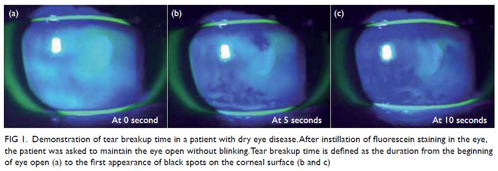
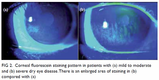
The diagnostic tests begin with triaging questions and risk factor analysis. These are followed by screening for symptoms using standardised questionnaires, including the five-item dry eye questionnaire or the ocular surface disease index. Markers of homeostasis used in diagnostic testing include measures of tear breakup time, staining of the ocular surface, Schirmer’s test, and tear osmolarity. Tear breakup time is a non-invasive measurement that is defined as the time required for the tear film to break up sufficiently that the patient can no longer refrain from blinking.31 A tear breakup time of <10 seconds is considered diagnostic for DED (Fig 1). Ocular surface staining is performed by fluorescein staining for corneal damage and lissamine green staining for conjunctival and lid margin damage (Fig 2).31 Schirmer’s test consists of the placement of a small strip of filter paper inside the lower fornix with the eye closed. After 5 minutes, the amount of moisture is measured as the distance that tear moisture has travelled on the paper, due to capillary action; a value of <5 mm indicates DED. Finally, tear osmolarity should be assessed with a calibrated device; a positive result is defined as ≥308 mOsm/L in the measured eye, or a difference of >8 mOsm/L between two eyes.32
 

Figure 1. Demonstration of tear breakup time in a patient with dry eye disease. After instillation of fluorescein staining in the eye, the patient was asked to maintain the eye open without blinking. Tear breakup time is defined as the duration from the beginning of eye open (a) to the first appearance of black spots on the corneal surface (b and c)
 

Figure 2. Corneal fluorescein staining pattern in patients with (a) mild to moderate and (b) severe dry eye disease. There is an enlarged area of staining in (b) compared with (a)
 
The Asia Dry Eye Society recommends diagnosis of DED by using a combination of symptoms assessed by standardised questionnaires (ocular surface disease index, McMonnies questionnaire, women’s health study questionnaire, or five-item dry eye questionnaire), together with a reduced tear breakup time (with a cut-off value of <5 s).3
 
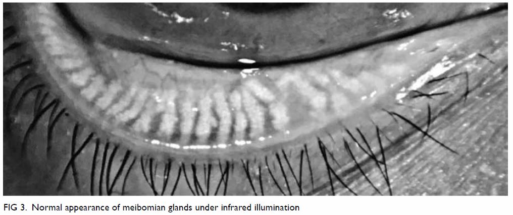
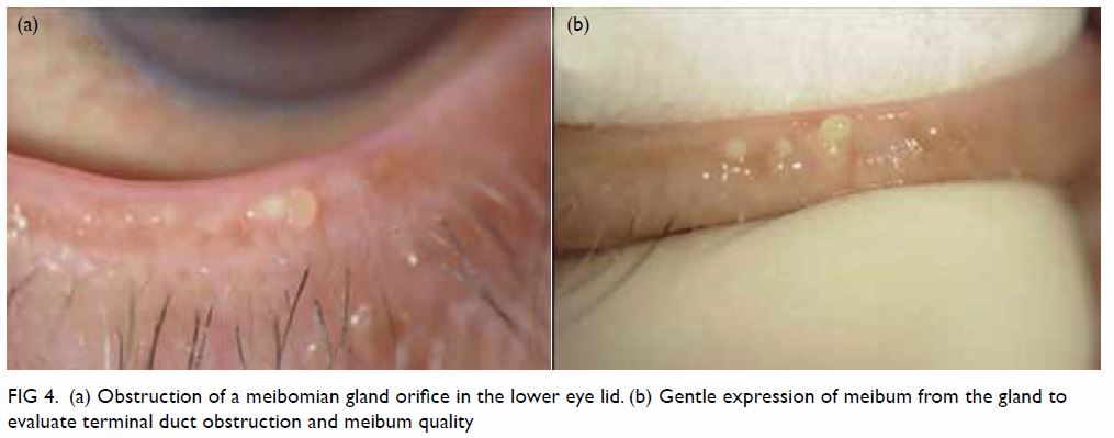
Clinical diagnosis of MGD is made based on the examination of altered anatomical features, such as meibomian gland dropout, altered meibum excretion, and changes to lid morphology, with plugging or pouting of the gland orifice. Meibomian glands with normal appearance are shown in Figure 3. Gentle gland expression with digital pressure to the central lower lid can evaluate terminal duct obstruction and meibum quality (Fig 4). Subtype classification tests, including identification of MGD features, as well as lipid thickness and tear volume assessment, are then performed to determine whether the disease constitutes evaporative dry eye or aqueous deficient dry eye. Lastly, the severity of disease is evaluated; for this purpose, the International Workshop on Meibomian Gland Dysfunction has provided a grading system that can be used to guide management of MGD.33
 

Figure 3. Normal appearance of meibomian glands under infrared illumination
 

Figure 4. (a) Obstruction of a meibomian gland orifice in the lower eye lid. (b) Gentle expression of meibum from the gland to evaluate terminal duct obstruction and meibum quality
 
Role of imaging in diagnosis
In recent years, multiple imaging modalities have been introduced to improve the diagnosis of DED and MGD.34 These modalities aim to facilitate the evaluation of the structural and dynamic properties of the tear film. In cases of DED with tear film instability, topographic systems have been used to determine changes in the edges of the mires of a Placido disc.35 Anterior segment optical coherence tomography aims to measure the height of the tear meniscus,36 while infrared meibography provides an objective evaluation of gland structures. Tear film lipid layer thickness can be measured by interferometry, which allows objective and quantitative measurement of tear film integrity.37
 
Both DED and MGD can lower the ocular surface temperature. In DED, the increased tear film evaporation rate causes heat loss, lowering ocular surface temperature.38 In MGD, lower tarsal conjunctival temperatures have been observed, increasing the viscosity of meibum; this change in viscosity leads to worsening of gland function.39 These advancements in imaging modalities have improved accuracy and standardised the diagnosis of DED and MGD.
 
Management
The aim of all treatment in MGD is to increase the quality and quantity of meibum expression. For this purpose, a stepwise staged approach is necessary to standardise management of the disease.40 The TFOS DEWS II created an algorithm to implement various management options, on the basis of disease severity.40 Initially, patients must be educated regarding environmental and dietary modifications, which include essential fatty acid supplements. Patients must also be guided to eliminate factors contributing to the onset of DED, including contact lens wear, as well as both topical and systemic medications. Several lifestyle modifications, such as ensuring sufficient sleep or rest, maintaining appropriate hydration, and discontinuing smoking habits, may help to relieve symptoms. Ocular lubricants are suggested for mild DED; preferably, these should not contain benzalkonium chloride preservatives. Some of these modification approaches are outlined below, along with an overview of the emerging available treatment devices and options.
 
Eyelid hygiene
In the presence of MGD, eyelid hygiene is the cornerstone of MGD management. This treatment modality consists of two components: eyelid warming and eyelid massage. Meibum in patients with MGD is more stagnant and viscous and has a higher melting temperature than that in a healthy individual; thus, warming the eyelid to melt pathologically altered meibum can improve its secretion.33 Warm compression provides further benefits by melting abnormal meibum. Secretions from meibomian glands in patients with MGD exhibit lower levels of lipids, esters, and free sterols.41 Potential involvement of microbes (ie, Staphylococcus spp, Propionibacterium acnes, Bacillus oleronius, and the Demodex species described above) contributes to the pathology of MGD-associated DED by increasing meibum melting temperature and enhancing inflammation. This illustrates the importance of eyelid hygiene in MGD management.42 For patients with MGD who exhibit demodicosis, many treatment options have been described, including the use of topical 2% metronidazole. Recently, the use of tea tree oil has also increased in popularity.43 Tea tree oil is a natural essential oil that includes 4-terpineol, which is antimicrobial, anti-inflammatory, and toxic to Demodex.44 Tea tree oil lid scrubs have shown promising results as management for Demodex-related MGD.45
 
Effective eyelid hygiene can be achieved by use of a hot compress (ie, soaking a clean towel in hot water, and applying the towel over the eyelids), which softens the meibum and allows better flow. After the application of the hot compress, lipid by-products can be removed gently by scrubbing both upper and lower lid margins via mild upward or downward compression of the eyelid, using a moist cotton bud; this compression begins from the nasal canthus and moves laterally. An additional therapeutic approach involves the use of mildly diluted baby shampoo for lid scrubs; this is a widely accepted therapy. Although eyelid warming and eyelid massage are efficacious for the management of MGD, they are often time-consuming and labour-intensive; thus, they encounter patient compliance issues.46 There are now a wide variety of lid cleansing products, which facilitate standardisation and simplification of treatment. Additional treatment options include warming of the lids and expression of meibomian glands, either manually (similar to above) or with the use of specially designed devices. One such device, LipiFlow (TearScience; Morrisville [NC], United States), is designed to transfer heat through the eyelid tissue to facilitate emptying of gland contents at a therapeutic temperature of 42.5°C.47 LipiFlow treatment has shown promising results, and may significantly improve symptoms.47
 
Intense pulsed light was first reported approximately 10 years ago for the treatment of MGD, and it has demonstrated an ability to improve tear film quality and quantity, as well as to promote reduction of dry eye symptoms.48 Intraductal meibomian gland probing provides another approach to remove abnormal meibum secretions.49 Oral tetracycline and macrolides are reportedly useful in the treatment of MGD-related DED.40 These compounds are used with the assumption that inhibition of lipase production results in reduction of lipid breakdown, which may contribute to improvement in MGD. Macrolides, azithromycin in particular, exhibit anti-inflammatory properties; moreover, these compounds increase cellular accumulation of cholesterol, which may promote a suitable outcome in patients with MGDrelated DED.50
 
Lipid-containing artificial lubricants
The majority of artificial tears are aqueous-based; however, these offer limited and short-term symptomatic relief, partly due to the lack of a lipid component. These artificial tears evaporate at a similar rate to that of natural tears.51 Addition of a lipid component to the artificial lubricant helps to replenish the lipid layer of the normal tear film.33 These lipid-containing lubricants exhibit long retention times and can stabilise the tear film lipid layer, reduce tear evaporation, and improve the signs of MGD.52 Additionally, lipid-containing lubricants have a longer-lasting effect and cause minimal interference of patient vision. Commercially available lipid-containing lubricants include mineral oil, high-purity castor oil, mixtures of light and standard mineral oil, and mixtures of polar phospholipid surfactant and mineral oil. A systematic review found that these lipid-containing eye drops are efficacious and safe alternatives to conventional tear lubricants in their abilities to relieve the signs and symptoms of DED.52
 
Anti-inflammatory medications
Because ocular surface inflammation plays an important role in the development of DED, anti-inflammatory mechanisms must be considered. For patients with moderate to severe DED, low-dose topical steroids have been advocated as a treatment choice, likely because the anti-inflammatory properties of this type of drug can improve ocular inflammation through suppression of inflammatory cytokines.53 Other anti-inflammatory options include non-glucocorticoid immunomodulatory drugs, such as topical cyclosporine A, which is an immunomodulatory drug that can reduce the expression of inflammatory markers.54 Notably, topical cyclosporine A has been proven efficacious in the treatment of DED.55 A randomised trial showed that cyclosporine A is beneficial in the stabilisation of tear film in patients with MGD.56 However, its anti-inflammatory effect is not as remarkable as that observed in DED, because the main pathophysiology (epithelial gland hyperkeratinisation) is not clearly resolved.
 
In severe cases of DED, autologous serum eye drops can be considered. Autologous serum, which is the fluid component of a patient’s own blood that remains after centrifugation, exhibits similar biochemical properties to those of tears.40 Autologous serum reportedly contains specific factors that enhance epithelial regeneration, and can inhibit the release of inflammatory cytokines.57 Another treatment option for patients with severe DED involves scleral contact lenses, which are rigid gas permeable lenses of large diameter that are supported by the sclera and serve as a bridge over the corneoscleral junction. A tear reservoir is maintained between the posterior surface of the scleral contact lens and the anterior corneal surface, improving tear osmolarity and relieving dry eye symptoms.58
 
Omega-3 dietary supplementation
Essential fatty acid supplementation has been proven beneficial in the treatment of DED and MGD, especially when administered by intake of foods rich in omega-3 fatty acids, such as flaxseed and fish oils.59 There is a speculative association between essential fatty acids and modifications in lipid profile, as well as reductions in the fatty acid content of meibomian gland secretions. In a randomised, placebo-controlled, masked trial, omega-3 fatty acid supplementation resulted in improving ocular surface disease index score, tear breakup time, and meibum score in patients with MGD.60 Essential fatty acids also enhance the lipid layer, slow tear evaporation, and reduce apoptosis of lacrimal gland cells.61 Essential fatty acids have been reported to exhibit anti-inflammatory properties, particularly by promoting the production of prostaglandin.62 These modifications improve the tear secretion rate and tear content. Further research is needed to enhance our understanding of the underlying mechanism by which fatty acid supplementation supports the management of MGD.63 64
 
Surgical and mechanical treatment options
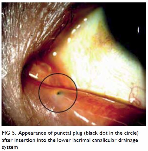
In cases where medical treatment is insufficient, surgical and mechanical treatment options include tear conservation via punctual occlusion or moisture chamber goggles. Punctal plugs retain tears on the ocular surface by blocking lacrimal drainage through the puncta (Fig 5). Permanent surgical closure may be useful when patients cannot tolerate punctual plugs. Surgical punctual occlusion blocks tear drainage and improves tear retention, and can be performed by cauterisation65 or lacrimal canalicular ligation.66 A systematic review showed that, when combined with other treatment for DED, punctual occlusion improves dry eye symptoms.67 A less invasive option, moisture chamber goggles provide a humid environment and minimise airflow to the ocular surface, thereby slowing the evaporation of tears.40
 

Figure 5. Appearance of punctal plug (black dot in the circle) after insertion into the lower lacrimal canalicular drainage system
 
Severe DED can lead to corneal erosion, persistent epithelial defects, corneal ulceration, and eventual corneal scarring. Amniotic membrane transplant is a reasonable option in such cases. Amniotic membrane has been shown to contain multiple neurotransmitters and neurotrophic factors, which are beneficial for the management of severe DED.68 For patients with severe DED with persistent epithelial defects that are refractive to medical treatment, tarsorrhaphy may be useful. Notably, tarsorrhaphy is a procedure that achieves partial or total closure of the eyelids, either temporarily or permanently. By reducing ocular surface exposure, the rate of tear evaporation decreases, such that DED can improve. Due to unfavourable aesthetic outcomes, this approach is typically one of the final methods used for management of severe DED.
 
Suggested treatment guideline for dry eye disease or meibomian gland dysfunction for non-ophthalmologists
Dry eye disease and MGD are two of the most common ocular conditions encountered by medical practitioners. To manage these conditions, risk factors must be identified and modified. Notably, several environmental and lifestyle modifications can help alleviate these conditions. Proper lighting, anti-glare filters, ergonomic positioning of computer monitors, and regular break time from work may help improve the symptoms.69 A modest increase in relative humidity, achieved by using a desktop-powered humidifier, has been shown to increase subjective comfort.70 Reduction or discontinuation of contact lens use, as well as enhancement of moisture within the surrounding environment, are possible risk factor modifications. Smoking cessation can also improve the ocular surface condition and tear function.71 Lubricants can be prescribed to be used as needed for symptomatic relief. If symptoms do not resolve, benzalkonium chloride–free lubricants should be considered. Low-dose topical steroids should be implemented with particular caution, owing to the risk of steroid-related complications (eg, cataract, glaucoma, and infection). In patients with recalcitrant disease, referral to an ophthalmologist is necessary to ensure regular monitoring. In the presence of MGD-related symptoms, lid hygiene and warm compression are strongly suggested for symptomatic control; careful manual expression of meibum should also be performed. If the above measures fail, or if the DED is secondary to other causes of aqueous deficiency (eg, Sjögren’s syndrome; graft versus host disease; or chronic inflammation in Stevens-Johnson’s disease, toxic epidermal necrolysis, or ocular cicatricial pemphigoid), referral to an ophthalmologist is warranted for further workup (eg, anti-SSA/Ro blood test for Sjögren’s syndrome) and management.
 
Conclusion
Dry eye disease is a common ophthalmic problem, with a cause that is often multifactorial. Meibomian gland dysfunction is an important contributor to DED, owing to an imbalance in lipid secretion that affects the rate of tear evaporation. When tears evaporate quickly, tear osmolarity increases, resulting in DED. There are many risk factors that contribute to onset of both DED and MGD, many of which may overlap between these diseases. A clear diagnosis is vital when managing DED. Various treatment options are available for DED and MGD, and a stepwise staged approach is often crucial for ensuring appropriate management.
 
Author contributions
All authors contributed to the concept and design, acquisition of data, analysis and interpretation of data, drafting of the manuscript, and critical revision for important intellectual content. All authors had full access to the data, contributed to the study, approved the final version for publication, and take responsibility for its accuracy and integrity.
 
Conflicts of interest
As an epidemiology advisor of the Editorial Board, HKL Yuen was not involved in the peer review process of the article. All authors have disclosed no conflicts of interest.
 
References
1. Craig JP, Nichols KK, Akpek EK, et al. TFOS DEWS II definition and classification report. Ocul Surf 2017;15:276-83. Crossref
2. The definition and classification of dry eye disease: report of the Definition and Classification Subcommittee of the International Dry Eye WorkShop (2007). Ocul Surf 2007;5:75-92. Crossref
3. Tsubota K, Yokoi N, Shimazaki J, et al. New perspectives on dry eye definition and diagnosis: a consensus report by the Asia Dry Eye Society. Ocul Surf 2017;15:65-76. Crossref
4. Stapleton F, Alves M, Bunya VY, et al. TFOS DEWS II epidemiology report. Ocul Surf 2017;15:334-65. Crossref
5. Schaumberg DA, Nichols JJ, Papas EB, Tong L, Uchino M, Nichols KK. The International Workshop on Meibomian Gland Dysfunction: report of the subcommittee on the epidemiology of, and associated risk factors for, MGD. Invest Ophthalmol Vis Sci 2011;52:1994-2005. Crossref
6. The epidemiology of dry eye disease: report of the Epidemiology Subcommittee of the International Dry Eye WorkShop (2007). Ocular Surf 2007;5:93-107. Crossref
7. Nichols KK, Foulks GN, Bron AJ, et al. The International Workshop on Meibomian Gland Dysfunction: executive summary. Invest Ophthalmol Vis Sci 2011;52:1922-9. Crossref
8. Jie Y, Xu L, Wu YY, Jonas JB. Prevalence of dry eye among adult Chinese in the Beijing Eye Study. Eye (Lond) 2009;23:688-93. Crossref
9. McCarty CA, Bansal AK, Livingston PM, Stanislavsky YL, Taylor HR. The epidemiology of dry eye in Melbourne, Australia. Ophthalmology 1998;105:1114-9. Crossref
10. Bron AJ, de Paiva CS, Chauhan SK, et al. TFOS DEWS II pathophysiology report. Ocul Surf 2017;15:438-510. Crossref
11. Chhadva P, Goldhardt R, Galor A. Meibomian gland disease: the role of gland dysfunction in dry eye disease. Ophthalmology 2017;124:S20-6. Crossref
12. Gutgesell VJ, Stern GA, Hood CI. Histopathology of meibomian gland dysfunction. Am J Ophthalmol 1982;94:383-7. Crossref
13. Goto E, Endo K, Suzuki A, Fujikura Y, Matsumoto Y, Tsubota K. Tear evaporation dynamics in normal subjects and subjects with obstructive meibomian gland dysfunction. Invest Ophthalmol Vis Sci 2003;44:533-9. Crossref
14. Sullivan DA, Rocha EM, Aragona P, et al. TFOS DEWS II sex, gender, and hormones report. Ocul Surf 2017;15:283-333. Crossref
15. Bron AJ, Tiffany JM. The contribution of meibomian disease to dry eye. Ocul Surf 2004;2:149-65. Crossref
16. Krenzer KL, Dana MR, Ullman MD, et al. Effect of androgen deficiency on the human meibomian gland and ocular surface. J Clin Endocrinol Metab 2000;85:4874-82. Crossref
17. Wan KH, Chen LJ, Young AL. Depression and anxiety in dry eye disease: a systematic review and meta-analysis. Eye (Lond) 2016;30:1558-67. Crossref
18. Pisella PJ, Pouliquen P, Baudouin C. Prevalence of ocular symptoms and signs with preserved and preservative free glaucoma medication. Br J Ophthalmol 2002;86:418-23. Crossref
19. Labbé A, Terry O, Brasnu E, Van Went C, Baudouin C. Tear film osmolarity in patients treated for glaucoma or ocular hypertension. Cornea 2012;31:994-9. Crossref
20. Agnifili L, Fasanella V, Costagliola C, et al. In vivo confocal microscopy of meibomian glands in glaucoma. Br J Ophthalmol 2013;97:343-9. Crossref
21. Doughty MJ, Fonn D, Richter D, Simpson T, Caffery B, Gordon K. A patient questionnaire approach to estimating the prevalence of dry eye symptoms in patients presenting to optometric practices across Canada. Optom Vis Sci 1997;74:624-31. Crossref
22. Yokoi N, Yamada H, Mizukusa Y, et al. Rheology of tear film lipid layer spread in normal and aqueous tear-deficient dry eyes. Invest Ophthalmol Vis Sci 2008;49:5319-24. Crossref
23. Arita R, Itoh K, Inoue K, Kuchiba A, Yamaguchi T, Amano S. Contact lens wear is associated with decrease of meibomian glands. Ophthalmology 2009;116:379-84. Crossref
24. Henriquez AS, Korb DR. Meibomian glands and contact lens wear. Br J Ophthalmol 1981;65:108-11. Crossref
25. Alghamdi WM, Markoulli M, Holden BA, Papas EB. Impact of duration of contact lens wear on the structure and function of the meibomian glands. Ophthalmic Physiol Opt 2016;36:120-31. Crossref
26. Nettune GR, Pflugfelder SC. Post-LASIK tear dysfunction and dysesthesia. Ocul Surf 2010;8:135-45. Crossref
27. Albietz JM, Lenton LM. Management of the ocular surface and tear film before, during, and after laser in situ keratomileusis. J Refract Surg 2004;20:62-71.
28. English FP, Nutting WB. Demodicosis of ophthalmic concern. Am J Ophthalmol 1981;91:362-72. Crossref
29. Liang L, Liu Y, Ding X, Ke H, Chen C, Tseng SC. Significant correlation between meibomian gland dysfunction and keratitis in young patients with Demodex brevis infestation. Br J Ophthalmol 2018;102:1098-102. Crossref
30. Randon M, Liang H, El Hamdaoui M, et al. In vivo confocal microscopy as a novel and reliable tool for the diagnosis of Demodex eyelid infestation. Br J Ophthalmol 2015;99:336-41. Crossref
31. Wolffsohn JS, Arita R, Chalmers R, et al. TFOS DEWS II diagnostic methodology report. Ocul Surf 2017;15:539-74. Crossref
32. Lemp MA, Bron AJ, Baudouin C, et al. Tear osmolarity in the diagnosis and management of dry eye disease. Am J Ophthalmol 2011;151:792-8.e1. Crossref
33. Geerling G, Tauber J, Baudouin C, et al. The International Workshop on Meibomian Gland Dysfunction: report of the subcommittee on management and treatment of meibomian gland dysfunction. Invest Ophthalmol Vis Sci 2011;52:2050-64. Crossref
34. Chan TC, Wan KH, Shih KC, Jhanji V. Advances in dry eye imaging: the present and beyond. Br J Ophthalmol 2018;102:295-301. Crossref
35. Goto T, Zheng X, Okamoto S, Ohashi Y. Tear film stability analysis system: introducing a new application for videokeratography. Cornea 2004;23:S65-70. Crossref
36. Ibrahim OM, Dogru M, Takano Y, et al. Application of visante optical coherence tomography tear meniscus height measurement in the diagnosis of dry eye disease. Ophthalmology 2010;117:1923-9. Crossref
37. Finis D, Pischel N, Schrader S, Geerling G. Evaluation of lipid layer thickness measurement of the tear film as a diagnostic tool for meibomian gland dysfunction. Cornea 2013;32:1549-53. Crossref
38. Purslow C, Wolffsohn J. The relation between physical properties of the anterior eye and ocular surface temperature. Optom Vis Sci 2007;84:197-201. Crossref
39. Arita R, Shirakawa R, Maeda S, Yamaguchi M, Ohashi Y, Amano S. Decreased surface temperature of tarsal conjunctiva in patients with meibomian gland dysfunction. JAMA Ophthalmol 2013;131:818-9. Crossref
40. Jones L, Downie LE, Korb D, et al. TFOS DEWS II management and therapy report. Ocul Surf 2017;15:575-628. Crossref
41. Shine WE, McCulley JP. Polar lipids in human meibomian gland secretions. Curr Eye Res 2003;26:89-94. Crossref
42. Knop E, Knop N, Millar T, Obata H, Sullivan DA. The International Workshop on Meibomian Gland Dysfunction: report of the subcommittee on anatomy, physiology, and pathophysiology of the meibomian gland. Invest Ophthalmol Vis Sci 2011;52:1938-78. Crossref
43. Gao YY, Di Pascuale MA, Elizondo A, Tseng SC. Clinical treatment of ocular demodecosis by lid scrub with tea tree oil. Cornea 2007;26:136-43. Crossref
44. Tighe S, Gao YY, Tseng SC. Terpinen-4-ol is the most active ingredient of tea tree oil to kill Demodex mites. Transl Vis Sci Technol 2013;2:2. Crossref
45. Gao YY, Xu DL, Huang IJ, Wang R, Tseng SC. Treatment of ocular itching associated with ocular demodicosis by 5% tea tree oil ointment. Cornea 2012;31:14-7. Crossref
46. Korb DR, Blackie CA. Meibomian gland therapeutic expression: quantifying the applied pressure and the limitation of resulting pain. Eye Contact Lens 2011;37:298-301. Crossref
47. Lane SS, DuBiner HB, Epstein RJ, et al. A new system, the LipiFlow, for the treatment of meibomian gland dysfunction. Cornea 2012;31:396-404. Crossref
48. Craig JP, Chen YH, Turnbull PR. Prospective trial of intense pulsed light for the treatment of meibomian gland dysfunction. Invest Ophthalmol Vis Sci 2015;56:1965-70. Crossref
49. Sik Sarman Z, Cucen B, Yuksel N, Cengiz A, Caglar Y. Effectiveness of intraductal meibomian gland probing for obstructive meibomian gland dysfunction. Cornea 2016;35:721-4. Crossref
50. Liu Y, Kam WR, Ding J, Sullivan DA. Can tetracycline antibiotics duplicate the ability of azithromycin to stimulate human meibomian gland epithelial cell differentiation? Cornea 2015;34:342-6. Crossref
51. Trees GR, Tomlinson A. Effect of artificial tear solutions and saline on tear film evaporation. Optom Vis Sci 1990;67:886-90. Crossref
52. Lee SY, Tong L. Lipid-containing lubricants for dry eye: a systematic review. Optom Vis Sci 2012;89:1654-61. Crossref
53. Djalilian AR, Nagineni CN, Mahesh SP, Smith JA, Nussenblatt RB, Hooks JJ. Inhibition of inflammatory cytokine production in human corneal cells by dexamethasone, but not cyclosporin. Cornea 2006;25:709-14. Crossref
54. Gao J, Sana R, Calder V, et al. Mitochondrial permeability transition pore in inflammatory apoptosis of human conjunctival epithelial cells and T cells: effect of cyclosporin A. Invest Ophthalmol Vis Sci 2013;54:4717-33. Crossref
55. Wan KH, Chen LJ, Young AL. Efficacy and safety of topical 0.05% cyclosporine eye drops in the treatment of dry eye syndrome: a systematic review and meta-analysis. Ocul Surf 2015;13:213-25. Crossref
56. Prabhasawat P, Tesavibul N, Mahawong W. A randomized double-masked study of 0.05% cyclosporine ophthalmic emulsion in the treatment of meibomian gland dysfunction. Cornea 2012;31:1386-93. Crossref
57. López-García JS, García-Lozano I, Rivas L, Giménez C, Acera A, Suárez-Cortés T. Effects of autologous serum eye drops on conjunctival expression of MUC5AC in patients with ocular surface disorders. Cornea 2016;35:336-41. Crossref
58. La Porta Weber S, Becco de Souza R, Gomes JÁ, Hofling-Lima AL. The use of the Esclera scleral contact lens in the treatment of moderate to severe dry eye disease. Am J Ophthalmol 2016;163:167-73.e1. Crossref
59. Liu Y, Kam WR, Sullivan DA. Influence of omega 3 and 6 fatty acids on human meibomian gland epithelial cells. Cornea 2016;35:1122-6. Crossref
60. Macsai MS. The role of omega-3 dietary supplementation in blepharitis and meibomian gland dysfunction (an AOS thesis). Trans Am Ophthalmol Soc 2008;106:336-56.
61. Rosenberg ES, Asbell PA. Essential fatty acids in the treatment of dry eye. Ocul Surf 2010;8:18-28. Crossref
62. Das UN. Essential fatty acids—a review. Curr Pharm Biotechnol 2006;7:467-82. Crossref
63. Barabino S, Rolando M, Camicione P, et al. Systemic linoleic and gamma-linolenic acid therapy in dry eye syndrome with an inflammatory component. Cornea 2003;22:97-101. Crossref
64. Aragona P, Bucolo C, Spinella R, Giuffrida S, Ferreri G. Systemic omega-6 essential fatty acid treatment and pge1 tear content in Sjögren’s syndrome patients. Invest Ophthalmol Vis Sci 2005;46:4474-9. Crossref
65. Ohba E, Dogru M, Hosaka E, et al. Surgical punctal occlusion with a high heat-energy releasing cautery device for severe dry eye with recurrent punctal plug extrusion. Am J Ophthalmol 2011;151:483-7.e1. Crossref
66. DeMartelaere SL, Blaydon SM, Tovilla-Canales JL, Shore JW. A permanent and reversible procedure to block tear drainage for the treatment of dry eye. Ophthalmic Plast Reconstr Surg 2006;22:352-5. Crossref
67. Ervin AM, Law A, Pucker AD. Punctal occlusion for dry eye syndrome. Cochrane Database Syst Rev 2017;(6):CD006775. Crossref
68. Sakuragawa N, Elwan MA, Uchida S, Fujii T, Kawashima K. Non-neuronal neurotransmitters and neurotrophic factors in amniotic epithelial cells: expression and function in humans and monkey. Jpn J Pharmacol 2001;85:20-3. Crossref
69. Blehm C, Vishnu S, Khattak A, Mitra S, Yee RW. Computer vision syndrome: a review. Surv Ophthalmol 2005;50:253-62. Crossref
70. Wang MT, Chan E, Ea L, et al. Randomized trial of desktop humidifier for dry eye relief in computer users. Optom Vis Sci 2017;94:1052-7. Crossref
71. Aktaş S, Tetikoğlu M, Koçak A, et al. Impact of smoking on the ocular surface, tear function, and tear osmolarity. Curr Eye Res 2017;42:1585-9. Crossref

Updated review: drug-resistant epilepsy and presurgical evaluation of epilepsy surgery

Hong Kong Med J 2018 Dec;24(6):610–6  |  Epub 27 Nov 2018
DOI: 10.12809/hkmj187285
© Hong Kong Academy of Medicine. CC BY-NC-ND 4.0
 
REVIEW ARTICLE
Updated review: drug-resistant epilepsy and presurgical evaluation of epilepsy surgery
TL Poon, FHKAM (Surgery), FRCS (SN) (Edin)1; Colin HT Lui, FHKAM (Medicine)2; Iris Chan, PhD3; Deyond YW Siu, FHKAM (Radiology)4; Subcommittee on the Consensus Statement of The Hong Kong Epilepsy Society
1 Department of Neurosurgery, Queen Elizabeth Hospital, Jordan, Hong Kong
2 Department of Medicine, Tseung Kwan O Hospital, Tseung Kwan O, Hong Kong
3 Department of Medicine, Queen Elizabeth Hospital, Jordan, Hong Kong
4 Department of Diagnostic and Interventional Radiology, Kwong Wah Hospital, Yaumatei, Hong Kong
 
Corresponding author: Dr TL Poon (poontaklap@yahoo.com.hk)
 
  A video clip showing insertion of depth electrode is available at www.hkmj.org
 
 
 Full paper in PDF
 
Abstract
Epilepsy is defined as drug-resistant after failure of two adequate trials of appropriately chosen and administered antiepileptic drugs. Approximately 30% of patients with epilepsy have drug-resistant epilepsy. Reasons for treatment failure include failure to recognise epilepsy syndrome, poor drug compliance, and lifestyle factors. Patients with drug-resistant epilepsy should be encouraged to have early referral to a tertiary epilepsy centre for presurgical evaluation. Comprehensive neurophysiology, structural neuroimaging, neuropsychological, and psychiatric assessments are regarded as essential for determining suitability for epilepsy surgery. Epilepsy surgery, whether resection, disconnection, or neuromodulation, should be recommended only after multidisciplinary consensus agreement based on these assessments.


Background
Patients with epilepsy whose seizures do not respond successfully to antiepileptic drug therapy are considered to have drug-resistant epilepsy (DRE). Prior equivalent terms include medically intractable epilepsy or pharmacoresistant epilepsy. Among patients with epilepsy, those with DRE have the greatest burden of epilepsy-related disabilities and of health care expenses.
 
In 2010, the task force of the International League Against Epilepsy Commission on Therapeutic Strategies proposed a framework of two levels for defining DRE.1 The diagnosis of DRE usually requires failure of two adequate trials of appropriately chosen and administered antiepileptic drugs (sequential monotherapy or combined polytherapy).1 2 It is also important to consider the effect of seizure factors (frequency, severity, associated behavioural problem) on individual psychosocial well-being. This effect will influence the physicians’ decision on drug options and the urgency of considering non-pharmacological therapy.
 
The important aspects of the clinical assessment of DRE are discussed in the sections below.
 
Epidemiology
A 2004 study estimated the prevalence of epilepsy among Hong Kong Chinese residents aged ≥15 years at about 3 to 5.7 per 1000 population, or approximately 40 000 Hong Kong residents in total.3 Approximately one third of these patients with epilepsy were expected to have DRE.3 A study in 2008 estimated the crude prevalence of active epilepsy and seizure disorder in Hong Kong to be 8.49 per 1000 population; therefore, the number of people in Hong Kong at that time with seizure disorder was approximately 62 000.4 A meta-analysis of international studies showed the point prevalence of epilepsy was 6.38 per 1000 population.5 Furthermore, a recent study regarding prevalence and incidence of epilepsy in the United States population showed that the overall age-adjusted prevalence of epilepsy was 8.5 per 1000 population.5 Previous studies have found that there is a treatment or referral gap of 20 years in the United States for patients with DRE.5 6
 
Pathogenesis
Prospective studies of patients with chronic epilepsy suggested that 70% to 80% of patients retain their status as either having intractable seizures or being in remission.7 In other words, around 20% of patients with seizure initially diagnosed as intractable will achieve seizure freedom in the long term. One proposed mechanism that might lead to intractability involves glial proliferation and dendritic sprouting with synaptic recognition in mesial temporal sclerosis.8 Paroxysmal depolarization shift refers to situations where the usual refractory period does not follow rapidly repeating action potentials at a cellular level, resulting in local high-frequency oscillations.9 10
 
Another compelling theory is the build-up of epileptic “neuronal network”, via alternation in neuronal circuitry.11 A well-defined neuronal network example is the limbic network with sequential propagation path via the hippocampus, amygdala, lateral temporal neocortex, entorhinal cortex, medial thalamus, and frontal inferior lobes. The interest on neuronal network analysis in epilepsy had gained strength with the use of high-resolution recording techniques. Different brain functional regions are interconnected with specific neuronal networks; therefore, a seizure in one part will spread quickly in a typical oscillatory manner.11
 
Clinical course
A recent study, which adopted the International League Against Epilepsy DRE criteria, observed different patterns of disease progression in an incident cohort of DRE.12 The authors of that study observed that 30% of patients eventually developed DRE. They also found a long delay from disease onset to failure of second antiepileptic drug. This finding might give insights into the pathogenesis of DLE mentioned above.
 
In general, the mortality and morbidity of DRE is higher than that of seizure-free patients or patients with good seizure control.13 14 15 Even when seizures are infrequent, daily life and subjective well-being are impaired in patients with DLE, to various extents.16
 
Classification
Refractory epilepsy can be subdivided into temporal lobe epilepsy (TLE) and non-temporal lobe epilepsy. Depending on the clinical and radiological manifestations, TLE can be divided further into two distinct groups: mesial TLE and neocortical TLE. Both mesial TLE and neocortical TLE share similar pathological substrates; the primary difference is that mesial temporal sclerosis is found only in mesial TLE.17 Patients with TLE usually present with complex partial seizures, with or without generalised seizures, depending on the neuronal network involved. A minority of patients with TLE will became seizure free after repeated drug trials.18 However, most patients with TLE require surgical intervention.18
 
For patients with non–temporal lobe epilepsy, the clinical and radiological features are diverse and also depend on the underlying aetiologies or pathological substrates. In general, the seizure semiology is poorly defined and variability in the associated magnetic resonance imaging (MRI) abnormalities makes seizure focus localisation challenging. Usually, a concerted effort and multimodality investigations (in phase 1 of the presurgical evaluation) are required.19
 
Approach after drug treatment failure
Before patients with DRE are referred to a tertiary centre for preparation for surgical intervention, potential factors for treatment failure should be considered. For example, failure to recognise a generalised epilepsy syndrome can result in an inappropriate choice of first-line antiepileptic drug (eg, carbamazepine) that will aggravate seizures. Poor drug compliance and other lifestyle-related factors can also contribute to seizure recurrence. These factors are often considered as pseudoresistance.20 21 22
 
Selection of eligible candidates
The longer the delay between the onset of DRE and surgery intervention, the lower the chance of a seizure-free and improved psychosocial postoperative outcome.5 Therefore, timely referral is crucial for quality care of patients with DRE.
 
Before recruiting the patient, the first step is to identify the criteria that indicate an appropriate candidate. Only patients who meet all three of these eligibility criteria should be considered for inclusion:
1. The patient and their family understand and accept the surgical treatment and any potential risks;
2. The seizures are disabling despite adequate and appropriate drug trials; and
3. The available imaging and neurophysiological data are consistent with surgically remediable epileptic syndrome.
 
Presurgical evaluations
The first objective of presurgical evaluation is to identify the epileptogenic zone (EZ) by invasive or non-invasive modalities of investigations. More sophisticated or invasive approaches might be required, depending on the clarity of structural identifiable pathologies on neuroimaging, and on the link with the clinical semiology.
 
The second objective, after successful identification and location of the EZ, is to develop strategies to ensure that the lesion can be safely resected without significant physical or cognitive sequelae.
 
It is also pragmatic to interview the patient and their family and friends, in order to obtain all relevant history and risk factors or aetiological factors. The latter will also give insight into the prognosis of the epileptic disorder after surgical treatment. For example, a case of post-encephalitic epilepsy would be less suitable for surgical intervention.23
 
A multidisciplinary evaluation team is required, and investigations should include neurophysiological evaluation, structural neuroimaging, neuropsychological assessment, and psychiatric assessment.
 
Neurophysiological evaluation
The neurophysiological evaluation includes interictal and ictal electroencephalography (EEG) sampling, which can be attained by non-invasive or invasive means in a long-term recording manner.
 
Interictal EEG provides important information on the lateralisation or localisation of the EZ. This is particularly true in cases of TLE, in which a solely unilateral anterior temporal spike is a strong predictor of a seizure-free postoperative outcome.24 Unilateral mesial temporal sclerosis with bitemporal interictal epileptiform discharges may also be found.25 Short bursts of low-voltage, high-frequency oscillations are valuable in localising focal cortical dysplasia.26
 
Video EEG recordings capture habitual seizures and ictal EEG discharges. The lateralisation and localisation of the ictal onset zone can be deduced from analysis of an adequate number of these captured events. After combined analysis of ictal and interictal EEG data, the irritative and ictal onset zones can be estimated.27
 
Invasive recording is indicated when there is a hypothesised EZ that is not fully supported by the results of non-invasive diagnostic evaluations. These difficult scenarios are more common in cases of non-lesional epilepsy.28 29
 
Structural neuroimaging
Brain MRI is the fundamental yet most important investigation for presurgical evaluation. This is particularly true for certain epileptic disorders, such as TLE in which mesial temporal sclerosis is the pathological substrate. The MRI features of hippocampal sclerosis include hippocampal atrophy on T1-weighted MRI images, increased signal on T2-weighted MRI images or fluid-attenuated inversion recovery MRI sequences, and decreased signal on inversion recovery MRI sequences.30 31 Detection of these abnormalities requires optimised imaging techniques, including angulated coronal sections obtained perpendicular to the long axis of the hippocampal structures.
 
For extratemporal substrates, MRI can also define hemimegaloencephaly, schizencephaly, and focal subcortical heterotopia. Focal cortical dysplasia is the most common developmental pathology in children with extratemporal lobe seizures, and there is an international classification to define the underlying histopathology and foretell the outlook of surgical success.32
 
Recently, 3T MRI systems have gained in importance for pre-surgical workup. Studies have shown that, for initially non-lesional cases scanned by a 1.5T system with standard MRI brain protocol, more than half had new findings after rescanning by a 3T MRI system with multichannel phased-array coils.33 34
 
Diffusion tensor imaging and tractography can be used for fibre tracking and non-invasive structural network mapping and is an optional imaging sequence to aid presurgical trajectory planning. A recent study reported using diffusion tensor imaging for successful identification of significant diffusion abnormalities of tract sections in the ipsilateral dorsal fornix and in the contralateral parahippocampal white matter bundle in patients with poor postoperative seizure control.35 Although more studies are required to draw a definitive conclusion, these results may help to understand the mechanisms of postoperative persistent seizure. Diffusion tensor imaging has potential prognostic value in predicting operation outcome.
 
Despite the strengths of MRI, there are pathological substrates that go beyond the detection ability of MRI analysis. As a result, multi-modality imaging of the brain will come into play. Functional neuroimaging modalities, such as positron emission tomography, single photon emission computed tomography, functional MRI and magnetoencephalography, can be co-registered with MRI to give a more detailed structural-functional correlated imaging analysis. These imaging modalities can aid the localisation of EZ, but the sensitivity largely depends on the epileptic syndrome. Magnetoencephalography is indicated in both localization of EZ and delineation of the relationship between the index lesion and the surroundings. Magnetoencephalography-guided excisional surgery provides more precise excision in subtle or MRI-negative cases, and in patients with multiple intra-cerebral lesions, such as cavernomata.36 37 38
 
Neuropsychological assessment
Neuropsychological tests rely on functional neuroanatomy, in which certain cognitive functions are ascribed to physical areas of the brain. The temporal lobe is associated with memory and language, with the left side representing verbal memory, and the right side representing visual memory. The frontal lobe is associated with executive actions and behaviour, and the posterior of the brain with perception and higher sensory faculties. It is controversial to state the prediction of postoperative cognitive outcome should be based on the side that was to be resected or the side that would remain following surgery.
 
The Wada test, first introduced in 1949, is used to determine cerebral language dominance.39 The Wada test is used to evaluate the risks of postoperative amnesia and task-specific memory deficits, to lateralise hemispheric dysfunction, and to predict postoperative seizure outcome.39 However, one study involving 145 patients showed that Wada test results were not predictive of outcome.40 Functional MRI is an alternate non-invasive method to determine cerebral language dominance. Functional MRI has been shown as eligible to replace Wada tests in the majority of patients with clearly lateralised language localisation; however, in patients who are agitated or mentally impaired and have bilateral functional MRI activations, Wada tests still provide additional information.41
 
Lower mental reserve and higher functional adequacy of the resected tissue preclude surgical feasibility.42 43
 
Psychiatric assessment
It is recommended that the presurgical evaluation should include a thorough psychiatric assessment.29 Psychiatric disorders are prevalent in epilepsy patients, and psychopathology is common in patients with TLE. Appropriate assessment can also help to anticipate acute anxiety, delusions, and other symptoms that might be aggravated in some temporal epilepsy cases, especially in the perioperative period. In addition, a history of psychiatric disorder is associated with the worst postoperative seizure outcome, although the existence of stable psychiatric disorder does not preclude epilepsy surgery.44 Psychiatric assessment should include four domains: behavioural, psychiatric, self-esteem profile, and quality of life.
 
Epilepsy surgery
The decision to proceed with surgical intervention is usually made by consensus agreement among the investigating clinicians. The decision is based on a rational estimation of the precision of the EZ (thus the success rate of seizure cure) and the risk-benefit analysis of the potential postoperative outcomes.
 
In general, outcomes are more favourable for lesional epileptic syndrome with concordance of investigation results and neuropsychological proof of “absence” of important cognitive function within the areas to be resected. In contrast, lack of concordance or evidence of important function in the pathological substrate will preclude surgical feasibility. In addition to the disease factor, patient factors such as seizure frequency, duration of illness, and co-morbidity govern the outcome.45
 
Conventionally, outcomes after epilepsy surgery are categorised into four classes as described by Engel.46 Epilepsy surgery is typically considered either curative, palliative, or modulatory.
 
Curative resective surgery involves temporal lobe surgery and extratemporal lobe surgery. Among the different epileptic syndromes, mesial temporal sclerosis has the most favourable outcomes. After curative resective surgery, outcomes in 70% of patients are reported as Engel Class I.47 48
 
Palliative disconnection surgery includes corpus callosotomy, hemispherectomy (anatomical/ functional), hemispherotomy, or multiple subpial transections. All of these procedures are commonly performed in paediatric patients with epilepsy and have been shown to reduce seizure frequency by 40% to 50%.49
 
Modulatory surgery includes gamma knife radiosurgery, vague nerve stimulation, and deep brain stimulation. Gamma knife radiosurgery has been shown to be effective in mesial temporal sclerosis patients. Seizure control is typically achieved about 10 months after radiation. Studies have shown the effectiveness of gamma knife radiosurgery in seizure control in patients with central region cavernomas and hypothalamic haemartomas.50 Vagal nerve stimulation is indicated for adults and adolescents aged >12 years with medically intractable partial seizures who are not candidates for potentially curative surgical resections. About 30% to 40% of patients have seizure reduction of ≥50%.51 Deep brain stimulation targets include the anterior nucleus and the centromedian nucleus of the thalamus, the subthalamic nucleus, the caudate nucleus, the hippocampus and the cerebellum.51 By 2 years, a median 56% reduction in seizure frequency was observed after stimulation of anterior nucleus of the thalamus. Among the patient study group, 54% had at least a 50% reduction in seizures, and 14 patients had seizure-free status for at least 6 months.52
 
Newer treatment entities include responsive or closed-loop cortical stimulation for patients with bitemporal lobe epilepsy or epilepsy originating from the eloquent cortex. The seizure reduction rate for these treatments is >50% at 12 weeks in approximately 30% of patients.51
 
Long-term outcomes after epilepsy surgery
Epilepsy surgery for TLE is usually recommended because of the promising results. One study from Wiebe et al showed that, for TLE patients, surgical treatment resulted in significantly higher seizure freedom (58%) than did medical treatment (8%) at 1-year follow-up.47 The Early Randomized Surgical Epilepsy Trial in 38 patients showed that 11 of 15 patients who received surgical treatment were seizure free at 2-year follow-up compared with 0 of 23 patients who received medical treatment.53 Another study including more than 3000 patients from Germany concluded that there is an increasing trend the number of patients with non-lesional epilepsy requiring intracranial recordings.54
 
Treatment considerations for non-lesional epilepsy
There is always difficulty in identification of the EZ in non-lesional neocortical epilepsy. Seizure-free outcomes have been reported in 55% of patients with non-lesional TLE and in 43% of patients with non-lesional extratemporal lobe epilepsy.55 Concordance with two or more presurgical evaluations, including interictal EEG, ictal EEG, 18-fluorodeoxyglucose positron emission tomography, or ictal single photon emission computed tomography, is significantly related to a seizure-free outcome.56 Another study showed that 9 out of 24 patients with non-lesional extratemporal epilepsy (38%) had Engel class I outcome at a mean follow-up time of 9 years.57 In patients with TLE with MRI-negative and positron emission tomography–positive findings, surgery could achieve Engel Class I surgical outcomes at postoperative 2 years in about 79.2%.58
 
Factors related to epilepsy surgery failure
Epilepsy surgery failure may be caused by incorrect localisation of the EZ, very widespread EZs, or very limited resection of the suspected EZ.
 
After mesial temporal resection, patients may experience seizures arising from neocortical regions instead of from the residual hippocampal structure. This may imply the existence of regional epileptogenicity. The hippocampus represents the area of cortex with the lowest threshold for seizure generation and any surrounding neocortical tissue also exhibiting epileptogenicity then becomes the site of ictal onset. About 25% of patients with seizure relapse after mesial temporal sclerosis may have seizure onset in the contralateral temporal region.59
 
Extensive re-evaluation of patients after epilepsy surgery failure is recommended, for consideration of reoperation if the EZ can be localised.
 
Recent advances in epilepsy monitoring and surgery
Current applications of EEG recordings are not limited to scalp EEG and intracranial EEG with subdural electrodes and depth electrodes. Minimally invasive intracranial endovascular EEG monitoring by means of nanowire, catheter, and stent-electrode recordings is evolving.60
 
Stereo EEG is gaining popularity owing to its ability to make precise recordings from deep cortical areas in bilateral and multiple lobes without subjecting the patient to bilateral large craniotomies. Stereo EEG has been shown to be a useful and relatively safe tool to localise the EZ, with a procedure-related morbidity as low as 5.6%.61 Other centres have incorporated stereo EEG into a neurorobotic surgical system with comparable results.62 63
 
High-frequency oscillations are a potential marker for epileptogenicity and a predictive factor for positive epilepsy surgery outcomes.64 A meta-analysis has shown a small but significant relationship between the removal of the high-frequency oscillation-generating brain region and favourable outcomes.65
 
Conclusions
The prerequisites of seizure origin in a well-circumscribed area of the brain and precise localisation of the EZ make modern epilepsy surgery a promising treatment modality for refractory epilepsy.
 
The presurgical assessment, which includes multiple disciplines, however, should be focused on two important aspects:
1. Data concordance: The individual seizure pattern is ascribed to the hypothetical brain lesions, as suggested by neurophysiological and radiological data.
2. Functional reserve: The brain pathological region, if being resected, will not leave the patient with significant morbidities.
 
The broad range of available diagnostic tests and surgical techniques has widened the applicability of surgical treatment. The success rates of these surgical interventions range depend strongly on different scenarios, ranging from seizure reductions of 10% to 20% to seizure-free outcomes in >70% of patients.
 
Epilepsy surgery for DRE involves close collaboration and cooperation by a multidisciplinary team. Epilepsy surgery can be performed in different epilepsy centres. Patients should be referred early in their refractory disease course to a higher-level epilepsy centre for evaluation of the complex surgical options.
 
Author contributions
All authors have made substantial contributions to the concept of this study; acquisition of data; analysis or interpretation of data; drafting of the article; and critical revision for important intellectual content.
 
Acknowledgement
This project was supported in part by an unrestricted grant of the Hong Kong Epilepsy Society.
 
Declaration
All authors have disclosed no conflicts of interest. All authors had full access to the data, contributed to the study, approved the final version for publication, and take responsibility for its accuracy and integrity.
 
References
1. Kwan P, Arzimanoglou A, Berg AT, et al. Definition of drug resistant epilepsy: consensus proposal by the ad hoc task force of the ILAE Commission on Therapeutic Strategies. Epilepsia 2010;51:1069-77. Crossref
2. Kwan P, Brodie MJ. Definition of refractory epilepsy: defining the indefinable? Lancet Neurology 2010;9:27-9. Crossref
3. Hui AC, Kwan P. Epilepsy in Hong Kong: a literature review. Hong Kong Med J 2004;10:185-9.
4. Fong GC, Kwan P, Hui AC, Lui CH, Fong JK, Wong V. An epidemiological study of epilepsy in Hong Kong SAR, China. Seizure 2008;17:457-64. Crossref
5. Fiest KM, Sauro KM, Wiebe S, Patten SB, Kwon S, Dykeman J, Pringsheim T, Lorenzetti DL, Jette N. Prevalence and incidence of epilepsy: A systemic review and meta-analysis of international studies. Neurology 2017;88:296-303. Crossref
6. Helmers SL, Thurman DJ, Durgin TL, Pai AK, Faught E. Descriptive epidemiology of epilepsy in the U.S. population: A different approach. Epilepsia 2015;56:942-8. Crossref
7. Brodie MJ, Kwan P. Staged approach to epilepsy management. Neurology 2002;58(8 Suppl 5):S2-8. Crossref
8. Pitkänen A, Sutula TP. Is epilepsy a progressive disorder? Prospects for new therapeutic approaches in temporal-lobe epilepsy. Lancet Neurol 2002;1:173-81. Crossref
9. Holmes GL, Ben-Ari Y. Seizing hold of seizures. Nat Med 2003;9:994-6. Crossref
10. Bragin A, Engel J Jr, Wilson CL, Fried I, Mathern GW. Hippocampal and entorhinal cortex high-frequency oscillations (100-500 Hz) in human epileptic brain and in kainic acid–treated rats with chronic seizures. Epilepsia 1990;40:127-37. Crossref
11. Stefan H, Lopes da Silva FH. Epileptic neuronal networks: methods of identification and clinical relevance. Front Neurol 2013;4:8. Crossref
12. Choi H, Hayat MJ, Zhang R, et al. Drug-resistant epilepsy in adults: outcome trajectories after failure of two medications. Epilepsia 2016;57:1152-60. Crossref
13. Tomson T. Mortality in epilepsy. J Neurol 2000;247:15-21. Crossref
14. Sperling MR, Feldman H, Kinman J, Liporace JD, O’Connor MJ. Seizure control and mortality in epilepsy. Ann Neurol 1999;46:45-50. Crossref
15. Shackleton DP, Westendorp RG, Kasteleijn-Nolst Trenité DG, de Craen AJ, Vandenbroucke JP. Survival of patients with epilepsy: an estimate of the mortality risk. Epilepsia 2002;43:445-50. Crossref
16. Luoni C, Bisuli F, Canevini MP, et al. Determinants of health-related quality of life in pharmacoresistant epilepsy: results from a large multicenter study of consecutively enrolled patients using validated quantitative assessment. Epilepsia 2011;52:2181-91. Crossref
17. Asadi-Pooya AA, Tajvarpour M, Vedadinezhad B, Emami M. Comparison of temporal lobe epilepsy with hippocampal sclerosis and temporal lobe epilepsies due to other etiologies. Med J Islamic Repub Iran 2015;29:263.
18. Spencer SS. When should temporal-lobe epilepsy be treated surgically? Lancet Neurol 2002;1:375-82. Crossref
19. Cascino GD. From the American Epilepsy Society 2009 Annual Course Non substrate-directed epilepsy and surgery: PRO and CON. Epilepsy Behav 2011;20:190-3. Crossref
20. Smith D, Defalla BA, Chadwick DW. The misdiagnosis of epilepsy and the management of refractory epilepsy in a specialist clinic. QJM 1999;92:15-23. Crossref
21. Perucca E, Gram L, Avanzini G, Dulac O. Antiepileptic drugs as a cause of worsening seizures. Epilepsia 1998;39:5-17. Crossref
22. Cramer JA, Glassman M, Rienzi V. The relationship between poor medication compliance and seizures. Epilepsy Behav 2002;3:338-42. Crossref
23. Trinka E, Dubeau F, Andermann F, et al. Successful epilepsy surgery in catastrophic postencephalitic epilepsy. Neurology 2000;54:2170-3. Crossref
24. Tonini C, Beghi E, Berg AT, et al. Predictors of epilepsy surgery outcome: a meta-analysis. Epilepsy Res 2004;62:75-87. Crossref
25. Ergene E, Shih JJ, Blum DE, So NK. Frequency of bilateral independent interictal epileptiform discharges in temporal lobe epilepsy. Epilepsia 2000;41:213-8. Crossref
26. Gambardella A, Palmini A, Andermann F, et al. Usefulness of focal rhythmic discharges on scalp EEG of patients with focal cortical dysplasia and intractable epilepsy. Electroencephalogr Clin Neurophysiol 1996;98:243-9. Crossref
27. Rosenow F, Lüders H. Presurgical evaluation of epilepsy. Brain 2001;124:1683-700. Crossref
28. Macrodimitris S, Sherman EM, Forde S, et al. Psychiatric outcomes of epilepsy surgery: a systemic review. Epilepsia 2011;52:880-90. Crossref
29. Jayakar P. Invasive EEG monitoring in children; when, where and what? J Clin Neurophysiol 1999;16:408-18. Crossref
30. Cascino GD, Jack CR Jr, Parisi JE, et al. Magnetic resonance imaging–based volume studies in temporal lobe epilepsy: pathological correlations. Ann Neurol 1991;30:31-6. Crossref
31. Cendes F, Andermann F, Glorr P, et al. MRI volumetric measurement of amygdala and hippocampus in temporal lobe epilepsy. Neurology 1993;43:719-25. Crossref
32. Bae YS, Kang HC, Kim HD, Kim SH. New classification of focal cortical dysplasia: application to practical diagnosis. J Epilepsy Res 2012;2:38-42. Crossref
33. Knake S, Triantafyllou C, Wald LL, et al. 3T phased array MRI improves the presurgical evaluation in focal epilepsies: a prospective study. Neurology 2005;65:1026-31. Crossref
34. Hiratsuka Y, Miki H, Kikuchi K, et al. Sensitivity of an eight-element phased array coil in 3 Tesla MR imaging: a basic analysis. Magn Reson Med Sci 2007;6:177-81. Crossref
35. Keller SS, Glenn GR, Weber B, et al. Preoperative automated fibre quantification predicts postoperative seizure outcome in temporal lobe epilepsy. Brain 2017;140:68-82. Crossref
36. Mamelak AN, Lopez N, Akhtari M, Sutherling WW. Magnetoencephalography-directed surgery in patients with neocortical epilepsy. J Neurosurg 2002;97:865-73. Crossref
37. Papanicolaou AC, Simos PG, Castillo EM, et al. Magnetocephalography: a noninvasive alternative to the Wada procedure. J Neurosurg 2004;100:867-76. Crossref
38. Mäkelä JP, Forss N, Jääskeläinen J, Kirveskari E, Korvenoja A, Paetau R. Magnetoencephalography in neurosurgery. Neurosurgery 2006;59:493-511. Crossref
39. Meador KJ, Loring DW. The Wada test: controversies, concerns, and insights. Neurology 1999;52:1535-6. Crossref
40. Chan S, Andrew A, Roberts D, et al. Wada memory performance does not predict memory and seizure outcome after epileptic surgery. Neurol 2017;88(16 Suppl):229.
41. Wagner K, Hader C, Metternich B, Buschmann F, Schwarzwald R, Schulze-Bonhage A. Who needs a Wada test? Present clinical indications for amobarbital procedures. J Neurol Neurosurg Psychiatry 2012;83:503-9. Crossref
42. Luders HO. Textbook of Epilepsy Surgery. London: Informa; 2008.
43. Schwartz TH, Jeha LE, Tanner A, Bingaman W, Sperling MR. Late seizures in patients initially seizure free after epilepsy surgery. Epilepsia 2006;47:567-73. Crossref
44. Kanner AM, Byrne R, Chicharro A, Wuu J, Frey M. A lifetime psychiatric history predicts a worse seizure outcome following temporal lobectomy. Neurology 2009;72:793-9. Crossref
45. Téllez-Zenteno JF, Dhar R, Wiebe S. Long-term seizure outcomes following epilepsy surgery: a systematic review and meta-analysis. Brain 2005;128:1188-98. Crossref
46. Engel J Jr. Surgical Treatment of the Epilepsies. New York: Raven Press; 1993.
47. Wiebe S. Blume WT, Girvin JP, et al. A randomized, controlled trial of surgery for temporal-lobe epilepsy. N Engl J Med 2001;345:311-8. Crossref
48. Engel J Jr. The timing of surgical intervention for mesial temporal lobe epilepsy: a plan for a randomized clinical trial. Arch Neurol 1999;56:1338-41. Crossref
49. Malaysian Society of Neurosciences. Consensus guidelines on the management of epilepsy 2010. Available from: http://www.neuro.org.my/MSN_GUIDELINE/MSN_GUIDELINE_Consensus%20Guidelines%20on%20the%20Management%20of%20Epilepsy%202010.pdf Accessed 15 Apr 2018.
50. Noacthar S, Borggraefe I. Epilepsy surgery: a critical review. Epilepsy Behav 2009;15:66-72. Crossref
51. Rolston JD, Englot DJ, Wang DD, Shih T, Chang EF. Comparison of seizure control outcomes and the safety of vagus nerve, thalamic deep brain, and responsive neurostimulation: evidence from randomized controlled trials. Neurosurg Focus 2012;32:E14. Crossref
52. Fisher R, Salanova V, Witt T, et al. Electrical stimulation of the anterior nucleus of thalamus for treatment of refractory epilepsy. Epilepsia 2010;51:899-908. Crossref
53. Engel J Jr, McDermott MP, Wiebe S, et al. Early surgical therapy for drug-resistant temporal lobe epilepsy: a randomized trial. JAMA 2012;307:922-30. Crossref
54. Cloppenborg T, May TW, Blümcke I, et al. Trends in epilepsy surgery: stable surgical numbers despite increasing presurgical volumes. J Neurol Neurosurg Psychiatry 2016;87:1322-9. Crossref
55. Lee SK. Surgical approaches in nonlesional neocortical epilepsy. J Epilepsy Res 2011;1:47-51. Crossref
56. Lee SK, Lee SY, Kim KK et al. Surgical outcome and prognostic factors of cryptogenic neocortical epilepsy. Ann Neurol 2005;58:525-32. Crossref
57. Noe K, Sulc V, Wong-Kisiel L, et al. Long-term outcomes after nonlesional extratemporal lobe epilepsy surgery. JAMA Neurol 2013;70:1003-8. Crossref
58. Capraz IY, Kurt G, Akdemir Ö, et al. Surgical outcome in patients with MRI-negative, PET-positive temporal lobe epilepsy. Seizure 2015;29:63-8. Crossref
59. Hennessy MJ, Elwes RD, Binnie CD, Polkey CE. Failed surgery for epilepsy: A study of persistence and recurrence of seizures following temporal resection. Brain 2000;123:2445-66. Crossref
60. Sefcik RK, Opie NL, John SE, Kellner CP, Mocco J, Oxley TJ. The evolution of endovascular electroencephalography: historical perspective and future applications. Neurosurg Focus 2016;40:E7. Crossref
61. Cossu M, Cardinale F, Castana L, et al. Stereoelectroencephalography in the presurgical evaluation of focal epilepsy: a retrospective analysis of 215 procedures. Neurosurgery 2005;57:706-18. Crossref
62. González-Martínez J, Bulacio J, Thompson S, et al. Technique, results, and complications related to robot-assisted stereoelectroencephalography. Neurosurgery 2016;78:169-80. Crossref
63. Cardinale F, Rizzi M, d’Orio P, et al. A new tool for touch-free patient registration for robot-assisted intracranial surgery: application accuracy from a phantom study and a retrospective surgical series. Neurosurg Focus 2017;42:E8. Crossref
64. Gloss D, Nevitt SJ, Staba R. The role of high-frequency oscillations in epilepsy surgery planning. Cochrane Database Syst Rev 2017;(10):CD010235. Crossref
65. Holler Y, Kutil R, Klaffenböck L, et al. High-frequency oscillations in epilepsy and surgical outcome. A meta-analysis. Front Hum Neurosci 2015;9:574. Crossref

Epidemiology of myopia and prevention of myopia progression in children in East Asia: a review

Hong Kong Med J 2018 Dec;24(6):602–9  |  Epub 3 Dec 2018
DOI: 10.12809/hkmj187513
© Hong Kong Academy of Medicine. CC BY-NC-ND 4.0
 
REVIEW ARTICLE
Epidemiology of myopia and prevention of myopia progression in children in East Asia: a review
CY Mak, MB, BS, MRCSEd (Ophth)1,2; Jason CS Yam, FRCS (Edin), FCOphth HK2; LJ Chen, MRCSEd (Ophth), PhD1,2; SM Lee, MB BCh BAO, DFM3; Alvin L Young, FRCOphth, FHKAM (Ophthalmology)1,2
1 Department of Ophthalmology and Visual Sciences, Prince of Wales Hospital, Shatin, Hong Kong
2 Department of Ophthalmology and Visual Sciences, The Chinese University of Hong Kong, Shatin, Hong Kong
3 Private Practice
 
Corresponding author: Prof Alvin L Young (youngla@ha.org.hk)
 
 Full paper in PDF
 
Abstract
Introduction: Myopia (short-sightedness) exhibits high prevalence in East Asia. Methods to mitigate myopia progression are important in preventing the vision-threatening complications associated with high myopia. In this review, we examine the regional epidemiology of myopia and provide updated evidence regarding interventions to slow myopia progression in children.
 
Methods: We performed a literature search using PubMed from the date of inception through 25 June 2018. Studies involving myopia epidemiology and control of myopia progression were selected; only studies published in English were reviewed. Preference was given to prospective studies, as well as those conducted in Hong Kong or East Asia.
 
Results: Atropine eye drops and pirenzepine eye gel are highly effective for controlling myopia progression in children. Orthokeratology, peripheral defocus contact lenses, bifocal or progressive addition spectacles, and increased involvement in outdoor activities are also effective for controlling myopia progression; however, myopia undercorrection and single vision contact lenses are ineffective.
 
Conclusion: Although various methods are effective for controlling myopia progression in children, no curative remedy exists for myopia. Health care professionals should be aware of the available methods, as well as their risks and benefits. Treatment should be individualised and based on the preferences of the patient’s family, after full discussion of the risks and benefits of each modality.


Introduction
Myopia is a prevalent eye disorder in children and adolescents in Hong Kong, which requires the use of spectacles or contact lenses for optimal vision. The complications of high myopia can be vision-threatening; therefore, clinicians and parents have great interest in controlling myopia progression in children, which may prevent the complications of myopia that can occur in adulthood. In this review, we examine recent publications regarding myopia epidemiology and interventions for controlling myopia progression in children.
 
Methods
We performed a literature search using PubMed from the date of inception through 25 June 2018. Studies involving myopia epidemiology and control of myopia progression were selected; only studies published in English were reviewed. In the selection of representative articles for each therapeutic intervention to control myopia progression, prospective studies were ranked higher than retrospective studies. Among prospective studies, preference was given to randomised and controlled trials, as well as studies conducted in Hong Kong or East Asia.
 
Definition
Myopia is a refractive disorder in which distant light entering a non-accommodating eye is focused in front of, rather than on, the retina. Myopia is categorised as axial or refractive myopia. Axial myopia is more common and results from an elongated eyeball; the onset and progression of axial myopia occur during childhood and adolescence. Conversely, refractive myopia is relatively uncommon, and involves the refractive elements of the eye; examples include index myopia, observed in patients with the nuclear sclerosis form of cataract, and curvature myopia, observed in patients with abnormal corneal curvature. Because most available studies were performed on axial myopia, this review solely focuses on axial myopia.
 
Myopia is optically measured in terms of dioptres (D). In Hong Kong, one dioptre of myopic refractive error (-1.0 D) is colloquially referred as 100 “degrees” by opticians and the general public. Another measure closely related to the refractive state of the eye is the axial length, which is the length of the eyeball from anterior to posterior poles. An adult emmetropic eye (without refractive error) typically exhibits an axial length of 22 to 25 mm1; as a reference value for East Asian populations, the mean axial length in a large cohort of Chinese adults was 23.3 mm.2
 
High myopia is commonly defined as myopic refraction greater than -6.0 D (600 “degrees”) or axial length >26 mm,3 whereas pathologic myopia is defined as high myopia with the presence of myopic maculopathy, as determined by an international photographic classification system.4
 
Regional epidemiology
The current epidemic of myopia is well-known in the developed countries of East Asia. A high prevalence of myopia has been reported by many countries with populations of Chinese ancestry.5 There is an increasing incidence of myopia in Hong Kong children: a local study showed that the rate of myopia in preschool children increased from 2.3% to 6.3% over 10 years.6 Additionally, there is a high prevalence of myopia in Hong Kong children: 18.3% at 6 years of age and 61.5% at 12 years of age7; the prevalence of high myopia (greater than -6.0 D) in the same cohort of Hong Kong children was 0.7% at 6 years of age and 3.8% at 12 years of age.7
 
There are minimal data regarding the prevalence of myopia in Hong Kong adults, with the exception of a small study performed 20 years prior to this review, which showed a prevalence of 41.1% in Hong Kong adults aged ≥40 years.8 Notably, the ongoing population-based epidemiological cohort of the Hong Kong Children Eye Study will provide updated information regarding myopia prevalence in schoolchildren 6 to 8 years of age, as well as their parents. For comparison, the Beijing Eye Study showed a myopia prevalence of 21.8% in adults aged ≥40 years9; in Taiwan, a study of male military conscripts, aged 18 to 24 years, demonstrated an extremely high prevalence of myopia (86.1%) with a mean refractive error of -3.66 D.10
 
Regional population genetics
Myopia comprises a multifactorial disease, which is affected by the interaction of environmental and genetic risk factors. Differences in myopia prevalence between East Asia and Western nations may arise as a result of lifestyle discrepancies, as well as differences in ethnicity and in population genetics. Thus far, a large number of myopia-linked genetic loci have been identified by genome-wide association studies and candidate gene-based association studies; detailed information regarding the prevalences of these loci in different populations has recently been reviewed.11 Genetic loci associated with myopia in the Hong Kong Chinese include PAX6,11 12 ZFHX1B,13 VIPR2,14 SNTB1,14 TGIF,15 13q12.12,16 and 5p15.17 The roles of these genes and loci in myopia pathogenesis and clinical manifestation are not yet known. Further investigations of the relationships between genotype and phenotype, as well as functional characterisations of these genes and loci, are warranted.
 
Complications of myopia
Myopia is associated with a wide range of complications, many of which are vision-threatening and may cause blindness. A large, population-based study in Beijing revealed that degenerative myopia was the most common cause of visual impairment and blindness in adults aged 40 to 49 years; it was the second most common cause (after cataract) in adults aged ≥50 years.18 The specific pathogenic mechanisms of myopia-related complications have not yet been elucidated. Mechanical stretching may play a major role, because progressive elongation of the globe in high myopia places strain on the sclera, peripapillary region, choroid, and retina.19 Thinning of these structures can cause many of the degenerative complications observed in high myopia; these complications are summarised in Table 1.
 

Table 1. Complications associated with high myopia
 
Pharmacological agents to control myopia progression
Atropine eye drops
Atropine eye drops have been widely used by ophthalmologists for pupil dilation and cycloplegic refraction. Atropine is a non-specific muscarinic acetylcholine receptor antagonist, available as a 1% topical solution; the earliest reports of its clinical effect on myopia progression were published in the 1970s—monocular application of atropine in children caused a significant reduction of myopia progression, compared with the fellow eye.20 21 The mechanism of atropine is not entirely known. Through experimental animal models, two theories have been hypothesised to explain the effect of atropine.22 The first theory is that atropine interacts with M1/M4 receptors in the retina and inhibits eyeball elongation via a neurochemical cascade; the second theory is that atropine directly inhibits glycosaminoglycan synthesis by scleral fibroblasts.
 
The Atropine for the Treatment of Myopia study (ATOM1) was a double-masked, randomised placebo-controlled trial of atropine eye drops, which involved 400 Singaporean children with myopia.23 Application of 1% atropine eye drops in one eye each night significantly reduced myopia progression and axial elongation over the 2-year study period, compared with control (-0.28 D vs -1.2 D and -0.02 mm vs +0.38 mm, respectively). No theoretical anticholinergic systemic adverse effects of dry mouth, skin flushing, constipation, or urinary difficulty were reported. However, adverse effects were reported, such that some children were withdrawn from the study; these included allergic reaction, glare due to pupil dilation, and blurred near vision due to cycloplegia. Of note, children in the ATOM1 study wore photochromatic spectacles, because the pupil dilation effect of 1% atropine can cause photophobia. A follow-up study of ATOM1 participants revealed that the effect of atropine on cycloplegia was fully reversible after cessation of eye drops.24
 
A subsequent trial, the ATOM2 study, evaluated lower concentrations of atropine: 0.5%, 0.1%, and 0.01%.25 Dose-related control of myopia was shown with administration of atropine, but the differences were clinically small. Two-year measurements of myopia progression were -0.30 D, -0.38 D and -0.49 D in the 0.5%, 0.1% and 0.01% atropine groups, respectively (P=0.02 between 0.5% and 0.01% groups; P>0.05 between other concentrations). However, 0.01% atropine showed a negligible effect with respect to accommodation and pupil size; its effect on near visual acuity was nearly absent. Therefore, the investigators concluded that 0.01% atropine had minimal adverse effects, compared with 0.1% and 0.5% atropine, and that it retained a comparable effect on myopia progression. Importantly, the ATOM2 study was limited by the lack of a placebo group; thus, the role of low-concentration atropine in myopia control is uncertain.
 
Recently, the Low-concentration Atropine for Myopia Progression study was conducted in Hong Kong.26 This was the first-ever placebo-controlled trial of low-concentration atropine eye drops to confirm their efficacy in myopia control. A total of 438 Chinese children, 4 to 12 years of age, were randomly assigned in a 1:1:1:1 ratio to receive 0.05%, 0.025% or 0.01% atropine, or placebo eye drops, respectively, once nightly to both eyes, for 1 year. After 1 year, the mean measurements of myopia progression were -0.27 D, -0.46 D, -0.59 D and -0.81 D in the atropine 0.05%, 0.025%, 0.01% and placebo groups, respectively (P<0.001); the respective mean increases in axial length were 0.20 mm, 0.29 mm, 0.36 mm, and 0.41 mm (P<0.001). There were no effects on distant or near visual acuity, or on vision-related quality of life, in any of the groups. The authors concluded that 0.05%, 0.025%, and 0.01% atropine eye drops could reduce myopia progression in a dose-dependent manner. Of the three concentrations used, 0.05% atropine was most effective for controlling myopia progression and axial elongation during the study period.
 
Following demonstration of the efficacy of various concentrations of atropine by ATOM1 and ATOM2, it was important to determine whether the treatment effect remained after cessation of therapy. A follow-up study of ATOM1 participants demonstrated that 1 year after cessation of 1% atropine drops, myopia progression in the atropine-treated group was -1.14 D, compared with -0.38 D in the placebo-treated group24; thus, there was a clear rebound phenomenon. A similar rebound phenomenon with respect to myopia progression was also observed among ATOM2 subjects; notably, this effect was dose-related—greater rebound was observed in groups that had used higher concentrations of atropine.27
 
To investigate prevention of the onset of myopia, a retrospective study in Taiwan compared pre-myopic children who received 0.025% atropine over a 1-year period with those who did not. Myopic shift was significantly lower in the atropine-treated group (-0.14 D vs -0.58 D).28
 
The ATOM1 and ATOM2 studies led to increased interest in the use of atropine among Hong Kong ophthalmologists. However, the sole concentration of atropine eye drops commercially available in Hong Kong public hospitals is 1%; the usage of this dose has been associated with significant adverse effects, as noted above. At this stage, the use of atropine eye drops as a measure to mitigate myopia progression is not yet readily available in the public sector, as none of the drugs are registered with the Department of Health. Lower-concentration eye drops either must be ordered on an individual patient basis, or prepared in collaboration with pharmacies and used in an off-label manner.
 
Pirenzepine eye gel
Pirenzepine is a selective muscarinic M1 acetylcholine receptor antagonist. A randomised placebo-controlled multicentre study, including 353 myopic children from Hong Kong, Singapore, and Thailand, showed myopia progression of -0.47 D, -0.70 D and -0.84 D in the 2% gel twice daily, 2% gel once daily and placebo twice daily groups, respectively.29 The difference between the pirenzepine 2% gel twice daily and placebo twice daily groups reached statistical significance (P<0.001). However, pirenzepine is not commercially available.
 
Optical means to control myopia progression
Myopia undercorrection
Early animal models30 showed that convex lens–induced myopic defocus could inhibit axial elongation of the globe; thus, researchers attempted to control myopia progression in humans via undercorrection with spectacles. Prospective randomised clinical trials showed that undercorrection of myopia, such as by under-prescription of spectacles by 0.5 D to 0.75 D, either did not significantly affect myopia control31 or worsened myopia progression32 compared with full spectacle prescription; this lack of effect was supported by data from a recent non-interventional large-scale prospective Chinese cohort.33
 
Contact lenses
In the 1990s, contact lens usage received substantial interest as a method to control myopia progression. At the time, it was speculated that by flattening the cornea, the contact lens might slow axial elongation. However, randomised controlled trials (RCTs) showed that both soft contact lenses34 and rigid gas permeable (RGP) lenses35 were ineffective in slowing myopia progression, compared with spectacles as control.
 
A randomised study comparing RGP lenses and soft contact lenses found significant reduction in myopia progression over 3 years in the RGP lens group (-1.56 D vs -2.19 D).36 However, axial elongation did not significantly differ between the two groups, suggesting that the apparent slowing of myopia progression might be a result of corneal flattening by RGP lenses, which is reversible upon discontinuation of RGP lens usage.
 
Bifocal or progressive addition spectacles
Bifocal or progressive addition spectacles contain multifocal lenses of two or more distinct optical powers. They are more commonly used in people aged ≥40 years with presbyopia, because differing optical powers allow clear vision at various distances. The use of these spectacles has also been evaluated for control of myopia in children. Notably, bifocal or progressive addition spectacles are speculated to reduce accommodative effort during near work, which may reduce peripheral retinal hyperopic defocus and slow myopia progression.
 
A meta-analysis from the Cochrane Library37 evaluated eight studies, including one from Hong Kong,38 which investigated the effect of bifocal or progressive addition spectacles in slowing myopia progression. Pooled data suggested that average myopia progression at 1 year was 0.16 D slower for wearers of multifocal spectacles than for wearers of single vision spectacles. Although the effect was statistically significant, it was regarded as insufficient for clinical use. A targeted trial was then designed to evaluate the effect of progressive addition lenses in children with a high lag of accommodation, a condition suspected to respond best to multifocal lenses39; the results showed a similar statistically significant, but clinically modest, 0.18 D reduction in myopia progression at 1 year in the progressive addition lens group, compared with the single vision lens group.
 
Despite its clinically modest effect, the use of multifocal lenses is a popular myopia control modality advocated by some Hong Kong optometrists, because it is readily available and safe.
 
Orthokeratology
Orthokeratology, known in Hong Kong as orthokeratology lens, consists of reverse geometry contact lenses which are worn by children overnight and removed upon waking. This method provides the convenience of spectacle-free vision during daytime, as it flattens the cornea during nighttime wear. An RCT conducted in Hong Kong, the Retardation of Myopia in Orthokeratology study,40 showed a significant effect of orthokeratology in controlling axial elongation at 2 years, compared with control (single vision glasses), in children with low-to-moderate myopia (+0.36 mm vs +0.63 mm). Another RCT in Hong Kong showed a significant effect of orthokeratology in children with high myopia, compared with control, in limiting axial elongation at 2 years (+0.19 mm vs +0.51 mm).41
 
Although orthokeratology has shown promising results in controlling axial elongation, the long-term effects of this method remain unknown. In particular, whether discontinuation may result in rebound of myopia is unclear. The application of contact lenses in children may be difficult; this difficulty is greater among younger children. In addition, during the nocturnal usage of any contact lens, there is an inherent increased risk of corneal infection. Moreover, the natural rebound to its original corneal contour during the daytime complicates the use of orthokeratology in high myopes, as there is a myopic shift towards the end of the day, which leads to deteriorating vision over time. The vision-threatening complication of infectious keratitis remains an important concern, as there have been reports of corneal ulcers in Hong Kong children using orthokeratology lenses.42 More than 160 cases of orthokeratology-associated infectious keratitis have been reported in the literature; Pseudomonas aeruginosa and Acanthamoeba are the most common aetiological agents.43 In addition, children with atopy are likely to be intolerant of contact lenses.
 
Orthokeratology is unavailable in public hospitals in Hong Kong, but is a common practice by many optometrists and some private ophthalmologists in Hong Kong. The potential benefits of orthokeratology in slowing myopia progression must be weighed against the risks of vision-threatening infectious keratitis.
 
Peripheral retinal defocus
Research in animal models has shown that the refractive state of the peripheral retina can affect eye growth.44 45 Peripheral retinal myopic defocus, achieved by imposing a convex refractive element, can inhibit axial elongation in growing animals.
 
An RCT was performed involving 221 myopic children in Hong Kong to evaluate the effect of defocus incorporated soft contact (DISC) lenses, compared with single vision contact lenses as control.46 The DISC lenses achieve peripheral retinal defocus by incorporating concentric rings that provide an addition of +2.5 D, in a manner that alternates with normal distance correction. Over a 1-year period, significant reductions in myopia progression and axial elongation were noted in the DISC group (-0.30 D vs -0.40 D and +0.13 mm vs +0.18 mm, respectively). Importantly, achievement of myopia progression slowing of 46% required wearing the DISC lenses for a minimum of 5 hours per day. However, there was a high dropout rate of 42% in the study, likely because of the practical difficulties associated with contact lens use in children. The potential risk of infectious keratitis was also a major concern for clinicians involved in the study.
 
Because of the inconvenience and risk of contact lens use in children, defocus incorporated multiple segments (DIMS) spectacle lenses have been developed by a local university in Hong Kong. The lenses comprise a central optical zone used to correct myopia, combined with multiple segments of myopic defocus around the central zone, which extend to the mid-periphery of the lens. Preliminary results suggest a significant effect of DIMS in slowing myopia progression and axial elongation, compared with control.47 Thus far, DIMS spectacles are not yet commercially available in Hong Kong.
 
Lifestyle modification for control of myopia progression
Outdoor activity
Epidemiological studies have demonstrated a difference in the prevalence of myopia in urban and rural areas; thus, outdoor activity has been hypothesised to affect myopia onset and progression, because much less time is spent outdoors in many urban areas. A meta-analysis of seven cross-sectional studies reported a 2% reduction in the odds of myopia per additional hour of time spent outdoors per week.48
 
Prospective studies have been conducted to examine the effect of outdoor activity as an intervention. A recent randomised study in Taiwan included 16 schools: intervention schools implemented a programme to remove children from the classroom during recess and encourage them to participate in outdoor activities.49 Myopia progression and axial elongation were significantly reduced in the intervention group, compared with control (-0.35 D vs -0.47 D and +0.28 mm vs +0.33 mm, respectively). A similar study in Taiwan, which compared two neighbouring schools, found a significantly lower rate of myopia onset in the intervention group (8.41% vs 17.65%).50 A similar significant effect on incident myopia was revealed in a prospective study in Guangzhou, China: intervention schools had a 40-minute outdoor activity class added to each school day.51 Three-year cumulative incident myopia was significantly lower in the intervention group (30.4% vs 39.5%); however, there were no significant differences in axial elongation between the intervention and control groups (+0.95 mm vs +0.98 mm; P=0.07). Thus, outdoor activity may slow myopia onset, but its effect on myopia progression is not yet clear.
 
The aetiology of this protective effect against myopia onset is uncertain. Animal models suggest that light exposure can stimulate retinal dopaminergic pathways, which then interfere with eye growth signalling pathways, preventing excessive elongation of the eyeball.52 Given the academic pressures and generally urban lifestyle among Hong Kong children, this intervention may be effective, but is likely to be difficult to implement in the greater context of public health.
 
Near work
Near work is regarded by the general public as a risk factor for myopia development and myopia progression. However, evidence is conflicting, and the quantification of near work is often difficult. A meta-analysis of 27 studies (including 14 from Asia) found higher odds of myopia (odds ratio=1.14) associated with longer periods of time devoted to near work.53 However, reports from Beijing54 and Singapore55 showed no significant effect of near work on myopia.
 
Thus far, there have been no prospective controlled trials regarding near work reduction as an intervention to control myopia progression.
 
Summary of interventions to control myopia progression
Cross-comparisons between interventions for controlling myopia progression are difficult, owing to intrinsic differences in study design and baseline patient characteristics. A recent network meta-analysis included 30 RCTs in attempt to cross-compare the efficacies of interventions for slowing myopia progression in children.56 The most effective interventions were atropine and pirenzepine, followed by orthokeratology and peripheral defocus contact lenses. Multifocal spectacles and increased periods of outdoor activity were also found to be useful, but these effects were modest. Notably, combinations of these modalities have not been assessed to determine whether they might have additional effects. Interventions for controlling myopia progression discussed in this review are summarised in Table 2.23 25 26 29 31 32 34 35 38 39 40 41 46 49 50 51 56
 

Table 2. Interventions for controlling myopia progression in children
 
Conclusion
There is an epidemic of myopia in East Asia, and local data suggest a high prevalence of myopia in Hong Kong children. Genetic studies have revealed a few loci associated with myopia in Hong Kong Chinese; however, the exact mechanisms of these loci are not yet known. Clinical trials have provided strong evidence regarding the efficacy of several interventions to control myopia progression in children. However, there are practical concerns, because some interventions (eg, concentrations of atropine <1%, as well as pirenzepine) are not currently available in Hong Kong. Moreover, some interventions (eg, orthokeratology and peripheral defocus contact lens) have potential visionthreatening adverse effects.
 
Author contributions
Concept and design: CY Mak, AL Young.
Acquisition of data: CY Mak, JCS Yam, LJ Chen.
Analysis and interpretation of data: CY Mak, JCS Yam, LJ Chen.
Drafting of the article: All authors.
Critical revision for important intellectual content: All authors.
 
Declaration
All authors have disclosed no conflicts of interest. All authors had full access to the data, contributed to the study, approved the final version for publication, and take responsibility for its accuracy and integrity.
 
References
1. Bhardwaj V, Rajeshbhai GP. Axial length, anterior chamber depth-a study in different age groups and refractive errors. J Clin Diagn Res 2013;7:2211-2. Crossref
2. Yin G, Wang YX, Zheng ZY, et al. Ocular axial length and its associations in Chinese: the Beijing Eye Study. PLoS One 2012;7:e43172. Crossref
3. Ohno-Matsui K. Pathologic myopia. Asia Pac J Ophthalmol (Phila) 2016;5:415-23. Crossref
4. Ohno-Matsui K, Kawasaki R, Jonas JB, et al. International photographic classification and grading system for myopic maculopathy. Am J Ophthalmol 2015;159:877-83.e7. Crossref
5. Wu PC, Huang HM, Yu HJ, Fang PC, Chen CT. Epidemiology of myopia. Asia Pac J Ophthalmol (Phila) 2016;5:386-93. Crossref
6. Fan DS, Lai C, Lau HH, Cheung EY, Lam DS. Change in vision disorders among Hong Kong preschoolers in 10 years. Clin Exp Ophthalmol 2011;39:398-403. Crossref
7. Lam CS, Lam CH, Cheng SC, Chan LY. Prevalence of myopia among Hong Kong Chinese schoolchildren: changes over two decades. Ophthalmic Physiol Opt 2012;32:17-24. Crossref
8. van Newkirk MR. The Hong Kong vision study: a pilot assessment of visual impairment in adults. Trans Am Ophthalmol Soc 1997;95:715-49.
9. Xu L, Li J, Cui T, et al. Refractive error in urban and rural adult Chinese in Beijing. Ophthalmology 2005;112:1676-83. Crossref
10. Lee YY, Lo CT, Sheu SJ, Lin JL. What factors are associated with myopia in young adults? A survey study in Taiwan military conscripts. Invest Ophthalmol Vis Sci 2013;54:1026-33. Crossref
11. Tang SM, Rong SS, Young AL, Tam PO, Pang CP, Chen LJ. PAX6 gene associated with high myopia: a meta-analysis. Optom Vis Sci 2014;91:419-29. Crossref
12. Tang SM, Ma L, Lu SY, et al. Association of the PAX6 gene with extreme myopia rather than lower grade myopias. Br J Ophthalmol 2018;102:570-4. Crossref
13. Khor CC, Miyake M, Chen LJ, et al. Genome-wide association study identifies ZFHX1B as a susceptibility locus for severe myopia. Hum Mol Genet 2013;22:5288-94. Crossref
14. Shi Y, Gong B, Chen L, et al. A genome-wide meta-analysis identifies two novel loci associated with high myopia in the Han Chinese population. Hum Mol Genet 2013;22:2325-33. Crossref
15. Lam DS, Lee WS, Leung YF, et al. TGFbeta-induced factor: a candidate gene for high myopia. Invest Ophthalmol Vis Sci 2003;44:1012-5. Crossref
16. Shi Y, Qu J, Zhang D, et al. Genetic variants at 13q12.12 are associated with high myopia in the Han Chinese population. Am J Hum Genet 2011;88:805-13. Crossref
17. Lam CY, Tam PO, Fan DS, et al. A genome-wide scan maps a novel high myopia locus to 5p15. Invest Ophthalmol Vis Sci 2008;49:3768-78. Crossref
18. Xu L, Wang Y, Li Y, et al. Causes of blindness and visual impairment in urban and rural areas in Beijing: the Beijing Eye Study. Ophthalmology 2006;113:1134.e1-11. Crossref
19. Ohno-Matsui K, Lai TY, Lai CC, Cheung CM. Updates of pathologic myopia. Prog Retin Eye Res 2016;52:156-87. Crossref
20. Bedrossian RH. The effect of atropine on myopia. Ann Ophthalmol 1971;3:891-7.
21. Bedrossian RH. The effect of atropine on myopia. Ophthalmology 1979;86:713-9. Crossref
22. Tan D, Tay SA, Loh KL, Chia A. Topical atropine in the control of myopia. Asia Pac J Ophthalmol (Phila) 2016;5:424-8. Crossref
23. Chua WH, Balakrishnan V, Chan YH, et al. Atropine for the treatment of childhood myopia. Ophthalmology 2006;113:2285-91. Crossref
24. Tong L, Huang XL, Koh AL, Zhang X, Tan DT, Chua WH. Atropine for the treatment of childhood myopia: effect on myopia progression after cessation of atropine. Ophthalmology 2009;116:572-9. Crossref
25. Chia A, Chua WH, Cheung YB, et al. Atropine for the treatment of childhood myopia: safety and efficacy of 0.5%, 0.1%, and 0.01% doses (Atropine for the Treatment of Myopia 2). Ophthalmology 2012;119:347-54. Crossref
26. Yam JC, Jiang Y, Tang SM, et al. Low-concentration Atropine for Myopia Progression (LAMP) study: a randomized, double-blinded, placebo-controlled trial of 0.05%, 0.025% and 0.01% atropine eye drops in myopia control. Ophthalmology. Available from: https://www.aaojournal.org/article/S0161-6420(18)30285-9/abstract. In press.
27. Chia A, Chua WH, Wen L, Fong A, Goon YY, Tan D. Atropine for the treatment of childhood myopia: changes after stopping atropine 0.01%, 0.1% and 0.5%. Am J Ophthalmol 2014;157:451-7.e1. Crossref
28. Fang PC, Chung MY, Yu HJ, Wu PC. Prevention of myopia onset with 0.025% atropine in premyopic children. J Ocul Pharmacol Ther 2010;26:341-5. Crossref
29. Tan DT, Lam DS, Chua WH, Shu-Ping DF, Crockett RS; Asian Pirenzepine Study Group. One-year multicenter, double-masked, placebo-controlled, parallel safety and efficacy study of 2% pirenzepine ophthalmic gel in children with myopia. Ophthalmology 2005;112:84-91. Crossref
30. Schaeffel F, Glasser A, Howland HC. Accommodation, refractive error and eye growth in chickens. Vision Res 1988;28:639-57. Crossref
31. Adler D, Millodot M. The possible effect of undercorrection on myopic progression in children. Clin Exp Optom 2006;89:315-21. Crossref
32. Chung K, Mohidin N, O’Leary DJ. Undercorrection of myopia enhances rather than inhibits myopia progression. Vision Res 2002;42:2555-9. Crossref
33. Sun YY, Li SM, Li SY, et al. Effect of uncorrection versus full correction on myopia progression in 12-year-old children. Graefes Arch Clin Exp Ophthalmol 2017;255:189-95. Crossref
34. Horner DG, Soni PS, Salmon TO, Swartz TS. Myopia progression in adolescent wearers of soft contact lenses and spectacles. Optom Vis Sci 1999;76:474-9. Crossref
35. Katz J, Schein OD, Levy B, et al. A randomized trial of rigid gas permeable contact lenses to reduce progression of children’s myopia. Am J Ophthalmol 2003;136:82-90. Crossref
36. Walline JJ, Jones LA, Mutti DO, Zadnik K. A randomized trial of the effects of rigid contact lenses on myopia progression. Arch Ophthalmol 2004;122:1760-6. Crossref
37. Walline JJ, Lindsley K, Vedula SS, Cotter SA, Mutti DO, Twelker JD. Interventions to slow progression of myopia in children. Cochrane Database Syst Rev 2011;(12):CD004916. Crossref
38. Edwards MH, Li RW, Lam CS, Lew JK, Yu BS. The Hong Kong progressive lens myopia control study: study design and main findings. Invest Ophthalmol Vis Sci 2002;43:2852-8.
39. Berntsen DA, Sinnott LT, Mutti DO, Zadnik K. A randomized trial using progressive addition lenses to evaluate theories of myopia progression in children with a high lag of accommodation. Invest Ophthalmol Vis Sci 2012;53:640-9. Crossref
40. Cho P, Cheung SW. Retardation of myopia in Orthokeratology (ROMIO) study: a 2-year randomized clinical trial. Invest Ophthalmol Vis Sci 2012;53:7077-85. Crossref
41. Charm J, Cho P. High myopia-partial reduction ortho-k: a 2-year randomized study. Optom Vis Sci 2013;90:530-9. Crossref
42. Young AL, Leung AT, Cheng LL, Law RW, Wong AK, Lam DS. Orthokeratology lens-related corneal ulcers in children: a case series. Ophthalmology 2004;111:590-5. Crossref
43. Kam KW, Yung W, Li GK, Chen LJ, Young AL. Infectious keratitis and orthokeratology lens use: a systematic review. Infection 2017;45:727-35. Crossref
44. Benavente-Pérez A, Nour A, Troilo D. Axial eye growth and refractive error development can be modified by exposing the peripheral retina to relative myopic or hyperopic defocus. Invest Ophthalmol Vis Sci 2014;55:6765-73. Crossref
45. Liu Y, Wildsoet C. The effective add inherent in 2-zone negative lenses inhibits eye growth in myopic young chicks. Invest Ophthalmol Vis Sci 2012;53:5085-93. Crossref
46. Lam CS, Tang WC, Tse DY, Tang YY, To CH. Defocus incorporated soft contact (DISC) lens slows myopia progression in Hong Kong Chinese schoolchildren: a 2-year randomised clinical trial. Br J Ophthalmol 2014;98:40-5. Crossref
47. The Hong Kong Polytechnic University. Spectacle lens designed by PolyU slows myopic progression by 60% and stops in 21.5% of children. Appendix I. Available from: https://www.polyu.edu.hk/web/filemanager/en/content_155/7027/Appendix.pdf. Accessed 20 May 2018.
48. Sherwin JC, Reacher MH, Keogh RH, Khawaja AP, Mackey DA, Foster PJ. The association between time spent outdoors and myopia in children and adolescents: a systematic review and meta-analysis. Ophthalmology 2012;119:2141-51. Crossref
49. Wu PC, Chen CT, Lin KK, et al. Myopia prevention and outdoor light intensity in a school-based cluster randomized trial. Ophthalmology 2018;125:1239-50. Crossref
50. Wu PC, Tsai CL, Wu HL, Yang YH, Kuo HK. Outdoor activity during class recess reduces myopia onset and progression in school children. Ophthalmology 2013;120:1080-5. Crossref
51. He M, Xiang F, Zeng Y, et al. Effect of time spent outdoors at school on the development of myopia among children in China: a randomized clinical trial. JAMA 2015;314:1142-8. Crossref
52. Feldkaemper M, Schaeffel F. An updated view on the role of dopamine in myopia. Exp Eye Res 2013;114:106-19. Crossref
53. Huang HM, Chang DS, Wu PC. The association between near work activities and myopia in children—a systematic review and meta-analysis. PLoS One 2015;10:e0140419. Crossref
54. Lin Z, Vasudevan B, Jhanji V, et al. Near work, outdoor activity, and their association with refractive error. Optom Vis Sci 2014;91:376-82. Crossref
55. Saw SM, Shankar A, Tan SB, et al. A cohort study of incident myopia in Singaporean children. Invest Ophthalmol Vis Sci 2006;47:1839-44. Crossref
56. Huang J, Wen D, Wang Q, et al. Efficacy comparison of 16 interventions for myopia control in children: a network meta-analysis. Ophthalmology 2016;123:697-708. Crossref

Group A Streptococcus disease in Hong Kong children: an overview

Hong Kong Med J 2018 Dec;24(6):593–601  |  Epub 9 Nov 2018
DOI: 10.12809/hkmj187275
© Hong Kong Academy of Medicine. CC BY-NC-ND 4.0
 
REVIEW ARTICLE  CME
Group A Streptococcus disease in Hong Kong children: an overview
Theresa NH Leung, FRCPCH (UK), FHKAM (Paediatrics)1; KL Hon, MD, FAAP2; Alexander KC Leung, FRCP (UK & Irel), FRCPCH3
1 Department of Paediatrics and Adolescent Medicine, The University of Hong Kong, Pokfulam, Hong Kong
2 Department of Paediatrics, The Chinese University of Hong Kong, Shatin, Hong Kong
3 Department of Paediatrics, Alberta Children’s Hospital, The University of Calgary, Calgary, Alberta, Canada
 
Corresponding author: Prof KL Hon (ehon@hotmail.com)
 
 Full paper in PDF
 
Abstract
Group A β-haemolytic Streptococcus or Streptococcus pyogenes is a gram-positive coccus that tends to grow in chains. Streptococcus pyogenes is the cause of many important human diseases, ranging from pharyngitis and mild superficial skin infections to life-threatening systemic diseases. Infections typically begin in the throat or skin. Mild Streptococcus pyogenes infections include pharyngitis (strep throat) and localised skin infections (impetigo). Erysipelas and cellulitis are characterised by multiplication and lateral spread of Streptococcus pyogenes in deep layers of the skin. Streptococcus pyogenes invasion and fascial involvement can lead to necrotising fasciitis, a life-threatening condition. Scarlet fever is characterised by a sandpaper-like rash in children with fever and is caused by a streptococcal toxin. Severe infections that lead to septicaemia or toxic shock syndrome are associated with high mortality. Autoimmune reactions cause characteristic syndromes such as rheumatic fever and nephritis. Epidemiology, disease presentation, diagnosis, and treatment of paediatric patients in Hong Kong with group A β-haemolytic Streptococcus are reviewed in this article. Streptococcus pyogenes disease is readily treatable, as the organism is invariably sensitive to penicillin. Delayed treatment of this common childhood pathogen is associated with significant mortality and morbidity.


Introduction
Group A β-haemolytic Streptococcus (GABHS) is a gram-positive, non-motile, non-spore-forming coccus that tends to grow in chains. It produces a large variety of extracellular enzymes and toxins and has β-haemolytic properties.1 Streptococcus pyogenes is the most important species of GABHS, causing many human diseases. The terms GABHS and S pyogenes are often used synonymously in the literature.2 Infections with GABHS typically begin in the throat or skin. The spectrum of infections ranges from mild pharyngitis (strep throat) and localised skin infections (impetigo) to moderate-to-severe manifestations in the forms of scarlet fever, pneumonia, bacteraemia, erysipelas, cellulitis, and life-threatening conditions such as necrotising fasciitis and toxic shock syndrome. Immune-mediated clinical conditions linked to GABHS infection include rheumatic fever, post-streptococcal glomerulonephritis, arthritis, and paediatric autoimmune neuropsychiatric disorders associated with streptococcal infections (PANDAS). The manifestations of acute GABHS infection mostly affect schoolchildren. There has been a surge of GABHS in Hong Kong and worldwide in recent years, and its epidemiology and clinical manifestations in Hong Kong children are reviewed in this article. A PubMed search for references was performed using the MeSH terms “Streptococcus pyogenes” or “group A Streptococcus” and “Hong Kong”, limited to ‘human’, with no filters for article type and publication time. This discussion is based on, but not limited to, the search results.
 
Epidemiology of group A β- haemolytic Streptococcus
Infections related to GABHS have long been described throughout history, but Streptococcus was not discovered to be the causative organism of erysipelas and wound infection until 1847. It was further named Streptococcus pyogenes (pyo = pus, genes = forming) in 1884 after the bacteria were found in suppurative conditions.3 Scarlet fever was first identified in the medical literature in 1675 and named “scarlatina”. Before the era of antibiotics, epidemics of scarlet fever leading to high mortality had been described for centuries.3 Despite significant improvements to socioeconomic conditions over the past 50 years, S pyogenes still remains one of the top ten infective causes contributing to global childhood mortality.4 5 A review published by the World Health Organization (WHO) in 2005 estimated that approximately 18.1 million people had serious GABHS-related disease, including rheumatic heart diseases, post-streptococcal glomerulonephritis, and over 650 000 cases of invasive diseases. The WHO review identified 1.78 million new cases of serious disease and over 500 000 deaths each year. In addition, the review indicated over 111 million new cases of streptococcal pyoderma and 616 million cases of GABHS pharyngitis each year.6
 
The prevalence of disease varies with age, time, season, and geographic location. Group A β- haemolytic Streptococcus infections predominantly affect children aged 5 to 15 years.7 8 With the wide range of diseases caused by GABHS and the lack of standard surveillance criteria, accurate data on its associated disease burden are limited. Over the years, reporting of scarlet fever and invasive GABHS diseases has become a statutory requirement in many countries.4 Scarlet fever has been a statutorily notifiable disease in Hong Kong since 1940, with its associated disease activity closely monitored by the Centre for Health Protection. However, reporting of invasive GABHS diseases not presenting as scarlet fever by individuals and institutions is voluntary.9
 
Scarlet fever outbreak
The incidence of scarlet fever has remained low in developed countries, in which health care and socioeconomic conditions have improved since the late 20th century. However, in 2011, a surge of scarlet fever was observed in East Asia, including in Hong Kong, China, South Korea, and Vietnam.2 10 11 12 Recently, a similar upsurge of reports has occurred in the United States, England, and European countries.13 In Hong Kong, there was an outbreak of 1535 total cases (21.7 cases per 100 000 population) of scarlet fever in 2011. This was almost 10 times the average annual incidence throughout the previous two decades, during which annual incidence ranged from 0.0351 to 3.37 cases per 100 000 population.14 15 During the outbreak, the age distribution was similar to that in past reports, with the majority of cases in children aged <10 years and a median age of 6 years.15 Two deaths were reported. The annual incidence has since remained high, with 1100 to 1500 cases reported in subsequent years, and there was a further increase to 2353 cases reported in 2017 (Fig).16 Epidemiological analysis of reported cases in Hong Kong from 2005 to 2015 indicated that the surge was more apparent among children aged <5 years, with the highest incidence at age 3 to 5 years. There was a 5-fold increase of annual incidence in children aged <5 years, from 3.3 per 10 000 population in 2005 to 2010 to 18.1 per 10 000 population in 2012 to 2015.17
 

Figure. Annual reports of scarlet fever in Hong Kong (1997-2017)16
 
The reason for the worldwide surge of scarlet fever is still mysterious. In the 2011 Hong Kong outbreak, the emergence of scarlet fever S pyogenes emm12 clones was associated with toxin acquisition and multidrug resistance, which could have contributed to the outbreak.18 19 Environmental alterations and host immunity are other possible factors. Similar seasonal trends were observed in the pre-surge (2005-2010) and post-surge (2011-2015) periods, with troughs in early September and rises after school holidays, with peaks in January. There was a slight bimodal elevation in June during the post-surge period. Reductions by 30% to 40% of cases after school holiday weeks were observed during both periods.17 Meteorological factors including humidity, rainfall, and temperature were found to be associated with scarlet fever, but these factors sometimes had opposite valences at different periods and in different places.20
 
Clinical manifestations of group A β-haemolytic Streptococcus infection in children
Clinical presentations of streptococcal infections vary according to bacterial virulence factors and individual host response. In 1993, an informal group of microbiologists, clinicians, and epidemiologists formed a working group on severe streptococcal infections and classified GABHS infections into five groups21: streptococcal toxic shock syndrome, invasive infections, scarlet fever, non-invasive infections (throat and skin infections), and non-suppurative sequelae.
 
Streptococcal toxic shock syndrome
Streptococcal toxic shock syndrome is a severe form of streptococcal infection due to toxin-mediated acute illness. Patients present with fever, rash, hypotension, and organ failure, and it can be life-threatening.9 Endotoxins produced by S pyogenes are associated with disease invasiveness. Streptococcal pyrogenic exotoxin A and streptococcal superantigen, are two of the superantigens that stimulate T-cell response and induce cytokine-mediated inflammatory reactions, thereby leading to shock and organ dysfunction. Some exotoxins can also induce shock through non-cytokine-mediated mechanisms. Streptococcal pyrogenic exotoxin B has been shown to release bradykinins, causing vasodilation, while streptolysin O can lead to dysfunction of cardiomyocytes.22 Mortality of streptococcal toxic shock syndrome in children has been reported to be 5% to 10%.23
 
Streptococcal toxic shock syndrome was defined by the working group on severe streptococcal infections according to the following criteria: (1) identification of GABHS in sterile sites as definite cases or in non-sterile sites as probable cases and (2) the presence of features of shock and organ dysfunction.21 Streptococcal toxic shock syndrome is characterised by the co-existence of shock and organ dysfunction in the early course of the disease. In certain cases, organ dysfunction can precede hypotension. Septic shock leading to organ dysfunction differentiates it from other invasive GABHS infections. Streptococcal toxic shock syndrome is rarely associated with pharyngitis; rather, it more commonly follows skin infections. In 50% of cases, no obvious bacterial portal of entry can be identified.22 Diagnosis is often delayed, as patients present with non-specific symptoms of fever, vomiting, and abdominal pain without localising signs in early stage of illness. The clinical course can progress rapidly to shock and organ dysfunction, and the typical signs of deep-seated infection or necrotising fasciitis usually become obvious in later stages. Streptococcal toxic shock syndrome can occur with coinfections of viral infections such as varicella and influenza B infections. Use of nonsteroidal anti-inflammatory drugs has been reported to be associated with severe necrotising fasciitis, possibly because they mask the signs and symptoms of inflammation.22 23 24 25 The clinical course of streptococcal toxic shock syndrome can be rapidly fatal, and early recognition is very important.25 Management is mainly supportive and consists of maintenance of haemodynamic stability, with administration of fluids and inotropic medications, prompt initiation of antibiotics for infection control, and surgical intervention as indicated in cases of necrotising fasciitis. Intravenous immunoglobulin may be considered as an adjunctive therapy to neutralise superantigens, although clinical evidence regarding mortality outcomes remains controversial.22 23
 
Invasive infections
Invasive GABHS diseases are usually defined as clinical diseases associated with identification of S pyogenes in sterile sites including blood, cerebrospinal or pleural fluids, deep wounds, and muscles. Before the introduction of antibiotics, bacteraemia was not uncommon, especially at extremes of age. In children, upper respiratory infections, varicella infections, and skin wounds are predisposing factors for invasive GABHS infections. Pneumonia usually starts as streptococcal pharyngitis, with 40% to 50% of cases complicated by empyema. Mortality is generally low with the appropriate use of antibiotics and management of empyema.
 
Scarlet fever
Scarlet fever is a clinical syndrome associated with S pyogenes pharyngitis or, less commonly, skin and soft tissue infections (the latter is known as “surgical” scarlet fever). It typically presents as abrupt onset of fever, sore throat, beefy red pharynx, and enlarged and erythematous tonsils (with or without exudates) followed by the development of sandpaper-like erythematous skin rashes on the first to second day of the fever.1 Skin rashes often start from the trunk and spread to the limbs, being more prominent over flexures, axilla, and the groin (Pastia lines) while sparing the palms and soles. The cheeks become flushed, leaving the perioral region looking pale (circumoral pallor). The papillae of the tongue become swollen, leading to a strawberry-like appearance of the tongue. The rashes usually resolve in about 1 week, followed by desquamation of the hands and feet.26
 
The clinical manifestations of scarlet fever are caused by streptococcal pyrogenic exotoxins produced by certain strains of S pyogenes. No single toxin but rather a combination of 11 identified exotoxins are implicated. Strains of S pyogenes producing streptococcal pyrogenic exotoxins A and C and streptococcal superantigen have been associated with a few outbreaks.26
 
Non-invasive infections
Pharyngitis and tonsillitis
Group A β-haemolytic Streptococcus infection is the most common bacterial cause of pharyngitis and tonsillitis both in children and adults. The typical symptoms include fever, sore throat, exudative tonsils or pharynx, and cervical lymphadenopathy. However, none of these can differentiate bacterial from viral infections. According to population studies in high-resource settings, about 4% to 10% of adults and 15% of schoolchildren had episodes of symptomatic GABHS pharyngitis each year. The rates were 5 to 10 times higher in low-resource settings.5 A meta-analysis involving studies on patients aged <18 years presenting with sore throat in out-patient settings showed that the pooled prevalence of positive throat swabs for GABHS was 37%. The prevalence in children aged <5 years was lower (24%), and it was only 5% to 10% in adults.7 In a 2000 study, patients presenting to the accident and emergency department of a Hong Kong public hospital complaining of sore throat or suspected to have acute pharyngitis had throat swabs taken for culture.8 The prevalence of GABHS pharyngitis in patients aged ≤14 years was 38.2%, which was similar to that in other countries, but the prevalence in patients aged >14 years was only 2.7%. No patient was aged <3 years, and only one patient was aged >60 years.8 The findings concurred with those of previous studies, which indicated that GABHS infections are uncommon at extremes of age.
 
Carrier status of S pyogenes in the throat is classically defined as isolation of the bacteria in subjects without clinical features of acute pharyngitis.27 Rates of S pyogenes carrier status among asymptomatic schoolchildren have been reported as up to 15% to 20% in some studies.5 7 In Hong Kong, the earliest epidemiological study of GABHS in a general practice setting was reported in 1968.26 It showed carrier rates of 6.1% in the general population, 7.6% to 8.3% in schoolchildren, and 27.5% in a residential children’s home. Approximately 50% of skin infections and 29% of tonsillitis cases were due to GABHS infection.28 With a high carrier rate of GABHS in children, it is sometimes difficult to determine in a child presenting with fever, sore throat, and a positive GABHS culture whether the symptoms are caused by a concurrent viral infection in a carrier or genuine GABHS pharyngitis. Clues in favour of GABHS pharyngitis include epidemiologic factors or clinical findings suggestive of GABHS pharyngitis, a marked clinical response to antimicrobial therapy, negative throat swab cultures between episodes of pharyngitis, and a serological response to GABHS extracellular antigens (eg, anti-streptolysin O, anti-DNase, and antihyaluronidase).2 3 In children with recurrent pharyngitis, it may be warranted to repeat throat cultures when they are asymptomatic after treatment with antibiotics to determine their carrier status and avoid indiscriminate use of antibiotics. Eradication of bacteria by use of antibiotics in asymptomatic carriers is generally not recommended, as the risks of development of complications and transmission to others are both low in these subjects.27 The American Academy of Pediatrics Committee on Infectious Diseases suggested the following indications for treatment of carriers: (1) family history of rheumatic fever or rheumatic heart disease; (2) parental anxiety or parents considering tonsillectomy solely because of the bacterial carriage; and (3) community outbreaks of S pyogenes acute pharyngitis.
 
Skin and soft tissue infections
Streptococcus pyogenes can cause infections in all layers of the skin and underlying soft tissues. Superficial infections include impetigo with localised infections of the keratin layer of the epidermis. Erysipelas are characterised by multiplication and lateral spread of S pyogenes in the superficial epidermis. They can spread through the lymphatic system, causing more widespread inflammation. In contrast, cellulitis results from deeper infections of subcutaneous tissues. Streptococcus pyogenes invasion and multiplication in the fascia can lead to necrotising fasciitis, which commonly involves all layers of the skin. Streptococcus pyogenes can also cause muscle infections, leading to myositis and myonecrosis.29
 
Impetigo is prevalent in developing countries and commonly affects young children aged 2 to 5 years. Infections are usually localised, rarely causing systemic spread. Post-streptococcal glomerulonephritis may follow impetigo caused by nephrogenic strains of GABHS, but rheumatic fever has not been found to be associated with impetigo.29 30 31
 
Cellulitis may result from infections of wounds or adjacent infections such as lymphadenitis or peritonsillar abscesses.32 Such infections may spread to involve large areas of subcutaneous tissue and be complicated by bacteraemia, toxic shock syndrome, and scarlet fever. Necrotising fasciitis is rare but has the potential to be rapidly fatal with high mortality.33 34 35 36 It may be preceded by only trivial injury or varicella infection in children.37 Early recognition with a low threshold for surgical debridement is of paramount importance in its management.34 36
 
Immune-mediated late complications
Streptococcus pyogenes can also cause post-infectious non-suppurative diseases. These immune-mediated complications follow a small percentage of infections and include acute rheumatic fever, post-streptococcal reactive arthritis, acute post-streptococcal glomerulonephritis, guttate psoriasis, and Henoch-Schönlein purpura.30 38 Different strains of GABHS have been found to be related to acute rheumatic fever (rheumatogenic types) and acute post-streptococcal glomerulonephritis (nephrogenic types). The incidence rates of these conditions have declined dramatically in the previous half-century with the use of antibiotics. These conditions appear several weeks following initial untreated or partially treated streptococcal infections, but it is relatively common for the preceding symptoms of infection not to be recognised. The Jones diagnostic criteria for acute rheumatic fever were first established in 1944, with recent modifications by the American Heart Association (AHA). The 1992 version stated that two out of the five major criteria for GABHS infection (carditis, arthritis, Sydenham chorea, subcutaneous nodules, and erythema marginatum) or one major with two minor criteria indicate a high probability of acute rheumatic fever.39 In 2015, the AHA incorporated the use of serial echocardiography with Doppler studies to evaluate and monitor for clinical or subclinical carditis in any confirmed or suspected cases of acute rheumatic fever. Subclinical carditis refers to mitral or aortic valvulitis revealed by echocardiography/Doppler studies without the classic auscultatory findings of valvar dysfunction.40
 
Acute post-streptococcal glomerulonephritis is caused by immune complexes deposited in the glomerulus. The typical clinical presentation includes acute nephritic syndrome, or less commonly, nephrotic syndrome approximately 7 to 10 days after streptococcal pharyngitis and 2 to 4 weeks after skin infections.41 Prognosis in children is usually excellent, and progressive renal failure rarely occurs. The clinical condition PANDAS was described by Swedo et al in 1998.42 The diagnostic criteria for PANDAS include: (1) the presence of obsessive–compulsive disorder and/or any other tic disorders, (2) pre-pubertal onset, (3) abrupt onset and relapsing remitting symptom course, (4) a distinct association with GABHS infection, and (5) association with neurological abnormalities, such as motoric hyperactivity and choreiform movements, during exacerbations. The pathogenesis of PANDAS is uncertain but likely linked to a post-infectious autoimmune phenomenon. Treatment is mainly symptomatic, and the efficacy of immunomodulatory therapies including plasmapheresis and intravenous immunoglobulin needs further evaluation.43
 
Laboratory diagnosis of group A β-haemolytic Streptococcus
Laboratory diagnosis of GABHS infection is classically based on bacterial culture of clinical specimens. Bacterial colonies of Streptococcus produce rings of β-haemolysis on agar plates and appear as Gram-positive cocci in chains on microscopic examination.1 Group A β-haemolytic Streptococcus can be identified by detection of Lancefield group A antigens on the bacterial surface. Further differentiation of S pyogenes from other GABHS requires pyrrolidonyl arylamidase colorimetric tests and tests of bacitracin susceptibility. Recently, more advanced automated laboratory systems based on molecular techniques have enhanced the identification of specific bacteria in blood cultures.44 The role of rapid antigen detection tests (RADTs) will be discussed in more detail below. Serology tests including anti-streptolysin O titre and anti-DNase are useful for diagnosis of immune-mediated late complications of GABHS infections.
 
Serotyping of group A β-haemolytic Streptococcus
Group A β-haemolytic Streptococcus is serotyped according to the streptococcal M protein coded by the emm gene. Since the 1990s, emm sequence typing has replaced classical M protein serotyping to define GABHS strains. The epidemiology of emm strains varies geographically and temporally. A meta-analysis identified 205 emm types worldwide, with the most common emm types being emm1 and emm12. There was a greater diversity of emm types in Africa and the Pacific region than in high-income countries.45 Local and overseas studies have shown a lack of association between GABHS emm type and invasiveness or disease severity.46 47 48 Presumably, an increase in the prevalence of strains in the general population, host immunity, and environmental factors rather than the strains’ virulence contributed to the surge of GABHS infections and severity of disease.
 
Diagnosis and management of group A β-haemolytic Streptococcus pharyngitis
Pharyngitis in children is commonly caused by viral infections, and GABHS is the most important bacterial cause. Throat swab culture is the gold standard for diagnosis, but the turnaround time is 2 to 3 days, limiting its utility for deciding whether antibiotics should be used for pharyngitis. The Centor score (0-4) is a 4-point clinical scale that was first reported in 1981 and is used for differentiation of GABHS pharyngitis from viral pharyngitis to decide about the use of antibiotics in adults presenting to emergency departments with sore throat. One point is scored for each of the following: fever >38°C, absence of coughing, presence of tonsillar exudates, and swollen and tender anterior cervical nodes.49 The modified Centor score or McIsaac score includes an age score applicable to children by adding one point for age 3 years to <15 years and subtracting one point for age ≥45 years while keeping the minimum score as 0 (0 or -1) and the maximum score as 4 (4 or 5).50 However, a large-scale validation study showed a positive predictive value of only about 35% to 55% for scores 3 and 4 and a negative predictive value of 80% for scores ≤2.51
 
Since the 1980s, commercially available RADT for detection of GABHS in throat swabs has become more widely available. Various techniques including latex agglutination, enzyme immunoassays, optical immunoassays, and molecular tests have been developed.52 In a Cochrane Review of RADT using enzyme immunoassay and optical immunoassay methods compared with throat swab cultures for diagnosis of streptococcal pharyngitis in children, the pooled sensitivity was 85.6% (95% confidence interval [CI]=83.3-87.6%), and the specificity was 95.5% (95% CI=94.5-96.2%).53 In another meta-analysis identified by the review that included molecular tests, the molecular technique category had higher sensitivity and specificity than the other methods had.54 Molecular tests are not truly point-of-care tests and require specialised equipment and personnel, limiting their application in primary care settings. International guidelines on management of acute pharyngitis for adults and children make varying recommendations about the use of the Centor or McIsaac scores and microbiological tests for diagnosis and management of acute pharyngitis.55 The United States guidelines recommend performing RADT only in patients with suspicion of GABHS pharyngitis based on clinical signs and symptoms or according to the Centor score, while the United Kingdom and some guidelines from Europe do not suggest the use of RADT.55 Practice recommendations published in the Hong Kong College of Paediatricians’ guidelines for management of acute pharyngitis do not recommend the use of any clinical scores for making management decisions. The guidelines suggest performing either a throat swab culture or RADT if clinical and epidemiological factors strongly suggest GABHS. Negative RADT is recommended to be confirmed by throat swab culture in these cases.56
 
Antibiotic treatment and resistance
Group A β-haemolytic Streptococcus is generally sensitive to penicillin and other members of the β-lactam group of antibiotics. Failure of treatment with penicillin is generally attributed to other local commensal organisms producing β-lactamase or failure to reach adequate tissue levels in the pharynx.57 In Hong Kong, macrolide-resistant strains of GABHS are common.48 According to surveillance data from the University of Hong Kong and the Public Health Laboratory Centre of the Centre for Health Protection, GABHS isolates from 2011 were all sensitive to penicillin, while more than 50% were resistant to macrolides, clindamycin, and tetracyclines.18 58 International and local management guidelines recommend either a 10-day course of penicillin or amoxicillin as first-line antibiotics for acute pharyngitis or scarlet fever due to GABHS. For patients allergic to penicillin, a narrow-spectrum cephalosporin (cephalexin, cefadroxil) is indicated, while macrolides such as azithromycin or clarithromycin are second-line options55 56 58 59 Oral penicillin V is highly sensitive, and its narrow spectrum makes it the drug of choice over amoxicillin for GABHS infection. Amoxicillin is often preferred in children because of the better taste of the suspension and its availability as chewable tablets.55 56 The Centre for Health Protection of Hong Kong recommends a shorter course of antibiotics of 5 to 7 days for streptococcal pharyngitis based on recent studies showing that shorter courses of antibiotics (mostly cephalosporins) are equally effective as a 10-day course of penicillins.60 61 However, for patients with positive GABHS cultures, scarlet fever, and in areas with high prevalence of rheumatic heart disease, a full 10-day course of treatment may still be needed.59 60 Effective treatment for GABHS infection is important for control of acute infection and prevention of late immunological manifestations including rheumatic heart disease, glomerulonephritis, and PANDAS.
 
Conclusion
Despite the availability of antibiotics, GABHS remains an important bacterial pathogen that causes a wide variety of diseases in children. Clinicians should be familiar with the clinical features of GABHS infections to decide on the appropriate use of microbiological tests and judicious use of empirical antibiotics. Early recognition of symptoms and signs of invasive infections and serious toxin-mediated conditions is the key to preventing mortality. The reasons for the recent surge of scarlet fever in Hong Kong and other parts of the world are still unclear. In addition to mandatory reporting of scarlet fever and laboratory surveillance of GABHS isolates according to current practice in Hong Kong, enhanced monitoring of severe acute GABHS infections and late immunological manifestations are important to provide a complete picture of the impact of the re-emergence of this pathogen. Further investigations on microbiological, environmental, and host factors are urgently needed to control the upsurge of GABHS infections.
 
Author contributions
All authors have made substantial contributions to the concept of this study, drafting of the article, and critical revision for important intellectual content.
 
Declarations
As an editor of the journal, KL Hon was not involved in the peer review process of the article. The authors have no conflicts of interest to disclose. All authors had full access to the data, contributed to the study, approved the final version for publication, and take responsibility for its accuracy and integrity. This paper has not been presented, published or posted before, in whole or in part.
 
Funding/support
This research received no specific grant from any funding agency in the public, commercial, or not-for-profit sectors.
 
References
1. Leung AK, Kellner JD. Group A beta-hemolytic streptococcal pharyngitis in children. Adv Ther 2004;21:277-87. Crossref
2. Wong SS, Yuen KY. Streptococcus pyogenes and re-emergence of scarlet fever as a public health problem. Emerg Microbes Infect 2012;1:e2. Crossref
3. Ferretti J, Köhler W. History of streptococcal research. In: Ferretti JJ, Stevens DL, Fischetti VA, editors. Streptococcus pyogenes: Basic Biology to Clinical Manifestations. Oklahoma City, OK: University of Oklahoma Health Sciences Center; 2016. Available from: https://www.ncbi.nlm.nih.gov/books/NBK333430/. Accessed 14 Jan 2018.
4. Efstratiou A, Lamagni T. Epidemiology of Streptococcus pyogenes. In: Ferretti JJ, Stevens DL, Fischetti VA, editors. Streptococcus pyogenes: Basic Biology to Clinical Manifestations. Oklahoma City, OK: University of Oklahoma Health Sciences Center; 2016. Available from: https://www.ncbi.nlm.nih.gov/books/NBK343616/. Accessed 14 Jan 2018.
5. Sims Sanyahumbi A, Colquhoun S, Wyber R, Carapetis JR. Global disease burden of group A Streptococcus. In: Ferretti JJ, Stevens DL, Fischetti VA, editors. Streptococcus pyogenes: Basic Biology to Clinical Manifestations. Oklahoma City, OK: University of Oklahoma Health Sciences Center; 2016. Available from: https://www.ncbi.nlm.nih.gov/books/NBK333415/. Accessed 14 Jan 2018.
6. Carapetis JR, Steer AC, Mulholland EK, Weber M. The global burden of group A streptococcal diseases. Lancet Infect Dis 2005;5:685-94. Crossref
7. Shaikh N, Leonard E, Martin JM. Prevalence of streptococcal pharyngitis and streptococcal carriage in children: a meta-analysis. Pediatrics 2010;126:e557-64. Crossref
8. Wong MC, Chung CH. Group A streptococcal infection in patients presenting with a sore throat at an accident and emergency department: prospective observational study. Hong Kong Med J 2002;8:92-8.
9. Hon KL, Fu A, Leung TF, et al. Cardiopulmonary morbidity of streptococcal infections in a PICU. Clin Respir J 2015;9:45-52. Crossref
10. Zhang Q, Liu W, Ma W, et al. Spatiotemporal epidemiology of scarlet fever in Jiangsu Province, China, 2005-2015. BMC Infect Dis 2017;17:596. Crossref
11. Mahara G, Chhetri JK, Guo X. Increasing prevalence of scarlet fever in China. BMJ 2016;353:i2689. Crossref
12. Park DW, Kim SH, Park JW, et al. Incidence and characteristics of scarlet fever, South Korea, 2008-2015. Emerg Infect Dis 2017;23:658-61. Crossref
13. Lamagni T, Guy R, Chand M, et al. Resurgence of scarlet fever in England, 2014-16: a population-based surveillance study. Lancet Infect Dis 2018;18:180-7. Crossref
14. Luk EY, Lo JY, Li AZ, et al. Scarlet fever epidemic, Hong Kong, 2011. Emerg Infect Dis 2012;18:1658-61. Crossref
15. Lau EH, Nishiura H, Cowling BJ, Ip DK, Wu JT. Scarlet fever outbreak, Hong Kong, 2011. Emerg Infect Dis 2012;18:1700-2. Crossref
16. Centre for Health Protection, Department of Health, Hong Kong SAR Government. Statistics on communicable diseases. Available from: https://www.chp.gov.hk/en/statistics/submenu/26/index.html. Accessed 25 Oct 2018.
17. Lee CF, Cowling BJ, Lau EH. Epidemiology of reemerging scarlet fever, Hong Kong, 2005-2015. Emerg Infect Dis 2017;23:1707-10. Crossref
18. Davies MR, Holden MT, Coupland P, et al. Emergence of scarlet fever Streptococcus pyogenes emm12 clones in Hong Kong is associated with toxin acquisition and multidrug resistance. Nat Genet 2015;47:84-7. Crossref
19. Tse H, Bao JY, Davies MR, et al. Molecular characterization of the 2011 Hong Kong scarlet fever outbreak. J Infect Dis 2012;206:341-51. Crossref
20. Duan Y, Yang LJ, Zhang YJ, Huang XL, Pan GX, Wang J. Effects of meteorological factors on incidence of scarlet fever during different periods in different districts of China. Sci Total Environ 2017;581-2:19-24. Crossref
21. The Working Group on Severe Streptococcal Infections. Defining the group A streptococcal toxic shock syndrome. Rationale and consensus definition. JAMA 1993;269:390-1. Crossref
22. Stevens DL, Bryant AE. Severe group A streptococcal infections. In: Ferretti JJ, Stevens DL, Fischetti VA, editors. Streptococcus pyogenes: Basic Biology to Clinical Manifestations. Oklahoma City, OK: University of Oklahoma Health Sciences Center; 2016. Available from: https://www.ncbi.nlm.nih.gov/books/NBK333425/. Accessed 14 Jan 2018.
23. Chuang YY, Huang YC, Lin TY. Toxic shock syndrome in children: epidemiology, pathogenesis, and management. Paediatr Drugs 2005;7:11-25. Crossref
24. Lam KW, Sin KC, Au SY, Yung SK. Uncommon cause of severe pneumonia: co-infection of influenza B and Streptococcus. Hong Kong Med J 2013;19:545-8. Crossref
25. Lau SK, Woo PC, Yuen KY. Toxic scarlet fever complicating cellulitis: early clinical diagnosis is crucial to prevent a fatal outcome. New Microbiol 2004;27:203-6.
26. Wessels MR. Pharyngitis and scarlet fever. In: Ferretti JJ, Stevens DL, Fischetti VA, editors. Streptococcus pyogenes: Basic Biology to Clinical Manifestations. Oklahoma City, OK: University of Oklahoma Health Sciences Center; 2016. Available from: https://www.ncbi.nlm.nih.gov/books/NBK333418/. Accessed 14 Jan 2018.
27. Martin J. The Streptococcus pyogenes carrier state. In: Ferretti JJ, Stevens DL, Fischetti VA, editors. Streptococcus pyogenes: Basic Biology to Clinical Manifestations. Oklahoma City, OK: University of Oklahoma Health Sciences Center; 2016. Available from: https://www.ncbi.nlm.nih.gov/books/NBK374206/. Accessed 14 Jan 2018.
28. Fischbacher E. Streptococcus pyogenes in general practice in Hong Kong. J R Coll Gen Pract 1968;16:345-52.
29. Stevens DL, Bryant AE. Impetigo, erysipelas and cellulitis. In: Ferretti JJ, Stevens DL, Fischetti VA, editors. Streptococcus pyogenes: Basic Biology to Clinical Manifestations. Oklahoma City, OK: University of Oklahoma Health Sciences Center; 2016. Available from: https://www.ncbi.nlm.nih.gov/books/NBK333408/. Accessed 14 Jan 2018.
30. Robson WL, Leung AK. Post-streptococcal glomerulonephritis: recent increase of incidence in southern Alberta. Can Fam Physician 1992;38:2883-7.
31. Robson WL, Leung AK. Post-streptococcal glomerulonephritis. Clin Nephrol 1995;43:139.
32. Leung AK, Robson WL. Childhood cervical lymphadenopathy. J Pediatr Health Care 2004;18:3-7. Crossref
33. Donaldson PM, Naylor B, Lowe JW, Gouldesbrough DR. Rapidly fatal necrotising fasciitis caused by Streptococcus pyogenes. J Clin Pathol 1993;46:617-20. Crossref
34. Leung AK, Eneli I, Davies HD. Necrotizing fasciitis in children. Pediatr Ann 2008;37:704-10. Crossref
35. Hon KL, Leung E, Burd DA, Leung AK. Necrotizing fasciitis and gangrene associated with topical herbs in an infant. Adv Ther 2007;24:921-5. Crossref
36. Cheung JP, Fung B, Tang WM, Ip WY. A review of necrotising fasciitis in the extremities. Hong Kong Med J 2009;15:44-52.
37. de Benedictis FM, Osimani P. Necrotising fasciitis complicating varicella. Arch Dis Child 2008;93:619. Crossref
38. Robson WL, Leung AK. Acute rheumatic fever and Henoch-Schönlein purpura. Acta Paediatr 2003;92:513. Crossref
39. Special Writing Group of the Committee on Rheumatic Fever, Endocarditis, and Kawasaki Disease of the Council on Cardiovascular Disease in the Young of the American Heart Association. Guidelines for the diagnosis of rheumatic fever. Jones Criteria, 1992 update. JAMA 1992;268:2069-73.
40. Gewitz MH, Baltimore RS, Tani LY, et al. Revision of the Jones Criteria for the diagnosis of acute rheumatic fever in the era of Doppler echocardiography: a scientific statement from the American Heart Association. Circulation 2015;131:1806-18. Crossref
41. Rodriguez-Iturbe B, Haas M. Post-streptococcal glomerulonephritis. In: Ferretti JJ, Stevens DL, Fischetti VA, editors. Streptococcus pyogenes: Basic Biology to Clinical Manifestations. Oklahoma City, OK: University of Oklahoma Health Sciences Center; 2016. Available from: https://www.ncbi.nlm.nih.gov/books/NBK333429/. Accessed 14 Jan 2018.
42. Swedo SE, Leonard HL, Garvey M, et al. Pediatric autoimmune neuropsychiatric disorders associated with streptococcal infections: clinical description of the first 50 cases. Am J Psychiatry 1998;155:264-71.
43. Esposito S, Bianchini S, Baggi E, Fattizzo M, Rigante D. Pediatric autoimmune neuropsychiatric disorders associated with streptococcal infections: an overview. Eur J Clin Microbiol Infect Dis 2014;33:2105-9. Crossref
44. Spellerberg B, Brandt C. Laboratory diagnosis of Streptococcus pyogenes (group A streptococci). In: Ferretti JJ, Stevens DL, Fischetti VA, editors. Streptococcus pyogenes: Basic Biology to Clinical Manifestations. Oklahoma City, OK: University of Oklahoma Health Sciences Center; 2016. Available from: https://www.ncbi.nlm.nih.gov/books/NBK343617/. Accessed 14 Jan 2018.
45. Steer AC, Law I, Matatolu L, Beall BW, Carapetis JR. Global emm type distribution of group A streptococci: systematic review and implications for vaccine development. Lancet Infect Dis 2009;9:611-6. Crossref
46. Rogers S, Commons R, Danchin MH, et al. Strain prevalence, rather than innate virulence potential, is the major factor responsible for an increase in serious group A Streptococcus infections. J Infect Dis 2007;195:1625-33. Crossref
47. Ho PL, Johnson DR, Yue AW, et al. Epidemiologic analysis of invasive and noninvasive group A streptococcal isolates in Hong Kong. J Clin Microbiol 2003;41:937-42. Crossref
48. Chan JC, Chu YW, Chu MY, Cheung TK, Lo JY. Epidemiological analysis of Streptococcus pyogenes infections in Hong Kong. Pathology 2009;41:681-6. Crossref
49. Centor RM, Witherspoon JM, Dalton HP, Brody CE, Link K. The diagnosis of strep throat in adults in the emergency room. Med Decis Making 1981;1:239-46. Crossref
50. McIsaac WJ, White D, Tannenbaum D, Low DE. A clinical score to reduce unnecessary antibiotic use in patients with sore throat. CMAJ 1998;158:75-83.
51. Fine AM, Nizet V, Mandl KD. Large-scale validation of the Centor and McIsaac scores to predict group A streptococcal pharyngitis. Arch Intern Med 2012;172:847-52. Crossref
52. Leung AK, Newman R, Kumar A, Davies HD. Rapid antigen detection testing in diagnosing group A beta-hemolytic streptococcal pharyngitis. Expert Rev Mol Diagn 2006;6:761-6. Crossref
53. Cohen JF, Bertille N, Cohen R, Chalumeau M. Rapid antigen detection test for group A Streptococcus in children with pharyngitis. Cochrane Database Syst Rev 2016;(7):CD010502. Crossref
54. Lean WL, Arnup S, Danchin M, Steer AC. Rapid diagnostic tests for group A streptococcal pharyngitis: a meta-analysis. Pediatrics 2014;134:771-81. Crossref
55. Chiappini E, Regoli M, Bonsignori F, et al. Analysis of different recommendations from international guidelines for the management of acute pharyngitis in adults and children. Clin Ther 2011;33:48-58. Crossref
56. Chan JY, Yau F, Cheng F, Chan D, Chan B, Kwan M. Practice recommendation for the management of acute pharyngitis. Hong Kong J Paediatr 2015;20:156-62.
57. Cattoir V. Mechanisms of antibiotic resistance. In: Ferretti JJ, Stevens DL, Fischetti VA, editors. Streptococcus pyogenes: Basic Biology to Clinical Manifestations. Oklahoma City, OK: University of Oklahoma Health Sciences Center; 2016. Available from: https://www.ncbi.nlm.nih.gov/books/NBK333414/. Accessed 14 Jan 2018.
58. Centre for Health Protection, Department of Health, Hong Kong SAR Government. Letter to doctor 13 June 2011: empirical antibiotic treatment of scarlet fever due to group A Streptococcus. Available from: https://www.chp.gov.hk/files/pdf/ltd_treatment_of_gas_20110613.pdf. Accessed 14 Feb 2018.
59. Centre for Health Protection, Department of Health, Hong Kong SAR Government. Acute pharyngitis. 2017. Available from: https://www.chp.gov.hk/files/pdf/guidance_notes_ acute_pharynitis_full.pdf. Accessed 13 May 2018.
60. Altamimi S, Khalil A, Khalaiwi KA, Milner RA, Pusic MV, Al Othman MA. Short-term late-generation antibiotics versus longer term penicillin for acute streptococcal pharyngitis in children. Cochrane Database Syst Rev 2012;(8):CD004872. Crossref
61. Falagas ME, Vouloumanou EK, Matthaiou DK, Kapaskelis AM, Karageorgopoulos DE. Effectiveness and safety of short-course vs long-course antibiotic therapy for group a beta hemolytic streptococcal tonsillopharyngitis: a meta-analysis of randomized trials. Mayo Clin Proc 2008;83:880-9. Crossref

Measles: a disease often forgotten but not gone

Hong Kong Med J 2018 Oct;24(5):512–20  |  Epub 24 Sep 2018
DOI: 10.12809/hkmj187470
© Hong Kong Academy of Medicine. CC BY-NC-ND 4.0
 
REVIEW ARTICLE
Measles: a disease often forgotten but not gone
Alexander KC Leung, FRCP (UK), FRCPCH1; KL Hon, MD, FAAP2; KF Leong, MB, BS, MRCPCH3; CM Sergi, FRCPC, FCAP4
1 Department of Pediatrics, The University of Calgary, Calgary, Alberta, Canada
2 Department of Paediatrics, The Chinese University of Hong Kong, Shatin, Hong Kong
3 Department of Pediatrics, Kuala Lumpur General Hospital, Kuala Lumpur, Malaysia
4 Department of Pediatrics and Department of Laboratory Medicine and Pathology, The University of Alberta, Edmonton, Alberta, Canada
 
Corresponding author: Prof Alexander KC Leung (aleung@ucalgary.ca)
 
 Full paper in PDF
 
Abstract
Measles (rubeola) is a highly contagious vaccine-preventable disease caused by the measles virus—a virus of the Paramyxoviridae family. The illness typically begins with fever, runny nose, cough, and pathognomonic enanthem (Koplik spots) followed by a characteristic erythematous, maculopapular rash. The rash classically begins on the face and becomes more confluent as it spreads cephalocaudally. Laboratory confirmation of measles virus infection can be based on a positive serological test for measles-specific immunoglobulin M antibody, a four-fold or greater increase in measles-specific immunoglobulin G between acute and convalescent sera, isolation of measles virus in culture, or detection of measles virus ribonucleic acid by reverse transcriptase-polymerase chain reaction. Complications occur in 10% to 40% of patients, and treatment is mainly symptomatic. Bacterial superinfections, if present, should be properly treated with antibiotics. To eradicate measles, universal childhood immunisation and vaccination of all susceptible individuals with measles vaccine would be ideal. In developed countries, routine immunisation with measles-containing vaccine is recommended, with the first and second doses at ages 12 to 15 months and 4 to 6 years, respectively. The World Health Organization recommends that the first and second doses of measles-containing vaccine be given at ages 9 months and 15 to 18 months, respectively, in countries with high rates of measles transmission.
 
 
Introduction
Measles (rubeola) is an extremely contagious, acute febrile viral illness. The illness typically begins with fever, runny nose, cough, and pathognomonic enanthem (Koplik spots) followed by a characteristic erythematous, maculopapular rash. Prior to the introduction of measles vaccine, measles was responsible for more than 2 million deaths worldwide annually.1 2 3 The incidence has declined dramatically over the past 20 years, and measles-associated mortality had decreased to slightly more than 100 000 by 2015 thanks to the increasingly widespread use of attenuated measles vaccines.2 Nevertheless, as of today, measles remains an important cause of morbidity and mortality in young children globally, especially in developing countries.4 As such, physicians should familiarise themselves with this disease and be able to recognise it early so that isolation measures can be promptly instituted to prevent its spread. This article provides an update on current knowledge about measles and outlines an approach to its evaluation and management.
 
A PubMed search was conducted in May 2018 using Clinical Queries with the key terms “Measles” and “Rubeola”. The search strategy included meta-analyses, randomised controlled trials, clinical trials, observational studies, and reviews. Discussion is based on, but not limited to, the search results.
 
Aetiology
The causative organism of measles is the measles virus, a paramyxovirus belonging to the genus Morbillivirus under the family Paramyxoviridae of the order Mononegavirales.2 5 The measles virus is spherical, with a diameter ranging from 100 to 200 nm, and shows pleomorphism.5 6 The virus contains a single strand of ribonucleic acid (RNA) of negative polarity enclosed within a lipid capsule.5 7 The non-segmented genome is approximately 16 000 nucleotides in length and contains six genes for eight viral proteins (six structural and two non-structural proteins).2 7 8 The six structural proteins are haemagglutinin protein, fusion protein, nucleocapsid protein, phosphoprotein, matrix protein, and large protein.4 The haemagglutinin protein binds to cellular receptors and enables the virus to attach to host cells.2 4 The fusion protein enables fusion of the viral envelope with the host cell plasma membranes, thereby allowing entry of viral ribonucleoproteins into the cytoplasm of the host cell.2 6 The phosphoprotein maintains connection with the nucleocapsid protein and large protein to ensure proper viral transcription and replication.5 The matrix protein interacts with the ribonucleoprotein complex and the cytoplasmic tails of haemagglutinin protein and fusion protein and thus plays a role in cell fusion.5 The two non-structural proteins, V protein and C protein, are encoded within the phosphoprotein gene.2 8 Although these two non-structural proteins have no role in maintaining viral structure, they act as virulence factors, facilitating suppression of the host’s innate immune response by suppressing interferon production and facilitating virus replication.2 4 5
 
Epidemiology
Humans are the only known hosts of measles.4 5 9 The virus can be transmitted by inhalation of virus-laden airborne droplets or small-particle aerosols that remain suspended in the air, direct contact with infected secretions, and less commonly, contact with contaminated fomites.2 9 10 11 Generally, the virus can survive in the air or on fomites for up to 2 hours.1 11 As such, disease transmission does not require direct contact with an infected person.7 Measles is a highly contagious disease: up to 90% of susceptible contacts develop the disease.11 12 13
 
Prior to the introduction of the measles vaccine, more than 90% of children contracted measles by age 15 years.6 14 Since the introduction of the vaccine, the disease has become increasing rare in North America and many developed countries.11 Globally, the number of reported cases of measles decreased from 146 cases per million in 2000 to 36 cases per million in 2015.14 Most reported cases in 2015 were from Africa. In decreasing order of frequency, the Western Pacific and South-East Asia regions had the next highest frequencies of measles cases.2 In the US, the annual incidence of measles was 0.08 and 2.06 per million population in 2001 and 2015, respectively.15 Currently, measles cases in developed countries are primarily “imported” from countries where measles is endemic and occur almost exclusively in unvaccinated or incompletely vaccinated individuals.11 14 Outbreaks of measles may occur because of immunity gaps in spite of high overall vaccine coverage.2 In developed countries, vaccine negligence or refusal is problematic and accounts for such outbreaks.16 17 18 19 In March 2018, there was an outbreak of measles in Okinawa, Japan. The measles virus is believed to have entered Japan via travellers in Taiwan.
 
Genetic characterisation of the measles virus has identified eight classes (A-H), which can be subdivided into 24 genotypes.20 Group A viruses circulate mainly in China, the US, the United Kingdom, Russia, and Argentina. Group B and C viruses circulate mainly in Japan, South Africa, and the Philippines. Group D and E viruses circulate mainly in Western Europe. Group F viruses circulate mainly in Africa. Group G viruses circulate mainly in Canada, Malaysia, and Indonesia. Group H viruses circulate mainly in China, Japan, and Korea.20
 
Measles affects both sexes equally,14 and young children are the most susceptible age-group.4 12 Infants born to mothers with vaccine-induced immunity become susceptible to measles at an earlier age than those born to mothers with naturally acquired immunity.2 Almost all infants lose their maternal immunity by age 6 months.21 However, breastfeeding has a protective effect: breast milk is more likely to contain measles haemagglutination antibodies than blood samples from infants.22
 
Other risk factors include being an infant who is too young to be vaccinated, being an unvaccinated or partially vaccinated individual, travelling to endemic areas, exposure to sick individuals with fever and respiratory symptoms from endemic areas, household exposure, immunodeficiency, malnutrition, and vitamin A deficiency.7 14
 
The period of infectivity is maximal in the prodromal phase, before the onset of rash; this coincides with peak levels of measles virus in the respiratory tract and viraemia, which facilitate transmission.12 21 Patients are infectious 4 days prior to through 4 days after the onset of the rash.12 21
 
In temperate climates, measles is most common in late winter and early spring.2
 
Pathophysiology
The inhaled virus from airborne droplets or small-particle aerosols initially infects dendritic cells, lymphocytes, and alveolar macrophages in the susceptible host’s respiratory tract.2 10 The haemagglutinin protein on the viral surface binds to host cell receptors such as human membrane cofactor protein (CD46), signalling lymphocytic activation molecule (CD150), and nectin 4 (PVRL4).2 3 4 5 6 8 10 The fusion protein on the viral surface induces fusion of the viral envelope with the host cell’s plasma membranes and fusion between infected host cells and neighbouring cells.8 Fusion of the viral envelope with the host cell’s plasma membranes facilitates the release of viral ribonucleoproteins into the cytoplasm of the host cell, while fusion between infected host cells and neighbouring cells results in the formation of multinucleated giant cells.2 4 6 8 The virus initially replicates locally in the epithelial cells of the upper respiratory tract and then spreads to local lymphatic tissue.2 10 Direct cell-to-cell transmission is responsible for dissemination of the virus within the host.8
 
The virus is then disseminated to other reticuloendothelial sites via the blood stream (primary viraemia).7 Secondary viraemia occurs several days after primary viraemia, facilitating circulation of the virus to multiple organs such as the skin, lymph nodes, trachea, nose, gastrointestinal tract, liver, kidney, and bladder.2 7 10 Virus replication in epithelial cells, endothelial cells, lymphocytes, monocytes, and macrophages may account for the clinical features and complications of measles virus infection.23 Infected lymphocytes and dendritic cells transfer the virus to the epithelial cells of the respiratory tract, which are then shed through the damaged epithelium and expelled as respiratory droplets during coughing and sneezing, thereby enabling respiratory transmission to susceptible individuals.2 4
 
Measles virus infection triggers both humoral and cellular immune responses. Cellular immune responses to the virus are vital for viral clearance and recovery, and individuals with T-cell deficiencies often develop severe complications or fatal disease.2
 
During the acute infection and for several weeks to months afterwards, humoral and cellular responses to new antigens are impaired. This can persist for weeks to months, rendering the individual more susceptible to infections caused by other pathogens.5 21 Immune amnesia results from replacement of the established memory cell repertoire by measles virus-specific lymphocytes.2 Delayed-type hypersensitivity is also decreased.
 
Histopathology
The architecture of measles-infected lymph nodes typically shows diffuse follicular, paracortical immunoblastic hyperplasia and diffuse effacement of the lymph node architecture. This pattern may give the appearance of a mottled (moth-eaten) pattern on haematoxylin-eosin staining. Warthin-Finkeldey multinucleated giant cells occur in the prodromal phase of measles in hyperplastic lymphatic tissues.23 When antibody titres increase, or at the time of cutaneous eruption, the Warthin-Finkeldey giant cells disappear. Thus, they are observed only sporadically by pathologists in nodal biopsies. Warthin-Finkeldey giant cells are syncytial cells with diameters of 25 to 150 μm, abundant oeosinophilic cytoplasm, and 4 to 50 hyperchromatic nuclei located at the centre of the syncytia. These cells may also be observed among buccal, conjunctival, or nasopharyngeal cells.23
 
Clinical manifestations
The incubation period of measles varies from 7 to 21 days, with a median of 13 days.12 18 The prodromal phase lasts 2 to 4 days.7 23 Prodromal illness caused by measles is characterised by increasing fever, anorexia, malaise, and the classic triad of the three “C”s: coryza (runny nose), cough, and conjunctivitis (red, watery eyes).24 Photophobia, peri-orbital oedema, and myalgias may also be present and suggestive of influenza virus infection. One to two days prior to the onset of the exanthem, 1- to 3-mm bluish-white papules with the appearance of “grains of sand or rice” on an erythematous base, which are called Koplik spots, appear on the buccal mucosa opposite the molars; these are pathognomonic for measles infection (Fig 1).23 However, Koplik spots are present in only 60% to 70% of patients and usually last 12 to 72 hours.7 21
 

Figure 1. Koplik spots (arrows) presenting as bluish-white papules on the buccal mucosa opposite the molars
 
Typically, morbilliform exanthem appears 3 to 4 days after the onset of fever and peaks with the appearance of exanthem,11 which consists of blanching, erythema, macules, and papules that classically begin on the face, around the hairline, on the sides of neck, and behind the ears.11 25 The rash becomes more confluent as it spreads downwards to the trunk and extremities (Figs 2 3 4).1 9 The lesions are more tense around the shoulders.7 The palms of the hands and soles of the feet are rarely affected.26 The lesions may be petechial or ecchymotic.7 The rash lasts for 5 to 10 days and fades in the same directional pattern in which it appears.7 Brownish discolouration (especially in patients of Caucasian descent; Fig 5) with fine desquamation (especially in malnourished patients; Fig 6) sometimes occurs as the rash fades.2 23 Fever usually subsides as the rash fades.12 Persistence of fever usually indicates complications.
 

Figure 2. Generalised, erythematous, blotchy rash on an 8-month boy with measles. The rash started on the face and became more confluent as it spread cephalocaudally to the trunk and extremities
 

Figure 3. Maculopapular measles rash spreading from the face to the trunk and extremities
 

Figure 4. Close-up view of the coalescing maculopapular rash of measles
 

Figure 5. Hyperpigmented lesions seen in areas of pre-existing maculopapular rash
 

Figure 6. Desquamation seen as the rash fades
 
Coughing is consistently present and may persist for weeks.7 Sore throat, abdominal pain, cervical lymphadenopathy, and (less commonly) splenomegaly may also be present.23
 
Modified measles occurs in those with pre-existing but incompletely protective immunity to measles from either vaccination, previous exposure to the measles virus, transplacental transfer of anti-measles antibody, or receipt of intravenous immunoglobulin.2 23 Patients with modified measles have a longer incubation period, milder and less characteristic clinical manifestations, and faster resolution.23 27 These patients might not have coryza, cough, or conjunctivitis, and they are less contagious.23 27
 
Atypical measles occurs in individuals who were vaccinated with the killed-virus measles vaccine and who are subsequently exposed to wild-type measles virus.23 The killed-virus measles vaccine was used in the US between 1963 and 1967.23 The vaccine sensitised individuals to measles virus antigens without providing full protection.23 Patients with atypical measles present with headache and high, prolonged fever.23 Typically, a maculopapular rash begins on the distal extremities (including the palms of the hands and soles of the feet) and spreads centripetally to the trunk.23 The rash may be vesicular, petechial, purpuric, or urticarial.23 Severe pneumonia can also occur. Bilateral pulmonary nodules and hilar lymphadenopathy are characteristic.23 Some patients may have oedema of the hands and feet, paraesthesia/hyperesthesia, and hepatosplenomegaly.1 12 22 Atypical measles is noncontagious.12
 
Complications
Complications occur in approximately 10% to 40% of patients and are more common and severe in very young, very old, pregnant, immunocompromised, and malnourished patients.1 11 12 21 Pneumonia accounts for 60% of measles-associated death.6 Pneumonia can be caused by the measles virus itself (Hecht giant cell pneumonia), or it may be caused by a secondary viral (eg, adenovirus, herpes simplex virus) or bacterial (eg, Streptococcus pneumoniae, Staphylococcus aureus) pathogen.4 Other respiratory tract complications include otitis media, sensorineural hearing loss, otosclerosis, tonsillitis, sinusitis, laryngotracheobronchitis (“measles croup”), bronchitis, and exacerbation of tuberculosis.12 28 29 Gastrointestinal complications include gastroenteritis, gingivostomatitis, pericoronitis, mesenteric lymphadenitis, hepatitis, pancreatitis, and appendicitis.23 28 30 Ophthalmological complications include keratoconjunctivitis, corneal ulceration, and blindness.18 Haematological complications include thrombocytopenia and disseminated intravascular coagulopathy.23 Cardiac complications include pericarditis and carditis.23 Renal complications include glomerulonephritis and acute renal failure.12
 
Neurological complications include febrile seizures, primary measles encephalitis, acute post-infectious encephalomyelitis, measles inclusion body encephalitis, and subacute sclerosing panencephalitis.12 31 32 33 Approximately one in 1000 patients with measles develop primary measles encephalitis, typically on day 5 of the rash (range: 1-14 days). That condition is fatal in approximately 10% of cases. Acute post-infectious encephalomyelitis is an autoimmune demyelinating disease that occurs approximately one in 1000 patients with measles. The condition typically manifests during the recovery phase, within 2 weeks of the rash. Measles inclusion body encephalitis occurs mainly in patients with impaired cellular immunity within months of the measles infection. That condition may cause progressive brain damage and has a high mortality rate. Subacute sclerosing panencephalitis is a fatal, progressive degenerative central nervous system disease that usually presents 5 to 10 years after the measles virus infection.
 
Measles in pregnancy is associated with an increased risk of spontaneous abortion, premature labour, low birth weight, intrauterine foetal death, stillbirth, serious measles infection in the neonate, and maternal death.4 11 21
 
Other problems can include absence from school by infected children and loss of income for parents who stay at home to care for them. Thus, the disease has an adverse effect on quality of life.2
 
Diagnosis and differential diagnosis
Measles should be suspected in the presence of all the following: fever ≥101°F (38.3°C); erythematous maculopapular (non-vesicular) rash spreading cephalocaudally from the face downwards and lasting 3 or more days; and at least one of the three “C”s: coryza, cough, or conjunctivitis.11 Suspicion should be particularly strong in individuals with no measles immunity if there is a history of exposure to measles, travel to endemic areas (eg, Africa, Western Pacific, and South-East Asia regions), or during an outbreak of measles. Koplik spots, if present, are pathognomonic. Diagnosis can be difficult in areas with low measles incidence.
 
Differential diagnosis includes rubella, roseola, varicella, erythema infectiosum, hand-foot-mouth disease, drug eruptions, scarlet fever, toxic shock syndrome, infectious mononucleosis, Rocky Mountain spotted fever, meningococcaemia, Henoch-Schönlein purpura, systemic lupus erythematosus, Kawasaki disease, and serum sickness.
 
Laboratory confirmation of measles virus infection can be based on a positive serological test for measles-specific immunoglobulin M (IgM) antibody; a four-fold or greater increase in measles-specific IgG titres between acute and convalescent sera; isolation of measles virus from cultures of blood mononuclear cells, urine, conjunctival swabs, or nasopharyngeal secretions; or detection of measles virus RNA by reverse transcriptase-polymerase chain reaction (RT-PCR) from blood, throat, nasal, nasopharyngeal, or urine samples.12 13 23 24 Serological testing for measles-specific IgM antibody is the most commonly used method for confirmation of measles virus infection.2 Unfortunately, measles-specific IgM antibody might not be detectable until 4 or more days after the onset of rash, which may result in false negative results if the test is conducted early.2 Only approximately 75% of affected individuals will have detectable measles-specific IgM antibody within the first 72 hours after rash onset, but almost all affected individuals will have detectable measles-specific IgM antibody 96 hours after rash onset.2 In addition, false positive results may rarely occur in patients with infectious mononucleosis, rubella, parvovirus B19 infection, and rheumatological diseases.12 Measles virus RNA testing by RT-PCR, if available, may be preferred to serological testing, as the test is more specific, becomes positive before measles-specific IgM antibody is detectable, and allows genotype identification.1 2 21
 
Management
Treatment is mainly symptomatic and consists of the use of antipyretics, prevention and control of dehydration, adequate nutrition, and infection control measures.6 Bacterial infections, if present, should be properly treated with appropriate antibiotics.6 A 2017 Cochrane systematic review showed that vitamin A supplementation is associated with a significant reduction in mortality and morbidity in children with measles.34 It is recommended that vitamin A be administered to all children with acute measles orally once daily for 2 consecutive days at age-specific doses (50 000 IU, 100 000 IU, and 200 000 IU to infants <6 months, infants aged 6 to 11 months, and children >12 months, respectively).11 23 For children with clinical evidence of vitamin A deficiency, a third age-specific dose is recommended 2 to 4 weeks later.11 23
 
There is no specific antiviral therapy for patients with measles. Although in vitro studies have shown that the measles virus is susceptible to ribavirin, and preliminary studies have shown the efficacy of ribavirin in treatment of patients with measles,35 36 no randomised controlled studies have assessed its clinical efficacy and safety profile. Hopefully, well-designed, large-scale, randomised, double-blind, placebo-controlled trials will provide more information on the efficacy and safety profile of ribavirin in children with measles in the future. Until such information is available, ribavirin cannot be routinely recommended.
 
Prevention
Active immunisation
To eliminate measles, population vaccination rates must be over 93%.4 7 Universal childhood immunisation and vaccination of all susceptible patients with measles vaccine is recommended.37 Measles vaccines in current use contain live attenuated measles strains that replicate within the host to induce both humoral and cellular immunity.2 25 A single dose of measles vaccine given at or after age 1 year is 93% to 95% effective at protecting against measles, whereas two doses given at appropriate intervals are nearly 100% effective.7 11 21 However, measles vaccine given at age 9 months is only 85% protective.21
 
Measles vaccines can be given as a single component (eg, in Russia and some African countries), but they are more often given as combination vaccines, such as measles-mumps-rubella (MMR) and measles-mumps-rubella-varicella vaccines.21 The measles-mumps-rubella-varicella vaccine has similar immunogenicity and safety profiles to those of the MMR vaccine, except that there is a two-fold increase in the relative risk of febrile seizures.21
 
In developed countries like the US and Canada, routine immunisation with MMR vaccine is recommended, with the first and second doses given at ages 12 to 15 months and 4 to 6 years, respectively.38 39 Measles-containing vaccine is not routinely given before age 12 months because of the less desirable immune response before that age.7 11 The World Health Organization recommends that the first and second doses of measles-containing vaccine be given at 9 months and 15 to 18 months, respectively, in countries with high rates of measles transmission.37
 
Measles-containing vaccine should be offered to susceptible individuals (including children aged 6-11 months) who are at higher risk of contracting measles: travellers to endemic areas, high school and college students, health care personnel, and those in the presence of a measles outbreak.12 Children who receive one dose of measles-containing vaccine prior to age 12 months should receive two additional doses, separated at least by 28 days, after age 12 months, as doses given before age 12 months should not count as valid doses.21 38
 
Measles vaccines are generally safe and well tolerated. Adverse effects usually occur 5 to 12 days post-vaccination and consist mainly of fever, rash, and arthralgia.11 25 There is a possible association between measles vaccine and acute disseminated encephalomyelitis, but the excess risk is not likely to be more than 1.16 cases of acute disseminated encephalomyelitis per million vaccines administered.40 The accusation that MMR vaccine may lead to autism spectrum disorders is baseless. In 1998, Wakefield et al41 reported 12 children with ileal-lymphoid-nodular hyperplasia, non-specific colitis, and pervasive developmental disorder. The authors hypothesised that MMR vaccine could trigger bowel dysfunction leading to gastrointestinal absorption of neurotoxic peptides, with resulting damage to the central nervous system and autism spectrum disorders. The article was found to be fraudulent and was retracted 12 years later. The speculative paper contained a hypothesis that was found by ethical and misconduct committees to have been investigated fraudulently because data were missing. Moreover, the hypothesis was not properly investigated, raising concerns about the quality of the peer reviewers. Unfortunately, the later retracted article was available online for almost 12 years and promoted parental concerns about the safety of MMR vaccine, leading to lower vaccination levels and outbreaks of measles infection in several countries. An evidence-based meta-analysis of five cohort studies (n=1 256 407) and five case-control studies (n=9920) found no evidence for a link between MMR vaccination and the subsequent development of autism or autistic spectrum disorders.42
 
Contra-indications for measles vaccination include hypersensitivity to any component of the vaccine, including gelatine and neomycin; confirmed history of an anaphylactic reaction to a previous measles-containing vaccine; cellular immune deficiency; moderate or severe illness; any febrile illness; and pregnancy.7 9 11 25 Measles vaccination should be deferred in individuals who have recently used high-dose corticosteroids, immunoglobulin, or blood products.9 Almost all states in the US require children to have two doses of measles-containing vaccine to enrol in public school kindergartens. Despite this requirement, the Centers for Disease Control and Prevention (CDC) state-level analysis of kindergarten MMR vaccination rates for the 2014 to 2015 school year found that the median state-level coverage was 94%.43
 
Postexposure prophylaxis
Measles vaccination given to susceptible contacts within 72 hours of exposure is effective at preventing illness or modifying illness severity and is preferred to immunoglobulin.1 9 37 With the exception of pregnant women and immunocompromised individuals, measles-containing vaccine should be given to individuals who cannot provide evidence of immunity to measles as postexposure prophylaxis.26
 
Passive immunisation with immunoglobulin administered intramuscularly or intravenously within 6 days of exposure can also be used to prevent or modify the clinical course of measles.1 33 A Cochrane systematic review of seven studies (n=1432) showed that passive immunisation with immunoglobulin is effective at preventing mortality from measles, reducing the risk by 76% compared with no treatment.44 Susceptible people for whom prophylaxis with immunoglobulin is indicated following significant exposure to measles include pregnant women, household contacts aged <12 months, and immunocompromised individuals.6 33 The recommended dose of immunoglobulin administered intramuscularly is 0.25 to 0.5 mL/kg of body weight (maximum 15 mL), and the recommended dose administered intravenously is 400 mg/kg of body weight.9 12 37 Individuals weighing >30 kg should receive intravenously administered immunoglobulin to achieve protective antibody levels.12 Measles vaccination should not be given within 6 months of immunoglobulin administration.12
 
Infection control
Suspected measles cases should be reported to local public health authorities, and patients with suspected measles should be seen in well-ventilated or negative pressure rooms isolated from other patients. Health care providers and patients should wear appropriate masks (eg, N95) or respirators.1 Only health care providers with immunity to measles should be involved in the care of such patients. Confirmed cases of uncomplicated measles can usually be managed at home.12 Proper hand washing technique should be emphasised.6 Patients with measles should remain in strict airborne isolation and should be excluded from school or work for at least 4 to 7 days after the onset of rash.1 6 23 Sick patients such as those with respiratory distress, dehydration, and immunodeficiency should be managed in hospitals with strict airborne transmission precautions for at least 4 days after the onset of rash in otherwise healthy patients or for the duration of illness in immunocompromised patients.12 23 Health care personnel should wear appropriate masks or respirators when entering the patient’s room and adhere to strict hand washing technique lasting several minutes. Depending on the number of air changes per hour, the patient’s room should not be used for up to 207 minutes after the patient’s departure.45 This is the time required for 99.9% efficient airborne contaminant removal with 2 air changes per hour. The times required for 99% and 99.9% efficient airborne contaminant removal with other numbers of air changes per hour can be found in the “Airborne contamination removal” table (Appendix B, Table B.1).45 Individuals susceptible to measles should be excluded from school or work from the 5th through 21st day after last exposure; the duration should be extended to 28 days if prophylactic immunoglobulin is given.46 Such individuals should report promptly to local public health authorities, and their care should adhere to the same airborne transmission precautions as that of suspected cases, should they develop any symptoms or signs of measles during that period.12
 
Prognosis
The case fatality ratio ranges from less than 0.1% to 5%, depending on the age of measles acquisition, nutritional status, vaccine coverage, underlying conditions (eg, immunodeficiency, chronic illness), and access to health care.8 47 Death is usually caused by pneumonia and diarrhoea in developing countries.47 Immunity following measles virus infection is life long and is caused by neutralising IgG antibodies to the haemagglutinin protein and creation of memory cells.2 21 Second primary attacks are extremely rare.23
 
Conclusions
Measles is highly contagious and can lead to serious and potentially fatal consequences among individuals who have not been vaccinated. To eliminate measles from a population, universal childhood immunisation and vaccination of all susceptible individuals with measles vaccine is recommended. Generally, vaccination against measles is effective and safe. Outbreaks of measles may occur because of immunity gaps despite high overall vaccine coverage. In developed countries, vaccine refusal is problematic and accounts for such outbreaks.
 
Author contributions
All authors have made substantial contributions to the concept or design of this study; acquisition of data; analysis or interpretation of data; drafting of the article; and critical revision for important intellectual content.
 
Declaration
As an editor of the journal, KL Hon was not involved in the peer review of the article. All authors have disclosed no conflicts of interest. All authors had full access to the data, contributed to the study, approved the final version for publication, and take responsibility for its accuracy and integrity.
 
References
1. Kumar D, Sabella C. Measles: back again. Cleve Clin J Med 2016;83:340-4. Crossref
2. Moss WJ. Measles. Lancet 2017;390:2490-502. Crossref
3. Strebel PM, Cochi SL, Hoekstra E, et al. A world without measles. J Infect Dis 2011;204(Suppl 1):S1-3. Crossref
4. Kondamudi NP, Whitten RA. Measles. Available from: https://www.ncbi.nlm.nih.gov/books/NBK448068/. Accessed 15 May 2018.
5. Bhattacharjee S, Yadava PK. Measles virus: background and oncolytic virotherapy. Biochem Biophys Rep 2018;13:58-62. Crossref
6. Dardis MR. A review of measles. J Sch Nurs 2012;28:9-12.Crossref
7. Bentley J, Rouse J, Pinfield J. Measles: pathology, management and public health issues. Nurs Stand 2014;28:51-8. Crossref
8. Rota PA, Moss WJ, Takeda M, de Swart RL, Thompson KM, Goodson JL. Measles. Nat Rev Dis Primers 2016;2:16049. Crossref
9. Caldararo S. Measles. Pediatr Rev 2007;28:352-4. Crossref
10. de Vries RD, Mesman AW, Geijtenbeek TB, Duprex WP, de Swart RL. The pathogenesis of measles. Curr Opin Virol 2012;2:248-55. Crossref
11. Lindberg C, Lanzi M, Lindberg K. Measles: still a significant health threat. MCN Am J Matern Child Nurs 2015;40:298-305. Crossref
12. Kobaidze K, Wallace G. Forgotten but not gone: update on measles infection for hospitalists. J Hosp Med 2017;12:472-6. Crossref
13. MacFadden DR, Gold WL. Measles. CMAJ 2014;186:450. Crossref
14. Gans H, Maldonado YA. Measles: epidemiology and transmission. Available from: https://www.uptodate.com/contents/measles-epidemiology-and-transmission. Accessed 25 May 2018.
15. Clemmons NS, Wallace GS, Patel M, Gastañaduy PA. Incidence of measles in the United States, 2001-2015. JAMA 2017;318:1279-81. Crossref
16. Zipprich J, Winter K, Hacker J, et al. Measles outbreak—California, December 2014-February 2015. MMWR Morb Mortal Wkly Rep 2015;64:153-4.
17. Hall V, Banerjee E, Kenyon C, et al. Measles outbreak—Minnesota April-May 2017. MMWR Morb Mortal Wkly Rep 2017;66:713-7. Crossref
18. Piccirilli G, Lazzarotto T, Chiereghin A, Serra L, Gabrielli L, Lanari M. Spotlight on measles in Italy: why outbreaks of a vaccine-preventable infection continue in the 21st century. Expert Rev Anti Infect Ther 2015;13:355-62. Crossref
19. Sá Machado R, Perez Duque M, Almeida S, et al. Measles outbreak in a tertiary level hospital, Porto, Portugal, 2018: challenges in the post-elimination era. Euro Surveill 2018;23. Crossref
20. Li S, Qian X, Yuan Z, et al. Molecular epidemiology of measles virus infection in Shanghai in 2000-2012: the first appearance of genotype D8. Braz J Infect Dis 2014;18:581-90. Crossref
21. Bester JC. Measles and measles vaccination: a review. JAMA Pediatr 2016;170:1209-15. Crossref
22. Adu FD, Adeniji JA. Measles antibodies in the breast milk of nursing mothers. Afr J Med Med Sci 1995;24:385-8.
23. Gans H, Maldonado YA. Measles: clinical manifestations, diagnosis, treatment, and prevention. Available from: https://www.uptodate.com/contents/measles-clinical-manifestations-diagnosis-treatment-and-prevention. Accessed 25 May 2018.
24. Campos-Outcalt D. Measles: why it’s still a threat. J Fam Pract 2017;66:446-9.
25. Sood SB, Suthar K, Martin K, Mather K. Vaccine-associated measles in an immunocompetent child. Clin Case Rep 2017;5:1765-7. Crossref
26. Gadler T, Martinez N, Ogg-Gress J. Recognizing measles, mumps, and rubella in the emergency department. Adv Emerg Nurs J 2018;40:110-8. Crossref
27. Roose J, Rohaert C, Jadoul A, Fölster-Holst R, van Gysel D. Modified measles: a diagnostic challenge. Acta Derm Venereol 2018;98:289-90. Crossref
28. Bhatt JM, Huoh KC. Otolaryngological manifestations of measles (rubeola): a case report and brief review. Laryngoscope 2016;126:1481-3. Crossref
29. Lai WS, Lin YY, Wang CH, Chen HC. Measles: a missed cause of acute tonsillitis. Ear Nose Throat J 2017;96:E54-5.
30. Nobili V, Pietro S, Stefania P. Fulminant hepatic failure following measles. Pediatr Infect Dis J 2007;26:766-7. Crossref
31. Colombo I, Forapani E, Spreafico C, Capraro C, Santilli I. Acute myelitis as presentation of a reemerging disease: measles. Neurol Sci 2018;39:1617-9. Crossref
32. Fisher DL, Defres S, Solomon T. Measles-induced encephalitis. QJM 2015;108:177-82. Crossref
33. Garg RK, Malhotra HS, Rizvi I, Kumar N, Jain A. An unusual case of acute encephalitic syndrome: is it acute measles encephalitis or subacute sclerosing panencephalitis? Neurol India 2017;65:1333-4. Crossref
34. Imdad A, Mayo-Wilson E, Herzer K, Bhutta ZA. Vitamin A supplementation for preventing morbidity and mortality in children from six months to five years of age. Cochrane Database Syst Rev 2017;(3):CD008524. Crossref
35. Bichon A, Aubry C, Benarous L, et al. Case report: ribavirin and vitamin A in a severe case of measles. Medicine (Baltimore) 2017;96:e9154. Crossref
36. Ortac Ersoy E, Tanriover MD, Ocal S, Ozisik L, Inkaya C, Topeli A. Severe measles pneumonia in adults with respiratory failure: role of ribavirin and high-dose vitamin A. Clin Respir J 2016;10:673-5. Crossref
37. World Health Organization. Measles vaccines: WHO position paper, April 2017—recommendations. Vaccine 2017;pii:S0264-410X(17)30974-X. Crossref
38. Drutz JE. Measles, mumps, and rubella immunization in infants, children, and adolescents. Available from: https://www.uptodate.com/contents/measles-mumps-and-rubella-immunization-in-infants-children-and-adolescents. Accessed 25 May 2018.
39. McLean HQ, Fiebelkorn AP, Temte JL, Wallace GS; Centers for Disease Control and Prevention. Prevention of measles, rubella, congenital rubella syndrome, and mumps, 2013: summary recommendations of the Advisory Committee on Immunization Practices (ACIP). MMWR Recomm Rep 2013;62:1-34.
40. Baxter R, Lewis E, Goddard K, et al. Acute demyelinating events following vaccines: a case-centered analysis. Clin Infect Dis 2016;63:1456-62. Crossref
41. Wakefield AJ, Murch SH, Anthony A, et al. Ileal-lymphoid-nodular hyperplasia, non-specific colitis, and pervasive developmental disorder in children. Lancet 1998;351:637-41. Erratum in: Lancet 2004;363:750. Retraction in: Lancet 2010;375:445. Crossref
42. Taylor LE, Swerdfeger AL, Eslick GD. Vaccines are not associated with autism: an evidence-based meta-analysis of case-control and cohort studies. Vaccine 2014;32:3623-9. Crossref
43. Seither R, Calhoun K, Knighton CL, et al. Vaccination coverage among children in kindergarten—United States, 2014-15 school year. MMWR Morb Mortal Wkly Rep 2015;64:897-904. Crossref
44. Young MK, Nimmo GR, Cripps AW, Jones MA. Post-exposure passive immunisation for preventing measles. Cochrane Database Syst Rev 2014;(4):CD010056.Crossref
45. US Centers for Disease Control and Prevention. Guidelines for environmental infection control in health-care facilities. Available from: https://www.cdc.gov/infectioncontrol/pdf/guidelines/environmental-guidelines.pdf. Accessed 20 Jul 2018.
46. US New Jersey Department of Health. Vaccine Preventable Disease Program. Measles Exposure. January 2015. Available from: https://www.state.nj.us/health/cd/documents/topics/measles/measles_exposures_guidance_01_2015.pdf. Accessed 20 Jul 2018.
47. Orenstein WA, Hinman A, Nkowane B, Olivé JM, Reingold A. Measles and rubella global strategic plan 2012-2020 midterm review. Vaccine 2018;36(Suppl 1):A1-34. Crossref

Systemic lupus erythematosus: what should family physicians know in 2018?

Hong Kong Med J 2018 Oct;24(5):501–11  |  Epub 28 Sep 2018
DOI: 10.12809/hkmj187319
© Hong Kong Academy of Medicine. CC BY-NC-ND 4.0
 
REVIEW ARTICLE  CME
Systemic lupus erythematosus: what should family physicians know in 2018?
CC Mok, MD, FRCP
Department of Medicine, Tuen Mun Hospital, Tuen Mun, Hong Kong
 
Corresponding author: Dr CC Mok (ccmok2005@yahoo.com)
 
 Full paper in PDF
 
Abstract
Systemic lupus erythematosus (SLE) is a complex multi-systemic autoimmune disease with considerable clinical and immunological heterogeneity. Family physicians should be familiar with the protean manifestations of SLE to aid early diagnosis and monitoring of disease progression. The role of family physicians in SLE includes education, counselling, psychological support, management of mild disease, and recognition of the need for referral to other specialists for more serious disease and complications. Surveillance of cardiovascular risk factors and osteoporosis and advice about vaccination and reproductive issues can be performed in the primary care setting under close collaboration with rheumatologists and other specialists. This review provides family physicians with the latest classification criteria for SLE, recommendations on SLE-related health issues, and pharmacological therapies for SLE.


Introduction
Systemic lupus erythematosus (SLE) is a prototypical multi-systemic autoimmune disease that predominantly affects women of childbearing age. The disease has considerable clinical and immunological heterogeneity; no two patients with SLE are exactly alike. The pathogenesis of SLE remains obscure, with multiple genetic, epigenetic, hormonal, and immunopathological pathways being involved.1 The course of SLE is largely unpredictable and characterised by periods of disease exacerbation and remission that lead to progressive organ damage and dysfunction.2 Compared with the age- and sex-matched general population, SLE is associated with at least a five-fold increase in mortality.3 Patients with SLE have reduced quality of life because of multiple factors, such as organ damage, anxiety, and depression.4 5
 
In Hong Kong, the prevalence and annual incidence of SLE are estimated to be 0.1% and 6.7 per 100 000 population, respectively.6 The 15-year cumulative survival of local Chinese patients with SLE managed in non-academic hospitals is 86%.7 Infections, cardiovascular events, and malignancies are their most common causes of death. Renal and musculoskeletal complications (eg, avascular bone necrosis and osteoporotic fracture) are the most important contributors to disease and treatment-related organ damage, respectively. One-third of such patients lose their ability to work within 5 years after disease onset; this is mainly attributed to musculoskeletal pain, fatigue, anxiety/depression symptoms, and memory deterioration.8
 
Strategies for management of SLE by family physicians should target early recognition and diagnosis, treatment and monitoring of mild disease, and referral to specialists to formulate an individualised plan based on age, disease severity, organ function, and other medical co-morbidities.9 In this article, the latest criteria for classification of SLE, use of autoantibodies for diagnosis and assessment, the role of family physicians, disease monitoring, advice on various SLE-related health issues from a local perspective, and pharmacological treatment of the disease will be reviewed.
 
Classification criteria for systemic lupus erythematosus
The manifestations of SLE are protean, and any body system can be involved during the course of the disease. In our experience with 803 Hong Kong Chinese patients with SLE, the most common features are arthritis, glomerulonephritis, facial rash, and haematological disease (Fig).7 In primary care practice, the most frequently encountered early symptoms of SLE include systemic upset (fatigue, fever, weight loss, loss of appetite, and prolonged influenza-like illness), arthralgia or arthritis, facial rash, photosensitivity, mouth sores, pleuritic chest pain, and Raynaud’s phenomenon.6 In hospital practice, more serious manifestations of SLE such as rapidly progressive glomerulonephritis, pulmonary haemorrhage, cardiac tamponade, severe cytopenia, and neuropsychiatric symptoms may be encountered.
 

Figure. Cumulative frequencies of clinical manifestations of systemic lupus erythematosus in Hong Kong Chinese
 
The American College of Rheumatology (ACR) classification criteria for SLE were established in 1982,10 and this set of criteria was revised in 1997,11 with the deletion of the LE cell phenomenon and the addition of antiphospholipid antibodies as a criterion. A classification of SLE is made when four or more of the 11 clinical or serological criteria are fulfilled serially or simultaneously. However, in real life practice, many patients with autoimmune cytopenia or hypocomplementaemia are treated as having SLE, even though they do not fulfil the 1997 criteria.11 Moreover, dermatological manifestations other than malar rash and discoid lesions and neuropsychiatric manifestations other than psychosis and seizure are not included in the criteria. Owing to these limitations, the Systemic Lupus International Collaborating Clinics (SLICC) group revised and validated a new set of classification criteria in 2012.12 A patient is classified as having SLE when at least four of the 17 SLICC/ACR criteria are fulfilled. A comparison of the 1997 ACR and 2012 SLICC criteria is shown in Table 1.
 

Table 1. Classification criteria for SLE
 
The SLICC group emphasises the absolute requirement of at least one clinical or immunologic criterion for a classification of SLE. Lupus malar rash and photosensitivity are no longer separated into two criteria. There is no need to demonstrate the absence of radiological erosion in lupus arthritis. A number of types of subacute and chronic lupus skin lesions are included, and diffuse non-scarring alopecia (excluding alopecia areata or other causes) is also regarded as a criterion for SLE classification. Haemolytic anaemia, leukopenia/lymphopenia, and thrombocytopenia are separated into three criteria, and more neuropsychiatric features are included in addition to psychosis and seizure. For the renal criteria, the dipstick test for urine protein is replaced by either the protein-to-creatinine ratio (spot urine test) or 24-hour urine protein quantification. Finally, an entity called “stand-alone” lupus nephritis is introduced, in which a patient has typical renal biopsy features of lupus nephritis and a positive ANA or anti-dsDNA antibody test in the absence of other features of SLE. The features in the SLICC criteria must be related to active SLE instead of other causes or differential diagnoses. Validation of the SLICC criteria has demonstrated higher sensitivity (97% vs 83%) but lower specificity (84% vs 96%) for SLE than the 1997 ACR criteria.12
 
To further improve the sensitivity and specificity of SLE classification, the ACR/EULAR (European League Against Rheumatism) is currently validating a new set of criteria consisting of an entry criterion and 10 domains (seven clinical and three immunologic). More items with different weighted scores are included. Applications on mobile devices and desktop computers will be devised to facilitate the calculation of summed scores for classification purposes.
 
These classification criteria for SLE are being developed to facilitate research and comparison among different cohorts of patients. Although they generally have good specificity to aid diagnosis, false positivity and negativity are bound to occur. The final diagnosis of SLE still requires the meticulous clinical judgement of attending physicians.
 
Antinuclear antibody for diagnosis of systemic lupus erythematosus
Antinuclear antibody (ANA) is the hallmark of SLE. Although this antibody shows extreme sensitivity for SLE (>98%), it has low specificity. As many as 20% to 23% of normal healthy individuals test positive for ANA, particularly older subjects.13 Other autoimmune and non-immune chronic illnesses also generate positivity for ANA, making it grossly unsuitable as a sole diagnostic test. However, ANA is an excellent screening test for SLE, and a negative result by indirect immunofluorescence assay (IIFA) may virtually exclude the diagnosis.
 
Antinuclear antibody is conventionally detected by the IIFA method, which involves initial screening, serial serum dilution, and determination of the distinct ANA staining patterns on human epithelial cell (HEp-2) slides.14 This is the most sensitive method of ANA detection, but it is labour-intensive and subject to inter-observer reading variability. Although IIFA remains the gold standard of ANA detection, automated and less laborious quantitative methods are often used by service laboratories. Enzyme-linked immunosorbent assay is commonly used to detect serum autoantibodies directed against antigens coated onto plates.15 As the antigens used may be derived from animal tissues or recombinant techniques, the specificity and sensitivity of the results for assessment of SLE vary among different commercial kits adopted by different laboratories. In general, higher ANA titres result in more specific predictions for SLE and related disorders. Therefore, ANA should be interpreted in the clinical context, and a diagnosis of SLE should not be based on a positive ANA result alone.
 
The dense fine speckle (DSF) pattern of ANA in IIFA is related to autoantibodies against a 70-kDa protein (DSF70).16 Interestingly, this anti-DSF70 antibody is present in around one-third16 17 18 19 of ANA-positive healthy subjects, in contrast to less than 1% of ANA-positive patients with SLE and other autoimmune diseases.20 Anti-DSF70 is becoming a part of the standard report along with the ANA result in public hospitals. Positive results for both ANA and anti-DSF70, in the absence of other autoantibodies such as anti-dsDNA and anti–extractable nuclear antigen (anti-ENA), may virtually exclude SLE or an ANA-related autoimmune disorder.
 
Systemic lupus erythematosus diathesis recognition
Table 2 shows a list of pointers that should alert family physicians to consider the possibility of SLE.21 When SLE is suspected, ANA should be included in the screening blood tests. If the patient is positive for ANA, more specific tests such as those for anti-dsDNA, anti-ENA, antiphospholipid antibodies (eg, anticardiolipin antibodies), and complements are needed to confirm the diagnosis. Other relevant investigations are also needed, such as urine analysis, cell counts, renal and liver function tests, and tests for inflammatory markers such as erythrocyte sedimentation rate or C-reactive protein (CRP). Diagnosis of SLE is based on a combination of compatible clinical features and the presence of relevant immunological abnormalities. Therefore, SLE should never be diagnosed by abnormal antibody tests alone.
 

Table 2. Diagnostic pointers to systemic lupus erythematosus21
 
The ANA titre is not useful for monitoring of SLE activity. The anti-dsDNA titre and complement levels (C3/4) are the standard serological tests for disease activity evaluation (“lupus serology”). The ENAs include a number of soluble cytoplasmic and nuclear antigens. The six main antigens used to detect anti-ENA antibodies are Ro, La, Sm, RNP, Scl-70, and Jo-1. The detected antibodies are associated with certain manifestations of SLE (eg, anti-Ro with cutaneous lupus and photosensitivity) and are relevant in pregnancy (eg, anti-Ro with congenital heart blockage and neonatal lupus). Anti-Sm is specific to SLE and is a criterion for its classification.11 12 As anti-ENA antibodies seldom sero-convert over time, repeating the tests during routine follow-up is not necessary. Table 3 summarises the assessment and monitoring of patients with SLE and includes general advice about various health-related issues.
 

Table 3. Assessment and monitoring of patients with SLE and general advice
 
Role of family physicians
According to our experience with Chinese patients with SLE, mood disorders are its most frequent psychiatric manifestations.22 The major self-reported symptoms that lead to impaired quality of life are problems with memory and concentration and symptoms of anxiety and depression.23 Therefore, patients with SLE should receive education about the disease, psychological counselling, and support in the primary care setting.
 
In addition to understanding the clinical presentation of SLE for early diagnosis, trained family physicians are able to treat and monitor mild SLE, which comprises the following characteristics: (1) diagnosis clearly established; (2) clinically stable; (3) absence of life-threatening manifestations; (4) stable function of organ systems; and (5) absence of significant complications related to disease activity or treatment. Patients with stable SLE should be followed at intervals of 3 to 6 months. Referral to specialists is indicated for worsening disease activity, involvement of major organs such as the kidneys, haematological and central nervous system complications, development of disease or treatmentrelated complications, antiphospholipid syndrome, and advice about pregnancy, surgery, and other special circumstances.21
 
Family physicians may help to monitor disease activity and the adverse effects of drug therapies. A complete blood count, renal function, SLE serology (anti-dsDNA and complements), and urinary protein analysis should be performed every 3 to 6 months for patients with stable disease. As patients with SLE are more prone to accelerated atherosclerosis as a result of disease activity and treatment,24 surveillance for vascular risk factors such as body mass index, blood pressure, fasting lipid profile, and glucose should be done at regular intervals. Erythrocyte sedimentation rate and CRP have little role in the monitoring of SLE activity and should only be performed in patients with active synovitis undergoing specific therapy.
 
Advice on photoprotection
The ultraviolet (UV) light spectrum can be divided into UVC (100-290 nm), UVB (290-320 nm), and UVA (320-400 nm) wavelengths. The superficial layers of the epidermis mainly absorb UVB irradiation, but longer-wavelength UVA can also penetrate the deeper dermis. Ultraviolet light may trigger a complicated process that includes the activation of keratinocytes to release pro-inflammatory cytokines, chemokines, and interferons, which may exacerbate local and systemic autoimmunity.25
 
Photosensitivity was poorly described in the ACR criteria as skin rash resulting from an unusual reaction to sunlight, as reported in patients’ history or physicians’ observation.10 11 Some clinicians regard photosensitivity as induction or exacerbation of skin lesions after extensive sun exposure, which also includes sunburn. Because of the broad definition of photosensitivity, its incidence in SLE ranges widely (27%-100% in different studies).25 The latency period between UV exposure and skin eruptions can range from several days to 3 weeks. In addition to UV exposure, photosensitivity in SLE can be caused by photosensitising medications and co-existing photodermatosis.
 
Avoidance of excessive sunshine, particularly during midday hours, is often advised to patients with SLE. Hats, protective clothing, and umbrellas are effective at blocking UV light. Ultraviolet-protective sunglasses and lip balms may also help. Topical sunscreen is a common means of reducing UV light penetration. A sunscreen with sun protection factor 30 absorbs/reflects 97% of UV light.26 Patients with SLE should apply sunscreen (sun protection factor ≥30) 30 minutes before going out into the sun to all exposed body parts and re-apply it after 1 to 2 hours if exposure is to continue. Patients should be reminded that sunscreen does not provide 100% protection from UV light or offer skin support for repair of photodamage. Therefore, avoidance of unnecessary sun exposure remains the most important behavioural modification.
 
Vitamin D supplementation and osteoporosis prevention
Vitamin D deficiency has recently been postulated to be an environmental trigger for autoimmune diseases, including SLE.27 Compared with age- and sex-matched healthy subjects, patients with SLE have significantly lower serum vitamin D levels, which correlate inversely with disease activity.28 29 30 Vitamin D insufficiency in SLE has multiple contributing factors, which include avoidance of UV exposure by using sunscreen, chronic kidney disease, long-term use of medications that hamper absorption or metabolism of vitamin D, and anti-vitamin D antibodies that may enhance plasma clearance of vitamin D.27 Although there is conflicting evidence regarding the efficacy of vitamin D supplementation at alleviating clinical SLE activity, such supplementation is recommended for prevention and treatment of glucocorticoid-induced osteoporosis.31 According to the updated ACR recommendations, patients receiving ≥3 months of prednisolone (≥2.5 mg/day) should receive elemental calcium (1000-1200 mg/day) and cholecalciferol (600-800 IU/day) along with lifestyle modification (weight bearing exercise, cessation of smoking, balanced diet, and maintaining optimal body weight).31 In patients with SLE aged >40 years, who have a moderate to high risk of a major osteoporotic (>10%) or hip fracture (>1%) within 10 years (as assessed by the fracture risk assessment tool), oral bisphosphonates are recommended. When oral bisphosphonates are inappropriate (eg, owing to intolerance or contra-indication), intravenous bisphosphonates (eg, zoledronate) are the next alternatives to be considered. Other treatment options include teriparatide (which is costly and inconvenient to inject daily), denosumab, and raloxifene (which has a lack of efficacy data regarding fractures). There is a general paucity of efficacy data of these agents in younger patients aged <40 years. The ACR recommends treatment for moderate–to-high-risk younger patients, defined as having a previous osteoporotic fracture; bone mineral density Z-score of <−3.0 at the hip or spine; or rapid loss of ≥10% bone mineral density over 1 year and continuous prednisolone treatment (≥7.5 mg/day for ≥6 months).31 The choice of drugs is the same as that for older patients, except for raloxifene, which is not indicated in premenopausal women or male patients.
 
Vaccination
Patients with SLE are prone to infections because of the underlying immune aberrations and therapies with immunosuppressive regimens.1 Vaccination offers the most cost-effective method of reducing infection risk in patients with SLE. Non-live vaccines such as influenza and pneumococcal vaccines are generally well tolerated in SLE, although they are less immunogenic than in age-matched individuals.32 Although there is conflicting evidence on whether influenza vaccine exacerbates SLE activity,33 seasonal influenza vaccination according to national guidelines is recommended.34 35 Influenza and pneumococcal vaccination is particularly recommended for patients with SLE before rituximab therapy. Additional vaccinations against Haemophilus influenzae and Neisseria meningitidis are suggested for patients with functional asplenia, splenectomy, or persistently very low complement levels.34 35 Hepatitis B vaccination can be safely administered to patients with SLE who are at risk of infection if it was not given at birth. Female patients with SLE are more prone to persistent genital human papillomavirus (HPV) infection, which predisposes them to cervical cancers. The HPV vaccine is recommended for patients with SLE, preferably prior to the beginning of sexual activity. There is no evidence of increased SLE flares after administration of the quadrivalent or bivalent HPV vaccines.36 In Hong Kong, the quadrivalent and nonavalent HPV vaccine is licensed for female and male patients aged ≥9 years.37 Non-live vaccines should be given to patients with SLE during periods of disease quiescence and minimal immunosuppression.
 
Live attenuated vaccines are generally not recommended for individuals who are heavily immunocompromised because of the risk of disseminated infections. Of relevance is the live attenuated herpes zoster vaccine, which has been licensed for patients aged >50 years. Patients with SLE are particularly prone to herpes zoster reactivation, with a pooled relative risk of 2.10 compared with the age- and sex-matched general population.38 According to the United States Advisory Committee on Immunization Practices, herpes zoster vaccine should not be given to individuals who are receiving heavy immunosuppressive therapies, such as prednisolone (>20 mg/day for ≥2 weeks), methotrexate (≥0.4 mg/kg/week), and azathioprine (≥3.0 mg/kg/day).39 However, in view of the high incidence of herpes zoster in patients with SLE, herpes zoster vaccine should be considered in those who have stable and remitted disease that does not require intense immunosuppression.34 35 The herpes zoster vaccine has been administered safely to SLE patients without subsequent development of herpetiform lesions or disease flares.40
 
Pregnancy counselling, assisted reproduction, and contraception
The fertility of patients with SLE is preserved, unless they develop chronic kidney disease or have been treated with cyclophosphamide. Patients with SLE should not be discouraged regarding pregnancy, provided that their disease has been under good control for at least 6 to 12 months.41 The outcomes of pregnancies have improved for patients with SLE in the past few decades as a result of better risk stratification, pre-conception counselling, and close multidisciplinary surveillance. However, the rates of pregnancy loss, preterm birth, pre-eclampsia, and intrauterine growth retardation remain higher in pregnancies of patients with SLE than in those of patients without.41 The main risk factors for poor maternal and fetal outcomes in pregnancies of patients with SLE are active disease at conception (particularly nephritis), the presence of strongly positive antiphospholipid antibodies (or a history of obstetric antiphospholipid syndrome), and a history of lupus nephritis.42 Some medications such as cyclophosphamide, mycophenolate mofetil, leflunomide, and angiotensin-converting enzyme inhibitors/angiotensin receptor blockers are teratogenic. High-dose glucocorticoid treatment may lead to intrauterine growth retardation and premature delivery. The risk of congenital heart blockage in anti-Ro-positive mothers with SLE is approximately 1% to 2%.42 Close liaison with obstetricians and paediatricians for monitoring of the cardiovascular status of the fetus during pregnancy and assessment of neonatal lupus syndrome is needed. In general, SLE patients with ≥6 months of disease remission who are in good general health may consider conception. Referral to specialists for adjustment of medications and prophylactic heparin/aspirin (in case of obstetric antiphospholipid syndrome) is needed.
 
The use of assisted reproductive technology is increasing. Despite increases in disease flares and thrombosis after hormonal ovulation stimulation,43 the current recommendation is to individualise the risk of these procedures in patients with SLE.44 Assisted reproductive technology procedures should be discouraged in female SLE patients who have active disease, severe renal insufficiency, serious valvulopathy or coronary heart disease, poorly controlled hypertension, history of major thrombotic events, or antiphospholipid syndrome.44 Counselling should also be given about other serious adverse effects of assisted reproductive technology procedures, such as ovarian hyperstimulation. In patients with SLE who are positive for antiphospholipid antibodies and have no history of thrombosis, aspirin and heparin prophylaxis is recommended during these procedures.45 Similar to naturally achieved pregnancies, the SLE of candidates for assisted reproductive technology should have been quiescent for ≥6 months.46
 
Patients with SLE should be counselled about contraception methods. Barrier methods are generally safe. Oestrogen-containing oral contraceptive pills were discouraged in the past. However, in a randomised double-blind placebo-controlled trial, a combination of oral contraceptive pills was not shown to increase SLE disease flares or thrombosis after 12 months’ administration as compared with placebo in patients with stable SLE and no antiphospholipid antibodies.47 Another randomised controlled trial did not reveal a difference in disease flares or adverse events in 12 months among patients with SLE who were assigned to receive combined oral contraceptive pill, intrauterine device, and progestogen-only pills for contraception.48 Thus, patients with stable SLE and no antiphospholipid antibodies or other contra-indications may use low-dose oestrogen oral contraceptive pills if they want to adopt a more reliable contraceptive method. When oral contraceptive pills are not appropriate, progestogens and intrauterine devices can be offered to patients with SLE as alternatives.44 Progestogen-impregnated intrauterine devices have the advantage of reducing the incidence of dysmenorrhoea and irregular vaginal bleeding.44
 
Conventional and novel therapeutics for systemic lupus erythematosus
Hydroxychloroquine is an antimalarial drug that exhibits immune-modulatory properties in addition to antithrombotic and lipid- and glucose-lowering properties.49 Hydroxychloroquine is mainly indicated for skin, joint, and serosal manifestations of SLE and has a glucocorticoid-sparing effect. The drug is compatible with pregnancy and breastfeeding and is relatively safe to be prescribed and monitored by trained family physicians. Allergy and acute ocular and neuromuscular toxicity are rare adverse drug reactions. Chronic use of hydroxychloroquine may lead to retinopathy, with the main risk factors being older age, pre-existing liver and renal dysfunction, higher daily dose, and longer duration of therapy.50 51 Early recognition of this adverse drug reaction is essential to minimise damage to vision. Referral to an ophthalmologist for baseline examination and regular retinopathy surveillance is recommended.50
 
In a recent study, 2361 patients received hydroxychloroquine for >5 years. In that study, the risk of retinopathy was <1% in the first 5 years and <2% in 10 years when the daily dose was <5 mg/ kg of real body weight.51 The risk of retinopathy increased sharply to 20% after 20 years. The daily dose of hydroxychloroquine was the most critical factor for the retinopathy risk, which correlated better with real rather than ideal body weight. The American Academy of Ophthalmology recommends a maximum daily hydroxychloroquine dose of <5.0 mg/kg of real weight to minimise retinal toxicity.50 A baseline ophthalmologic examination within the first year of commencement of drug administration is recommended, and annual screening should start after 5 years of exposure in patients using a lower dosage and without major risk factors. Patients with major risk factors for retinopathy (older age, renal or liver dysfunction, or pre-existing macular or retinal disease) should be screened annually if not more frequently.50
 
Short courses of non-steroidal anti-inflammatory drugs (NSAIDs) are indicated for control of SLE symptoms such as arthritis, myalgia, serositis, and fever. The risk of allergic and skin reactions, aseptic meningitis, and renal and liver toxicity is increased in SLE patients, despite their younger age. Ovulation may be affected by NSAIDs, and they should be used cautiously during pregnancy. Patients with SLE who have renal insufficiency, bleeding tendency, and pre-existing coronary heart disease should avoid NSAIDs. Except for their lower risk of gastrointestinal toxicity, selective Cox II inhibitors share similar renal, hepatological, and neurological adverse effects with non-selective Cox inhibitors.52 Among the NSAIDs, naproxen appears to be associated with the lowest risk of cardiovascular events and is the preferred NSAID for patients with multiple cardiovascular risk factors.53 Diclofenac is associated with the highest risk and should be avoided in these patients. A recent randomised controlled trial reported that celecoxib was non-inferior to ibuprofen or naproxen with regard to cardiovascular safety in patients with rheumatoid arthritis and osteoarthritis.54 The lowest effective dose of NSAIDs should be used, and their indications should be periodically reviewed. Monitoring of fluid status, kidney function, liver transaminases, and blood pressure is necessary.
 
Glucocorticoids and a number of non-glucocorticoid immunosuppressive agents are often used to treat more serious organ manifestations of SLE. Systemic glucocorticoids are a major cause of treatment-related organ damage in patients with SLE and contribute significantly to mortality and co-morbidities.7 55 Therefore, the use of systemic glucocorticoids in SLE has to be fully justified, judicious, and closely monitored. Other treatment modalities used for severe SLE include intravenous immunoglobulin and plasmapheresis. A biological agent called belimumab has recently been approved for mild to moderate SLE manifestations that are refractory to standard therapies.56 Although rituximab has not been proven to be more effective than placebo in randomised controlled trials, it is often used off-label for refractory lupus manifestations.1 Many other biological and targeted synthetic agents are being tested in patients with SLE. While it is outside the scope of this review to describe these therapies in detail, they are summarised in Table 4 for quick reference.
 

Table 4. Pharmacological therapies for SLE
 
Conclusions
Systemic lupus erythematosus is a prototypical autoimmune disease that affects primarily young women of reproductive age. The new SLICC classification has expanded the clinical and serological criteria for its classification. Systemic lupus erythematosus should never be diagnosed based solely on positive test results for antibodies, particularly ANA, which is highly non-specific and should be interpreted in conjunction with clinical signs and symptoms. In view of the disease’s multisystemic involvement, holistic care is necessary to formulate treatment plans for individual patients. Family physicians play an important role in establishing an early diagnosis, treatment and monitoring of mild disease, and making referrals to specialists when appropriate. Education, counselling, and psychological support are equally important to improve treatment adherence and alleviate mood symptoms. General advice about photoprotection, vaccination, prevention of osteoporosis, and reproductive issues may be given in the primary care setting. Hydroxychloroquine is a relatively safe drug that can be commenced and monitored by family physicians. For patients with stable SLE, screening for cardiovascular risk factors and osteoporosis may also be performed periodically in family clinics.
 
Author contributions
The author has made substantial contributions to the concept or design, acquisition of data, analysis or interpretation of data, drafting of the article, and critical revision for important intellectual content.
 
Declaration
The author has disclosed no conflicts of interest. The author had full access to the data, contributed to the study, approved the final version for publication, and take responsibility for its accuracy and integrity.
 
References
1. Mok CC. Biological and targeted therapies of systemic lupus erythematosus: evidence and the state of the art. Expert Rev Clin Immunol 2017;13:677-92. Crossref
2. Mok CC. Epidemiology and survival of systemic lupus erythematosus in Hong Kong Chinese. Lupus 2011;20:767-71. Crossref
3. Mok CC, Kwok CL, Ho LY, Chan PT, Yip SF. Life expectancy, standardized mortality ratios, and causes of death in six rheumatic diseases in Hong Kong, China. Arthritis Rheum 2011;63:1182-9. Crossref
4. Mok CC, Kosinski M, Ho LY, Chan KL, Jolly M. Validation of the LupusPRO in Chinese patients from Hong Kong with systemic lupus erythematosus. Arthritis Care Res (Hoboken) 2015;67:297-304. Crossref
5. Mok CC, Ho LY, Cheung MY, Yu KL, To CH. Effect of disease activity and damage on quality of life in patients with systemic lupus erythematosus: a 2-year prospective study. Scand J Rheumatol 2009;38:121-7. Crossref
6. Mok CC. Emerging biological therapies for systemic lupus erythematosus. Expert Opin Emerg Drugs 2014;19:303-22. Crossref
7. Mok CC, Tse SM, Chan KL, Ho LY. Effect of immunosuppressive therapies on survival of systemic lupus erythematosus: a propensity score analysis of a longitudinal cohort. Lupus 2018;27:722-7. Crossref
8. Mok CC, Cheung MY, Ho LY, Yu KL, To CH. Risk and predictors of work disability in Chinese patients with systemic lupus erythematosus. Lupus 2008;17:1103-7. Crossref
9. Lam NC, Ghetu MV, Bieniek ML. Systemic lupus erythematosus: primary care approach to diagnosis and management. Am Fam Physician 2016;94:284-94.
10. Tan EM, Cohen AS, Fries JF, et al. The 1982 revised criteria for the classification of systemic lupus erythematosus. Arthritis Rheum 1982;25:1271-7. Crossref
11. Hochberg MC. Updating the American College of Rheumatology revised criteria for the classification of systemic lupus erythematosus. Arthritis Rheum 1997;40:1725. Crossref
12. Petri M, Orbai AM, Alarcón GS, et al. Derivation and validation of the Systemic Lupus International Collaborating Clinics classification criteria for systemic lupus erythematosus. Arthritis Rheum 2012;64:2677-86. Crossref
13. Pisetsky DS. Antinuclear antibody testing—misunderstood or misbegotten? Nat Rev Rheumatol 2017;13:495-502. Crossref
14. Rigon A, Infantino M, Merone M, et al. The inter-observer reading variability in anti-nuclear antibodies indirect (ANA) immunofluorescence test: A multicenter evaluation and a review of the literature. Autoimmun Rev 2017;16:1224-9.Crossref
15. Emlen W, O’Neill L. Clinical significance of antinuclear antibodies: comparison of detection with immunofluorescence and enzyme-linked immunosorbent assays. Arthritis Rheum 1997;40:1612-8. Crossref
16. Dellavance A, Viana VS, Leon EP, Bonfa ES, Andrade LE, Leser PG. The clinical spectrum of antinuclear antibodies associated with the nuclear dense fine speckled immunofluorescence pattern. J Rheumatol 2005;32:2144-9.
17. Infantino M, Meacci F, Grossi V, et al. The clinical impact of anti-DFS70 antibodies in undifferentiated connective tissue disease: case reports and a review of the literature. Immunol Res 2017;65:293-5. Crossref
18. Mahler M, Hanly JG, Fritzler MJ. Importance of the dense fine speckled pattern on HEp-2 cells and anti-DFS70 antibodies for the diagnosis of systemic autoimmune diseases. Autoimmun Rev 2012;11:642-5. Crossref
19. Mariz HA, Sato EI, Barbosa SH, Rodrigues SH, Dellavance A, Andrade LE. Pattern on the antinuclear antibody-HEp-2 test is a critical parameter for discriminating antinuclear antibody-positive healthy individuals and patients with autoimmune rheumatic diseases. Arthritis Rheum 2011;63:191-200. Crossref
20. Seelig CA, Bauer O, Seelig HP. Autoantibodies against DFS70/LEDGF exclusion markers for systemic autoimmune rheumatic diseases (SARD). Clin Lab 2016;62:499-517. Crossref
21. Mok CC. Systemic lupus erythematosus: when to refer? HK Pract 2002;24:444-9.
22. Mok CC, To CH, Mak A. Neuropsychiatric damage in Southern Chinese patients with systemic lupus erythematosus. Medicine (Baltimore) 2006;85:221-8. Crossref
23. Mok CC, Ho LY, Tse SM, Chan KL. Prevalence of remission and its effect on damage and quality of life in Chinese patients with systemic lupus erythematosus. Ann Rheum Dis 2017;76:1420-5. Crossref
24. Mok CC. Accelerated atherosclerosis, arterial thromboembolism, and preventive strategies in systemic lupus erythematosus. Scand J Rheumatol 2006;35:85-95. Crossref
25. Kuhn A, Wenzel J, Bijl M. Lupus erythematosus revisited. Semin Immunopathol 2016;38:97-112. Crossref
26. Obermoser G, Zelger B. Triple need for photoprotection in lupus erythematosus. Lupus 2008;17:525-7. Crossref
27. Mok CC. Vitamin D and systemic lupus erythematosus: an update. Expert Rev Clin Immunol 2013;9:453-63. Crossref
28. Shahin D, El-Farahaty RM, Houssen ME, et al. Serum 25-OH vitamin D level in treatment-naïve systemic lupus erythematosus patients: relation to disease activity, IL-23 and IL-17. Lupus 2017;26:917-26. Crossref
29. Mok CC, Birmingham DJ, Leung HW, Hebert LA, Song H, Rovin BH. Vitamin D levels in Chinese patients with systemic lupus erythematosus: relationship with disease activity, vascular risk factors and atherosclerosis. Rheumatology (Oxford) 2012;51:644-52. Crossref
30. Amital H, Szekanecz Z, Szücs G, et al. Serum concentrations of 25-OH vitamin D in patients with systemic lupus erythematosus (SLE) are inversely related to disease activity: is it time to routinely supplement patients with SLE with vitamin D? Ann Rheum Dis 2010;69:1155-7. Crossref
31. Buckley L, Guyatt G, Fink HA, et al. 2017 American College of Rheumatology Guideline for the prevention and treatment of glucocorticoid-induced osteoporosis. Arthritis Rheumatol 2017;69:1521-37. Crossref
32. Pugès M, Biscay P, Barnetche T, et al. Immunogenicity and impact on disease activity of influenza and pneumococcal vaccines in systemic lupus erythematosus: a systematic literature review and meta-analysis. Rheumatology (Oxford) 2016;55:1664-72. Crossref
33. Murdaca G, Orsi A, Spanò F, et al. Vaccine-preventable infections in systemic lupus erythematosus. Hum Vaccin Immunother 2016;12:632-43. Crossref
34. van Assen S, Agmon-Levin N, Elkayam O, et al. EULAR recommendations for vaccination in adult patients with autoimmune inflammatory rheumatic diseases. Ann Rheum Dis 2011;70:414-22. Crossref
35. Heijstek MW, Ott de Bruin LM, Bijl M, et al. EULAR recommendations for vaccination in paediatric patients with rheumatic diseases. Ann Rheum Dis 2011;70:1704-12. Crossref
36. Mok CC, Ho LY, Fong LS, To CH. Immunogenicity and safety of a quadrivalent human papillomavirus vaccine in patients with systemic lupus erythematosus: a case-control study. Ann Rheum Dis 2013;72:659-64. Crossref
37. Cervical screening programme, Department of Health, Hong Kong SAR Government. Available from: https://www.cervicalscreening.gov.hk/english/hum/hum_ccv.html#4. Accessed 1 May 2018.
38. Kawai K, Yawn BP. Risk factors for herpes zoster: a systematic review and meta-analysis. Mayo Clin Proc 2017;92:1806-21. Crossref
39. Harpaz R, Ortega-Sanchez IR, Seward JF; Advisory Committee on Immunization Practices (ACIP), Centers for Disease Control and Prevention. Prevention of herpes zoster: recommendations of the Advisory Committee on Immunization Practices (ACIP). MMWR Recomm Rep 2008;57:1-30.
40. Guthridge JM, Cogman A, Merrill JT, et al. Herpes zoster vaccination in SLE: a pilot study of immunogenicity. J Rheumatol 2013;40:1875-80. Crossref
41. Fischer-Betz R, Specker C. Pregnancy in systemic lupus erythematosus and antiphospholipid syndrome. Best Pract Res Clin Rheumatol 2017;31:397-414. Crossref
42. Peart E, Clowse ME. Systemic lupus erythematosus and pregnancy outcomes: an update and review of the literature. Curr Opin Rheumatol 2014;26:118-23. Crossref
43. Orquevaux P, Masseau A, Le Guern V, et al. In vitro fertilization in 37 women with systemic lupus erythematosus or antiphospholipid syndrome: a series of 97 procedures. J Rheumatol 2017;44:613-8. Crossref
44. Andreoli L, Crisafulli F, Tincani A. Pregnancy and reproductive aspects of systemic lupus erythematosus. Curr Opin Rheumatol 2017;29:473-9. Crossref
45. Andreoli L, Bertsias GK, Agmon-Levin N, et al. EULAR recommendations for women’s health and the management of family planning, assisted reproduction, pregnancy and menopause in patients with systemic lupus erythematosus and/or antiphospholipid syndrome. Ann Rheum Dis 2017;76:476-85. Crossref
46. Levine AB, Lockshin MD. Assisted reproductive technology in SLE and APS. Lupus 2014;23:1239-41. Crossref
47. Petri M, Kim MY, Kalunian KC, et al. Combined oral contraceptives in women with systemic lupus erythematosus. N Engl J Med 2005;353:2550-8. Crossref
48. Sánchez-Guerrero J, Uribe AG, Jiménez-Santana L, et al. A trial of contraceptive methods in women with systemic lupus erythematosus. N Engl J Med 2005;353:2539-49. Crossref
49. Ponticelli C, Moroni G. Hydroxychloroquine in systemic lupus erythematosus (SLE). Expert Opin Drug Saf 2017;16:411-9. Crossref
50. Marmor MF, Kellner U, Lai TY, Melles RB, Mieler WF; American Academy of Ophthalmology. Recommendations on screening for chloroquine and hydroxychloroquine retinopathy (2016 revision). Ophthalmology 2016;123:1386-94. Crossref
51. Melles RB, Marmor MF. The risk of toxic retinopathy in patients on long-term hydroxychloroquine therapy. JAMA Ophthalmol 2014;132:1453-60. Crossref
52. Ostensen M, Villiger PM. Nonsteroidal anti-inflammatory drugs in systemic lupus erythematosus. Lupus 2000;9:566-72. Crossref
53. Pelletier JP, Martel-Pelletier J, Rannou F, Cooper C. Efficacy and safety of oral NSAIDs and analgesics in the management of osteoarthritis: evidence from real-life setting trials and surveys. Semin Arthritis Rheum 2016;45(4 Suppl):S22-7. Crossref
54. Nissen SE, Yeomans ND, Solomon DH, et al. Cardiovascular safety of celecoxib, naproxen, or ibuprofen for arthritis. N Engl J Med 2016;375:2519-29. Crossref
55. Tarr T, Papp G, Nagy N, Cserép E, Zeher M. Chronic high-dose glucocorticoid therapy triggers the development of chronic organ damage and worsens disease outcome in systemic lupus erythematosus. Clin Rheumatol 2017;36:327-33. Crossref
56. Hahn BH. Belimumab for systemic lupus erythematosus. N Engl J Med 2013;368:1528-35. Crossref

Alzheimer’s disease: insights for risk evaluation and prevention in the Chinese population and the need for a comprehensive programme in Hong Kong/China

Hong Kong Med J 2018 Oct;24(5):492–500  |  Epub 20 Sep 2018
DOI: 10.12809/hkmj187244
© Hong Kong Academy of Medicine. CC BY-NC-ND 4.0
 
REVIEW ARTICLE
Alzheimer’s disease: insights for risk evaluation and prevention in the Chinese population and the need for a comprehensive programme in Hong Kong/China
Anita Yee, PhD1; Nancy BY Tsui, PhD1,2; YN Chang1; Clarea SM Au3; Manson Fok, MB, BS, FRCS1,2,4; LT Lau, PhD2; Teresa Chung, MPhil2; Gregory Cheng, MD, PhD4; Rick YC Kwan, PhD5; Angela YM Leung, PhD, FHKAN5; Johnson YN Lau, MD, FRCP1,2; David LK Dai, FRCP, FHKAM (Medicine)6
1 Avalon Genomics (HK) Limited, Shatin, Hong Kong
2 Department of Applied Biology and Chemical Technology, The Hong Kong Polytechnic University, Hunghom, Hong Kong
3 Yan Oi Tong Clarea Au Eldergarten, Kwun Tong, Hong Kong
4 Faculty of Health Sciences, Macau University of Science and Technology, Macau
5 Centre for Gerontological Nursing, School of Nursing, The Hong Kong Polytechnic University, Hunghom, Hong Kong
6 Hong Kong Alzheimermer’s Disease Association, Hong Kong
 
Corresponding author: Dr David LK Dai (davidlkdai@gmail.com)
 
 Full paper in PDF
 
Abstract
With the ageing of the global population, China is projected to be impacted significantly by the rising number of patients with Alzheimer’s disease (AD). A cure for AD is not yet available, so society should be prepared for an increasing AD-related burden. In this review, we examine this impending problem and provide overviews on (a) the magnitude of the problem of AD in Hong Kong/China in the near future; (b) the genetic and lifestyle risk factors that contribute to AD; (c) current diagnostic approaches and the potential of newly discovered genetic biomarkers for early detection; (d) medications, non-pharmacological interventions, and possible preventive measures; and (e) the need for social and psychological care from the community. In Hong Kong, primary care and AD-related support for at-risk individuals, patients, and caregivers are inadequate. A joint effort from the medical community, government, universities, non-governmental organisations/charities, and industry should initiate the development of a long-term programme for AD. Finally, we outline recommendations for the relevant parties to consider.
 
 
Introduction
Alzheimer’s disease (AD) is the most common form of dementia among older adults. It is an age-related chronic condition characterised by gradual decline in memory, cognitive function, and physical status. Patients with AD lose their self-management abilities and require long-term care as the disease progresses. The average survival of patients after diagnosis is approximately 8 to 10 years. Currently, no treatment effectively reverses or halts the disease’s progression.
 
There are two main types of AD. Early-onset familial AD occurs before age 60 years. It accounts for around 1% of AD cases and typically has strong familial aggregation. Causative variants have been identified in genes encoding amyloid precursor protein (APP), presenilin-1, and presenilin-2. Late-onset AD (the focus of this review) is often called sporadic AD. It is the more common type of AD, and it usually occurs after age 60 years. Both genetic and environmental factors contribute to the disease’s development. Before the disease manifests, a continuum of biological and molecular changes has accumulated. Clinical stages that precede AD, including preclinical AD and mild cognitive impairment (MCI), have been proposed.1 By establishing biomarkers associated with the pre-symptomatic changes, at-risk individuals can be identified for preventive interventions to delay further cognitive decline.
 
Given the ageing population and the potential impact of AD, the Hong Kong Alzheimer’s Disease Association has assembled a number of experts to identify information critical to Hong Kong/China and to prepare appropriate recommendations. This review aimed to provide key information about AD, from prevention, diagnostics, and treatment to continuous care. The review also recommended an implementable plan for the medical community, government, universities, non-government organisations (NGOs), charities, and industry to consider.
 
Prevalence and incidence
The global prevalence of dementia among people aged ≥60 years is 5% to 7%.2 In 2010, approximately 35.6 million people lived with dementia, and this number is expected to double every 20 years. Approximately two-thirds of dementia cases are attributed to AD. There are regional differences in the disease’s prevalence, with estimates of 6.9% in Western Europe, 6.5% in North America,2 and 4.6% among the Chinese population, including Hong Kong.3 The prevalence of dementia increases exponentially with age. For the age-group of 60 to 64 years, the prevalence is 2% and 0.6% for Caucasian and Chinese people, respectively; while for the age-group of 80 to 84 years, it increases to 13% and 9.4%, respectively. The annual incidence rate (per 1000 individuals) of dementia worldwide was estimated to be 7.5, with regional rates of 8.0 in China, 8.8 in Western Europe, and 10.5 in North America.4
 
China has one of the largest elderly populations. Between 2015 and 2030, the proportion of the population aged ≥60 years is estimated to increase from 15% to 25% in China and Macau and from 22% to 34% in Hong Kong.5 With this population ageing, the number of patients with AD is expected to increase substantially in the near future.
 
Progression from asymptomatic to disease manifestation
Pathophysiology
A notable pathological feature of AD is the aggregation of amyloid-β (Aβ) peptides in the brain. Amyloid-β peptides are derived from proteolytic cleavage of APP by β- and γ-secretases. This process produces diverse types of Aβ peptides, among which the Aβ42 peptide is strongly self-aggregating. When the clearance mechanism is impaired, the level of Aβ peptides in the brain rises, and the peptides assemble into insoluble extracellular amyloid plaques, which are neurotoxic.6 Other pathological features include the formation of intracellular neurofibrillary tangles from abnormally hyperphosphorylated and cleaved tau proteins, neuroinflammatory responses triggered by the presence of aggregated Aβ, and oxidative stress induced by reactive oxygen species generated in Aβ-altered cells.6 7 Although the specific sequences of pathological events remain uncertain, the consequences of synaptic/neuronal dysfunction, neuronal degeneration, reduced neural network connectivity, and brain atrophy are well established and commonly found in the hippocampus, which is the key functional location of memory.
 
Diagnostic criteria
The criteria for diagnosing AD include significant decline in at least two cognitive domains, one of which is learning and memory, and the deficits’ interference with daily abilities and independence.8 The characteristics of MCI include concern about change in cognition and evidence of lower performance in one or more cognitive domains, while the abilities of independent living are preserved.9 Validated assessment tools, such as the Mini-Mental State Examination, Montreal Cognitive Assessment, Clinical Dementia Rating, and Alzheimer Disease Assessment Scale–Cognitive Subscale (ADAS-Cog), are commonly used. In the preclinical AD stage, elderly people are asymptomatic with normal cognitive performance, but adverse molecular changes in the brain may have accumulated significantly, which may lead to subsequent disease development.1
 
Conversion from mild cognitive impairment to Alzheimer’s disease
Not all subjects with MCI convert to AD. A meta-analysis revealed that the cumulative proportion of subjects with MCI who progressed to AD was 33.6% in studies conducted in specialist clinical settings and 28.9% in population studies.10 The adjusted annual conversion rates from MCI to AD were 8.1% and 6.8% in specialist settings and population studies, respectively. In Hong Kong, 15.9% of subjects with MCI had developed dementia at the end of a 2-year follow-up, according to a prospective study.11 Subjects with MCI were more prone to AD progression if they had the risk factors of apolipoprotein E (APOE) ε4 allele, abnormal cerebrospinal fluid (CSF) tau level, hippocampal and medial temporal lobe atrophy, entorhinal atrophy, depression, diabetes, hypertension, older age, female sex, lower Mini-Mental State Examination score, or higher ADAS-Cog score.12 Some of these factors will be further elaborated in the next section.
 
Risk and protective factors in Alzheimer’s disease
Late-onset AD has contributions from both genetic and environmental factors. Genetics predisposes individuals to susceptibility to AD before birth. Environmental exposure modifies the risk of disease development. In addition, co-morbidity of AD with other diseases appears to occur frequently, probably due to interactions between disease pathways.
 
Genetic factors
The estimated heritability of AD ranges from 60% to 80%.13 Among the reported AD-associated genetic variants, the APOE ε4 allele is most prominent. In Caucasians, the odds ratios (ORs) of developing AD for individuals carrying one and two copies of ε4 allele have been reported as 2.8 and 11.8, respectively.14 Comparable results have also been obtained in Chinese people, with ORs of 3.1 and 11.7, respectively.15
 
The population frequency of the ε4 allele is relatively low in Chinese people (Fig a), particularly Southern Chinese people (Fig b). Among patients with AD, the allele frequencies of ε4 vary geographically, with the lowest frequency observed in Chinese patients (heterozygous: 32.8%; homozygous: 5.5%) [Fig c].16 This observation indicates potential variation in the genetic makeup of patients with AD in different ethnic groups. The relatively low ε4 allele frequency in Chinese patients with AD also suggests the existence of genetic factors in addition to ε4 allele in the Chinese population.
 

Figure. Prevalence of the APOE ε4 allele in the general population and in patients with Alzheimer’s disease. (a) Prevalence of the APOE ε4 allele is compared among Chinese, European, and American samples. (b) Prevalence of the APOE ε4 allele compared among the in-house Macau database and CHS and CHB from the 1000 Genomes Project. (c) Prevalence of the APOE ε4 allele in patients with AD compared among Northern Europe, North America, and China16
 
Genome-wide association studies (GWASs) allow the identification of disease-associated variants without a priori hypotheses about potential candidates. More than 10 novel genetic markers for AD have been identified by GWASs thus far. They have modest effect sizes (ORs around 1.2, or 0.83 for alleles with protective effects). Meta-analyses of validation studies have confirmed the contributions of CR1 (rs6656401)17 and CD33 (rs3865444)18 to AD susceptibility in Chinese people. We have also retrieved relevant articles from PubMed about each of the GWAS-identified single nucleotide polymorphisms (SNPs) (using the keywords “Alzheimer’s disease”, “Chinese”, and the gene name or SNP identifier) and conducted a meta-analysis (methodology is shown in the online supplementary Appendix). We found a significant association of rs610932 in the MS4A gene cluster in Chinese patients with AD (Fig S1a). Negative results were obtained for other reported SNPs (Fig S1b-f).
 
Recent advances in next-generation sequencing have allowed investigation of rare variants on a genome-wide scale with single-base resolution. Using this technology, a rare missense mutation in TREM2 (rs75932628), which has an allelic frequency of 0.64%, was found to confer a significant risk of AD in Caucasians (OR: 2.9).19
 
In the future, large-scale GWASs focusing on Chinese patients would be worthwhile, to facilitate comprehensive identification of novel Chinese-specific SNP markers. This represents an important medical research area for Hong Kong/China.
 
Environmental factors
Diet
The traditional Mediterranean diet is widely accepted as optimal for health. Higher adherence to this diet is associated with reduced total mortality and incidence of cardiovascular, neoplastic, and neurodegenerative diseases. The Mediterranean diet is characterised by high intake of vegetables, legumes, fruits, nuts, and cereals; moderate consumption of fish; low to moderate intake of dairy products; olive oil as the major source of fat; low consumption of meat; and regular but moderate intake of wine during meals. With regard to cognitive health, greater adherence to the Mediterranean diet has been related to better cognitive function and lower risk of AD.20 Among older Chinese women, the “vegetables-fruits” dietary pattern has been associated with reduced risk of cognitive impairment. It includes frequent intake of vegetables, fruits, soy, and soy products and low consumption of fats and oils.21 This shares some food items with the Mediterranean diet and may suggest the potential importance of foods with anti-oxidant and anti-inflammatory properties for improvement of cognitive health.
 
Sleep
Sleep disturbances are common in patients with AD—difficulty falling asleep, more disrupted nocturnal sleep, and increased wakefulness after sleep onset. These lead to reduction of total sleep time and excessive daytime sleepiness.22 Sleep disturbances can result from neurodegenerative changes and emergent AD, and conversely, they can increase the risk of AD. Increased sleep fragmentation has been related to lower baseline cognitive performance and a more rapid rate of cognitive decline.23 Animal studies have revealed a potential mechanism of the influence of sleep on AD. During sleep, there is more convective exchange of CSF and interstitial fluid in the brain due to increased interstitial space surrounding brain cells.24 This convective flux increases clearance of Aβ peptides and other toxic compounds compared with that during wakefulness. Poor-quality or insufficient sleep may slow down the removal of neurotoxic substances from the brain, leading to increased susceptibility to AD. This reciprocal relationship between sleep and AD forms a vicious cycle that may cause further pathological changes in patients.
 
Physical activity
Physical activity attenuates the risk of cerebrovascular diseases and improves attention, processing speed, executive function, and memory. Moreover, aerobic exercise reversed age-related volume loss of the hippocampus in older adults without dementia.25 Any frequency of moderate exercise performed in midlife and late life has been shown to reduce the risk of MCI (ORs: 0.61 and 0.68, respectively).26 In a randomised controlled trial on older adults with memory problems, subjects assigned to an intervention group that performed moderate-intensity physical activity over a 6-month period showed improvement in ADAS-Cog scores, and such effect was sustained even after 18 months.27 Taken together, studies have indicated a positive impact of physical activity on cognitive function, probably via improving cerebral metabolism, circulation, and endurance towards oxidative stress. All of these are important in brain plasticity and thus potentially prevent AD.
 
Cognitive reserve
This hypothesis proposes that individuals with greater cognitive reserve can tolerate more pathological changes, thus delaying the onset of AD. However, at the time of onset, these individuals may show more rapid cognitive decline because more pathological changes have been accumulated.28 With greater cognitive reserve, the brain may be more resilient to cognitive damage or use compensatory networks more effectively when coping with pathology. Education is a major contributor to cognitive reserve. Higher education has been shown to reduce the risk of dementia and protect against further cognitive decline for an additional 7 years after the first signs appear, as compared with less-educated counterparts.29 Occupational attainment and engaging in leisure activities also reduce the risk of developing dementia.28
 
Co-morbidity with other diseases
Diabetes mellitus
The incidence of AD is 50% to 100% higher in people with type 2 diabetes mellitus (T2DM) than that in those without.30 Higher blood glucose level has been associated with increased risk of dementia, even among people without T2DM.31 The pathological features of T2DM and AD are similar in many ways. While T2DM is characterised by aggregation of islet amyloid polypeptide in the pancreas and loss of β-cells, Aβ plaques and neuronal loss occur in the brains of patients with AD. Impairment of insulin signalling may be an underlying pathological process common to both diseases. Chronic hyperglycaemia, activation of inflammatory pathways, oxidative stress, and accumulation of advanced glycation end products could alter insulin receptor sensitivity and lead to peripheral insulin resistance in T2DM.32 A similar disturbance in the brain could account for the abnormalities in AD.
 
Depression
A meta-analysis revealed that late-life depression was associated with an increased risk of AD (OR: 1.65).33 People with MCI were also depressed more often than normal controls. Reciprocally, patients with depression had higher deficiencies of executive function, memory, and attention.34 Molecular mechanisms proposed to link depression with AD include activation of the hypothalamic-pituitary-adrenal axis and elevation of glucocorticoid production levels in depression. Prolonged dysregulation of these pathways can cause damage to the hippocampus.
 
Biomarkers for early detection of Alzheimer’s disease
Imaging
Brain imaging enables characterisation of pathological progression. Hippocampal atrophy is a relevant marker of memory loss and can be assessed by structural magnetic resonance imaging (MRI). Reduction in hippocampal volume by 10% to 15% and 15% to 30% were found in people with amnestic MCI and AD, respectively, relative to healthy controls.35 Longitudinal analysis has also shown a higher rate of atrophy in patients with AD and MCI-to-AD converters.36 Amyloid imaging can be conducted using positron emission tomography (PET) with Pittsburgh compound B (PIB) tracer. Retention of 11C-PIB correlates with brain amyloid level and can differentiate patients with AD from normal individuals. Among patients with MCI, those who exhibited higher 11C-PIB retention were more likely to convert to AD than those with lower retention.37 Brain glucose metabolism can be assessed using 18F-fluorodeoxyglucose (FDG) PET scanning. Reduction in glucose metabolism is associated with decline in cognitive ability. Glucose metabolic reduction has been shown to be particularly prominent in the medial temporal lobe of patients with MCI, while such reduction has been observed in the parietotemporal, frontal, and posterior cingulate cortices in patients with AD.38 Imaging techniques to detect tau deposition, inflammation, and neurotransmitter alterations have also been developed and may serve as biomarkers after careful evaluation.
 
Cerebrospinal fluid characterisation
Cerebrospinal fluid is considered as a highly relevant sampling source for AD biomarkers because it directly interacts with the brain. Reduced levels of Aβ42 peptide in CSF have been detected in patients with AD, probably due to deposition into amyloid plaques in the brain.39 Elevated levels of phosphorylated tau in CSF may reflect neurofibrillary tangles in the brain, while total tau level is linked to cognitive decline. Integrating various biomarkers may further enhance the identification of elderly people who are at risk of developing AD. For example, a study in Hong Kong has shown that the AD-CSF Indices, an approach that combines Aβ42, total tau, and phosphorylated tau, were able to differentiate patients with AD from controls without dementia with high sensitivity and specificity.40 Another example utilised data extracted from whole-brain structural MRI and 18F-FDG PET scans, CSF biomarkers, and clinical variables including age, education, APOE genotype, and ADAS-Cog score; these were combined into a model that greatly reduced the misclassification rate of MCI-to-AD converters than that using clinical variables alone.41
 
Despite advances in brain imaging and CSF biomarkers for early detection, variability in measurement methods, the availability and cost of imaging, and the invasiveness of the lumbar puncture procedure impose limitations on their widespread use. Increasing efforts are being made to search for surrogate markers that serve similar functions.
 
Circulating biomarkers in blood
Blood samples can be obtained easily with standardised and minimally invasive methods. One of the earliest proposed blood biomarkers was homocysteine, the level of which is increased in AD.42 Recent studies have taken advantage of “omics” approaches to derive a signature of biomarkers. DNA methylation profiling of blood samples has revealed several AD-associated differential methylation sites43 that may represent blood-specific epigenetic changes due to AD. The blood transcriptome approach revealed a blood RNA signature of 170 oligonucleotide probe sets that can differentiate patients with AD from controls with high sensitivity and specificity.44 In another study, an RNA signature involving 48 genes was derived and applied to subjects with MCI to predict their cognitive changes.45 Biomarkers may also be developed from the blood proteome, such as a panel of 18 out of 120 signalling proteins that serves as a classifier of AD.46 Although further evaluations on the utility of proposed signatures are needed, these studies have demonstrated the feasibility of using blood as a sampling source for identification of surrogate AD biomarkers for early detection.
 
Current treatment
Medications
Drugs approved for AD treatment include acetylcholinesterase inhibitors (donepezil, rivastigmine, galantamine) and an N-methyl-D-aspartate receptor antagonist (memantine). They slow down cognitive decline by targeting cholinergic transmission and glutamate release in the brain, respectively. Antidepressant, antipsychotic, and anti-anxiety drugs can be prescribed for behavioural symptoms.
 
Non-pharmacological intervention
Cognitive stimulation improves both general cognition and specific cognitive domains, such as attention and memory. Cognitive interventions are also beneficial to elderly people with MCI, with improvements to memory and delay of cognitive decline. Among the cognitive approaches of cognitive stimulation, cognitive training, and cognitive rehabilitation, clinical guidelines have recommended cognitive stimulation for all people with mild dementia47 because of its efficacy (standardised mean difference of 0.41 for cognition), which is similar to that of cholinesterase inhibitor medication.48 Cultural appropriateness should be considered when applying evidence-based non-pharmacological interventions, particularly to older adults. Multimodal activities can be mapped against domains within the Chinese culture. For instance, Six Arts, a core set of Confucian philosophical teachings comprising six disciplines (rites, music, archery, charioteering, literacy, and numeracy), corresponds to the major mind-body functional domains of social functioning, music and rhythm, visuospatial skills, and fine motor skills.49
 
Social and psychological management
Patients with AD require continuous, integrated health care after diagnosis. Families are the major care providers outside clinical or institutional settings. Caring for a patient with AD is associated with significant risks to the caregiver’s health and well-being.50 Stress and anxiety may arise when caregivers perceive that caregiving demands exceed available resources. Promoting help-seeking through increasing awareness, scaling up the supply of diagnostic and care services, and reducing barriers to access resources can enhance both social and psychological support to the patients and their caregivers. In addition, public education about dementia can reduce stigmatising attitudes and alleviate any hindrances to early help-seeking and intervention.51
 
In view of public expectation and demand, the Dementia Community Support Scheme, a pilot scheme funded by the Community Care Fund of Hong Kong, was launched in February 2017 to provide dementia-related community support services in District Elderly Community Centres.52 The scheme provides elderly people with health care, training, and support services based on their individual care plans to enhance their cognitive function, knowledge of home safety, self-care ability, physical functioning, social skills, and adherence to medication instructions. It also provides caregivers with training and support services, such as stress management, knowledge about taking care of elderly people with dementia, counselling services, and formation of carer support groups to alleviate their burden.
 
Our recommendations
The ageing society of Hong Kong and the upcoming ageing of the population have motivated the preparation of a dementia-friendly community. Alzheimer’s Disease International has defined a framework for dementia-friendly communities that includes the four components of people, organisations, partnerships, and communities and advocates timely diagnosis and post-diagnostic support by primary health care and appropriate professionals.53 The World Alzheimer Report 2016 criticised the over-specialisation of overall dementia care and emphasised the role of primary care in early detection, diagnosis, disclosure, treatment, collaboration with social care, and continuing support to patients’ families.54 We propose that the medical community, government, universities, NGOs/charities, and industry in Hong Kong/China should collaborate closely to develop measures to cope with the medical and social impacts of AD.
 
Increase public awareness
The general public should be educated about the symptoms of dementia, including AD, to increase their awareness. Mass media, such as soap operas and television programmes, could be used for public education, while health care professionals will be key supportive information providers.
 
Early detection
Health education allows people to be familiarised with the symptoms of AD and initiate assessment when they suspect that their relatives have the disease, which facilitates early detection. Training for medical professionals needs to be focused on enhancing the perceptions of suitability and ability to arrive at a diagnosis and the value of doing so in a timely manner.54
 
Prevention
The benefits of healthy diet, regular physical activity, sufficient sleep, and cognitive stimulation for cognitive health should be promoted. The public should also be aware that diseases such as T2DM and depression can increase the risk of developing AD. The Department of Health and NGOs could collaborate on promotion of preventive measures.
 
Diagnosis with genetic biomarker consideration
Identification of blood-based biomarkers may provide insight into the development of AD. Systematic and large-scale research is worthy of support, especially that on the identification of biomarkers that are unique to the Chinese population. Collaborations between academic institutions and industry will speed up research progress. A carefully designed plan should also be developed to ensure the appropriate utilisation of these biomarkers and environmental factors for early detection. Governmental support is essential to push forward the utilisation of research findings on AD diagnosis.
 
Potential therapeutics
Compounds targeting Aβ pathology, tau pathology, mitochondrial dysfunction, and neuroinflammation have entered clinical trials.55 Keeping track of these trials can help to bring the latest information to the local community. Identification of genetic markers, aberrant gene expression patterns, and epigenetic profiles could provide insight on the aetiology of AD and thus may provide novel therapeutic targets. For example, a recent study using an AD mouse model revealed that interleukin-33 treatment could reverse synaptic plasticity and cognitive deficits.56 Treatment with interleukin-33 reduced soluble Aβ and amyloid plaque deposition and decreased proinflammatory response in the brain. It may serve as a therapeutic candidate.
 
Continuous integrated care
A system of continuing care in the context of function preservation and living support must be easily accessible to patients with AD. It should guide the standards of patient care at different phases of the disease condition. A comprehensive integrated care programme can assist patients with AD in slowing down cognitive decline and preservation of function. It can also reduce unnecessary hospitalisation. Non-government organisations can help to moderate continual care pathways, such as caregiver training and support, day care, and residential services. Support and care for families and caregivers should also be readily available. Mutual help among patients, their families, and caregivers can enhance their coping ability with situations related to the condition.
 
An example: “Project Sunrise”
The myth of the inadequacy of primary care for dementia care in Hong Kong surrounds the accuracy of diagnosis, missing secondary diagnoses, accessibility of medicine, investigations, caseload, time, and remuneration. In the past 3 years, the Hong Kong Alzheimer’s Disease Association has spearheaded a project named “Project Sunrise” at Tsuen Wan and Kwun Tong. The project trains family physicians in diagnosis of early AD and alerts them about unusual presentations that warrant further investigation and referrals. The resulting shortening of the period from seeking medical attention to diagnosis and initiation of treatment was demonstrated to be 2.1 months. The crux of the project lies in a pre-diagnostic assessment protocol and capacity building of primary care doctors and related health care professionals. Community awareness and training and service industries to be alerted about the needs of persons with dementia are other important elements of the project, which aspires to the dementia-friendly community concept. A Mental Health Review led by the Food and Health Bureau has investigated dementia-related needs and provided 10 recommendations in its 2016 report.57 Moving forward, the task is shifting from secondary to primary care, and when a broad-based primary foundation is built, we will enter the era of task sharing, when early diagnosis and treatments are initiated promptly in the community. Longer-term case management for people with dementia and care paths to facilitate appropriate care can then be provided.
 
Author contributions
Concept and design of study: CSM Au, JYN Lau, DLK Dai.
Acquisition of data: YN Chang, T Chung.
Analysis and interpretation of data: A Yee, NBY Tsui, LT Lau.
Drafting of the article: A Yee.
Critical revision of important intellectual content: M Fok, LT Lau, G Cheng, RYC Kwan, AYM Leung, JYN Lau, DLK Dai.
 
Acknowledgements
We thank Maggie Lee of Hong Kong Alzheimer’s Disease Association for discussion input; Prof Claudia Lai of the School of Nursing, The Hong Kong Polytechnic University for research coordination; Prof Terry Lum and Dr Gloria Wong of the Sau Po Centre on Ageing, The University of Hong Kong for discussion input about non-pharmacological interventions for Alzheimer’s disease; and Ms Polly Chan, Ms Connie Leung, Mr Nelson Li, and Ms Rebecca Shiu of Yan Oi Tong for valuable discussion about the potential roles of non-governmental organisations/charities in Alzheimer’s disease research.
 
Declaration
JYN Lau is the managing director of Avalon Genomics and a shareholder of its parent company, Avalon Biomedical Management. He is also an executive of Athenex Corporation and a board member of C-Mer Eye Care Holdings Limited, Porton Fine Chemicals, Aiviva, and Avagenex. All other authors have disclosed no conflicts of interest. All authors had full access to the data, contributed to the study, approved the final version for publication, and take responsibility for its accuracy and integrity.
 
Funding/support
This work was supported by the Moonchu Foundation, a private donation from CSM Au, and The Hong Kong Polytechnic University (grant account number: 5.ZJL7).
 
Ethical approval
The Macau database was established with ethics approval from the Clinical Research Ethics Committee of the University Hospital, Macau University of Science and Technology. All subjects were recruited with written informed consent. All experiments were performed in accordance with the relevant guidelines and regulations.
 
References
1. Langbaum JB, Fleisher AS, Chen K, et al. Ushering in the study and treatment of preclinical Alzheimer disease. Nat Rev Neurol 2013;9:371-81. Crossref
2. Prince M, Bryce R, Albanese E, Wimo A, Ribeiro W, Ferri CP. The global prevalence of dementia: a systematic review and metaanalysis. Alzheimers Dement 2013;9:63-75.e2. Crossref
3. Wu YT, Lee HY, Norton S, et al. Prevalence studies of dementia in Mainland China, Hong Kong and Taiwan: a systematic review and meta-analysis. PLoS One 2013;8:e66252. Crossref
4. Ferri CP, Prince M, Brayne C, et al. Global prevalence of dementia: a Delphi consensus study. Lancet 2005;366:2112-7. Crossref
5. World Population Ageing, 2015. Population Division, Department of Economic and Social Affairs, United Nations; 2015.
6. Huang Y, Mucke L. Alzheimer mechanisms and therapeutic strategies. Cell 2012;148:1204-22. Crossref
7. Heneka MT, Carson MJ, El Khoury J, et al. Neuroinflammation in Alzheimer’s disease. Lancet Neurol 2015;14:388-405. Crossref
8. McKhann GM, Knopman DS, Chertkow H, et al. The diagnosis of dementia due to Alzheimer’s disease: recommendations from the National Institute on Aging-Alzheimer’s Association workgroups on diagnostic guidelines for Alzheimer’s disease. Alzheimers Dement 2011;7:263-9. Crossref
9. Albert MS, DeKosky ST, Dickson D, et al. The diagnosis of mild cognitive impairment due to Alzheimer’s disease: recommendations from the National Institute on Aging-Alzheimer’s Association workgroups on diagnostic guidelines for Alzheimer’s disease. Alzheimers Dement 2011;7:270-9. Crossref
10. Mitchell AJ, Shiri-Feshki M. Rate of progression of mild cognitive impairment to dementia—meta-analysis of 41 robust inception cohort studies. Acta Psychiatr Scand 2009;119:252-65. Crossref
11. Chan WC, Lam LC, Tam CW, et al. Neuropsychiatric symptoms are associated with increased risks of progression to dementia: a 2-year prospective study of 321 Chinese older persons with mild cognitive impairment. Age Ageing 2011;40:30-5. Crossref
12. Li JQ, Tan L, Wang HF, et al. Risk factors for predicting progression from mild cognitive impairment to Alzheimer’s disease: a systematic review and meta-analysis of cohort studies. J Neurol Neurosurg Psychiatry 2016;87:476-84. Crossref
13. Gatz M, Reynolds CA, Fratiglioni L, et al. Role of genes and environments for explaining Alzheimer disease. Arch Gen Psychiatry 2006;63:168-74. Crossref
14. Bertram L, McQueen MB, Mullin K, Blacker D, Tanzi RE. Systematic meta-analyses of Alzheimer disease genetic association studies: the AlzGene database. Nat Genet 2007;39:17-23. Crossref
15. Liu M, Bian C, Zhang J, Wen F. Apolipoprotein E gene polymorphism and Alzheimer’s disease in Chinese population: a meta-analysis. Sci Rep 2014;4:4383. Crossref
16. Ward A, Crean S, Mercaldi CJ, et al. Prevalence of apolipoprotein E4 genotype and homozygotes (APOE e4/4) among patients diagnosed with Alzheimer’s disease: a systematic review and meta-analysis. Neuroepidemiology 2012;38:1-17. Crossref
17. Jin C, Li W, Yuan J, Xu W, Cheng Z. Association of the CR1 polymorphism with late-onset Alzheimer’s disease in Chinese Han populations: a meta-analysis. Neurosci Lett 2012;527:46-9. Crossref
18. Li X, Shen N, Zhang S, et al. CD33 rs3865444 polymorphism contributes to Alzheimer’s Disease susceptibility in Chinese, European, and North American populations. Mol Neurobiol 2015;52:414-21. Crossref
19. Jonsson T, Stefansson H, Steinberg S, et al. Variant of TREM2 associated with the risk of Alzheimer’s disease. N Engl J Med 2013;368:107-16. Crossref
20. Lourida I, Soni M, Thompson-Coon J, et al. Mediterranean diet, cognitive function, and dementia: a systematic review. Epidemiology 2013;24:479-89. Crossref
21. Chan R, Chan D, Woo J. A cross sectional study to examine the association between dietary patterns and cognitive impairment in older Chinese people in Hong Kong. J Nutr Health Aging 2013;17:757-65. Crossref
22. Peter-Derex L, Yammine P, Bastuji H, Croisile B. Sleep and Alzheimer’s disease. Sleep Med Rev 2015;19:29-38. Crossref
23. Lim AS, Kowgier M, Yu L, Buchman AS, Bennett DA. Sleep fragmentation and the risk of incident Alzheimer’s disease and cognitive decline in older persons. Sleep 2013;36:1027-32. Crossref
24. Xie L, Kang H, Xu Q, et al. Sleep drives metabolite clearance from the adult brain. Science 2013;342:373-7. Crossref
25. Erickson KI, Voss MW, Prakash RS, et al. Exercise training increases size of hippocampus and improves memory. Proc Natl Acad Sci U S A 2011;108:3017-22. Crossref
26. Geda YE, Roberts RO, Knopman DS, et al. Physical exercise, aging, and mild cognitive impairment: a population-based study. Arch Neurol 2010;67:80-6. Crossref
27. Lautenschlager NT, Cox KL, Flicker L, et al. Effect of physical activity on cognitive function in older adults at risk for Alzheimer disease: a randomized trial. JAMA 2008;300:1027-37. Crossref
28. Stern Y. Cognitive reserve in ageing and Alzheimer’s disease. Lancet Neurol. 2012;11:1006-12. Crossref
29. Amieva H, Mokri H, Le Goff M, et al. Compensatory mechanisms in higher-educated subjects with Alzheimer’s disease: a study of 20 years of cognitive decline. Brain 2014;137:1167-75. Crossref
30. Biessels GJ, Staekenborg S, Brunner E, Brayne C, Scheltens P. Risk of dementia in diabetes mellitus: a systematic review. Lancet Neurol 2006;5:64-74. Crossref
31. Crane PK, Walker R, Hubbard RA, et al. Glucose levels and risk of dementia. N Engl J Med 2013;369:540-8. Crossref
32. De Felice FG, Ferreira ST. Inflammation, defective insulin signaling, and mitochondrial dysfunction as common molecular denominators connecting type 2 diabetes to Alzheimer disease. Diabetes 2014;63:2262-72. Crossref
33. Diniz BS, Butters MA, Albert SM, Dew MA, Reynolds CF 3rd. Late-life depression and risk of vascular dementia and Alzheimer’s disease: systematic review and meta-analysis of community-based cohort studies. Br J Psychiatry 2013;202:329-35. Crossref
34. Rock PL, Roiser JP, Riedel WJ, Blackwell AD. Cognitive impairment in depression: a systematic review and meta-analysis. Psychol Med 2014;44:2029-40. Crossref
35. Frisoni GB, Fox NC, Jack CR Jr, Scheltens P, Thompson PM. The clinical use of structural MRI in Alzheimer disease. Nat Rev Neurol 2010;6:67-77. Crossref
36. Risacher SL, Shen L, West JD, et al. Longitudinal MRI atrophy biomarkers: relationship to conversion in the ADNI cohort. Neurobiol Aging 2010;31:1401-18. Crossref
37. Okello A, Koivunen J, Edison P, et al. Conversion of amyloid positive and negative MCI to AD over 3 years: an 11C-PIB PET study. Neurology 2009;73:754-60. Crossref
38. Mosconi L. Brain glucose metabolism in the early and specific diagnosis of Alzheimer’s disease. FDG-PET studies in MCI and AD. Eur J Nucl Med Mol Imaging 2005;32:486-510.Crossref
39. Blennow K, Dubois B, Fagan AM, Lewczuk P, de Leon MJ, Hampel H. Clinical utility of cerebrospinal fluid biomarkers in the diagnosis of early Alzheimer’s disease. Alzheimers Dement 2015;11:58-69. Crossref
40. Chu LW, Shea YF, Ha J. Validation of AD-CSF-Index in Chinese patients with Alzheimer’s disease and nondemented controls. Am J Alzheimers Dis Other Demen 2015;30:522-6. Crossref
41. Shaffer JL, Petrella JR, Sheldon FC, et al. Predicting cognitive decline in subjects at risk for Alzheimer disease by using combined cerebrospinal fluid, MR imaging, and PET biomarkers. Radiology 2013;266:583-91. Crossref
42. Seshadri S, Beiser A, Selhub J, et al. Plasma homocysteine as a risk factor for dementia and Alzheimer’s disease. N Engl J Med 2002;346:476-83. Crossref
43. Lunnon K, Smith R, Hannon E, et al. Methylomic profiling implicates cortical deregulation of ANK1 in Alzheimer’s disease. Nat Neurosci 2014;17:1164-70. Crossref
44. Fehlbaum-Beurdeley P, Jarrige-Le Prado AC, Pallares D, et al. Toward an Alzheimer’s disease diagnosis via highresolution blood gene expression. Alzheimers Dement 2010;6:25-38. Crossref
45. Lunnon K, Sattlecker M, Furney SJ, et al. A blood gene expression marker of early Alzheimer’s disease. J Alzheimers Dis 2013;33:737-53. Crossref
46. Ray S, Britschgi M, Herbert C, et al. Classification and prediction of clinical Alzheimer’s diagnosis based on plasma signaling proteins. Nat Med 2007;13:1359-62. Crossref
47. National Collaborating Centre for Mental Health (UK). Dementia: A NICE-SCIE guideline on supporting people with dementia and their carers in health and social care. In: National Institute for Health and Clinical Excellence: Guidance. Leicester, UK: British Psychological Society; 2007.
48. Woods B, Aguirre E, Spector AE, Orrell M. Cognitive stimulation to improve cognitive functioning in people with dementia. Cochrane Database Syst Rev 2012;(2):CD005562. Crossref
49. Wong GH, Ng CK, Lai CK, et al. Development of Six Arts, a culturally appropriate multimodal nonpharmacological intervention in dementia. Gerontologist 2015;55:865-74. Crossref
50. Richardson TJ, Lee SJ, Berg-Weger M, Grossberg GT. Caregiver health: health of caregivers of Alzheimer’s and other dementia patients. Curr Psychiatry Rep 2013;15:367. Crossref
51. Cheng ST, Lam LC, Chan LC, et al. The effects of exposure to scenarios about dementia on stigma and attitudes toward dementia care in a Chinese community. Int Psychogeriatr 2011;23:1433-41. Crossref
52. Food and Health Bureau, Hong Kong SAR Government. Dementia Community Support Scheme. Available from: http://www.hpdo.gov.hk/en/dcss_index.html. Accessed 10 Apr 2018.
53. Alzheimer’s Disease International. Dementia friendly communities: key principles. 2016. Available from: https://www.alz.co.uk/adi/pdf/dfc-principles.pdf. Accessed 1 Feb 2018.
54. Prince M, Comas-Herrera A, Knapp M, Guerchet M, Karagiannidou M. World Alzheimer Report 2016. Improving Healthcare for People Living with Dementia: Coverage, Quality and Costs Now and in the Future. London, Alzheimer’s Disease International; 2016.
55. Mangialasche F, Solomon A, Winblad B, Mecocci P, Kivipelto M. Alzheimer’s disease: clinical trials and drug development. Lancet Neurol 2010;9:702-16. Crossref
56. Fu AK, Hung KW, Yuen MY, et al. IL-33 ameliorates Alzheimer’s disease-like pathology and cognitive decline. Proc Natl Acad Sci U S A 2016;113:E2705-13. Crossref
57. Food and Health Bureau, Hong Kong SAR Government. Mental Health Review Report. 2017. Available from: https://www.fhb.gov.hk/download/press_and_publications/otherinfo/180500_mhr/e_mhr_full_report.pdf. Accessed 1 Feb 2018.

Mixed methods study on elimination of tuberculosis in Hong Kong

Hong Kong Med J 2018 Aug;24(4):400–7  |  Epub 27 Jul 2018
DOI: 10.12809/hkmj177141
© Hong Kong Academy of Medicine. CC BY-NC-ND 4.0
 
REVIEW ARTICLE
Mixed methods study on elimination of tuberculosis in Hong Kong
Greta Tam, MB, BS, MS; H Yang, MPH; Tammy Meyers, MB, BS, PhD
Jockey Club School of Public Health and Primary Care, The Chinese University of Hong Kong, Shatin, Hong Kong
 
Corresponding author: Prof Greta Tam (gretatam@cuhk.edu.hk)
 
 Full paper in PDF
 
Abstract
Introduction: Tuberculosis (TB) commonly affects developing countries. Several developed regions in Asian still have a stagnant intermediate TB burden. Information to adequately inform TB strategies is lacking. We conducted a mixed methods study to fill this information gap in Hong Kong.
 
Methods: Data from the Hong Kong government were used to analyse trends of TB notification rates compared with World Health Organization (WHO) targets. A review of policy documents and literature was conducted to evaluate TB control and elimination in Hong Kong.
 
Results: Extrapolated trends showed that Hong Kong will be unable to meet the WHO target of a 90% drop in incidence rate by 2030. The policy review showed that the Hong Kong government has not set a clear strategy and timeline for specific goals in TB control and elimination. The literature review found that older adults are largely responsible for the stagnant TB prevalence because of reactivation of latent TB infection, while mortality of hospitalised patients with TB is still high because of delayed diagnosis and treatment.
 
Conclusion: Tuberculosis incidence is currently under control in Hong Kong, but further actions are needed if the elimination targets are to be achieved. Improved diagnostic tools are required, and policies targeting latent TB infection in older adults should be implemented to achieve the WHO target by 2030.


Introduction
Tuberculosis (TB) is a major global health burden that ranks with human immunodeficiency virus (HIV)/acquired immune deficiency syndrome (AIDS) as a leading cause of death worldwide. The World Health Organization (WHO) estimated that 9.6 million people were sickened by TB and 1.5 million died as a result in 2014, with 58% of global TB cases occurring in the South-East Asia and Western Pacific regions.1 2 As a part of the global response to TB, the sixth Millennium Development Goal (MDG) set out to halve TB prevalence and mortality rates by 2015 compared with the 1990 baseline.3 Following significant declines in TB mortality and prevalence rates, in 2015, the third Sustainable Development Goals contained targets to end the epidemics of AIDS, TB, malaria, and neglected tropical disease by 2030.4 The TB target for 2030 is to reduce the number of TB deaths by 90% compared with 2015 numbers. The WHO established the End TB Strategy in 2014, aiming to reduce the TB burden by 2030 and eliminate TB entirely by 2050.5 6 Advanced economies such as the US and Australia7 typically have low TB incidence, and TB is commonly known as a disease of poverty that more heavily affects developing countries.8 The Global Fund is conducting country case studies on HIV/AIDS, TB, and malaria in several developing countries, including Haiti, Pakistan, and the Philippines.9 No country case studies have yet been conducted in developed Asian countries/regions such as Hong Kong, Japan, Singapore, Taiwan, or South Korea, which have good health infrastructure and stable economic growth, but where intermediate levels of TB incidence persist.10 11
 
Reaching the WHO targets in Asia will require strategies specific to TB epidemiology in this setting. However, information to adequately inform strategies is lacking. The last report of comparative data between Asian countries was published 10 years ago by the WHO.5 The reasons for the gap between the TB burden in Asian countries and that in their equally developed counterparts in other regions need to be understood. The TB burden in low-incidence countries is attributable mostly to immigrants.12 In contrast, the stagnant intermediate incidence in developed Asian countries is ascribed mainly to latent TB infection in ageing populations.13
 
Compared with that of Singapore, Japan, or Western countries with similar gross domestic products, the notification rate of TB in Hong Kong is relatively high (60 per 100 000 population in 2016).11 14 Presently, TB is the second most common notifiable disease in Hong Kong, following chickenpox.15 The TB notification rate in Hong Kong has declined slowly since 1995, although the notification rate only dropped below 100 per 100 000 population in 2002, and it took until 2011 for the notification rate to decline below 70 per 100 000 population.16
 
The present case study of the TB situation in Hong Kong highlights successful policies intended to achieve WHO goals and identifies areas for further research or intervention in gaps that could prevent attainment of these targets. This could facilitate useful comparisons with the situation in other developed Asian countries.
 
Methods
Secondary data analysis of publicly available data
A document review including both policy and literature was conducted. Statistics on TB notification in Hong Kong were obtained from the official website of the Tuberculosis and Chest Service, Department of Health of Hong Kong SAR Government.14 The TB notification rates were analysed in terms of immigrant status, age-group, and gender and presented in line graphs. The notification trend was extrapolated to 2030 by using Microsoft Excel’s FORECAST function on the trend in the past 10 years (2005-2015).
 
Policy review
Existing documents from the Tuberculosis and Chest Service, Department of Health of Hong Kong SAR Government, such as the TB manual (2006),17 TB annual reports (2007-2013),18 19 20 21 22 23 24 information and guidelines (2006-2015),25 26 27 28 29 30 31 and other recommendations were obtained. Reports and strategies regarding TB control and elimination from the WHO were also reviewed to analyse how the strategy had been operationalised, how this may affect implementation of local programmes, and to identify the policy gap between the Hong Kong government’s and WHO’s strategies.
 
Literature review
Two electronic databases, PubMed and Google Scholar, were searched to identify articles related to TB control and elimination in Hong Kong. The key words ‘tuberculosis’ or ‘TB’ in combination with the terms ‘Hong Kong’, ‘epidemiology’, ‘risk factors’, ‘prevention’, ‘treatment’, ‘Latent TB’, ‘MDR-TB’, or ‘XDR-TB’ were used to search for relevant articles.
 
Selected publications included studies (a) carried out in Hong Kong; (b) published in the past 10 years; (c) related to TB prevalence, at-risk populations, and TB control measures/interventions in Hong Kong; (d) with full-text articles in English; (e) with no overlapping data; and (f) qualitative studies with sufficient sample size, significant results (P<0.05), and specified outcomes/outputs.
 
Results
Tuberculosis notification in Hong Kong
Since 1947, a downward trend in total TB notification in Hong Kong has been observed. From 1970 to 1977, TB notification rapidly declined but remained stagnant thereafter (Fig 1). The oldest age-group (≥75 years) had much higher TB notification rates (Fig 2). Between 1995 and 2015, reductions in notification rates occurred in younger age-groups but increased sharply at the turn of the millennium in the oldest group, whose notification rate had only gradually decreased by 2015. Notification rates in both genders showed downward trends, although men had a higher notification rate than women (data not shown).
 

Figure 1. Notification rates of TB in Hong Kong (1947-2015) with extrapolated trend to 2030 and estimated trend according to the WHO target
 

Figure 2. Notification rates of tuberculosis by age-group in Hong Kong (1995-2015)
 
Tuberculosis notification rates dropped rapidly after the Bacille Calmette–Guérin (BCG) vaccine was introduced in 1952, with a further decline after the introduction of directly observed treatment short course (DOTS) [Fig 1]. The incidence in 2015 had almost halved compared with that in 1990. Extrapolated trends showed that at the current rate, Hong Kong would be unable to meet the WHO target of a 90% drop in incidence rate by 2030. By then, Hong Kong’s TB notification rate is predicted to drop by only 60.2%, compared with that in 2015. The analysis shows that Hong Kong could become a low-incidence country (10 cases per 100 000 population) by 2036.
 
Comparison of reviewed policy between the World Health Organization and Hong Kong
A comparison between the WHO’s and Hong Kong’s TB policies is shown in Table 1.4 6 17 30 32 33 In 2015, the Sustainable Development Goal 3 included a target to end the TB epidemic by 2030,4 and the End TB Strategy aims to achieve a 90% drop in TB incidence rate and up to 95% reduction in number of TB-related deaths by 2035 compared with those in 2015.6 Yet, the Hong Kong government has not set a clear strategy and timeline for specific goals in TB control and elimination.
 

Table 1. Summary of reviewed policy4 6 17 30 32 33
 
The WHO guidelines for management of latent TB infection (LTBI) strongly recommended that high-income and upper-middle income countries with TB incidence less than 100 per 100 000 population per year perform systematic testing and treatment of LTBI in specific groups, and Hong Kong was listed among these.32 Hong Kong follows the WHO recommendations for LTBI screening in high-risk groups. However, conditional recommendations for a number of target populations to be included in active case finding are not included in the local Hong Kong policy documents. According to the Hong Kong TB Manual, active case finding in high-risk groups was not very effective, as only 1% of active TB was found in household contacts in 2004.17
 
Summary of reviewed literature
We reviewed the TB literature about studies conducted in Hong Kong published in the past 10 years (Table 2).34 35 36 37 38 39 40 41 42 43 44 45 46 Thirteen published studies were included: two on older adults in old age homes, one on migrant populations, two on drug-resistant TB, two on HIV-related TB, two on primary school children, three on TB treatment outcomes, and one on TB prevalence in Hong Kong.
 

Table 2. Articles on TB in Hong Kong included in the literature review34 35 36 37 38 39 40 41 42 43 44 45 46
 
Among the included studies, three indicated that Hong Kong’s TB prevalence rate is stagnating because of high TB prevalence in older adults and a high risk of TB reactivation34 35 caused by high prevalence of latent infection among older adults in old age homes.36 Some immigrants come from countries with higher TB incidence and drug resistance rates, particularly mainland China. These migrants may also be at increased risk of TB reactivation.37 However, TB in the migrant population is likely to decrease as migration from China is reduced and living conditions for those entering the city improve.38
 
Multidrug-resistant TB (MDR-TB) is a threat that is more likely in patients diagnosed with TB at younger ages.39 Extensively drug-resistant TB (XDR-TB) significantly increases household TB transmission, demonstrating a need for prolonged household surveillance.40 Treatment of LTBI is recommended to control TB, especially among people with HIV. Two studies reported outcomes of treating LTBI in patients with HIV in Hong Kong, one confirming the usefulness of LTBI treatment,41 while the other doubted the utility of LTBI tests in annual screening of patients with HIV because of discordant results between different tests.42 Identification of children with LTBI is also useful: in a study that described the use of tuberculin tests to screen primary school children, strong tuberculin reactions (>15 mm) predicted TB in adolescence.43 44 Diagnosis of TB is still problematic, and new methods are needed to prevent delayed diagnosis and treatment,35 as mortality of hospitalised TB patients is still high.45 However, one Hong Kong study demonstrated that although early diagnosis and treatment are recommended, TB therapy carried a high risk of side-effects in the study population.36 Directly observed treatment short course has significantly decreased TB incidence,38 46 although not all patients in Hong Kong completed the first 2 months of treatment, with failure to complete treatment predicting poorer outcomes than undergoing the full course.46
 
Discussion
In Hong Kong’s older adult population, TB accounts for the majority of the city’s high burden from the disease. In Hong Kong, those aged >75 years showed an especially high TB incidence rate. Migrants and people with HIV also have higher TB prevalence but contribute significantly less to the burden than do older adults. Children with a strong purified protein derivative reaction indicating infection were more prone to develop TB in adolescence. Also, MDR-TB and XDR-TB pose a relatively rare but important threat in Hong Kong. Late or underdiagnosis results in high TB-related mortality in those who present symptoms late and require hospitalisation.
 
High rates of LTBI in Hong Kong have been documented in other Asian countries with low and intermediate TB burden.47 The BCG vaccine was introduced to Hong Kong in April 195217; therefore, by 1995, 2005 and 2015, those aged >43, >53 and >63 years, respectively, would not have been vaccinated in infancy. The higher prevalence of LTBI and active TB in old age homes compared with that in older adults living in the community is a trend shared with other countries, including low-burden countries such as the US.48 49 50 Despite the higher prevalence of LTBI in institutionalised older adults in Hong Kong (68.6%)36 compared with their American counterparts (5.5%),51 52 53 54 55 56 Hong Kong has not followed the US policy of LTBI testing in this population.57 Further research is needed to explore the feasibility and cost-effectiveness of screening and providing prophylaxis to older adults and other populations.
 
In contrast to countries with low TB burden, where infections in migrants primarily contribute to the burden,58 the infection rate in Hong Kong’s migrant population is declining.16 However, MDR-TB rates are higher in migrants and younger age-groups in Hong Kong and countries with low TB burden.59 60 A systematic review also concurred with a Hong Kong study’s findings that patients with HIV had a higher risk of MDR-TB.41 61 Meanwhile, the findings on transmission of XDR-TB in Hong Kong differ from those in Peru, where household contacts reported a very high prevalence of XDR-TB.62 It has been postulated that in Hong Kong, XDR-TB is mainly transmitted outside the household setting because of the high population density.40 The Peru study’s different findings may support this idea, as the population density of Hong Kong is more than double that of Lima.63 64
 
The WHO has called for improved tests to diagnose LTBI, as the current ones lack accuracy.65 This was echoed by findings in the study of patients with HIV by Leung et al.42 The finding that a strong tuberculin reaction in 6-to-10-year-old schoolchildren in Hong Kong predicted TB in adolescence was reinforced by a similar study in Singapore, which is also a developed city with an intermediate TB burden.43 66 67 However, Hong Kong schoolchildren are not routinely screened for LTBI.30 It may be advisable to extend LTBI testing to cover schoolchildren.
 
The high mortality of hospitalised patients with TB in Hong Kong is also seen in many other countries,45 68 emphasising the need for early detection and treatment. The DOTS strategy is an important cornerstone of TB treatment; however, there is room for improvement in compliance with DOTS in Hong Kong.46 Other developed Asian countries have similar DOTS treatment success rates to Hong Kong.69 Without improvement in medication adherence, treatment success rates are unlikely to rise.
 
Policy recommendations
Hong Kong reached the MDG target of reducing TB incidence, with a declining notification rate. However, according to the extrapolated trend, if improvements are not instituted, there will likely be only a 60% reduction in TB notification by 2030 compared with the 2015 baseline. To achieve the goal of 80% reduction in TB incidence proposed by the End TB Strategy,70 an improved supportive protocol targeting older adults with a clear timeline is needed. In addition, the Hong Kong government should consider screening high-risk groups included in the WHO’s conditional recommendations. More research needs to be done to explore whether screening these groups would be beneficial.
 
Limitations
This study has several limitations. First, some key literature and important policies or strategies may have been missed, as no systematic review was conducted. This may have imposed error on the screening and article selection. Second, some patients that did not seek health care may have been missed by the system. Despite these limitations, this research has provided helpful suggestions and valuable insights for future research and implementation of TB-related policy.
 
Conclusion
The TB incidence rate is currently under control in Hong Kong, but further actions are warranted if the elimination targets are to be achieved. More accurate diagnostic tools are required, and policies targeting LTBI in older adults and children should be implemented to achieve the WHO goal by 2030.
 
Author contributions
Concept or design: G Tam.
Acquisition of data: H Yang.
Analysis or interpretation of data: H Yang.
Drafting of the article: All authors.
Critical revision for important intellectual content: G Tam, T Meyers.
 
Funding/support
This research received no specific grant from any funding agency in the public, commercial, or not-for-profit sectors.
 
Declaration
All authors have disclosed no conflicts of interest. All authors had full access to the data, contributed to the study, approved the final version for publication, and take responsibility for its accuracy and integrity. Abstract of this article was presented at Infection 2016 (13th Annual Scientific Meeting), 22 June 2016, The Chinese University of Hong Kong, Hong Kong.
 
References
1. World Health Organization. Tuberculosis, Fact sheet N104. Available from: http://www.who.int/mediacentre/factsheets/fs104/en/. Accessed 11 Jul 2018.
2. World Health Organization. Global tuberculosis report 2015. Available from: http://www.who.int/tb/publications/global_report/gtbr15_main_text.pdf. Accessed 11 Jul 2018.
3. World Health Organization. Global tuberculosis report 2014. Available from: http://www.who.int/tb/publications/global_report/gtbr14_main_text.pdf. Accessed 11 Jul 2018.
4. World Health Organization. Health in 2015: from MDGs to SDGs. WHO report 2015. Available from: http://www.who.int/gho/publications/mdgs-sdgs/en/. Accessed 11 Jul 2018.
5. World Health Organization. Framework towards TB elimination in low-incidence countries. 2014. Available from: http://www.who.int/tb/publications/Towards_TB_Eliminationfactsheet.pdf. Accessed 11 Jul 2018.
6. World Health Organization. The End TB strategy; global strategy and targets for tuberculosis prevention, care and control after 2015. 2016. Available from: http://www.who.int/tb/post2015_TBstrategy.pdf. Accessed 11 Jul 2018.
7. International Monetary Fund. World economic outlook: adjusting to lower commodity prices. 2015. Available from: http://www.imf.org/external/pubs/ft/weo/2015/02/pdf/text.pdf. Accessed 11 Jul 2018.
8. World Health Organization. Addressing poverty in TB control. Options for national TB control programmes. Available from: http://www.who.int/tb/areas-of-work/population-groups/poverty/en/ Accessed 11 Jul 2018.
9. The Global Fund. Where we invest, The Global Fund. Available from: http://www.theglobalfund.org/en/overview/. Accessed 11 Jul 2018.
10. The World Bank. Incidence of tuberculosis (per 100,000 people). Available from: http://data.worldbank.org/indicator/SH.TBS.INCD. Accessed 11 Jul 2018.
11. World Health Organization. Tuberculosis (TB), tuberculosis country profiles. Available from: http://www.who.int/tb/country/data/profiles/en/. Accessed 11 Jul 2018.
12. Pareek M, Abubakar I, White PJ, Garnett GP, Lalvani A. Tuberculosis screening of migrants to low-burden nations: insights from evaluation of UK practice. Eur Respir J 2011;37:1175-82. Crossref
13. World Health Organization. Tuberculosis control in the South-East Asia and Western Pacific regions 2005: a biregional report. Available from: http://iris.wpro.who.int/bitstream/handle/10665.1/5519/9290611960_eng.pdf.Accessed 11 Jul 2018.
14. Tuberculosis and Chest Service, Department of Health, Hong Kong SAR Government. Statistics on tuberculosis, 2015. Available from: http://www.info.gov.hk/tb_chest/index_2.htm. Accessed 11 Jul 2018.
15. Centre for Health Protection, Department of Health, Hong Kong SAR Government. Number of notifiable infectious disease by month in 2015, notifiable infectious disease, Statistics on communicable diseases. Available from: http://www.chp.gov.hk/en/data/1/10/26/43/3829.html. Accessed 11 Jul 2018.
16. Centre for Health Protection, Department of Health, Hong Kong SAR Government. Notification and death rate of tuberculosis (all forms), 1947-2015. Available from: http://www.chp.gov.hk/en/data/4/10/26/43/88.html. Accessed 11 Jul 2018.
17. Tuberculosis and Chest Service, Department of Health, Hong Kong SAR Government. Tuberculosis manual 2006. 2006. Available from: http://www.info.gov.hk/tb_chest/doc/Tuberculosis_Manual2006.pdf. Accessed 11 Jul 2018.
18. Tuberculosis and Chest Service, Department of Health, Hong Kong SAR Government. Annual report 2007. 2007. Available from: http://www.info.gov.hk/tb_chest/doc/Annual Report 2007.pdf. Accessed 11 Jul 2018.
19. Tuberculosis and Chest Service, Department of Health, Hong Kong SAR Government. Annual report 2008. 2008. Available from: http://www.info.gov.hk/tb_chest/doc/Annual_Report_2008.pdf. Accessed 11 Jul 2018.
20. Tuberculosis and Chest Service, Department of Health, Hong Kong SAR Government. Annual report 2009. 2009. Available from: http://www.info.gov.hk/tb_chest/doc/AnnualReport2009.pdf. Accessed 11 Jul 2018.
21. Tuberculosis and Chest Service, Department of Health, Hong Kong SAR Government. Annual report 2010. 2010. Available from: http://www.info.gov.hk/tb_chest/doc/AnnualReport2010.pdf. Accessed 11 Jul 2018.
22. Tuberculosis and Chest Service, Department of Health, Hong Kong SAR Government. Annual report 2011. 2011. Available from: http://www.info.gov.hk/tb_chest/doc/AnnualReport2011.pdf. Accessed 11 Jul 2018.
23. Tuberculosis and Chest Service, Department of Health, Hong Kong SAR Government. Annual report 2012. 2012. Available from: http://www.info.gov.hk/tb_chest/doc/AnnualReport2012.pdf. Accessed 11 Jul 2018.
24. Tuberculosis and Chest Service, Department of Health, Hong Kong SAR Government. Annual report 2013. 2013. Available from: http://www.info.gov.hk/tb_chest/doc/AnnualReport2013.pdf. Accessed 11 Jul 2018.
25. Tuberculosis and Chest Service, Department of Health, Hong Kong SAR Government. Recommendations on the management of human immunodeficiency virus and tuberculosis coinfection. 2008. Available from: http://www.chp.gov.hk/files/pdf/recommendations_on_management_of_human_immunodeficiency_virus_and_tuberculosis_coinfection_r.pdf. Accessed 11 Jul 2018.
26. Tuberculosis and Chest Service, Department of Health, Hong Kong SAR Government. Guidelines on the management of multidrug-resistant and extensively drugresistant tuberculosis in Hong Kong. 2008. Available from: http://www.info.gov.hk/tb_chest/doc/MDRTBguideline_0812.pdf. Accessed 11 Jul 2018.
27. Centre for Health Protection, Department of Health, Hong Kong SAR Government. The use of BCG vaccine in HIV infected patients. 2009. Available from: http://www.chp.gov.hk/files/pdf/the_use_of_bcg_vaccine_in_hiv_infected_patients_r.pdf. Accessed 11 Jul 2018
28. Tuberculosis and Chest Service, Department of Health, Hong Kong SAR Government. Ambulatory treatment and public health measures for a patient with uncomplicated pulmonary tuberculosis—an information paper. 2013. Available from: http://www.info.gov.hk/tb_chest/doc/Information_paper_ambulatory_tb_2013.pdf. Accessed 11 Jul 2018.
29. Tuberculosis and Chest Service, Department of Health, Hong Kong SAR Government. Guidance on logistical arrangement for targeted screening and treatment of latent tuberculosis infection among immunocompetent household contacts of smear-positive pulmonary tuberculosis patients in TB and Chest Service. 2013. Available from: http://www.info.gov.hk/tb_chest/doc/Guidance_Notes_Contact_TBCS_upated 1 NOV 2013.pdf. Accessed 11 Jul 2018.
30. Tuberculosis and Chest Service, Department of Health, Hong Kong SAR Government. Guidelines on targeted tuberculin testing and treatment of latent tuberculosis infection. 2015. Available from: http://www.info.gov.hk/tb_chest/doc/LTBI_guide_TBCS_2012_update 1 Nov2013_ADD_31March2015.pdf. Accessed 11 Jul 2018.
31. Tuberculosis and Chest Service, Department of Health, Hong Kong SAR Government. Scheme for investigation of close contacts (household) in the Tuberculosis & Chest Service, Department of Health. 2015. Available from: http://www.info.gov.hk/tb_chest/doc/TST_contacts of smear-negative sources.pdf. Accessed 11 Jul 2018.
32. Getahun H, Matteelli A, Abubakar I, et al. Management of latent mycobacterium tuberculosis infection: WHO guidelines for low tuberculosis burden countries. Eur Respir J 2015;46:1563-76. Crossref
33. World Health Organization. Weekly epidemiological record. 2004. Available from: http://www.who.int/wer/2004/en/wer7904.pdf?ua=1. Accessed 11 Jul 2018.
34. Chan-Yeung M, Cheung AH, Dai DL, et al. Prevalence and determinants of positive tuberculin reactions of residents in old age homes in Hong Kong. Int J Tuberc Lung Dis 2006;10:892-8.
35. Vynnycky E, Borgdorff MW, Leung CC, Tam CM, Fine PE. Limited impact of tuberculosis control in Hong Kong: attributable to high risks of reactivation disease. Epidemiol Infect 2008;136:943-52. Crossref
36. Chan-Yeung M, Chan FH, Cheung AH, et al. Prevalence of tuberculous infection and active tuberculosis in old age homes in Hong Kong. J Am Geriatr Soc 2006;54:1334-40. Crossref
37. Leung CC, Chan CK, Chang KC, et al. Immigrants and tuberculosis in Hong Kong. Hong Kong Med J 2015;21:318-26. Crossref
38. Wu P, Cowling BJ, Schooling CM, et al. Age-period-cohort analysis of tuberculosis notifications in Hong Kong from 1961 to 2005. Thorax 2008;63:312-6. Crossref
39. Law WS, Yew WW, Chiu Leung C, et al. Risk factors for multidrug-resistant tuberculosis in Hong Kong. Int J Tuberc Lung Dis 2008;12:1065-70.
40. Leung EC, Leung CC, Kam KM, et al. Transmission of multidrug-resistant and extensively drug-resistant tuberculosis in a metropolitan city. Eur Respir J 2013;41:901-8. Crossref
41. Chan CK, Alvarez Bognar F, Wong KH, et al. The epidemiology and clinical manifestations of human immunodeficiency virus–associated tuberculosis in Hong Kong. Hong Kong Med J 2010;16:192-8.
42. Leung CC, Chan K, Yam WC, et al. Poor agreement between diagnostic tests for latent tuberculosis infection among HIV-infected persons in Hong Kong. Respirology 2016;21:1322-9. Crossref
43. Leung CC, Yew WW, Au KF, et al. A strong tuberculin reaction in primary school children predicts tuberculosis in adolescence. Pediatr Infect Dis J 2012;31:150-3. Crossref
44. Leung CC, Yew WW, Chang KC, et al. Risk of active tuberculosis among schoolchildren in Hong Kong. Arch Pediatr Adolesc Med 2006;160:247-51. Crossref
45. Lui G, Wong RY, Li F, et al. High mortality in adults hospitalized for active tuberculosis in a low HIV prevalence setting. PLoS One 2014;9:e92077. Crossref
46. Wong MY, Leung CC, Tam CM, Lee SN. Directly observed treatment of tuberculosis in Hong Kong. Int J Tuberc Lung Dis 2005;9:443-9.
47. Mori T, Leung CC. Tuberculosis in the global aging population. Infect Dis Clin North Am 2010;24:751-68. Crossref
48. Stead WW, Dutt AK. Tuberculosis in elderly persons. Annu Rev Med 1991;42:267-76. Crossref
49. Stead WW, Lofgren JP, Warren E, Thomas C. Tuberculosis as an endemic and nosocomial infection among the elderly in nursing homes. N Engl J Med 1985;312:1483-7.Crossref
50. Macarthur C, Enarson DA, Fanning EA, Hessel PA, Newman S. Tuberculosis among institutionalized elderly in Alberta, Canada. Int J Epidemiol 1992;21:1175-9. Crossref
51. Shea KM, Kammerer JS, Winston CA, Navin TR, Horsburgh CR Jr. Estimated rate of reactivation of latent tuberculosis infection in the United States, overall and by population subgroup. Am J Epidemiol 2014;179:216-25. Crossref
52. Welty C, Burstin S, Muspratt S, Tager IB. Epidemiology of tuberculous infection in a chronic care population. Am Rev Respir Dis 1985;132:133-6.
53. Dorken E, Grzybowski S, Allen EA. Significance of the tuberculin test in the elderly. Chest 1987;92:237-40. Crossref
54. Stead WW, To T. The significance of the tuberculin skin test in elderly persons. Ann Intern Med 1987;107:837-42. Crossref
55. Nisar M, Williams CS, Ashby D, Davies PD. Tuberculin testing in residential homes for the elderly. Thorax 1993;48:1257-60. Crossref
56. Creditor MC, Smith EC, Gallai JB, Baumann M, Nelson KE. Tuberculosis, tuberculin reactivity, and delayed cutaneous hypersensitivity in nursing home residents. J Gerontol 1988;43:M97-100. Crossref
57. Centers for Disease Control and Prevention. Latent tuberculosis infection: a guide for primary health care providers. 2013. Available from: http://www.cdc.gov/tb/publications/ltbi/targetedtesting.htm. Accessed 11 Jul 2018.
58. Pareek M, Greenaway C, Noori T, Munoz J, Zenner D. The impact of migration on tuberculosis epidemiology and control in high-income countries: a review. BMC Med 2016;14:48. Crossref
59. Hargreaves S, Lönnroth K, Nellums LB, et al. Multidrugresistant tuberculosis and migration to Europe. Clin Microbiol Infect 2017;23:141-6. Crossref
60. Faustini A, Hall AJ, Perucci CA. Risk factors for multidrug resistant tuberculosis in Europe: a systematic review. Thorax 2006;61:158-63.Crossref
61. Mesfin YM, Hailemariam D, Biadgilign S, Kibret KT. Association between HIV/AIDS and multi-drug resistance tuberculosis: a systematic review and meta-analysis. PLoS One 2014;9:e82235. Crossref
62. Becerra MC, Appleton SC, Franke MF, et al. Tuberculosis burden in households of patients with multidrug-resistant and extensively drug-resistant tuberculosis: a retrospective cohort study. Lancet 2011;377:147-52. Crossref
63. World population review. Lima population 2017. 2018. Available from: http://worldpopulationreview.com/worldcities/lima-population/. Accessed 11 Jul 2018.
64. World population review. Hong Kong population 2017. 2018. Available from: http://worldpopulationreview.com/countries/hong-kong-population/#. Accessed 11 Jul 2018.
65. World Health Organization. WHO guidelines on the management of latent tuberculosis infection launched today. Available from: http://www.who.int/tb/features_archive/LTBI/en/. Accessed 11 Jul 2018.
66. Chee CB, Soh CH, Boudville IC, Chor SS, Wang YT. Interpretation of the tuberculin skin test in Mycobacterium bovis BCG-vaccinated Singaporean schoolchildren. Am J Respir Crit Care Med 2001;164:958-61. Crossref
67. World Health Organization. Tuberculosis. 2007. Available from: http://www.wpro.who.int/mediacentre/factsheets/fs_20060829/en/. Accessed 11 Jul 2018.
68. Waitt CJ, Squire SB. A systematic review of risk factors for death in adults during and after tuberculosis treatment. Int J Tuberc Lung Dis 2011;15:871-85. Crossref
69. Tam CM. The DOTS strategy in Hong Kong. Med Bull 2006;11:3-4.
70. World Health Organization. The End TB Strategy. Available from: http://www.who.int/tb/End_TB_brochure.pdf?ua=1. Accessed 11 Jul 2018.

Palliative care: the need of the modern era

Hong Kong Med J 2018 Aug;24(4):391–9  |  Epub 30 Jul 2018
DOI: 10.12809/hkmj187310
© Hong Kong Academy of Medicine. CC BY-NC-ND 4.0
 
REVIEW ARTICLE
Palliative care: the need of the modern era
KS Chan, FRCP, FHKAM (Medicine)
Department of Medicine, Haven of Hope Hospital, Tseung Kwan O, Hong Kong
 
Corresponding author: Dr KS Chan (chanks@ha.org.hk)
 
 Full paper in PDF
 
Abstract
There is a great need for palliative care in the modern era of medicine. Despite medical advances, patients with life-limiting illnesses still suffer significantly. Palliative care emerged a half century ago as an ethos based on compassion and care for patients and their families to relieve their suffering. It entails a paradigm shift from the biomedical model to the biopsychospiritual model. Palliative care is recognised by the World Health Organization as an essential part of the continuum of universal health coverage. In 2014, the World Health Assembly approved a resolution on “Strengthening of palliative care as a component of comprehensive care throughout the life course”. Despite Hong Kong’s relatively good local palliative care service coverage for patients who died of cancer and end-stage renal failure, service gaps for palliative care do exist among our ageing population with non-malignant life-limiting illnesses. We strongly urge the Hong Kong Government to develop our local palliative care policy in response to the World Health Assembly’s resolution. Growing international and local evidence demonstrates the impacts of palliative care on patient outcomes, caregivers, and health care. Such outcomes can be service-based, disease-based, or symptom/suffering-based. The goal of palliative care is to relieve health-related suffering. Evidence-based management of pain, breathlessness, and psychospiritual suffering are discussed. Care in the end-of-life phase should be an integral part of palliative care, promoting patient choice, advance care planning, and good death.
 
 
The need for palliative care
Doctors commonly encounter patients who experience distress and suffering. New medical technologies bring new hope for cures but do not always alleviate the distress of patients with life-limiting illnesses. A systematic review of 38 studies that included 1.2 million older subjects in 10 countries showed that an average of 33% to 38% of patients received non-beneficial treatments in the final 6 months of life.1 In this unprecedented era of “personalised medicine,” health care has become increasingly depersonalised. Nobel Prize laureate Dr Bernard Lown succinctly declared that “Science contributes to abandoning healing”.2 The Cartesian dualism of body and mind has profoundly influenced the development of modern medicine, which is predominantly rooted in the biomedical model.2 The modern hospice care movement was advanced a half century ago by Dame Cicely Saunders as an ethos based on compassion and care for patients and their families to relieve their suffering. It entails a paradigm shift from the biomedical model to the biopsychospiritual model, from a disease-centred approach to person-centred care, and from prolongation of survival to enhancement of the journey’s meaningfulness. Palliative care has evolved from hospice care and moved upstream from the end-of-life (EOL) phase to earlier stages of illnesses.3 Palliative medicine was established as an independent subspecialty by the Hong Kong College of Physicians and the Hong Kong College of Radiologists in 1998 and 2002, respectively. The year 2017 marked the 35th anniversary of hospice palliative care services in Hong Kong.
 
The World Health Organization (WHO) defines palliative care as “an approach that improves the quality of life of patients (adults and children) and their families facing the problems associated with life-threatening illness through the prevention and relief of suffering by means of early identification and impeccable assessment and treatment of pain and other problems, physical, psychosocial, or spiritual”. Subsequently, its scope was clarified to explain its comprehensiveness, including: (1) no prognostic limitation on palliative care; and (2) the need for palliative care to address both chronic and life-threatening/limiting illness.4 5 Palliative care is recognised by the WHO as an essential component of the continuum of universal health coverage, which includes promotion, prevention, cure, rehabilitation, and palliation.5 It should be delivered as an integrated approach with concurrent curative or disease-modifying treatment.5 Palliative care should be provided at three levels according to patient needs: (1) a “palliative care approach” adopted by all health care professionals; (2) “general palliative care” provided by primary care professionals; and (3) “specialist palliative care” provided by a specialised palliative care team for patients with complex problems.4
 
The WHO estimated that out of 40 million patients at the EOL who need palliative care, only about 14% currently receive it.4 In 2014, the World Health Assembly (WHA) approved resolution WHA 67.19 on “strengthening of palliative care as a component of comprehensive care throughout the life course”.6 Its top recommendation for member states is to develop, strengthen, and implement palliative care policies in the continuum of care, across all levels, where appropriate.6 In 2017, the Hospital Authority released the “Strategic Service Framework for Palliative Care”.7 Nevertheless, the Hong Kong Government is strongly urged to develop a local palliative care policy in response to the WHA resolution.
 
In the US, there has been rapid growth of palliative care to improve the quality of dying. In 2000, only 25% of US hospitals with more than 50 beds had a palliative care team, whereas in 2015, 75% of such hospitals offered a palliative care programme.8 In Hong Kong, around 68% of patients who died of cancer under the Hospital Authority in 2012 to 2013 received palliative care.7 Since 2010, palliative care has also been systematically extended to patients with advanced non-cancer diseases in Hong Kong. These predominantly include patients with end-stage renal failure, advanced chronic obstructive pulmonary disease (COPD), heart failure, and neurodegenerative conditions such as motor neuron disease. In 2015, a cohort review performed by the Central Committee on Palliative Care of the Hospital Authority indicated that 44% of patients who died from end-stage renal failure had received palliative care.7 However, service gaps for palliative care exist among patients with many non-malignant life-limiting illnesses, especially with the ageing of the local population.7 A collaborative approach between palliative care teams and specialist teams is becoming an established model of care for patients entering the palliative care phase of chronic illnesses.7
 
Palliative care improves health outcomes
There is growing evidence that shows the impacts of palliative care on patient outcomes, caregivers, and health care, which can be service-based, disease-based, or symptom/suffering-based.
 
A recent systematic review reported the association between palliative care and the outcomes of patients with life-limiting illness and their caregivers.9 A total of 43 investigated randomised controlled trials (RCTs) included 12 731 patients and 2479 caregivers. Among these trials, 14 (32.5%) were in ambulatory settings, 18 (41.8%) were home-based, and 11 (25.6%) were hospital-based. A meta-analysis showed that palliative care was associated with statistically and clinically significant improvements in patients’ quality of life (QOL) (standardised mean difference [SMD]=0.46; 95% confidence intervals [CI]=0.08-0.83) and symptom burden (SMD=−0.66; 95% CI= −1.25 to −0.07) at 1-to-3-month follow-up. 9 There was no association between palliative care and survival (hazard ratio=0.90; 95% CI=0.69-1.17). Palliative care was consistently associated with improvements in advance care planning, patient and caregiver satisfaction, and lower health care utilisation.9
 
Early palliative care delivered within 12 weeks of diagnosis of incurable cancers10 is a recent service model for patients with advanced cancer. A Cochrane review10 on early palliative care for adults with advanced cancer reported on seven RCTs with 1614 participants. Compared with usual/standard cancer care alone, early palliative care significantly improved health-related QOL with a small effect size (SMD=0.27; 95% CI=0.15-0.38), and there was a small but significant effect of lower symptom intensity (SMD= −0.23; 95% CI= −0.35 to −0.10). A meta-analysis of four studies did not indicate differences in survival (death hazard ratio=0.85; 95% CI=0.56-1.28).10
 
Home care is an important component of care at advanced stages of illness. A Cochrane review evaluated 23 studies including 16 RCTs on the effectiveness and cost-effectiveness of home palliative care services for adults with advanced illness and their caregivers.11 A meta-analysis showed that home palliative care increased the odds of dying at home (odds ratio [OR]=2.21; 95% CI=1.31-3.71).11 Narrative synthesis showed evidence of small but statistically significant beneficial effects of home palliative care services compared with usual care: reduction of symptom burden for patients and no effect on caregiver grief. The evidence on cost-effectiveness was inconclusive.11
 
A review of the economic impact of in-patient palliative care, which included nine observational studies and one RCT from the US, has also been published.12 Each of the studies demonstrated a clear cost-saving impact of in-patient consultation programmes (range of cost savings: 5%-32% compared with usual care).12
 
The outcomes of palliative care services in Hong Kong have also been reported. Two instruments assessing the QOL of local Chinese patients at the EOL were validated and applied locally: the McGill Quality of Life Questionnaire–Hong Kong13 and the Quality of Life Concerns in the End of Life Questionnaire (QOLC-E).14
 
The impacts of palliative care on cancer deaths in four Hong Kong hospitals have also been reported.15 Palliative care was administered in two thirds of 494 cancer deaths, and half died in a palliative care setting. A total of 247 patients received palliative care service and died in palliative care units (PCS-PCD group), 86 received palliative care but died in non-palliative care wards, and 161 never received palliative care and died in non-palliative care wards. During the last 6 months of life, patients in the PCS-PCD group had less admission to acute care wards, shorter duration of stay in acute care wards, and less admission to intensive care units. Within the last 2 weeks of life, the PCS-PCD group had fewer interventions initiated; a higher frequency of symptoms documented in patients’ records; and a higher likelihood of receiving analgesics, adjuvant analgesics, and sedatives.15
 
An RCT was conducted in Hong Kong on home-based transitional palliative care for patients with end-stage heart failure after hospital discharge.16 The interventions consisted of weekly palliative home visits/telephone calls in the first 4 weeks, then monthly follow-up provided by a nurse case manager supported by a multidisciplinary palliative team. The intervention group (n=43) had a significantly lower readmission rate and mean number of readmissions than the control group (n=41) at 12 weeks. The intervention group experienced significantly greater clinical improvement in depression, dyspnoea, and total Edmonton Symptom Assessment Scale score than the control group did at 4 weeks.16
 
Fewer studies have reported palliative care outcomes in patients with non-cancer illnesses than in those with cancer. A recent review reported positive outcomes of various troublesome symptoms for people with COPD.17 A recent meta-analysis of three studies of heart failure patients found that home-based palliative care consultations reduce the risk of re-hospitalisation by 42% (relative risk=0.58; 95% Cl=0.44-0.77).18
 
A study in Hong Kong explored the symptom burden and QOL of patients with end-stage renal disease who received chronic dialysis or palliative care.19 There was significant impairment of QOL in the palliative care and dialysis groups. The authors concluded that patients with end-stage renal disease who received palliative care and dialysis had overlapping symptom prevalence and intensity, significant symptom burden, and impaired QOL.19
 
Relief of suffering as the goal of palliative care
Pain has been routinely addressed in modern medicine, but suffering has not. The global burden of serious health-related suffering (SHS) from 20 life-threatening diseases was estimated for the first time by the Lancet Commission on Global Access to Palliative Care and Pain Relief.20 In 2015, an estimated 25.5 million people died with SHS, and 35.5 million people who did not die also experienced SHS.20 The Commission stated that alleviating SHS is imperative for global health and equity and that palliative care should focus on relieving SHS associated with life-limiting conditions or at the EOL.20
 
“Total pain” was described by Dame Cicely Saunders to illustrate suffering that encompasses all of a person’s physical, psychological, social, and spiritual struggles. Closely related to total pain are the concepts of “distress” and “suffering”. Suffering was defined by Eric Cassel as “the state of severe distress associated with events that threaten the intactness of person”.21 Suffering is personal and individual and can only be communicated by empathetic listening.21
 
All domains of patient suffering, including physical, psychosocial, and spiritual distress, should be assessed and addressed. Communication with the patient is important, especially about pain in relation to disease progression and side-effects of drugs. The National Australian Palliative Care Outcomes Collaboration analysed 19 747 patients who showed significant improvements of palliative care outcomes across all domains of symptom control, family care, and psychological and spiritual care, except pain.22 Pain, breathlessness, and depression are prevalent and distressing EOL experiences for patients and families and will be reviewed below.
 
Evidence-based pain management
The second edition of the WHO Guideline on Cancer Pain Relief, issued in 1996, remains the most widely used pain guideline based on expert opinion instead of evidence-based review. Its purpose is to facilitate and legitimise the use of “strong” opioids in regions where these medications are illegal. The greatest barrier against pain control worldwide is opioid availability.20
 
Cancer pain guidelines based on evidence have been published.23 24 Despite the wide use of paracetamol for cancer pain, the evidence supporting its use in combination with step III25 or step II/III26 opioids is weak. A Cochrane review on non-steroidal anti-inflammatory drugs in 2017 concluded there is no high-quality evidence to support or refute the use of non-steroidal anti-inflammatory drugs alone or in combination with opioids in the WHO analgesic ladder’s three steps.27 There is low-quality evidence that some people (26% and 51%) with moderate or severe cancer pain can obtain substantial benefits within 1 or 2 weeks.27
 
There is evidence supporting the use of codeine,28 but there is only weak evidence supporting the use of tramadol for cancer pain. A Cochrane review on tramadol with or without paracetamol for cancer pain in 201729 mentioned limited, low-quality evidence from RCTs that tramadol relieved cancer pain. The authors concluded that its role in step II of the WHO analgesic ladder is unclear.29 Some national cancer pain guidelines prefer the use of codeine over tramadol as a choice of weak opioids.23
 
A systematic review by Caraceni et al30 suggested that oral morphine, hydromorphone, oxycodone, and methadone offer similar pain relief with similar patterns of side-effects. The choice among these drugs can be influenced by availability, cost, and other local considerations.30 Transdermal fentanyl may be associated with less constipation and good patient compliance but may not be a first choice in opioid-naïve patients.30 A 2017 Cochrane systemic review on oxycodone confirmed that oxycodone offers similar levels of pain relief and overall adverse events to other opioids, including morphine.31
 
The analgesic effects of opioids are affected by genetic variation: for example, variations in μ-opioid receptor gene OPRM1, catechol-O-methyltransferase, and opioid transporter ABCB1 are associated with variation of analgesic response to morphine, and genetic variation of CYP2D6 is associated with different responses to codeine and tramadol.32 In the future, pharmacogenomics will guide personalised medicine selection in pain control.
 
Based on Bennett’s review,33 amitriptyline and gabapentin are recommended by the European Association for Palliative Care24 for patients with neuropathic cancer pain that is only partially responsive to opioid analgesia. However, the review concluded that adjuvant antidepressants and anti-epileptics were unlikely to reduce pain of intensity greater than 1 out of 10 on a numerical rating scale but were likely to increase the frequency of adverse events.33 A recent review by Kane et al included seven RCTs and a meta-analysis of four RCTs comparing opioids in combination with either gabapentin or pregabalin with opioid monotherapy for cancer pain. There was no significant difference in pain relief between the groups (SMD=0.16; 95% CI= −0.19 to 0.51).34
 
Bisphosphonates and denosumab are well-established therapies to reduce the frequency and severity of skeletal-related events in patients with bone metastasis. A recent systematic review reported that 22 of 28 placebo-controlled trials (79%) found no analgesic benefit of bisphosphonates, and none of the investigated denosumab studies found the drug to be associated with direct pain relief.35
 
Evidence-based management of breathlessness
Breathlessness has been described as an invisible form of suffering. Dyspnoea is an important factor that predicts will to live in terminally ill patients while approaching death. Assessment of the underlying causes and impact of breathlessness is of paramount importance. Addressing cancer- or non-cancer–related breathlessness follows a tripartite approach that includes disease management, non-pharmacological management, and pharmacological management.
 
Non-pharmacological management of breathlessness consists of single-component and complex interventions. Strong evidence indicates that neuroelectric muscle stimulation and chest wall vibration can relieve breathlessness, and there is moderately strong evidence supporting the use of walking aids and breathing training.36 An RCT study indicated that airflow delivered to the face by a handheld fan was an effective and simple means to alleviate breathlessness.37 To investigate complex interventions, Farquhar et al38 reported an RCT involving 67 patients with cancer who received either a home-based multidisciplinary non-pharmacological Breathlessness Intervention Service (BIS) or usual care. The results showed that BIS reduced patient distress due to breathlessness (−1.29; 95% CI= −2.57 to −0.005; P=0.049), and was associated with a 66% likelihood of better outcomes in terms of reduced distress due to breathlessness at lower health/social care costs than standard care.38 Higginson et al39 reported an RCT involving 105 patients with mixed advanced disease who received either usual care or Breathlessness Support Service (BSS), which is an out-patient and home-based multiprofessional integrated service that combines respiratory therapy, physiotherapy, occupational therapy, and palliative care. The results showed that BSS improved mastery (mean difference: 0.58; 95% CI=0.01-1.15, P=0.048; effect size: 0.44) compared with usual care, and the BSS group had improved survival at 6 months compared with the control group.39 In Hong Kong, a multidisciplinary “SOB program” was launched in a cancer palliative care service, leading to improvement in breathlessness scores.40
 
Regarding the use of supplemental oxygen, dyspnoea in terminally ill patients is not significantly correlated with the degree of hypoxaemia.41 For non-hypoxaemic, breathless patients with cancer and mixed diagnosis, meta-analysis42 and an RCT43 did not show that supplemental palliative oxygen was superior to air; thus, there is no evidence to support its routine use. The largest RCT to date of long-term home oxygen therapy for patients with chronic heart failure was reported by Clark et al.44 As only 11% of patients reported that they used the oxygen for the full 15 hours daily, the trial was stopped early. There was no evidence that home oxygen improved patients’ QOL, symptoms, or any other measurement of severity of heart failure, and there was only a non-significant small improvement in survival with oxygen.44
 
The primary pharmacological treatment of breathlessness is the use of systemic opioids. A Cochrane review on opioids for palliation of refractory breathlessness recruited 26 studies of patients with advanced disease and terminal illness.45 The meta-analysis demonstrated a small effect of treatment on breathlessness (change from baseline in 7 studies: SMD= −0.09, 95% CI= −0.36 to 0.19; P=0.54; post-treatment score in 11 studies: SMD= −0.28, 95% CI= −0.50 to −0.05; P=0.02).45 The authors concluded that there is low-quality evidence illustrating benefits of oral or parenteral opioids to palliate breathlessness.45 However, this review has been challenged for its inappropriate method of statistical analysis (ie, using a fixed effect model for crossover designs).46 When this review’s data were re-analysed using a random effects model that accounted for crossover data, opioids decreased breathlessness (SMD= −0.32; 95% CI= −0.47 to −0.18; P<0.001) compared with placebo, consistent with the findings of earlier studies.46 Moreover, a recent systematic review on COPD showed similar positive effects of opioids on breathlessness.47 The results showed that breathlessness was reduced by systemic opioids (SMD= −0.34, 95% CI= −0.58 to −0.10, moderate-quality evidence) and less consistently by nebulised opioids (SMD= −0.39, 95% CI= −0.07 to 0.71, low-quality evidence), and opioids did not affect exercise capacity.47
 
Regarding the use of benzodiazepines for relief of breathlessness in advanced diseases, a 2016 Cochrane review concluded that there is no evidence for or against benzodiazepines’ effectiveness for relief of breathlessness in people with advanced cancer and COPD. Benzodiazepines may be considered as a second- or third-line treatment when opioids and non-pharmacological measures have failed to control breathlessness.48
 
Addressing psychospiritual suffering
Psychospiritual suffering can present as mood disturbances expressed verbally or through body language or behaviour, wishes to hasten death, or suboptimal pain control.49 Suffering can be perceived as an intrapersonal process consisting of an irrevocable past, unbearable present, and incomprehensible future with the source of suffering embedded within loss that threatens the sufferer’s sense of self.50 It involves common elements such as loss of meaning, hope, and relationships and other associated losses.49 A systematic review showed that meaning in life and sense of coherence were significantly negatively associated with distress in patients with cancer.51 The challenge of diagnosing psycho-spiritual distress for patients with life-limiting illness will be differentiation between normal grief, adjustment disorders, anxiety, depression, and demoralisation syndrome. Grief should be distinguished from depression.52 Demoralisation presents symptoms of hopelessness and helplessness caused by a loss of purpose and meaning in life, with loss of anticipatory pleasure rather than loss of pleasure in the present moment and general anhedonia, as in depresson.53 A systematic review showed that demoralisation syndrome is clinically significant in 13% to 18% of patients with progressive disease or cancer53 and that up to 25% of patients with high demoralisation do not have clinical depression.53
 
Various modalities of psychotherapeutic interventions54 and antidepressants have been shown to be efficacious for depression in palliative care.55 An RCT investigating Individual Meaning-Centered Psychotherapy56 and Group Meaning-Centered Psychotherapy57 in patients with advanced cancer demonstrated significantly greater improvement of spiritual well-being, QOL, and symptom burden compared with the control group. An RCT investigating a meaning of life intervention in Hong Kong showed significant improvements in the existential distress subscale, the total score, and the single-item global QOL scale of the QOLC-E.58 Moreover, studies of dignity and EOL care have shown a strong association between undermining of dignity and depression, anxiety, desire for death, hopelessness, the feeling of being a burden on others, and overall poorer QOL.59 On the basis of Chochinov’s empirically based dignity model, patients were offered the opportunity to address the issues that mattered most to them as death drew near to decrease suffering, enhance QOL, and provide a sense of meaning and dignity.60 Two Dignity Therapy RCTs have been conducted on patients with high levels of baseline psychological distress. One showed statistically significant decreases in patients’ anxiety and depression scores measured at 4, 15, and 30 days compared with baseline scores. The other RCT comparing measurements pre–post-Dignity Therapy showed a statistically significant decrease in anxiety scores but not depression.60
 
Palliative and end-of-life care
There is no unified international definition of the term EOL, and it may have different meanings in different places. In the United Kingdom and Australia, EOL care includes people who are likely to die within 12 months and people whose death is imminent, either from advanced, progressive, incurable conditions or life-threatening acute conditions. It also covers support for their families and caregivers and care provided by health and social care staff in all settings.61 Care at the EOL is thus an integral part of palliative care. There is no standardised definition of EOL In Hong Kong, so the term should be defined whenever it is used. No matter which definition is adopted, a person’s choice should determine the contents of person-centred care. A survey on “What choices are important to me at the EOL and after my death?” performed in the United Kingdom highlighted the following seven key themes62:
• I want to be cared for and die in a place of my choice
• I want involvement in and control over decisions about my care
• I want the right people to know my wishes at the right time
• I want access to high-quality care given by welltrained staff
• I want access to the right services when I need them
• I want support for my physical, emotional, social, and spiritual needs
• I want the people who are important to me to be supported and involved in my care
 
A survey on EOL care options in the community in Hong Kong was conducted in 2011. The results of 1015 completed questionnaires showed that about 90% of respondents were willing to discuss EOL issues with family members and health care professionals,63 and over 30% of them had actually discussed these issues with family and health care professionals. The five most important attributes of a “good death” were: (1) no acute suffering at the moment of death (89.3%), (2) difficult symptoms under control (83.6%), (3) recognising one’s disease and prognosis (82.6%), (4) not being a burden to family members (81.0%), and (5) feeling satisfied with one’s life (80.1%).63
 
Advance care planning is critical for the achievement of patients’ EOL goals. Advance care planning enables individuals to define goals and preferences regarding future medical treatment and care when a person is mentally competent, to discuss these goals and preferences with family and health care providers, and to record and review these preferences if appropriate.64 A systematic review and meta-analysis showed that advance care planning interventions increase the frequency of completion of advance directives (OR=3.26, 95% CI=2.00-5.32; P<0.001), discussions about advance care planning (OR=2.82, 95% CI=2.09-3.79; P<0.001), and concordance between preferences for care and delivered care (OR=4.66, 95% CI=1.20-18.08; P=0.03).65 In Hong Kong, advance directives were promoted under the existing common law framework instead of by legislation.66 The Hospital Authority of Hong Kong issued the Guidance for HA Clinicians on Advance Directives in Adults in 2010 and revised it in 2014.66 With an increasing number of patients with advanced irreversible illnesses staying at home or in residential care homes, the Hospital Authority updated and extended the Guidelines on Do-Not-Attempt Cardiopulmonary Resuscitation to seriously ill non-hospitalised patients (2014/2016).66 A population-based telephone survey of 1067 adults in Hong Kong showed 85.7% of participants had not heard of advance directives, but that 60.9% would prefer to make their own advance directives if legislation facilitated it.67 In terms of life-sustaining treatments, 87.6% preferred to receive appropriate palliative care rather than prolong life if diagnosed with a terminal illness, whereas 31.2% of the participants would choose to die at home.67 However, a survey performed among palliative patients revealed that only 13.8% wished to die at home.68 Despite the large amount of work needed to align the legal and logistics issues, care at the EOL for older people in residential care homes has been initiated through collaboration between geriatric outreach teams and palliative care teams.69
 
Conclusion
There is a great need for palliative care both globally and locally in the current era of medicine. The paradigm shift in medicine generated by palliative care alleviates patient suffering and is supported by both science and art.
 
Author contributions
The author made substantial contributions to the concept or design, acquisition of data, analysis or interpretation of data, drafting of the article, and critical revision for important intellectual content.
 
Funding/support
This research received no specific grant from any funding agency in the public, commercial, or not-for-profit sectors.
 
Declaration
The author has disclosed no conflicts of interest. The author had full access to the data, contributed to the study, approved the final version for publication, and takes responsibility for its accuracy and integrity.
 
References
1. Cardona-Morrell M, Kim J, Turner RM, Anstey M, Mitchell IA, Hillman K. Non-beneficial treatments in hospital at the end of life: a systematic review on extent of the problem. Int J Qual Health Care 2016;28:456-69. Crossref
2. Lown B. The commodification of health care. Physicians for a national health program. 2007 Newsletter. Available from: http://www.pnhp.org/publications/the_commodification_of_health_care.php?page=all. Accessed 14 Mar 2018.
3. Chan KS, Tse DM. Palliative medicine. In: Yu R, editor. Sapientia et Humanitas: A History of Medicine in Hong Kong. Hong Kong: Hong Kong Academy of Medicine Press; 2011: 177-80.
4. Worldwide Palliative Care Alliance and World Health Organization. Global atlas of palliative care at the end of life. Jan 2014. Available from: http://www.who.int/nmh/Global_Atlas_of_Palliative_Care.pdf. Accessed 14 Mar 2018.
5. Gómez-Batiste X, Connor S, editors. Building integrated palliative care programs and services. May 2017. Available from: http://kehpca.org/wp-content/uploads/Go%CC%81mez-Batiste-X-Connor-S-Eds.-Building-Integrated-Palliative-Care-Programs-and-Services.-2017-b.pdf. Accessed 14 Mar 2018.
6. Sixty-seventh World Health Assembly. Strengthening of palliative care as a component of comprehensive care throughout the life course (Agenda item 15.5). 24 May 2014. Available from: http://apps.who.int/gb/ebwha/pdf_files/WHA67/A67_R19-en.pdf. Accessed 14 Mar 2018.
7. Hospital Authority, Hong Kong SAR Government. Strategic service framework for palliative care. 2017. Available from: http://www.ha.org.hk/haho/ho/ap/PCSSF_1.pdf. Accessed 14 Mar 2018.
8. Centre to advance palliative care. Growth of palliative care in US hospitals 2016 snapshot. 2000-2015. Available from: https://registry.capc.org/wp-content/uploads/2017/02/Growth-of-Palliative-Care-in-U.S.-Hospitals-2016-Snapshot.pdf. Accessed 14 Mar 2018.
9. Kavalieratos D, Corbelli J, Zhang D, et al. Association between palliative care and patient and caregiver outcomes: a systematic review and meta-analysis. JAMA 2016;316:2104-14. Crossref
10. Haun MW, Estel S, Rücker G, et al. Early palliative care for adults with advanced cancer. Cochrane Database Syst Rev 2017;(6):CD011129. Crossref
11. Gomes B, Calanzani N, Curiale V, McCrone P, Higginson IJ. Effectiveness and cost-effectiveness of home palliative care services for adults with advanced illness and their caregivers. Cochrane Database Syst Rev 2013;(6):CD007760. Crossref
12. May P, Normand C, Morrison RS. Economic impact of hospital inpatient palliative care consultation: review of current evidence and directions for future research. J Palliat Med 2014;17:1054-63. Crossref
13. Lo RS, Woo J, Zhoc KC, et al. Cross-cultural validation of the McGill Quality of Life questionnaire in Hong Kong Chinese. Palliat Med 2001;15:387-97. Crossref
14. Pang SM, Chan KS, Chung BP, et al. Assessing quality of life of patients with chronic obstructive pulmonary disease in the end of life. J Palliat Care 2005;21:180-7.
15. Tse DM, Chan KS, Lam WM, Leu K, Lam PT. The impact of palliative care on cancer deaths in Hong Kong: a retrospective study of 494 cancer deaths. Palliat Med 2007;21:425-33. Crossref
16. Wong FK, Ng AY, Lee PH, et al. Effects of a transitional palliative care model on patients with end-stage heart failure: a randomised controlled trial. Heart 2016;102:1100-8. Crossref
17. Maddocks M, Lovell N, Booth S, Man WD, Higginson IJ. Palliative care and management of troublesome symptoms for people with chronic obstructive pulmonary disease. Lancet 2017;390:988-1002. Crossref
18. Diop MS, Rudolph JL, Zimmerman KM, Richter MA, Skarf LM. Palliative care interventions for patients with heart failure: a systematic review and meta-analysis. J Palliat Med 2017;20:84-92. Crossref
19. Yong DS, Kwok AO, Wong DM, Suen MH, Chen WT, Tse DM. Symptom burden and quality of life in end-stage renal disease: a study of 179 patients on dialysis and palliative care. Palliat Med 2009;23:111-9. Crossref
20. Knaul FM, Farmer PE, Krakauer EL, et al. Alleviating the access abyss in palliative care and pain relief-an imperative of universal health coverage: the Lancet Commission report. Lancet 2018;391:1391-454. Crossref
21. Cassell EJ. Diagnosing suffering: a perspective. Ann Intern Med 1999;131:531-4. Crossref
22. Currow DC, Allingham S, Yates P, Johnson C, Clark K, Eagar K. Improving national hospice/palliative care service symptom outcomes systematically through point-of-care data collection, structured feedback and benchmarking. Support Care Cancer 2015;23:307-15. Crossref
23. National Clinical Effectiveness Committee. Pharmacological management of cancer pain in adults, National Clinical Guideline No. 9. Ireland, 2015. Available from: http://health.gov.ie/wp-content/uploads/2015/11/Pharma-Mgmt-Cancer-Pain_web.pdf. Accessed 14 Mar 2018.
24. Caraceni A, Hanks G, Kaasa S, et al. Use of opioid analgesics in the treatment of cancer pain: evidence-based recommendations from the EAPC. Lancet Oncol 2012;13:e58-68. Crossref
25. Nabal M, Librada S, Redondo MJ, Pigni A, Brunelli C, Caraceni A. The role of paracetamol and nonsteroidal anti-inflammatory drugs in addition to WHO Step III opioids in the control of pain in advanced cancer. A systematic review of the literature. Palliat Med 2012;26:305-12. Crossref
26. Wiffen PJ, Derry S, Moore RA, et al. Oral paracetamol (acetaminophen) for cancer pain. Cochrane Database Syst Rev 2017;(7):CD012637. Crossref
27. Derry S, Wiffen PJ, Moore RA, et al. Oral nonsteroidal anti-inflammatory drugs (NSAIDs) for cancer pain in adults. Cochrane Database Syst Rev 2017;(7):CD012638. Crossref
28. Straube C, Derry S, Jackson KC, et al. Codeine, alone and with paracetamol (acetaminophen), for cancer pain. Cochrane Database Syst Rev 2014;(9):CD006601. Crossref
29. Wiffen PJ, Derry S, Moore RA. Tramadol with or without paracetamol (acetaminophen) for cancer pain. Cochrane Database Syst Rev 2017;(5):CD012508. Crossref
30. Caraceni A, Pigni A, Brunelli C. Is oral morphine still the first choice opioid for moderate to severe cancer pain? A systematic review within the European Palliative Care Research Collaborative guidelines project. Palliat Med 2011;25:402-9. Crossref
31. Schmidt-Hansen M, Bennett MI, Arnold S, Bromham N, Hilgart JS. Oxycodone for cancer-related pain. Cochrane Database Syst Rev 2017;(8):CD003870. Crossref
32. Bell GC, Donovan KA, McLeod HL. Clinical implication of opioid pharmacogenomics in patients with cancer. Cancer Control 2015;22:426-32. Crossref
33. Bennett MI. Effectiveness of antiepileptic or antidepressant drugs when added to opioids for cancer pain: systematic review. Palliat Med 2011;25:553-9. Crossref
34. Kane CM, Mulvey MR, Wright S, Craigs C, Wright JM, Bennett MI. Opioids combined with anti-depressants or antiepileptic drugs for cancer pain: systematic review and meta-analysis. Palliat Med 2018;32:276-286. Crossref
35. Porta-Sales J, Garzón-Rodríguez C, Llorens-Torromé S, Brunelli C, Pigni A, Caraceni A. Evidences on the analgesic role of bisphosphonates and denosumab in the treatment of pain due to bone metastases: a systematic review within the European Association for Palliative Care guidelines project. Palliat Med 2017;31:5-25. Crossref
36. Bausewein C, Booth S, Gysels M, Higginson I. Non-pharmacological interventions for breathlessness in advanced stages of malignant and non-malignant diseases. Cochrane Database of Syst Rev 2008;(2):CD005623. Crossref
37. Galbraith S, Fagan P, Perkins P, Lynch A, Booth S. Does the use of a handheld fan improve chronic dyspnea? A randomized, controlled, crossover trial. J Pain Symptom Manage 2010;39:831-8. Crossref
38. Farquhar MC, Prevost AT, McCrone P, et al. Is specialist breathlessness service more effective and cost-effective for patients with advanced cancer and their carers than standard care? Findings of a mixed-method randomized controlled trial. BMC Med 2014;12:194. Crossref
39. Higginson IJ, Bausewein C, Reilly CC, et al. An integrated palliative and respiratory care service for patients with advanced disease and refractory breathlessness: a randomised controlled trial. Lancet Respir Med 2014;2:979-87. Crossref
40. Chan WL, Ng CW, Lee C, et al. Effective management of breathlessness in advanced cancer patients with a program-based, multidisciplinary approach: The “SOB program” in Hong Kong. J Pain Symptom Manage 2016;51:623-7.e2. Crossref
41. Bruera E, Schmitz B, Pither J, Neumann CM, Hanson J. The frequency and correlates of dyspnea in patients with advanced cancer. J Pain Symptom Manage 2000;19:357-62. Crossref
42. Uronis HE, Ekström MP, Currow DC, McCrory DC, Samsa GP, Abernethy AP. Oxygen for relief of dyspnoea in people with chronic obstructive pulmonary disease who would not qualify for home oxygen: a systematic review and meta-analysis. Thorax 2015;70:492-4. Crossref
43. Abernethy AP, McDonald CF, Frith PA, et al. Effect of palliative oxygen versus room air in relief of breathlessness in patients with refractory dyspnoea: a double-blind, randomised controlled trial. Lancet 2010;376:784-93. Crossref
44. Clark AL, Johnson M, Fairhurst C, et al. Does home oxygen therapy (HOT) in addition to standardcare reduce disease severity and improve symptoms in people with chronic heart failure? A randomised trial of home oxygen therapy for patients with chronic heart failure. Health Technol Assess 2015;19:1-120. Crossref
45. Barnes H, McDonald J, Smallwood N, Manser R. Opioids for the palliation of refractory breathlessness in adults with advanced disease and terminal illness. Cochrane Database Syst Rev 2016;(3):CD011008. Crossref
46. Ekström M, Bajwah S, Bland JM, Currow DC, Hussain J, Johnson MJ. One evidence base; three stories: do opioids relieve chronic breathlessness? Thorax 2018;73:88-90. Crossref
47. Ekström M, Nilsson F, Abernethy AA, Currow DC. Effects of opioids on breathlessness and exercise capacity in chronic obstructive pulmonary disease. A systematic review. Ann Am Thorac Soc 2015;12:1079-92. Crossref
48. Simon ST, Higginson IJ, Booth S, Harding R, Weingärtner V, Bausewein C. Benzodiazepines for the relief of breathlessness in advanced malignant and non-malignant diseases in adults. Cochrane Database Syst Rev 2016;(10):CD007354. Crossref
49. Cancer Australia, Australian Government. Clinical guidance for responding to suffering in adults with cancer. Jun 2014. Available from: https://canceraustralia.gov.au/publications-and-resources/clinical-practice-guidelines/clinical-guidance-responding-suffering-adults-cancer. Accessed 14 Mar 2018.
50. Sacks JL. Suffering at end of life: a systematic review of the literature. J Hosp Palliat Nurs 2013:15;286-97. Crossref
51. Winger JG, Adams RN, Mosher CE. Relations of meaning in life and sense of coherence to distress in cancer patients: a meta-analysis. Psychooncology 2016;25:2-10. Crossref
52. Block SD. Psychological issues in end-of-life care. J Palliat Med 2006;9:751-72. Crossref
53. Robinson S, Kissane DW, Brooker J, Burney S. A systematic review of the demoralization syndrome in individuals with progressive disease and cancer: a decade of research. J Pain Symptom Manage 2015;49:595-610. Crossref
54. Kissane DW, Levin T, Hales S, Lo C, Rodin G. Psychotherapy for depression in cancer and palliative care. In: Kissane DW, Maj M, Sartorius N, editors. Depression and Cancer. Chichester: Wiley-Blackwell; 2011: 177-206.
55. Rayner L, Price A, Evans A, Valsraj K, Hotopf M, Higginson IJ. Antidepressants for the treatment of depression in palliative care: systematic review and meta-analysis. Palliat Med 2011;25:36-51. Crossref
56. Breitbart W, Rosenfeld B, Pessin H, Applebaum A, Kulikowski J, Lichtenthal WG. Meaning-centered group psychotherapy: an effective intervention for improving psychological well-being in patients with advanced cancer. J Clin Oncol 2015;33:749-54. Crossref
57. Breitbart W, Poppito S, Rosenfeld B, et al. Pilot randomized controlled trial of individual meaning-centered psychotherapy for patients with advanced cancer. J Clin Oncol 2012;30:1304-9. Crossref
58. Mok E, Lau KP, Lai T, Ching S. The meaning of life intervention for patients with advance-stage cancer: development and pilot study. Oncol Nurs Forum 2012;39:E480-8. Crossref
59. Chochinov HM, Hack T, Hassard T, Kristjanson LJ, MsClement S, Harlos M. Dignity in the terminally ill: a cross-sectional, cohort study. Lancet 2002;360:2026-30. Crossref
60. Martínez M, Arantzamendi M, Belar A, et al. ‘Dignity therapy’, a promising intervention in palliative care: a comprehensive systematic literature review. Palliat Med 2017;31:492-509. Crossref
61. Australian Commission on Safety and Quality in Health Care. National consensus statement: essential elements for safe and high quality end-of-life care. Sydney: ACSQHC; 2015.
62. The Choice in End of Life Care Programme Board, Department of Health and Social Care. United Kingdom. What’s important to me: a review of choice in end of life care. Feb 2015. Available from: https://www.gov.uk/government/uploads/system/uploads/attachment_data/file/407244/CHOICE_REVIEW_FINAL_for_web.pdf. Accessed 14 Mar 2018.
63. Society for the Promotion of Hospice Care. Survey on endof- life care opinions in Hong Kong. 2011. Available from: http://hospicecare.cloudhost8.com/wp-content/uploads/2017/09/20111003acpsurvey-whd11.pdf. Accessed 14 Mar 2018.
64. Rietjens JA, Sudore RL, Connolly M, et al. Definition and recommendations for advance care planning: an international consensus supported by the European Association for Palliative Care. Lancet Oncol 2017;18:e543-51. Crossref
65. Houben CH, Spruit MA, Groenen MT, Wouters EF, Janssen DJ. Efficacy of advance care planning: a systematic review and meta-analysis. J Am Med Dir Assoc 2014;15:477-89. Crossref
66. Tse CY. Reflections on the development of advance directives in Hong Kong. Asian Bioeth Rev 2016;8:211-23. Crossref
67. Chung RY, Wong EL, Kiang N, et al. Knowledge, attitudes, and preference of advance decisions, end-of-life care, and place of care and death in Hong Kong. A population-based telephone survey of 1067 adults. J Am Med Dir Assoc 2017;18:367.e19-27. Crossref
68. Woo RK, Kwok AO, Tse DM. Preferred place of care and death among the terminally ill: Asian perspectives and implications for Hong Kong. In: Fong B, Ng A, Yuen P, editors. Sustainable Health and Long-term Care Solutions for an Aging Population. Hershey: IGI Global; 2018: 277-93.
69. Luk JK. End-of-life services for older people in residential care homes in Hong Kong. Hong Kong Med J 2018;24:63-7. Crossref

Pages