Persistent hypoglossal artery with a contralateral hypoglossal canal venous lake: a case report

© Hong Kong Academy of Medicine. CC BY-NC-ND 4.0
 
CASE REPORT
Persistent hypoglossal artery with a contralateral hypoglossal canal venous lake: a case report
Nuno Vaz, MD1; WL Poon, FRCR, FHKCR2; SS Cheng, FRCR, FHKCR2
1 Center for Diagnostic Imaging, Barcelona Clinic Hospital, Barcelona, Spain
2 Department of Radiology and Imaging, Queen Elizabeth Hospital, Jordan, Hong Kong
 
Corresponding author: Dr WL Poon (poonwl@ha.org.hk)
 
 Full paper in PDF
 
Case report
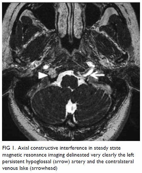
A 44-year-old woman presented to the emergency department in December 2014 with acute severe right-sided headache that began at the occipital region and spread to the right temporal and frontal regions. Pain was only partially relieved by analgesics. The patient had no history of altered mental state or focal neurological deficits. Her medical history was unremarkable except for a road traffic accident a few months previously ago with consequent right lower limb trauma. Neurological assessment revealed no gross abnormality. An urgent non-contrast brain computed tomography (CT) scan showed no intracranial haemorrhage or other abnormalities. Due to persistence of symptoms the patient underwent brain magnetic resonance (MR) imaging at a private centre. A small skull lesion was evident on the right basiocciput for which further imaging study was requested at our hospital. A contrast 3-T MR scan with angiography sequences revealed that the previously reported lesion corresponded to a 0.7-cm T1-weighted isointense and T2-weighted hyperintense structure located at the right hypoglossal canal, which was expanded. It exhibited intense contrast enhancement and was in direct continuity with the inferior petrosal sinus and the internal venous plexus around the foramen magnum, all findings suggestive of a venous lake at the right hypoglossal canal (Fig 1).
 

Figure 1. Axial constructive interference in steady state magnetic resonance imaging delineated very clearly the left persistent hypoglossal (arrow) artery and the contralateral venous lake (arrowhead)
 
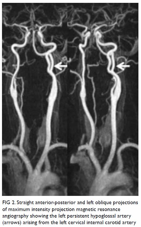
Additionally, an anomalous vessel arising from the left internal carotid artery at C2 level was noted, entering the cranium through the left hypoglossal canal and joining the basilar artery. This anomalous vessel corresponded to a left persistent hypoglossal artery (PHA; Figs 1 and 2). The bilateral cervical vertebral arteries were diminutive in calibre and did not serve as major arterial supplies to the basilar artery. No intracranial aneurysms were detected and no infarction or other abnormality was noted. The patient’s symptoms later substantially improved with symptomatic treatment.
 

Figure 2. Straight anterior-posterior and left oblique projections of maximum intensity projection magnetic resonance angiography showing the left persistent hypoglossal artery (arrows) arising from the left cervical internal carotid artery
 
Discussion
Bony venous lakes of the skull are common and asymptomatic, and they are typically parasagittal in location. In CT scans, they appear as lucent lesions with corticated/sclerotic margins. In MR imaging, they exhibit the same signal characteristics as veins. However, it is rare to find venous lakes located at the hypoglossal canal, and other entities must be excluded such as a neurinoma or even a dural arteriovenous fistula of the hypoglossal canal, another rare but potentially symptomatic condition that may follow head trauma.1 In this case, there was no apparent arteriovenous shunt detected in the MR angiography sequences.
 
A PHA results from failure of regression of a primitive hypoglossal artery, one of the several anastomoses that exist between the carotid and vertebrobasilar arteries during embryogenesis. Although rare, it is the second most common persistent carotid-vertebrobasilar anastomosis after the trigeminal artery, with a prevalence of up to 0.29%,2 usually representing an incidental finding. However, diagnosis of PHA is important because it is often the only blood supply to the basilar trunk, as vertebral arteries are usually hypoplastic. Moreover, PHA is associated with intracranial arterial aneurysms, ischaemic cerebrovascular attacks, subarachnoid haemorrhage and arteriovenous malformations.3 Recognition of PHA is extremely important before any endovascular procedure, carotid endarterectomy or skull base surgery is performed. Exposure of the basilar trunk to an unusual haemodynamic stress could be the underlying mechanism that predisposes an individual to the development of aneurysms.4 On the contrary, there is an increased risk of ischaemia caused by embolism from the internal carotid artery to the posterior circulation through the PHA.5
 
Both vascular anomalies in this patient were most likely incidental findings; however, owing to the reported association of PHA with intracranial aneurysm development and ischaemic events, any new episode or the development of neurological symptoms should have triggered immediate imaging study.
 
To the best of our knowledge, this is the first report of a PHA with a contralateral hypoglossal canal venous lake, both representing rare vascular variants.
 
Author contributions
All authors contributed to the concept of study, acquisition and analysis of data, drafting of the article, and critical revision for important intellectual content. All authors had full access to the data, contributed to the study, approved the final version for publication, and take responsibility for its accuracy and integrity.
 
Conflicts of interest
All authors have disclosed no conflicts of interest.
 
Funding/support
This case report received no specific grant from any funding agency in the public, commercial, or not-for-profit sectors.
 
Ethics approval
The patient was treated in accordance with the Declaration of Helsinki. The patient provided informed consent for all procedures.
 
References
1. Manabe S, Satoh K, Matsubara S, Satomi J, Hanaoka M, Nagahiro S. Characteristics, diagnosis and treatment of hypoglossal canal dural arteriovenous fistula: report of nine cases. Neuroradiology 2008;50:715-21. Crossref
2. Uchino A, Saito N, Okada Y, et al. Persistent hypoglossal artery and its variants diagnosed by CT and MR angiography. Neuroradiology 2013;55:17-23. Crossref
3. Srinivas MR, Vedaraju KS, Manjappa BH, Nagaraj BR. Persistent primitive hypoglossal artery (PPHA)—a rare anomaly with literature review. J Clin Diagn Res 2016;10:TD13-4.
4. Terayama R, Toyokuni Y, Nakagawa S, et al. Persistent hypoglossal artery with hypoplasia of the vertebral and posterior communicating arteries. Anat Sci Int 2011;86:58-61. Crossref
5. Conforto AB, de Souza M, Puglia P Jr, Yamamoto FI, da Costa Leite C, Scaff M. Bilateral occipital infarcts associated with carotid atherosclerosis and a persistent hypoglossal artery. Clin Neurol Neurosurg 2007;109:364-7. Crossref

Pulmonary metastasis from a World Health Organization grade I intracranial parasagittal meningioma: a case report

© Hong Kong Academy of Medicine. CC BY-NC-ND 4.0
 
CASE REPORT
Pulmonary metastasis from a World Health Organization grade I intracranial parasagittal meningioma: a case report
Peter YM Woo, MB, BS, FRCS; Remy SL Hung, MB, BS, MRCS; Saori Takemura, MB, ChB; KY Chan, MB, ChB, FRCS; John CK Kwok, MB, ChB, FRCS
Department of Neurosurgery, Kwong Wah Hospital, Yaumatei, Hong Kong
 
Corresponding author: Dr Peter YM Woo (wym307@ha.org.hk)
 
 Full paper in PDF
 
Case report
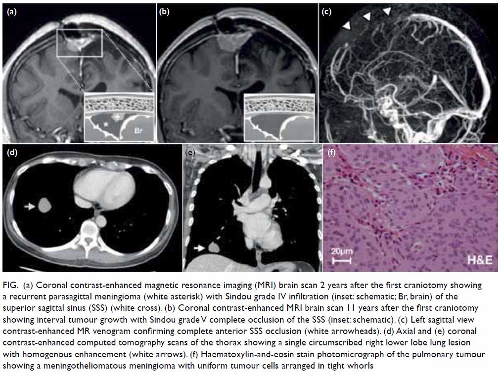
A 37-year-old woman presented to our neurosurgical centre in January 2003 with a 2-month history of progressive blurred vision and was found to have papilloedema. Magnetic resonance imaging (MRI) scan of the brain revealed a large left frontal parasagittal extra-axial dural-based tumour with homogenous gadolinium contrast-enhancement (6.3 cm × 4.3 cm × 3.7 cm) and Sindou grade II invasion (ie, into the lateral recess) into the junction of the anterior-to-middle third superior sagittal sinus (SSS). The patient underwent preoperative polyvinyl alcohol particle catheter tumour embolisation and a subsequent craniotomy was performed for Simpson’s grade III excision (macroscopic complete excision without resection of the tumour’s extra-dural extension into the SSS). The histological diagnosis was a World Health Organization (WHO) grade I meningothelial meningioma with a Ki-67 proliferation index of 5%. A 2-year surveillance MRI scan (Fig 1a) revealed an asymptomatic local recurrence with further invasion into the SSS (Sindou grade IV, ie, involvement of the roof and lateral wall). The patient was asymptomatic and reluctant to undergo further treatment, opting for regular observation of the lesion. A new MRI scan performed 11 years after the first operation revealed interval tumour growth with complete occlusion of the SSS (Sindou grade V) that was confirmed with MR venography (Fig 1b and c). A second craniotomy was performed in October 2014, 11 years after the first, but only subtotal excision could be achieved because of dense tumour adhesions to a large posterior frontal cortical draining vein. The histology remained that of a WHO grade I meningioma.
 

Figure. (a) Coronal contrast-enhanced magnetic resonance imaging (MRI) brain scan 2 years after the first craniotomy showing a recurrent parasagittal meningioma (white asterisk) with Sindou grade IV infiltration (inset: schematic; Br, brain) of the superior sagittal sinus (SSS) (white cross). (b) Coronal contrast-enhanced MRI brain scan 11 years after the first craniotomy showing interval tumour growth with Sindou grade V complete occlusion of the SSS (inset: schematic). (c) Left sagittal view contrast-enhanced MR venogram confirming complete anterior SSS occlusion (white arrowheads). (d) Axial and (e) coronal contrast-enhanced computed tomography scans of the thorax showing a single circumscribed right lower lobe lung lesion with homogenous enhancement (white arrows). (f) Haematoxylin-and-eosin stain photomicrograph of the pulmonary tumour showing a meningotheliomatous meningioma with uniform tumour cells arranged in tight whorls
 
From a preoperative chest X-ray, performed in preparation for the patient’s second craniotomy, a new opacity in the right lower lobe was incidentally discovered. Computed tomography scan of the thorax revealed a single right lower lobe lung nodule (2.4 cm × 2.8 cm × 2.3 cm) with a well-defined border and vivid homogenous contrast enhancement (Fig 1d and e). Video-assisted thoracoscopic wedge resection of the right lower lobe was performed 8 weeks after the craniotomy with gross total excision achieved. The final pathological diagnosis was a metastatic WHO grade I meningioma with a Ki-67 proliferation index of 1% and clear margins (Fig 1f).
 
In view of residual intracranial disease, the patient underwent adjuvant fractionated radiotherapy (50.4 Gy). At 2 years after the second craniotomy, surveillance MRI brain scans and chest X-rays showed no detectable tumour.
 
Discussion
Meningiomas are the most frequently diagnosed primary brain tumour in adults, accounting for 13% to 26% of all lesions.1 The population incidence is estimated to be four to six per 100 000 with a female:male ratio of 2:1. Despite this high prevalence, distant (extracranial) metastasis is extremely rare with fewer than 120 cases reported.1
 
The grading of meningiomas is principally determined by light microscopy of haematoxylin-eosin sections in accordance with WHO criteria. Grade I intracranial meningiomas comprise 80% of tumours and are generally considered benign, slow-growing lesions that have no demonstrable malignant behaviour such as distant metastasis. However, contrary to this belief, one third of meningiomas with distant metastases originate from grade I tumours with 31% identified incidentally.1 In contrast, grade III lesions, which demonstrate overt aggressive behaviour, represent only 1% of meningiomas and account for 40% of documented metastases.1 The true incidence of metastatic meningiomas is unknown, but given the frequent occurrence of grade I tumours, that metastatic lesions are often asymptomatic and that routine whole-body imaging is seldom performed, the stated figure of 0.1% is likely an underestimation.1 In our case, the interval between primary resection and metastasis detection was 11 years, considerably longer than the cited median duration of 58 months (range, 4 months to 15 years), reflecting the slow-growing nature of grade I tumours.1
 
Three quarters of metastatic WHO grade I meningiomas involve a single organ, primarily the lung (42%) followed by the spine (12%), bone (10%), liver (10%), and cervical lymph nodes (10%).1 Although conventional histological studies such as the Ki-67 proliferation index have failed to identify a subgroup of meningiomas predisposed to metastasis, loss of heterozygosity of 1p, 9p, 14q and 22q may be characteristic of these lesions.2 Clinical risk factors for metastasis include repeated surgery, local recurrence and invasion of the dural venous sinuses.1 The non-collapsible and valve-less nature of the dural venous sinuses, such as the SSS, may permit seeding of tumour cells into the internal jugular vein and subsequently into the pulmonary microcirculation, an indication that tumour location is pivotal in determining haematogenous metastasis.1 Parasagittal meningiomas, comprising 20% to 34% of lesions, are perhaps most susceptible because of their propensity to invade the SSS, technically hindering their complete resection.3 4 Our case illustrates the importance of treating the SSS infiltrating portion of these tumours, but there is little consensus on the appropriate management strategy. When the posterior SSS is patent, prohibiting its ligation and excision, some neurosurgeons prefer subtotal resection followed by adjuvant radiosurgery or radiotherapy.3 Others advocate the more technically demanding surgical approach of gross total resection with sinus reconstruction, to spare the patient the long-term adverse effects of irradiation.4 Both strategies offer comparable tumour control rates although multimodality treatment may be associated with fewer procedure-related complications.3
 
Bronchogenic carcinoma is the most important differential diagnosis to exclude in patients with pulmonary meningioma metastasis, but it is difficult to distinguish on computed tomography imaging. Meningioma metastases are usually single, non-calcified well-circumscribed lesions that may display strong homogenous contrast-enhancement.1 111Indium-octreotide imaging is useful in identifying meningiomas, exhibiting avid uptake, but its restricted availability limits its use.5 Excision of the pulmonary lesion is recommended to establish the diagnosis and in some instances the meningioma metastasis may manifest a more aggressive grading than the primary lesion warranting adjuvant radiotherapy.5 When multiple disseminated metastases preclude surgical excision, systemic treatments such as octreotide acetate or bevacizumab, an anti-angiogenic therapy directed against vascular-endothelial growth factor have shown some promise for tumour control.5 However, in a case series of patients with recurrent meningioma refractory to surgery, radiotherapy and chemotherapy, pulmonary metastasis was identified as an unfavourable prognostic factor for overall survival.5
 
Distant metastasis from a WHO grade I meningioma is a rare phenomenon and can occur more than a decade after the initial diagnosis of the primary tumour. This case demonstrates that, regardless of grading and especially when the patient is young, meningiomas that infiltrate the dural venous sinuses require proactive management, either by adjuvant irradiation or by gross total resection.
 
Author contributions
All authors had full access to the data, contributed to the study, approved the final version for publication, and take responsibility for its accuracy and integrity.
 
Concept of study: PYM Woo, RSL Hung.
Acquisition of data: PYM Woo, RSL Hung.
Analysis of data: PYM Woo, RSL Hung, S Takemura.
Drafting of the article: PYM Woo, RSL Hung, S Takemura.
Critical revision for important intellectual content: All authors.
 
Conflicts of interest
All authors have disclosed no conflicts of interest.
 
Funding/support
This research received no specific grant from any funding agency in the public, commercial, or not-for-profit sectors.
 
Ethics approval
Ethics committee approval was waived because this is a case report and no personal identifying information was disclosed. A signed patient consent statement was obtained.
 
References
1. Surov A, Gottschling S, Bolz J, et al. Distant metastases in meningioma: an underestimated problem. J Neurooncol 2013;112:323-7. Crossref
2. Gladin CR, Salsano E, Menghi F, et al. Loss of heterozygosity studies in extracranial metastatic meningiomas. J Neurooncol 2007;85:81-5. Crossref
3. Gatterbauer B, Gevsek S, Höftberger R, et al. Multimodal treatment of parasagittal meningiomas: a single-center experience. J Neurosurg 2017;127:1249-56. Crossref
4. Ricci A, Di Vitantonio H, De Paulis D, et al. Parasagittal meningiomas: our surgical experience and the reconstruction technique of the superior sagittal sinus. Surg Neurol Int 2017;8:1. Crossref
5. Alexandru D, Glantz MJ, Kim L, Chamberlain MC, Bota DA. Pulmonary metastases in patients with recurrent, treatment-resistant meningioma: prognosis and identification by 111Indium-octreotide imaging. Cancer 2011;117:4506-11. Crossref

Lysergic acid diethylamide–associated intoxication in Hong Kong: a case series

© Hong Kong Academy of Medicine. CC BY-NC-ND 4.0
 
CASE REPORT
Lysergic acid diethylamide–associated intoxication in Hong Kong: a case series
C Li, MB, BS1,2; Magdalene HY Tang, PhD1; YK Chong, FHKCPath, FHKAM (Pathology)1,2; Tina YC Chan, MB, ChB, PhD1,2; Tony WL Mak, FRCPath, FHKAM (Pathology)1,2
1 Hospital Authority Toxicology Reference Laboratory, Hong Kong
2 Chemical Pathology Laboratory, Department of Pathology, Princess Margaret Hospital, Laichikok, Hong Kong
 
Corresponding author: Dr Tony WL Mak (makwl@ha.org.hk)
 
 Full paper in PDF
 
Case series
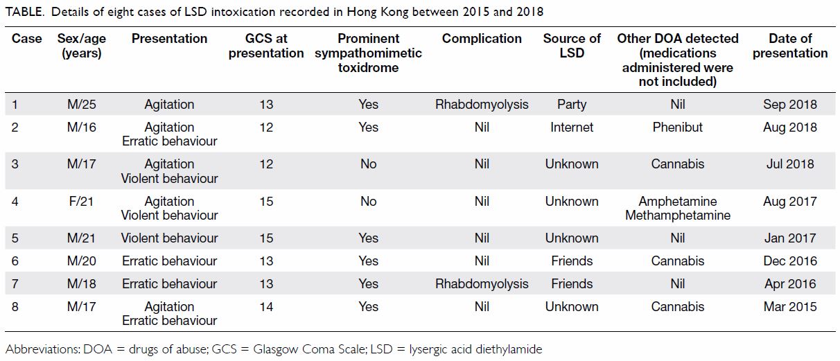
Lysergic acid diethylamide (LSD) is a powerful hallucinogenic drug that was first synthesised in 1938.1 Although LSD is considered a conventional drug of abuse, cases of LSD intoxication are scarce in Hong Kong. The Hospital Authority Toxicology Reference Laboratory—the only tertiary referral laboratory for toxicological analysis in Hong Kong, established in 2004—did not encounter cases of LSD intoxication until 2015. Between 2015 and 2018, eight cases of LSD-associated intoxication were identified at five acute hospitals in Hong Kong when LSD and its metabolites were detected in patient urine samples. Details of these eight patients (7 male, 1 female; age range, 16-25 years) are presented in the Table. The most common presentation of LSD intoxication in these patients was agitation (63%), followed by erratic behaviour (50%) and violent behaviour (38%). Impaired level of consciousness (75%) and apparent sympathomimetic toxidrome (75%) were documented in most patients. History of LSD use was elicited in all cases. However, only four patients were willing to volunteer the sources of LSD: one bought LSD from the Internet, one obtained LSD at a party, and two obtained LSD from friends. The most common co-ingestant was cannabis, which was detected in three cases. Amphetamine and methamphetamine were detected in one case. In one patient, phenibut (3-phenyl-4-aminobutyric acid), a central nervous system depressant structurally related to gamma-aminobutyric acid, was detected. Two cases were complicated by rhabdomyolysis and one of them required intensive care unit admission. The clinical details of these two cases are presented below.
 

Table. Details of eight cases of LSD intoxication recorded in Hong Kong between 2015 and 2018
 
Case 1
A 25-year-old man who had a history of childhood asthma presented to Princess Margaret Hospital, Hong Kong, in September 2018 with agitation after sublingual use of LSD on a stamp at a party. At presentation, his Glasgow Coma Scale was 13/15 (E4, V4, M5). The patient’s blood pressure was 116/80 mm Hg, heart rate was 150 beats per minute, body temperature was 39.2℃, and pupil sizes were 5 mm bilaterally. Biochemical investigations showed a peak creatine kinase (CK) value of 6260 U/L, and urine myoglobin was positive. The patient was intubated and treated with alkaline diuresis in the intensive care unit. The urine specimen was analysed in the Toxicology Reference Laboratory, where LSD, its metabolite (2-oxo-3-hydroxy-LSD), diazepam, nordiazepam, temazepam, midazolam and propofol were detected.
 
Case 2
An 18-year-old man who enjoyed good past health presented to United Christian Hospital, Hong Kong, in April 2016 with erratic behaviour after sublingual use of LSD on a stamp obtained from his friends. At presentation, his Glasgow Coma Scale was 13/15 (E4, V4, M5). The patient’s blood pressure was 127/50 mm Hg, heart rate was 167 beats per minute, body temperature was 38.7℃, and pupil sizes were 6 mm bilaterally. Biochemical investigations showed metabolic acidosis, a peak CK value of 14 732 U/L, and urine myoglobin was positive. In the urine specimen, LSD, its metabolite (2-oxo-3-hydroxy-LSD), and lidocaine were detected.
 
Discussion
Classically described as a psychedelic or hallucinogenic agent, LSD is structurally similar to serotonin (5-hydroxytryptamine), an important neurotransmitter in the central nervous system. It acts as a serotonin receptor agonist in the central nervous system and may produce prominent visual hallucinations, audiovisual synaesthesia, and derealisation. Significant sympathomimetic stimulation has also been observed.2 The effects typically begin within 30 to 60 minutes, peak at around 2 hours and can last for up to 12 hours after intake.2 These effects are consistent with the current findings that most patients presented with apparent sympathomimetic toxidrome, characterised by tachycardia, hypertension, mydriasis, and pyrexia.
 
There is a general impression promulgated over the Internet and by celebrities that LSD is harmless and even beneficial to personal development. Recently, LSD has re-emerged as a micro-dosing psychedelic. People consume regular low doses of LSD in an attempt to boost their creativity.3 However, this practice, also described as the Silicon Valley trend, lacks scientific evidence. These factors appear to have misled the public into underestimating the potential sequelae of LSD abuse. In contrast, our case series clearly demonstrates that LSD intoxication is associated with severe sequelae. In both cases complicated with rhabdomyolysis, no other stimulant-class drugs of abuse were detected, including conventional and emerging drugs of abuse.4 No better alterative causes of rhabdomyolysis were identified from the medical or drug history and biochemical investigations; LSD intoxication was the major contributing factor to rhabdomyolysis in both cases. Other cases of LSD-associated rhabdomyolysis have been reported in the literature.5 6 Fortunately, all patients in our series recovered uneventfully. However, at least one fatal case has been reported elsewhere.7
 
Frontline clinicians should be aware that LSD has re-appeared, disguised as a “safe” drug of abuse associated with multiple local intoxication cases with severe sequelae including rhabdomyolysis. Thorough investigations and serial monitoring are required to detect complications. Urine toxicology is useful to confirm the exposure to drugs of abuse. However, owing to its high potency, the dosage of LSD taken is small (in micrograms) and urine levels may be very low.2 A sensitive analytical system is required to detect the presence of LSD and its metabolite. Public education on the dangers or LSD abuse, and effective regulatory control by the government sectors are recommended.
 
Author contributions
All authors had full access to the data, contributed to the study, approved the final version for publication, and take responsibility for its accuracy and integrity.
 
Concept or design of study: All authors.
Acquisition of data: All authors.
Analysis or interpretation of data: All authors.
Drafting of the article: C Li, MHY Tang, TWL Mak.
Critical revision for important intellectual content: All authors.
 
Conflicts of interest
All authors have disclosed no conflicts of interest.
 
Funding/support
This research received no specific grant from any funding agency in the public, commercial, or not-for-profit sectors.
 
Ethics approval
Ethics approval for this study was granted by the Hong Kong Hospital Authority Kowloon West Cluster Research Ethics Committee (Ref KW/EX-19-003). The Committee exempted the study group from obtaining patient consent.
 
References
1. Nichols DE, Grob CS. Is LSD toxic? Forensic Sci Int 2018;284:141-5. Crossref
2. Dolder PC, Schmid Y, Steuer AE, et al. Pharmacokinetics and pharmacodynamics of lysergic acid diethylamide in healthy subjects. Clin Pharmacokinet 2017;56:1219-30. Crossref
3. Anderson T, Petranker R, Rosenbaum D, et al. Microdosing psychedelics: personality, mental health, and creativity differences in microdosers. Psychopharmacology (Berl) 2019;236:731-40. Crossref
4. Tang M, Ching CK, Tse ML, et al. Surveillance of emerging drugs of abuse in Hong Kong: validation of an analytical tool. Hong Kong Med J 2015;21:114-23. Crossref
5. Mercieca J, Brown EA. Acute renal failure due to rhabdomyolysis associated with use of a straitjacket in lysergide intoxication. Br Med J (Clin Res Ed) 1984;288:1949-50. Crossref
6. Berrens Z, Lammers J, White C. Rhabdomyolysis after LSD ingestion. Psychosomatics 2010;51:356-356.e3. Crossref
7. Fysh RR, Oon MC, Robinson KN, Smith RN, White PC, Whitehouse MJ. A fatal poisoning with LSD. Forensic Sci Int 1985;28:109-13. Crossref

Per urethral insertion of foreign body for erotism: case reports

© Hong Kong Academy of Medicine. CC BY-NC-ND 4.0
 
CASE REPORT
Per urethral insertion of foreign body for erotism: case reports
Christy WH Mak, MB, ChB; CL Cho, FRCSEd (Urol), FHKAM (Surgery); Wayne KW Chan, FRCSEd (Urol), FHKAM (Surgery); Ringo WH Chu, FRCSEd (Urol), FHKAM (Surgery); IC Law, FRCSEd (Urol), FHKAM (Surgery)
Division of Urology, Department of Surgery, Kwong Wah Hospital, Yaumatei, Hong Kong
 
Corresponding author: Dr CL Cho (chochaklam@yahoo.com.hk)
 
 Full paper in PDF
 
Case reports
Patient 1
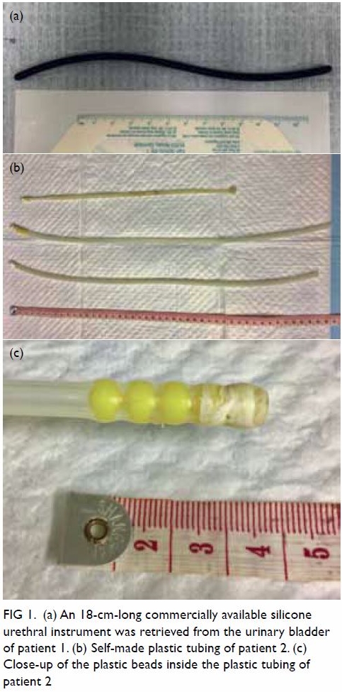
A 31-year-old man presented in December 2016 with a 1-day history of retained foreign body in the urinary bladder. He reported regular insertion of a commercially available silicone urethral instrument for autoerotism. He customarily evacuated the instrument by voiding but he had failed to do so on this occasion. There was no dysuria, haematuria, abdominal pain or fever. Laboratory parameters were within normal limits and no foreign body could be identified on X-ray. Flexible cystoscopy revealed an 18-cm length of soft tubing inside the urinary bladder (Fig 1a). Successful retrieval was achieved with endoscopic forceps. He was discharged home on the same day.
 

Figure 1. (a) An 18-cm-long commercially available silicone urethral instrument was retrieved from the urinary bladder of patient 1. (b) Self-made plastic tubing of patient 2. (c) Close-up of the plastic beads inside the plastic tubing of patient 2
 
Patient 2
A 57-year-old man was admitted in January 2017 with a 6-week history of intermittent fever and right scrotal swelling. He had no lower urinary tract symptoms except for a yellowish urethral discharge. He revealed a history of regular per urethral insertion of self-customised plastic tubing with plastic beads inside the lumen for sexual gratification (Fig 1b and c). The patient cleaned the tubing with tap water after each use. Physical examination revealed a tender and swollen right testis with fluctuance, indicative of abscess formation. Serum white blood cell count was 15.1 × 10⁹/L. Drainage of scrotal abscess was performed and the patient was discharged home with a course of antibiotics.
 
Patient 3
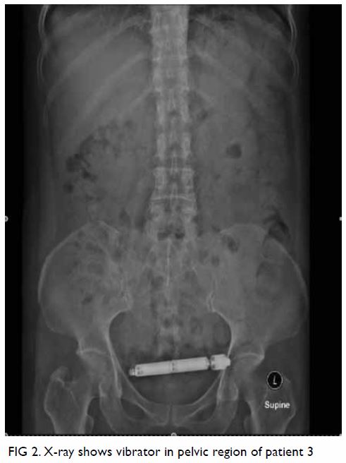
A 55-year-old woman was admitted in May 2017 with a 1-day history of retained vibrator in the urinary bladder. The vibrator was inserted into her urethra during sexual activity with her husband. She complained of subsequent dysuria and haematuria. Serum white blood cell count was mildly elevated. X-ray revealed a 12.5-cm foreign body in the pelvic region (Fig 2). Retrieval of the vibrator via flexible cystoscopy failed owing to inadequate size and strength of the cystoscopic forceps. Semi-rigid ureteroscopy was used to locate the vibrator with Kocher forceps placed alongside for removal. Removal of the vibrator was achieved and the patient was discharged home with antibiotics.
 

Figure 2. X-ray shows vibrator in pelvic region of patient 3
 
Discussion
Polyembolokoilamania refers to the act of inserting a foreign body into body orifices. Urethral polyembolokoilamania is not uncommon and several cases have been reported in the literature. Different types of foreign bodies have been reportedly introduced into the urethra, including electric wires, thermometers, hairpins, toothpicks, eyeglass rims, straws, pencils, paperclips, and fishhooks.1 2 Autoerotism is one of the most common reasons given for polyembolokoilamania, although others include curiosity, psychiatric illness, iatrogenic causes, and an attempt to relieve urinary symptoms.3 The prevalence of the condition is unknown and probably underreported owing to embarrassment of patients so a high index of suspicion and targeted history taking play an important role in correctly diagnosing the condition. There are reports suggesting the possible role of psychiatric assessment in revealing an underlying psychiatric disease and preventing repeated occurrences of foreign body insertion.4 Nonetheless psychiatric assessment is not considered mandatory for all patients. The prevalence of psychiatric diagnoses in this group of patients and the efficacy of psychiatric interventions are still largely unknown. There is no consensus on the role of psychiatric consultation for patients with urethral polyembolokoilamania.
 
Various complications may arise from insertion of foreign bodies per urethra including urinary tract infection, urethral injury and, most commonly, retained foreign body. The presentation of retained foreign body in the lower urinary tract varies widely although dysuria and haematuria are the most common presentation.3 5 The severity of the condition depends on the nature of the foreign body and, more importantly, time to medical attention. Early diagnosis and removal of the foreign body are of prime importance. In our case series, patients 1 and 3 sought early medical advice and experienced no complications. Late presentation may lead to complications such as recurrent urinary tract infection and urolithiasis or, less commonly, erosion of foreign body to adjacent organs.5 Presenting time is highly variable, ranging from hours to months.1 Delay in seeking medical consultation may stem from ignorance, psychiatric illness, or embarrassment.1
 
X-ray is often performed but is helpful in identifying only radio-opaque objects. Instead, flexible cystoscopy should be performed early for all patients with suspected retained foreign body in the lower urinary tract, in view of the relative simplicity and low risk of the procedure. Flexible cystourethroscopy can achieve accurate diagnosis and treatment simultaneously. A high success rate of 50% to 93% has been reported for endoscopic removal of foreign body.1 3 Computed tomography scan is indicated only in patients with peritonitis or suspected foreign body migration to adjacent organs.3
 
A surgical approach for retrieval of retained foreign body depends on the size, shape and location of the object. Simple measures, for example milking of the urethra, usually have a high success rate for expulsion of foreign body located in the distal penile urethra.3 Invasive approaches including open cystostomy and perineal urethrostomy have been reported when retrieving larger size, encrusted, or sharp objects after failed endoscopic manoeuvres.1 More complex procedures, including laparotomy and urinary diversion may be indicated for complicated cases. The surgical approach should be individualised.5
 
Insertion of foreign bodies into the urethra is predominantly reported in men. Palmer et al3 reported that 97% of their patients were men during their 15 years’ experience of managing self-insertion of urethral foreign bodies and six of their 27 patients had a recurrent history. This male-dominated phenomenon is also evidenced by the wide availability of male sexual urethral instruments in shops and online platforms. These instruments, namely urethral sounds and dilators, generally constitute a 15- to 20- cm-long stainless steel or silicone tube that is used for deep penetration to the level of the prostatic urethra. Deep prostate stimulation-associated orgasms are thought to be more intense than those with direct penile stimulation, and involve more pelvic muscle contractions.6 However, hygiene of the instrument is a concern as instrumentation without proper disinfection subjects a patient to further risk of infective complications.
 
In conclusion, insertion of a foreign body into the lower urinary tract for erotism is not uncommon, especially in men. Early diagnosis and endoscopic removal of the foreign body achieve a high success rate and should be considered the initial management. Delayed presentation may lead to complications that require further imaging and more complex surgical procedures.
 
Author contributions
All authors had full access to the data, contributed to the study, approved the final version for publication, and take responsibility for its accuracy and integrity.
 
Concept or design: CL Cho.
Acquisition of data: CWH Mak.
Analysis or interpretation of data: CL Cho, CWH Mak.
Drafting of the article: CWH Mak, CL Cho.
Critical revision for important intellectual content: All authors.
 
Conflicts of interest
The authors have disclosed no conflict of interest.
 
Funding/support
This research received no specific grant from any funding agency in the public, commercial, or not-for-profit sectors.
 
Ethics approval
Verbal consent was obtained for the purpose of case study.
 
References
1. Rafique M. Intravesical foreign bodies: review and current management strategies. Urol J 2008;5:223-31.
2. Rahman NU, Elliott SP, McAninch JW. Self-inflicted male urethral foreign body insertion: endoscopic management and complications. BJU Int 2004;94:1051-3. Crossref
3. Palmer CJ, Houlihan M, Psutka SP, Ellis KA, Vidal P, Hollowell CM. Urethral foreign bodies: clinical presentation and management. Urology 2016;97:257-60. Crossref
4. Unruh BT, Nejad SH, Stern TW, Stern TA. Insertion of foreign bodies (polyembolokoilamania): underpinnings and management strategies. Prim Care Companion CNS Disord 2012;14(1). pii: PCC.11f01192. Crossref
5. Cury J, Coelho RF, Srougi M. Retroperitoneal migration of a self-inflicted ballpoint pen via the urethra. Int Braz J Urol 2006;32:193-5. Crossref
6. Alwaal A, Breyer BN, Lue TF. Normal male sexual function: emphasis on orgasm and ejaculation. Fertil Steril 2015;104:1051-60. Crossref

Next-generation sequencing panel for diagnosis and management of chronic neutrophilic leukaemia: a case report

© Hong Kong Academy of Medicine. CC BY-NC-ND 4.0
 
CASE REPORT
Next-generation sequencing panel for diagnosis and management of chronic neutrophilic leukaemia: a case report
KY Mak, MB, ChB1; CH Au, PhD2; TL Chan, PhD2; Edmond SK Ma, MD, FHKAM (Pathology)2; Eudora YD Chow, MB, BS, FHKAM (Pathology)1; SY Lin, MB, BS, FHKAM (Medicine)3; William WL Choi, MB, BS, FHKAM (Pathology)1
1 Department of Pathology, United Christian Hospital, Kwun Tong, Hong Kong
2 Department of Pathology, Hong Kong Sanatorium & Hospital, Happy Valley, Hong Kong
3 Department of Medicine and Geriatrics, United Christian Hospital, Kwun Tong, Hong Kong
 
Corresponding author: Dr William WL Choi (wlchoi@hotmail.com)
 
 Full paper in PDF
 
Case report
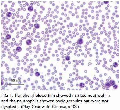
An 80-year-old woman presented to our hospital with mild headache in November 2015. She had a history of hypertension, diabetes mellitus, and hyperlipidaemia. She was febrile (38.6°C) but did not appear septic. Abdominal examination revealed mild splenomegaly but no hepatomegaly and there were no focal neurological signs or suggestions of other organ involvement. A full blood count showed leukocytes 124.0 × 10⁹/L (neutrophils 120.3 × 10⁹/L, lymphocytes 2.5 × 10⁹/L, monocytes 1.2 × 10⁹/L); haemoglobin 7.8 g/dL, mean corpuscular volume 89.1 fL; and platelets 384 × 109/L. The blood film showed marked neutrophilia, occasional myelocytes, and absolute basophilia but no blasts. The neutrophils showed toxic granules and were not dysplastic (Fig 1). Plain radiographs of the chest, kidney, ureter, and urinary bladder did not reveal any abnormalities. Bacterial cultures of the blood and urine did not reveal any septic foci. Because of the marked neutrophilia, the patient was initially treated for bacterial sepsis with empirical intravenous amoxicillin with clavulanic acid. Subsequent ultrasonography of the abdomen confirmed the splenomegaly (15.1 cm) but no other space-occupying lesions. The low-grade fever soon subsided after admission and she remained afebrile and non-septic, although the marked neutrophilia persisted.
 

Figure 1. Peripheral blood film showed marked neutrophilia, and the neutrophils showed toxic granules but were not dysplastic (May-Grünwald-Giemsa, ×400)
 
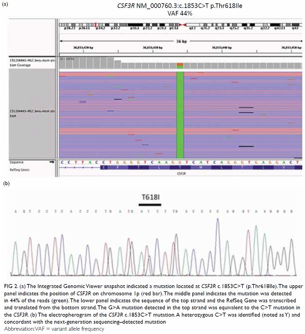
A bone marrow biopsy revealed marked hypercellularity, primarily due to markedly increased granulopoiesis with left-shift in maturation but no increase in blasts. Erythropoiesis was active and normoblastic. Megakaryocytes were moderately increased with some being large and hyperlobulated. No overt dysplasia was seen. Cytogenetic karyotyping showed a normal karyotype. Reverse transcription polymerase chain reaction (PCR) for BCR-ABL1 and allele-specific PCR for Janus kinase 2 (JAK2) V617F mutation analysis were negative. In view of the clinical and laboratory picture of a possible myeloproliferative neoplasm, and the lack of a clonal marker detected by the molecular tests, we sought to utilise a next-generation sequencing (NGS) panel (TruSight Myeloid Sequencing Panel; Illumina, San Diego [CA], United States) to look for possible mutations in selected exons of 54 different genes commonly implicated in myeloid neoplasms, in accordance with a previously published protocol.1 Deep sequencing by this panel returned three pathogenic mutations: colony-stimulating factor 3 receptor (CSF3R) c.1853C>T; p.Thr618Ile or T618I at variant allele frequency (VAF) of 43.9% (Fig 2a), serine/arginine-rich splicing factor 2 (SRSF2) c.284C>T; p.Pro95Leu (NM_003016.4) or P95L at VAF of 49.6%, and additional sex combs like 1 (ASXL1) truncating mutation c.1934dupG; p.Gly646Trpfs*12 (NM_015338.5) at VAF of 34.1%. The CSF3R T618I mutation was further confirmed by Sanger sequencing (Fig 2b), thus prompting the diagnosis of chronic neutrophilic leukaemia (CNL). Although the CSF3R mutation rendered the disease amenable to ruxolitinib, the patient deteriorated rapidly and died of sudden severe gastrointestinal bleeding 12 days after admission and before specific therapy could be contemplated.
 

Figure 2. (a) The Integrated Genomic Viewer snapshot indicated a mutation located at CSF3R c.1853C>T (p.Thr618Ile). The upper panel indicates the position of CSF3R on chromosome 1p (red bar). The middle panel indicates the mutation was detected in 44% of the reads (green). The lower panel indicates the sequence of the top strand and the RefSeq Gene was transcribed and translated from the bottom strand. The G>A mutation detected in the top strand was equivalent to the C>T mutation in the CSF3R. (b) The electropherogram of the CSF3R c.1853C>T mutation. A heterozygous C>T was identified (noted as Y) and concordant with the next-generation sequencing–detected mutation
 
Discussion
Several clinical features in this patient hinted at differential diagnoses other than bacterial infection or acute inflammation. Apart from fever, there were no other clinical features related to the presenting symptom of headache. Splenomegaly, a common feature of myeloproliferative neoplasms, was present. The absolute neutrophil count was very high, yet no clinical features of sepsis were found on physical examination or from investigations.
 
Since 2013, understanding of the molecular genetics of CNL has been dramatically changed by the discovery of CSF3R mutations in around 80% of cases.2 The CSF3R encodes a transmembrane receptor for granulocyte colony-stimulating factor 3, and plays a crucial role in the differentiation and maturation of neutrophils.2 The CNL-associated mutations in CSF3R activate the receptor and promote the proliferation and differentiation of neutrophils, leading to the marked neutrophilia that characterises the CNL disease phenotype.3 There are two major types of CSF3R mutations in CNL. The first encompasses point mutations in the extracellular or transmembrane domains, of which the T618I mutation is the most common and comprises the majority of mutations in CNL. The second type of CSF3R mutation comprises nonsense or frameshift mutations leading to a premature stop codon and truncation of the cytoplasmic domain of the receptor.2
 
Mutations of other genes have also been reported in CNL. These can be grouped as SET binding protein 1 (SETBP1) mutations, spliceosome mutations (eg, SRSF2), epigenetic modifier mutations (eg, ASXL1), and signalling mutations (eg, JAK2).3 One previous study found that SRSF2 mutations occurred in three of 14 cases of CNL (21%). The SRSF2 mutations were previously associated with a worse prognosis in chronic myelomonocytic leukaemia, but its effects on CNL are unclear.3 A significant proportion of CNL patients have been shown to harbour ASXL1 (30%-60%). Similar to mutations in other myeloid malignancies, ASXL1 mutations in CNL have been shown to confer a poor prognosis.3
 
Although these other mutations have not been incorporated into the World Health Organization 2016 diagnostic criteria for CNL, these data suggest that some may show prognostic value. Additionally, different CSF3R mutations may allow different therapeutic approaches (see below). Because Sanger sequencing of an increasing number of genes leads to substantial increases in the required time, resources, and necessary amount of DNA, we sought to explore an NGS panel to interrogate these genes simultaneously more efficiently and cost-effectively. Of note, the panel includes genes that are important for the diagnosis of myeloproliferative neoplasm (JAK2, calreticulin [CALR] and myeloproliferative leukaemia protein [MPL]), plus genes that are frequently reported in CNL (CSF3R, SETBP1, SRSF2, ASXL1). Therefore, compared with Sanger sequencing, NGS panels are a more efficient and powerful means to enable comprehensive genomic profiling of individual CNL cases, utilising a smaller amount of DNA.
 
With recent discoveries in the molecular pathogenesis of CNL, new therapeutic approaches that target the CSF3R signalling pathway–related SRC family and JAK-kinase pathways have emerged. The SRC signalling pathway is activated by truncation mutations of CSF3R leading to sensitivity to dasatinib, while the JAK-STAT pathway is activated by membrane proximal mutations of CSF3R leading to sensitivity to ruxolitinib.2 Although there are few reported cases of these agents,2 4 they represent significant breakthroughs in the management of CNL. This case of a rare myeloproliferative neoplasm demonstrates how advances in understanding of the molecular pathogenesis of a disease open up new routes for the development of effective novel therapeutic strategies.
 
Author contributions
All authors had full access to the data, contributed to the study, approved the final version for publication, and take responsibility for its accuracy and integrity.
 
Concept and design of study: WWL Choi.
Acquisition of data: KY Mak, CH Au, TL Chan, ESK Ma, SY Lin, WWL Choi.
Analysis or interpretation of data: KY Mak, CH Au, TL Chan, ESK Ma, WWL Choi.
Drafting of the manuscript: KY Mak, WWL Choi.
Critical revision for important intellectual content: CH Au, TL Chan, ESK Ma, EYD Chow, SY Lin.
 
Conflicts of interest
All authors have no conflicts of interest to declare.
 
Funding/support
This research received no specific grant from any funding agency in the public, commercial, or not-for-profit sectors.
 
References
1. Au CH, Wa A, Ho DN, Chan TL, Ma ES. Clinical evaluation of panel testing by next-generation sequencing (NGS) for gene mutations in myeloid neoplasms. Diagn Pathol 2016;11:11. Crossref
2. Maxson JE, Gotlib J, Pollyea DA, et al. Oncogenic CSF3R mutations in chronic neutrophilic leukemia and atypical CML. N Engl J Med 2013;368:1781-90. Crossref
3. Maxson JE, Tyner JW. Genomics of chronic neutrophilic leukemia. Blood 2017;129:715-22. Crossref
4. Stahl M, Xu ML, Steensma DP, Rampal R, Much M, Zeidan AM. Clinical response to ruxolitinib in CSF3R T618-mutated chronic neutrophilic leukemia. Ann Hematol 2016;95:1197-200. Crossref

Parapharyngeal abscess presenting as masticatory otorrhoea-persistent foramen tympanicum as a route of drainage: a case report

© Hong Kong Academy of Medicine. CC BY-NC-ND 4.0
 
CASE REPORT
Parapharyngeal abscess presenting as masticatory otorrhoea-persistent foramen tympanicum as a route of drainage: a case report
KH Lee, FRCR, FHKCR; YL Li, MB, BS, FRCR; ML Yu, MB, ChB, FRCR
Department of Radiology, Queen Mary Hospital, Pokfulam, Hong Kong
 
Corresponding author: Dr KH Lee (viclkh88@gmail.com)
 
 Full paper in PDF
 
Case report
A 26-year-old woman presented to the emergency department of Queen Mary Hospital in August 2014 with fever and right otorrhea following extraction of her right lower third molar 5 days previously. Physical examination revealed right buccal swelling, limited mouth opening, and pus-like discharge in the right external auditory canal (EAC) exacerbated by mastication. There was no hearing loss or facial nerve palsy. Otoscopy revealed granulation tissue and pus arising from the anterior wall of the inner right EAC.
 
Contrast-enhanced computed tomography scan showed a right parapharyngeal rim enhancing collection (Fig 1) tracking into the right temporomandibular fossa. Enhancing soft tissue was seen at the inner one-third of the right EAC. A bony defect was present at its anterior wall, compatible with a persistent foramen tympanicum (also known as foramen of Huschke), allowing communication between the EAC and the temporomandibular fossa (Fig 2). Overall findings were compatible with right parapharyngeal abscess discharging via a persistent foramen tympanicum into the right EAC.
 

Figure 1. Axial contrast-enhanced computed tomography of the neck of a 26-year-old woman showing rim-enhancing right parapharyngeal abscess extending to the right pterygomandibular space (white arrow)
 

Figure 2. (a) Coronal and (b) sagittal computed tomography images of a 26-year-old woman demonstrating superior extension of the right parapharyngeal abscess (white arrows) into the right external auditory canal (EAC) via a bony defect at the anterior wall of the inner EAC, compatible with persistent foramen tympanicum (dashed black arrow). The enhancing soft tissue mass seen within the EAC corresponds to the granulation tissue visualised on otoscopy (black arrow). Mottled contents compatible with exudates can be seen within the right EAC
 
The patient was treated with a course of antibiotics in view of her stable condition and lack of airway compromise. The patient responded clinically and repeat computed tomography scan 1 week after initial presentation showed complete resolution of the right parapharyngeal abscess and enhancing soft tissue within the right EAC. Subsequent bacterial culture of the right ear discharge yielded Streptococcus anginosus, a common cause of oral infection.
 
Discussion
Parapharyngeal abscesses are deep cervical infections with potential serious complications such as shock, mediastinitis, jugular vein thrombosis, upper airway obstruction, and death. Tonsillitis and odontogenic infection are the most common aetiologies. The clinical presentation typically involves fever, neck pain, odynophagia, neck oedema, and upper airway obstruction.1
 
Otorrhea is typically caused by external or middle ear pathologies such as otitis. Otorrhoea as a presenting symptom for a neck abscess is highly unusual. In our literature review, only two cases were found. Biron et al2 described a patient with a submandibular abscess that tracked into the ipsilateral external auditory meatus via the parapharyngeal and masticator spaces. Pepato et al3 reported a case of lower third molar infection presenting as purulent ear discharge, with persistent foramen tympanicum found in a follow-up cone-beam computed tomography study. The route of spread was postulated to be either via the Santorini fissures (the tiny defects in the anterior wall of the cartilaginous EAC) or via a persistent foramen tympanicum.
 
Persistent foramen tympanicum, first described by Emil Huschke in 1844, represents a failure of ossification of the tympanic part of the temporal bone and normally occurs from birth with completion by age 5 years. The foramen is located at the anteroinferior aspect of the EAC, just posterior to the temporomandibular joint. Incidence is quoted from 4.6% to 22.7% based on radiological and cadaveric studies.4
 
The majority of individuals with the foramen are asymptomatic although various complications have been reported. Most are benign, such as salivation from the ear during mastication and spontaneous herniation of the temporomandibular joint into the EAC leading to otalgia and tinnitus. Iatrogenic middle ear injury is possible when the foramen is inadvertently traversed during temporomandibular joint arthroscopy.5 It is possible that the lack of bony integrity reduces mechanical resistance to pathological processes, such as the spreading of parapharyngeal abscess in our case. Parotid pleomorphic adenomas have also been reported to herniate through the foramen to present as an EAC mass.
 
To the best of our knowledge, this is the first case to provide direct radiological evidence of persistent foramen tympanicum as a route for drainage leading to masticatory otorrhoea. It is important for doctors to be aware of the clinical presentation to permit diagnosis and subsequent treatment.
 
Author contributions
All authors contributed to the concept, image acquisition, image and data interpretation, manuscript drafting, and critical revision for important intellectual content. All authors had full access to the data, contributed to the study, approved the final version for publication, and take responsibility for its accuracy and integrity.
 
Conflicts of interest
The authors have no conflicts of interest to disclose.
 
Funding/support
This research received no specific grant from any funding agency in the public, commercial, or not-for-profit sectors.
 
Ethics approval
Patient consent was obtained for the purpose of this case study.
 
References
1. Brito TP, Hazboun IM, Fernandes FL, et al. Deep neck abscesses: study of 101 cases. Braz J Otorhinolaryngol 2017;83:341-8. Crossref
2. Biron A, Halperin D, Sichel JY, Eliashar R. Deep neck abscess of dental origin draining through the external ear canal. Otolaryngol Head Neck Surg 2005;133:166-7. Crossref
3. Pepato AO, Yamaji MA, Sverzut CE, Trivellato AE. Lower third molar infection with purulent discharge through the external auditory meatus. Case report and review of literature. Int J Oral Maxillofac Surg 2012;41:380-3. Crossref
4. Akbulut N, Kursun S, Aksoy S, Kurt H, Orhan K. Evaluation of foramen tympanicum using cone-beam computed tomography in orthodontic malocclusions. J Craniofac Surg 2014;25:e105-9. Crossref
5. Nakasato T, Nakayama T, Kikuchi K et al. Spontaneous temporomandibular joint herniation into the external auditory canal through a persistent foramen tympanicum (Huschke): radiographic features. J Comput Assist Tomogr 2013;37:111-3. Crossref

Multifocal mucosa–associated lymphoid tissue lymphoma involving the lungs and the stomach: a rare clinical entity: a case report

© Hong Kong Academy of Medicine. CC BY-NC-ND 4.0
 
CASE REPORT
Multifocal mucosa–associated lymphoid tissue lymphoma involving the lungs and the stomach: a rare clinical entity: a case report
Carla PM Lam, MB, BS1; CF Wong, FHKCP, FRCP (Edin)2
1 Department of Medicine, Queen Mary Hospital, Pokfulam, Hong Kong
2 Tuberculosis and Chest Medicine Unit, Grantham Hospital, Wong Chuk Hang, Hong Kong
 
Corresponding author: Dr CF Wong (meicarlalam@gmail.com)
 
 Full paper in PDF
 
Case report
A 77-year-old male ex-smoker initially presented to Queen Mary Hospital, Hong Kong in April 2009 with an abnormal lung lesion at the left lower lobe on computed tomography (CT) scan of the abdomen during workup for abdominal aorta aneurysm (Fig 1). The lung lesion persisted despite antibiotic treatment. Flexible bronchoscopy, bronchial brushings and aspirate were unremarkable. It was decided to keep the patient under observation.
 

Figure 1. Computer tomography of the thorax of a 77-year-old man on initial presentation showing left lower lobar consolidation
 
One year later he developed per-rectal bleeding. Colonoscopy was unremarkable but upper endoscopy revealed a 5-cm ulcerative growth in the posterior wall of the proximal gastric body. Histology showed sheets of small abnormal lymphoid cells with pale cytoplasm and cleaved nuclear outline, strongly positive for CD20 and CD79a, and negative for CD3, CD5, CD23, CD10, CD43, and Cyclin-D1. There was lambda light chain restriction. The Ki-67 proliferation index was less than 10%. Lymphoepithelial lesions were identified. Helicobacter pylori was not detected and the patient was treated as a case of isolated primary H pylori–negative gastric mucosa-associated lymphoid tissue (MALT) lymphoma with H pylori eradication followed by rituximab for eight cycles. The response was suboptimal and he was subsequently prescribed three cycles of rituximab, cyclophosphamide, vincristine, and prednisolone.
 
He was admitted to our chest unit in July 2017 for treatment of chest infection. Review of serial chest X-ray and CT scans showed progressive deterioration of the lung lesion that had extended to the whole left lower lobe with consolidative changes, air bronchogram, multiple cystic changes and bronchial dilatation (Fig 2). Serial positron emission tomography–CT scans showed that the lung lesion was metabolically active with partial improvement upon chemotherapy correlating with that of the gastric MALT lymphoma. Repeat fibre-optic bronchoscopy and transbronchial lung biopsy revealed respiratory mucosa with diffuse dense lymphoid proliferation in the stroma. Lymphoepithelial lesions were again observed, positive for CD20 and negative for CD3, CD5, CD10, CD23, and Cyclin-D1. Lambda light chain restriction was demonstrated, compatible with MALT lymphoma.
 

Figure 2. Reassessment computer tomography of the thorax of a 77-year-old man showing bilateral multiple cystic lung lesions, bronchial dilatation, and air-bronchogram
 
Discussion
Mucosa-associated lymphoid tissue lymphoma is a relatively rare disease with an annual incidence estimated at 1/313 000; MALT lymphoma accounts for 6% to 8% of all non-Hodgkin lymphomas. Histologically, MALT lymphoma is characterised by neoplastic cell infiltration around reactive secondary lymphoid follicles in a marginal zone distribution and centrocyte-like cells that are small-to-medium in size with small irregular nuclei. Neoplastic cells frequently have abundant pale cytoplasm and a distinct cell border, resembling small mature lymphocytes. Lymphoepithelial lesions have been frequently described. The immunophenotype of MALT lymphoma is virtually identical to that of non-neoplastic marginal-zone B cells. They are positive for CD20 but negative for IgD, CD5, CD10, Bcl6, and Cyclin-D1. Demonstration of immunoglobulin light chain restriction is also helpful to exclude reactive lymphoid infiltrate. No specific immunohistochemical marker has been identified for MALT lymphoma with different tissues of origin.1
 
The most common site of involvement of MALT lymphoma is the stomach, accounting for half of all cases. Other sites include the small intestine (20%-30%), colon (10%), salivary glands, thyroid, lung, bladder, and skin. Gastric MALT lymphoma is strongly associated with H pylori that has been implicated in its pathogenesis. The majority (92%-98.3%) of gastric MALT lymphomas are positive for H pylori, and H pylori eradication alone achieves complete remission of gastric MALT lymphoma in 80% of cases.
 
Pulmonary MALT lymphoma is a very rare condition, accounting for aproximately 1% of cases. Pulmonary MALT lymphoma is usually an indolent disease and has no association with H pylori but is associated with chronic inflammatory conditions instead. Radiological features of pulmonary MALT lymphoma are diverse and include air bronchogram, bronchial dilation, nodular lesions, lung mass, ground-glass opacities, and cystic lung lesions. Lesions are likely multiple, bilateral without lobar predilection with maximum standardised uptake value varying from 2.8 to 9.4.2
 
Although MALT lymphoma was once thought to be an indolent disease due to its tendency to remain localised for a prolonged period to the tissue of origin, multifocal involvement of MALT lymphoma at presentation has been increasingly reported in recent years.3 4 In a case series of 304 patients with MALT lymphoma in Japan,3 seven (2%) had multifocal involvement, mostly involving the gastrointestinal tract. In another case series in Austria involving 72 patients with non-gastric MALT lymphoma, 23 (32%) had multifocal disease either on presentation or during the study period. Site-specific involvement was reported. The stomach was involved at staging upper endoscopy and histologically confirmed (P<0.0001) in seven of 13 patients with primary lung MALT lymphoma.4
 
There are no specific histological or immunostaining characteristics for MALT lymphoma at different sites. Sequence analysis of immunoglobulin heavy chain gene (IgH) may help demonstrate multifocality of MALT lymphoma. In a study that recruited 170 patients with MALT lymphoma over 8 years, 11 had multifocal involvement and paired tumour biopsy samples were analysed in four.5 Monoclonal rearrangement of the IgH gene was detected in all four tumour pairs of which three had different VDJ sequences, indicating that there was no clonal relationship between the tumour pairs whereas the fourth demonstrated clonal identity. That study implied that MALT lymphomas involving different organ systems more often represent different clones and arise independently instead of disseminating from one system to another.5
 
In our case, we believe that the MALT lymphoma was multifocal in origin and the pulmonary lesion preceded that of the stomach based on the temporal sequence (appearance of lung lesion long before clinical manifestation of gastric MALT lymphoma). Further analysis of the VDJ sequence of the IgH of the tumour samples is needed to demonstrate the clonal relationship between them.
 
Conclusion
Mucosa-associated lymphoid tissue lymphoma was once thought to be an indolent disease localised to one tissue origin, but occurrence of multifocal disease has been increasingly reported. Our case illustrates multifocal MALT lymphoma involving the lungs and the stomach with classic histology, radiological features, and clinical behaviour.
 
Author contributions
All authors had full access to the data, contributed to the study, approved the final version for publication, and take responsibility for its accuracy and integrity.
 
Concept or design: All authors.
Acquistition of data: CPM Lam.
Analysis or interpretation of data: CPM Lam.
Drafting of the manuscript: CPM Lam.
Critical revision for important intellectual content: All authors.
 
Conflicts of interest
The authors have disclosed no conflict of interest.
 
Funding/support
This research received no specific grant from any funding agency in the public, commercial, or not-for-profit sectors.
 
Ethics approval
This study was conducted in accordance with the principles outlined in the Declaration of Helsinki. The patient provided verbal informed consent.
 
References
1. Bacon CM, Du MQ, Dogan A. Mucosa-associated lymphoid tissue (MALT) lymphoma: a practical guide for pathologists. J Clin Pathol 2007;60:361-72. Crossref
2. Zhang WD, Guan YB, Li CX, Huang XB, Zhang FJ. Pulmonary mucosa-associated lymphoid tissue lymphoma: computed tomography and 18F fluorodeoxyglucose-positron emission tomography/computed imaging findings and follow-up. J Comput Assist Tomogr 2011;35:608-13. Crossref
3. Yoshino T, Ichimura K, Mannami T, et al. Multiple organ mucosa-associated lymphoid tissue lymphomas often involve the intestine. Cancer 2001;91:346-53. Crossref
4. de Boer JP, Hiddink RF, Raderer M, et al. Dissemination patterns in non-gastric MALT lymphoma. Haematologica 2008;93:201-6. Crossref
5. Konoplev S, Lin P, Qiu X, Medeiros LJ, Yin CC. Clonal relationship of extranodal marginal zone lymphomas of mucosa-associated lymphoid tissue involving different sites. Am J Clin Pathol 2010;134:112-8. Crossref

Hashimoto’s encephalopathy with partial response to steroid therapy: a case report

© Hong Kong Academy of Medicine. CC BY-NC-ND 4.0
 
CASE REPORT
Hashimoto’s encephalopathy with partial response to steroid therapy: a case report
Bahar Kaymakamzade, MD1; Senem Ertugrul Mut, MD1; Amber Eker, MD1; Hanife Özkayalar, MD2
1 Department of Neurology, Near East University Faculty of Medicine, Nicosia, Cyprus
2 Department of Pathology, Near East University Faculty of Medicine, Nicosia, Cyprus
 
Corresponding author: Dr Senem Ertugrul Mut (senemertugrul@yahoo.com)
 
 Full paper in PDF
 
Case report
Hashimoto’s encephalopathy (HE), also termed as steroid responsive encephalopathy associated with autoimmune thyroiditis, is a rare and highly variable clinical spectrum. The clinical presentation includes seizures, stroke-like episodes, cognitive decline, neuropsychiatric symptoms, and myoclonus.1 We report a rare and unusual case of HE in which there was a partial response to steroid therapy.
 
A 76-year-old man was admitted to the Department of Neurology of the Near East University Hospital, Cyprus, in October 2016 with the chief complaint of myoclonic jerks and walking difficulty for the past 6 weeks. The patient’s family had observed no significant cognitive or behavioural change. He did not experience any seizure. His medical history included hypertension and diabetes. On neurological examination he was alert and fully oriented. Motor weakness was noted at the left lower extremity with Babinski sign. He had myoclonus in all limbs and bilateral postural tremor, which was predominantly left sided. Magnetic resonance imaging (MRI) scan of the brain revealed widespread T2 hyperintensities mainly in the juxtacortical areas (Fig 1). Blood studies including complete blood count and electrolyte count; liver, renal and thyroid function tests; tumour markers; and paraneoplastic antibody analysis (anti-Hu, -Yo, -Ri, -Ma, -CV2) were all normal. Vasculitis markers including antinuclear antibodies, anti-ds DNA, anticardiolipin immunoglobulin (Ig) M, IgG antibodies, antiphosphatidylserine IgM, IgG antibodies, perinuclear antineutrophil cytoplasmic antibodies, cytoplasmic antineutrophil cytoplasmic antibodies, anti-La and anti-Ro antibodies, and rheumatoid factor were within normal limits. He tested negative for human immunodeficiency virus. His cognitive status worsened during his first week of hospitalisation and he rapidly developed delusions and aggressive behaviour. He was not able to cooperate with the neurocognitive assessment. Electroencephalogram showed 5 to 6 Hz diffuse slow-wave activity without any epileptiform discharge (Fig 2). Cerebrospinal fluid (CSF) analysis showed a very high protein content (1.95 g/L). The CSF/serum glucose ratio was normal. Cerebrospinal fluid investigation and culture was negative, excluding central nervous system infection. Whole-body positron emission tomography was performed to exclude paraneoplastic processes and the result was normal. Consequently, steroid-responsive encephalopathy and associated autoimmune thyroiditis was suspected, and antithyroglobulin antibody (anti-TG-Ab) and antithyroperoxidase antibody (anti-TPO-Ab) levels were studied. Serum level of both was increased (431 IU/mL and 40 IU/mL respectively). He was not taking any medication (eg, lithium, amiodarone, etc) that could account for the positivity of thyroid antibodies. Thyroid ultrasonography did not show any pathological findings. Intravenous pulse steroid treatment (IVPS, methylprednisolone 1 g/day) was started. Myoclonus resolved on the fourth day of treatment. Because the cognitive status of the patient was not adequately changed, IVPS was extended to 10 days. A partial response was obtained in cognition and Mini-Mental State Examination score was 11/30 after IVPS. Another electroencephalogram showed mild improvement. Consequently, oral methylprednisolone was continued at a dosage of 1 mg/kg/day. Afterwards, intravenous Ig treatment was given at a dose of 0.4 g/kg for 5 days. No additional improvement was seen. Lumbar puncture and thyroid autoantibody testing were repeated. The anti-TPO-Ab and anti-TG-Ab levels were normalised, and CSF was acellular at that time and protein content decreased (1.45 g/L). Azathioprine 100 mg/day was gradually added to his treatment. The patient was discharged from the hospital with oral steroid and azathioprine treatment. His neurological status was stable. He died 3 months later due to a lung infection.
 

Figure 1. (a, b) Magnetic resonance images of the brain revealing widespread T2 hyperintensities. (c, d) Magnetic resonance image of the brain revealing widespread T2 hyperintensities mainly in the juxtacortical areas
 

Figure 2. Electroencephalogram showing 5-to-6-Hz diffuse slow wave activity without any epileptiform discharge
 
Discussion
The clinical findings, CSF analysis, and MRI of our patient were compatible with HE. The challenging conditions we faced were the lack of prominent cognitive or psychiatric change at the beginning and normal thyroid functions. The differential diagnosis included vasculitis, paraneoplastic limbic encephalitis, and Creutzfeldt-Jakob disease (CJD). The MRI was very helpful for differential diagnosis. The pattern of isolated cortical hyperintensity with concomitant combined cortical and deep grey matter (basal ganglia) hyperintensity on fluid attenuation inversion recovery along with restricted diffusion can differentiate CJD from other rapidly progressive dementias with a high sensitivity and specificity.2 Because the consecutive diffusion-weighted images of the patient were not compatible with CJD, 14-3-3 assay in the CSF was not studied. In addition, it is not specific for CJD and its positivity is also reported in HE.3 The laboratory and imaging findings were not compatible with limbic encephalitis. We assessed his objective clinical recovery (dramatic disappearance of myoclonus and partial cognitive-behavioural improvement) following pulse steroid therapy. Response to treatment may also exclude the diagnosis of CJD.
 
The pathophysiology of HE is not well understood. Autoimmune cerebral vasculitis and antibody-mediated neuronal reaction are the most accepted mechanisms. Most patients are euthyroid at the time of diagnosis.4 Antithyroperoxidase antibody is known as a positive predictor of responsiveness to steroid therapy and higher titres are associated with a more favourable outcome.5 Most cases in the literature treated with steroids make a complete recovery.5 Since it is a rare condition, the optimum treatment for steroid-resistant cases is unknown. Response to intravenous Ig or plasmapheresis treatments in steroid non-responsive cases has also been reported.5 Despite the normalisation of anti-TPO-Ab and anti-TG-Ab levels and somewhat improved inflammatory findings of CSF after treatment, we observed a complete response in myoclonus and only a partial improvement in cognition in our patient. This supports the hypothesis that thyroid autoantibodies are not the only pathogenic mechanism in HE. Other causes, the role of the thyroid gland, and other antibodies should be clarified by future studies.
 
Author contributions
Concept or design: B Kaymakamzade, S Ertugrul Mut.
Acquisition of data: H Özkayalar, A Eker, S Ertugrul Mut.
Analysis or interpretation of data: B Kaymakamzade.
Drafting of the manuscript: All authors.
Critical revision for important intellectual content: S Ertugrul Mut, B Kaymakamzade.
 
All authors had full access to the data, contributed to the study, approved the final version for publication, and take responsibility for its accuracy and integrity.
 
Acknowledgements
We thank Dr Mustafa Canatan and Dr Fehim Türktan for their contribution to the editing of the manuscript.
 
Conflicts of interest
All authors have disclosed no conflicts of interest.
 
Funding/support
This research received no specific grant from any funding agency in the public, commercial, or not-for-profit sectors.
 
Ethics approval
This study was conducted in accordance with the principles outlined in the Declaration of Helsinki. The patient provided written informed consent.
 
References
1. Mocellin R, Walterfang M, Velakoulis D. Hashimoto’s encephalopathy: epidemiology, pathogenesis and management. CNS Drugs 2007;21:799-811. Crossref
2. Vitali P, Maccagnano E, Caverzasi E, et al. Diffusion-weighted MRI hyperintensity patterns differentiate CJD from other rapid dementias. Neurology 2011;76:1711-9. Crossref
3. Hernández Echebarría LE, Saiz A, et al. Detection of 14-3-3 protein in the CSF of a patient with Hashimoto’s encephalopathy. Neurology 2000;54:1539-40. Crossref
4. Oide T, Tokuda T, Yazaki M, et al. Anti-neuronal autoantibody in Hashimoto’s encephalopathy: neuropathological, immunohistochemical, and biochemical analysis of two patients. J Neurol Sci 2004;217:7-12. Crossref
5. Litmeier S, Prüss H, Witsch E, Witsch J. Initial serum thyroid peroxidase antibodies and long-term outcomes in SREAT. Acta Neurol Scand 2016;134:452-7. Crossref

Malignant otitis externa complicated by multiple cervical-petrous internal carotid artery pseudoaneurysms: a case report

© Hong Kong Academy of Medicine. CC BY-NC-ND 4.0
 
CASE REPORT
Malignant otitis externa complicated by multiple cervical-petrous internal carotid artery pseudoaneurysms: a case report
James SK Lau, BSc, MB, BS1,2; Jane CY Wong, MB, BS1; Rebecca YT Ng, FCSHK, FHKAM (Surgery)1; Vincent KY Pang, FRCS, FHKAM (Surgery)1; CK Wong, FRCS, FHKAM (Surgery)1
1 Department of Neurosurgery, Pamela Youde Nethersole Eastern Hospital, Chai Wan, Hong Kong
2 Accident and Emergency Department, Ruttonjee Hospital, Wan Chai, Hong Kong
 
Corresponding author: Dr Jane CY Wong (janewongcy@gmail.com)
 
 Full paper in PDF
 
Case report
Pseudoaneurysms that arise from malignant otitis externa (MOE) are a rare and potentially fatal condition. This is the first reported case of cervical-petrous internal carotid artery (ICA) pseudoaneurysm due to secondary MOE.
 
A 59-year-old man with poorly controlled diabetes mellitus and end-stage renal failure presented with a 1-week history of right ear hearing loss and tinnitus. Tympanic examination was normal and he was treated with intratympanic steroids. Three weeks later he was diagnosed with chronic suppurative otitis media and completed a course of oral ciprofloxacin and ototopic ofloxacin. Despite treatment, his condition deteriorated and he developed otitis externa that was complicated by a grade IV facial nerve palsy after 6 weeks. A pus swab from the ear grew Pseudomonas aeruginosa and biopsy of an aural polyp was compatible with infection. Computed tomography of the temporal bone excluded the presence of osteomyelitis. Over the following 3 months of out-patient consultations, he complained of intermittent otorrhea and was prescribed multiple courses of ciprofloxacin, the longest course lasting 3 weeks.
 
The patient developed fifth and twelfth cranial nerve palsies in addition to facial nerve palsy that persisted for 16 weeks. Computed tomography of the brain revealed lytic changes along the skull base (Fig a and b) and he was diagnosed with grade II MOE. Six weeks of meropenem was commenced but he developed epistaxis and blood-stained otorrhea after 2 weeks. Computed tomography angiogram revealed multiple pseudoaneurysms at the right ICA situated between the subpetrous and proximal cavernous segments (Fig c). His blood pressure was stable throughout with no drop in haemoglobin. Balloon occlusion test (BOT) demonstrated sufficient crosss-hunting to the right anterior circulation via the anterior communicating artery as well as from the right external carotid artery even under hypotensive challenge. The patient remained neurologically stable throughout the test that lasted 40 minutes. Trapping of the right ICA was then performed with coil embolisation of the cervical segment and the horizontal part of the cavernous segment separately (Fig d and e), in order to minimise deposition of any foreign body at the infected region.
 

Figure. (a) Plain computed tomography brain (axial, bone window) showing extensive bony erosion over right temporomandibular joint, sphenoid, petrous apex, and clivus. (b) Brain window of Figure a. (c) Three-dimensional reconstruction of DSA showing multiple pseudoaneurysms at subpetrous and subcavernous segments of the right ICA. (d) Lateral view of the right ICA DSA showing multiple pseudoaneurysms. The subpetrous one (arrow) measuring 4.67 mm × 3.76 mm is believed to be the source of the haemorrhage. A few more were at the subcavernous segment. (e) Control injection of right ICA after trapping showing no forward flow to distal ICA but reflux to right external carotid artery. The infected ICA segment was trapped by two Ruby Coils (Penumbra, Alameda [CA], US) and an Interlock-18 Fibered IDC Occlusion System (Boston Scientific Corporation, Malborough [MA], US) proximally, and three Target Detachable Coils (Stryker, Kalamazoo [MI], US) distally (arrows)
 
Postoperatively, he was stable and was discharged home with an 8-week course of ciprofloxacin and amoxillin clavulanate. Gallium scan was repeated at 3 and 8 weeks postoperatively and revealed further interval decrease in uptake over the right skull with mild residual gallium activity. His otalgia and fifth and twelfth cranial nerve palsy subsided subsequently. Three months after embolisation, he continues to suffer residual grade III 7th nerve palsy and otorrhea.
 
Discussion
Cervical-petrous internal carotid artery pseudoaneurysms can arise from different aetiologies including congenital, trauma, malignancy, radiation therapy, and infection. Cases of ruptured ICA pseudoaneurysm due to otogenic infection are rare with only four cases reported1 2 3 4; these cases were MOE complicated by pseudoaneurysm on only one ICA segment. This is the first reported case of pseudoaneurysm occurring on multiple segments of the ICA with rupture due to MOE, and demonstrates the features that predict development of complex vascular complications of unresolved otogenic infection, the timing of appropriate imaging modalities, and appropriate algorithm and duration of treatments to prevent or rescue a fatal carotid blowout.
 
Malignant otitis externa is becoming an increasingly common condition as a consequence of the prevalence of diabetes and other immunocompromised states. It is an aggressive infection that involves the external ear canal, either primarily originating from the external auditory canal or secondary to chronic suppurative otitis media. Pseudomonas aeruginosa is the most common causative organism.1 Conventionally, MOE is classified according to the structural involvement (Table).5 In stage II MOE, complications can arise as it invades the temporal bone and surrounding nerve and vascular structures causing multiple cranial nerve palsies and carotid pseudoaneurysms. Ruptured mycotic pseudoaneurysm has an alarming mortality rate of 54% due to shock, aspiration and ultimately cardiopulmonary arrest.4 Furthermore, cavernous sinus thrombosis and cerebral venous sinus thrombosis can occur when the internal jugular vein is affected.


Table. Stages, clinical features, and management of malignant otitis externa
 
In our patient, the time to diagnosis of MOE was 12 weeks compared with an average of 13 weeks.1 Because the possible mechanisms of the palsies include contiguous infectious spread or direct compression by the pseudoaneurysm, it is paramount that we recognise the warning signs of pseudoaneurysm and instigate appropriate investigations and treatment (Table).
 
Treatment of malignant otitis externa and its complications
A prolonged course of culture-directed antibiotics remains the mainstay treatment for osteomyelitis, especially when surgical debridement at the skull base is not feasible. Because Pseudomonas aeruginosa is the most common bacterial organism in MOE, an antipseudomonal antibiotic such as ceftazidime is preferred. With regard to duration, 4 to 6 weeks is optimal with the rationale that bone revascularisation takes 3 to 4 weeks. Gallium-67 citrate scintigraphy and indium scintigraphy scans are sensitive to active infection and can be used to monitor treatment progress, guide duration of antibiotic administration, and prevent recurrence.
 
Other than antibiotics and local debridement and drainage of abscesses, adjuvant hyperbaric oxygen therapy has been reported to improve the clinical course of MOE. Despite the absence of randomised controlled trials, adjuvant hyperbaric oxygen therapy has been shown since the 1980s to be effective in numerous patients. It should be strongly considered in stage II MOE5 to minimise intracranial involvement that brings high mortality. Hyperbaric oxygen therapy is postulated to enhance phagocytic action via free radicals, minimise tissue hypoxia that otherwise leads to further infection and augment antibiotic activity.
 
An endovascular approach is frequently adopted in the treatment of ICA pseudoaneurysm since expertise and accessibility in open surgery are limited.6 Depending on the anatomy of the aneurysm, collateral flow sufficiency and the segment involved, either a reparative or destructive approach is used.6 In a life-threatening scenario, we prefer a more aggressive approach of occluding the parent artery/trapping as it protects both the aneurysm and the frequently diseased ICA segment. A BOT can identify those who cannot tolerate permanent carotid occlusion that has a complication rate of 1.6% for neurological deficits.7 Alternatively, a failure rate of 4.7% and permanent stroke can occur after passing BOT.7 For patients who fail BOT, a reparative approach such as stenting or stent-assisted coiling may be considered but there is a risk of further infection due to foreign body deposition around the infected segment. In this case, we avoided such infection by not implanting coils in the diseased ICA segment. Coils were instead deployed at segments of the ICA both proximal (cervical) and distal (cavernous) to the diseased segment as illustrated in Figure e. Definitively, performing a high-flow bypass prior to surgical ligation of the parent artery can avoid the issues mentioned.
 
The patient in this case had multiple risk factors including longstanding uncontrolled diabetes mellitus and end-stage renal failure. In retrospect, the use of intratympanic steroids along with intermittent antibiotics could have further worsened his clinical course. With progression of symptoms despite antibiotics, we should have had a high clinical suspicion of MOE and treated appropriately before complications developed. In the presence of multiple cranial nerve palsies or even facial nerve palsy alone, timely vascular imaging is crucial to exclude the presence of pseudoaneurysms.
 
In conclusion, this case highlights the rare but important complication of MOE and the warning symptoms associated with pseudoaneurysms. Early involvement of ear, nose, and throat specialists and neurosurgeons can expedite the time to diagnosis and allow for prompt investigations and intervention.
 
Author contributions
All authors had full access to the data, contributed to the study, approved the final version for publication, and take responsibility for its accuracy and integrity.
 
Concept or design: All authors.
Acquisition of data: JSK Lau, JCY Wong, RYT Ng.
Analysis or interpretation of data: All authors.
Drafting of the manuscript: JSK Lau, JCY Wong, RYT Ng.
Critical revision for important intellectual content: All authors.
 
Conflicts of interest
All authors have disclosed no conflicts of interest.
 
Funding/support
This research received no specific grant from any funding agency in the public, commercial, or not-for-profit sectors.
 
Ethics approval
The patient was treated in accordance with the Declaration of Helsinki. The patient provided informed consent for all procedures.
 
References
1. Baker A, Rizk H, Carroll W, Lambert P. Cervical internal carotid artery pseudoaneurysm complicating malignant otitis externa: first case report. Laryngoscope 2015;125:733-5. Crossref
2. Oyama H, Hattori K, Tanahashi S, Kito A, Maki H, Tanahashi K. Ruptured pseudoaneurysm of the petrous internal carotid artery caused by chronic otitis media. Neurol Med Chir (Tokyo) 2010;50:578-80. Crossref
3. Telmesani LM. Ruptured petrous carotid pseudoaneurysm complicating malignant otitis externa. J Otolaryngol 2004;33:278-80.
4. Yagci AB, Ardiç FN, Oran I, Bir F, Karabulut N. Ruptured petrous carotid pseudoaneurysm due to tuberculous otitis: endovascular treatment. Interv Neuroradiol 2006;12:53-6. Crossref
5. Davis JC, Gates GA, Lerner C, Davis MG Jr, Mader JT, Dinesman A. Adjuvant hyperbaric oxygen in malignant external otitis. Arch Otolaryngol Head Neck Surg 1992;118:89-93. Crossref
6. Powitzky R, Vasan N, Krempl G, Medina J. Carotid blowout in patients with head and neck cancer. Ann Otol Rhinol Laryngol 2010;119:476-84. Crossref
7. Mathis JM, Barr JD, Jungreis CA, et al. Temporary balloon test occlusion of the internal carotid artery: experience in 500 cases. AJNR Am J Neuroradiol 1995:16:749-54.

Rosai-Dorfman disease presenting as a solitary soft-tissue mass in the thigh: a case report

© Hong Kong Academy of Medicine. CC BY-NC-ND 4.0
 
CASE REPORT
Rosai-Dorfman disease presenting as a solitary soft-tissue mass in the thigh: a case report
Alice KY Au, FHKCR, FHKAM (Radiology)1; HM Cheng, FHKCR, FHKAM (Radiology)1; KY Cho, FHKCR, FHKAM (Radiology)1; CW Tam, FHKCR, FHKAM (Radiology)1; Jennifer LS Khoo, FHKCR, FHKAM (Radiology)1; Joshua HY Ng, MB, BS2; Vincent TW Hau, MB, ChB, FHKAM (Orthopaedic Surgery)3
1 Department of Radiology, Pamela Youde Nethersole Eastern Hospital, Chai Wan, Hong Kong
2 Department of Clinical Pathology, Pamela Youde Nethersole Eastern Hospital, Chai Wan, Hong Kong
3 Department of Orthopaedics and Traumatology, Pamela Youde Nethersole Eastern Hospital, Chai Wan, Hong Kong
 
Corresponding author: Dr Alice KY Au (augar520@gmail.com)
 
 Full paper in PDF
 
Case report
A 46-year-old man presented to the Department of Orthopaedics and Traumatology, Pamela Youde Nethersole Eastern Hospital in February 2004 with a 3-month history of self-detected left thigh mass. It was of spontaneous onset with no history of trauma, associated pain, weakness, or numbness. The patient had full range of movement and no lymphadenopathy was noted. Magnetic resonance imaging (MRI) [Fig 1] revealed a large area of infiltrative soft-tissue thickening at the medial aspect of the left distal thigh and involved the subcutaneous layer. The lesion measured 8.4 × 3.4 × 11.2 cm (anteroposterior × transverse × longitudinal) and was characterised by T1-weighted (T1W) hypointense to isointense and T2-weighted (T2W) fat-suppressed hyperintense signals with internal heterogeneity. Internal foci of hypointensity in the T2W fat-suppressed images were noted. An internal reticular pattern of septal thickening was also found. There was enhancement after gadolinium contrast administration. The margin of the lesion was well delineated from the underlying vastus medialis and sartorius muscles with no features of muscular invasion or destruction. The knee joint was unremarkable and bone marrow signal was normal. The neurovascular bundle was also intact. Overall features were non-specific for either inflammatory or neoplastic pathology.
 

Figure 1. (a) Axial T1-weighted magnetic resonance (MR) image showing a large area of infiltrative soft tissue thickening involving the subcutaneous layer at the medial aspect of the thigh with hypo- to iso-intense signals (arrow). (b) Axial T2-weighted fat-suppressed MR image showing hyperintense signals with internal heterogeneity and foci of hypointensities. Internal reticular pattern of septal thickening was present (arrow). (c, d) Axial and coronal T1-weighted fat-suppressed post-contrast MR images showing gadolinium enhancement of the lesion (arrow). The margin of the lesion was well delineated from the underlying vastus medialis and sartorius muscles without features of muscular invasion or destruction (arrow)
 
Microscopic examination of an incisional biopsy over the left vastus medialis with a wedge of skin and subcutaneous tissue revealed infiltrate in the subcutis and to a lesser extent the deep dermis. The infiltrate consisted of lymphocytes and a low number of plasma cells. Immunohistochemical stains showed mainly T-cells and some B-cells. Occasional areas with aggregates of paler histiocytic cells were present and suggested granuloma formation. Stains for acid-fast bacilli and fungus were negative. The paler histiocytic cells were S100-positive and showed lymphophagocytosis (Fig 2). Molecular study by polymerase chain reaction showed no clonal T-cell proliferation. The overall features were suggestive of Rosai-Dorfman disease (RDD).
 

Figure 2. (a) Pale areas of histiocytes with emperipolesis and adjacent areas rich in lymphocytes and plasma cells are evident (haematoxylin and eosin, × 400). (b) Histiocytes are S100-positive on immunostaining. Emperipolesis can also be demonstrated (immunostain for S100, × 600)
 
Radical excision of the lesion was performed subsequently and included the epimysium of the gracilis, sartorius and aponeurosis of the vastus medialis. The excision margin in the radial excision of the lesion was 2 cm. Microscopic examination revealed that the mass in the subcutis was composed of nodules or aggregates of lymphohistiocytic cells separated by areas of fibrosis. The cellular aggregates were composed of dark areas with plasma cells and lymphocytes and pale areas with clusters of histiocytes. The histiocytes showed round vesicular nuclei, distinct nucleoli, and abundant foamy cytoplasm with presence of emperipolesis (phagocytosis of plasma cells and lymphocytes). The histiocytes showed positive immunostaining for S100. Special stains for acid-fast bacilli and fungus were again negative. The overall features were consistent with RDD. The resection margins were unremarkable. The patient recovered well postoperatively.
 
Eight years after the operation, the patient detected a nodular swelling over the inferior margin of the surgical site. Serial MRI showed a static nodular T1W hypointense, and T2W isointense to mildly hyperintense soft-tissue lesion with contrast enhancement, measuring approximately 8 mm in diameter (Fig 3). Features could represent postoperative change or tumour recurrence. The patient was otherwise asymptomatic and he opted for follow-up scans to monitor the lesion instead of surgical excision.
 

Figure 3. (a) Axial T1-weighted (T1W) magnetic resonance (MR) image showing an 8-mm nodular hypointense lesion at the inferior margin of the operative site. (b) Axial T2-weighted fat-suppressed MR image showing an isointense to mildly hyperintense lesion (arrow). (c, d) Axial and coronal T1-weighted fat-suppressed post-contrast MR images showing nodular enhancement of the lesion (arrow). Features could represent postoperative change or tumour recurrence (arrow)
 
Discussion
Rosai-Dorfman disease is also known as sinus histiocytosis with massive lymphadenopathy and was first described by Rosai and Dorfman in 1969.1 It is a rare non-malignant histiocytic proliferative disorder. Although the disease may develop at any age, it is more common in young adults with a mean age of onset of 20 years and a slight male predominance (1.4:1).2 3
 
The aetiology of RDD is unknown, although previous studies have attempted to relate RDD to infectious agents including Epstein-Barr virus, human herpesvirus 6, herpes simplex virus, Brucella, Klebsiella rhinoscleromatis, and Nocardia.4 The disease involves a wide distribution and can affect a multitude of organ systems, including nodal involvement and extranodal involvement. The majority of patients present with painless massive cervical lymphadenopathy. Most patients have a complete and spontaneous remission, but some may experience recurrent or persistent albeit stable lymphadenopathy. In rare cases, the disease may follow an aggressive course and be fatal.
 
Pure cutaneous RDD is a distinct clinical entity that has an older age of onset (median 43.5 years) and a male-to-female ratio of 1:2.3 In contrast to systemic RDD that is commonly seen in blacks and rarely reported in Asians, most patients with purely cutaneous RDD are Asians or whites. The lesion remains localised to the skin even after long-term follow-up.5
 
Histologically, RDD is characterised by sheets of large pale histiocytes with large, round, vesicular nuclei. Phagocytosis of lymphoid cells or neutrophils by histiocytes may be found (“emperipolesis”). Immunohistochemical stains are useful when diagnosing RDD and the most consistent and reliable phenotype for RDD is S100 positive and CD1a negative.
 
Relative to the wide disease spectrum, there are variable radiographic features. Although no specific imaging characteristics allow differentiation of lymphadenopathy in RDD from the myriad other disease processes, massive painless bilateral cervical lymph node enlargement, particularly when it occurs in children and adolescents, should prompt consideration of RDD as a differential diagnosis. Nodal involvement may be evidenced as lymphadenopathy. In computed tomography scan of the sinuses and brain, polypoid masses, mucosal thickening, soft-tissue lesion of the paranasal sinuses or nasal cavity with or without associated osseous erosion can be seen. Features of brain involvement include a hyperattenuating meningeal-based mass showing contrast enhancement or parenchymal oedema surrounding the lesion. In MRI of the sinuses and brain, sinus lesions may also demonstrate hypointensity on T2W images. Meningeal-based mass lesions may demonstrate T1W isointensity to grey matter, T2W hyperintensity to grey matter and homogeneous contrast enhancement.6 Gallium scanning may show increased uptake and increased metabolism with fluorodeoxyglucose positron emission tomography. The differential diagnosis is broad and includes infectious (granulomatous) disease, Wegener’s granulomatosis, other histiocytosis, Hodgkin’s and non-Hodgkin’s lymphoma, and fibroinflammatory lesions. In general, RDD does not show bone or soft-tissue destruction as in cases of Wegener’s granulomatosis and T-cell lymphoma.
 
Rosai-Dorfman disease usually follows a benign and self-limiting course with treatment largely targeted at controlling local manifestations. Surgical options may be warranted for symptomatic control.
 
Author contributions
All authors contributed to the concept or design, acquisition of data, analysis or interpretation of data, drafting of the manuscript, and critical revision for important intellectual content. All authors had full access to the data, contributed to the study, approved the final version for publication, and take responsibility for its accuracy and integrity.
 
Conflicts of interest
All authors have disclosed no conflicts of interest.
 
Funding/support
This research received no specific grant from any funding agency in the public, commercial, or not-for-profit sectors.
 
Ethics approval
This study was conducted in accordance with the Declaration of Helsinki. The patient provided written informed consent.
 
References
1. Rosai J, Dorfman RF. Sinus histiocytosis with massive lymphadenopathy. A newly recognized benign clinical pathologic entity. Arch Pathol 1969;87:63-70.
2. Annessi G, Giannetti A. Purely cutaneous Rosai-Dorfman disease. Br J Dermatol 1996;134:749-53. Crossref
3. Brenn T, Calonje E, Granter SR, et al. Cutaneous Rosai-Dorfman disease is a distinct clinical entity. Am J Dermatopathol 2002;24:385-91. Crossref
4. Lu CI, Kuo TT, Wong WR, Hong HS. Clinical and histopathologic spectrum of cutaneous Rosai-Dorfman disease in Taiwan. J Am Acad Dermatol 2004;51:931-9. Crossref
5. Farooq U, Chacon A, Vincek V, Elgart GW. Purely cutaneous Rosai-Dorfman disease with immunohistochemistry. Indian J Dermatol 2013;58:447-50. Crossref
6. Symss NP, Cugati G, Vasudevan MC, Ramamurthi R, Pande A. Intracranial Rosai Dorfman disease: report of three cases and literature review. Asian J Neurosurg 2010;5:19-30.

Pages