Congenital infections in Hong Kong: an overview of TORCH

Hong Kong Med J 2020 Apr;26(2):127–38  |  Epub 2 Apr 2020
© Hong Kong Academy of Medicine. CC BY-NC-ND 4.0
 
REVIEW ARTICLE  CME
Congenital infections in Hong Kong: an overview of TORCH
Karen KY Leung, MB, BS, MRCPCH1; KL Hon, MB, BS, MD1; Alice Yeung2; Alexander KC Leung, FRCP (UK), FRCPCH3; Elim Man, MB, BS, MRCPCH1
1 Department of Paediatrics and Adolescent Medicine, The Hong Kong Children’s Hospital, Kowloon Bay, Hong Kong
2 Faculty of Medicine, The Chinese University of Hong Kong, Shatin, Hong Kong
3 Department of Pediatrics, University of Calgary and Alberta Children’s Hospital, Calgary, Canada
 
Corresponding author: Dr KL Hon (ehon@hotmail.com)
 
 Full paper in PDF
 
Abstract
Congenital infections refer to a group of perinatal infections that may have similar clinical presentations, including rash and ocular findings. TORCH is the acronym that covers these infections (toxoplasmosis, other [syphilis], rubella, cytomegalovirus, herpes simplex virus). There are, however, other important causes of intrauterine/perinatal infections, including enteroviruses, varicella zoster virus, Zika virus, and parvovirus B19. Intrauterine and perinatal infections are significant causes of fetal and neonatal mortality and important contributors to childhood morbidity. A high index of suspicion for congenital infections and awareness of the prominent features of the most common congenital infections can help to facilitate early diagnosis, tailor appropriate diagnostic evaluation, and if appropriate, initiate early treatments. In the absence of maternal laboratory results diagnostic of intrauterine infections, congenital infections should be suspected in newborns with certain clinical features or combinations of clinical features, including hydrops fetalis, microcephaly, seizures, cataract, hearing loss, congenital heart disease, hepatosplenomegaly, jaundice, or rash. Primary prevention of maternal infections during pregnancy is the cornerstone of prevention of congenital infection. Available resources should focus on the promotion of public health.


Introduction
Congenital infections are those that can cross the placenta and damage the fetus in utero or transmit to the infant during the peripartum period of birth, resulting in neonatal infection.1 Apart from miscarriage, stillbirths, and neonatal deaths, congenital infections account for 2% to 3% of all congenital anomalies and are a significant cause of childhood morbidity.2 3 4 Immunologist Andres Nahmias first used the acronym ToRCH in 1971 to describe perinatal infections associated with toxoplasma (To), rubella (R), cytomegalovirus (C), and herpes simplex virus (H); these infections are difficult to differentiate from one another clinically.5 In 1975, Harold Fuerst proposed adding syphilis, another important congenital infection, to the list and revising the acronym into STORCH.6 Also in 1975, Roger Brumback recommended replacing STORCH with TORCHES, as the latter term was more readily accepted and recognised by paediatricians familiar with the older acronym.7 Subsequently, the ‘O’ in TORCH has been broadened and now stands for ‘Others’ to include the following pathogens: syphilis, parvovirus, coxsackievirus, listeriosis, hepatitis virus, varicella-zoster virus, Trypanosoma cruzi, enterovirus, human immunodeficiency virus (HIV), and the latest addition, Zika virus.1 4
 
Congenital infections have remained a major public health issue globally, especially in developing countries. These infections can lead to significant consequences, such as severe disabilities or even fetal deaths, but most of them are preventable by preventing primary maternal infection during pregnancy. In view of this, public awareness is crucial. The World Health Organization has proposed strategies to eliminate mother-to-child transmission of HIV by 2020 and syphilis and hepatitis B by 2030.8
 
This review discusses congenital infections in terms of the clinical features, medical management, implications to public health, and neurodevelopmental outcomes pertinent to the local situation. This review will focus on the classic TORCH infections: toxoplasmosis, syphilis, rubella, cytomegalovirus (CMV), and herpes simplex virus (HSV). References were searched using key terms ‘congenital infection’ and ‘Hong Kong’ or ‘TORCH’ and ‘Hong Kong’ in PubMed, limited to ‘human’, with no filters on article type or publication date. Discussion is based on, but not limited to, the search results.
 
T – Congenital toxoplasmosis
Toxoplasma gondii is the protozoan parasite responsible for congenital toxoplasmosis. Toxoplasma infection usually occurs through contact with faeces of infected cats or consumption of raw or undercooked meat, raw oysters, clams, mussels, fruits, vegetables, goat’s milk, or water contaminated with the parasite.9 Approximately 30% of primary Toxoplasma infections during pregnancy result in vertical transmission to the fetus through the transplacental route.9 10 Congenital infection occurs predominantly after primary infection during the parasitemic phase in a pregnant woman. However, cases of transmission from women infected shortly before pregnancy and reactivation from immunosuppressed women have also been reported.11 The sequelae tend to be more frequent and severe if the mother is infected during the first or second trimester of pregnancy, which may result in intrauterine death, spontaneous abortion, or premature birth.11 The risk of developing the classic triad of congenital toxoplasmosis (intracranial calcifications, hydrocephalus, and chorioretinitis) decreases from 61% at 13 weeks to 25% at 26 weeks and 9% at 36 weeks. However, if maternal infection occurs later during gestation, the risk of transmission to the fetus is higher, increasing sharply from 6% at 13 weeks to 40% at 26 weeks and 72% at 36 weeks.10 12
 
The seroprevalence of toxoplasmosis also varies greatly by geographical area, ranging from less than 1% to more than 95%. The highest rates are found in Latin American countries while the lowest rates are reported in the Southeast Asia.9 In 1980, the overall seroprevalence of toxoplasmosis in Hong Kong was reported to be 9.8%; this low rate is probably attributed to the local habit of eating well-cooked meat as part of traditional Chinese meals and the relatively low number of households that keep cats as pets.13 The estimated global incidence of congenital toxoplasmosis is approximately 190 100 cases annually, with an incidence rate of approximately 1.5 cases per 1000 live births.14
 
Although congenital toxoplasmosis is asymptomatic in approximately 75% of affected newborns, common manifestations in symptomatic neonates include fever, maculopapular rash, chorioretinitis, intracranial calcification, hydrocephalus, abnormal cerebrospinal fluid (CSF), jaundice, thrombocytopenia, anaemia, hepatosplenomegaly, lymphadenopathy, pneumonitis, seizures, microphthalmia, and microcephaly.10 15 The classic triad of chorioretinitis, hydrocephalus, and intracranial calcifications occur in less than 10% of cases of congenital toxoplasmosis.16 Infants who are asymptomatic at birth may develop chorioretinitis, blindness, cerebral palsy, cerebellar dysfunction, microcephaly, seizures, mental retardation, sensorineural deafness, or growth retardation later in life, and untreated cases are at higher risk of developing these serious sequelae.17 18
 
Acute maternal infection with T gondii is usually asymptomatic. Symptoms include transient, mild fever, headaches, myalgias, maculopapular rash, sore throat, lymphadenopathy, and hepatomegaly. When toxoplasmosis is suspected, three tests can be performed for prenatal diagnosis. Serological tests to detect the levels of Toxoplasma-specific immunoglobulin G (IgG) and IgM antibodies in the maternal serum are widely used to assess immunity to the parasite and any recent infection in the pregnant mother. After infection, IgG appears in 1 to 2 weeks and persists throughout life, therefore leading to immunity in the mother. In contrast, IgM becomes detectable earlier after infection than IgG and persists for a variable period from months to years, such that positive results for both IgG and IgM antibodies can be difficult to interpret.9 Ultrasonography of affected fetuses can be normal or non-specific, but intracranial calcifications, ventricular dilatations, hepatic enlargement, ascites, and increased placental thickness are present in approximately 6% of infected fetuses.19 20 Because maternal infection does not necessarily result in fetal infection, suspected or established maternal infection should be confirmed prenatally by polymerase chain reaction (PCR) amplification of Toxoplasma DNA in amniotic fluid. Diagnostic performance after 18 weeks of gestation and at least 4 weeks after maternal seroconversion is likely to be more reliable.10 21
 
Postnatal diagnosis of congenital toxoplasmosis can be confirmed by detection of T gondii in the infant’s umbilical cord blood, urine, peripheral blood, or CSF; Toxoplasma DNA in the infant’s amniotic fluid, peripheral blood, urine, or CSF; IgG, IgM, and IgA antibodies in peripheral blood or CSF; or Toxoplasma IgG antibody at 12 months of life. For infants positive for IgG but negative for IgM and IgA, follow-up serology testing for IgG should be repeated every 4 to 6 weeks until complete disappearance of IgG.9 21 22
 
When primary maternal infection is diagnosed before 18 weeks of gestation, antiparasitic treatment with spiramycin should be initiated as soon as possible to prevent transplacental transmission.9 10 If PCR on amniotic fluid is positive for T gondii DNA after 18 weeks of gestation, treatment should be replaced by pyrimethamine-sulfadiazine with leucovorin (folinic acid). If PCR is negative, following the prophylaxis regimen in the US and France, continuation of spiramycin is recommended until delivery; or following the prophylaxis regimen in Austria and Germany, spiramycin is used by a 4-week course of pyrimethamine-sulfadiazine at 17 weeks of gestation.10
 
For infants with symptomatic congenital toxoplasmosis, a 12-month treatment with pyrimethamine-sulfadiazine is indicated. Folinic acid is also given to minimise pyrimethamine toxicity.10 22 As sulfadiazine is not available in Hong Kong, clindamycin can be used instead.23 The same regimen is used for asymptomatic infants, but the treatment duration is 3 months.22
 
Education on prevention of Toxoplasma infection is important for pregnant mothers. They should be advised to avoid eating raw or undercooked meat/shellfish or drinking unfiltered water; to clean fruits and vegetables thoroughly before consumption24; and to employ proper hand hygiene to reduce the risk of infection.25 If the family keeps cats as pets, the cats should not be fed raw or undercooked meat and contact with cat litter should be avoided.26
 
Universal screening for maternal toxoplasmosis is incorporated into the maternal-child care programme in some countries, including Austria, France, and Italy.27 However, such screening is not practised in the US, Canada, or the United Kingdom, mainly because of the lower prevalence of toxoplasmosis, uncertainty about the effectiveness of maternal treatment at preventing congenital infection, high cost of frequent testing required for early detection of infections, low screening sensitivity, cost-ineffectiveness of treating women with false positive results, and possible low adherence to frequent rescreening.28 29 In Hong Kong, as the prevalence of toxoplasmosis and the cost effectiveness of antenatal screening for toxoplasmosis are relatively low, patient education for prevention of Toxoplasma infection should be sufficient to reduce the risk of congenital toxoplasmosis.
 
O – Congenital syphilis
Syphilis is a sexually transmitted disease caused by Treponema pallidum, a spirochete. Congenital syphilis affects approximately 2 million pregnancies annually, and approximately 25% of these pregnancies result in spontaneous abortion or stillbirths.30
 
Syphilis is not a notifiable disease in Hong Kong. The Social Hygiene Service reported only 100 new cases in 1991, increasing to 1095 cases in 2018, accounting for 14.6 cases per 100 000 population.31 This is much higher than the 9.5 cases per 100 000 population reported in the US in 2017.32 In Hong Kong, congenital syphilis decreased from over 100 new cases annually in the early 1970s to three cases of congenital syphilis in 2017, 0 cases in 2018, and one case in 2019 (through August). All recent reported cases were late congenital syphilis.31
 
Congenital syphilis usually results from transplacental transmission of T pallidum, mostly in untreated or inadequately treated mothers, especially with concomitant HIV infection. The risk of fetal infection increases along with the progression of gestation.9 In contrast, the risk of vertical transmission in untreated mothers is highest during the first stage and lowest in the late stage.33
 
Congenital syphilis can result in spontaneous abortion (usually after the first trimester), stillbirth, premature birth, impaired fetal growth, and neonatal mortality.9,34 Approximately two thirds of infected neonates born alive are asymptomatic at birth.9 34 However, symptoms usually develop by the third month if these infants are left untreated.35
 
Congenital syphilis can be divided into two stages: early congenital syphilis with symptoms onset during the first 2 years of life and late congenital syphilis with manifestations after age 2 years.35 Hepatomegaly with or without splenomegaly, jaundice, syphilitic rhinitis (snuffles), maculopapular and vesicular rash, generalised lymphadenopathy, osteochondritis, and periostitis are common manifestations of early congenital syphilis.9 36 Other manifestations of early congenital syphilis include non-immune hydrops fetalis, fever, pneumonia, secondary sepsis, myocarditis, inability to move an extremity because of pain (“pseudoparalysis of Parrot”), chorioretinitis, cataract, glaucoma, loss of eyebrows, uveitis, nephrotic syndrome, rectal bleeding from ileitis, malabsorption, keratoderma of the hands and feet, and onychauxis of the fingernails and toenails.37 38 39 Laboratory abnormalities may include anaemia, thrombocytopenia, leukopenia, and leukocytosis. Radiological abnormalities may include erosions (osseous destruction) and lucencies (demineralisation) of the proximal medial tibial metaphysis (Wimberger sign), metaphyseal lucent bands, metaphyseal serrated appearance at the epiphyseal margin of long bones (Wegner sign), irregular areas of increased density and rarefaction (‘moth-eaten’ appearance), diaphyseal periostitis, and multiple sites of osteochondritis.40
 
Late congenital syphilis occurs in approximately 40% of untreated infants and is often related to scarring and deformities resulting from early infection.9 Manifestations include saddle nose; perioral fissures (rhagades); frontal bossing; Clutton joints (symmetrical, sterile, and painless synovial effusions); thickening of sternoclavicular joint (Higoumenakis sign); scaphoid scapula; anterior bowing of shins (saber shins); perforation of the hard palate; multicusped first molars (mulberry molars); peg-shaped, notched, widely spaced permanent upper central incisors (Hutchinson’s teeth); interstitial keratitis; glaucoma; mental retardation; sensorineural deafness; and hydrocephalus.9 36 37 Hutchinson’s triad, including Hutchinson’s teeth, interstitial keratitis, and sensorineural deafness, specific to late congenital syphilis, is rather rare.36
 
Diagnosis of gestational syphilis can be established by serological tests, including both non-treponemal (rapid plasma regain [RPR] and venereal disease research laboratory [VDRL]) and treponemal tests (T pallidum particle agglutination and automated treponemal assay, eg enzyme immunoassay). Non-treponemal assays are recommended for screening, followed by a treponemal test if the screening result is positive. If the reverse sequence screening algorithm is used and the pregnant woman is reactive to the treponemal test, confirmatory testing with a non-treponemal test should be performed. However, if these two results are discordant, a different treponemal test using a different T pallidum should be performed.22 Nonspecific ultrasonographic abnormalities include hepatomegaly, placentomegaly, polyhydramnios, and hydrops fetalis.36 Congenital syphilis should be suspected in infants with a history of untreated or inadequately treated maternal syphilis, especially when they have a reactive treponemal test, and the physical examination showing signs of infection, abnormal long bone radiography, elevated cell count, protein in the CSF, or a quantitative non-treponemal test with titre at least four-fold higher than that of the mother. Diagnosis can be established by the presence of T pallidum in body fluids or samples from lesions when viewed by dark-field microscopy or fluorescent antibody staining.41
 
Parenterally administered penicillin G is the standard treatment for syphilis.41 Pregnant patients with immediate-type penicillin allergy should be treated with penicillin after desensitisation because there is no satisfactory alternative for treating syphilis in pregnancy.41 The overall success rate of maternal treatment at all gestational ages in preventing congenital syphilis is as high as 98% and maternal secondary syphilis has the highest risk of fetal treatment failure compared with other stages of maternal infection.42 Mother-to-child infection is more likely to occur in adequately treated mothers in the conditions that the interval from treatment to delivery is short (<30 days), stage of maternal infection is early, delivery occurs before 36 weeks of gestation, or non-treponemal titre is high at the initiation of treatment and delivery.43
 
If congenital syphilis is diagnosed or suspected in an infant, treatment can be made with reference to the mother’s treatment history for syphilis, the infant’s physical examination findings, and maternal and infant RPR/VDRL titres. For proven or highly probable congenital syphilis, penicillin G should be given intravenously for 10 days.22 41 For possible congenital syphilis cases with either incomplete or abnormal evaluations, the same regimen should be administered. In cases where congenital syphilis is less likely positive (eg, infant RPR/VDRL are less than four-fold of the maternal RPR/VDRL), the infant should be followed-up every 3 months until nontreponemal tests become non-reactive; alternatively, a single dose of penicillin G can be given.22
 
Screening for syphilis during early pregnancy remains an important preventive measure against congenital syphilis since early diagnosis facilitates timely treatment that can prevent perinatal loss and potential severe disabilities.44 In Hong Kong and many other countries, the VDRL test for syphilis is performed during the first antenatal visit.
 
R – Congenital rubella
Rubella, also known as German measles, is a viral illness that is often mild or even asymptomatic when acquired. However, catastrophic consequences may result from congenital infections and is therefore of particular concern in pregnant women.
 
Rubella has been a notifiable disease in Hong Kong since 1994, and the number of cases has fluctuated drastically, from eight cases in 1994 up to 4958 cases in 1997.45 After a spike of 2338 cases recorded in 2000, the number of rubella cases has decreased and remained low in recent years. There were 11 reported cases in 2018 and 46 cases in 2019 (through August).46 From 2001 to 2019, there were only four reported cases of congenital rubella syndrome (CRS): one in 2008 and three in 2012.46 The three cases in 2012 were mothers born in mainland China who had either uncertain proof or no history of rubella vaccination.45
 
Although rubella virus can spread by a respiratory route, vertical transmission from an infected mother to her fetus can occur via haematogenous spread during maternal viraemia. The risk of transmission differs depending on the timing of maternal infection.47 A study conducted in England from 1976 to 1978 found that when maternal infection occurred within the first 12 weeks of gestation, the fetal infection rate reached >80%; at the end of second trimester, it could drop to 25%; at 27 to 30 weeks, it could be 35%; and when it occurred during the last month of gestation, the rate could get close to 100%.48
 
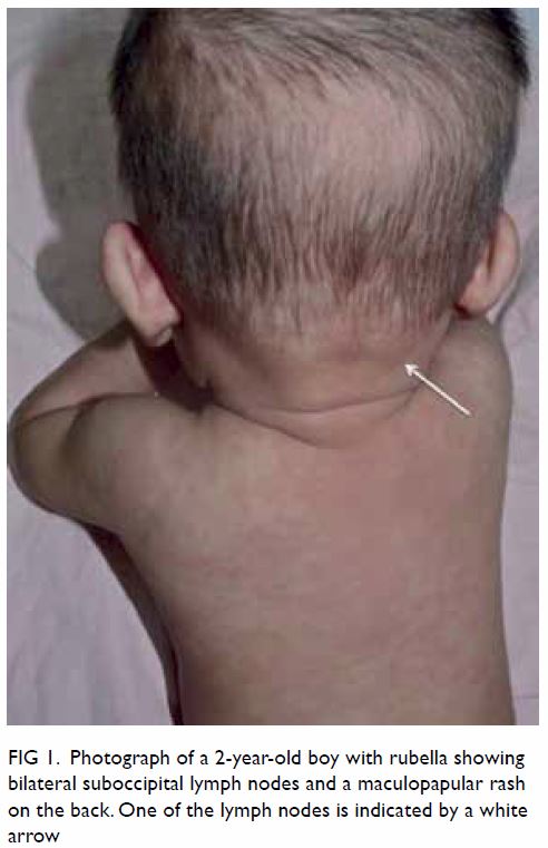
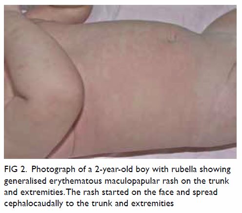
Rubella infection has an incubation period of 14 to 21 days and can be asymptomatic (ie, subclinical) in 25% to 50% of individuals.49 When symptoms occur, they are usually mild and self-limiting. Prodromal symptoms include low-grade fever, malaise, anorexia, nausea, non-exudative conjunctivitis, coryza, cough, sore throat, headache, petechiae on the soft palate (Forchheimer spots) and postauricular area, and occipital and/or posterior cervical lymphadenopathy.47 49 These symptoms, though common in adolescents and adults, are unusual in children.47 Prodromal symptoms are usually followed by the characteristic pinpoint, erythematous, maculopapular rash in 50% to 80% of cases, starting on the face, later spreading to the trunk and limbs, and becoming generalised within 24 hours.47 The rash usually subsides in 3 days and fades in the same directional pattern as it appears. Some individuals, mostly adolescents and adult women, may also experience polyarthritis and polyarthralgia approximately 1 week after the rash.49
 
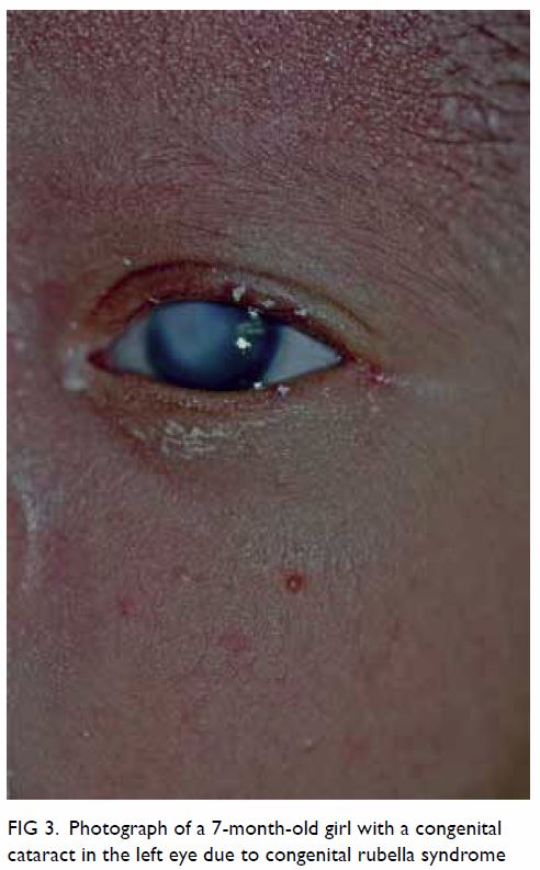
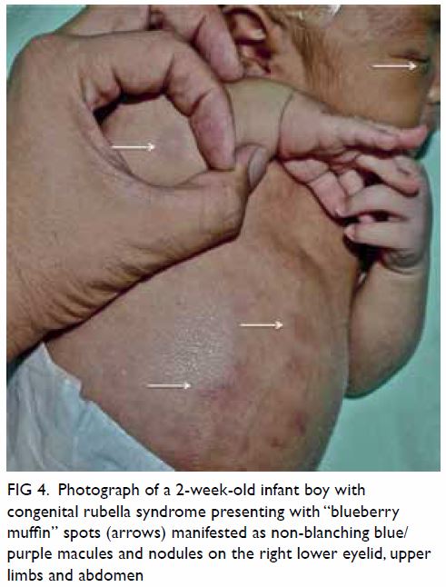
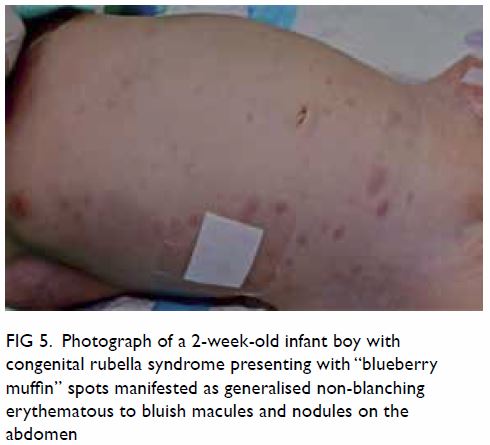
Maternal rubella infection during the first 8 to 10 weeks of gestation can lead to catastrophic consequences, including spontaneous abortion, stillbirth, and prematurity.9 50 51 Cataracts, congenital heart defects (eg, patent ductus arteriosus, branch pulmonary artery hypoplasia/stenosis), and sensorineural hearing loss are the classic triad of CRS.47 Other manifestations of CRS include intrauterine growth retardation, retinopathy, infantile glaucoma, mental retardation, microcephaly, meningoencephalitis, hepatitis, hepatomegaly, splenomegaly, haemolytic anaemia, thrombocytopenia, and purpura (“blueberry muffin spots”).9 50 51 52 Some of these clinical manifestations are transient and non-specific; others may evolve over time into adulthood, even when the infection is subclinical at birth.9 50 51 52 The risk of congenital defects after maternal infection is essentially limited to the first 16 weeks of gestation. Beyond 20 weeks of gestation, focal growth restriction seems to be the only significant sequela.49
 
Diagnosis of maternal infection can be confirmed by using serological tests when there is a four-fold increase in rubella-specific IgG titre between acute and convalescent serum samples, a positive test for rubella-specific IgM antibodies, or a positive culture for the rubella virus.49 Fetal infection can be confirmed with a positive PCR assay on a chorionic villus sample when other clinical findings are consistent with the features of congenital rubella.49 If congenital rubella infection is suspected, the diagnosis can be confirmed by the presence of rubella-specific IgM antibodies in the cord blood or neonatal serum within the first 6 months of life, detection of rubella virus RNA by reverse transcription PCR on nasopharyngeal swab or urine, or isolation of the rubella virus.47 53
 
No specific antiviral treatment for rubella is now available. When maternal rubella is confirmed before 20 weeks of gestation, treatment with Ig and termination of pregnancy should be discussed as options based on local legislation.47 Treatment of CRS mainly involves long-term interdisciplinary supportive care for managing clinical manifestations, close monitoring of neurodevelopmental progress, and long-term audiological and ophthalmic follow-up. 47
 
Vaccination is the only practical and effective way of preventing congenital rubella infection; a single dose of vaccine can offer long-lasting immunity in >95% of cases, while two doses given at an appropriate interval offers close to 100% protection.47 52 While the rubella vaccine is available as an isolated vaccine, it is often administered as a combined vaccine with measles (MR), both measles and mumps (MMR), or together with varicella (MMRV). Yet the vaccine is contra-indicated in pregnant women because of potential transplacental transfer of live rubella virus. Since 1969 the immunisation against rubella with live, attenuated virus was first introduced, some countries have implemented national immunisation programmes against rubella although no cases of CRS have been reported due to rubella vaccination during early pregnancy,54 55 the number of cases of rubella infection has declined globally.
 
Under the Hong Kong Childhood Immunisation Programme, the inclusion of eligible children who are entitled to free immunisation against 11 infectious diseases including rubella has been incorporated into the programme progressively since 1978.45 Starting from 1982, the two-dose protocol has covered all eligible children in Hong Kong. The first dose of MMR vaccine is given at age 1 year, and the second dose at primary one. The second dose of MMR vaccine has been replaced with MMRV vaccine for children born on or after 1 January 2013. The estimated coverage of the first dose of MMR vaccine in children from aged 2 to 5 years remains high (over 98%).56
 
Screening for rubella IgG antibodies is performed in pregnant women during their first antenatal visit. If the pregnant woman is found to be non-immune to rubella during the screening, postpartum vaccination is arranged at the Maternal Child Health Centre to protect the mother and her future pregnancies. With universal immunisation against rubella implemented in Hong Kong, the seronegativity rate among resident women who delivered their babies in a local hospital was 8.1%, less than half of the non-residents being 19.9%, who were mostly Chinese from the mainland China where there is no such immunisation programme implemented.57
 
C – Congenital cytomegalovirus
Cytomegalovirus is a ubiquitous herpesvirus that can reside in the body after a primary infection throughout the person’s life.9 Non-primary infection can result from reactivation of the latent virus or reinfection by a different strain of CMV. Approximately 1% to 7% of pregnant women acquire primary CMV infection; of these, about 30% to 40% transmit the infection to the fetus.58 59 Congenital CMV infection represents the most common congenital viral infection worldwide and is a leading cause of hearing loss and neurological disabilities in children.60 61
 
According to the Centre for Health Protection of Hong Kong, the seroprevalence rates of CMV antibodies in local women aged between 20 and 39 years ranged from 50% to 86% in 2015, with the highest rate in the age-group of 35 to 39 years.62 A local study of Chinese women in Hong Kong found a 7.4% prevalence rate of CMV in cervical excretions during the third trimester, which was low relative to other Asian countries.63 A 1994 report indicated that the seroprevalence rate of CMV in Hong Kong increased steadily from 37% at aged 1 year to 51% by aged 10 years and increased drastically to almost 100% by aged 21 years, implying that CMV infection is usually acquired early in life.64
 
Cytomegalovirus can be transmitted through direct or indirect contact with infectious body fluids like saliva, urine, blood, semen, or cervical or vaginal secretions.60 Maternal CMV infection is mainly acquired through contact with the urine or saliva of infected individuals or sexual contact.60 Cytomegalovirus can be transmitted transplacentally, resulting in congenital infection, with the transmission rate reaching approximately 32.3% in mothers with primary infection but only 1.4% in those with non-primary infection.65 Despite the higher rate of vertical transmission and the fact that many women of childbearing age are seropositive for CMV, only one-quarter of congenital CMV infections are caused by primary maternal infection, with the rest three-quarters resulting from non-primary maternal infections.66 The risk of vertical transmission is approximately 36% when the primary maternal infection occurs during the first trimester, whereas in the third trimester67 rise to 77.6%.
 
Primary CMV infection in immunocompetent individuals is asymptomatic including 75% to 95% of pregnant women with infection.9 68 In others, it may present as a mild mononucleosis- or flu-like syndrome with non-specific symptoms such as fever and fatigue.68 Nevertheless, congenital CMV infections can have severe disabling consequences.
 
Congenital CMV infections from primary maternal infections are more likely to result in symptoms and long-term defects in neonates than those from non-primary maternal infections.58 59 60 61 62 63 64 65 66 67 68 69 Approximately 10% of newborns congenitally infected with CMV are symptomatic.65 The most common manifestations are jaundice at birth, petechiae, hepatosplenomegaly, small size for age, and microcephaly.60 Other clinical manifestations include premature birth, hypotonia, poor feeding, lethargy, sensorineural hearing loss, chorioretinitis, hydrocephalus, seizures, thrombocytopenia, anaemia, and pneumonitis.60 69
 
The risk of developing central nervous system (CNS) sequelae is higher if the congenital infection results from primary maternal infection occurring in the first trimester than later stage in pregnancy.70 The development of permanent sequelae is more frequent in symptomatic newborns, while 10% to 15% of those who are asymptomatic at birth have developmental abnormalities including sensorineural hearing loss, microcephaly, motor defects, mental retardation, chorioretinitis, and dental defects, usually appearing before the age of 2 years.9 61 71 For instance, while nearly 50% of symptomatic neonates develop sensorineural hearing loss, only 7% of asymptomatic ones develop it.72 Fetal and neonatal deaths can also result from congenital CMV infections in approximately 0.5% of cases.71
 
A history of maternal primary CMV infection, together with ultrasonography findings such as echogenic bowel, bilateral periventricular cerebral calcifications, hydrocephalus, microcephaly, fetal growth retardation, oligohydramnios, polyhydramnios, placentomegaly, hepatosplenomegaly, hepatic calcifications, ascites, or hydrops, are suggestive of fetal infection.71 Fetal CMV infection can be diagnosed by PCR for CMV DNA and virus isolation from the amniotic fluid, but PCR does not predict adverse fetal outcomes.68 As for newborns, isolation of CMV in the infant’s urine within the first 2 weeks of life is the standard for diagnosing congenital infection.59
 
While no prenatal treatment has yet been shown to reduce in-utero infections or sequelae, antiviral treatment with ganciclovir and valganciclovir has been proven to reduce the risk of sensorineural hearing loss and improve neurodevelopmental outcomes, especially when treatment is initiated within the first month after birth in symptomatic infants and continued for 6 months.73 Meanwhile, more evidence is needed to justify the risks and benefits of antiviral treatment in asymptomatic infants to devise a suitable treatment scheme that helps to minimise the risk of development of severe sequelae later in life.
 
Currently, systemic screening for CMV primary infection is not performed in any one country.59 Risk reduction strategies should focus on preventing maternal primary infection during pregnancy. Thus, proper hygiene practices (eg, hand washing), reducing maternal exposure to body fluids that contain the virus (eg, saliva and urine of children with infection), and access to clean running water are effective measures to reduce CMV infections.60
 
H – Congenital herpes simplex virus
Herpes simplex viruses types 1 and 2 are herpesviruses, which can establish lifelong latency after the primary infection.74 75 Both types of HSV can cause genital herpes, which when acquired during pregnancy may pose a risk of vertical transmission to the neonate.76 Neonatal HSV infection is estimated to occur in 1 in 3000 to 20 000 livebirths, but it is rare in Hong Kong, with only one case of neonatal HSV type 1 infection reported from 2008 to 2010.75 77
 
In Hong Kong, HSV infection tends to be acquired early in life, with a gradual rise in seropositivity in childhood and a drastic rise in young adults.64 Infection with HSV occurs through direct contact of mucosal or abraded skin surfaces with infectious secretions or lesions, commonly found in the oral and genital regions.9 Approximately 5% of neonatal HSV infections occur in utero, 85% during the peripartum period, and the remaining 10% postnatally, through direct contact with infectious lesions or secretions.78 Peripartum infection is mainly caused by direct contact with infected genital lesions in the birth canal.79 80 The risk of vertical transmission is higher in mothers with newly acquired genital HSV infections than those with HSV reactivation, with 57% and 2% increases in risk, respectively.79 80 81 The presence of maternal antibodies to either HSV subtype seems to offer a certain degree of protection against neonatal transmission. Even antibodies against HSV-2 seem to have a protective effect against neonatal transmission of both HSV subtypes, women of all HSV serological statuses can transmit the virus to neonates.79 Genital infection occurring closer to term incurs a higher risk of neonatal transmission, with 50% to 80% risk of neonatal infections resulting from maternal genital HSV infections close to term.79
 
The majority of both newly acquired or recurrent genital HSV infections are asymptomatic or too mild to be recognised or properly diagnosed, such that no known history of genital HSV infection was found in close to 80% of mothers who delivered an infected infant.9 82
 
In-utero HSV infection accounts for only 5% of all neonatal HSV infections.9 The triad of cutaneous, CNS, and ophthalmic findings are typical in this group of infants. Common cutaneous presentations include vesicles, scarring, aplasia cutis, and hypopigmentation/hyperpigmentation. Neurological findings include microcephaly, intracranial calcifications, and hydranencephaly, while ophthalmic manifestations typically include chorioretinitis, microphthalmia, and optic atrophy.9 76
 
Peripartum and postnatal HSV infections share similar clinical manifestations. They can generally be categorised into three main groups: disease localised to the skin, eyes, or mouth (SEM disease); disease localised to the CNS (CNS disease); and disseminated disease.78 Approximately 45% of all cases are categorised as SEM disease, and these cases typically include vesicles on the skin, keratoconjunctivitis of the eye, and infection of the oropharynx. Approximately 30% of cases are categorised as CNS disease, which is mainly caused by meningoencephalitis and can present with focal or generalised seizures, fever, lethargy, irritability, tremors, poor oral intake, temperature instability, bulging fontanelle, or pyramidal tract signs.9 78 Disseminated disease involving multiple visceral organs, such as the lungs, liver, heart, and brain, occurs in the remaining 25% of cases.78 It presents with irritability, seizures, respiratory failure, hepatic failure, jaundice, disseminated intravascular coagulopathy, and shock. Disseminated disease is associated with a mortality rate of 29%.78
 
Diagnosis of neonatal HSV infection is challenging because of its non-specific presentation. All of the following specimens should be obtained and sent for PCR assay: surface specimens (mouth, nasopharynx, conjunctivae, anus), skin vesicles, CSF, and whole blood.22 Other organ involvement can be screened by laboratory tests and imaging, such as alanine transaminase levels as indicator of hepatic involvement; chest X-ray for lung involvement; and neuroimaging and electroencephalogram as indicators of CNS disease.
 
Antiviral treatment with intravenous acyclovir is recommended for neonates with HSV infection. For infants with disseminated or CNS disease, 21 days of antiviral therapy are indicated. For those with SEM disease, 14 days of treatment is recommended.22 The 14-day treatment is also recommended for asymptomatic neonates born to mothers who are infected with HSV close to term.22 The emergence of antiviral treatment for HSV infection has contributed to a drastic reduction in the 12-month mortality rate of infants with disseminated HSV disease, from 85% to 29% overall, and from 50% to only 4% in those with CNS disease.78 80 Morbidity rates and long-term outcomes have also been improved with the use of antiviral agents, especially when treatment is initiated as soon as possible with an early diagnosis.9 78
 
As the majority of neonatal HSV infections are peripartum, reducing neonates’ exposure to active genital lesions during delivery is important for prevention of transmission. Active genital HSV lesions or presence of prodromal symptoms at the time of delivery are indications for delivery by Caesarean section, which has been found to be effective in preventing neonatal transmission.76 Oral acyclovir or valacyclovir as antiviral suppression therapy, initiated at 36 weeks of gestation, is associated with reduced genital lesions and decreased viral detection by viral culture or PCR at the time of delivery.76 78 More studies are required to establish its safety and effectiveness as prophylaxis against neonatal HSV infection.
 
Counselling on prevention of unprotected sexual contact during pregnancy, especially late pregnancy, may reduce the risk of maternal HSV infection and thus neonatal infection through vertical transmission.80
 
Diagnosing TORCH infection
‘TORCH titres’ are often ordered by clinicians who suspect congenital infections; however, various studies have shown that the diagnostic yield is very low if they are not used appropriately.83 84 Maternal IgG antibodies can cross the placenta and IgG antibody titres from one blood sample might not be adequate for diagnosis. Therefore, appropriate follow-up testing should be arranged to evaluate the levels of IgG-specific antibody of concern, and whenever possible, isolation of the organism should be attempted.83 Targeted specimens and tests rather than a TORCH screen should be sent if the clinical suspicion for a specific congenital infection is strong after reviewing the maternal history and infant’s clinical features. The clinical features, antenatal screening, diagnostic testing, and treatment of congenital toxoplasmosis, syphilis, rubella, CMV, and HSV are summarised in the Table.9 10 13 15 18 19 20 22 31 36 4147 50 53 57 69 71 81 84
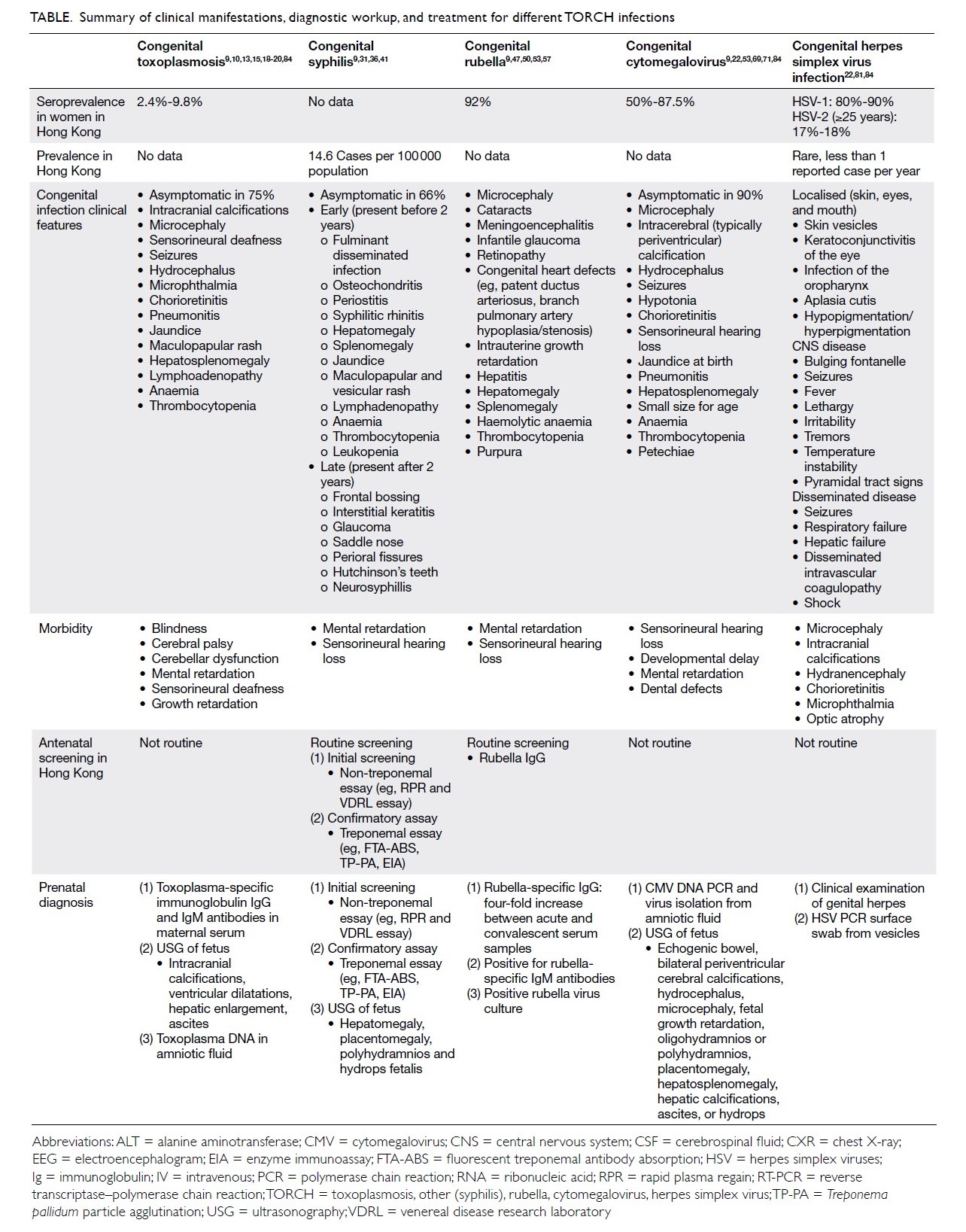
 

Table. Summary of clinical manifestations, diagnostic workup, and treatment for different TORCH infections
 
Conclusion
Despite being densely populated, Hong Kong has relatively low rates of congenital infections. Nevertheless, it is important to stay vigilant for infections during pregnancy that may lead to severe disabilities or even death of the fetus. In Hong Kong, comprehensive antenatal services that are available to eligible mothers have been provided free of charge by the public sector in collaboration with the Maternal and Child Health Centres (under the Department of Health) and hospital Obstetrics Departments (under the Hospital Authority). Pregnant women registered for the shared care system are entitled to receive regular antenatal check-ups, postnatal services, and close monitoring of the pregnancy.
 
Prevention is better than intervention, especially in the context of congenital infections. Primary prevention of maternal infection during pregnancy is the key to preventing congenital infections. Effective measures for primary prevention are available, and during the first antenatal visit, screening for rubella, syphilis, and HIV are performed. Available resources should focus on promoting public health.
 
Author contributions
All authors had full access to the data, contributed to the study, approved the final version for publication, and take responsibility for its accuracy and integrity.
 
Concept or design: KL Hon.
Acquisition of data: KKY Leung, A Yeung.
Analysis or interpretation of data: KKY Leung, A Yeung.
Drafting of the article: KL Hon, KKY Leung, A Yeung.
Critical revision for important intellectual content: KL Hon, KKY Leung, AKC Leung, E Man.
 
Conflicts of interest
As an editor of the journal, KL Hon was not involved in the peer review process. Other authors have disclosed no conflicts of interest.
 
Funding/support
This research received no specific grant from any funding agency in the public, commercial, or not-for-profit sectors.
 
References
1. DeVore NE, Jackson VM, Piening SL. TORCH infections. Am J Nurs 1983;83:1660-5. Crossref
2. Neu N, Duchon J, Zachariah P. TORCH infections. Clin Perinatol 2015;42:77-103. Crossref
3. Stegmann BJ, Carey JC. TORCH infections. Toxoplasmosis, other (syphilis, varicella-zoster, parvovirus B19), rubella, cytomegalovirus (CMV), and herpes infections. Curr Womens Health Rep 2002;2:253-8.
4. Schwartz DA. The origins and emergence of Zika virus, the newest torch infection: what’s old is new again. Arch Pathol Lab Med 2017;141:18-24. Crossref
5. Nahmias AJ, Walls KW, Stewart, JA, Herrmann KL, Flynt WJ Jr. The ToRCH complex-perinatal infections associated with toxoplasma and rubella, cytomegol- and herpes simplex viruses. Pediatr Res 1971;5:405-6. Crossref
6. Fuerst HT. Flame or bird? Pediatrics 1975;56:107. Crossref
7. Brumback RA. TORCHES. Pediatrics 1976;58:916.
8. World Health Organization. Global validation of elimination of mother-to child transmission (EMTCT) of HIV and syphilis. 2019. Available from: https://www.who. int/reproductivehealth/congenital-syphilis/emtc-gvac/ en/. Accessed 3 Oct 2019.
9. Wilson CB, Nizet V, Maldonado Y, Remington JS, Klein JO. Remington and Klein’s Infectious Diseases of the Fetus and Newborn Infant. 8th ed. Philadelphia: Elsevier Saunders; 2016: 513, 520-5, 675, 677-9, 685-90, 693-4, 729, 732.
10. Montoya JG, Liesenfeld O. Toxoplasmosis. Lancet 2004;363:1965-76. Crossref
11. McAuley JB. Congenital toxoplasmosis. J Pediatric Infect Dis Soc 2014;3 Suppl 1:S30-5. Crossref
12. Dunn D, Wallon M, Peyron F, Petersen E, Peckham C, Gilbert R. Mother-to-child transmission of toxoplasmosis: risk estimates for clinical counselling. Lancet 1999;353:1829-33. Crossref
13. Ko RC, Wong FW, Todd D, Lam KC. Prevalence of Toxoplasma gondii antibodies in the Chinese population of Hong Kong. Trans R Soc Trop Med Hyg 1980;74:351-4. Crossref
14. Torgerson PR, Mastroiacovo P. The global burden of congenital toxoplasmosis: a systematic review. Bull World Health Organ 2013;91:501-8. Crossref
15. Serranti D, Buonsenso D, Valentini P. Congenital toxoplasmosis treatment. Eur Rev Med Pharmacol Sci 2011;15:193-8.
16. Tamma P. Toxoplasmosis. Pediatr Rev 2007;28:470-1. Crossref
17. Wilson CB, Remington JS, Stagno S, Reynolds DW. Development of adverse sequelae in children born with subclinical congenital Toxoplasma infection. Pediatrics 1980;66:767-74.
18. Sever JL, Ellenberg JH, Ley AC, et al. Toxoplasmosis: maternal and pediatric findings in 23,000 pregnancies. Pediatrics 1988;82:181-92.
19. Gay-Andrieu F, Marty P, Pialat J, Sournies G, Drier de Laforte T, Peyron F. Fetal toxoplasmosis and negative amniocentesis: necessity of an ultrasound follow-up. Prenat Diagn 2003;23:558-60. Crossref
20. Cortina-Borja M, Tan HK, Wallon M, et al. Prenatal treatment for serious neurological sequelae of congenital toxoplasmosis: an observational prospective cohort study. PLoS Med 2010;7(10). pii: e1000351. Crossref
21. Pomares C, Montoya JG. Laboratory diagnosis of congenital toxoplasmosis. J Clin Microbiol 2016;54:2448-54. Crossref
22. Kimberlin DW, Brady MT, Jackson MA, editors. Red Book 2018: Report of the Committee on Infectious Diseases. 31st ed. Itasca; American Academy of Pediatrics; 2018: 310-7, 437-48, 459-76, 773-88, 809-19, 8698.
23. Chik TS, Tsang OT. Toxoplasmosis. HIV manual. 4th ed. 2019. Available from: https://hivmanual.hk/d27/. Accessed 7 Oct 2019.
24. Cook AJ, Gilbert RE, Buffolano W, et al. Sources of toxoplasma infection in pregnant women: European multicentre case-control study. European Research Network on Congenital Toxoplasmosis. BMJ 2000;321:142- 7. Crossref
25. Halonen SK, Weiss LM. Toxoplasmosis. Handb Clin Neurol 2013;114:125-45. Crossref
26. Jones JL, Krueger A, Schulkin J, Schantz PM. Toxoplasmosis prevention and testing in pregnancy, survey of obstetriciangyn-aecologists. Zoonoses Public Health 2010;57:27-33. Crossref
27. Sagel U, Krämer A. Screening of maternal toxoplasmosis in pregnancy: laboratory diagnostics from the perspective of public health requirements. J Bacteriol Parasitol 2013;S5.
28. Cornu C, Bissery A, Malbos C, et al. Factors affecting the adherence to an antenatal screening programme: an experience with toxoplasmosis screening in France. Euro Surveill 2009;14:21-5.
29. Paquet C, Yudin MH; Society of Obstetricians and Gynaecologists of Canada. Toxoplasmosis in pregnancy: prevention, screening, and treatment [in English, French]. J Obstet Gynaecol Can 2013;35:78-81. Crossref
30. World Health Organization. The global elimination of congenital syphilis: rationale and strategy for action. 2017. Available from: https://apps.who.int/iris/ handle/10665/43782. Accessed 8 Oct 2019.
31. Special Preventive Programme, Centre for Health Protection, Department of Health, Hong Kong SAR Government. Hong Kong STD/AIDS update—a quarterly surveillance report. Vol 23 No 4, 2017. Available from: https://www.chp.gov.hk/files/pdf/std17q4.pdf. Accessed 8 Oct 2019.
32. Centers for Disease Control and Prevention, US Government. Sexually transmitted disease surveillance. Primary and secondary syphilis—rates of reported cases by state, United States and outlying areas, 2017. Available from: https://www.cdc.gov/std/stats17/figures/37.htm. Accessed 3 Oct 2019.
33. Fiumara NJ. Syphilis in newborn children. Clin Obstet Gynecol 1975;18:183-9. Crossref
34. Jenson HB. Congenital syphilis. Semin Pediatr Infect Dis 1999;10:183-94. Crossref
35. Herremans T, Kortbeek L, Notermans DW. A review of diagnostic tests for congenital syphilis in newborns. Eur J Clin Microbiol Infect Dis 2010;29:495-501. Crossref
36. De Santis M, De Luca C, Mappa I, et al. Syphilis infection during pregnancy: fetal risks and clinical management. Infect Dis Obstet Gynecol 2012;2012:430585. Crossref
37. Leung AK, Leong KF, Lam JM. A case of congenital syphilis presenting with unusual skin eruptions. Case Rep Pediatr 2018;2018:1761454. Crossref
38. Lago EG, Vaccari A, Fiori RM. Clinical features and follow-up of congenital syphilis. Sex Transm Dis 2013;40:85-94. Crossref
39. Woods CR. Syphilis in children: congenital and acquired. Semin Pediatr Infect Dis 2005;16:245-57. Crossref
40. Mannelli L, Perez FA, Parisi MT, Giacani L. A case of congenital syphilis. Emerg Radiol 2013;20:337-9. Crossref
41. Public Health Agency of Canada. Government of Canada. Section 5-10: Canadian guidelines on sexually transmitted infections—management and treatment of specific infections—syphilis. 2019. Available from: https://www. canada.ca/en/public-health/services/infectious-diseases/ sexual-health-sexually-transmitted-infections/canadianguidelines/ sexually-transmitted-infections/canadianguidelines- sexually-transmitted-infections-27.html. Accessed 3 Oct 2019.
42. Alexander JM, Sheffield JS, Sanchez PJ, Mayfield J, Wendel GD Jr. Efficacy of treatment for syphilis in pregnancy. Obstet Gynecol 1999;93:5-8. Crossref
43. Sheffield JS, Sánchez PJ, Morris G, et al. Congenital syphilis after maternal treatment for syphilis during pregnancy. Am J Obstet Gynecol 2002;186:569-73. Crossref
44. Duthie SJ, King PA, Yung GL, Ma HK. Routine serological screening for syphilis during pregnancy—disposable anachronism or fundamental necessity? Aust N Z J Obstet Gynaecol 1990;30:29-31. Crossref
45. Ho FW. Update on rubella in Hong Kong. Commun Dis Watch 2017;14:66-8.
46. Centre for Health Protection, Department of Health, Hong Kong SAR Government. Number of notifiable infectious diseases by month. 2019. Available from: https://www.chp. gov.hk/en/static/24012.html. Accessed 3 Oct 2019.
47. Leung AK, Hon KL, Leong KF. Rubella (German measles) revisited. Hong Kong Med J 2019;25:134-41. Crossref
48. Miller E, Cradock-Watson JE, Pollock TM. Consequences of confirmed maternal rubella at successive stages of pregnancy. Lancet 1982;2:781-4. Crossref
49. Dontigny L, Arsenault MY, Martel MJ; Clinical Practice Obstetrics Committee. Rubella in pregnancy. J Obstet Gynaecol Can 2008;30:152-8. Crossref
50. Reef SE, Plotkin S, Cordero JF, et al. Preparing for elimination of congenital rubella syndrome (CRS): summary of a workshop on CRS elimination in the United States. Clin Infect Dis 2000;31:85-95. Crossref
51. Watson JC, Hadler SC, Dykewicz CA, Reef S, Phillips L. Measles, mumps, and rubella—vaccine use and strategies for elimination of measles, rubella, and congenital rubella syndrome and control of mumps: recommendations of the Advisory Committee on Immunization Practices (ACIP). MMWR Recomm Rep 1998;47:1-57.
52. Rubella vaccines: WHO position paper [editorial] [in English, French]. Wkly Epidemiol Rec 2011;86:301-16.
53. World Health Organization. Manual for the laboratory diagnosis of measles and rubella virus infection Second edition. 2017. Available from: https://www.who.int/ihr/ elibrary/manual_diagn_lab_mea_rub_en.pdf. Accessed 8 Oct 2019.
54. Bart SW, Stetler HC, Preblud SR, et al. Fetal risk associated with rubella vaccine: an update. Rev Infect Dis 1985;7 Suppl 1:S95-102. Crossref
55. Centers for Disease Control (CDC). Rubella vaccination during pregnancy—United States, 1971-1988. MMWR Morb Mortal Wkly Rep 1989;38:289-93.
56. Food and Health Bureau, Hong Kong SAR Government. Hong Kong reference framework for preventive care for children in primary care settings. 2012. Available from: https://www.fhb.gov.hk/pho/english/health_ professionals/professionals_preventive_children_pdf. html. Accessed 10 Oct 2019.
57. Lao TT, Suen SS, Leung TY, Sahota DS, Lau TK. Universal rubella vaccination programme and maternal rubella immune status: a tale of two systems. Vaccine 2010;28:2227-30. Crossref
58. Boppana SB, Rivera LB, Fowler KB, Mach M, Britt WJ. Intrauterine transmission of cytomegalovirus to infants of women with preconceptional immunity. N Engl J Med 2001;344:1366-71. Crossref
59. Lazzarotto T, Guerra B, Gabrielli L, Lanari M, Landini MP. Update on the prevention, diagnosis and management of cytomegalovirus infection during pregnancy. Clin Microbiol Infect 2011;17:1285-93. Crossref
60. Cannon MJ. Congenital cytomegalovirus (CMV) epidemiology and awareness. J Clin Virol 2009;46 Suppl 4:S6-10. Crossref
61. Boppana SB, Ross SA, Fowler KB. Congenital cytomegalovirus infection: clinical outcome. Clin Infect Dis 2013;57 Suppl 4:S178-81. Crossref
62. Centre for Health Protection, Department of Health, Hong Kong SAR Government. Seroprevalence rates of cytomegalovirus antibodies. 2018. Available from: https:// www.chp.gov.hk/en/statistics/data/10/641/701/6445.html. Accessed 5 Oct 2019.
63. Leung AK, Loong EP, Chan RC, Murray HG, Chang AM. Prevalence of cytomegalovirus cervical excretion in pregnant women in Hong Kong. Asia Oceania J Obstet Gynaecol 1989;15:77-8. Crossref
64. Kangro HO, Osman HK, Lau YL, Heath RB, Yeung CY, Ng MH. Seroprevalence of antibodies to human herpesviruses in England and Hong Kong. J Med Virol 1994;43:91-6. Crossref
65. Kenneson A, Cannon MJ. Review and meta-analysis of the epidemiology of congenital cytomegalovirus (CMV) infection. Rev Med Virol 2007;17:253-76. Crossref
66. Wang C, Zhang X, Bialek S, Cannon MJ. Attribution of congenital cytomegalovirus infection to primary versus non-primary maternal infection. Clin Infect Dis 2011;52:e11-3. Crossref
67. Bodéus M, Hubinont C, Goubau P. Increased risk of cytomegalovirus transmission in utero during late gestation. Obstet Gynecol 1999;93(5 Pt 1):658-60. Crossref
68. Nigro G. Maternal-fetal cytomegalovirus infection: from diagnosis to therapy. J Matern Fetal Neonatal Med 2009;22:169-74. Crossref
69. Leung AK, Sauve RS, Davies HD. Congenital cytomegalovirus infection. J Natl Med Assoc 2003;95:213- 8.
70. Pass RF, Fowler KB, Boppana SB, Britt WJ, Stagno S. Congenital cytomegalovirus infection following first trimester maternal infection: symptoms at birth and outcome. J Clin Virol 2006;35:216-20. Crossref
71. Dollard SC, Grosse SD, Ross DS. New estimates of the prevalence of neurological and sensory sequelae and mortality associated with congenital cytomegalovirus infection. Rev Med Virol 2007;17:355-63. Crossref
72. Dahle AJ, Fowler KB, Wright JD, Boppana SB, Britt WJ, Pass RF. Longitudinal investigation of hearing disorders in children with congenital cytomegalovirus. J Am Acad Audiol 2000;11:283-90.
73. Demmler-Harrison GJ. Congenital cytomegalovirus infection: management and outcome. Available from: https://www.uptodate.com/contents/congenital-cytomegalovirus-infection-management-and-outcome. Accessed 10 Oct 2019.
74. Leung AK, Barankin B. Herpes labialis: an update. Recent Pat Inflamm Allergy Drug Discov 2017;11:107-13. Crossref
75. Hon KL, Leung TF, Cheung HM, Chan PK. Neonatal herpes: what lessons to learn. Hong Kong Med J 2012;18:60- 2.
76. James SH, Sheffield JS, Kimberlin DW. Mother-to-child transmission of herpes simplex virus. J Pediatric Infect Dis Soc 2014;3 Suppl 1:S19-23. Crossref
77. Sauerbrei A. Genital herpes. In: Diagnostics to Pathogenomics of Sexually Transmitted Infections. West Sussex; John Wiley & Sons Ltd: 2018: 83-99. Crossref
78. Kimberlin DW. Herpes simplex virus infections of the newborn. Semin Perinatol 2007;31:19-25. Crossref
79. Brown ZA, Selke S, Zeh J, et al. The acquisition of genital herpes during pregnancy. N Engl J Med 1997;337:509-15. Crossref
80. Corey L, Wald A. Maternal and neonatal herpes simplex virus infections. N Engl J Med 2009;361:1376-85. Crossref
81. Lee NL, Tse IC. V. Major opportunistic infections. Herpes simplex and zoster. 2019. Available from: https://www.aids. gov.hk/pdf/g190htm/25.htm. Accessed 1 Jan 2020.
82. Whitley RJ, Corey L, Arvin A, et al. Changing patterns of herpes simplex virus infection in neonates. J Infect Dis 1988;158:109-16. Crossref
83. Leland D, French ML, Kleiman MB, Schreiner RL. The use of TORCH titers. Pediatrics 1983;72:41-3.
84. Lim WL, Wong DA. TORCH screening: time for abolition? J Hong Kong Med Assoc 1994;46:306.

Echocardiography update for primary care physicians: a review

Hong Kong Med J 2020 Feb;26(1):44–55  |  Epub 12 Feb 2020
© Hong Kong Academy of Medicine. CC BY-NC-ND 4.0
 
REVIEW ARTICLE
Echocardiography update for primary care physicians: a review
Jeffrey SK Chan, MB, ChB1; Gary Tse, MPH, PhD1; H Zhao, MD2; XX Luo, MB3; CN Jin, PhD4; Kevin Kam, MB, ChB, MRCP1; YT Fan, MD, PhD1,5; Alex PW Lee, MD, FRCP1,5
1 Division of Cardiology, Department of Medicine and Therapeutics, Prince of Wales Hospital, Hong Kong
2 Department of Cardiology, Renji Hospital, School of Medicine, Shanghai Jiao Tong University, Shanghai, China
3 Department of Ultrasonography, Shenzhen Hospital, Southern Medical University, Shenzhen, China
4 Department of Cardiology, The Second Affiliated Hospital of Zhejiang University, Zhejiang, China
5 Laboratory for Cardiac Imaging and 3D Printing, Li Ka Shing Institute of Health Sciences, The Chinese University of Hong Kong, Hong Kong
 
Corresponding author: Prof Alex PW Lee (alexpwlee@cuhk.edu.hk)
 
 Full paper in PDF
 
Abstract
Echocardiography is a key evaluation tool for the diagnosis, prognosis, and guidance of interventional management of numerous cardiovascular conditions, including ischaemia, heart failure, and structural heart diseases. Recent technological advancements have also seen the exploration of artificial intelligence, intracardiac vortex imaging, and three-dimensional printing in echocardiography. With cardiovascular diseases increasing in prevalence worldwide, it is important for clinicians including general practitioners to have updated knowledge of appropriate use of echocardiography. As such, this article reviews the current literature and summarises the latest developments and the general clinical usage of echocardiography.


Introduction
Cardiovascular diseases are a major burden to healthcare globally and a leading cause of death in industrialised countries.1 In 2015, cardiovascular disorders accounted for 77 600 inpatient discharges in Hong Kong, including 6190 deaths, making them the third most common cause of death. The prevalence of cardiovascular disease is increasing and the age-standardised mortality is decreasing.2 Thus, patients with heart diseases are increasingly likely to present to primary care physicians as their first point of care or for a follow-up examination: up to 15% of coronary heart disease patients are treated exclusively by primary care physicians.3 Furthermore, a long-term follow-up study has proven the viability of general practitioners providing care for low-risk chronic heart failure patients.4 As such, clinical care for patients with heart diseases has become increasingly relevant to primary care physicians.
 
Echocardiography does not involve any radiation and may be performed as a point-of-care diagnostic tool in both outpatient and inpatient settings. Doppler echocardiography allows haemodynamic assessment of stroke volume, pressure gradients, valvular regurgitations, and intracardiac shunts. The treatment of patients with cardiovascular diseases almost invariably involves the use of echocardiography as a means of initial and follow-up investigation. Many patients undergo cardiac surgery and/or catheter-based interventions guided by perioperative or intraprocedural echocardiography. Point-of-care echocardiography in primary care settings improves outcomes in some patients,5 and may identify otherwise-undiagnosed valvular heart diseases.6
 
Echocardiographic technologies remain a vibrant field of research. The rapid advancement of echocardiographic technologies requires primary care physicians to have an updated understanding of its appropriate use and referral. According to the American College of Cardiology, appropriate echocardiography “likely contributes to improving patient’s clinical outcomes” and inappropriate echocardiography may be “potentially harmful to patients and generate unwarranted costs to the healthcare system”.7 Recent technological developments have expanded the use of echocardiography dramatically. Therefore, the aim of this study was to review the contemporary literature on clinical indications and emerging technologies of echocardiography and to provide a summary for primary care physicians.
 
Basic echocardiography and the echocardiographic report
Transthoracic echocardiography (TTE) is non-invasive, readily available, and is generally the initial imaging option. In contrast, transoesophageal echocardiography (TEE) requires insertion of a probe into the oesophagus and thus involves sedation and risks of complications such as major bleeding (<0.01%) and hoarseness of voice (12%).8 However, TEE offers better resolution, especially of the left heart, mitral and aortic valves, and the aorta. A routine echocardiography involves several methods of imaging, including two-dimensional (2D; previously known as B mode), Doppler, and M mode echocardiography. Two-dimensional echocardiography, the most commonly used and intuitive method, is used for visualisation and dimensional measurements of the cardiac structures. Two-dimensional echocardiography is often the initial method of imaging, by which cardiac structures are identified and other methods applied. The left ventricular ejection fraction (LVEF) is also obtainable from 2D echocardiography, with the normal value being 50% to 70%. This is mainly used in the classification of heart failure.
 
Colour Doppler imaging allows visualisation of the blood flow, the conventional colour scheme being red for flow towards the probe and blue for away. This allows intuitive and easy assessment of valvular assessments, where stenotic valves generally show turbulent flow and often thickened valve leaflets, whereas regurgitating valves show a regurgitant jet. Trace or mild regurgitation is common even in normal, healthy individuals. When an abnormal, turbulent flow is detected by colour Doppler imaging, continuous and pulse wave spectral Doppler techniques should be used to further evaluate the nature and severity of the abnormal flow.
 
Continuous and pulse wave Doppler imaging provides quantitative measurement of flow velocity, volume, and pressure gradient at operator-defined locations. These allow calculation of stroke volume and haemodynamic assessment of the heart valves. Tissue Doppler provides information about movement of cardiac tissues, such as the diastolic motion of the mitral annulus that reflects left ventricular diastolic function. By means of transmitral flow assessment (ie, E, A, E/A ratio) and tissue Doppler imaging of the mitral annulus (ie, e′, a′, s′, E/e′), filling pressure and diastolic function of the left ventricle can also be assessed.9 Interpretation of these parameters is complex and requires correlation with the clinical picture; in general, the normal E/A ratio is 0.8-2 but may be high in young, fit persons; a E/e′ (medial mitral annulus) ratio <8 generally indicates normal left ventricular filling pressure. Further details may be found in the joint recommendation of the European Association for Cardiovascular Imaging and the American Society of Echocardiography for diastolic echocardiographic assessment.10
 
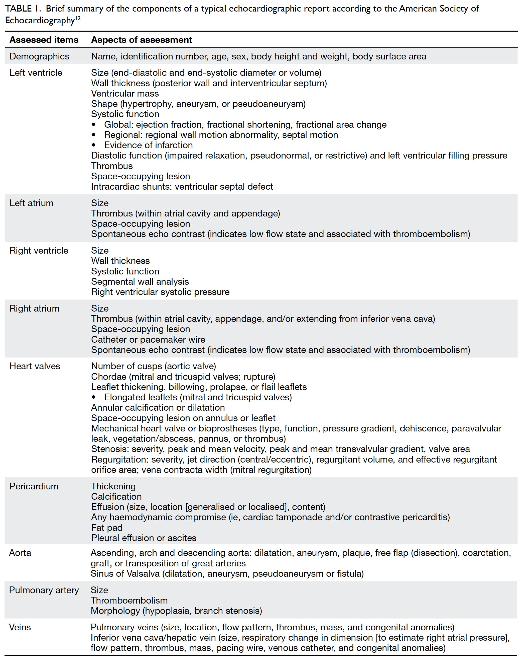
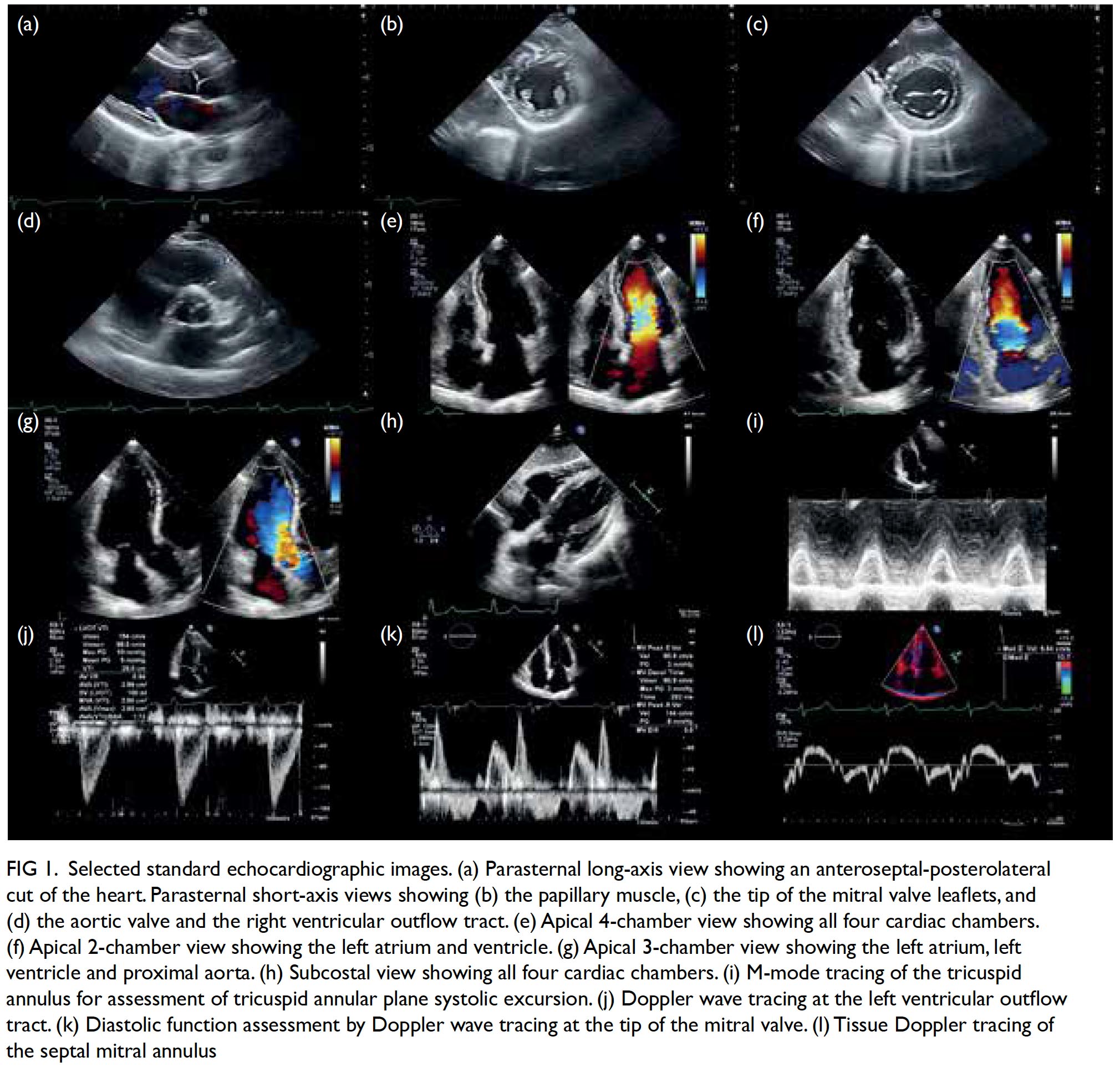
Lastly, M mode tracks over time the movement of tissues along a straight line extending from the probe and has the advantage of high temporal resolution. The M mode is most commonly used for the assessment of right ventricular function by means of tricuspid annular plane systolic excursion, which is typically ≥17 mm. The M mode is also useful in the assessment of constrictive pericarditis, pericardial effusion and cardiac tamponade, hypertrophic obstructive cardiomyopathy, pulmonary hypertension, and left ventricular outflow tract obstruction.11 An echocardiographic report thus contains information about dimensions and functions of the heart chambers and the proximal aorta. Table 1 summarises the typical components of an echocardiographic report.12 A set of selected standard echocardiographic images is shown in Figure 1.
 

Table 1. Brief summary of the components of a typical echocardiographic report according to the American Society of Echocardiography12
 

Figure 1. Selected standard echocardiographic images. (a) Parasternal long-axis view showing an anteroseptal-posterolateral cut of the heart. Parasternal short-axis views showing (b) the papillary muscle, (c) the tip of the mitral valve leaflets, and (d) the aortic valve and the right ventricular outflow tract. (e) Apical 4-chamber view showing all four cardiac chambers. (f) Apical 2-chamber view showing the left atrium and ventricle. (g) Apical 3-chamber view showing the left atrium, left ventricle and proximal aorta. (h) Subcostal view showing all four cardiac chambers. (i) M-mode tracing of the tricuspid annulus for assessment of tricuspid annular plane systolic excursion. (j) Doppler wave tracing at the left ventricular outflow tract. (k) Diastolic function assessment by Doppler wave tracing at the tip of the mitral valve. (l) Tissue Doppler tracing of the septal mitral annulus
 
Common clinical scenarios where echocardiography is indicated
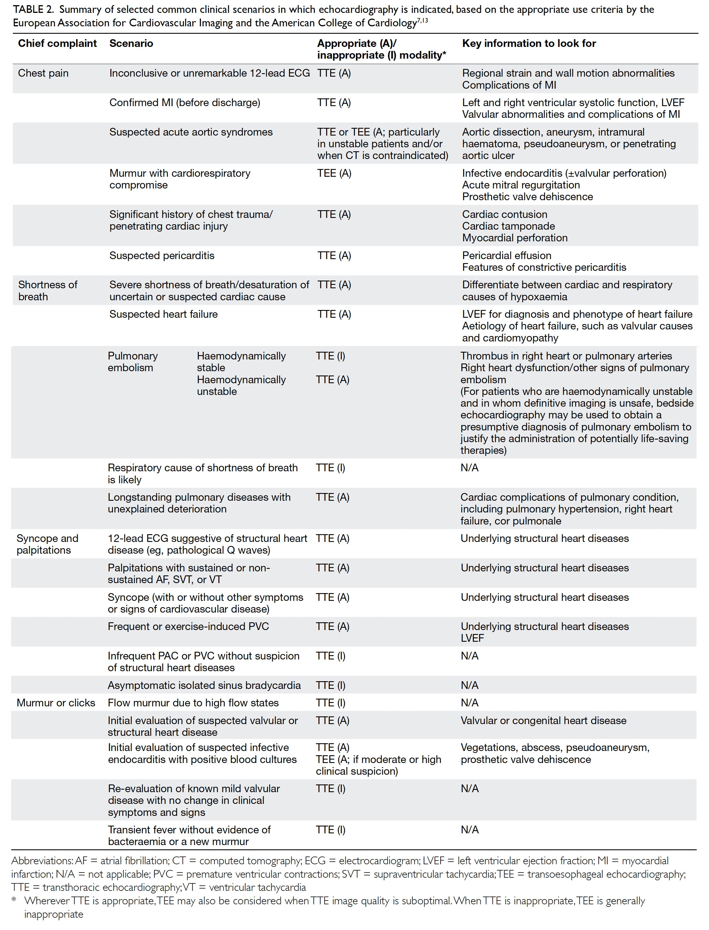
The clinical indications for echocardiography are detailed in the appropriate use criteria from the European Association for Cardiovascular Imaging13 and the American College of Cardiology.7 This section summarises the key recommendations for the use of echocardiography in some common chief complaints encountered by clinicians. Key points are summarised in Table 2. In general, echocardiography is indicated for evaluating symptoms or conditions potentially of cardiac aetiology, including chest pain, shortness of breath, palpitations, transient ischaemic attack, stroke, or peripheral embolic event. Echocardiography is also considered appropriate when heart disease or structural abnormality are suspected on prior testing including but not limited to chest X-ray, electrocardiogram (ECG), or cardiac biomarkers, whether the patient is symptomatic or not. As a general principle, echocardiography is accepted as appropriate for initial diagnosis when there is a change in clinical status or when the results of the echocardiography are anticipated to change patient treatment. In contrast, routine testing when there is no change in clinical status or when test results are unlikely to lead to changes in treatment are generally considered inappropriate.
 

Table 2. Summary of selected common clinical scenarios in which echocardiography is indicated, based on the appropriate use criteria by the European Association for Cardiovascular Imaging and the American College of Cardiology7 13
 
Chest pain
The differential diagnoses of chest pain are broad, and reaching a definitive diagnosis is often difficult when relying on the clinical history and physical examination only. The most important diagnosis in patients presenting with acute chest pain is acute coronary syndrome (ACS). As such, 12-lead ECG and troponin assay are the first-line investigations for acute chest pain suggestive of ACS. Echocardiography aids or confirms ischaemic diagnoses, and is used to screen for non-ischaemic structural, aortic, and pericardial pathology in selected patients. For evaluation of acute chest pain with suspected myocardial ischaemia/infarction and non-diagnostic ECG, resting TTE is appropriate and can be performed while the patent has chest pain. The absence of regional wall motion abnormality on 2D TTE during chest pain virtually excludes acute myocardial infarction, with a sensitivity of 93% for any infarct and 100% for ST-elevation myocardial infarction.14 All patients with chest pain should have initial evaluation of ventricular function by TTE after ACS. After ACS, re-evaluation of ventricular function during the recovery phase is appropriate when results will guide therapy.15 For detecting other causes of chest pain, including acute aortic syndromes, valvular diseases, pulmonary embolism, and pericarditis, TTE is valuable and can be done in emergency settings. Although TTE can be used as a first-line test to assess suspected aortic pathologies, TEE allows clearer and more complete assessment of the thoracic aorta, with diagnostic accuracy equivalent to that of cardiac computed tomography (CT) scans or magnetic resonance imaging (MRI).16 Even though CT is the imaging modality of choice in acute aortic syndromes, TTE and TEE remain valuable for acute assessment and follow-up, with the advantage of not requiring radiation, and especially for hemodynamically unstable patients and situations where CT contrast is contraindicated (eg, severe renal impairment).17
 
Shortness of breath
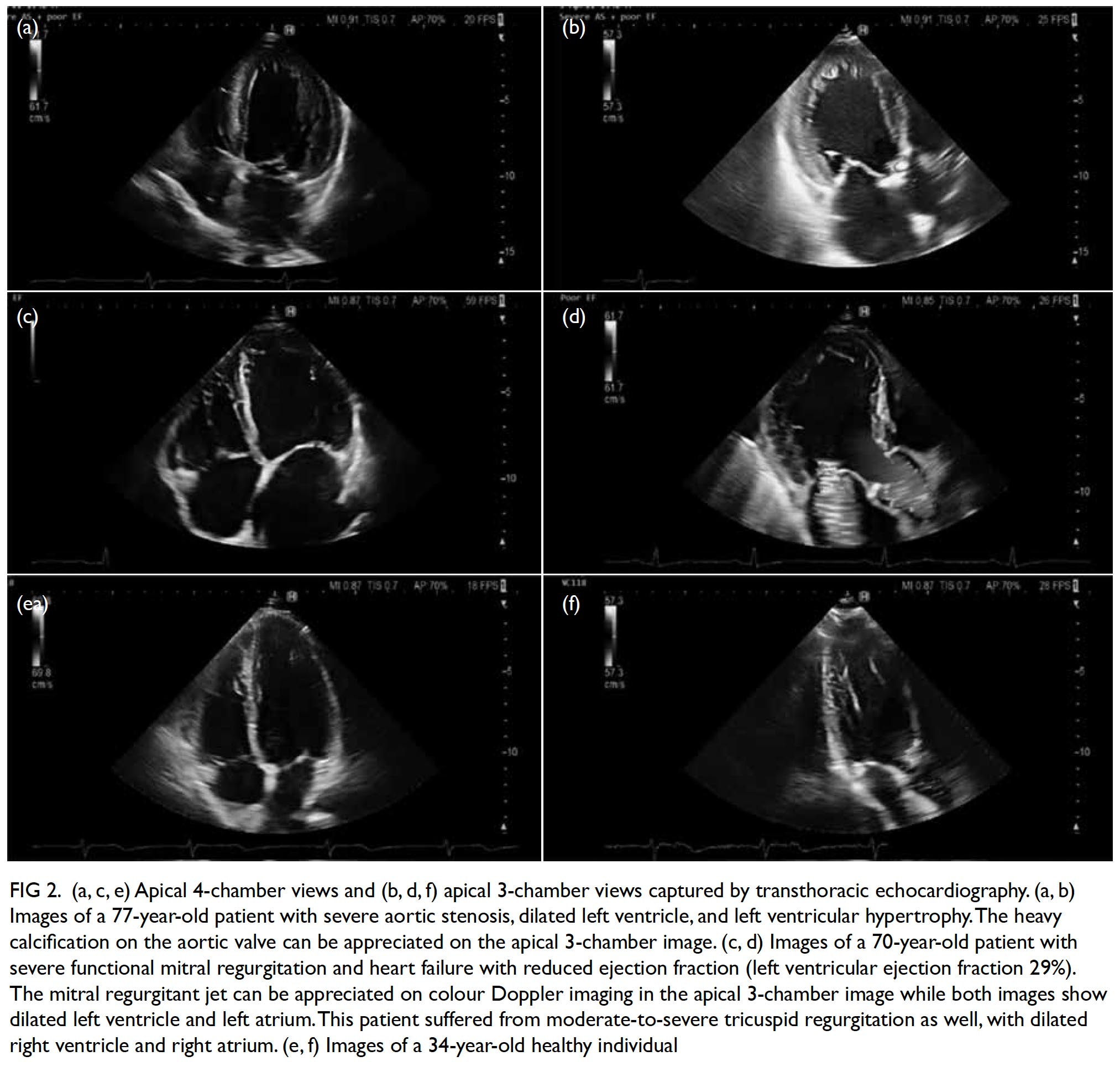
Shortness of breath is a common symptom among patients presenting to primary and emergency care physicians. Differential diagnoses of breathlessness include cardiovascular disease, lung disease, anaemia, obesity, and deconditioning. In any setting, clinical history and examination are the first step in evaluation.13 When heart failure is suspected after initial clinical evaluation, TTE is mandatory to confirm or exclude the diagnosis; to quantify chamber volumes, systolic and diastolic function, and wall thickness; and to identify the aetiology of heart failure (eg, cardiomyopathy, valvular disease, or prior myocardial infarction). The TTE also serves to classify heart failure into heart failure with preserved, mid-range or reduced EF (Fig 2).18 Such classification guides management of the condition. For patients with heart failure with preserved or mid-range EF, assessment of diastolic function is important as diastolic dysfunction is known to be key in the pathogenesis. Although some patients have resting echocardiographic evidence of diastolic dysfunction, left ventricular filling pressure, which is the main haemodynamic parameter that explains exertional dyspnoea, may only be elevated during exertion. This may be demonstrated by diastolic stress echocardiography, providing additional haemodynamic information about the patient and aiding diagnosis and treatment.19
 

Figure 2. (a, c, e) Apical 4-chamber views and (b, d, f) apical 3-chamber views captured by transthoracic echocardiography. (a, b) Images of a 77-year-old patient with severe aortic stenosis, dilated left ventricle, and left ventricular hypertrophy. The heavy calcification on the aortic valve can be appreciated on the apical 3-chamber image. (c, d) Images of a 70-year-old patient with severe functional mitral regurgitation and heart failure with reduced ejection fraction (left ventricular ejection fraction 29%). The mitral regurgitant jet can be appreciated on colour Doppler imaging in the apical 3-chamber image while both images show dilated left ventricle and left atrium. This patient suffered from moderate-to-severe tricuspid regurgitation as well, with dilated right ventricle and right atrium. (e, f) Images of a 34-year-old healthy individual
 
Patients who have undergone chemotherapy or radiotherapy are at risk of developing heart failure, and baseline echocardiography with regular follow-up examinations are required. The precise algorithm depends on the cardiotoxic agent prescribed. Traditionally, LVEF has been used as the main marker of cardiac function and cancer therapeutics- related cardiac dysfunction is defined as a drop in LVEF of ≥5% in symptomatic patients or a drop in LVEF of ≥10% to an LVEF of <53% in asymptomatic patients.20 However, significant damage has occurred when a drop in LVEF is detected. Earlier detection can potentially reverse myocardial dysfunction before irreparable damage occurs.21 Thus, global longitudinal strain assessment is recommended for these patients (see section “Advanced and emerging imaging technologies”).20
 
In the acute setting of respiratory failure where patients present with acute shortness of breath and hypoxaemia of uncertain aetiology, or with hypotension of uncertain or suspected cardiac aetiology, TTE is useful to confirm or exclude cardiac diseases. However, if pulmonary or other noncardiac causes of respiratory distress are established, TTE is unnecessary. Right ventricular function can be evaluated by TTE to determine prognosis and predict recurrence of pulmonary embolism.7 13 However, CT, ventilation-perfusion imaging, and pulmonary angiography remain the imaging modalities of choice in the diagnosis of patients with suspected pulmonary embolism.22 In particular, the triple-rule-out CT—which uses contrast enhancement to evaluate the coronary arteries, aorta, and pulmonary arteries—may be used reliably to detect or exclude pulmonary embolism.23 24 TTE is not sensitive nor specific for diagnosing acute pulmonary embolism. Conversely, in a haemodynamically compromised patient with high clinical probability of pulmonary embolism, unequivocal signs of right ventricular pressure overload and/or right-heart/pulmonary artery thrombi on bedside TTE may support a presumptive diagnosis of pulmonary embolism and may justify emergency reperfusion treatment if immediate CT angiography is not feasible. Although sudden-onset dyspnoea is the most common presenting complaint in pulmonary embolism (80%), chest pain is also common (49%) and a low threshold of clinical suspicion should be maintained.25
 
For patients with known chronic pulmonary diseases presenting with acute deterioration, careful physical exam and TTE can identify complicating factors such as pulmonary hypertension and right heart failure (cor pulmonale).
 
Syncope, dizziness, and cardiac source of embolism
The differential diagnoses for syncope or dizziness include vasovagal and orthostatic syncope, arrhythmias, cardiac structural abnormalities, and neurological causes.26 A TTE is indicated when clinical symptoms, signs, or ECG findings are consistent with a structural cardiac diagnosis known to cause syncope/dizziness, such as aortic stenosis (Fig 2), left ventricular dysfunction, hypertrophic cardiomyopathy, intracardiac tumours, or pericardial effusions. A normal TTE in this setting is still an important finding that helps to direct the clinician in diagnosis and treatment.7 13
 
In patients with suspected cardioembolic stroke, echocardiography especially TEE is useful for identifying intracardiac thrombi in the left atrial appendage (typically in patients with atrial fibrillation) and aortic atherosclerosis. In younger patients with cryptogenic stroke, patent foramen ovale, right-to-left interatrial shunt, and/ or atrial septal aneurysm should be looked for on echocardiography with agitated saline contrast at rest and on Valsalva manoeuvre. TEE is also indicated in patients with high clinical suspicion of septic emboli associated with infective endocarditis.27
 
Palpitation and abnormal electrocardiogram findings
Patients presenting with palpitation should be evaluated with a careful history and physical examination followed by a 12-lead ECG and, where appropriate, ambulatory ECG monitor (eg, Holter, loop recorder). If frequent ventricular premature complexes, atrial fibrillation, supraventricular, or ventricular tachycardia are diagnosed, TTE is appropriate to identify and evaluate underlying structural heart abnormalities.7 13 28 However, it is inappropriate to routinely order TTE for evaluation of infrequent atrial premature complexes, ventricular premature complexes without other evidence of heart disease, or asymptomatic isolated sinus bradycardia.13
 
Patients with new right bundle branch block should be assessed with a careful history focusing on potential causes of right ventricular strain (eg, pulmonary embolism, pulmonary hypertension, obstructive sleep apnoea). Echocardiographic examination should be performed if there is suspicion of pulmonary disease potentially impacting the right ventricle. Asymptomatic isolated chronic right bundle branch block generally does not require further evaluation or treatment. In contrast, left bundle branch block needs to be assessed carefully, mostly by history taking and physical examination for any associated disorders, including but not limited to hypertension, coronary artery disease, heart failure, myocarditis, valvular heart disease, and cardiomyopathies. Typically, an echocardiographic examination for LVEF is indicated. A subset of patients may be indicated for further evaluation by stress testing with imaging, depending on the clinical contexts.29
 
Murmur or clicks
Murmur or clicks on auscultation may suggest the presence of valvular or congenital heart diseases. Initial evaluation with TTE is appropriate when there is a reasonable suspicion of valvular (Fig 2) or structural heart disease.7 Conversely, echocardiography is inappropriate for routine evaluation of murmur when there are no other symptoms or signs of valvular or structural heart disease. An innocent murmur is typically a short and soft systolic ejection murmur, with normal S1 and S2, normal cardiac impulse, and no evidence for any haemodynamic abnormality. Hand grip usually decreases the intensity of an innocent murmur but increases that of a regurgitant murmur. Initial evaluation of suspected infective endocarditis with positive blood cultures or a new murmur with TTE is appropriate. Initial evaluation with TEE for suspected endocarditis is appropriate if pretest probability is moderate or high (eg, staphylococcal bacteraemia, fungaemia, prosthetic heart valve, or intracardiac device).30 31 However, echocardiography should not be routinely ordered for evaluation of transient fever without evidence of bacteraemia or a new murmur.
 
Re-evaluation of known valvular heart disease with echocardiography is considered appropriate where there is any change in clinical status or cardiac examination, or to guide therapy (eg, detecting asymptomatic ventricular dysfunction that would prompt early surgical or transcatheter valve intervention). Routinely repeating echocardiography is inappropriate in patients with no or mild valve disease on prior echocardiography with no change in clinical symptoms or cardiac exam. In patients with exercise-induced symptoms but only moderate valvular dysfunction on resting echocardiography, an exercise or pharmacological stress echocardiography may be necessary to reveal the true severity of valve dysfunction during exertion.28 A TEE is indicated in some patients where TTE is inconclusive, or when intervention is planned.32
 
Screening asymptomatic patients for hereditary structural heart diseases
This is a complex topic and a full discussion is beyond the scope of this review. Thus, this subsection shall focus on the two most common congenital diseases affecting the heart with the highest inheritability: bicuspid aortic valve (BAV) and Marfan syndrome. Although BAV is not always syndromic, 9% of first-degree relatives of BAV patients also have BAV,33 and echocardiographic screening in these patients detects a substantial number of BAV cases.34 In most cases, a single TTE is sufficient to diagnose BAV. Once diagnosed, the patient should be referred for specialist cardiology treatment and follow-up.
 
In contrast, Marfan syndrome is an autosomal dominant disorder of the fibrillin gene on chromosome 15. Its main effects on the cardiovascular system include aortic aneurysms (especially thoracic) and aortic regurgitation. The diagnosis of Marfan syndrome is made with the Ghent nosology, a set of diagnostic criteria last revised in 2010 that includes clinical, laboratory, and imaging criteria.35 The imaging criteria includes significant aortic root dilatation (Z score of ≥2). The European Society of Cardiology recommends at-risk relatives without a definite diagnosis of Marfan syndrome to be screened every 5 years until a definitive diagnosis is established or excluded.17 Once diagnosed, yearly echocardiography is required for the stable patient, as well as baseline and at least 5-yearly cardiac MRI scan. If aneurysmal changes are detected beyond the aortic root, MRI scan should be repeated at least yearly.36
 
Advanced and emerging imaging technologies
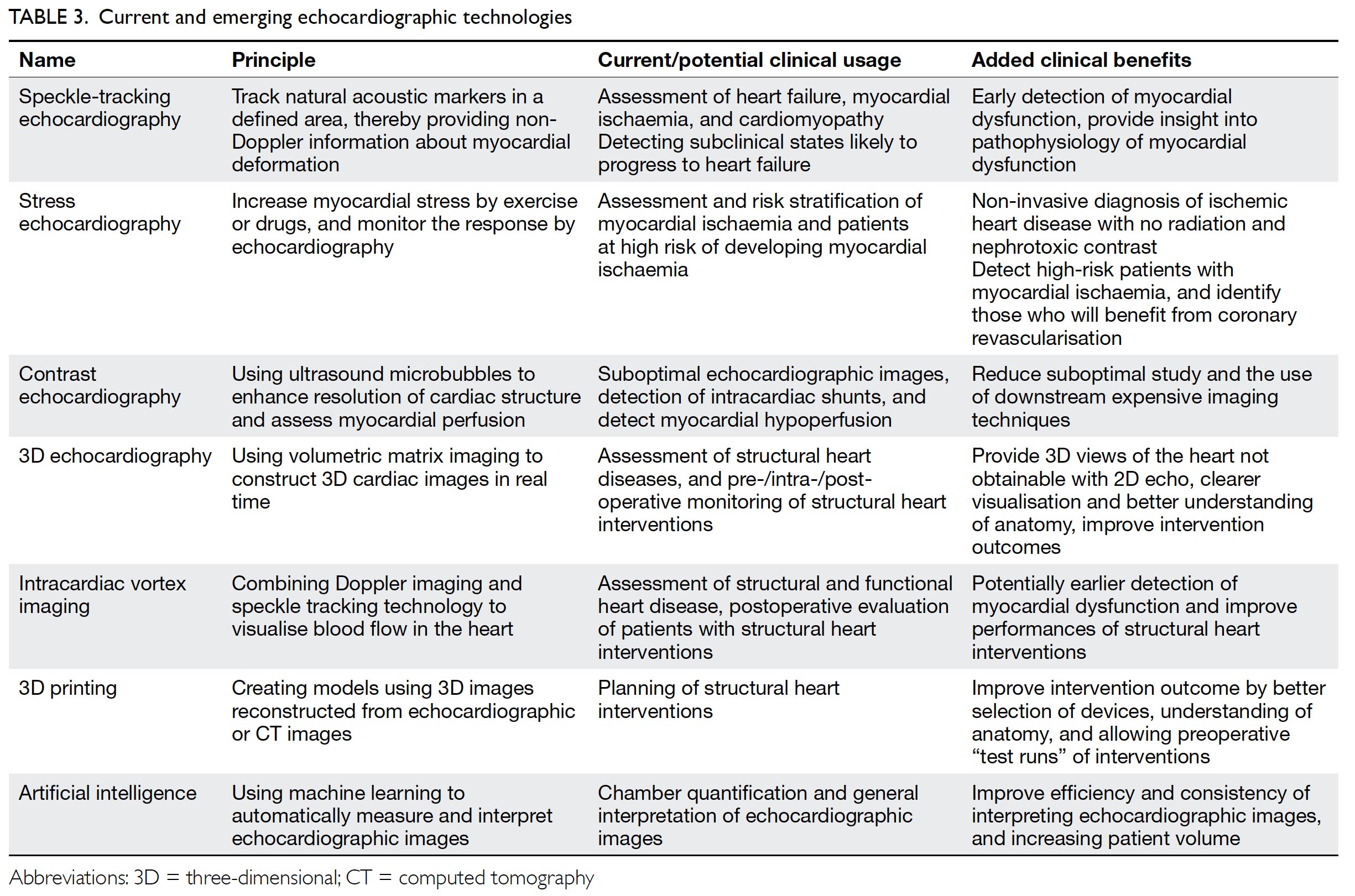
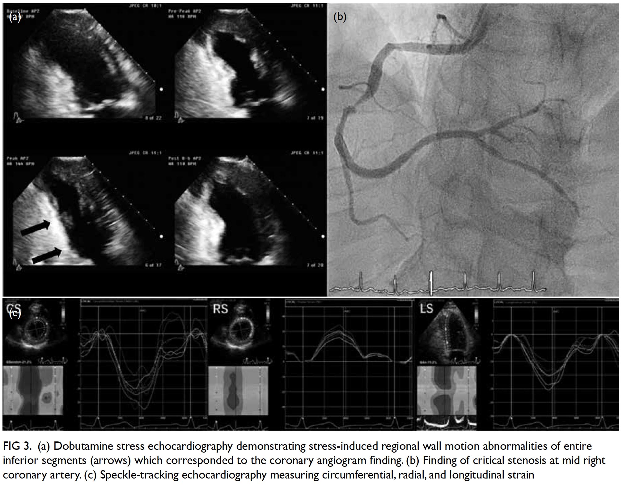
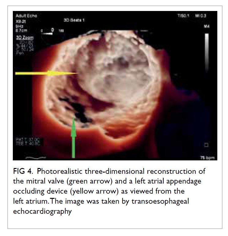
This section summarises advanced echocardiographic technologies that are relevant to common cardiovascular diseases, as well as emerging imaging technologies (Table 3).
 

Table 3. Current and emerging echocardiographic technologies
 
Stress echocardiography is valuable for evaluation of suspected coronary artery disease. Myocardial ischaemia causes reduced systolic wall thickening and excursion during stress, constituting stress-induced regional wall motion abnormalities (Fig 3). These may precede angina and ST-T changes on ECG. The sensitivity of exercise and dobutamine stress echocardiography for detection of coronary artery disease are 85% and 80% with specificity of 77% and 86%, respectively.37 Using cardiac MRI as reference standard, three-dimensional (3D) echocardiography provides more accurate and reproducible left ventricle volume and ejection fraction measurements than 2D echocardiography.38 39 40 41 42 The American Society of Echocardiography recommends the use of 3D echocardiography in chamber quantification when such technique is available in the echocardiography laboratory.43 Three-dimensional TEE has brought new insights to the mechanisms of mitral regurgitation,44 45 and has become essential in guiding transcatheter structural interventions such as transcatheter edge-to-edge mitral46 or tricuspid47 repair, transcatheter aortic valve implantation,48 and left atrial appendage occlusion (Fig 4).49 50 51 More recently, 3D printing has been used to create 3D models from 3D TEE images for preprocedural simulation.52 53 54 Speckle-tracking echocardiography allows measurement of myocardial strain (deformation; Fig 3).55 56 Assessment of the global longitudinal strain is useful for detection of chemotherapy-related cardiotoxicity in cancer patients, better than the assessment of LVEF.20
 

Figure 3. (a) Dobutamine stress echocardiography demonstrating stress-induced regional wall motion abnormalities of entire inferior segments (arrows) which corresponded to the coronary angiogram finding. (b) Finding of critical stenosis at mid right coronary artery. (c) Speckle-tracking echocardiography measuring circumferential, radial, and longitudinal strain
 

Figure 4. Photorealistic three-dimensional reconstruction of the mitral valve (green arrow) and a left atrial appendage occluding device (yellow arrow) as viewed from the left atrium. The image was taken by transoesophageal echocardiography.
 
More recently, echocardiography has been used to visualise intracardiac blood flow.57 Intracardiac vortex patterns of various pathological conditions have been shown to differ from those of healthy patients, including heart failure,58 dilated and hypertrophic cardiomyopathy,59 coronary heart disease,60 and valvular diseases.61
 
Conclusion
Echocardiography is essential in a wide range of clinical scenarios. Appropriate use of echocardiography improves clinical outcomes by increasing diagnostic accuracy, providing non-invasive or minimally invasive assessment of disease status and risk stratification, and enabling real-time monitoring and guidance of interventional procedures. Technological advancements have extended echocardiography beyond 2D imaging, allowing advanced structural visualisation and functional measurement of the heart. Echocardiography remains a vibrant field of research, and recent developments are promising clinically. Echocardiography will continue to grow as a core clinical investigation for a wide spectrum of disorders, and its relevance to primary care physicians cannot be overstated.
 
Author contributions
All authors are responsible for writing the article. APW Lee had critical revision for important intellectual content. All authors had full access to the data, contributed to the study, approved the final version for publication, and take responsibility for its accuracy and integrity.
 
Funding/support
This work was funded by the Hong Kong Special Administrative Region Government Health and Medical Research Fund (Ref 05160976).
 
Disclosure of potential conflicts of interest
APW Lee receives speaker honorarium and research grant from Philips Healthcare.
 
References
1. Roth GA, Johnson C, Abajobir A, et al. Global, regional, and national burden of cardiovascular diseases for 10 causes, 1990 to 2015. J Am Coll Cardiol 2017;70:1-25. Crossref
2. Centre for Health Protection. Heart diseases. Available from: https://www.chp.gov.hk/en/healthtopics/ content/25/57.html. Accessed 11 Mar 2019.
3. Sigurdsson EL, Jónsson JS, Thorgeirsson G. Medical treatment and secondary prevention of coronary heart disease in general practice in Iceland. Scand J Prim Health Care 2002;20:10-5. Crossref
4. Luttik ML, Jaarsma T, van Geel PP, et al. Long-term follow-up in optimally treated and stable heart failure patients: primary care vs. heart failure clinic. Results of the COACH-2 study. Eur J Heart Fail 2014;16:1241-8. Crossref
5. Senni M, Rodeheffer RJ, Tribouilloy CM, et al. Use of echocardiography in the management of congestive heart failure in the community. J Am Coll Cardiol 1999;33:164-70. Crossref
6. d’Arcy JL, Coffey S, Loudon MA, et al. Large-scale community echocardiographic screening reveals a major burden of undiagnosed valvular heart disease in older people: the OxVALVE Population Cohort Study. Eur Heart J 2016;37:3515-22. Crossref
7. American College of Cardiology Foundation Appropriate Use Criteria Task Force, American Society of Echocardiography, American Heart Association, et al. ACCF/ASE/AHA/ASNC/HFSA/HRS/SCAI/ SCCM/SCCT/SCMR 2011 Appropriate Use Criteria for Echocardiography. J Am Soc Echocardiogr 2011;24:229-67. Crossref
8. Hahn RT, Abraham T, Adams MS, et al. Guidelines for performing a comprehensive transesophageal echocardiographic examination: recommendations from the American Society of Echocardiography and the Society of Cardiovascular Anesthesiologists. J Am Soc Echocardiogr 2013;26:921-64. Crossref
9. Anavekar NS, Oh JK. Doppler echocardiography: a contemporary review. J Cardiol 2009;54:347-58. Crossref
10. Nagueh SF, Smiseth OA, Appleton CP, et al. Recommendations for the evaluation of left ventricular diastolic function by echocardiography: an update from the American Society of Echocardiography and the European Association of Cardiovascular Imaging. J Am Soc Echocardiogr 2016;29:277-314. Crossref
11. Feigenbaum H. Role of M-mode technique in today’s echocardiography. J Am Soc Echocardiogr 2010;23:240-57. Crossref
12. Gardin JM, Adams DB, Douglas PS, et al. Recommendations for a standardized report for adult transthoracic echocardiography: a report from the American Society of Echocardiography’s Nomenclature and Standards Committee and Task Force for a Standardized Echocardiography Report. J Am Soc Echocardiogr 2002;15:275-90.Crossref
13. Steeds RP, Garbi M, Cardim N, et al. EACVI appropriateness criteria for the use of transthoracic echocardiography in adults: a report of literature and current practice review. Eur Heart J Cardiovasc Imaging 2017;18:1191-204. Crossref
14. Sabia P, Afrookteh A, Touchstone DA, Keller MW, Esquivel L, Kaul S. Value of regional wall motion abnormality in the emergency room diagnosis of acute myocardial infarction. A prospective study using two-dimensional echocardiography. Circulation 1991;84(3 Suppl):I85-92.
15. Ibanez B, James S, Agewall S, et al. 2017 ESC Guidelines for the management of acute myocardial infarction in patients presenting with ST-segment elevation: The Task Force for the management of acute myocardial infarction in patients presenting with ST-segment elevation of the European Society of Cardiology (ESC). Eur Heart J 2018;39:119-77. Crossref
16. Shiga T, Wajima Z, Apfel CC, Inoue T, Ohe Y. Diagnostic accuracy of transesophageal echocardiography, helical computed tomography, and magnetic resonance imaging for suspected thoracic aortic dissection: systematic review and meta-analysis. Arch Intern Med 2006;166:1350-6. Crossref
17. Erbel R, Aboyans V, Boileau C, et al. 2014 ESC Guidelines on the diagnosis and treatment of aortic diseases: Document covering acute and chronic aortic diseases of the thoracic and abdominal aorta of the adult. The Task Force for the Diagnosis and Treatment of Aortic Diseases of the European Society of Cardiology (ESC). Eur Heart J 2014;35:2873-926. Crossref
18. Ponikowski P, Voors AA, Anker SD, et al. 2016 ESC Guidelines for the diagnosis and treatment of acute and chronic heart failure: The Task Force for the diagnosis and treatment of acute and chronic heart failure of the European Society of Cardiology (ESC) Developed with the special contribution of the Heart Failure Association (HFA) of the ESC. Eur Heart J 2016;37:2129-200. Crossref
19. Ha JW, Andersen OS, Smiseth OA. Diastolic stress test: invasive and noninvasive testing. JACC Cardiovasc Imaging 2019;pii:S1936-878X(19)30444-9.
20. Plana JC, Galderisi M, Barac A, et al. Expert consensus for multimodality imaging evaluation of adult patients during and after cancer therapy: a report from the American Society of Echocardiography and the European Association of Cardiovascular Imaging. J Am Soc Echocardiogr 2014;27:911-39. Crossref
21. Cardinale D, Colombo A, Lamantia G, et al. Anthracycline- induced cardiomyopathy: clinical relevance and response to pharmacologic therapy. J Am Coll Cardiol 2010;55:213- 20. Crossref
22. Konstantinides SV, Torbicki A, Agnelli G, et al. 2014 ESC Guidelines on the diagnosis and management of acute pulmonary embolism. Eur Heart J 2014;35:3033-69,3069a- 3069k. Crossref
23. Wnorowski AM, Halpern EJ. Diagnostic yield of triple- rule-out CT in an emergency setting. AJR Am J Roentgenol 2016;207:295-301. Crossref
24. Schertler T, Frauenfelder T, Stolzmann P, et al. Triple rule- out CT in patients with suspicion of acute pulmonary embolism: findings and accuracy. Acad Radiol 2009;16:708- 17. Crossref
25. Miniati M, Cenci C, Monti S, Poli D. Clinical presentation of acute pulmonary embolism: survey of 800 cases. PLoS One 2012;7:e30891. Crossref
26. Brignole M, Moya A, de Lange FJ, et al. 2018 ESC Guidelines for the diagnosis and management of syncope. Eur Heart J 2018;39:1883-948. Crossref
27. Saric M, Armour AC, Arnaout MS, et al. Guidelines for the use of echocardiography in the evaluation of a cardiac source of embolism. J Am Soc Echocardiogr 2016;29:1-42. Crossref
28. Baumgartner H, Falk V, Bax JJ, et al. 2017 ESC/EACTS Guidelines for the management of valvular heart disease. Eur Heart J 2017;38:2739-91. Crossref
29. Kusumoto FM, Schoenfeld MH, Barrett C, et al. 2018 ACC/AHA/HRS Guideline on the evaluation and management of patients with bradycardia and cardiac conduction delay. J Am Coll Cardiol 2019;74:e51-e156. Crossref
30. Habib G, Lancellotti P, Antunes MJ, et al. 2015 ESC Guidelines for the management of infective endocarditis. Eur Heart J 2015;36:3075-128. Crossref
31. Nishimura RA, Otto CM, Bonow RO, et al. 2014 AHA/ACC Guideline for the management of patients with valvular heart disease. Circulation 2014;129:e521-643. Crossref
32. Stout KK, Daniels CJ, Aboulhosn JA, et al. 2018 AHA/ACC Guideline for the management of adults with congenital heart disease. Circulation 2019;139:e698-e800. Crossref
33. Freeze SL, Landis BJ, Ware SM, Helm BM. Bicuspid aortic valve: a review with recommendations for genetic counseling. J Genet Couns 2016;25:1171-8. Crossref
34. Cozijnsen L, van der Zaag-Loonen HJ, Braam RL, et al. Yield of family screening in patients with isolated bicuspid aortic valve in a general hospital. Int J Cardiol 2018;255:55-8. Crossref
35. Loeys BL, Dietz HC, Braverman AC, et al. The revised Ghent nosology for the Marfan syndrome. J Med Genet 2010;47:476-85. Crossref
36. Baumgartner H, Bonhoeffer P, De Groot NM, et al. ESC Guidelines for the management of grown-up congenital heart disease (new version 2010). Eur Heart J 2010;31:2915-57. Crossref
37. Beleslin BD, Ostojic M, Djordjevic-Dikic A, et al. Integrated evaluation of relation between coronary lesion features and stress echocardiography results: the importance of coronary lesion morphology. J Am Coll Cardiol 1999;33:717-26. Crossref
38. Thavendiranathan P, Liu S, Verhaert D, et al. Feasibility, accuracy, and reproducibility of real-time full-volume 3D transthoracic echocardiography to measure LV volumes and systolic function: a fully automated endocardial contouring algorithm in sinus rhythm and atrial fibrillation. JACC Cardiovasc Imaging 2012;5:239-51. Crossref
39. Grewal J, Majdalany D, Syed I, Pellikka P, Warnes CA. Three-dimensional echocardiographic assessment of right ventricular volume and function in adult patients with congenital heart disease: comparison with magnetic resonance imaging. J Am Soc Echocardiogr 2010;23:127-33. Crossref
40. Jacobs LD, Salgo IS, Goonewardena S, et al. Rapid online quantification of left ventricular volume from real-time three-dimensional echocardiographic data. Eur Heart J 2006;27:460-8. Crossref
41. Corsi C, Lang RM, Veronesi F, et al. Volumetric quantification of global and regional left ventricular function from real-time three-dimensional echocardiographic images. Circulation 2005;112:1161-70. Crossref
42. Kühl HP, Schreckenberg M, Rulands D, et al. High-resolution transthoracic real-time three-dimensional echocardiography: quantitation of cardiac volumes and function using semi-automatic border detection and comparison with cardiac magnetic resonance imaging. J Am Coll Cardiol 2004;43:2083-90. Crossref
43. Lang RM, Badano LP, Mor-Avi V, et al. Recommendations for cardiac chamber quentification by echocardiography in adults: an update from the American Society of Echocardiography and the European Association of Cardiovasular Imaging. J Am Soc Echocardiogr 2015;28:1-39.e14. Crossref
44. Lee AP, Hsiung MC, Salgo IS, et al. Quantitative analysis of mitral valve morphology in mitral valve prolapse with real-time 3-dimensional echocardiography: importance of annular saddle shape in the pathogenesis of mitral regurgitation. Circulation 2013;127:832-41. Crossref
45. Tang Z, Fan YT, Wang Y, Jin CN, Kwok KW, Lee AP. Mitral annular and left ventricular dynamics in atrial functional mitral regurgitation: a three-dimensional and speckle- tracking echocardiographic study. J Am Soc Echocardiogr 2019;32:503-13. Crossref
46. Stone GW, Lindenfeld J, Abraham WT, et al. Transcatheter mitral-valve repair in patients with heart failure. N Engl J Med 2018;379:2307-18. Crossref
47. Cheung GS, Kam KK, Lam YY, Lee AP. Transcatheter tricuspid valve edge-to-edge repair for severe tricuspid regurgitation in a Chinese patient. Heart Asia 2018;10:e010997. Crossref
48. Mack MJ, Leon MB, Thourani VH, et al. Transcatheter aortic-valve replacement with a balloon-expandable valve in low-risk patients. N Engl J Med 2019;380:1695-705. Crossref
49. Yu CM, Khattab AA, Bertog SC, et al. Mechanical antithrombotic intervention by LAA occlusion in atrial fibrillation. Nat Rev Cardiol 2013;10:707-22. Crossref
50. Zamorano JL, Badano LP, Bruce C, et al. EAE/ASE recommendations for the use of echocardiography in new transcatheter interventions for valvular heart disease. Eur Heart J 2011;32:2189-214. Crossref
51. Lee AP, Lam YY, Yip GW, Lang RM, Zhang Q, Yu CM. Role of real time three-dimensional transesophageal echocardiography in guidance of interventional procedures in cardiology. Heart 2010;96:1485-93. Crossref
52. So KC, Fan Y, Sze L, et al. Using multimaterial 3-dimensional printing for personalized planning of complex structural heart disease intervention. JACC Cardiovasc Interv 2017;10:e97-e98. Crossref
53. Fan Y, Yang F, Cheung GS, et al. Device sizing guided by echocardiography-based three-dimensional printing is associated with superior outcome after percutaneous left atrial appendage occlusion. J Am Soc Echocardiogr 2019;32:708-19.e1. Crossref
54. Fan Y, Kwok KW, Zhang Y, Cheung GS, Chan AK, Lee AP. Three-dimensional printing for planning occlusion procedure for a double-lobed left atrial appendage. Circ Cardiovasc Interv 2016;9:e003561. Crossref
55. Leitman M, Lysyansky P, Sidenko S, et al. Two-dimensional strain-a novel software for real-time quantitative echocardiographic assessment of myocardial function. J Am Soc Echocardiogr 2004;17:1021-9. Crossref
56. Blessberger H, Binder T. NON-invasive imaging: Two dimensional speckle tracking echocardiography: basic principles. Heart 2010;96:716-22. Crossref
57. Sengupta PP, Korinek J, Belohlavek M, et al. Left ventricular structure and function: basic science for cardiac imaging. J Am Coll Cardiol 2006;48:1988-2001. Crossref
58. Poh KK, Lee LC, Shen L, et al. Left ventricular fluid dynamics in heart failure: echocardiographic measurement and utilities of vortex formation time. Eur Heart J Cardiovasc Imaging 2012;13:385-93. Crossref
59. Tang C, Zhu Y, Zhang J, et al. Analysis of left ventricular fluid dynamics in dilated cardiomyopathy by echocardiographic particle image velocimetry. Echocardiography 2018;35:56-63. Crossref
60. Zhang H, Ren X, Song J, et al. Intraventricular isovolumic relaxation flow patterns studied by using vector flow mapping. Echocardiography 2016;33:902-9. Crossref
61. Dyverfeldt P, Kvitting J, Carlhäll CJ, et al. Hemodynamic aspects of mitral regurgitation assessed by generalized phase-contrast MRI. J Magn Reson Imaging 2011;33:582-8. Crossref

Assessment and diagnosis of dementia: a review for primary healthcare professionals

Hong Kong Med J 2019 Dec;25(6):473–82  |  Epub 4 Dec 2019
© Hong Kong Academy of Medicine. CC BY-NC-ND 4.0
 
REVIEW ARTICLE  CME
Assessment and diagnosis of dementia: a review for primary healthcare professionals
K Lam, FRCP (Edin), FHKAM (Medicine)1; Windy SY Chan, MClinPharm, DHSc2; James KH Luk, FHKCP, FHKAM (Medicine)3; Angela YM Leung, PhD, FHKAN (Geron)4
1 Cheshire Home (Shatin), Hospital Authority, Hong Kong
2 School of Health Sciences, Caritas Institute of Higher Education, Hong Kong
3 Department of Medicine, Fung Yiu King Hospital, Hong Kong
4 Centre for Gerontological Nursing, The Hong Kong Polytechnic University, Hong Kong
 
Corresponding author: Dr Angela YM Leung (angela.ym.leung@polyu.edu.hk)
 
 Full paper in PDF
 
Abstract
Dementia is one of the most costly, disabling diseases associated with ageing, yet it remains underdiagnosed in primary care. In this article, we present the comprehensive approach illustrated with a classical case for diagnosing dementia which can be applied by healthcare professionals in primary care. This diagnostic approach includes history taking and physical examination, cognitive testing, informant interviews, neuropsychological testing, neuroimaging, and the utility of cerebrospinal fluid biomarkers. For the differential diagnosis of cognitive impairment, the differences and similarities among normal ageing, mild cognitive impairment, depression, and delirium are highlighted. As primary care physicians are playing an increasingly prominent role in the caring of elderly patients in an ageing population, their role in the diagnosis of dementia should be strengthened in order to provide a quality care for patients with dementia.


Introduction
Among people aged >65 years, the global prevalence of dementia has been estimated as 5%,1 with an overall prevalence of 3.9% in Asia.2 The prevalence increases with ageing, with more than one third of people aged >85 years having dementia.1 In 2015, it was estimated that 46.8 million people lived with dementia worldwide, with those numbers expected to almost double every 20 years until reaching 131.5 million in 2050.3 In China, the burden of dementia is increasing much more rapidly than previously assumed by the international health community.4 Earlier and more accurate detection of dementia is critical because it allows patients to plan their future care while they still have the capacity to make important decisions.5 Only through receiving a diagnosis can a person with dementia obtain access to cognitive and pharmacological therapies. However, dementia is underdiagnosed in primary care. Evidence from a primary care-based screening and diagnosis programme in the United States revealed that only 19% of patients with a confirmed dementia diagnosis had been checked for dementia during routine medical care.6
 
A population-based study showed that approximately 20% of family informants failed to recognise memory problems in elderly subjects who were found to have dementia on a standardised examination.7 A United Kingdom study showed that earlier diagnosis may also ease caregiver concerns.8 Planning by families of elderly patients is most effective when dementia is diagnosed early in the course of the illness. Accurate diagnosis with subtyping is the prerequisite for providing optimal therapies specific to different dementia diagnoses. In addition, reversible causes of cognitive impairment, eg, depression, should be looked for.9
 
This article reviews recent approaches in dementia diagnosis and discusses their applications by healthcare professionals, particularly those working in the primary care setting.
 
Definition of dementia
The fifth edition of the Diagnostic and Statistical Manual of Mental Disorders (DSM-5) provides a common framework for the diagnosis of neurocognitive disorders, including dementia, which is named ‘major neurocognitive disorder’ in this edition.10 11 It serves as a common linguistic framework to deal with neurocognitive disorders, thus promoting efficient communication among clinicians and researchers.11 The criteria for major neurocognitive disorders are summarised in Box 1.
 

Box 1. Criteria for major neurocognitive disorders
 
The major limitation of the DSM-5 is that it requires intellectual deficits to be sufficiently severe to impair social or occupational functioning. Therefore, it necessarily draws an arbitrary line between dementia and the lack thereof. In clinical settings, patients usually pass through stages of intellectual decline, including cognitive deficits thought to occur with normal ageing, mild cognitive impairment (MCI), and early dementia.12
 
Differential diagnosis of cognitive impairment
Accurately diagnosing dementia remains a challenge for healthcare professionals, as there is no definitive diagnostic test to identify it.
 
Does Mrs Wong have dementia?
Mrs Wong, aged 83 years, has a history of hypertension and diabetes mellitus. She lives with her daughter’s family and her activities of daily living are independent. In the past half year, her family members have noticed that Mrs Wong sometimes misses appointments with friends and her favourite Chinese music classes. Mrs Wong denies physical discomfort but cannot recall the details of her appointments. Her daughter helps by marking every appointment on a calendar to remind her. Mrs Wong is unable to handle her own banking and has become lost on the street when she goes shopping on her own. She occasionally becomes irritable when she encounters difficulties in recalling memories.
 
Not all patients with memory loss complaints have dementia. There are four common conditions that primary care doctors need to differentiate from dementia.
 
Cognitive changes with normal ageing
The normal cognitive decline associated with ageing consists primarily of mild changes in memory and the rate of information processing and does not usually affect daily functioning. Normative data from cross-sectional studies examining neuropsychological performance demonstrate that the ability to perform new learning or acquisition declines with age, whereas cued recall remains stable.12 13 14 Agerelated declines are not inevitable, and when they do occur, careful evaluation for underlying age-related diseases is warranted.
 
Mild cognitive impairment
Mild cognitive impairment is defined by the presence of memory difficulty and objective memory impairment but preserved ability to function in daily life.
 
While cognitive changes with normal age refer to normal age-associated memory and cognitive changes in older adults compared with young normal adults, MCI refers to abnormal changes in cognitive functioning, and the criteria include a measurable cognitive deficit in at least one domain.15 16 17
 
This condition is considered to be a transitional stage between normal ageing and dementia. There is no pharmacological treatment for MCI at this time. Studies have shown that subjects with MCI followed for up to 4 years have an annual conversion rate ranging from 6% to 25%, but not all of those with MCI evolve to dementia.18 Subjects with MCI may fluctuate between different trajectories of MCI, including normal cognition, MCI, and dementia.19 Progression from ‘normal to MCI’ or ‘normal to MCI to dementia’ is not always linear: subjects who develop MCI and later return to normal may develop dementia later.20 Longitudinal follow-up of MCI subjects is indicated to avoid missing a diagnosis of dementia conversion.20 The criteria for minor neurocognitive disorders are summarised in Box 2.
 

Box 2. Criteria for minor neurocognitive disorders
 
Depression
Memory impairment is commonly associated with major depression in elderly people, and it can cause pseudo-dementia syndrome. Patients with depression usually present with persistent sadness, loss of interest in their usual activities, sleep and appetite disturbances, or feelings of worthlessness or guilt.10 The most commonly used clinical screening tool for depression is the Geriatric Depression Scale.21 Patients with depression may have signs of psychomotor slowing and apply little effort to testing, while those with dementia often try hard but respond with incorrect answers. Antidepressants may improve patients’ mood and cognitive symptoms. However, dementia can sometimes co-exist with depression, and treatments may be required for both.
 
Delirium
Delirium, or acute confusional state, is another common condition in elderly people. It is usually acute or subacute in onset and is associated with sensory clouding; patients have fluctuations in their consciousness level and have difficulty maintaining attention and concentration.22 Delirium is associated with a variety of systemic illnesses, infections, and toxic and metabolic disturbances.
 
Delirium is uncommon among community-dwelling elderly people. The Canadian Study of Health and Aging found that the prevalence of delirium is <0.5% among elderly people living outside of acute care.23 Nevertheless, hospitalised elderly patients are more prone to delirium. Studies have shown that approximately 11% to 25% of hospitalised older patients have delirium upon admission, and an additional 29% to 31% of older patients admitted without delirium will develop it.24 25 Elderly patients have a decreased level of brain reserve, which makes them more prone to decompensation during acute stress and the development of delirium.
 
It is important to recognise that patients with dementia are at increased risk of delirium and that delirium and dementia may coexist. The most commonly used instrument for screening and identifying delirium is the Confusion Assessment Method.26 The updated standard diagnostic criteria for delirium are stipulated in the DSM-5.
 
Aetiology of dementia
Once a patient is diagnosed with dementia, it is important to determine the underlying aetiologies. These include various neurodegenerative diseases as well as metabolic and toxic causes. Among them, Alzheimer’s disease (AD) is the most common cause of dementia in elderly individuals, accounting for 60% to 80% of all cases, followed by vascular dementia (VaD), which accounts for about 20%.4 27 The distribution of dementia varies across geographical locations and cultural and socio-economic differences. In the Chinese population, the median proportions of AD and VaD in all forms of dementia were around 70% and 24%, respectively; other forms constituted 7.5%.4 Dementia with Lewy bodies (DLB) is among the most common forms of degenerative dementia, accounting for 4.2% of all diagnosed dementia cases in the community.28 Frontotemporal dementia (FTD), Parkinson-plus syndromes, alcohol-related dementia, chronic traumatic encephalopathy, and other central nervous system illnesses are uncommon causes and are responsible for the majority of the remaining chronic dementia cases.12
 
Common subtypes of dementia
Alzheimer’s disease is usually a disease of ageing, and its incidence increases exponentially with age >65 years. The onset and progress of AD are insidious, and memory impairment is its most frequent feature. Deficits in other cognitive domains such as executive and visuospatial tend to occur relatively early, while language deficits and behavioural symptoms often manifest later in the course of the disease.29 30
 
Vascular dementia is primarily caused by cerebrovascular disease and/or impaired cerebral blood flow. There are two main syndromes of VaD: post-stroke dementia and VaD without recent stroke.
 
Patients with post-stroke dementia experience a stepwise cognitive decline that may be accompanied by other cortical stroke signs, including aphasia and apraxia, after a clinically diagnosed stroke. In contrast, patients with VaD without recent stroke present with progressive or stepwise cognitive decline with prominent impairment in executive functioning and processing speed. Brain imaging reveals silent cerebrovascular disease including infarction or haemorrhage and is a required element of some of the commonly used diagnostic criteria for VaD, including the criteria of the NINDS-AIREN (National Institute of Neurological Disorders and Stroke).31 32
 
Dementia with Lewy bodies is a form of dementia caused by abnormal protein structures called Lewy bodies, which co-occur with symptoms of Parkinsonism such as trembling, stiffness, and slowness. Other classical features of DLB include rapid eye movement sleep behavioural disorder and fluctuation of cognition (both are easy to elicit from history). This disorder often causes vivid and long-lasting visual hallucinations. Differential diagnosis of DLB includes other degenerative dementias, especially if complicated by superimposed delirium, medication toxicity, or seizures. Diagnosis of DLB is made primarily by the revised criteria for clinical diagnosis.33
 
Frontotemporal dementia is a heterogeneous neurodegenerative disorder characterised by frontal and/or temporal lobe degeneration with early-onset dementia presenting with prominent changes in social behaviour, personality, or aphasia.34 There are several clinical presentations: behavioural variant FTD, two forms of primary progressive aphasia (PPA), non-fluent variant PPA, and semantic variant PPA.34 Among these, behavioural variant FTD is the most common form, characterised by progressive personality and behavioural changes including disinhibition, apathy, loss of empathy, hyperorality, and compulsive behaviours. The diagnostic criteria for FTD created by an international consortium synthesise clinical features, neuroimaging, neuropathology, and genetic testing.35
 
Behavioural and psychological symptoms of dementia
Behavioural and psychological symptoms of dementia (BPSD) were defined as ‘symptoms of disturbed perception, thought content, mood, and behaviour frequently occurring in patients with dementia’ by consensus among clinicians in 1996.36 37 More than 50% of people with dementia have BPSD, and these symptoms affect both patients and their relatives.38 A wide variety of affective, psychotic, and behavioural symptoms and signs can be signs of BPSD presentation, including verbal and physical aggression, agitation, hallucinations, delusions, sleep disturbances, oppositional behaviour, and wandering.39 Non-pharmacological interventions have been beneficial and should be offered as the first-line management.40 Indoor therapeutic gardening is an effective non-pharmacological intervention to reduce BPSD, medicine intake, and stress levels among patients with AD.41 Antipsychotic drugs are the common treatment for BPSD,42 but evidencebased clinical practice guidelines recommend deprescribing the drugs when the symptoms have been stabilised or treated for 3 months or more.43 Caregivers’ burden has been associated with patients’ BPSD44; therefore, treating BPSD brings benefits to both patients and caregivers.
 
Diagnostic approach
There is no single test to confirm the diagnosis of dementia. Initial assessment should include careful histories from both the patient and caregivers, physical examinations, cognitive assessments, laboratory tests, and neuroimaging.
 
History and physical examination
Clinical evaluation of patients with suspected dementia should start with a thorough and detailed history, taken from both the patient and a relative or close friend.45 It is important for primary care practitioners to rule out other causes of memory and cognitive impairments and refer appropriate patients for specialist assessment, especially those with unusual symptoms.46 A review is necessary of the patient’s family history and medical and psychiatric history, including obstructive sleep apnoea, cardiovascular disease, remote head trauma, alcohol use, and depression treatment that may contribute to cognitive decline, as well as the use of drugs that impair cognition (eg, analgesics, anticholinergics, psychotropic medications, and sedative-hypnotics).12 47 It is important to note the age of onset of cognitive impairment and to ask about family history for patients with cognitive impairment developing at or before age 65 years. Familial AD has been reported in Hong Kong, and these patients may need to be referred for genetic counselling and testing.48 49 This should be followed by a complete physical examination, including a neurological examination, to identify any clinical features of obstructive sleep apnoea, focal neurologic signs that may suggest vascular aetiology, or bradykinesia, rigidity, or tremors that would suggest a Parkinsonian syndrome.12
 
Cognitive testing
Cognitive tests provide objective evidence about cognitive deficits. Nevertheless, a single test will not suffice for all assessments. Different classes of cognitive tests are better suited to different tasks—short questionnaires are useful for rapid screening, but multidomain tests are more useful to support a clinical diagnosis of dementia.50 In the primary care setting, abbreviated or brief cognitive instruments such as abbreviated versions of the Montreal Cognitive Assessment (MoCA) and Mini-Cog are recommended in persons with symptoms of dementia.46 51
 
Mini-Mental State Examination
The Mini-Mental State Examination (MMSE) has been the most commonly used cognitive screening instrument, although patent protection has led to its decreased use in recent years. It has the advantages of being brief, easy to administer, and inclusive of multiple domains, including orientation, recall, attention, calculation, language manipulation, and constructional praxis.52 53 Published normative data allow interpretation of MMSE scores according to patient age and education. The maximum score on the MMSE is 30 points. The Cantonese version of the MMSE has been locally validated, and its cut-off points are categorised according to the patient’s education level. For patients with more than 2 years of schooling, the cut-off score is 22.54 55 The pattern of clinical deficit shown by the MMSE test is also important for the diagnosis of dementia.56 However, the test is not sensitive to mild dementia, and scores may be influenced by age, education, and language, motor and visual impairments.12 57 The MMSE has limited ability to assess progressive cognitive decline in individual patients over time.58
 
Although many ‘free’ versions of the MMSE are available online, the official version is copyrighted.
 
Montreal Cognitive Assessment
The MoCA has become the more widespread initial screening test for dementia. In early 2018, news of the President of the United States having passed the MoCA triggered widespread interest in this test. The MoCA is a 30-point test designed to detect cognitive impairment in older adults. Compared with MMSE, it is more sensitive for detection of MCI and includes items that sample a wider range of cognitive domains, including memory, language, attention, visuospatial, and executive functions.59 Cut-offs should be adjusted based on education level and other appropriate norms.60 The original MoCA takes a longer time (approximately 15 minutes) to complete compared with the MMSE. Free access with registration is available from the official website at www.mocatest.org. The MoCA has been validated in different languages, including Chinese. The Hong Kong–MoCA (HK-MoCA) has been shown to have comparable sensitivity to the Cantonese version of the MMSE for detection of MCI.61 The optimal cut-off score for HK-MoCA to detect dementia was 18/19 (sensitivity: 0.923; specificity: 0.918).61 To facilitate screening in busy clinical settings, a few abbreviated versions of the MoCA have been validated, including the MoCA 5-min protocol, which is feasible for telephone administration.62
 
Clinical Dementia Rating
Clinical Dementia Rating (CDR) was designed to assess the severity of AD in longitudinal studies and clinical trials. In a semi-structured interview with the patient and caregiver, impairments in six domains (memory, orientation, judgement and problem solving, community affairs, home and hobbies, and personal care) are assessed.63 A caregiver who knows the patient well should be present for an accurate and valid CDR assessment. The global CDR score is assigned based on performance in each domain. It is time-consuming, but the test has established validity and inter-rater reliability and is useful for following disease progression over time.64
 
Mini-Cog test
The Mini-Cog test consists of two components: a three-item recall test for memory and a simply scored clock drawing test. The assessment and instructions can be accessed at www.mocatest.org. The results of the clock drawing test are considered normal if all numbers are present in the correct sequence and the hands display the correct time in a readable way.65 The advantages of the Mini-Cog include high sensitivity for predicting dementia status, short testing time relative to the MMSE, ease of administration, and diagnostic value not limited by the subject’s education or language. Nevertheless, these tests are not appropriate when assessing patients with aphasic or anomic disorders, and more prospective data are required for further validation of this test.66
 
Informant interview
The AD8 Dementia Screening Interview is a brief, eight-item questionnaire for informants to detect dementia and cognitive impairment. Informants are asked whether the patient has exhibited any increase in eight deficits or behaviours. A positive response to two or more questions had a sensitivity of 93% and a specificity of 46%.67 The AD8 is readily administered by nurses and is useful as a screening tool by primary healthcare doctors.68 The interview questions and scoring guidelines can be accessed from Washington University in St Louis at http://alzheimer.wustl.edu/cdr/ad8.htm.
 
Neuropsychological testing
Neuropsychological testing usually involves an extensive evaluation of multiple cognitive domains (eg, attention, orientation, executive function, verbal memory, spatial memory, language, calculations, mental flexibility, and conceptualisation) and may be necessary when the bedside assessment fails to differentiate between the changes associated with normal ageing and early dementia. Moreover, neuropsychological testing can identify patterns of deficits that suggest a particular cause of dementia and assist in narrowing the differential diagnosis of dementia syndrome. However, these scores can also be influenced by education and age.18 69
 
Diagnosing Mrs Wong’s dementia
Mrs Wong scored 18 on the HK-MoCA assessment, which was the cut-off score for dementia adjusted by her age and education level.
 
Mrs Wong presented with a memory complaint evidenced by the collateral history from her family members. In her daily life, she was noted to have impaired executive functioning (inability to handle banking) and topographic disorientation (getting lost while shopping alone). Her HK-MoCA score also confirms the presence of memory impairment. Moreover, her impairment has reached the point of interfering with her social functioning. Therefore, Mrs Wong meets the clinical definition of dementia.
 
Neuroimaging
Although the literature regarding indications for neuroimaging in evaluating dementia remains inconclusive, most dementia specialists suggest that a structural brain image be obtained for newly diagnosed patients to assess any cerebrovascular lesions, neoplasms, subdural haematomas, or hydrocephalus. Imaging analysis techniques that quantify the volume of brain structures or lesions may be useful in the future for diagnosing AD. Fluorodeoxyglucose positron emission tomography (FDG-PET) and other functional neuroimaging techniques are usually used by specialists to assess complex or unclear cases of neurodegenerative dementia.18 70 Occasionally, PET imaging with different tracers for biomarkers (eg, amyloid tracers with 18F-florbetapir) in dementia may be indicated for the differential diagnosis of dementing disorders, especially in the presence of overlapping clinical features.71 Scanning with 18F-florbetapir is not available in Hong Kong, but imaging with Pittsburgh Compound B and 18F-flutametamol are available in Hong Kong. A local study has shown that 18FDG-PET with or without 11C-PIB brain imaging improved the accuracy of diagnosis of dementia subtype in 36% of a case series of Chinese dementia patients.72
 
Volumetric analysis of magnetic resonance imaging (MRI) may identify patterns of regional atrophy that are more specific in certain subtypes of dementia. In some studies, volumetric MRI has shown that patients with AD had a greater degree of hippocampal atrophy than patients with DLB had, and patients with DLB had more pronounced cortical atrophy than patients with Parkinson disease dementia.73 74
 
Although basic structural brain imaging (computed tomography or MRI) can be obtained in the primary care setting, highly specialised imaging is recommended for specialist settings only, as most studies using specialised modalities have focused on subtyping rather than the existence versus absence of dementia.46
 
Laboratory tests
Laboratory tests should be performed to identify infectious, metabolic, toxic, and inflammatory disorders that can cause neuropsychological impairment. The American Academy of Neurology recommends screening for vitamin B12 deficiency and hypothyroidism in patients with dementia. Screening for neurosyphilis is not recommended unless there is a high clinical suspicion. Because of the potential for both false positives and false negatives, genetic testing is not currently recommended unless a specific characteristic family history is present. Lumbar puncture, electroencephalography, and/or serologic tests may be useful in patients with dementia who are aged <55 years or in those with rapid progression, unusual dementia, or immunosuppression.12 18 75
 
Cerebrospinal fluid biomarkers (total-tau and p-tau)
Cerebrospinal fluid biomarkers (including Aβ42 protein, total tau, and phospho-tau) are widely investigated biomarkers of AD and can be supportive of a diagnosis of AD but are not yet recommended for routine diagnostic purposes.30 76 None of these tests are valid as a standalone diagnostic test. These cerebrospinal fluid biomarkers can be measured only in private laboratory settings on a self-paid basis. Such tests are not well accepted in Hong Kong because of the invasiveness of collecting cerebrospinal fluid.
 
Determining the underlying cause(s) of Mrs Wong’s dementia
Delirium or depression, which may mimic symptoms of dementia, should first be excluded. Medication history is checked to rule out any drugs with potential anticholinergic properties, which may also cause cognitive impairment.
 
With vascular risk factors (a history of hypertension and diabetes mellitus), there is a potential contributing cerebrovascular component to Mrs Wong’s dementia. Mrs Wong did not have a history of clinical stroke and her dementia is of a slow progressive course, not one of the stroke-like episodes of stepwise decline, as might be the case in VaD.
 
On physical examination, Mrs Wong was not found to have any Parkinsonism features or abnormal neurological signs that would be suggestive of dementia related to Parkinsonism. There were no features suggestive of DLB. Screening for other reversible causes of dementia, including thyroid disorder and vitamin B12 deficiency, were negative. Mrs Wong underwent a computed tomography scan of the brain, which showed mild cerebral atrophy and some periventricular ischaemia. On the basis of these findings, Mrs Wong met the clinical diagnostic criteria for AD.
 
Role of primary healthcare professionals
The World Alzheimer Report 2016 indicated that with the rising prevalence of dementia, the usual specialist-led approach may not expand rapidly enough to keep up with the increased need for care. Thus, primary and community care should have a more prominent role in improving the coverage of diagnosis and continuing dementia care.77
 
Screening for dementia among people at risk in primary care practices was found to significantly promote the recognition of dementia, but because of the risk of receiving a false-positive diagnosis, additional diagnostic assessment should be mandatory.78 Other perspectives exist on systematic consideration of the respective roles of primary and specialty care in long-range dementia care.79 Following recent research on the causes of and treatments for cognitive impairment, changes in clinical practice have occurred, and there is increased awareness of cognitive impairment detection during routine health check-ups in the primary care setting. The objectives of primary care physicians (PCPs) are to identify AD and other types of dementia, arrive at early diagnoses, and prevent and treat AD complications such as falls and malnutrition. It is also important for PCPs to develop close interactions with specialists, including geriatricians, psychogeriatricians, and neurologists.80 For complex case diagnosis and management, a prompt referral to a specialist is required.
 
In one study, the collaborative care delivered by an interdisciplinary team resulted in a significant improvement in both the quality of care and BPSD among both primary care patients and their caregivers.81 Structured partnerships among primary healthcare professionals may serve as bridges between primary care, specialty care, and community-based services.82 To facilitate such management, it is advised that PCPs include a relevant history in the referral letter to facilitate subtyping of the patient’s dementia (online supplementary Appendix).
 
Because of primary care providers’ growing role, there is a need for better professional education and training in both diagnosis and management of dementia. In the United Kingdom, general practitioners work one session per week in their local Memory Clinics, where they can learn directly from experts and establish working relationships with secondary dementia caregivers. It is highly recommended that we adopt the same educational programme in Hong Kong. In addition, primary doctors can refer to the Hong Kong Reference Framework for Preventive Care for Older Adults in Primary Care Settings–Module for Cognitive Impairment (2017) written by the Primary Care Office, Department of Health.83
 
Conclusion
It is important that dementia is recognised at the earliest stage and that timely evaluations are carried out to initiate appropriate therapy, with patients able to participate in management decisions. The initial step in evaluation of a patient with suspected dementia should be the taking of a focused history of cognitive and behavioural changes, followed by a complete physical examination. Delirium, depression, and MCI should be considered in the differential diagnosis of memory impairment. Many useful screening tools are now available for dementia. Once a diagnosis of dementia has been made, neuroimaging (eg, brain computed tomography or MRI scan) and laboratory tests should be performed to rule out any reversible causes of dementia. Advanced imaging, such as FDG-PET imaging or amyloid PET scanning with special tracers, may be indicated for specific cases to reach a definitive diagnosis. In the future, primary care will play a more prominent role in dementia management. Close interactions between PCPs and specialists are needed for complex case management, especially those requiring pharmacological management.
 
Author contributions
All authors have made substantial contributions to the concept or design of the study, acquisition of data, analysis or interpretation of data, drafting of the article, and critical revision for important intellectual content. All authors had full access to the data, contributed to the study, approved the final version for publication, and take responsibility for its accuracy and integrity.
 
Acknowledgement
The authors would like to thank Mr David CH Lau, who offered professional advice on cognitive tests.
 
Conflicts of interest
As an editor of the journal, JKH Luk was not involved in the peer review process of the article. Other authors have declared no conflicts of interest.
 
Funding/support
This research received no specific grant from any funding agency in the public, commercial, or not-for-profit sectors.
 
References
1. Qiu C, Kivipelto M, von Strauss E. Epidemiology of Alzheimer’s disease: occurrence, determinants, and strategies toward intervention. Dialogues Clin Neurosci 2009;11:111-28.
2. Catindig JA, Venketasubramanian N, Ikram MK, Chen C. Epidemiology of dementia in Asia: insights on prevalence, trends and novel risk factors. J Neurol Sci 2012;321:11-6. Crossref
3. Prince M, Wimo A, Guerchet M, et al. World Alzheimer Report 2015—The Global Impact of Dementia. London: Alzheimer’s Disease International; 2015.
4. Chan KY, Wang W, Wu JJ, et al. Epidemiology of Alzheimer’s disease and other forms of dementia in China, 1990-2010: a systematic review and analysis. Lancet 2013;381:2016-23. Crossref
5. Prince M, Bryce R, Ferri C. World Alzheimer report 2011: The benefits of early diagnosis and intervention. London: Alzheimer’s Disease International; 2011.
6. Boustani M, Callahan CM, Unverzagt FW, et al. Implementing a screening and diagnosis program for dementia in primary care. J Gen Intern Med 2005;20:572-7. Crossref
7. Ross GW, Abbott RD, Petrovitch H, et al. Frequency and characteristics of silent dementia among elderly Japanese-American men. The Honolulu-Asia Aging Study. JAMA 1997;277:800-5. Crossref
8. Fahy M, Wald C, Walker Z, Livingston G. Secrets and lies: the dilemma of disclosing the diagnosis to an adult with dementia. Age Ageing 2003;32:439-41. Crossref
9. Tripathi M, Vibha D. Reversible dementias. Indian J Psychiatry 2009;51 Suppl 1:S52-5.
10. American Psychiatric Association. Diagnostic and Statistical Manual of Mental Disorders (DSM-5). 5th ed. Arlington, VA: Arlington; 2013. Crossref
11. Sachdev PS, Blacker D, Blazer DG, et al. Classifying neurocognitive disorders: the DSM-5 approach. Nat Rev Neurol 2014;10:634-42. Crossref
12. Ross GW, Bowen JD. The diagnosis and differential diagnosis of dementia. Med Clin North Am 2002;86:455-76. Crossref
13. Howieson DB, Holm LA, Kaye JA, Oken BS, Howieson J. Neurologic function in the optimally healthy oldest old. Neuropsychological evaluation. Neurology 1993;43:1882-6. Crossref
14. Small SA, Stern Y, Tang M, Mayeux R. Selective decline in memory function among healthy elderly. Neurology 1999;52:1392-6. Crossref
15. Crook T, Bartus RT, Ferris SH, Whitehouse P, Cohen GD, Gershon S. Age-associated memory impairment: proposed diagnostic criteria and measures of clinical change—report of a national institute of mental health work group. Dev Neuropsychol 1986;2:261-76. Crossref
16. Levy R. Aging-associated cognitive decline. Working Party of the International Psychogeriatric Association in collaboration with the World Health Organization. Int Psychogeriatr 1994;6:63-8. Crossref
17. Petersen RC, Smith GE, Waring SC, Ivnik RJ, Tangalos EG, Kokmen E. Mild cognitive impairment: clinical characterization and outcome. Arch Neurol 1999;56:303-8. Crossref
18. Petersen RC, Stevens JC, Ganguli M, Tangalos EG, Cummings JL, DeKosky ST. Practice parameter: early detection of dementia: mild cognitive impairment (an evidence-based review). Report of the Quality Standards Subcommittee of the American Academy of Neurology. Neurology 2001;56:1133-42. Crossref
19. Pandya SY, Clem MA, Silva LM, Woon FL. Does mild cognitive impairment always lead to dementia? A review. J Neurol Sci 2016;369:57-62. Crossref
20. Lopez OL, Becker JT, Chang YF, et al. Incidence of mild cognitive impairment in the Pittsburgh Cardiovascular Health Study–Cognition Study. Neurology 2012;79:1599-606. Crossref
21. Yesavage JA, Brink TL, Rose TL, et al. Development and validation of a geriatric depression screening scale: a preliminary report. J Psychiatr Res 1982-1983;17:37-49. Crossref
22. Jorm AF, Fratiglioni L, Winblad B. Differential diagnosis in dementia. Principal components analysis of clinical data from a population survey. Arch Neurol 1993;50:72-7. Crossref
23. Andrew MK, Freter SH, Rockwood K. Prevalence and outcomes of delirium in community and non-acute care settings in people without dementia: a report from the Canadian Study of Health and Aging. BMC Med 2006;4:15. Crossref
24. Levkoff S, Cleary P, Liptzin B, Evans DA. Epidemiology of delirium: an overview of research issues and findings. Int Psychogeriatr 1991;3:149-67. Crossref
25. Vasilevskis EE, Han JH, Hughes CG, Wesley Ely E. Epidemiology and risk factors for delirium across hospital settings. Best Pact Res Clin Anaesthesiol 2012;26:277-87. Crossref
26. Leung JL, Leung VC, Leung CM, Pan PC. Clinical utility and validation of two instruments (the Confusion Assessment Method Algorithm and the Chinese version of Nursing Delirium Screening Scale) to detect delirium in geriatric inpatients. Gen Hosp Psychiatry 2008;30:171-6. Crossref
27. Rizzi L, Rosset I, Roriz-Cruz M. Global epidemiology of dementia: Alzheimer’s and vascular types. Biomed Res Int 2014;2014:908915. Crossref
28. Vann Jones SA, O’Brien JT. The prevalence and incidence of dementia with Lewy bodies: a systematic review of population and clinical studies. Psychol Med 2014;44:673- 83. Crossref
29. Ballard C, Gauthier S, Corbett A, Brayne C, Aarsland D, Jones E. Alzheimer’s disease. Lancet 2011;377:1019-31. Crossref
30. McKhann GM, Knopman DS, Chertkow H, et al. The diagnosis of dementia due to Alzheimer’s disease: recommendations from the National Institute on Aging-Alzheimer’s Association workgroups on diagnostic guidelines for Alzheimer’s disease. Alzheimers Dement 2011;7:263-9.Crossref
31. Hachinski V, Iadecola C, Petersen RC, et al. National Institute of Neurological Disorders and Stroke–Canadian Stroke Network vascular cognitive impairment harmonization standards. Stroke 2006;37:2220-41. Crossref
32. Román GC, Tatemichi TK, Erkinjuntti T, et al. Vascular dementia: diagnostic criteria for research studies. Report of the NINDS-AIREN International Workshop. Neurology 1993;43:250-60. Crossref
33. McKeith IG, Boeve BF, Dickson DW, et al. Diagnosis and management of dementia with Lewy bodies: fourth consensus report of the DLB Consortium. Neurology 2017;89:88-100. Crossref
34. Olney NT, Spina S, Miller BL. Frontotemporal dementia. Neurol Clin 2017;35:339-74. Crossref
35. Rascovsky K, Hodges JR, Knopman D, et al. Sensitivity of revised diagnostic criteria for the behavioural variant of frontotemporal dementia. Brain 2011;134(Pt 9):2456-77.
36. Finkel SI, Silva JC, Cohen GD, Miller S, Sartorius N. Behavioral and psychological symptoms of dementia: a consensus statement on current knowledge and implications for research and treatment. Am J Geriatr Psychiatry 1998;6:97-100. Crossref
37. International Psychogeriatric Association. The IPA complete guides to behavioral and psychological symptoms of dementia. Milwaukee, WI; 2010.
38. Hersch EC, Falzgraf S. Management of the behavioral and psychological symptoms of dementia. Clin Interv Aging 2007;2:611-21. Crossref
39. Tible OP, Riese F, Savaskan E, von Gunten A. Best practice in the management of behavioural and psychological symptoms of dementia. Ther Adv Neurol Disord 2017;10:297-309. Crossref
40. Bessey LJ, Walaszek A. Management of behavioral and psychological symptoms of dementia. Curr Psychiatry Rep 2019;21:66. Crossref
41. Pedrinolla A, Tamburin S, Brasioli A, et al. An indoor therapeutic garden for behavioral symptoms in Alzheimer’s disease: a randomized controlled trial. J Alzheimers Dis 2019;71:813-23. Crossref
42. Luk JH. Pharmacological management of behavioural and psychological symptoms of dementia. Asian J Gerontol Geriatr 2017;12:65-8.
43. Bjerre LM, Farrell B, Hogel M, et al. Deprescribing antipsychotics for behavioural and psychological symptoms of dementia and insomnia: evidence-based clinical practice guideline. Can Fam Physician 2018;64:17-27.
44. Huang SS, Lee MC, Liao YC, Wang WF, Lai TJ. Caregiver burden associated with behavioral and psychological symptoms of dementia (BPSD) in Taiwanese elderly. Arch Gerontol Geriatr 2012;55:55-9. Crossref
45. Knopman DS, Boeve BF, Petersen RC. Essentials of the proper diagnoses of mild cognitive impairment, dementia, and major subtypes of dementia. Mayo Clin Proc 2003;78:1290-308. Crossref
46. Pink J, O’Brien J, Robinson L, Longson D; Guideline Committee. Dementia: assessment, management and support: summary of updated NICE guidance. BMJ 2018;361:k2438. Crossref
47. Campbell NL, Boustani MA, Lane KA, et al. Use of anticholinergics and the risk of cognitive impairment in an African American population. Neurology 2010;75:152-9. Crossref
48. Shea YF, Chu LW, Chan AO, Ha J, Li Y, Song YQ. A systematic review of familial Alzheimer’s disease: differences in presentation of clinical features among three mutated genes and potential ethnic differences. J Formos Med Assoc 2016;115:67-75. Crossref
49. Shea YF, Chu LW, Lee SC, Chan AO. The first case series of Chinese patients in Hong Kong with familial Alzheimer’s disease compared with those with biomarker-confirmed sporadic late-onset Alzheimer’s disease. Hong Kong Med J 2017;23:579-85. Crossref
50. Brown J. The use and misuse of short cognitive tests in the diagnosis of dementia. J Neurol Neurosurg Psychiatry 2015;86:680-5. Crossref
51. Creavin S, Wisniewski S, Noel-Storr A, Cullum S; MMSE review team. Cognitive tests to help diagnose dementia in symptomatic people in primary care and the community. Br J Gen Pract 2018;68:149-50. Crossref
52. Folstein MF, Folstein SE, McHugh PR. “Mini-mental state”. A practical method for grading the cognitive state of patients for the clinician. J Psychiatr Res 1975;12:189-98. Crossref
53. Tangalos EG, Smith GE, Ivnik RJ, et al. The Mini-Mental State Examination in general medical practice: clinical utility and acceptance. Mayo Clin Proc 1996;71:829-37. Crossref
54. Chiu HF, Lee HC, Chung WS, Kwong PK. Reliability and validity of the Cantonese version of Mini-Mental State Examination—a preliminary study. JHKC Psych 1994;4:25-8.
55. Chiu HF, Lam C, Chi I, et al. Prevalence of dementia in Chinese elderly in Hong Kong. Neurology 1998;50:1002-9. Crossref
56. Anthony JC, LeResche L, Niaz U, von Korff MR, Folstein MF. Limits of the ‘Mini-Mental State’ as a screening test for dementia and delirium among hospital patients. Psychol Med 1982;12:397-408. Crossref
57. Crum RM, Anthony JC, Bassett SS, Folstein MF. Population-based norms for the Mini-Mental State Examination by age and educational level. JAMA 1993;269:2386-91. Crossref
58. Creavin ST, Wisniewski S, Noel-Storr AH, et al. Mini-Mental State Examination (MMSE) for the detection of dementia in clinically unevaluated people aged 65 and over in community and primary care populations. Cochrane Database Syst Rev 2016;(1):CD011145.
59. Nasreddine ZS, Phillips NA, Bédirian V, et al. The Montreal Cognitive Assessment, MoCA: a brief screening tool for mild cognitive impairment. J Am Geriatr Soc 2005;53:695-9. Crossref
60. Rossetti HC, Lacritz LH, Cullum CM, Weiner MF. Normative data for the Montreal Cognitive Assessment (MoCA) in a population-based sample. Neurology 2011;77:1272-5. Crossref
61. Yeung PY, Wong LL, Chan CC, Leung JL, Yung CY. A validation study of the Hong Kong version of Montreal Cognitive Assessment (HK-MoCA) in Chinese older adults in Hong Kong. Hong Kong Med J 2014;20:504-10. Crossref
62. Wong A, Nyenhuis D, Black SE, et al. Montreal Cognitive Assessment 5-minute protocol is a brief, valid, reliable, and feasible cognitive screen for telephone administration. Stroke 2015;46:1059-64. Crossref
63. Washington University in St Louis. CDR® Dementia Staging Instrument. Available from: https://knightadrc. wustl.edu/cdr/cdr.htm. Accessed 30 Aug 2019.
64. Morris JC. The Clinical Dementia Rating (CDR): current version and scoring rules. Neurology 1993;43:2412-4. Crossref
65. Mini-Cog©–Screening for cognitive impairment in older adults. Available from: https://mini-cog.com/. Accessed 30 Aug 2019.
66. Borson S, Scanlan J, Brush M, Vitaliano P, Dokmak A. The Mini-Cog: a cognitive ‘vital signs’ measure for dementia screening in multi-lingual elderly. Int J Geriatr Psychiatry 2000;15:1021-7. Crossref
67. Galvin JE, Roe CM, Xiong C, Morris JC. Validity and reliability of the AD8 informant interview in dementia. Neurology 2006;67:1942-8. Crossref
68. Shaik MA, Khoo CH, Thiagarajah AG, et al. Pilot evaluation of a dementia case finding clinical service using the informant AD8 for at-risk older adults in primary health care: a brief report. J Am Med Dir Assoc 2016;17:673.e5-8. Crossref
69. Cahn DA, Salmon DP, Butters N, et al. Detection of dementia of the Alzheimer type in a population-based sample: neuropsychological test performance. J Int Neuropsychol Soc 1995;1:252-60. Crossref
70. Corey-Bloom J, Thal LJ, Galasko D, et al. Diagnosis and evaluation of dementia. Neurology 1995;45:211-8. Crossref
71. Berti V, Pupi A, Mosconi L. PET/CT in diagnosis of dementia. Ann N Y Acad Sci 2011;12281:81-92. Crossref
72. Shea YF, Ha J, Lee SC, Chu LW. Impact of 18FDG PET and 11C-PIB PET brain imaging on the diagnosis of Alzheimer’s disease and other dementias in a regional memory clinic in Hong Kong. Hong Kong Med J 2016;22:327-33. Crossref
73. Barber R, Ballard C, McKeith IG, Gholkar A, O’Brien JT. MRI volumetric study of dementia with Lewy bodies; a comparison with AD and vascular dementia. Neurology 2000;54:1304-9. Crossref
74. Beyer MK, Larsen JP, Aarsland D. Gray matter atrophy in Parkinson disease with dementia and dementia with Lewy bodies. Neurology 2007;69:747-54. Crossref
75. Geschwind MD, Shu H, Haman A, Sejvar JJ, Miller BL. Rapidly progressive dementia. Ann Neurol 2008;64:97-108. Crossref
76. Shea YF, Chu LW, Zhou L, et al. Cerebrospinal fluid biomarkers of Alzheimer’s disease in Chinese patients: a pilot study. Am J Alzheimers Dis Other Demen 2013;28:769-75. Crossref
77. Prince M, Comas-Herrera A, Knapp M, Guerchet M, Karagiannidou M. World Alzheimer Report 2016—improving healthcare for people living with dementia: coverage, quality and costs now and in the future. London: Alzheimer’s Disease International; 2016.
78. Eichler T, Thyrian JR, Hertel J, et al. Rates of formal diagnosis of dementia in primary care: the effect of screening. Alzheimers Dement (Amst) 2015;1:87-93. Crossref
79. Borson S, Frank L, Bayley PJ, et al. Improving dementia care: the role of screening and detection of cognitive impairment. Alzheimers Dement 2013;9:151-9. Crossref
80. Cohen CA, Pringle D, LeDuc L. Dementia caregiving: the role of the primary care physician. Can J Neurol Sci 2001;28 Suppl 1:S72-6. Crossref
81. Callahan CM, Boustani MA, Unverzagt FW, et al. Effectiveness of collaborative care for older adults with Alzheimer disease in primary care: a randomized controlled trial. JAMA 2006;295:2148-57. Crossref
82. Reuben DB, Roth CP, Frank JC, et al. Assessing care of vulnerable elders—Alzheimer’s disease: a pilot study of a practice redesign intervention to improve the quality of dementia care. J Am Geriatr Soc 2010;58:324-9. Crossref
83. Hong Kong Reference Framework for Preventive Care for Older Adults in Primary Care Settings. Module on Cognitive Impairment. Department of Health, Hong Kong SAR Government; 2017.

In vitro fertilisation in Hong Kong: the situation in 2019

Hong Kong Med J 2019 Dec;25(6):468–72  |  Epub 4 Dec 2019
© Hong Kong Academy of Medicine. CC BY-NC-ND 4.0
 
REVIEW ARTICLE  CME
In vitro fertilisation in Hong Kong: the situation in 2019
MW Lui, MB, BS; William SB Yeung, PhD; PC Ho, MB, BS, MD; Ernest HY Ng, MB, BS, MD
Department of Obstetrics and Gynaecology, The University of Hong Kong, Pokfulam, Hong Kong
 
Corresponding author: Prof Ernest HY Ng (nghye@hku.hk)
 
 Full paper in PDF
 
Abstract
The popularity of in vitro fertilisation has continuously increased throughout the past 40 years owing to an increased incidence of infertility and delayed planning for pregnancy. The aim of this paper is to review the current situation of in vitro fertilisation in Hong Kong. In Hong Kong, in 2018, 7995 women underwent 5055 fresh and 5050 frozen-thawed embryo in vitro fertilisation cycles, resulting in an ongoing pregnancy rate of 33.7% per transfer. However, in vitro fertilisation is associated with several problems, including a high rate of multiple pregnancies and risks associated with cross-border reproductive care. Single embryo transfer is a simple strategy to reduce multiple pregnancies without compromising the cumulative live birth rate.


Introduction
The incidence of infertility has increased in recent decades, and women are more often delaying marriage and pregnancy. The mean age at which women deliver their first baby has increased from 27.9 years in 1995 to 30.5 years in 2017.1 Owing to the natural decline in fertility with age, more and more couples seek help to conceive through IVF. The number of IVF cycles performed in Europe increased from 203 225 cycles in 1997 to 776 556 in 2014, with 170 163 babies born.2 A similar trend has been observed worldwide.3 The aim of this paper is to review the current situation of IVF in 2019.
 
History of the in vitro fertilisation process
Last year in 2018, the first “test tube” baby Louise Brown celebrated her 40th birthday. In vitro fertilisation was pioneered by Patrick Steptoe and Robert Edwards. They first obtained an oocyte in a natural cycle using a laparoscope. After it was fertilised in the laboratory, they transferred the embryo back to the donor’s uterus. The techniques of IVF have progressed rapidly since then. The success rate has also greatly increased from lower than 1% per cycle to around 35% per cycle. The success rate has increased owing to several factors, including ovarian stimulation, assisted fertilisation using intracytoplasmic sperm insemination, improvements in culture conditions, and more advanced embryo transfer techniques.
 
Modern IVF cycles include ovarian stimulation, oocyte retrieval, fertilisation and culture in vitro, and embryo transfer to the uterus. Ovarian stimulation involves administration of gonadotropin, coupled with suppression of endogenous gonadotropin release by either gonadotropin-releasing hormone (GnRH) agonist or antagonist. The super-physiological doses of gonadotropins enhance growth of multiple follicles, in contrast to the development of a single follicle in a natural cycle. The use of GnRH agonist or antagonist reduces the risk of premature ovulation. Women undergoing ovarian stimulation need close monitoring, with pelvic scanning to determine the number and size of the follicles and hormonal tests to measure the serum oestradiol level. When the leading follicles reach >17 to 18 mm, human chorionic gonadotropin is usually given to the patient to trigger the final maturation of the oocytes.
 
At 36 to 38 hours after the trigger, oocyte retrieval is typically performed through the transvaginal route under ultrasound guidance. An aspiration needle is used to puncture through the vagina to aspirate the follicular fluid with the oocytes and the follicles are aspirated till complete collapse of the follicles. Oocyte retrieval involves a small risk of bleeding, infection, pain, and risk of injury to visceral organs.
 
In vitro fertilisation refers to the overnight co-culture of oocytes with sperm. For couples with severe male factor infertility or fertilisation problems in previous IVF cycles, intracytoplasmic sperm insemination involving injection of a single sperm into an oocyte is advised.4 The fertilised embryo is transferred back to the uterine cavity either at cleavage (Day 2 or 3) or blastocyst stage (Day 5). Surplus high quality fertilised embryos can be frozen at the cleavage or blastocyst stage for subsequent transfer by slow freezing or more commonly by vitrification, which involves rapid freezing in liquid nitrogen. Vitrification enables a rapid transition of the liquid form of water to a “glass” status, which avoids crystallisation that can damage the cells.
 
In vitro fertilisation is not without risks, even in healthy couples. The most common complications of IVF are multiple pregnancy followed by ovarian hyperstimulation syndrome, which can lead to ascites, pleural effusion, venous thromboembolism, or even death. Although severe ovarian hyperstimulation syndrome has a low incidence, it can be potentially life-threatening and is avoidable. In cycles using GnRH antagonists to prevent premature ovulation, agonist can be used instead of human chorionic gonadotropin as a trigger for oocyte final maturation in order to lower the risk of ovarian hyperstimulation syndrome.2 5
 
Overall, the success of IVF greatly depends on the woman’s age. As shown by the latest data by ESHRE (European Society of Human Reproduction and Embryology),2 in 2014, live birth rates after fresh transfer IVF in women aged <35, 35 to 39, and ≥40 years were 23.8%, 18.8%, and 8.1%, respectively. For women aged <30 years, the cumulative live birth rate after completion of a cycle can be nearly 70%, compared with 17.3% in women aged ≥40 years.4
 
In vitro fertilisation in Hong Kong
History and development
In Hong Kong, IVF programmes were initiated in the Department of Obstetrics and Gynaecology, The Chinese University of Hong Kong in December 1984, in the Hong Kong Sanatorium & Hospital in January 1986, and in the Department of Obstetrics and Gynaecology, The University of Hong Kong in July 1986.7 The Hong Kong Sanatorium & Hospital programme achieved the first successful IVF pregnancy and delivery in Hong Kong.7
 
By August 1987, 522 IVF cycles had been initiated in Hong Kong and the overall clinical pregnancy rate was 5.2% per cycle initiated.6 From January 1992 to December 1993, 912 IVF cycles, 158 cycles of gamete intrafallopian transfer, 87 cycles of zygote intrafallopian transfer, and 233 cycles of frozen-thawed embryo transfer (FET) were initiated, with delivery rates per cycle started of 8.4%, 29.1%, 13.8%, and 11.2%, respectively.8
 
More recently, according to the Council on Human Reproductive Technology (HRT Council) Annual Report,9 the number of women undergoing IVF increased from 2415 in 2009 to 7995 in 2018, with the number of fresh and FET IVF cycles increasing from 2768 to 10 105 over the same period (Table 1). The ongoing pregnancy rate per transfer increased slightly from 29.9% in 2009 to 32.8% in 2015 and remained steady in 2015 to 2018 (Table 2). The ongoing pregnancy rate per transfer was 33.7% in 2018. However, there was no significant increase in live birth rates throughout the years. Both ongoing and live birth rates are markedly reduced in women aged ≥41 years.
 

Table 1. In vitro fertilisation cycles from 2009 to 2018 in Hong Kong, data from the Council on Human Reproductive Technology Annual Report11
 

Table 2. Ongoing pregnancy rate and live birth rate of both fresh and frozen cycles according to age for women who underwent in vitro fertilisation in 201711
 
The number of embryos transferred has reduced gradually over the years, though double embryo transfer still is the most common. The single embryo transfer rate has increased from 12.9% (321 cycles) in 2009 to 49.2% (3043 cycles) in 2017. As a result, the multiple pregnancy rate per ongoing pregnancy has decreased, but remained high at 15.2% in 2017.
 
Regulation of assisted reproduction
Assisted reproduction in Hong Kong is regulated by the Human Reproductive Technology Ordinance (Cap. 561).10 Centres that provide IVF must obtain a treatment licence from the HRT Council. At the time of writing, there are 18 licensed treatment centres in Hong Kong and of these, 13 provide IVF. Of these 13 treatment centres, 10 are in the private sector and three are in the public sector.11
 
As stated in the HRT Council Code of Practice,6 in Hong Kong, IVF can be provided only to legally married couples in monogamous relationship. The maximum number of embryos that can be transferred to the woman is three per cycle. Frozen embryos can be stored for up to 10 years from the day of freezing, and gametes can be stored until the patient is aged 55 years. All IVF data must be documented clearly and reported promptly and accurately to the HRT Council. Commercial oocyte donation is not allowed in Hong Kong.11 No payment to the oocyte donor is allowed apart from reimbursing the loss of earnings and the expenses, such as transportation and appointment fee. Sex selection, unless medically indicated, is also prohibited in Hong Kong. Although surrogacy is legal in Hong Kong, none of the centres in Hong Kong has a licence for surrogacy arrangements, and surrogacy agreements have been found to be unenforceable under the law.11
 
Public in vitro fertilisation service
Currently, IVF service is provided in three public hospitals in Hong Kong: Kwong Wah Hospital, Prince of Wales Hospital, and Queen Mary Hospital. Eligible couples can receive partial subsidy funding from the Hong Kong Hospital Authority for up to three IVF cycles. The number of publicly funded cycles has increased to around 1000 cycles in total in 2018. The criteria for funded IVF cycles include women who are permanent Hong Kong residents, aged <40 years, and with no biological children. The waiting time for IVF in public hospitals in Hong Kong is up to 3 years. The provision of IVF in public hospitals is not entirely free of charge. Couples pay approximately HK$20 000 for all medication, procedures, and embryo storage for up to three cycles, but this is lower than the HK$80 000 to HK$100 000 required for just one IVF cycle in the private sector.
 
Recent in vitro fertilisation technology developments
Chromosome aneuploidy
Chromosome aneuploidy is an error in cell division that results in the “daughter” cells having the wrong number of chromosomes. Chromosome aneuploidy is a major reason for failure of conception, pregnancy loss, and congenital anomalies following both natural conception and IVF pregnancies and its prevalence increases exponentially with maternal age. The need to assess embryo quality and select those with the highest potential for implantation on the basis of morphology has led to preimplantation genetic testing for aneuploidy (PGT-A).9 This involves biopsy of a few cells from an embryo at the blastocyst stage and assessment of the comprehensive chromosome copy numbers. Although PGT-A cannot create a healthy embryo or improve the health of an embryo, it can provide an accurate method of selecting of embryos with a normal number of chromosomes for transfer. This in turn has the potential to increase the chance of having a healthy live birth per each transfer and to reduce the risk of miscarriage or abnormal fetus caused by an abnormal number of chromosomes.12 However, the potential damage of trophectoderm biopsy on the developing embryos remains unknown. In addition, the concordance of trophectoderm biopsy and blastocyst is questionable especially in those with segmental aneuploidy13 and mosaicism.14 Gene editing may provide a new perspective in future, but there is still a long way ahead. Gene editing technologies are immature and imprecise, and involve unknown long-term risks.
 
Oocyte freezing
Oocyte freezing by vitrification is no longer considered to be experimental and is offered to women who desire preservation of their fertility potential before chemotherapy or radiotherapy for cancer treatment. In Hong Kong, the indication of oocyte freezing has been extended to single women who wish to delay parenthood for education or career purposes. In general, fertility preservation for cancer patients is underutilised, partly owing to lack of funding in the public sector and inadequate information for patients. Fertility preservation for cancer patients requires a close cooperation between fertility specialists, oncologists, paediatricians, and surgeons. Oocyte or embryo freezing provides more promising results in fertility preservation, but it involves a delay of treatment for at least 2 weeks and it is possible only in postpubertal women. Owing to the lack of funding, patients often need to pay for their own treatment.
 
Problems facing in vitro fertilisation service in Hong Kong
Multiple pregnancy
There is an international goal aiming to reduce the incidence of multiple pregnancy as a result of IVF to <10%. Multiple pregnancies after IVF create a huge burden on the healthcare system. The chance of multiple pregnancy increases exponentially with the number of embryos transferred. Multiple pregnancies are associated with high risk of prematurity, low birth rate, and tremendous support from neonatal intensive care unit. It has been estimated that one premature baby at 29 weeks can cost up to US$122 000 and prematurity cost a total of US$4567 billion in United Kingdom in 2006.15 Owing to improvements in embryo culture and cryopreservation techniques, with the use of vitrification, the cumulative pregnancy rate is similar in single or double embryo transfer.16 In Kwong Wah Hospital and Queen Mary Hospital, women can have only one embryo transferred at a time unless they are aged ≥38 years, have failed two IVF cycles, and have had no live births. Although single embryo transfer results in a slightly longer interval to pregnancy, it can significantly lower the multiple pregnancy rate to <2%.16
 
Cross-border reproductive care
The reasons for Hong Kong residents seeking cross-border reproductive care are multifactorial. Reasons may include the long waiting times for publicly funded IVF, the high cost of private IVF, lack of oocyte donors, and ineligibility for IVF in Hong Kong. Those ineligible for IVF in Hong Kong include unmarried couples, same-sex couples, and those seeking IVF for surrogacy, sex selection, or social reasons. Women seeking IVF overseas face risks including lack of regulations in some centres and lack of medical care or insurance coverage in case of complications.
 
Other problems
Other problems include the risks of ovarian hyperstimulation syndrome, the high cost of IVF, and difficulty in getting gamete and embryo donation.
 
Conclusion
In vitro fertilisation provides hope for infertile couples. However, there are many unresolved issues, especially the high rate of multiple pregnancies and potential risks associated with cross-border reproductive care.
 
Author contributions
All authors had full access to the data, contributed to the study, approved the final version for publication, and take responsibility for its accuracy and integrity.
 
Concept or design: EHY Ng.
Acquisition of data: MW Lui.
Analysis or interpretation of data: MW Lui.
Drafting of the article: MW Lui.
Critical revision for important intellectual content: WSB Yeung, PC Ho, EHY Ng.
 
Conflicts of interest
All authors have disclosed no conflicts of interest.
 
Funding/support
This research received no specific grant from any funding agency in the public, commercial, or not-for-profit.
 
References
1. Organisation for Economic Co-operation and Development. OECD Family Database. SF2.3.B. Age of mothers at childbirth and age-specific fertility. Available from: http://www.oecd.org/els/family/database.htm. Accessed 26 Jul 2019.
2. De Geyter C, Calhaz-Jorge C, Kupka MS, et al. ART in Europe, 2014: results generated from European registries by ESHRE: The European IVF-monitoring Consortium (EIM) for the European Society of Human Reproduction and Embryology (ESHRE). Hum Reprod 2018;33:1586-601. Crossref
3. Chambers GM, Wand H, Macaldowie A, Chapman MG, Farquhar CM, Bowman M, Molloy, D, Ledger W. Population trends and live birth rates associated with common ART treatment strategies. Hum Reprod 2016;31:2632-41. Crossref
4. Babayev SN, Park CW, Bukulmez O. Intracytoplasmic sperm injection indications: how rigorous? Semin Reprod Med 2014;32:283-90. Crossref
5. McLernon DJ, Maheshwari A, Lee AJ, Bhattacharya S. Cumulative live birth rates after one or more complete cycles of IVF: a population-based study of linked cycle data from 178,898 women. Hum Reprod 2016;31:572-81. Crossref
6. Council on Human Reproductive Technology. Code of Practice on Reproductive Technology and Embryo Research. January 2013. Available from: https://www.chrt.org.hk/english/service/service_cod.html. Accessed 26 Jul 2019.
7. Mao KR, Loong EP, Lee HC, et al. Current status of in-vitro fertilization (IVF) in Hong Kong. J Hong Kong Med Assoc 1987;39:144-6.
8. Hong Kong IVF Study Group. Assisted reproduction in Hong Kong: status in the 1990s. Hong Kong Med J 1996;2:253-6.
9. Lehmann L, El-Haddad A, Barr RD. Global approach to hematologic malignancies. Hematol Oncol Clin North Am 2016;30:417-32. Crossref
10. Cap. 561 Human reproductive technology ordinance 2000 (Hong Kong SAR). Available from: https://www.elegislation.gov.hk/hk/cap561. Accessed 26 Jul 2019.
11. Council on Human Reproductive Technology. Available from: https://www.chrt.org.hk/. Accessed 26 Jul 2019.
12. Maxwell SM, Grifo JA. Should every embryo undergo preimplantation genetic testing for aneuploidy? A review of the modern approach to in vitro fertilization. Best Pract Res Clin Obstet Gynaecol 2018;53:38-47. Crossref
13. Victor AR, Griffin DK, Brake AJ, et al. Assessment of aneuploidy concordance between clinical trophectoderm biopsy and blastocyst. Hum Reprod 2019;34:181-92. Crossref
14. Capalbo A, Ubaldi FM, Rienzi L, Scott R, Treff N. Detecting mosaicism in trophectoderm biopsies: current challenges and future possibilities. Hum Reprod 2017;32:492-8. Crossref
15. Saha S, Gerdtham UG. Cost of illness studies on reproductive, maternal, newborn, and child health: a systematic literature review. Health Econ Rev 2013;3:24. Crossref
16. Pandian Z, Marjoribanks J, Ozturk O, Serour G, Bhattacharya S. Number of embryos for transfer following in vitro fertilisation or intra-cytoplasmic sperm injection. Cochrane Database Syst Rev 2013;(7):CD003416. Crossref

Assessment and diagnosis of dementia: a review for primary healthcare professionals

Hong Kong Med J 2019;25:Epub 4 Dec 2019
© Hong Kong Academy of Medicine. CC BY-NC-ND 4.0
 
REVIEW ARTICLE
Assessment and diagnosis of dementia: a review for primary healthcare professionals
K Lam, FRCP (Edin), FHKAM (Medicine)1; Windy SY Chan, MClinPharm, DHSc2; James KH Luk, FHKCP, FHKAM (Medicine)3; Angela YM Leung, PhD, FHKAN (Geron)4
1 Cheshire Home (Shatin), Hospital Authority, Hong Kong
2 School of Health Sciences, Caritas Institute of Higher Education, Hong Kong
3 Department of Medicine, Fung Yiu King Hospital, Hong Kong
4 Centre for Gerontological Nursing, The Hong Kong Polytechnic University, Hong Kong
 
Corresponding author: Dr Angela YM Leung (angela.ym.leung@polyu.edu.hk)
 
 Full paper in PDF
 
Abstract
Dementia is one of the most costly, disabling diseases associated with ageing, yet it remains underdiagnosed in primary care. In this article, we present the comprehensive approach illustrated with a classical case for diagnosing dementia which can be applied by healthcare professionals in primary care. This diagnostic approach includes history taking and physical examination, cognitive testing, informant interviews, neuropsychological testing, neuroimaging, and the utility of cerebrospinal fluid biomarkers. For the differential diagnosis of cognitive impairment, the differences and similarities among normal ageing, mild cognitive impairment, depression, and delirium are highlighted. As primary care physicians are playing an increasingly prominent role in the caring of elderly patients in an ageing population, their role in the diagnosis of dementia should be strengthened in order to provide a quality care for patients with dementia.


Introduction
Among people aged >65 years, the global prevalence of dementia has been estimated as 5%,1 with an overall prevalence of 3.9% in Asia.2 The prevalence increases with ageing, with more than one third of people aged >85 years having dementia.1 In 2015, it was estimated that 46.8 million people lived with dementia worldwide, with those numbers expected to almost double every 20 years until reaching 131.5 million in 2050.3 In China, the burden of dementia is increasing much more rapidly than previously assumed by the international health community.4 Earlier and more accurate detection of dementia is critical because it allows patients to plan their future care while they still have the capacity to make important decisions.5 Only through receiving a diagnosis can a person with dementia obtain access to cognitive and pharmacological therapies. However, dementia is underdiagnosed in primary care. Evidence from a primary care-based screening and diagnosis programme in the United States revealed that only 19% of patients with a confirmed dementia diagnosis had been checked for dementia during routine medical care.6
 
A population-based study showed that approximately 20% of family informants failed to recognise memory problems in elderly subjects who were found to have dementia on a standardised examination.7 A United Kingdom study showed that earlier diagnosis may also ease caregiver concerns.8 Planning by families of elderly patients is most effective when dementia is diagnosed early in the course of the illness. Accurate diagnosis with subtyping is the prerequisite for providing optimal therapies specific to different dementia diagnoses. In addition, reversible causes of cognitive impairment, eg, depression, should be looked for.9
 
This article reviews recent approaches in dementia diagnosis and discusses their applications by healthcare professionals, particularly those working in the primary care setting.
 
Definition of dementia
The fifth edition of the Diagnostic and Statistical Manual of Mental Disorders (DSM-5) provides a common framework for the diagnosis of neurocognitive disorders, including dementia, which is named ‘major neurocognitive disorder’ in this edition.10 11 It serves as a common linguistic framework to deal with neurocognitive disorders, thus promoting efficient communication among clinicians and researchers.11 The criteria for major neurocognitive disorders are summarised in Box 1.
 

Box 1. Criteria for major neurocognitive disorders
 
The major limitation of the DSM-5 is that it requires intellectual deficits to be sufficiently severe to impair social or occupational functioning. Therefore, it necessarily draws an arbitrary line between dementia and the lack thereof. In clinical settings, patients usually pass through stages of intellectual decline, including cognitive deficits thought to occur with normal ageing, mild cognitive impairment (MCI), and early dementia.12
 
Differential diagnosis of cognitive impairment
Accurately diagnosing dementia remains a challenge for healthcare professionals, as there is no definitive diagnostic test to identify it.
 
Does Mrs Wong have dementia?
Mrs Wong, aged 83 years, has a history of hypertension and diabetes mellitus. She lives with her daughter’s family and her activities of daily living are independent. In the past half year, her family members have noticed that Mrs Wong sometimes misses appointments with friends and her favourite Chinese music classes. Mrs Wong denies physical discomfort but cannot recall the details of her appointments. Her daughter helps by marking every appointment on a calendar to remind her. Mrs Wong is unable to handle her own banking and has become lost on the street when she goes shopping on her own. She occasionally becomes irritable when she encounters difficulties in recalling memories.
 
Not all patients with memory loss complaints have dementia. There are four common conditions that primary care doctors need to differentiate from dementia.
 
Cognitive changes with normal ageing
The normal cognitive decline associated with ageing consists primarily of mild changes in memory and the rate of information processing and does not usually affect daily functioning. Normative data from cross-sectional studies examining neuropsychological performance demonstrate that the ability to perform new learning or acquisition declines with age, whereas cued recall remains stable.12 13 14 Agerelated declines are not inevitable, and when they do occur, careful evaluation for underlying age-related diseases is warranted.
 
Mild cognitive impairment
Mild cognitive impairment is defined by the presence of memory difficulty and objective memory impairment but preserved ability to function in daily life.
 
While cognitive changes with normal age refer to normal age-associated memory and cognitive changes in older adults compared with young normal adults, MCI refers to abnormal changes in cognitive functioning, and the criteria include a measurable cognitive deficit in at least one domain.15 16 17
 
This condition is considered to be a transitional stage between normal ageing and dementia. There is no pharmacological treatment for MCI at this time. Studies have shown that subjects with MCI followed for up to 4 years have an annual conversion rate ranging from 6% to 25%, but not all of those with MCI evolve to dementia.18 Subjects with MCI may fluctuate between different trajectories of MCI, including normal cognition, MCI, and dementia.19 Progression from ‘normal to MCI’ or ‘normal to MCI to dementia’ is not always linear: subjects who develop MCI and later return to normal may develop dementia later.20 Longitudinal follow-up of MCI subjects is indicated to avoid missing a diagnosis of dementia conversion.20 The criteria for minor neurocognitive disorders are summarised in Box 2.
 

Box 2. Criteria for minor neurocognitive disorders
 
Depression
Memory impairment is commonly associated with major depression in elderly people, and it can cause pseudo-dementia syndrome. Patients with depression usually present with persistent sadness, loss of interest in their usual activities, sleep and appetite disturbances, or feelings of worthlessness or guilt.10 The most commonly used clinical screening tool for depression is the Geriatric Depression Scale.21 Patients with depression may have signs of psychomotor slowing and apply little effort to testing, while those with dementia often try hard but respond with incorrect answers. Antidepressants may improve patients’ mood and cognitive symptoms. However, dementia can sometimes co-exist with depression, and treatments may be required for both.
 
Delirium
Delirium, or acute confusional state, is another common condition in elderly people. It is usually acute or subacute in onset and is associated with sensory clouding; patients have fluctuations in their consciousness level and have difficulty maintaining attention and concentration.22 Delirium is associated with a variety of systemic illnesses, infections, and toxic and metabolic disturbances.
 
Delirium is uncommon among community-dwelling elderly people. The Canadian Study of Health and Aging found that the prevalence of delirium is <0.5% among elderly people living outside of acute care.23 Nevertheless, hospitalised elderly patients are more prone to delirium. Studies have shown that approximately 11% to 25% of hospitalised older patients have delirium upon admission, and an additional 29% to 31% of older patients admitted without delirium will develop it.24 25 Elderly patients have a decreased level of brain reserve, which makes them more prone to decompensation during acute stress and the development of delirium.
 
It is important to recognise that patients with dementia are at increased risk of delirium and that delirium and dementia may coexist. The most commonly used instrument for screening and identifying delirium is the Confusion Assessment Method.26 The updated standard diagnostic criteria for delirium are stipulated in the DSM-5.
 
Aetiology of dementia
Once a patient is diagnosed with dementia, it is important to determine the underlying aetiologies. These include various neurodegenerative diseases as well as metabolic and toxic causes. Among them, Alzheimer’s disease (AD) is the most common cause of dementia in elderly individuals, accounting for 60% to 80% of all cases, followed by vascular dementia (VaD), which accounts for about 20%.4 27 The distribution of dementia varies across geographical locations and cultural and socio-economic differences. In the Chinese population, the median proportions of AD and VaD in all forms of dementia were around 70% and 24%, respectively; other forms constituted 7.5%.4 Dementia with Lewy bodies (DLB) is among the most common forms of degenerative dementia, accounting for 4.2% of all diagnosed dementia cases in the community.28 Frontotemporal dementia (FTD), Parkinson-plus syndromes, alcohol-related dementia, chronic traumatic encephalopathy, and other central nervous system illnesses are uncommon causes and are responsible for the majority of the remaining chronic dementia cases.12
 
Common subtypes of dementia
Alzheimer’s disease is usually a disease of ageing, and its incidence increases exponentially with age >65 years. The onset and progress of AD are insidious, and memory impairment is its most frequent feature. Deficits in other cognitive domains such as executive and visuospatial tend to occur relatively early, while language deficits and behavioural symptoms often manifest later in the course of the disease.29 30
 
Vascular dementia is primarily caused by cerebrovascular disease and/or impaired cerebral blood flow. There are two main syndromes of VaD: post-stroke dementia and VaD without recent stroke.
 
Patients with post-stroke dementia experience a stepwise cognitive decline that may be accompanied by other cortical stroke signs, including aphasia and apraxia, after a clinically diagnosed stroke. In contrast, patients with VaD without recent stroke present with progressive or stepwise cognitive decline with prominent impairment in executive functioning and processing speed. Brain imaging reveals silent cerebrovascular disease including infarction or haemorrhage and is a required element of some of the commonly used diagnostic criteria for VaD, including the criteria of the NINDS-AIREN (National Institute of Neurological Disorders and Stroke).31 32
 
Dementia with Lewy bodies is a form of dementia caused by abnormal protein structures called Lewy bodies, which co-occur with symptoms of Parkinsonism such as trembling, stiffness, and slowness. Other classical features of DLB include rapid eye movement sleep behavioural disorder and fluctuation of cognition (both are easy to elicit from history). This disorder often causes vivid and long-lasting visual hallucinations. Differential diagnosis of DLB includes other degenerative dementias, especially if complicated by superimposed delirium, medication toxicity, or seizures. Diagnosis of DLB is made primarily by the revised criteria for clinical diagnosis.33
 
Frontotemporal dementia is a heterogeneous neurodegenerative disorder characterised by frontal and/or temporal lobe degeneration with early-onset dementia presenting with prominent changes in social behaviour, personality, or aphasia.34 There are several clinical presentations: behavioural variant FTD, two forms of primary progressive aphasia (PPA), non-fluent variant PPA, and semantic variant PPA.34 Among these, behavioural variant FTD is the most common form, characterised by progressive personality and behavioural changes including disinhibition, apathy, loss of empathy, hyperorality, and compulsive behaviours. The diagnostic criteria for FTD created by an international consortium synthesise clinical features, neuroimaging, neuropathology, and genetic testing.35
 
Behavioural and psychological symptoms of dementia
Behavioural and psychological symptoms of dementia (BPSD) were defined as ‘symptoms of disturbed perception, thought content, mood, and behaviour frequently occurring in patients with dementia’ by consensus among clinicians in 1996.36 37 More than 50% of people with dementia have BPSD, and these symptoms affect both patients and their relatives.38 A wide variety of affective, psychotic, and behavioural symptoms and signs can be signs of BPSD presentation, including verbal and physical aggression, agitation, hallucinations, delusions, sleep disturbances, oppositional behaviour, and wandering.39 Non-pharmacological interventions have been beneficial and should be offered as the first-line management.40 Indoor therapeutic gardening is an effective non-pharmacological intervention to reduce BPSD, medicine intake, and stress levels among patients with AD.41 Antipsychotic drugs are the common treatment for BPSD,42 but evidencebased clinical practice guidelines recommend deprescribing the drugs when the symptoms have been stabilised or treated for 3 months or more.43 Caregivers’ burden has been associated with patients’ BPSD44; therefore, treating BPSD brings benefits to both patients and caregivers.
 
Diagnostic approach
There is no single test to confirm the diagnosis of dementia. Initial assessment should include careful histories from both the patient and caregivers, physical examinations, cognitive assessments, laboratory tests, and neuroimaging.
 
History and physical examination
Clinical evaluation of patients with suspected dementia should start with a thorough and detailed history, taken from both the patient and a relative or close friend.45 It is important for primary care practitioners to rule out other causes of memory and cognitive impairments and refer appropriate patients for specialist assessment, especially those with unusual symptoms.46 A review is necessary of the patient’s family history and medical and psychiatric history, including obstructive sleep apnoea, cardiovascular disease, remote head trauma, alcohol use, and depression treatment that may contribute to cognitive decline, as well as the use of drugs that impair cognition (eg, analgesics, anticholinergics, psychotropic medications, and sedative-hypnotics).12 47 It is important to note the age of onset of cognitive impairment and to ask about family history for patients with cognitive impairment developing at or before age 65 years. Familial AD has been reported in Hong Kong, and these patients may need to be referred for genetic counselling and testing.48 49 This should be followed by a complete physical examination, including a neurological examination, to identify any clinical features of obstructive sleep apnoea, focal neurologic signs that may suggest vascular aetiology, or bradykinesia, rigidity, or tremors that would suggest a Parkinsonian syndrome.12
 
Cognitive testing
Cognitive tests provide objective evidence about cognitive deficits. Nevertheless, a single test will not suffice for all assessments. Different classes of cognitive tests are better suited to different tasks—short questionnaires are useful for rapid screening, but multidomain tests are more useful to support a clinical diagnosis of dementia.50 In the primary care setting, abbreviated or brief cognitive instruments such as abbreviated versions of the Montreal Cognitive Assessment (MoCA) and Mini-Cog are recommended in persons with symptoms of dementia.46 51
 
Mini-Mental State Examination
The Mini-Mental State Examination (MMSE) has been the most commonly used cognitive screening instrument, although patent protection has led to its decreased use in recent years. It has the advantages of being brief, easy to administer, and inclusive of multiple domains, including orientation, recall, attention, calculation, language manipulation, and constructional praxis.52 53 Published normative data allow interpretation of MMSE scores according to patient age and education. The maximum score on the MMSE is 30 points. The Cantonese version of the MMSE has been locally validated, and its cut-off points are categorised according to the patient’s education level. For patients with more than 2 years of schooling, the cut-off score is 22.54 55 The pattern of clinical deficit shown by the MMSE test is also important for the diagnosis of dementia.56 However, the test is not sensitive to mild dementia, and scores may be influenced by age, education, and language, motor and visual impairments.12 57 The MMSE has limited ability to assess progressive cognitive decline in individual patients over time.58
 
Although many ‘free’ versions of the MMSE are available online, the official version is copyrighted.
 
Montreal Cognitive Assessment
The MoCA has become the more widespread initial screening test for dementia. In early 2018, news of the President of the United States having passed the MoCA triggered widespread interest in this test. The MoCA is a 30-point test designed to detect cognitive impairment in older adults. Compared with MMSE, it is more sensitive for detection of MCI and includes items that sample a wider range of cognitive domains, including memory, language, attention, visuospatial, and executive functions.59 Cut-offs should be adjusted based on education level and other appropriate norms.60 The original MoCA takes a longer time (approximately 15 minutes) to complete compared with the MMSE. Free access with registration is available from the official website at www.mocatest.org. The MoCA has been validated in different languages, including Chinese. The Hong Kong–MoCA (HK-MoCA) has been shown to have comparable sensitivity to the Cantonese version of the MMSE for detection of MCI.61 The optimal cut-off score for HK-MoCA to detect dementia was 18/19 (sensitivity: 0.923; specificity: 0.918).61 To facilitate screening in busy clinical settings, a few abbreviated versions of the MoCA have been validated, including the MoCA 5-min protocol, which is feasible for telephone administration.62
 
Clinical Dementia Rating
Clinical Dementia Rating (CDR) was designed to assess the severity of AD in longitudinal studies and clinical trials. In a semi-structured interview with the patient and caregiver, impairments in six domains (memory, orientation, judgement and problem solving, community affairs, home and hobbies, and personal care) are assessed.63 A caregiver who knows the patient well should be present for an accurate and valid CDR assessment. The global CDR score is assigned based on performance in each domain. It is time-consuming, but the test has established validity and inter-rater reliability and is useful for following disease progression over time.64
 
Mini-Cog test
The Mini-Cog test consists of two components: a three-item recall test for memory and a simply scored clock drawing test. The assessment and instructions can be accessed at www.mocatest.org. The results of the clock drawing test are considered normal if all numbers are present in the correct sequence and the hands display the correct time in a readable way.65 The advantages of the Mini-Cog include high sensitivity for predicting dementia status, short testing time relative to the MMSE, ease of administration, and diagnostic value not limited by the subject’s education or language. Nevertheless, these tests are not appropriate when assessing patients with aphasic or anomic disorders, and more prospective data are required for further validation of this test.66
 
Informant interview
The AD8 Dementia Screening Interview is a brief, eight-item questionnaire for informants to detect dementia and cognitive impairment. Informants are asked whether the patient has exhibited any increase in eight deficits or behaviours. A positive response to two or more questions had a sensitivity of 93% and a specificity of 46%.67 The AD8 is readily administered by nurses and is useful as a screening tool by primary healthcare doctors.68 The interview questions and scoring guidelines can be accessed from Washington University in St Louis at http://alzheimer.wustl.edu/cdr/ad8.htm.
 
Neuropsychological testing
Neuropsychological testing usually involves an extensive evaluation of multiple cognitive domains (eg, attention, orientation, executive function, verbal memory, spatial memory, language, calculations, mental flexibility, and conceptualisation) and may be necessary when the bedside assessment fails to differentiate between the changes associated with normal ageing and early dementia. Moreover, neuropsychological testing can identify patterns of deficits that suggest a particular cause of dementia and assist in narrowing the differential diagnosis of dementia syndrome. However, these scores can also be influenced by education and age.18 69
 
Diagnosing Mrs Wong’s dementia
Mrs Wong scored 18 on the HK-MoCA assessment, which was the cut-off score for dementia adjusted by her age and education level.
 
Mrs Wong presented with a memory complaint evidenced by the collateral history from her family members. In her daily life, she was noted to have impaired executive functioning (inability to handle banking) and topographic disorientation (getting lost while shopping alone). Her HK-MoCA score also confirms the presence of memory impairment. Moreover, her impairment has reached the point of interfering with her social functioning. Therefore, Mrs Wong meets the clinical definition of dementia.
 
Neuroimaging
Although the literature regarding indications for neuroimaging in evaluating dementia remains inconclusive, most dementia specialists suggest that a structural brain image be obtained for newly diagnosed patients to assess any cerebrovascular lesions, neoplasms, subdural hematomas, or hydrocephalus. Imaging analysis techniques that quantify the volume of brain structures or lesions may be useful in the future for diagnosing AD. Fluorodeoxyglucose positron emission tomography (FDG-PET) and other functional neuroimaging techniques are usually used by specialists to assess complex or unclear cases of neurodegenerative dementia.18 70 Occasionally, PET imaging with different tracers for biomarkers (eg, amyloid tracers with 18F-florbetapir) in dementia may be indicated for the differential diagnosis of dementing disorders, especially in the presence of overlapping clinical features.71 Scanning with 18F-florbetapir is not available in Hong Kong, but imaging with Pittsburgh Compound B and 18F-flutametamol are available in Hong Kong. A local study has shown that 18FDG-PET with or without 11C-PIB brain imaging improved the accuracy of diagnosis of dementia subtype in 36% of a case series of Chinese dementia patients.72
 
Volumetric analysis of magnetic resonance imaging (MRI) may identify patterns of regional atrophy that are more specific in certain subtypes of dementia. In some studies, volumetric MRI has shown that patients with AD had a greater degree of hippocampal atrophy than patients with DLB had, and patients with DLB had more pronounced cortical atrophy than patients with Parkinson disease dementia.73 74
 
Whereas basic structural brain imaging (computed tomography or MRI) can be obtained in the primary care setting, highly specialised imaging is recommended for specialist settings only, as most studies using specialised modalities have focused on subtyping rather than the existence versus absence of dementia.46
 
Laboratory tests
Laboratory tests should be performed to identify infectious, metabolic, toxic, and inflammatory disorders that can cause neuropsychological impairment. The American Academy of Neurology recommends screening for vitamin B12 deficiency and hypothyroidism in patients with dementia. Screening for neurosyphilis is not recommended unless there is a high clinical suspicion. Because of the potential for both false positives and false negatives, genetic testing is not currently recommended unless a specific characteristic family history is present. Lumbar puncture, electroencephalography, and/or serologic tests may be useful in patients with dementia who are aged <55 years or in those with rapid progression, unusual dementia, or immunosuppression.12 18 75
 
Cerebrospinal fluid biomarkers (total-tau and p-tau)
Cerebrospinal fluid biomarkers (including Aβ42 protein, total tau, and phospho-tau) are widely investigated biomarkers of AD and can be supportive of a diagnosis of AD but are not yet recommended for routine diagnostic purposes.30 76 None of these tests are valid as a standalone diagnostic test. These cerebrospinal fluid biomarkers can only be measured in private laboratory settings on a self-paid basis, and their investigation is not well accepted in Hong Kong because of its invasiveness.
 
Determining the underlying cause(s) of Mrs Wong’s dementia
Delirium or depression, which may mimic symptoms of dementia, should first be excluded. Medication history is checked to rule out any drugs with potential anticholinergic properties, which may also cause cognitive impairment.
 
With vascular risk factors (a history of hypertension and diabetes mellitus), there is a potential contributing cerebrovascular component to Mrs Wong’s dementia. Mrs Wong did not have a history of clinical stroke and her dementia is of a slow progressive course, not one of the stroke-like episodes of stepwise decline, as might be the case in VaD.
 
On physical examination, Mrs Wong was not found to have any Parkinsonism features or abnormal neurological signs that would be suggestive of dementia related to Parkinsonism. There were no features suggestive of DLB. Screening for other reversible causes of dementia, including thyroid disorder and vitamin B12 deficiency, were negative. Mrs Wong underwent a computed tomography scan of the brain, which showed mild cerebral atrophy and some periventricular ischaemia. On the basis of these findings, Mrs Wong met the clinical diagnostic criteria for AD.
 
Role of primary healthcare professionals
The World Alzheimer Report 2016 pointed out that with the rising prevalence of dementia, the usual specialist-led approach may not expand rapidly enough to keep up with the increased need for care. Thus, primary and community care should have a more prominent role in improving the coverage of diagnosis and continuing dementia care.77
 
Screening for dementia among people at risk in primary care practices was found to significantly promote the recognition of dementia, but because of the risk of receiving a false-positive diagnosis, additional diagnostic assessment should be mandatory.78 Other perspectives exist on systematic consideration of the respective roles of primary and specialty care in long-range dementia care.79 Following recent research on the causes of and treatments for cognitive impairment, changes in clinical practice have occurred, and there is increased awareness of cognitive impairment detection during routine health check-ups in the primary care setting. The objectives of primary care physicians (PCPs) are to identify AD and other types of dementia, arrive at early diagnoses, and prevent and treat AD complications such as falls and malnutrition. It is also important for PCPs to develop close interactions with specialists, including geriatricians, psychogeriatricians, and neurologists.80 For complex case diagnosis and management, a prompt referral to a specialist is required.
 
In one study, the collaborative care delivered by an interdisciplinary team resulted in a significant improvement in both the quality of care and BPSD among both primary care patients and their caregivers.81 Structured partnerships among primary healthcare professionals may serve as bridges between primary care, specialty care, and community-based services.82 To facilitate such management, it is advised that PCPs include a relevant history in the referral letter to facilitate subtyping of the patient’s dementia (online supplementary Appendix).
 
Because of primary care providers’ growing role, there is a need for better professional education and training in both diagnosis and management of dementia. In the United Kingdom, general practitioners work one session per week in their local Memory Clinics, where they can learn directly from experts and establish working relationships with secondary dementia caregivers. It is highly recommended that we adopt the same educational programme in Hong Kong. In addition, primary doctors can refer to the Hong Kong Reference Framework for Preventive Care for Older Adults in Primary Care Settings–Module for Cognitive Impairment (2017) written by the Primary Care Office, Department of Health.83
 
Conclusion
It is important that dementia be recognised at the earliest stage and that timely evaluations be carried out to initiate appropriate therapy, with patients able to participate in management decisions. The initial step in evaluation of a patient with suspected dementia should be the taking of a focused history of cognitive and behavioural changes, followed by a complete physical examination. Delirium, depression, and MCI should be considered in the differential diagnosis of memory impairment. Many useful screening tools are now available for dementia. Once a diagnosis of dementia has been made, neuroimaging (eg, brain computed tomography or MRI scan) and laboratory tests should be performed to rule out any reversible causes of dementia. Advanced imaging, such as FDG-PET imaging or amyloid PET scanning with special tracers, may be indicated for specific cases to reach a definitive diagnosis. In the future, primary care will play a more prominent role in dementia management. Close interactions between PCPs and specialists are needed for complex case management, especially those requiring pharmacological management.
 
Author contributions
All authors have made substantial contributions to the concept or design of the study, acquisition of data, analysis or interpretation of data, drafting of the article, and critical revision for important intellectual content. All authors had full access to the data, contributed to the study, approved the final version for publication, and take responsibility for its accuracy and integrity.
 
Acknowledgement
The authors would like to thank Mr David CH Lau, who offered professional advice on cognitive tests.
 
Conflicts of interest
As an editor of the journal, JKH Luk was not involved in the peer review process of the article. Other authors have declared no conflicts of interest.
 
Funding/support
This research received no specific grant from any funding agency in the public, commercial, or not-for-profit sectors.
 
References
1. Qiu C, Kivipelto M, von Strauss E. Epidemiology of Alzheimer’s disease: occurrence, determinants, and strategies toward intervention. Dialogues Clin Neurosci 2009;11:111-28.
2. Catindig JA, Venketasubramanian N, Ikram MK, Chen C. Epidemiology of dementia in Asia: insights on prevalence, trends and novel risk factors. J Neurol Sci 2012;321:11-6. Crossref
3. Prince M, Wimo A, Guerchet M, et al. World Alzheimer Report 2015—The Global Impact of Dementia. London: Alzheimer’s Disease International; 2015.
4. Chan KY, Wang W, Wu JJ, et al. Epidemiology of Alzheimer’s disease and other forms of dementia in China, 1990-2010: a systematic review and analysis. Lancet 2013;381:2016-23. Crossref
5. Prince M, Bryce R, Ferri C. World Alzheimer report 2011: The benefits of early diagnosis and intervention. London: Alzheimer’s Disease International; 2011.
6. Boustani M, Callahan CM, Unverzagt FW, et al. Implementing a screening and diagnosis program for dementia in primary care. J Gen Intern Med 2005;20:572-7. Crossref
7. Ross GW, Abbott RD, Petrovitch H, et al. Frequency and characteristics of silent dementia among elderly Japanese-American men. The Honolulu-Asia Aging Study. JAMA 1997;277:800-5. Crossref
8. Fahy M, Wald C, Walker Z, Livingston G. Secrets and lies: the dilemma of disclosing the diagnosis to an adult with dementia. Age Ageing 2003;32:439-41. Crossref
9. Tripathi M, Vibha D. Reversible dementias. Indian J Psychiatry 2009;51 Suppl 1:S52-5.
10. American Psychiatric Association. Diagnostic and Statistical Manual of Mental Disorders (DSM-5). 5th ed. Arlington, VA: Arlington; 2013. Crossref
11. Sachdev PS, Blacker D, Blazer DG, et al. Classifying neurocognitive disorders: the DSM-5 approach. Nat Rev Neurol 2014;10:634-42. Crossref
12. Ross GW, Bowen JD. The diagnosis and differential diagnosis of dementia. Med Clin North Am 2002;86:455-76. Crossref
13. Howieson DB, Holm LA, Kaye JA, Oken BS, Howieson J. Neurologic function in the optimally healthy oldest old. Neuropsychological evaluation. Neurology 1993;43:1882-6. Crossref
14. Small SA, Stern Y, Tang M, Mayeux R. Selective decline in memory function among healthy elderly. Neurology 1999;52:1392-6. Crossref
15. Crook T, Bartus RT, Ferris SH, Whitehouse P, Cohen GD, Gershon S. Age-associated memory impairment: proposed diagnostic criteria and measures of clinical change—report of a national institute of mental health work group. Dev Neuropsychol 1986;2:261-76. Crossref
16. Levy R. Aging-associated cognitive decline. Working Party of the International Psychogeriatric Association in collaboration with the World Health Organization. Int Psychogeriatr 1994;6:63-8. Crossref
17. Petersen RC, Smith GE, Waring SC, Ivnik RJ, Tangalos EG, Kokmen E. Mild cognitive impairment: clinical characterization and outcome. Arch Neurol 1999;56:303-8. Crossref
18. Petersen RC, Stevens JC, Ganguli M, Tangalos EG, Cummings JL, DeKosky ST. Practice parameter: early detection of dementia: mild cognitive impairment (an evidence-based review). Report of the Quality Standards Subcommittee of the American Academy of Neurology. Neurology 2001;56:1133-42. Crossref
19. Pandya SY, Clem MA, Silva LM, Woon FL. Does mild cognitive impairment always lead to dementia? A review. J Neurol Sci 2016;369:57-62. Crossref
20. Lopez OL, Becker JT, Chang YF, et al. Incidence of mild cognitive impairment in the Pittsburgh Cardiovascular Health Study–Cognition Study. Neurology 2012;79:1599-606. Crossref
21. Yesavage JA, Brink TL, Rose TL, et al. Development and validation of a geriatric depression screening scale: a preliminary report. J Psychiatr Res 1982-1983;17:37-49. Crossref
22. Jorm AF, Fratiglioni L, Winblad B. Differential diagnosis in dementia. Principal components analysis of clinical data from a population survey. Arch Neurol 1993;50:72-7. Crossref
23. Andrew MK, Freter SH, Rockwood K. Prevalence and outcomes of delirium in community and non-acute care settings in people without dementia: a report from the Canadian Study of Health and Aging. BMC Med 2006;4:15. Crossref
24. Levkoff S, Cleary P, Liptzin B, Evans DA. Epidemiology of delirium: an overview of research issues and findings. Int Psychogeriatr 1991;3:149-67. Crossref
25. Vasilevskis EE, Han JH, Hughes CG, Wesley Ely E. Epidemiology and risk factors for delirium across hospital settings. Best Pact Res Clin Anaesthesiol 2012;26:277-87. Crossref
26. Leung JL, Leung VC, Leung CM, Pan PC. Clinical utility and validation of two instruments (the Confusion Assessment Method Algorithm and the Chinese version of Nursing Delirium Screening Scale) to detect delirium in geriatric inpatients. Gen Hosp Psychiatry 2008;30:171-6. Crossref
27. Rizzi L, Rosset I, Roriz-Cruz M. Global epidemiology of dementia: Alzheimer’s and vascular types. Biomed Res Int 2014;2014:908915. Crossref
28. Vann Jones SA, O’Brien JT. The prevalence and incidence of dementia with Lewy bodies: a systematic review of population and clinical studies. Psychol Med 2014;44:673- 83. Crossref
29. Ballard C, Gauthier S, Corbett A, Brayne C, Aarsland D, Jones E. Alzheimer’s disease. Lancet 2011;377:1019-31. Crossref
30. McKhann GM, Knopman DS, Chertkow H, et al. The diagnosis of dementia due to Alzheimer’s disease: recommendations from the National Institute on Aging-Alzheimer’s Association workgroups on diagnostic guidelines for Alzheimer’s disease. Alzheimers Dement 2011;7:263-9.Crossref
31. Hachinski V, Iadecola C, Petersen RC, et al. National Institute of Neurological Disorders and Stroke–Canadian Stroke Network vascular cognitive impairment harmonization standards. Stroke 2006;37:2220-41. Crossref
32. Román GC, Tatemichi TK, Erkinjuntti T, et al. Vascular dementia: diagnostic criteria for research studies. Report of the NINDS-AIREN International Workshop. Neurology 1993;43:250-60. Crossref
33. McKeith IG, Boeve BF, Dickson DW, et al. Diagnosis and management of dementia with Lewy bodies: fourth consensus report of the DLB Consortium. Neurology 2017;89:88-100. Crossref
34. Olney NT, Spina S, Miller BL. Frontotemporal dementia. Neurol Clin 2017;35:339-74. Crossref
35. Rascovsky K, Hodges JR, Knopman D, et al. Sensitivity of revised diagnostic criteria for the behavioural variant of frontotemporal dementia. Brain 2011;134(Pt 9):2456-77.
36. Finkel SI, Silva JC, Cohen GD, Miller S, Sartorius N. Behavioral and psychological symptoms of dementia: a consensus statement on current knowledge and implications for research and treatment. Am J Geriatr Psychiatry 1998;6:97-100. Crossref
37. International Psychogeriatric Association. The IPA complete guides to behavioral and psychological symptoms of dementia. Milwaukee, WI; 2010.
38. Hersch EC, Falzgraf S. Management of the behavioral and psychological symptoms of dementia. Clin Interv Aging 2007;2:611-21. Crossref
39. Tible OP, Riese F, Savaskan E, von Gunten A. Best practice in the management of behavioural and psychological symptoms of dementia. Ther Adv Neurol Disord 2017;10:297-309. Crossref
40. Bessey LJ, Walaszek A. Management of behavioral and psychological symptoms of dementia. Curr Psychiatry Rep 2019;21:66. Crossref
41. Pedrinolla A, Tamburin S, Brasioli A, et al. An indoor therapeutic garden for behavioral symptoms in Alzheimer’s disease: a randomized controlled trial. J Alzheimers Dis 2019;71:813-23. Crossref
42. Luk JH. Pharmacological management of behavioural and psychological symptoms of dementia. Asian J Gerontol Geriatr 2017;12:65-8.
43. Bjerre LM, Farrell B, Hogel M, et al. Deprescribing antipsychotics for behavioural and psychological symptoms of dementia and insomnia: evidence-based clinical practice guideline. Can Fam Physician 2018;64:17-27.
44. Huang SS, Lee MC, Liao YC, Wang WF, Lai TJ. Caregiver burden associated with behavioral and psychological symptoms of dementia (BPSD) in Taiwanese elderly. Arch Gerontol Geriatr 2012;55:55-9. Crossref
45. Knopman DS, Boeve BF, Petersen RC. Essentials of the proper diagnoses of mild cognitive impairment, dementia, and major subtypes of dementia. Mayo Clin Proc 2003;78:1290-308. Crossref
46. Pink J, O’Brien J, Robinson L, Longson D; Guideline Committee. Dementia: assessment, management and support: summary of updated NICE guidance. BMJ 2018;361:k2438. Crossref
47. Campbell NL, Boustani MA, Lane KA, et al. Use of anticholinergics and the risk of cognitive impairment in an African American population. Neurology 2010;75:152-9. Crossref
48. Shea YF, Chu LW, Chan AO, Ha J, Li Y, Song YQ. A systematic review of familial Alzheimer’s disease: differences in presentation of clinical features among three mutated genes and potential ethnic differences. J Formos Med Assoc 2016;115:67-75. Crossref
49. Shea YF, Chu LW, Lee SC, Chan AO. The first case series of Chinese patients in Hong Kong with familial Alzheimer’s disease compared with those with biomarker-confirmed sporadic late-onset Alzheimer’s disease. Hong Kong Med J 2017;23:579-85. Crossref
50. Brown J. The use and misuse of short cognitive tests in the diagnosis of dementia. J Neurol Neurosurg Psychiatry 2015;86:680-5. Crossref
51. Creavin S, Wisniewski S, Noel-Storr A, Cullum S; MMSE review team. Cognitive tests to help diagnose dementia in symptomatic people in primary care and the community. Br J Gen Pract 2018;68:149-50. Crossref
52. Folstein MF, Folstein SE, McHugh PR. “Mini-mental state”. A practical method for grading the cognitive state of patients for the clinician. J Psychiatr Res 1975;12:189-98. Crossref
53. Tangalos EG, Smith GE, Ivnik RJ, et al. The Mini-Mental State Examination in general medical practice: clinical utility and acceptance. Mayo Clin Proc 1996;71:829-37. Crossref
54. Chiu HF, Lee HC, Chung WS, Kwong PK. Reliability and validity of the Cantonese version of Mini-Mental State Examination—a preliminary study. JHKC Psych 1994;4:25-8.
55. Chiu HF, Lam C, Chi I, et al. Prevalence of dementia in Chinese elderly in Hong Kong. Neurology 1998;50:1002-9. Crossref
56. Anthony JC, LeResche L, Niaz U, von Korff MR, Folstein MF. Limits of the ‘Mini-Mental State’ as a screening test for dementia and delirium among hospital patients. Psychol Med 1982;12:397-408. Crossref
57. Crum RM, Anthony JC, Bassett SS, Folstein MF. Population-based norms for the Mini-Mental State Examination by age and educational level. JAMA 1993;269:2386-91. Crossref
58. Creavin ST, Wisniewski S, Noel-Storr AH, et al. Mini-Mental State Examination (MMSE) for the detection of dementia in clinically unevaluated people aged 65 and over in community and primary care populations. Cochrane Database Syst Rev 2016;(1):CD011145.
59. Nasreddine ZS, Phillips NA, Bédirian V, et al. The Montreal Cognitive Assessment, MoCA: a brief screening tool for mild cognitive impairment. J Am Geriatr Soc 2005;53:695-9. Crossref
60. Rossetti HC, Lacritz LH, Cullum CM, Weiner MF. Normative data for the Montreal Cognitive Assessment (MoCA) in a population-based sample. Neurology 2011;77:1272-5. Crossref
61. Yeung PY, Wong LL, Chan CC, Leung JL, Yung CY. A validation study of the Hong Kong version of Montreal Cognitive Assessment (HK-MoCA) in Chinese older adults in Hong Kong. Hong Kong Med J 2014;20:504-10. Crossref
62. Wong A, Nyenhuis D, Black SE, et al. Montreal Cognitive Assessment 5-minute protocol is a brief, valid, reliable, and feasible cognitive screen for telephone administration. Stroke 2015;46:1059-64. Crossref
63. Washington University in St Louis. CDR® Dementia Staging Instrument. Available from: https://knightadrc. wustl.edu/cdr/cdr.htm. Accessed 30 Aug 2019.
64. Morris JC. The Clinical Dementia Rating (CDR): current version and scoring rules. Neurology 1993;43:2412-4. Crossref
65. Mini-Cog©–Screening for cognitive impairment in older adults. Available from: https://mini-cog.com/. Accessed 30 Aug 2019.
66. Borson S, Scanlan J, Brush M, Vitaliano P, Dokmak A. The Mini-Cog: a cognitive ‘vital signs’ measure for dementia screening in multi-lingual elderly. Int J Geriatr Psychiatry 2000;15:1021-7. Crossref
67. Galvin JE, Roe CM, Xiong C, Morris JC. Validity and reliability of the AD8 informant interview in dementia. Neurology 2006;67:1942-8. Crossref
68. Shaik MA, Khoo CH, Thiagarajah AG, et al. Pilot evaluation of a dementia case finding clinical service using the informant AD8 for at-risk older adults in primary health care: a brief report. J Am Med Dir Assoc 2016;17:673.e5-8. Crossref
69. Cahn DA, Salmon DP, Butters N, et al. Detection of dementia of the Alzheimer type in a population-based sample: neuropsychological test performance. J Int Neuropsychol Soc 1995;1:252-60. Crossref
70. Corey-Bloom J, Thal LJ, Galasko D, et al. Diagnosis and evaluation of dementia. Neurology 1995;45:211-8. Crossref
71. Berti V, Pupi A, Mosconi L. PET/CT in diagnosis of dementia. Ann N Y Acad Sci 2011;12281:81-92. Crossref
72. Shea YF, Ha J, Lee SC, Chu LW. Impact of 18FDG PET and 11C-PIB PET brain imaging on the diagnosis of Alzheimer’s disease and other dementias in a regional memory clinic in Hong Kong. Hong Kong Med J 2016;22:327-33. Crossref
73. Barber R, Ballard C, McKeith IG, Gholkar A, O’Brien JT. MRI volumetric study of dementia with Lewy bodies; a comparison with AD and vascular dementia. Neurology 2000;54:1304-9. Crossref
74. Beyer MK, Larsen JP, Aarsland D. Gray matter atrophy in Parkinson disease with dementia and dementia with Lewy bodies. Neurology 2007;69:747-54. Crossref
75. Geschwind MD, Shu H, Haman A, Sejvar JJ, Miller BL. Rapidly progressive dementia. Ann Neurol 2008;64:97-108. Crossref
76. Shea YF, Chu LW, Zhou L, et al. Cerebrospinal fluid biomarkers of Alzheimer’s disease in Chinese patients: a pilot study. Am J Alzheimers Dis Other Demen 2013;28:769-75. Crossref
77. Prince M, Comas-Herrera A, Knapp M, Guerchet M, Karagiannidou M. World Alzheimer Report 2016—improving healthcare for people living with dementia: coverage, quality and costs now and in the future. London: Alzheimer’s Disease International; 2016.
78. Eichler T, Thyrian JR, Hertel J, et al. Rates of formal diagnosis of dementia in primary care: the effect of screening. Alzheimers Dement (Amst) 2015;1:87-93. Crossref
79. Borson S, Frank L, Bayley PJ, et al. Improving dementia care: the role of screening and detection of cognitive impairment. Alzheimers Dement 2013;9:151-9. Crossref
80. Cohen CA, Pringle D, LeDuc L. Dementia caregiving: the role of the primary care physician. Can J Neurol Sci 2001;28 Suppl 1:S72-6. Crossref
81. Callahan CM, Boustani MA, Unverzagt FW, et al. Effectiveness of collaborative care for older adults with Alzheimer disease in primary care: a randomized controlled trial. JAMA 2006;295:2148-57. Crossref
82. Reuben DB, Roth CP, Frank JC, et al. Assessing care of vulnerable elders—Alzheimer’s disease: a pilot study of a practice redesign intervention to improve the quality of dementia care. J Am Geriatr Soc 2010;58:324-9. Crossref
83. Hong Kong Reference Framework for Preventive Care for Older Adults in Primary Care Settings. Module on Cognitive Impairment. Department of Health, Hong Kong SAR Government; 2017.

In vitro fertilisation in Hong Kong: the situation in 2019

Hong Kong Med J 2019;25:Epub 4 Dec 2019
© Hong Kong Academy of Medicine. CC BY-NC-ND 4.0
 
REVIEW ARTICLE
In vitro fertilisation in Hong Kong: the situation in 2019
MW Lui, MB, BS; William SB Yeung, PhD; PC Ho, MB, BS, MD; Ernest HY Ng, MB, BS, MD
Department of Obstetrics and Gynaecology, The University of Hong Kong, Pokfulam, Hong Kong
 
Corresponding author: Prof Ernest HY Ng (nghye@hku.hk)
 
 Full paper in PDF
 
Abstract
The popularity of in vitro fertilisation has continuously increased throughout the past 40 years owing to an increased incidence of infertility and delayed planning for pregnancy. The aim of this paper is to review the current situation of in vitro fertilization in Hong Kong. In Hong Kong, in 2018, 7995 women underwent 5055 fresh and 5050 frozen-thawed embryo in vitro fertilisation cycles, resulting in an ongoing pregnancy rate of 33.7% per transfer. However, in vitro fertilisation is associated with several problems, including a high rate of multiple pregnancies and risks associated with cross-border reproductive care. Single embryo transfer is a simple strategy to reduce multiple pregnancies without compromising the cumulative live birth rate.


Introduction
The incidence of infertility has increased in recent decades, and women are more often delaying marriage and pregnancy. The mean age at which women deliver their first baby has increased from 27.9 years in 1995 to 30.5 years in 2017.1 Owing to the natural decline in fertility with age, more and more couples seek help to conceive through IVF. The number of IVF cycles performed in Europe increased from 203 225 cycles in 1997 to 776 556 in 2014, with 170 163 babies born.2 A similar trend has been observed worldwide.3 The aim of this paper is to review the current situation of IVF in 2019.
 
History of the in vitro fertilisation process
Last year in 2018, the first “test tube” baby Louise Brown celebrated her 40th birthday. In vitro fertilisation was pioneered by Patrick Steptoe and Robert Edwards. They first obtained an oocyte in a natural cycle using a laparoscope. After it was fertilised in the laboratory, they transferred the embryo back to the donor’s uterus. The techniques of IVF have progressed rapidly since then. The success rate has also greatly increased from lower than 1% per cycle to around 35% per cycle. The success rate has increased owing to several factors, including ovarian stimulation, assisted fertilisation using intracytoplasmic sperm insemination, improvements in culture conditions, and more advanced embryo transfer techniques.
 
Modern IVF cycles include ovarian stimulation, oocyte retrieval, fertilisation and culture in vitro, and embryo transfer to the uterus. Ovarian stimulation involves administration of gonadotropin, coupled with suppression of endogenous gonadotropin release by either gonadotropin-releasing hormone (GnRH) agonist or antagonist. The super-physiological doses of gonadotropins enhance growth of multiple follicles, in contrast to the development of a single follicle in a natural cycle. The use of GnRH agonist or antagonist reduces the risk of premature ovulation. Women undergoing ovarian stimulation need close monitoring, with pelvic scanning to determine the number and size of the follicles and hormonal tests to measure the serum oestradiol level. When the leading follicles reach >17 to 18 mm, human chorionic gonadotropin is usually given to the patient to trigger the final maturation of the oocytes.
 
At 36 to 38 hours after the trigger, oocyte retrieval is typically performed through the transvaginal route under ultrasound guidance. An aspiration needle is used to puncture through the vagina to aspirate the follicular fluid with the oocytes and the follicles are aspirated till complete collapse of the follicles. Oocyte retrieval involves a small risk of bleeding, infection, pain, and risk of injury to visceral organs.
 
In vitro fertilisation refers to the overnight co-culture of oocytes with sperm. For couples with severe male factor infertility or fertilisation problems in previous IVF cycles, intracytoplasmic sperm insemination involving injection of a single sperm into an oocyte is advised.4 The fertilised embryo is transferred back to the uterine cavity either at cleavage (Day 2 or 3) or blastocyst stage (Day 5). Surplus high quality fertilised embryos can be frozen at the cleavage or blastocyst stage for subsequent transfer by slow freezing or more commonly by vitrification, which involves rapid freezing in liquid nitrogen. Vitrification enables a rapid transition of the liquid form of water to a “glass” status, which avoids crystallisation that can damage the cells.
 
In vitro fertilisation is not without risks, even in healthy couples. The most common complications of IVF are multiple pregnancy followed by ovarian hyperstimulation syndrome, which can lead to ascites, pleural effusion, venous thromboembolism, or even death. Although severe ovarian hyperstimulation syndrome has a low incidence, it can be potentially life-threatening and is avoidable. In cycles using GnRH antagonists to prevent premature ovulation, agonist can be used instead of human chorionic gonadotropin as a trigger for oocyte final maturation in order to lower the risk of ovarian hyperstimulation syndrome.2 5
 
Overall, the success of IVF greatly depends on the woman’s age. As shown by the latest data by ESHRE (European Society of Human Reproduction and Embryology),2 in 2014, live birth rates after fresh transfer IVF in women aged <35, 35 to 39, and ≥40 years were 23.8%, 18.8%, and 8.1%, respectively. For women aged <30 years, the cumulative live birth rate after completion of a cycle can be nearly 70%, compared with 17.3% in women aged ≥40 years.4
 
In vitro fertilisation in Hong Kong
History and development
In Hong Kong, IVF programmes were initiated in the Department of Obstetrics and Gynaecology, The Chinese University of Hong Kong in December 1984, in the Hong Kong Sanatorium & Hospital in January 1986, and in the Department of Obstetrics and Gynaecology, The University of Hong Kong in July 1986.7 The Hong Kong Sanatorium & Hospital programme achieved the first successful IVF pregnancy and delivery in Hong Kong.7
 
By August 1987, 522 IVF cycles had been initiated in Hong Kong and the overall clinical pregnancy rate was 5.2% per cycle initiated.6 From January 1992 to December 1993, 912 IVF cycles, 158 cycles of gamete intrafallopian transfer, 87 cycles of zygote intrafallopian transfer, and 233 cycles of frozen-thawed embryo transfer (FET) were initiated, with delivery rates per cycle started of 8.4%, 29.1%, 13.8%, and 11.2%, respectively.8
 
More recently, according to the Council on Human Reproductive Technology (HRT Council) Annual Report,9 the number of women undergoing IVF increased from 2415 in 2009 to 7995 in 2018, with the number of fresh and FET IVF cycles increasing from 2768 to 10 105 over the same period (Table 1). The ongoing pregnancy rate per transfer increased slightly from 29.9% in 2009 to 32.8% in 2015 and remained steady in 2015 to 2018 (Table 2). The ongoing pregnancy rate per transfer was 33.7% in 2018. However, there was no significant increase in live birth rates throughout the years. Both ongoing and live birth rates are markedly reduced in women aged ≥41 years.
 

Table 1. In vitro fertilisation cycles from 2009 to 2018 in Hong Kong, data from the Council on Human Reproductive Technology Annual Report11
 

Table 2. Ongoing pregnancy rate and live birth rate of both fresh and frozen cycles according to age for women who underwent in vitro fertilisation in 201711
 
The number of embryos transferred has reduced gradually over the years, though double embryo transfer still is the most common. The single embryo transfer rate has increased from 12.9% (321 cycles) in 2009 to 49.2% (3043 cycles) in 2017. As a result, the multiple pregnancy rate per ongoing pregnancy has decreased, but remained high at 15.2% in 2017.
 
Regulation of assisted reproduction
Assisted reproduction in Hong Kong is regulated by the Human Reproductive Technology Ordinance (Cap. 561).10 Centres that provide IVF must obtain a treatment licence from the HRT Council. At the time of writing, there are 18 licensed treatment centres in Hong Kong and of these, 13 provide IVF. Of these 13 treatment centres, 10 are in the private sector and three are in the public sector.11
 
As stated in the HRT Council Code of Practice,6 in Hong Kong, IVF can only be provided to legally married couples in monogamous relationship. The maximum number of embryos that can be transferred to the woman is three per cycle. Frozen embryos can be stored for up to 10 years from the day of freezing, and gametes can be stored until the patient is aged 55 years. All IVF data must be documented clearly and reported promptly and accurately to the HRT Council. Commercial oocyte donation is not allowed in Hong Kong.11 No payment to the oocyte donor is allowed apart from reimbursing the loss of earnings and the expenses, such as transportation and appointment fee. Sex selection, unless medically indicated, is also prohibited in Hong Kong. Although surrogacy is legal in Hong Kong, none of the centres in Hong Kong has a licence for surrogacy arrangements, and surrogacy agreements have been found to be unenforceable under the law.11
 
Public in vitro fertilisation service
Currently, IVF service is provided in three public hospitals in Hong Kong: Kwong Wah Hospital, Prince of Wales Hospital, and Queen Mary Hospital. Eligible couples can receive partial subsidy funding from the Hong Kong Hospital Authority for up to three IVF cycles. The number of publicly funded cycles has increased to around 1000 cycles in total in 2018. The criteria for funded IVF cycles include women who are permanent Hong Kong residents, aged <40 years, and with no biological children. The waiting time for IVF in public hospitals in Hong Kong is up to 3 years. The provision of IVF in public hospitals is not entirely free of charge. Couples pay approximately HK$20 000 for all medication, procedures, and embryo storage for up to three cycles, but this is lower than the HK$80 000 to HK$100 000 required for just one IVF cycle in the private sector.
 
Recent in vitro fertilisation technology developments
Chromosome aneuploidy
Chromosome aneuploidy is an error in cell division that results in the “daughter” cells having the wrong number of chromosomes. Chromosome aneuploidy is a major reason for failure of conception, pregnancy loss, and congenital anomalies following both natural conception and IVF pregnancies and its prevalence increases exponentially with maternal age. The need to assess embryo quality and select those with the highest potential for implantation on the basis of morphology has led to preimplantation genetic testing for aneuploidy (PGT-A).9 This involves biopsy of a few cells from an embryo at the blastocyst stage and assessment of the comprehensive chromosome copy numbers. Although PGT-A cannot create a healthy embryo or improve the health of an embryo, it can provide an accurate method of selecting of embryos with a normal number of chromosomes for transfer. This in turn has the potential to increase the chance of having a healthy live birth per each transfer and to reduce the risk of miscarriage or abnormal fetus caused by an abnormal number of chromosomes.12 However, the potential damage of trophectoderm biopsy on the developing embryos remains unknown. In addition, the concordance of trophectoderm biopsy and blastocyst is questionable especially in those with segmental aneuploidy13 and mosaicism.14 Gene editing may provide a new perspective in future, but there is still a long way ahead. Gene editing technologies are immature and imprecise, and involve unknown long-term risks.
 
Oocyte freezing
Oocyte freezing by vitrification is no longer considered to be experimental and is offered to women who desire preservation of their fertility potential before chemotherapy or radiotherapy for cancer treatment. In Hong Kong, the indication of oocyte freezing has been extended to single women who wish to delay parenthood for education or career purposes. In general, fertility preservation for cancer patients is underutilised, partly owing to lack of funding in the public sector and inadequate information for patients. Fertility preservation for cancer patients requires a close cooperation between fertility specialists, oncologists, paediatricians, and surgeons. Oocyte or embryo freezing provides more promising results in fertility preservation, but it involves a delay of treatment for at least 2 weeks and it is only possible in postpubertal women. Owing to the lack of funding, patients often need to pay for their own treatment.
 
Problems facing in vitro fertilisation service in Hong Kong
Multiple pregnancy
There is an international goal aiming to reduce the incidence of multiple pregnancy as a result of IVF to <10%. Multiple pregnancies after IVF create a huge burden on the healthcare system. The chance of multiple pregnancy increases exponentially with the number of embryos transferred. Multiple pregnancies are associated with high risk of prematurity, low birth rate, and tremendous support from neonatal intensive care unit. It has been estimated that one premature baby at 29 weeks can cost up to US$122 000 and prematurity cost a total of US$4567 billion in United Kingdom in 2006.15 Owing to improvements in embryo culture and cryopreservation techniques, with the use of vitrification, the cumulative pregnancy rate is similar in single or double embryo transfer.16 In Kwong Wah Hospital and Queen Mary Hospital, women can only have one embryo transferred at a time unless they are aged ≥38 years, have failed two IVF cycles, and have had no live births. Although single embryo transfer results in a slightly longer interval to pregnancy, it can significantly lower the multiple pregnancy rate to <2%.16
 
Cross-border reproductive care
The reasons for Hong Kong residents seeking cross-border reproductive care are multifactorial. Reasons may include the long waiting times for publicly funded IVF, the high cost of private IVF, lack of oocyte donors, and ineligibility for IVF in Hong Kong. Those ineligible for IVF in Hong Kong include unmarried couples, same-sex couples, and those seeking IVF for surrogacy, sex selection, or social reasons. Women seeking IVF overseas face risks including lack of regulations in some centres and lack of medical care or insurance coverage in case of complications.
 
Other problems
Other problems include the risks of ovarian hyperstimulation syndrome, the high cost of IVF, and difficulty in getting gamete and embryo donation.
 
Conclusion
In vitro fertilisation provides hope for infertile couples. However, there are many unresolved issues, especially the high rate of multiple pregnancies and potential risks associated with cross-border reproductive care.
 
Author contributions
All authors had full access to the data, contributed to the study, approved the final version for publication, and take responsibility for its accuracy and integrity.
 
Concept or design: EHY Ng.
Acquisition of data: MW Lui.
Analysis or interpretation of data: MW Lui.
Drafting of the article: MW Lui.
Critical revision for important intellectual content: WSB Yeung, PC Ho, EHY Ng.
 
Conflicts of interest
All authors have disclosed no conflicts of interest.
 
Funding/support
This research received no specific grant from any funding agency in the public, commercial, or not-for-profit.
 
References
1. Organisation for Economic Co-operation and Development. OECD Family Database. SF2.3.B. Age of mothers at childbirth and age-specific fertility. Available from: http://www.oecd.org/els/family/database.htm. Accessed 26 Jul 2019.
2. De Geyter C, Calhaz-Jorge C, Kupka MS, et al. ART in Europe, 2014: results generated from European registries by ESHRE: The European IVF-monitoring Consortium (EIM) for the European Society of Human Reproduction and Embryology (ESHRE). Hum Reprod 2018;33:1586-601. Crossref
3. Chambers GM, Wand H, Macaldowie A, Chapman MG, Farquhar CM, Bowman M, Molloy, D, Ledger W. Population trends and live birth rates associated with common ART treatment strategies. Hum Reprod 2016;31:2632-41. Crossref
4. Babayev SN, Park CW, Bukulmez O. Intracytoplasmic sperm injection indications: how rigorous? Semin Reprod Med 2014;32:283-90. Crossref
5. McLernon DJ, Maheshwari A, Lee AJ, Bhattacharya S. Cumulative live birth rates after one or more complete cycles of IVF: a population-based study of linked cycle data from 178,898 women. Hum Reprod 2016;31:572-81. Crossref
6. Council on Human Reproductive Technology. Code of Practice on Reproductive Technology and Embryo Research. January 2013. Available from: https://www.chrt.org.hk/english/service/service_cod.html. Accessed 26 Jul 2019.
7. Mao KR, Loong EP, Lee HC, et al. Current status of in-vitro fertilization (IVF) in Hong Kong. J Hong Kong Med Assoc 1987;39:144-6.
8. Hong Kong IVF Study Group. Assisted reproduction in Hong Kong: status in the 1990s. Hong Kong Med J 1996;2:253-6.
9. Lehmann L, El-Haddad A, Barr RD. Global approach to hematologic malignancies. Hematol Oncol Clin North Am 2016;30:417-32. Crossref
10. Cap. 561 Human reproductive technology ordinance 2000 (Hong Kong SAR). Available from: https://www.elegislation.gov.hk/hk/cap561. Accessed 26 Jul 2019.
11. Council on Human Reproductive Technology. Available from: https://www.chrt.org.hk/. Accessed 26 Jul 2019.
12. Maxwell SM, Grifo JA. Should every embryo undergo preimplantation genetic testing for aneuploidy? A review of the modern approach to in vitro fertilization. Best Pract Res Clin Obstet Gynaecol 2018;53:38-47. Crossref
13. Victor AR, Griffin DK, Brake AJ, et al. Assessment of aneuploidy concordance between clinical trophectoderm biopsy and blastocyst. Hum Reprod 2019;34:181-92. Crossref
14. Capalbo A, Ubaldi FM, Rienzi L, Scott R, Treff N. Detecting mosaicism in trophectoderm biopsies: current challenges and future possibilities. Hum Reprod 2017;32:492-8. Crossref
15. Saha S, Gerdtham UG. Cost of illness studies on reproductive, maternal, newborn, and child health: a systematic literature review. Health Econ Rev 2013;3:24. Crossref
16. Pandian Z, Marjoribanks J, Ozturk O, Serour G, Bhattacharya S. Number of embryos for transfer following in vitro fertilisation or intra-cytoplasmic sperm injection. Cochrane Database Syst Rev 2013;(7):CD003416. Crossref

Common urological problems in children: primary nocturnal enuresis

Hong Kong Med J 2019 Aug;25(4):305–11  |  Epub 5 Aug 2019
© Hong Kong Academy of Medicine. CC BY-NC-ND 4.0
 
REVIEW ARTICLE  CME
Common urological problems in children: primary nocturnal enuresis
Ivy HY Chan, MB, BS, FHKAM (Surgery); Kenneth KY Wong, PhD, FHKAM (Surgery)
Department of Surgery, The University of Hong Kong, Pokfulam, Hong Kong
 
Corresponding author: Dr Kenneth KY Wong (kkywong@hku.hk)
 
 Full paper in PDF
 
Abstract
Enuresis is a common complaint in children, with a prevalence of around 15% at age 6 years. Evidence suggests that enuresis could affect neuropsychiatric development. The condition may represent an entire spectrum of underlying urological conditions. It is important to understand the difference between monosymptomatic and non-monosymptomatic enuresis. Primary monosymptomatic enuresis can be managed efficaciously with care in different settings, like primary care, specialist nursing, or paediatric specialists, while non-monosymptomatic enuresis requires more complex evaluation and treatment. The diagnosis, investigation, and management of the two types of enuresis are discussed in this review.


Introduction
Enuresis is a common problem in children worldwide. Despite this, many parents underestimate its impact. The reactions from parents of children with enuresis are diverse, with various misconceptions among patients and parents.1 Some parents delay seeking medical assessment to avoid stigmatisation. Many parents believe that enuresis will be cured with age and does not need any treatment. Yet, left untreated, this problem may persist into adulthood.2
 
The most common enuresis condition is primary monosymptomatic nocturnal enuresis (PMNE). It can also be a symptom of other urological conditions like detrusor overactivity, neurogenic bladder, or posterior urethral valve.
 
The underlying causes of PMNE and non-monosymptomatic nocturnal enuresis are different. Their treatment and prognosis thus differ. Both conditions nonetheless can affect patients’ psychological development, self-confidence, and participation in social events.
 
What is enuresis?
The term ‘enuresis’ refers to nocturnal urinary incontinence, while urinary incontinence is involuntary leakage of urine. This differentiation is very important in establishing diagnosis and hence formulating treatment plans. The American Psychiatric Association defines enuresis as recurrent urine leakage in bed >2 nights per week for >3 weeks in patients aged ≥5 years.
 
In contrast, PMNE refers to those patients who have never been dry for >6 months and have urinary leakage during sleep time only. They do not have any other urinary symptoms like daytime urinary leakage, urinary frequency, or urgency. Hence, patients with symptoms in addition to isolated bedwetting warrant different evaluation and should be regarded as having non-monosymptomatic nocturnal enuresis. The International Children Continence Society (ICCS) has listed the definitions of different terminology for all related symptoms and signs (Table 3).
 

Table. List of terminology by International Children Continence Society3
 
Prevalence
The prevalence of enuresis in children is usually quoted as 10% to 15% at age 6 years, 5% at age 10 years, and 0.5% to 1% among teenagers/young adults.4 A large-scale epidemiological study in Hong Kong carried out in 2006 showed the prevalence of nocturnal enuresis as 16.1% at age 5 years, 3.14% at age 9 years, and 2.2% at age 19 years.5 It is more common in boys in all age-groups. The prevalence of nocturnal enuresis decreases as age increases. However, patients who have daytime urinary incontinence and those with enuresis >3 nights per week seem to have symptoms that persist to adulthood, according to a 2004 study that showed the static prevalence of enuresis over different age-groups from age 16 to 40 years.6 Thus, the phenomenon of ‘growing out of enuresis’ may not apply in all patients.
 
Aetiology and risk factors
Enuresis is a complex condition to which genetic, physiological, and psychological factors contribute. Some patients have a definite cause of enuresis or urinary incontinence, like neurogenic bladder, detrusor overactivity, vaginal reflux, or stress incontinence. Presence of daytime symptoms definitely warrants a detailed investigation. it has been hypothesised that small bladder volume in children plays a role in PMNE. Nonetheless, further studies showed that there was a genetic component to this condition. Indeed, a Finnish twin study showed a higher concordance rate of enuresis for monozygotic than dizygotic twins.7 Furthermore, there is a strong association with parental history of childhood enuresis.8 In this study, the overall odds ratio of having nocturnal enuresis and urinary incontinence was 10.1 times higher if the father or mother also had a history of nocturnal enuresis or urinary incontinence.
 
It is also known that antidiuretic hormone, or vasopressin, is very important in controlling and concentrating the urine volume. The normal physiological increase in night-time vasopressin level/diurnal level has been shown to be absent in some enuretic children.9 10 There is a phenomenon of ‘nocturnal polyuria’, characterised by increased nocturnal urine output due to a dysregulated diurnal rhythm of antidiuretic hormone, with a larger volume of urine production at night time.11 Adults with nocturnal polyuria syndrome will need to get up to urinate a few times every night. This group of children will benefit from desmopressin treatment.
 
Enuresis is classified as a sleep disorder by the American Psychiatric Association in the fifth edition of the Diagnostic and Statistical Manual of Mental Disorders. Enuresis is correlated with the quality of sleep and the arousability of the child. Poor sleep quality, periodic movement during sleep, and inadequate sleep are factors associated with enuresis and night-time diuresis.12 13 The awareness of the relationship between enuresis and obstructive sleep apnoea is also increasing.13 In 2014, Kovacevic et al14 showed resolution of nocturnal enuresis after adenotonsillectomy. Indeed, a systematic review on this topic also showed that after adenotonsillectomy, an improvement of nocturnal enuresis was seen in >60% of patients, with a complete resolution rate in excess of 50%.15
 
There is also strong association between attention deficit hyperactivity disorder (ADHD) and enuresis. Children with enuresis had 2.88 times increased odds of having ADHD compared with those without enuresis in a population study in the United States.16 Conversely, children with ADHD have also been shown to have a 2.1 times higher risk of enuresis.17 In addition, treating ADHD with medications like atomoxetine can decrease the number of wet nights in children with co-morbid enuresis.18
 
Until now, no conclusions about the causal relationship between enuresis and childhood behavioural problems could be drawn. Yet, patients with refractory enuresis may warrant neuropsychiatric evaluation.19
 
Why do we need to treat enuresis?
The prevalence of enuresis decreases with age. Parents may therefore ask whether enuresis really needs to be treated. The answer is yes because, first, enuresis is a symptom but not a diagnosis. After specialist medical assessment, other underlying urological problems may be suspected and further investigations instigated. Second, enuresis is often associated with poor school performance and decreased quality of life, self-esteem, and psychosocial development.19 20 A study showed significant improvement in self-esteem with higher self-concept scores after 6 months of treatment with desmopressin or alarm therapy.21 Finally, enuresis can persist to adulthood if not treated properly.
 
About one third of the patients with childhood enuresis reported symptoms of nocturia despite resolution of enuresis. About one quarter of them still reported some kind of urinary incontinence.
 
Initial assessment and evaluation
Detailed history taking is the key to success in treating enuresis or urinary incontinence. Bladder function and cognitive control of voiding take time to develop. By definition, we define enuresis as persistent urinary incontinence in children aged >5 years. Before the detailed history, there are three important questions that one should ask (Fig).
 

Figure. Diagnostic flow for a paediatric patient aged ≥5 years with urinary incontinence
 
Does the patient suffer continuous or intermittent urinary incontinence?
‘Intermittent incontinence’ is discrete leakage of urine, whereas ‘continuous incontinence’ is constant urine leakage (both day and night time). Patients with continuous urinary incontinence usually have congenital anatomical anomalies, eg, ectopic ureter, exstrophy variant, etc. These patients will need to be referred to a specialist for further assessment.
 
Does the patient have daytime symptoms?
Enuresis per se is simply involuntary leakage of urine at night time. If patients have symptoms like excessive daytime urinary frequency (urination >8 times/day), urinary urgency (sudden and unexpected experience of an immediate and compelling need to void), daytime urinary incontinence (urine leakage during the awake period), interrupted urine stream, hesitancy, weak urine stream, or other lower urinary tract symptoms, they should be referred to a specialist for further assessment. The list of symptoms, terminology, and definitions is described in the Table.3
 
Is this ‘primary enuresis’?
Primary enuresis is defined by the ICCS as a patient not having been dry for >6 months. If the patient has been dry for >6 months and then has enuresis again, it is termed secondary, and the patient should be referred to a specialist for further assessment.
 
History taking
The aim of history taking is to formulate a correct diagnosis. As mentioned above, it is important to see whether the patient has PMNE. If not, the patient may have other conditions like detrusor overactivity or neurogenic bladder, which may need referral for further investigation and management. The management of this group of patients is out of the scope of this review. Questions asked during history taking should also help to exclude underlying neurological or anatomical anomalies. Information about bowel function and constipation should be obtained, as should a brief assessment of psychological history. The following aspects should be asked.
 
Urinary symptoms
The following symptoms of urinary storage and emptying are important.
1. Night-time symptoms: Frequency of bedwetting (times/week), precipitating factors for wetting (eg, long holiday/sleep hours, drinking before bed). This is used to identify the severity of bedwetting.
2. Daytime symptoms: Voiding frequency in daytime (times/day), sudden urgent need to void, habit of voiding postponement, holding manoeuvres (tips of heel pressing on perineum, leg crossing), abdominal straining to void, and interrupted urine stream.
3. Urinary frequency of 3 to 8 times per day is defined as normal by the ICCS. If it is out of this range, the patient may have other problems. The presence of other daytime symptoms may signify that the patient is having non-monosymptomatic enuresis.
 
History taking of urinary or voiding symptoms is often difficult in a child. Adequate time and patience in out-patient clinics are therefore absolutely necessary. The use of a bladder diary and enuresis chart is helpful in providing vital information, such as bladder capacity and the presence of urinary leakage, and helps to identify underlying bladder dysfunction.
 
Bowel symptoms
The frequency of bowel movements and the presence of faecal incontinence should be asked. Bladder and bowel movements are closely related. Constipation affects the treatment of enuresis: it is often difficult to completely stop enuresis in a patient with chronic constipation. Faecal incontinence can also be a symptom of constipation or underlying spinal cord anomalies.22
 
Drinking habits
The quantity and quality of drinking during the day and fluid intake in the evening are important. Evidence of diabetes insipidus may be obtained from this element of history. With heavy workloads and tight school schedules in Hong Kong, many patients do not have adequate fluid intake during daytime. They prefer to drink in the evening. It is logical to deduce that a large volume of fluid intake in the evening increases the risk of enuresis. Patients should also be advised to void before bed. A bladder diary is a vital instrument for the patient and parents to monitor fluid intake and observe voiding habits. This diary is shown to the clinician during clinic visits for progress monitoring.
 
Possible underlying problems and general medical health
These symptoms include: (a) history of urinary tract infections, (b) known urological or spinal cord problems, (c) evidence of ADHD, autism, or other psychological problems, (d) history of motor development or learning disability, (e) sleep habits, sleep quality and presence of heavy snoring, and (f) family history, living environment, or evidence of sexual abuse.
 
Physical examination
A general physical examination including body weight and height can help to identify patients with growth retardation or failure to thrive, which may be suggestive of an underlying disorder. An abdominal examination can reveal faecal impaction. Examination of the genitalia and inspection of underwear can identify signs of sexual abuse, stress incontinence, or faecal incontinence. Dimples or hair tufts in the lumbosacral area may be caused by occult spinal dysraphism.
 
A simple urine dipstick test may be helpful when diabetes mellitus or urinary tract infection is suspected.
 
Management of primary monosymptomatic nocturnal enuresis
Once the diagnosis of PMNE is made, advice and treatment should be given accordingly. The patient and parents should be educated about the prevalence of enuresis in children and the importance of treatment. They should know that enuresis is common and that the child should not be blamed or labelled as lazy. Yet, they should understand that indolent psychological stress may result if the problem is not dealt with seriously.
 
Discussion and general advice
The child should be advised to avoid large amounts of fluid intake at least 1 to 2 hours before bed, avoid heavy loads of salt and protein during dinner time, void before bed, and drink adequately during daytime. Understanding of the patient’s drinking, voiding, and eating habits is important to treatment adherence and hence success. It is ideal to keep records on an enuresis chart to illustrate the frequency of the enuresis and a bladder diary for possible daytime symptoms. Sometimes it is difficult or unreliable for the patient or caretaker to recall the pattern of enuresis; keeping a calendar of bed wetting nights should help to monitor treatment progress. Rewards can also be given for dry nights.
 
Constipation
Urinary and bowel functions are closely related. Children with functional constipation are 6.8 times more likely to have lower urinary tract symptoms, and these patients have higher Voiding Dysfunction Symptom Scores.23 The rate of constipation is also much higher in children with urinary incontinence. After treating the constipation, urinary incontinence improves, especially in adolescents.24 Bowel habits should be asked, and daily passage of soft stool without discomfort should be the aim. Principles of treatment should include a four-step approach: Education, dis-impaction of faeces, prevention of re-accumulation, and follow-up.25 Advice on fluid intake, fibre intake, and use of laxatives are the common first-line treatment options. A meta-analysis from the Cochrane library showed good results of using polyethylene glycol on paediatric patients with functional constipation.26
 
Alarm therapy
Enuresis alarms are an effective measure to improve or cure bedwetting. This is a behavioural therapy that works by conditioning on arousal. The rationale is to wake up the child with the alarm once the sensor attached to the child’s pants is wet. The child will thus gradually learn to wake up before wetting the bed. This requires the patient to wake up, cease voiding in bed, go to void in the toilet, and reattach the alarm when they return. In a 2005 meta-analysis, about two thirds of the children became dry during the treatment period, and nearly half who persisted with alarm use remained dry after treatment finished.27 Its effect is similar to the use of desmopressin.28 29 However, its long-term success rate is significantly higher than that of desmopressin, with 68.8% of one study’s patients who used enuresis alarms continuing to have dry nights, whereas only 46% of the desmopressin group did.29
 
The ICCS recommends that the parent or caregiver attend the child each time the alarm rings to make sure the child does not just turn the alarm off to maximise the effect.30 As with other behavioural therapies, this is not an immediate cure. It requires a trial of a minimum of 2 to 3 months and requires compliance from the child and family. Despite the promising long-term success rate of enuresis alarms, the initial dropout rate can be up to 30%.29 This is probably caused by the awkwardness of using the alarm and the sleep disruption to the whole family. The ICCS also recommends that the patient be reviewed on the first day of using the alarm and 2 to 3 weeks after starting use of the alarm to improve compliance.
 
Desmopressin
Desmopressin is a synthetic analogue of arginine vasopressin antidiuretic hormone. It acts on the renal tubules to concentrate urine. It has been hypothesised that loss of diurnal rhythm of vasopressin is associated with enuresis in children. In the clinical setting, desmopressin can rapidly decrease the number of wet nights.31 It has been reported that up to 70% of patients have dry nights after treatment; yet, 50% of the “successful” patients reported relapse after stopping the medication.32 Gradual withdrawal of desmopressin is thus advocated to help reduce the relapse rate.33 Functional bladder capacity has also been shown to predict the response to desmopressin. Patients with functional bladder capacity >70% of predicted bladder capacity were 2 times more likely to respond to desmopressin.34
 
Desmopressin can be prescribed as tablet or melt (oral lyophilisate) formulation. It should be taken around 1 hour before sleep. The melt preparation can help to further decrease fluid intake before bed35: the patient should be instructed not to drink any fluid after the medication until the next morning.
 
According to ICCS recommendations, the effects of desmopressin can be evaluated after 2 to 6 weeks with an enuresis diary.36 If there is significant improvement, treatment can be continued to a maximum of 3 months. If the patient does not respond well, re-evaluation of symptoms and diagnosis and referral to a specialist may be needed.
 
Treatment resistance and other treatment options
Apart from enuresis alarms and desmopressin, there are other second-line treatment options, but these should ideally be initiated at the specialist level. It is important to ensure that the patient is using the enuresis alarm correctly or taking desmopressin daily before proceeding to second-line treatments. Inability to adhere to the advice of limiting evening fluid or voiding before bed can also decrease the overall treatment success rate.
 
For those patients who respond poorly to the treatment, re-evaluation should be considered. Questions about bowel habits and psychiatric conditions should be asked, and a full urine frequency/volume chart should be recorded. Referral to specialists should also be considered.
 
Second-line treatment options include combination therapy using both enuresis alarm and desmopressin, use of tricyclic antidepressants, indomethacin, and even daytime diuretics.
 
Conclusion
Primary monosymptomatic nocturnal enuresis is a common condition that can impair a child’s psychosocial development. Affected children and caretakers should be educated about its prevalence, potential associated problems, associated co-morbidities, and the necessity of treatment. It should be distinguished from non-monosymptomatic nocturnal enuresis. Advice about drinking and voiding habits, use of enuresis alarms, and desmopressin are common treatment options. Patients with poor response to treatment should be referred to specialists for re-evaluation.
 
Author contributions
Concept or design: All authors.
Acquisition of data: IHY Chan.
Analysis or interpretation of data: All authors.
Drafting of the article: IHY Chan.
Critical revision for important intellectual content: KKY Wong.
 
Conflicts of interest
As an editor of the journal, KKY Wong was not involved in the peer review process. The other author has no conflicts of interest to disclose.
 
Funding/support
This research received no specific grant from any funding agency in the public, commercial, or not-for-profit sectors.
 
References
1. Cederblad M, Nevéus T, Åhman A, Österlund Efraimsson E, Sarkadi A. “Nobody asked us if we needed help”: Swedish parents experiences of enuresis. J Pediatr Urol 2014;10:74-9. Crossref
2. Goessaert AS, Schoenaers B, Opdenakker O, Hoebeke P, Everaert K, Vande Walle J. Long-term followup of children with nocturnal enuresis: increased frequency of nocturia in adulthood. J Urol 2014;191:1866-70. Crossref
3. Austin PF, Bauer SB, Bower W, et al. The standardization of terminology of lower urinary tract function in children and adolescents: update report from the standardization committee of the International Children’s Continence Society. Neurourol Urodyn 2016;35:471-81. Crossref
4. Franco I, von Gontard A, De Gennaro M; International Children’s Continence Society. Evaluation and treatment of nonmonosymptomatic nocturnal enuresis: a standardization document from the International Children’s Continence Society. J Pediatr Urol 2013;9:234-43. Crossref
5. Yeung CK, Sreedhar B, Sihoe JD, Sit FK, Lau J. Differences in characteristics of nocturnal enuresis between children and adolescents: a critical appraisal from a large epidemiological study. BJU Int 2006;97:1069-73. Crossref
6. Yeung CK, Sihoe JD, Sit FK, Bower W, Sreedhar B, Lau J. Characteristics of primary nocturnal enuresis in adults: an epidemiological study. BJU Int 2004;93:341-5. Crossref
7. Hublin C, Kaprio J, Partinen M, Koskenvuo M. Nocturnal enuresis in a nationwide twin cohort. Sleep 1998;21:579-85. Crossref
8. von Gontard A, Heron J, Joinson C. Family history of nocturnal enuresis and urinary incontinence: results from a large epidemiological study. J Urol 2011;185:2303-6. Crossref
9. Nørgaard JP, Pedersen EB, Djurhuus JC. Diurnal anti-diuretic-hormone levels in enuretics. J Urol 1985;134:1029-31. Crossref
10. Rittig S, Knudsen UB, Nørgaard JP, Pedersen EB, Djurhuus JC. Abnormal diurnal rhythm of plasma vasopressin and urinary output in patients with enuresis. Am J Physiol 1989;256(4 Pt 2):F664-71. Crossref
11. Rittig S, Schaumburg HL, Siggaard C, Schmidt F, Djurhuus JC. The circadian defect in plasma vasopressin and urine output is related to desmopressin response and enuresis status in children with nocturnal enuresis. J Urol 2008;179:2389-95. Crossref
12. Dhondt K, Raes A, Hoebeke P, Van Laecke E, Van Herzeele C, Vande Walle J. Abnormal sleep architecture and refractory nocturnal enuresis. J Urol 2009;182(4 Suppl):1961-5. Crossref
13. Alexopoulos EI, Malakasioti G, Varlami V, Miligkos M, Gourgoulianis K, Kaditis AG. Nocturnal enuresis is associated with moderate-to-severe obstructive sleep apnea in children with snoring. Pediatr Res 2014;76:555-9. Crossref
14. Kovacevic L, Wolfe-Christensen C, Lu H, et al. Why does adenotonsillectomy not correct enuresis in all children with sleep disordered breathing? J Urol 2014;191(5 Suppl):1592-6. Crossref
15. Lehmann KJ, Nelson R, MacLellan D, Anderson P, Romao RL. The role of adenotonsillectomy in the treatment of primary nocturnal enuresis in children: a systematic review. J Pediatr Urol 2018;14:53.e1-8. Crossref
16. Shreeram S, He JP, Kalaydjian A, Brothers S, Merikangas KR. Prevalence of enuresis and its association with attention-deficit/hyperactivity disorder among U.S. children: results from a nationally representative study. J Am Acad Child Adolesc Psychiatry 2009;48:35-41. Crossref
17. Mellon MW, Natchev BE, Katusic SK, et al. Incidence of enuresis and encopresis among children with attention-deficit/hyperactivity disorder in a population-based birth cohort. Acad Pediatr 2013;13:322-7. Crossref
18. Ohtomo Y. Atomoxetine ameliorates nocturnal enuresis with subclinical attention-deficit/hyperactivity disorder. Pediatr Int 2017;59:181-4. Crossref
19. Gulisano M, Domini C, Capelli M, Pellico A, Rizzo R. Importance of neuropsychiatric evaluation in children with primary monosymptomatic enuresis. J Pediatr Urol 2017;13:36.e1-6. Crossref
20. Theunis M, Van Hoecke E, Paesbrugge S, Hoebeke P, Vande Walle J. Self-image and performance in children with nocturnal enuresis. Eur Urol 2002;41:660-7. Crossref
21. Longstaffe S, Moffatt ME, Whalen JC. Behavioral and self-concept changes after six months of enuresis treatment: a randomized, controlled trial. Pediatrics 2000;105(4 Pt 2):935-40.
22. Rasquin A, Di Lorenzo C, Forbes D, et al. Childhood functional gastrointestinal disorders: child/adolescent. Gastroenterology 2006;130:1527-37. Crossref
23. Sampaio C, Sousa AS, Fraga LG, Veiga ML, Bastos Netto JM, Barroso U Jr. Constipation and lower urinary tract dysfunction in children and adolescents: a population-based study. Front Pediatr 2016;4:101. Crossref
24. Hodges SJ, Anthony EY. Occult megarectum—a commonly unrecognized cause of enuresis. Urology 2012;79:421-4. Crossref
25. Burgers RE, Mugie SM, Chase J, et al. Management of functional constipation in children with lower urinary tract symptoms: report from the Standardization Committee of the International Children’s Continence Society. J Urol 2013;190:29-36. Crossref
26. Gordon M, MacDonald JK, Parker CE, Akobeng AK, Thomas AG. Osmotic and stimulant laxatives for the management of childhood constipation. Cochrane Database Syst Rev 2016;(8):CD009118. Crossref
27. Glazener CM, Evans JH, Peto RE. Alarm interventions for nocturnal enuresis in children. Cochrane Database Syst Rev 2005;(2):CD002911. Crossref
28. Perrin N, Sayer L, While A. The efficacy of alarm therapy versus desmopressin therapy in the treatment of primary mono-symptomatic nocturnal enuresis: a systematic review. Prim Health Care Res Dev 2015;16:21-31. Crossref
29. Önol FF, Guzel R, Tahra A, Kaya C, Boylu U. Comparison of long-term efficacy of desmopressin lyophilisate and enuretic alarm for monosymptomatic enuresis and assessment of predictive factors for success: a randomized prospective trial. J Urol 2015;193:655-61. Crossref
30. Neveus T, Eggert P, Evans J, et al. Evaluation of and treatment for monosymptomatic enuresis: a standardization document from the International Children’s Continence Society. J Urol 2010;183:441-7. Crossref
31. Glazener CM, Evans JH. Desmopressin for nocturnal enuresis in children. Cochrane Database Syst Rev 2002;(3):CD002112. Crossref
32. Kwak KW, Lee YS, Park KH, Baek M. Efficacy of desmopressin and enuresis alarm as first and second line treatment for primary monosymptomatic nocturnal enuresis: prospective randomized crossover study. J Urol 2010;184:2521-6. Crossref
33. Dalrymple RA, Wacogne ID. Gradual withdrawal of desmopressin in patients with enuresis leads to fewer relapses than an abrupt withdrawal. Arch Dis Child Educ Pract Ed 2017;102:335. Crossref
34. Rushton HG, Belman AB, Zaontz MR, Skoog SJ, Sihelnik S. The influence of small functional bladder capacity and other predictors on the response to desmopressin in the management of monosymptomatic nocturnal enuresis. J Urol 1996;156(2 Pt 2):651-5. Crossref
35. De Bruyne P, De Guchtenaere A, Van Herzeele C, et al. Pharmacokinetics of desmopressin administered as tablet and oral lyophilisate formulation in children with monosymptomatic nocturnal enuresis. Eur J Pediatr 2014;173:223-8. Crossref
36. Vande Walle J, Rittig S, Bauer S, et al. Practical consensus guidelines for the management of enuresis. Eur J Pediatr 2012;171:971-83. Crossref

Statins role in preventing contrast-induced acute kidney injury: a scoping review

Hong Kong Med J 2019 Jun;25(3):216–21  |  Epub 29 May 2019
© Hong Kong Academy of Medicine. CC BY-NC-ND 4.0
 
REVIEW ARTICLE  CME
Statins role in preventing contrast-induced acute kidney injury: a scoping review
Ibrar Anjum, MB, BS, MD1; Manahil Akmal, MB, BS2; Nimra Hasnain, MB, BS2; Maha Jahangir, MB, BS2; Wafa Sohail, MB, BS2
1 Department of Medicine, California Institute of Behavioral Neurosciences and Psychology, United States
2 Department of Medicine, Dow Medical College, Karachi, Pakistan
 
Corresponding author: Dr Ibrar Anjum (ibrar.anjum@gmail.com)
 
 Full paper in PDF
 
Abstract
Background: Acute renal failure secondary to contrast-induced acute kidney injury (CI-AKI) is one of the most commonly encountered problems in hospitalised patients. The CI-AKI may lead to the development of persistent renal disease, causing significant morbidity and mortality in high-risk patients. Statins are increasingly recognised as effective in preventing CI-AKI. In this review, we reviewed the literature on statin use for prophylaxis of CI-AKI, its potential benefits, and adverse effects. The aim of the present review was to reveal gaps and discrepancies in the available literature, and to identify areas for future research.
 
Methods: We searched PubMed for articles published up to 2018, using keywords including: “Statins AND contrast-induced kidney injury”, “3-hydroxy-3-methyl-glutaryl-CoA reductase inhibitors AND contras-induced kidney injury”, and “HMG-CoA reductase inhibitors AND contrast induced nephropathy”.
 
Results: Various trials and reviews have yielded promising results in terms of statin efficacy. However, conflicting results and a lack of homogeneity in the protocols of these trials have limited the applicability of statin-based therapy in clinical practice. Despite the reported beneficial therapeutic effects of short-term high-dosage statin use in preventing CI-AKI, statin therapy is not yet the standard prophylactic regimen due to widespread heterogeneity in the clinical trials.
 
Conclusion: Statin therapy can be used as an adjunct to usual prophylactic measures such as adequate hydration and use of low-volume contrast media. Large well-designed trials on the effects of short-term high-dose statin use in preventing CI-AKI should be conducted, to eliminate any form of discrepancy among results, and to clarify any potential adverse effects.


Introduction
Acute renal failure secondary to contrast-induced acute kidney injury (CI-AKI) is one of the most commonly encountered problems in hospitalised patients.1 2 The CI-AKI is generally defined as an increase of at least 0.5 mg/dL in the plasma creatinine level from the basal value within 24 to 48 hours of contrast exposure.3 The rising incidence of CI-AKI in recent decades is concurrent with the increasing use of diagnostic and therapeutic procedures requiring contrast administration, such as coronary angiography (CAG) and percutaneous coronary intervention (PCI). Recent studies have suggested that CI-AKI may lead to the development of persistent renal disease4 and is thus a cause of significant morbidity and mortality,5 particularly in patients with pre-existing chronic diseases.6 Thus, there is an urgent need to design effective prophylactic regimens for CI-AKI to improve the long-term outcomes in patients undergoing such procedures.
 
The complex pathways involved in the pathogenesis of CI-AKI are not fully understood. Contrast media are known to cause reduced perfusion of renal medulla, owing to an increase in the release of vasoconstrictive mediators and decrease in the vasodilator substances.7 8 This leads haemodynamic changes in the renal vasculature that contribute to CI-AKI. Other mechanisms include direct tubular injury by contrast agents and free-radical mediated injury.9 Both direct cellular injury and ischaemia act in concert to increase the production of free radicals that can cause cellular injury themselves and thus cause cumulative damage to tubular cells.10
 
An appropriate preventative strategy is the sole means of lowering the risk of contrast-induced nephropathy in high-risk patients, because no intervention is effective once exposure to the contrast medium has already occurred.3 Despite extensive research, the best prophylactic approach for acute kidney injury is yet to be discovered. The current recommended strategy is adequate hydration and intravascular volume expansion prior to contrast administration.3 Although routinely practised, this strategy has not resulted in substantial reduction of CI-AKI; thus, research on optimal prophylactic regimens for CI-AKI is necessary.
 
In recent years, there has been increasing interest in statins for prophylaxis of CI-AKI, although the results to date are controversial.11
 
The aim of the present scoping review was to summarise existing evidence on the efficacy of statins in preventing CI-AKI, to identify discrepancies and gaps in the available literature, and to recommend areas for future research.
 
Methods
We conducted a review of the literature from PubMed for articles published up to May 2018. A variety of keywords were employed including “Statins AND contrast-induced kidney injury”, “3-hydroxy-3-methyl-glutaryl-CoA reductase inhibitors AND contras-induced kidney injury”, “HMG-CoA reductase inhibitors AND contrast-induced kidney injury”, “Statins AND contrast-induced nephropathy”, “3-hydroxy-3-methyl-glutaryl-CoA reductase inhibitors AND contrast-induced nephropathy”, “HMG-CoA reductase inhibitors AND contrast induced nephropathy”, “Statins AND CI-AKI”, “HMG-CoA reductase inhibitors AND CI-AKI”, and “3-hydroxy-3-methyl-glutaryl-CoA reductase inhibitors AND CI-AKI”. No language restriction was placed. The abstracts of all the articles were reviewed to assess their relevance to the aims of our study. Our review was conducted in accordance with the PRISMA Extension for Scoping Reviews.12
 
Discussion
In the past decade, there has been growing interest in discovering the renoprotective effects of statins. The cholesterol-lowering properties of statins are well recognised, supporting their widespread use for the prevention of cardiovascular diseases.13 However, studies have also revealed a number of pleiotropic effects of statins which are not directly related to lipid metabolism.14 Though the exact mechanism has not been discovered, it has been speculated that statins act by modulation of immune and inflammatory responses, reduction of oxidative stress, prevention of plaque rupture, and improvement in endothelial function.15 16 Since aberrations in these responses are thought to be responsible for CI-AKI, statins can potentially be used to effectively prevent CI-AKI.
 
The efficacy of statins in preventing CI-AKI has been demonstrated in various trials; however, results have been contradictory. One of the earliest meta-analyses by Zhang et al17 showed no statistically significant benefits (relative risk [RR]=0.76) of statin pretreatment in preventing CI-AKI. The only significant difference between the treatment arm and the control arm in that study was serum creatinine levels, which were slightly more elevated in patients treated with statins. However, this meta-analysis included only four trials and a total of 752 subjects, increasing the likelihood of bias and rendering the results of the analysis inconclusive. In contrast, several meta-analyses, with greater numbers of trials and patients included, have yielded promising results with pre-procedural statin administration.18 19 20
 
A recent meta-analysis by Li et al21 that included 21 randomised controlled trials and 7746 patients observed a significant decrease (RR=0.57) in the likelihood of CI-AKI with statin pretreatment in patients undergoing CAG and PCI. This analysis had some major strengths over previous analyses; for example, the risk of bias in all included studies was gauged using Cochrane Collaboration’s tool. This contrasts with earlier analyses22 23 which used the Jadad scoring system.24 The Jadad scoring system has several inherent shortcomings; for example, it does not account for bias arising from the absence of allocation concealment, which limits its reliability for the assessment of methodological quality of trials.
 
Another major strength of the meta-analysis by Li et al21 was the use of subgroup analysis to discover differences in the effectiveness of statin use with respect to statin type, dosages, duration of therapy, pre-existing diseases, hydration protocols, and definition of CI-AKI. Although the need for better quality trials was emphasised, the authors of that study recommended the use of statins in patients undergoing CAG and PCI, particularly those at high risk of CI-AKI.
 
The most recent review on statins by Verdoodt et al11 concluded that, although statins are useful for the prevention of cardiovascular diseases, their effectiveness in acute kidney injury and chronic kidney disease (CKD) remains unclear. There is a considerable lack of homogeneity in trials of statin use for preventing CI-AKI, not only in the protocols and methodological designs, but also in the clinical settings in which contrast administration was required. This has limited the validity and reliability of the results of the meta-analyses that have been performed using these trials.
 
Type, dosage, duration, and timing of statin therapy
There are inadequate data regarding the differences in the efficacy of different types of statins, mainly because many trials have only compared one type of statin versus placebo. Li et al21 used subgroup analysis to determine discrepancies among different statins and found that the prophylactic effect of statins in CI-AKI was seen irrespective of the type of statin used. Most studies have used one of three statins: rosuvastatin, atorvastatin, or simvastatin. Of these, an appreciable amount of evidence exists for rosuvastatin and atorvastatin, but data on simvastatin are insufficient to draw any reliable conclusions. Liu et al22 did not find any difference in the incidence of CI-AKI between patients treated with rosuvastatin and atorvastatin. However, in another experimental study, rosuvastatin was found to yield better outcomes than simvastatin and atorvastatin.25 Yang et al26 performed a meta-analysis of five randomised controlled trials including a total of 4045 patients that compared the effects of rosuvastatin versus placebo and discovered that rosuvastatin administration prior to cardiac catheterisation caused a notable decrease in the risk of CI-AKI. However, since only one type of statin was used, comparisons could not be made among different types of statins. Current evidence indicates that all statin types have similar effects in the prophylaxis of CI-AKI.
 
Statins have consistently demonstrated higher efficacy at higher doses. In the meta-analysis by Li et al,21 compared with lower-dose statin, high-dose statins were associated with an absolute risk reduction of 63% although the quality of the evidence was reportedly low. Cheungpasitporn et al23 reported that only three out of 13 trials compared high- versus low-dose statins; however, all trials that showed a decreased risk of CI-AKI with statin pretreatment used moderately high-dose statins. Another meta-analysis that evaluated the statin efficacy in patients undergoing CAG found high-dose statins to be more effective.27 However, significant heterogeneity in the protocols of the trials and differing baseline characteristics of the patients render it difficult to decide on a single-best dosing regimen. This highlights the need to ensure homogeneity in the protocols of the future trials, so that more reliable conclusions can be drawn.
 
Timing and duration of statin therapy differed markedly across the trials. Most meta-analyses did not perform subgroup analyses based on these parameters. Li et al21 studied the effect of short-term statin treatment on the incidence of CI-AKI, but the duration of therapy that qualified as ‘short-term’ was not specified. This was also the case with the meta-analysis by Ukaigwe et al,27 which had several strengths but did not elaborate on the timing and duration of statin therapy separately. A meta-analysis by Barbieri et al,20 which demonstrated half the risk of CI-AKI in the statin group versus the control group specified the duration of statin therapy (12 hours to 3 days); however, the effect of therapy duration on outcomes was not determined.
 
Influence of hydration protocols and volume and strength of contrast media
Most studies have shown that the combination of hydration with statins yields optimal results. Verdoodt et al11 concluded that adequate intravenous hydration with iso-osmolar crystalloids is the best preventative measure for acute coronary syndrome or for patients undergoing CAG and PCI. The meta-analysis by Barbieri et al20 reported the administration of periprocedural hydration in all but one study, in which hydration was administered only in patients with serum creatinine level <1.5 mg/dL or creatinine clearance >60 mL/min.28 Quintavalle et al29 primarily used sodium bicarbonate solution for hydration. In contrast, most other studies have reported the use of isotonic saline instead of sodium bicarbonate solution for hydration, with30 31 32 or without33 34 35 36 N-acetylcysteine. However, a recent large-scale multi-centre prospective randomised trial (the ‘Acetylcysteine for Contrast-induced nephropathy Trial37’) demonstrated the ineffectiveness of N-acetylcysteine in cases of CI-AKI.38 39
 
High-volume contrast media (>100 mL) are associated with a particularly high risk of adverse renal events and the current recommendation is the use of low-volume contrast media.40 However, Li et al21 found that the benefit of statin therapy was observed even in patients administered with contrast media volumes as high as ≥140 mL, although the evidence was of moderate quality for this subgroup of patients.
 
Statins were found to be useful in cases of both low-osmolar (RR=0.42) and iso-osmolar (RR=0.59) contrast media. The quality of evidence for both subgroups was high.21 An earlier meta-analysis by Barbieri et al20 also demonstrated the efficacy of statins in CI-AKI which was independent of the strength of contrast media.
 
Measures of assessment
To assess the efficacy of statin use, different studies assessed renal function via different measures. For example, a number of studies28 32 41 42 used an increase in serum creatinine of ≥0.5 mg/dL or >25% from baseline within 48 h after procedure, whereas others31 32 43 used the same criteria but within 72 h. Acikel et al42 and Toso et al44 assessed renal function by an increase in serum creatinine of ≥0.5 mg/dL within 5 days after contrast exposure. Those findings suggest significantly lower postprocedural serum creatinine level among patients in the statin-use group than among those in the control group (P<0.0001).21 A few studies have excluded patients on the criteria of serum creatinine level of >3 mg/dL.28 30 Quintavalle et al29 reported the incidence of CI-AKI on the basis of increases in serum cystatin C ≥10% from baseline within 24 hours after contrast exposure. Creatinine clearance is another parameter used to enrol patients in different studies, for example with a creatinine clearance of <60 mL/min8 21 22 or <70 mL/min.32 A meta-analysis indicated that, in some studies, postprocedural estimated glomerular filtration rate was higher among patients in the statin-use group than among those in the control group (P=0.001).21 However, no restrictions on the basis of renal function were imposed by Li et al.31
 
Most articles, including meta-analyses and reviews, have not commented on whether different effects of statins were observed in different populations. However, a meta-analysis conducted by Mao and Huang19 included trials consisting of Caucasian and Asian populations. The authors reported that the effect of statins in both groups was equally significant.19 A meta-analysis by Li et al21 was the first to report better outcomes in East Asian and statin-naïve patients. Another meta-analysis that performed subgroup analysis for different populations found that there were no differences in the efficacy of statins among different racial populations, suggesting that genetic polymorphisms may not have an important role in determining the efficacy of statins in CI-AKI.27
 
Effect of underlying diseases/risk factors on statin efficacy
Most previous studies recruited patients who already had some underlying disease or precipitating risk factor for CI-AKI. Advanced age, type and volume of contrast, pre-existing disease such as congestive heart failure and CKD, and haemodynamic instability are reportedly more likely to influence the development of CI-AKI.11 Chyou et al45 demonstrated that increased age, diabetes mellitus, acute coronary syndrome, and CKD are the factors responsible for precipitating the hazard for contrast-induced nephropathy. Further, Chung et al46 confirmed that there is a 13% increased risk of developing severe renal failure with statin treatment among the high-risk population. Quintavalle et al29 found lower rates of CI-AKI occurrence in patients with CKD, whereas Toso et al44 did not find decrease in the occurrence of CI-AKI in patients with existing CKD with high-dose atorvastatin; however, high-dose rosuvastatin was effective in these patients.44
 
The study by Li et al21 was the first to assess the benefits of statin therapy in patients with diabetes mellitus, acute coronary syndrome, CKD, or congestive heart failure and those requiring higher-volume contrast media. The authors observed that statins proved useful regardless of these risk factors, although the quality of evidence varied from low to high. The authors found that the risk of CI-AKI was 4.4% in the diabetes mellitus subgroup compared with 6.5% in control group (RR=0.70).21 The overall risk reduction was 5.0% in the statin pretreatment arm compared with 8.4% in the control arm (RR=0.61).21 Thus, the results of these meta-analysis indicate that statins are effective prophylactic agents for CI-AKI even in patients with risk factors such as CKD and diabetes mellitus.
 
Adverse effects
As most trials reviewed did not have a long-term follow-up, the frequency of adverse events with statin use for prophylaxis of CI-AKI is not accurately known.43 A recent updated review published by Verdoodt et al11 was sceptical of the beneficial effects of statins owing to the wide range of potential adverse effects. A large retrospective cohort study from Taiwan found that high-efficacy statins increased the risk of severe renal failure by 13% compared with low-efficacy statins, such as lovastatin, pravastatin, simvastatin, and fluvastatin.46 Myopathy is a common adverse effect of statin use and its risk is further increased by concomitant CKD. Statin-induced myopathy clinically manifests as a mild increase in creatinine kinase levels, myalgia, myositis, and rhabdomyolysis. The incidences of these effects have not been reported in most trials that have focused on CI-AKI. Thompson et al47 reviewed data from two databases and revealed that the incidence of myalgia ranged from 6% to 14% in one database and from 19% to 25% in the other. However, the clinical trials rarely report on the incidence of myalgia. In almost all trials and meta-analyses that explored the efficacy of statin use for the prevention of CI-AKI, adverse effects of statin use were not documented. This highlights the need for long-term follow-up of patients undergoing statin prophylaxis, so that the potential adverse effects of statin use can be clarified.
 
Conclusion
Although several studies have implied beneficial therapeutic effects of short-term high-dose statin use in preventing CI-AKI, statin therapy is not yet the standard prophylactic regimen. Widespread heterogeneity in clinical trials has resulted in inconclusive and contradictory findings regarding the efficacy of statin use for preventing CI-AKI. Large and well-designed trials with more homogeneous protocols should be conducted to minimise discrepancies among the results. Statin therapy can be used as an adjunct to usual prophylactic measures such as adequate hydration and use of low-volume contrast media. However, further controlled trials are required to clarify the harmful potential of statin use in the context of CI-AKI, before this treatment is adopted in clinical practice.
 
Author contributions
All authors had full access to the data, contributed to the study, approved the final version for publication, and take responsibility for its accuracy and integrity.
 
Concept and design of study: I Anjum.
Acquisition of data: M Akmal, N Hasnain, M Jahangir.
Analysis or interpretation of data: W Sohail.
Drafting of the manuscript: M Akmal, N Hasnain, M Jahangir.
Critical revision for important intellectual content: I Anjum, W Sohail.
 
Conflicts of interest
All authors have declared no conflicts of interest.
 
Funding/support
This research received no specific grant from any funding agency in the public, commercial, or not-for-profit sectors.
 
References
1. Nash K, Hafeez A, Hou S. Hospital-acquired renal insufficiency. Am J Kidney Dis 2002;39:930-6. Crossref
2. McCullough PA, Wolyn R, Rocher LL, Levin RN, O’Neill WW. Acute renal failure after coronary intervention: incidence, risk factors, and relationship to mortality. Am J Med 1997;103:368-75. Crossref
3. Ozkok S, Ozkok A. Contrast-induced acute kidney injury: a review of practical points. World J Nephrol 2017;6:86-99. Crossref
4. Maioli M, Toso A, Leoncini M, Gallopin M, Musilli N, Bellandi F. Persistent renal damage after contrast-induced acute kidney injury: incidence, evolution, risk factors, and prognosis. Circulation 2012;125:3099-107. Crossref
5. Rudnick M, Feldman H. Contrast-induced nephropathy: what are the true clinical consequences? Clin J Am Soc Nephrol 2008;3:263-72. Crossref
6. Parfrey PS, Griffiths SM, Barrett BJ, et al. Contrast material-induced renal failure in patients with diabetes mellitus, renal insufficiency, or both. A prospective controlled study. N Engl J Med 1989;320:143-9. Crossref
7. Sendeski M, Patzak A, Pallone TL, Cao C, Persson AE, Persson PB. Iodixanol, constriction of medullary descending vasa recta, and risk for contrast medium-induced nephropathy. Radiology 2009;251:697-704. Crossref
8. Liu ZZ, Schmerbach K, Lu Y, et al. Iodinated contrast media cause direct tubular cell damage, leading to oxidative stress, low nitric oxide, and impairment of tubuloglomerular feedback. Am J Physiol Renal Physiol 2014;306:F864-72. Crossref
9. Katzberg RW. Contrast medium-induced nephrotoxicity: which pathway? Radiology 2005;235:752-5. Crossref
10. Geenen RW, Kingma HJ, van der Molen AJ. Pathophysiology of contrast-induced acute kidney injury. Interv Cardiol Clin 2014;3:363-7. Crossref
11. Verdoodt A, Honore PM, Jacobs R, et al. Do statins induce or protect from acute kidney injury and chronic kidney disease: an update review in 2018. J Transl Int Med 2018;6:21-5. Crossref
12. Tricco AC, Lillie E, Zarin W, et al. PRISMA Extension for Scoping Reviews (PRISMA-ScR): checklist and explanation. Ann Intern Med 2018;169:467-73. Crossref
13. Davies JT, Delfino SF, Feinberg CE, et al. Current and emerging uses of statins in clinical therapeutics: a review. Lipid Insights 2016;9:13-29. Crossref
14. Liao JK, Laufs U. Pleiotropic effects of statins. Annu Rev Pharmacol Toxicol 2005;45:89-118. Crossref
15. Hernández-Perera O, Pérez-Sala D, Navarro-Antolin J, et al. Effects of the 3-hydroxy-3-methylglutaryl-CoA reductase inhibitors, atorvastatin and simvastatin, on the expression of endothelin-1 and endothelial nitric oxide synthase in vascular endothelial cells. J Clin Invest 1998;101:2711-9. Crossref
16. Al-Otaibi KE, Al Elaiwi AM, Tariq M, Al-Asmari AK. Simvastatin attenuates contrast-induced nephropathy through modulation of oxidative stress, proinflammatory myeloperoxidase, and nitric oxide. Oxid Med Cell Longev 2012;2012:831748. Crossref
17. Zhang L, Zhang L, Lu Y, et al. Efficacy of statin pretreatment for the prevention of contrast-induced nephropathy: a meta-analysis of randomised controlled trials. Int J Clin Pract 2011;65:624-30. Crossref
18. Liu LY, Liu Y, Wu MY, Sun YY, Ma FZ. Efficacy of atorvastatin on the prevention of contrast-induced acute kidney injury: a meta-analysis. Drug Des Devel Ther 2018;12:437-44. Crossref
19. Mao S, Huang S. Statins use and the risk of acute kidney injury: a meta-analysis. Ren Fail 2014;36:651-7. Crossref
20. Barbieri L, Verdoia M, Schaffer A, Nardin M, Marino P, De Luca G. The role of statins in the prevention of contrast induced nephropathy: a meta-analysis of 8 randomized trials. J Thromb Thrombolysis 2014;38:493-502. Crossref
21. Li H, Wang C, Liu C, Li R, Zou M, Cheng G. Efficacy of short-term statin treatment for the prevention of contrast-induced acute kidney injury in patients undergoing coronary angiography/percutaneous coronary intervention: a meta-analysis of 21 randomized controlled trials. Am J Cardiovasc Drugs 2016;16:201-19. Crossref
22. Liu YH, Liu Y, Duan CY, et al. Statins for the prevention of contrast-induced nephropathy after coronary angiography/percutaneous interventions: a meta-analysis of randomized controlled trials. J Cardiovasc Pharmacol Ther 2015;20:181-92. Crossref
23. Cheungpasitporn W, Thongprayoon C, Kittanamongkolchai W, et al. Periprocedural effects of statins on the incidence of contrast-induced acute kidney injury: a systematic review and meta-analysis of randomized controlled trials. Ren Fail 2015;37:664-71. Crossref
24. Jadad AR, Moore RA, Carroll D, et al. Assessing the quality of reports of randomized clinical trials: is blinding necessary? Control Clin Trials 1996;17:1-12. Crossref
25. Ferreira TS, Lanzetti M, Barroso MV, et al. Oxidative stress and inflammation are differentially affected by atorvastatin, pravastatin, rosuvastatin, and simvastatin on lungs from mice exposed to cigarette smoke. Inflammation 2014;37:1355-65. Crossref
26. Yang Y, Wu YX, Hu YZ. Rosuvastatin treatment for preventing contrast-induced acute kidney injury after cardiac catheterization: a meta-analysis of randomized controlled trials. Medicine (Baltimore) 2015;94:e1226. Crossref
27. Ukaigwe A, Karmacharya P, Mahmood M, et al. Meta-analysis on efficacy of statins for prevention of contrast-induced acute kidney injury in patients undergoing coronary angiography. Am J Cardiol 2014;114:1295-302. Crossref
28. Patti G, Ricottini E, Nusca A, et al. Short-term, high-dose atorvastatin pretreatment to prevent contrast-induced nephropathy in patients with acute coronary syndromes undergoing percutaneous coronary intervention (from the ARMYDA-CIN [atorvastatin for reduction of myocardial damage during angioplasty–contrast-induced nephropathy] trial. Am J Cardiol 2011;108:1-7. Crossref
29. Quintavalle C, Fiore D, De Micco F, et al. Impact of a high loading dose of atorvastatin on contrast-induced acute kidney injury. Circulation 2012;126:3008-16. Crossref
30. Leoncini M, Toso A, Maioli M, Tropeano F, Villani S, Bellandi F. Early high-dose rosuvastatin for contrast-induced nephropathy prevention in acute coronary syndrome: Results from the PRATO-ACS Study (Protective Effect of Rosuvastatin and Antiplatelet Therapy On contrast-induced acute kidney injury and myocardial damage in patients with Acute Coronary Syndrome). J Am Coll Cardiol 2014;63:71-9. Crossref
31. Li W, Fu X, Wang Y, et al. Beneficial effects of high-dose atorvastatin pretreatment on renal function in patients with acute ST-segment elevation myocardial infarction undergoing emergency percutaneous coronary intervention. Cardiology 2012;122:195-202. Crossref
32. Özhan H, Erden I, Ordu S, et al. Efficacy of short-term high-dose atorvastatin for prevention of contrast-induced nephropathy in patients undergoing coronary angiography. Angiology 2010;61:711-4. Crossref
33. Jia X, Fu X, Zhang J, et al. Comparison of usefulness of simvastatin 20 mg versus 80 mg in preventing contrast-induced nephropathy in patients with acute coronary syndrome undergoing percutaneous coronary intervention. Am J Cardiol 2009;104:519-24. Crossref
34. Yun KH, Lim JH, Hwang KB, et al. Effect of high dose rosuvastatin loading before percutaneous coronary intervention on contrast-induced nephropathy. Korean Circ J 2014;44:301-6. Crossref
35. Abaci O, Arat Ozkan A, Kocas C, et al. Impact of rosuvastatin on contrast-induced acute kidney injury in patients at high risk for nephropathy undergoing elective angiography. Am J Cardiol 2015;115:867-71. Crossref
36. Qiao B, Deng J, Li Y, Wang X, Han Y. Rosuvastatin attenuated contrast-induced nephropathy in diabetes patients with renal dysfunction. Int J Clin Exp Med 2015;8:2342-9.
37. ACT Investigators. Acetylcysteine for prevention of renal outcomes in patients undergoing coronary and peripheral vascular angiography: main results from the randomized Acetylcysteine for Contrast-induced nephropathy Trial (ACT). Circulation 2011;124:1250-9. Crossref
38. Akyuz S, Yaylak B, Altay S, Kasikcioglu H, Cam N. The role of statins in preventing contrast-induced acute kidney injury: a narrative review. Angiology 2015;66:701-7. Crossref
39. O’Sullivan S, Healy DA, Moloney MC, Grace PA, Walsh SR. The role of N-acetylcysteine in the prevention of contrast-induced nephropathy in patients undergoing peripheral angiography: a structured review and meta-analysis. Angiology 2013;64:576-82. Crossref
40. McCullough PA. Contrast-induced acute kidney injury. J Am Coll Cardiol 2008;51:1419-28. Crossref
41. Jo SH, Koo BK, Park J, et al. Prevention of radiocontrast medium-induced nephropathy using short-term high-dose simvastatin in patients with renal insufficiency undergoing coronary angiography (PROMISS) trial—a randomized controlled study. Am Heart J 2008;155:499.e1-8. Crossref
42. Acikel S, Muderrisoglu H, Yildirir A, et al. Prevention of contrast-induced impairment of renal function by short-term or long-term statin therapy in patients undergoing elective coronary angiography. Blood Coagul Fibrinolysis 2010;21:750-7.
43. Han Y, Zhu G, Han L, et al. Short-term rosuvastatin therapy for prevention of contrast-induced acute kidney injury in patients with diabetes and chronic kidney disease. J Am Coll Cardiol 2014;63:62-70. Crossref
44. Toso A, Maioli M, Leoncini M, et al. Usefulness of atorvastatin (80 mg) in prevention of contrast-induced nephropathy in patients with chronic renal disease. Am J Cardiol 2010;105:288-92. Crossref
45. Chyou AC, Thodge A, Feldman DN, Swaminathan RV. Statins in the prevention of contrast-induced nephropathy. Curr Treat Options Cardiovasc Med 2015;17:375. Crossref
46. Chung YH, Lee YC, Chang CH, Lin MS, Lin JW, Lai MS. Statins of high versus low cholesterol-lowering efficacy and the development of severe renal failure. Pharmacoepidemiol Drug Saf 2013;22:583-92.
47. Thompson PD, Clarkson P, Karas RH. Statin-associated myopathy. JAMA 2003;289:1681-90. Crossref

Non-surgical treatment of knee osteoarthritis

Hong Kong Med J 2019 Apr;25(2):127–33  |  Epub 28 Mar 2019
© Hong Kong Academy of Medicine. CC BY-NC-ND 4.0
 
REVIEW ARTICLE  CME
Non-surgical treatment of knee osteoarthritis
HS Kan1; PK Chan, FHKCOS, FHKAM (Orthopaedic Surgery)2; KY Chiu, FHKCOS, FHKAM (Orthopaedic Surgery)1; CH Yan, FHKCOS, FHKAM (Orthopaedic Surgery)1; SS Yeung, MScHC(PT), PDPT3; YL Ng, BSc, MHSc4; KW Shiu, BNurs(Aust)5; Tegan Ho, BMedSc1
1 Department of Orthopaedics and Traumatology, The University of Hong Kong, Pokfulam, Hong Kong
2 Department of Orthopaedics and Traumatology, Queen Mary Hospital, Pokfulam, Hong Kong
3 Department of Physiotherapy, MacLehose Medical Rehabilitation Centre, Pokfulam, Hong Kong
4 Department of Occupational Therapy, MacLehose Medical Rehabilitation Centre, Pokfulam, Hong Kong
5 Department of Nursing, MacLehose Medical Rehabilitation Centre, Pokfulam, Hong Kong
 
Corresponding author: Dr PK Chan (lewis@ortho.hku.hk)
 
 Full paper in PDF
 
Abstract
Knee osteoarthritis is one of the most common degenerative diseases causing disability in elderly patients. Osteoarthritis is an increasing problem for ageing populations, such as that in Hong Kong. It is important for guidelines to be kept up to date with the best evidence-based osteoarthritis management practices available. The aim of this study was to review the current literature and international guidelines on non-surgical treatments for knee osteoarthritis and compared these with the current guidelines in Hong Kong, which were proposed in 2005. Internationally, exercise programmes for non-surgical management of osteoarthritis have been proven effective, and a pilot programme in Hong Kong for comprehensive non-surgical knee osteoarthritis management has been successful. Long-term studies on the effectiveness of such exercise programmes are required, to inform future changes to guidelines on osteoarthritis management.


Introduction
Conventionally, osteoarthritis (OA) is considered as progressive wear and tear of articular cartilage. However, recent evidence has suggested that it is an inflammatory disease of the entire synovial joint, comprising not only mechanical degeneration of articular cartilage but also concomitant structural and functional change of the entire joint, including the synovium, meniscus (in the knee), periarticular ligament, and subchondral bone.1
 
Knee osteoarthritis (KOA) is one of the most common degenerative diseases that causes disability in elderly people. An epidemiological study by Felson et al2 showed that about 30% of all adults have radiological signs of OA; 8.9% of the adult population has clinically significant OA of the knee or hip, of which KOA was the most common type. Another study also showed that the likelihood of OA increases with age.3 The Chinese population has a similar prevalence rate. A nationwide population-based study in China showed an 8.1% total incidence rate of symptomatic KOA and increasing prevalence of KOA with age.4 A study in Hong Kong showed that 7% of men and 13% of women had KOA.5 It is estimated that the percentage of older adults in the Hong Kong population will increase from 16.6% in 2016 to 31.1% by 2036.6
 
Although clinical guidelines for managing lower limb osteoarthritis (LLOA) in the primary care setting were proposed in Hong Kong in 2004,7 comparison with recently updated international guidelines shows some differences from management in Hong Kong.
 
In ageing populations, such as that in Hong Kong, the prevalence of OA is expected to increase. Therefore, it is of paramount importance to keep updating OA management guidelines so as to provide the best possible evidence-based management in the primary setting. This may help to delay progression into end-stage OA and thus decrease the need for arthroplasty and alleviate long waiting times (the average waiting time for arthroplasty in public hospitals in Hong Kong is 66 months).5 The aim of the present study was to compare and contrast the LLOA management guidelines proposed in Hong Kong7 with international guidelines, including the Osteoarthritis Research Society International (OARSI),8 the American Academy of Orthopedic Surgeons (AAOS),9 and the American College of Rheumatology (ACR) [Table 1].10
 

Table 1. Summary table of comparison between the Hong Kong LLOA guidelines5 and the AAOS,9 the ACR,10 and the OARSI8 guidelines
 
Overview of treatment of knee osteoarthritis
Treatment of KOA can be divided into non-surgical or surgical treatment. Non-surgical treatment comprises non-pharmacological and pharmacological treatment, and non-pharmacological treatment comprises core first-line treatment for all patients with OA, including education, self-management, exercise, and weight reduction. Other primary non-pharmacological treatments for KOA include walking canes and biomechanical interventions like braces and orthosis. Pharmacological therapy may include the use of paracetamol, topical or oral non-steroidal anti-inflammatory drugs (NSAIDs), or intra-articular corticosteroids. Surgical procedures are a last resort for end-stage KOA, the most effective type of which is total knee arthroplasty with rehabilitation (Fig).
 

Figure. Summary of treatment of knee osteoarthritis
 
Non-pharmacological treatment of knee osteoarthritis
Education
Both the Hong Kong LLOA and OARSI guidelines aim to teach patients more about OA; provide them with information about the disease process, nature, prognosis, investigation, and treatment options for OA; facilitate changes in health behaviour; and improve compliance with doctor advice. Counselling can take the form of telephone-based or group sessions or spouse-assisted training programmes, and this counselling works in combination with other treatment approaches.
 
Weight management
The Hong Kong LLOA and other international guidelines agree that weight reduction plays a key role in treating all OA patients. Obesity is strongly associated with an increased risk of developing OA, the requirement of arthroplasty, and physical disability.11 A meta-analysis reported that obesity increases the risk of developing KOA five-fold, and overweight increases the risk two-fold.12 The main form of weight management is lifestyle modification, which may include a low-calorie diet, increased physical activity, or anti-obesity drugs; in severe cases, surgery like gastric bypass, adjustable gastric banding, or sleeve gastrectomy should be recommended. Recent studies have proven the significance of weight modification: evidence has shown that knee pain is reduced by over 50% after body weight reduction by around 10%,13 and weight reduction may drop the risk of developing symptomatic KOA by 50%.2 It is expected that weight management would also be effective in Hong Kong. A local study investigating the risk factors of KOA showed that overweight was the greatest risk factor for KOA in Hong Kong and that 64% of the investigated Hong Kong patients with KOA were overweight.5
 
Exercise
Exercise aims to reduce pain and improve general mobility and joint function; more intensive exercise can strengthen the muscle around the knee joint. Exercise is one form of first-line treatment advocated by the Hong Kong LLOA guidelines. There is no recommendation regarding the type of exercise to do, suggesting that it has lower efficacy in reduction of pain and disability compared with weight loss. Exercise is now universally recommended by the other international guidelines. Recent studies suggest the important role of exercise in OA management, and different types of exercise have different benefits in KOA treatment. Targeted strengthening exercises, aerobic exercise, stretching, and flexibility exercises are recommended by AAOS, ACR, and OARSI.
 
A meta-analysis found that land-based exercise (especially exercise like Tai Chi) has the strongly favourable benefits of improving pain and physical function in patients with KOA; the duration and type of exercise programme in the meta-analyses varied widely, but the general components of the programmes are strength training, active range of motion exercise, and aerobic activity. Although positive results were obtained for land-based exercise, they did not favour any specific exercise regimen or duration.14
 
A study in 2016 found that water-based exercise has short-term benefits for function but minor benefits for pain.15 It is suggested for patients with functional or mobility limitations.
 
Strength training exercises include primarily resistance-based lower limb and quadriceps strengthening exercises. A meta-analysis in 2011 showed moderate benefits of reducing pain and improving physical function. However, the duration of exercise varied among these programmes.16
 
Biomechanical intervention and walking canes
Biomechanical intervention and walking canes are not included in the Hong Kong LLOA guidelines but are regarded as appropriate and effective by the OARSI guidelines. A literature review suggests that knee braces and foot orthoses could have a positive impact on decreasing pain and stiffness and improving physical function. However, conclusions about their effectiveness have yet to be made because of the lack of clinical trials and the heterogeneity of interventions among the studies reviewed.17 Both the OARSI and ACR guidelines suggest that walking canes are appropriate for KOA but not appropriate for multi-joint OA because they may increase weight loading on other affected joints.18 In contrast, the AAOS guidelines are inconclusive about this topic.
 
Pharmacological treatment of knee osteoarthritis
Paracetamol
The Hong Kong LLOA guidelines stipulate that paracetamol is a key medication for knee OA. It is regarded as the first-line treatment for mild to moderate OA pain because of its efficacy, safety, and cost, and it is also the preferred essential component of long-term pain control. However, it is no longer the first-line treatment suggested by OARSI, as a meta-analysis showed that paracetamol has low efficacy for pain management.19 The OARSI guidelines recommend that paracetamol be given in conservative doses and durations, as there is concern regarding an increasing risk of gastrointestinal disturbance and multi-organ failure.20
 
Non-steroidal anti-inflammatory drugs
The Hong Kong LLOA and ACR guidelines suggest NSAIDs as an alternative to paracetamol, whereas the OARSI guidelines suggest NSAIDs as the preferred first-line pharmacological treatment, because systematic reviews have found that NSAIDs are superior to paracetamol for resting and overall pain.21 Although NSAIDs are recommended in patients without risk, OARSI is reserved in its recommendation for NSAID use in patients with a high risk of co-morbidities. Non-selective NSAIDs have greater associated upper gastrointestinal risks, whereas selective NSAIDs have more cardiovascular side-effects like myocardial infarction; in addition, both selective and non-selective NSAIDs cause side-effects like hypertension, congestive heart failure, and renal toxicity. The AAOS recommendations about NSAIDs are inconclusive for symptomatic KOA. The Hong Kong LLOA guidelines and all international guidelines strongly suggest that topical NSAIDs (eg, topical diclofenac) be considered as an option for knee-only OA, but their applicability for multiple joint OA is still uncertain. Both topical and oral NSAIDs have similar efficacy and significant benefits over placebo. Topical ones have less gastrointestinal risk but a higher risk of dermatological side-effects.22
 
Intra-articular steroids
The Hong Kong LLOA, ACR, and OARSI guidelines recommend that steroids only be used in acute exacerbations of joint inflammation, as frequent use can result in cartilage or joint damage and increase infection risk. The AAOS recommendation on this topic is inconclusive.
 
Intra-articular hyaluronic acid
The Hong Kong LLOA guidelines recommend hyaluronic acid for management of KOA for both pain reduction and functional improvement, as it is considered to have effects comparable to those of oral NSAIDs or steroid injections. However, the AAOS and ACR guidelines do not recommend the use of hyaluronic acid because of the lack of data from randomised controlled trials on either its benefits or safety. The OARSI recommendation is also uncertain because of the inconclusive results of recent meta-analyses. A meta-analysis with blinded trials found only small benefits for pain.23
 
Glucosamine
The Hong Kong LLOA guidelines consider glucosamine to have moderate to large effects on pain and disability in LLOA compared with placebo, and it is associated with few side-effects. It is used commonly as an alternative treatment, especially for mild to moderate KOA. However, all the international guidelines strongly recommend against the use of glucosamine because recent randomised controlled trials showed similar effects to placebo, with independent trials showing smaller effects than commercially funded ones.24
 
Opioids
In Hong Kong, opioid analgesics are considered if paracetamol is inadequate and NSAIDs are contraindicated, ineffective, or poorly tolerated. The ACR also suggested that opioids may be an alternative in failed initial therapy. However, with reference to international guidelines for OA management, we should consider the long-term overall usefulness of opioids. Although they have benefits for pain and physical function, compared with those who are not, patients taking opioids have a chance of adverse withdrawal effects that is 4 times higher, and a risk of developing serious side events, including fractures and cardiovascular events, that is 3 times higher.25 International guidelines provide a similar recommendation, AAOS makes an inconclusive recommendation, and OARSI is uncertain about opioid use because of the increased risk of side-effects.
 
Duloxetine
The use of duloxetine is not suggested by the Hong Kong LLOA guidelines or AAOS. However, OARSI and AAOS suggest that co-existing depression and neuropathic pain contribute to the overall pain syndrome, as the pain experienced in OA is multifactorial. A study showed that duloxetine has pain reduction benefits over placebo.26 Therefore, it is recommended as a potential adjunct to conventional OA treatment for pain reduction.27
 
Models of knee osteoarthritis management and comparison
As mentioned above, an increasing number of studies has proven the effectiveness of exercise and physiotherapy on OA management; with the increasing ageing of the population, it would be ideal for Hong Kong to develop a well-established exercise programme for patients with OA as both a non-surgical treatment and follow-up. There have already been different well-established exercise programmes for non-surgical OA management throughout the world, and they have achieved great outcomes. Successful examples include the Osteoarthritis Chronic Care Program in Australia (OACCP),28 Better management of patients with OsteoArthritis in Sweden,29 and Good Life with osteoArthritis in Denmark (Table 2).30 All of these programmes have been proven to improve patients’ pain, mobility, physical function, and quality of life. The OACCP has also proven that an exercise programme helps to decrease the demand for arthroplasty; 11% of knee and 4% of hip OA participants who had been waiting for arthroplasty agreed they no longer needed surgery. Different programmes may have minor arrangements targeting their patients, but their content and training duration are generally similar; these programmes consist of education delivered by physiotherapists and sharing from “expert” patients, supported self-management, and supervised neuromuscular exercise sessions of progressive intensity. These programmes usually last at least 3 months with follow-up for 12 months.
 

Table 2. Comparison between Hong Kong (COME31) and international exercise programmes (OACCP,28 BOA,29 GLA:D30)
 
Comprehensive Osteoarthritis ManagEment (COME) initiated in 2016 is a pioneering programme for Hong Kong.31 The COME programme is a multidisciplinary exercise programme for non-surgical KOA that consists of a 6-week intensive training programme: the components include a 3-hour nurse-led education session, 12 sessions of physiotherapist-supervised exercises; and five to eight sessions of an occupational therapist–led management programme with emphasis on disease coping strategies and fatigue management. After 1 year, patients enrolled in the COME programme reported short-term improvement in Pain Self-Efficacy Questionnaire, Functional Assessment of Chronic Illness Therapy–Fatigue Scale, physical capacity assessed by quadriceps strength, and physical function assessed by one-minute chair test. One-year improvement showed in Patient-Specific Functional Scale, Chinese version of Self-Efficacy of Exercise, and weekly time spent for exercise (Table 3).
 

Table 3. Patients receiving Comprehensive Osteoarthritis ManagEment (COME) showed consecutive significant improvements over each assessment time point (n=55)31
 
Conclusion
In ageing populations, the prevalence of KOA is expected to increase; thus, there is a need for consensus on non-surgical OA management, so as to improve outcomes for patients with OA and to decrease the burden of arthroplasty. Various exercise programmes for non-surgical OA management have been shown to be effective for improvement of pain, physical function, mobility, and quality of life, and these programmes have even decreased the need and waiting times for arthroplasty. Long-term follow-up of such exercise programmes should be considered to further assess their outcomes.
 
Author contributions
All authors had full access to the data, contributed to the study, approved the final version for publication, and take responsibility for its accuracy and integrity.
 
Concept or design: RHS Kan, PK Chan, KY Chiu, CH Yan.
Acquisition of data: T Ho.
Analysis or interpretation of data: SS Yeung, YL Ng, KW Shiu.
Drafting of the manuscript: RHS Kan.
Critical revision for important intellectual content: RHS Kan, PK Chan, KY Chiu, CH Yan, T Ho.
 
Conflicts of interest
No conflicts of interest are declared by the authors.
 
Funding/support
This research received no specific grant from any funding agency in the public, commercial, or not-for-profit sectors.
 
References
1. Mobasheri A, Batt M. An update on the pathophysiology of osteoarthritis. Ann Phys Rehabil Med 2016;59:333-9. Crossref
2. Felson DT, Zhang Y, Anthony JM, Naimark A, Anderson JJ. Weight loss reduces the risk for symptomatic knee osteoarthritis in women. The Framingham Study. Ann Intern Med 1992;116:535-9. Crossref
3. Michael JW, Schlüter-Brust KU, Eysel P. The epidemiology, etiology, diagnosis, and treatment of osteoarthritis of the knee. Dtsch Arztebl Int 2010;107:152-62. Crossref
4. Tang X, Wang S, Zhang Y, Niu J, Tao K, Lin J. The prevalence of symptomatic knee osteoarthritis in China: results from China health and retirement longitudinal study. Osteoarthritis Cartilage 2015;23(Suppl 2):A176-7. Crossref
5. The Chinese University of Hong Kong. Osteoarthritis in Hong Kong Chinese—prevalence, aetiology and prevention. 2001. Available from: http://www.cuhk.edu.hk/ipro/010306e.htm. Accessed 14 Dec 2018.
6. Census and Statistics Department, Hong Kong SAR Government. Hong Kong population projections for 2017-2066. Available from: https://www.censtatd.gov.hk/media_workers_corner/pc_rm/hkpp2017_2066/index.jsp. Accessed 14 Dec 2018.
7. Department of Community and Family Medicine, Chinese University of Hong Kong; Centre for Health Education and Health Promotion. Clinical guidelines for managing lower-limb osteoarthritis in Hong Kong primary care setting. Department of Community and Family Medicine, Chinese University of Hong Kong; 2004.
8. McAlindon TE, Bannuru RR, Sullivan MC, et al. OARSI guidelines for the non-surgical management of knee osteoarthritis. Osteoarthritis Cartilage 2014;22:363-88. Crossref
9. Jevsevar DS. Treatment of osteoarthritis of the knee: evidence-based guideline, 2nd edition. J Am Acad Orthop Surg 2013;21:571-6. Crossref
10. Hochberg MC, Altman RD, April KT, et al. American College of Rheumatology 2012 recommendations for the use of nonpharmacologic and pharmacologic therapies in osteoarthritis of the hand, hip, and knee. Arthritis Care and Res (Hoboken) 2012;64:465-74. Crossref
11. Wluka AE, Lombard CB, Cicuttini FM. Tackling obesity in knee osteoarthritis. Nat Rev Rheumatol 2013;9:225-35. Crossref
12. Zheng H, Chen C. Body mass index and risk of knee osteoarthritis: systematic review and meta-analysis of prospective studies. BMJ Open 2015;5:e007568. Crossref
13. Vincent HK, Heywood K, Connelly J, Hurley RW. Obesity and weight loss in the treatment and prevention of osteoarthritis. PM R 2012;4(5 Suppl):S59-67. Crossref
14. Kang JW, Lee MS, Posadzki P, Ernst E. T’ai chi for the treatment of osteoarthritis: a systematic review and meta-analysis. BMJ Open 2011;1:e000035. Crossref
15. Bartels EM, Juhl CB, Christensen R, et al. Aquatic exercise for the treatment of knee and hip osteoarthritis. Cochrane Database Syst Rev 2016;(3):CD005523. Crossref
16. Jansen MJ, Viechtbauer W, Lenssen AF, Hendriks EJ, de Bie RA. Strength training alone, exercise therapy alone, and exercise therapy with passive manual mobilisation each reduce pain and disability in people with knee osteoarthritis: a systematic review. J Physiother 2011;57:11-20. Crossref
17. Raja K, Dewan N. Efficacy of knee braces and foot orthoses in conservative management of knee osteoarthritis: a systematic review. Am J Phys Med Rehabil 2011;90:247-62. Crossref
18. Jones A, Silva PG, Silva AC, et al. Impact of cane use on pain, function, general health and energy expenditure during gait in patients with knee osteoarthritis: a randomised controlled trial. Ann Rheum Dis 2012;71:172-9. Crossref
19. Bannuru RR, Dasi UR, McAlindon TE. Reassessing the role of acetaminophen in osteoarthritis: systematic review and meta-analysis. Osteoarthritis Cartilage 2010;18(Suppl 2):S250. Crossref
20. Craig DG, Bates CM, Davidson JS, Martin KG, Hayes PC, Simpson KJ. Staggered overdose pattern and delay to hospital presentation are associated with adverse outcomes following paracetamol-induced hepatotoxicity. Br J Clin Pharmacol 2012;73:285-94. Crossref
21. Towheed TE, Maxwell L, Judd MG, Catton M, Hochberg MC, Wells G. Acetaminophen for osteoarthritis. Cochrane Database Syst Rev 2006;(1):CD004257. Crossref
22. Chou R, Helfand M, Peterson K, Dana T, Roberts C. Comparative effectiveness and safety of analgesics for osteoarthritis. Rockville, MD: Agency for Healthcare Research and Quality (US); 2006.
23. Rutjes AW, Jüni P, da Costa BR, Trelle S, Nüesch E, Reichenbach S. Viscosupplementation for osteoarthritis of the knee: a systematic review and meta-analysis. Ann Intern Med 2012;157:180-91. Crossref
24. Zhang W, Nuki G, Moskowitz RW, et al. OARSI recommendations for the management of hip and knee osteoarthritis: part III: changes in evidence following systematic cumulative update of research published through January 2009. Osteoarthritis Cartilage 2010;18:476-99. Crossref
25. da Costa BR, Nüesch E, Kasteler R, et al. Oral or transdermal opioids for osteoarthritis of the knee or hip. Cochrane Database Syst Rev 2014;(9):CD003115. Crossref
26. Chappell AS, Desaiah D, Liu-Seifert H, et al. A double-blind, randomized, placebo-controlled study of the efficacy and safety of duloxetine for the treatment of chronic pain due to osteoarthritis of the knee. Pain Pract 2011;11:33-41. Crossref
27. Yu SP, Hunter DJ. Managing osteoarthritis. Aust Prescr 2015;38:115-9. Crossref
28. Deloitte Access Economics. Osteoarthritis Chronic Care Program evaluation. Agency for Clinical Innovation. 2014. Available from: https://www.aci.health.nsw.gov.au/__data/assets/pdf_file/0009/259794/oaccp-evaluation-feb-2015.pdf. Accessed 15 Dec 2018.
29. Thorstensson CA, Garellick G, Rystedt H, Dahlberg LE. Better management of patients with OsteoArthritis: development and nationwide implementation of an evidence-based supported osteoarthritis self-management programme. Musculoskeletal Care 2015;13:67-75. Crossref
30. Skou ST, Roos EM. Good Life with osteoarthritis in Denmark (GLA:DTM): a nationwide implementation of clinical recommendations of education and supervised exercise in knee and hip osteoarthritis. Osteoarthritis Cartilage 2017;25(Suppl 1):S389-90. Crossref
31. Chan PK, Yeung SS, Siu SW, et al. Comprehensive Osteoarthritis Management (COME) Programme—Multidisciplinary exercise training programme for patients with age-related osteoarthritis of knee. Hong Kong. Proceedings of the 13th International Symposium on Healthy Aging “Aging, Health, Happiness”; 2018 Mar 10-11; Hong Kong. Hong Kong: LKS Faculty of Medicine, The University of Hong Kong; 2018: 70.

Rubella (German measles) revisited

Hong Kong Med J 2019 Apr;25(2):134–41  |  Epub 10 Apr 2019
© Hong Kong Academy of Medicine. CC BY-NC-ND 4.0
 
REVIEW ARTICLE
Rubella (German measles) revisited
Alexander KC Leung, FRCP (UK), FRCPCH1; KL Hon, MD, FAAP2; KF Leong, MB, BS, MRCPCH3
1 Department of Pediatrics, University of Calgary, Canada
2 Department of Paediatrics, The Chinese University of Hong Kong, Hong Kong
3 Department of Pediatric Institute, Kuala Lumpur General Hospital, Malaysia
 
Corresponding author: Prof Alexander KC Leung (aleung@ucalgary.ca)
 
 Full paper in PDF
 
Abstract
Rubella is generally a mild and self-limited disease in children. During pregnancy, rubella can have potentially devastating effects on the developing fetus. Postnatal rubella is transmitted primarily by inhalation of virus-laden airborne droplets or direct contact with infected nasopharyngeal secretions. In susceptible pregnant women, the virus may cross the placenta and spread through the vascular system of the developing fetus. Postnatally acquired rubella typically begins with fever and lymphadenopathy, followed by an erythematous, maculopapular rash. The rash classically begins on the face, spreads cephalocaudally, becomes generalised within 24 hours, and disappears within 3 days. Maternal rubella, especially during early pregnancy, may lead to miscarriage, intrauterine fetal death, premature labour, intrauterine growth retardation, and congenital rubella syndrome. Cataracts, congenital heart defects, and sensorineural deafness are the classic triad of congenital rubella syndrome and they typically occur if the fetal infection occurs in the first 11 weeks of gestation. Laboratory confirmation of rubella virus infection can be based on a positive serological test for rubella-specific immunoglobulin M antibody; a four-fold or greater increase in rubella-specific immunoglobulin G titres between acute and convalescent sera; or detection of rubella virus RNA by reverse transcriptase-polymerase chain reaction. Treatment is mainly symptomatic. Universal childhood immunisation and vaccination of all susceptible patients with rubella vaccine to decrease circulation of the virus are cornerstones to prevention of rubella and, more importantly, congenital rubella syndrome.


Introduction
Rubella, also called German measles, is a communicable viral illness that typically begins with mild fever and lymphadenopathy followed by a characteristic brief appearance of a generalised erythematous, maculopapular rash.1 The disease was described by two German physicians, De Bergen in 1752 and Orlow in 1758, leading to the term “German measles”.2 The term “rubella” was coined by Henry Veale, a Scottish physician in 1866.2 The word “rubella” is derived from the Latin word “rubellus” meaning “reddish” or “little red”. In the absence of pregnancy, rubella is generally a mild, self-limited, and relatively benign infection. However, maternal rubella infection during the first trimester of pregnancy can be catastrophic and can result in fetal death or the birth of an infant with a constellation of congenital anomalies referred to as congenital rubella syndrome.3 This article provides an update on current knowledge about rubella and outlines an approach to its evaluation, prevention, and management.
 
A PubMed search was conducted in November 2018 using Clinical Queries with the key terms “Rubella” and “German measles”. The search strategy included randomised controlled trials, meta-analyses, observational studies, clinical trials, and reviews. Only papers published in the English literature were included in this review. The information retrieved from the above search was used in the compilation of the present article.
 
Aetiology
The causative organism is the rubella virus, which is a non-arthropod-borne member of the family Togaviridae and the sole member of the genus Rubivirus.4 5 It is an enveloped, single-stranded, positive sense ribonucleic acid (RNA) virus.6 The virus is easily destroyed by detergents, heat (temperature >56°C), ultraviolet light, and extremes of pH (pH <6.8 or >8.1).4
 
The rubella virus is spherical, measuring 50 to 85 nm in diameter and shows pleomorphism.5 The viral genome encodes three structural proteins (C, E1, and E2) and two non-structural proteins (p90 and p150).6 7 The nucleocapsid is composed of the capsid protein C which surrounds the single-stranded RNA.6 8 The outer envelope contains two glycosylated lipoproteins E1 and E2 which form transmembrane spikes anchored to the external layer of the membrane.5 The humoral response is induced particularly by these two glycosylated proteins which are important for the virulence of the virus.9 The E1 protein contains antigenic determinants that induce major immune responses.7 The protein is responsible for receptor binding, receptor-mediated endocytosis, and induction of membrane fusion.5 6 The E2 protein forms connections between rows of E1 proteins.5 The two non-structural proteins are related to transcription and replication which take place in the cytoplasm of the host cells.5 6
 
Epidemiology
Humans are the only known reservoir for rubella infection.7 10 Postnatal rubella is transmitted primarily by inhalation of virus-laden airborne droplets or direct contact with infected nasopharyngeal secretions.1 Peak infection rates tend to occur in late winter and early spring.3 4 9
 
Prior to the introduction of the rubella vaccine, rubella was endemic worldwide, epidemics occurred at 6- to 9-year intervals, and major pandemics occurred every 10 to 30 years.6 11 During the last major pandemic from 1962 to 1965, approximately 10% of pregnant women were infected and 30% of infants born to the infected mothers ultimately manifested features of congenital rubella syndrome.11 In the US alone, there were at least 12.5 million cases of clinically acquired rubella with more than 13 000 fetal or early infant deaths, and 20 000 cases of congenital rubella syndrome during the pandemic from 1962 to 1965.11 Since the introduction of the live attenuated rubella vaccine in 1969, rubella has become increasingly rare in North America and many developed countries.8 From 1969 to 1989, the annual number of reported cases of rubella in the US decreased 99.6% and the annual number of reported cases of congenital rubella syndrome decreased 97.4%.11 From 1998 to 2000, 2001 to 2004, and 2005 to 2011, the median number of reported rubella cases in the US was 272, 13, and 11 per year, respectively.10 On 29 April 2015, the Pan American Health Organization officially declared elimination of rubella from the whole of the American region.12 Nowadays, rubella cases in developed countries are mainly “imported” from those countries where rubella is endemic and occur mostly in incompletely vaccinated or unvaccinated individuals.7 13
 
Globally, rubella continues to occur, with more than 100 000 cases reported worldwide, especially in countries where routine childhood rubella vaccination is either not available or has just been recently introduced.14 In 2011, the World Health Organization (WHO) updated its guidance on the preferred strategy for introduction of rubella-containing vaccine into the national immunisation schedules and recommended a vaccination campaign, targeting mainly children aged 9 months to 14 years.15 The number of WHO member states that include a rubella-containing vaccine in their routine childhood immunisation schedule has increased from 83 (43%) of 193, to 132 (68%) of 194, to 141 (72.7%) of 194, and to 152 (78.4%) of 194, in 1996, 2012, 2014, and 2016, respectively.16 17 This is encouraging as there was a net increase of 20 countries that introduced the rubella-containing in their routine childhood immunisation schedule from 2012 to 2016. Despite this, intermittent rubella outbreaks continue to occur in some parts of the world, even in countries with a national immunisation if there is a substantial proportion of the population that are susceptible.8 18 The recent outbreak of rubella in Japan can be primarily attributed to susceptible men who were not included in the initial rubella immunisation schedule as the initial immunisation strategy provided rubella vaccine only to adolescent girls.5 8 In 2018, there were 2186 cases of rubella in Japan as of 29 November with more than 70% of cases reported in Tokyo and its surrounding prefectures. This is a significant increase from 1103 cases of rubella reported on 7 October 2018.19 As of 22 October 2018, the Centre for Health Protection of the Department of Health in Hong Kong recorded only six local cases of rubella infection in 2018 affecting two male and four female patients with age ranging from 3 to 65 years.20
 
In children, rubella affects both sexes equally whereas in adults, rubella affects more women than men. In the pre-vaccine era, rubella was most common in children aged 5 to 9 years.6 7 16 Currently, individuals aged ≥20 years account for most reported cases.11 Risk factors for rubella include partially vaccinated or unvaccinated individuals, travelling to endemic areas, exposure to household members with rubella, and immunodeficiency.21 22
 
Pathogenesis
Postnatally acquired rubella is transmitted mainly via inhalation of aerosolised particles from the respiratory tract secretions of an infected individual.8 The virus infects cells in the upper respiratory tract of the susceptible host through receptor-mediated endocytosis.6 23 Initial replication occurs in the nasopharyngeal cells and lymphoid tissue of the nasopharynx and upper respiratory tract.6 8 23 Infected individuals may shed virus from the oropharynx and are contagious before the infection becomes clinically evident.8 Viraemia occurs 5 to 7 days after inoculation, disseminating the virus to multiple organs including the skin, lymph nodes, and, in a gravid patient, the placenta.8 9 The maculopapular rash occurs 2 to 8 days after the onset of viraemia and resolves as the humoral immune response develops and at this stage, the viraemia is terminated.6 23
 
Maternal immunity, either naturally derived or after vaccination, is generally protective against intrauterine rubella infection.24 In a susceptible gravid patient, after infecting the placenta, the virus may cross the placenta, and spread through the vascular system of the developing fetus.6 14 25 Fetal damage may result from necrosis in the epithelium of chorionic villi, direct viral damage of infected cells by apoptosis, viral inhibition of mitosis and restricted development of precursor cells, and cytopathic damage to endothelial cells of blood vessels with resultant ischaemia in developing organs.14 23 24 25 Maternal rubella infection during pregnancy does not always result in vertical transmission of the virus to the fetus.9 The risk of fetal infection varies depending upon the time of maternal infection.11 25 Fetal infection rates are approximately 80% in the first trimester, 25% in the late second trimester, 35% at 27 to 30 weeks’ gestation, and close to 100% beyond 36 weeks’ gestation.10 11 24 It is important to note that although a fetus becomes infected, fetal malformation may not necessarily develop.11 The estimated risk for fetal malformation is approximately 90%, 33%, 11%, 24% and 0% when maternal infection occurs before 11 weeks, at 11 to 12 weeks, at 13 to 14 weeks, at 15 to 16 weeks, and after 20 weeks of gestation, respectively.10 24 25 This can be explained by passive transfer of maternal antibodies and by development of fetal humoral and cell-mediated immune responses with time.23
 
Clinical manifestations
Postnatally acquired rubella
The incubation period for postnatally acquired rubella is approximately 14 to 21 days (usually 16 to 18 days) after exposure to an individual with rubella.1 Approximately 25% to 50% of patients are asymptomatic (subclinical infection).1 4 10 24 The period of infectivity is maximal 7 days before to 7 days after onset of rash, coinciding with peak levels of rubella virus in the respiratory tract and viraemia which facilitate transmission.1 6 24
 
Prodromal symptoms typically precede the rash by 1 to 5 days.6 24 The symptoms, though common in adolescents and adults, are unusual in young children.3 6 8 The prodromal illness is characterised by low-grade fever, anorexia, nausea, malaise, lethargy, coryza, cough, headaches, non-exudative conjunctivitis, sore throat, myalgia, and tender lymph nodes.4 The lymphadenopathy typically involves retroauricular, suboccipital, and posterior cervical lymph nodes which often become more pronounced with the onset of rash (Fig 1).1 3 8 At times, the lymphadenopathy can be generalised.1 10 In approximately 20% of cases, petechiae can be observed on the soft palate (Forchheimer spots).6 8
 

Figure 1. Photograph of a 2-year-old boy with rubella showing bilateral suboccipital lymph nodes and a maculopapular rash on the back. One of the lymph nodes is indicated by a white arrow
 
The exanthem consists of pinpoint erythematous, maculopapules which classically begin on the face, spread caudally to the trunk and extremities, and become generalised within 24 hours (Fig 2).24 It is not unusual for the rash to be mildly pruritic.24 Occasionally, the rash is scarlatiniform or purpuric.7 The rash usually lasts for 3 days and fades in the same directional pattern as it appears.3 24
 

Figure 2. Photograph of a 2-year-old boy with rubella showing generalised erythematous maculopapular rash on the trunk and extremities. The rash started on the face and spread cephalocaudally to the trunk and extremities
 
Congenital rubella syndrome
Congenital rubella syndrome may result from maternal rubella infection during embryogenesis.14 Cataracts, congenital heart defects, and sensorineural deafness are the classic triad of congenital rubella syndrome and they typically occur if the fetal infection occurs in the first 11 weeks of gestation.7 25 26
 
Ophthalmic abnormalities including cataracts (Fig 3), pigmentary retinopathy, infantile glaucoma, cloudy cornea, chorioretinitis, iris hypoplasia, lacrimal drainage anomalies, and microphthalmia occur in approximately 40% of cases.4 5 7 25 26 27 28 Cataracts occur in approximately 25% of children with congenital rubella syndrome and they are bilateral in approximately 50% of cases.4 25 Congenital rubella syndrome is the most common cause of congenital cataracts.13 Pigmentary retinopathy is traditionally characterised by a salt and pepper appearance or a mottled, blotchy, irregular pigmentation in the fundus.11 27
 

Figure 3. Photograph of a 7-month-old girl with a congenital cataract in the left eye due to congenital rubella syndrome
 
Patent ductus arteriosus and peripheral pulmonary artery stenosis occur in approximately 20% and 12% of patients with congenital rubella syndrome, respectively.25 Other cardiovascular defects such as pulmonary artery hypoplasia, pulmonary valvular stenosis, aortic valve stenosis, coarctation of the aorta, atrial septal defect, ventricular septal defect, and tetralogy of Fallot have also been reported.5 14 25
 
Hearing impairment occurs in approximately 60% of patients and is usually sensorineural and bilateral.7 25 At times, hearing impairment may be the sole manifestation of congenital rubella syndrome.29 The hearing impairment ranges from mild to severe, may not be apparent until the second year of life and beyond, and may progress over time.25
 
During the neonatal period, congenital rubella may produce a myriad of clinical features and conditions. These include prematurity, intrauterine growth retardation, microcephaly, haemolytic anaemia, thrombocytopenia, purpuric rash, jaundice, hepatitis, hepatomegaly, splenomegaly, “blueberry muffin” spots (sites of dermal erythropoiesis, Figs 4 and 5), hypotonia, bulging anterior fontanelle, constricted maxillary arch, high palate, interstitial pneumonia, myocarditis, myositis, nephritis, meningoencephalitis, and striated radiolucencies in the long bone (“celery stalk” lesions).4 7 25 30 31 32 33 34 35 Many of these features are transient and may resolve spontaneously over days or weeks.25
 

Figure 4. Photograph of a 2-week-old infant boy with congenital rubella syndrome presenting with “blueberry muffin” spots (arrows) manifested as non-blanching blue/purple macules and nodules on the right lower eyelid, upper limbs and abdomen
 

Figure 5. Photograph of a 2-week-old infant boy with congenital rubella syndrome presenting with “blueberry muffin” spots manifested as generalised non-blanching erythematous to bluish macules and nodules on the abdomen
 
Delayed manifestations, in addition to sensorineural hearing impairment, include mental retardation, psychomotor retardation, speech delay, attention deficit hyperactivity syndrome, autism, behavioural disorders, progressive encephalopathy, insulin-dependent diabetes mellitus, thyroid dysfunction (hypothyroidism, hyperthyroidism, thyroiditis), Addison disease, growth hormone deficiency, and immunological defects.4 5 25 36 37 38
 
Complications
The most common complication of postnatal rubella is arthralgia/arthritis, which occurs in 60% to 70% of teenagers and adult women about 1 week after the rash.8 23 24 The arthralgia/arthritis usually lasts 3 to 4 days but may persist for 1 month.7 Typically, the wrists, fingers, knees, and ankles are affected symmetrically.8 24 Morning stiffness may also occur.10 Arthralgia/arthritis is uncommon in children and adult men.4 8 Other rare complications include carpal tunnel syndrome, tenosynovitis, thrombocytopenia, purpura, haemolytic anaemia, haemolytic uraemia syndrome, myocarditis, pericarditis, hepatitis, orchitis, retinopathy, uveitis, Guillain-Barré syndrome, and post-infectious encephalopathy.7 23 24 39 40 41 42
 
In addition to congenital rubella syndrome, maternal rubella, especially during early pregnancy, may lead to miscarriage, intrauterine fetal death, premature labour, and intrauterine growth retardation.14 25 Individuals with congenital rubella syndrome are at risk for deafness, blindness, hypertension, cardiac failure, academic failure, reduced life expectancy and, in female patients, early menopause and osteoporosis.37 The disabilities and financial burden associated with congenital rubella syndrome have an adverse effect on quality of life.
 
Diagnosis and laboratory investigations
Rubella should be suspected in a patient with fever, erythematous maculopapular (non-vesicular) rash spreading cephalocaudally from the face downwards, and retroauricular/suboccipital/posterior cervical lymphadenopathy, especially in the presence of arthralgia/arthritis. This is especially so if there is a history of exposure to rubella, travel to an endemic area, or during an outbreak of rubella in an individual without immunity to rubella.
 
Generally, clinical diagnosis of rubella is unreliable because the clinical manifestations can be mild and non-specific especially in young children. In addition, there are many other viral infections having similar clinical features. Laboratory confirmation of rubella virus infection is therefore essential. The diagnosis of a recent postnatal rubella infection can be based on a positive serological test for rubella-specific immunoglobulin M (IgM) antibody in a single sample or a four-fold or greater increase in rubella-specific immunoglobulin (IgG) titres between acute and convalescent sera drawn 2 to 3 weeks apart.5 6 8 24 Among all the serologic tests available, enzyme linked immunoassays (ELISA) are most commonly used to measure rubella-specific IgG and IgM because they are very sensitive, highly specific, technically easy to perform, rapid, and relatively inexpensive.8 10 Rubella-specific IgM antibody is present in approximately 50% of patients on the day of appearance of the rash but in almost all the cases 5 days after the onset of rash; the IgM antibody tends to persist ≥8 weeks.5 8 As such, rubella-specific IgM antibody might be falsely negative if the test is conducted early. In contrast, false positive results may rarely occur in patients with heterophile antibodies, rheumatoid factors, parvovirus B19 infection, and cytomegalovirus infection.1 8 The use of IgM-capture ELISA rather than indirect IgM ELISA may reduce the occurrence of false positive results.1 8 When the first serum sample was collected months after clinical symptoms, avidity (strength of antigen-antibody binding) test of rubella-specific IgG antibody, if available, can be used to differentiate a recent primary infection from a past infection or reinfection.3 5 8 Low avidity anti-rubella IgG suggests recent primary rubella infection while high avidity is consistent with previous rubella vaccination, past rubella infection, or reinfection.3 5 7 8
 
Although rubella virus can be isolated most consistently from nasopharyngeal and throat specimens, viral culture is generally not necessary because viral culture is expensive, time-consuming, and not readily available. Rubella virus culture is done mainly for academic and epidemiological purposes to facilitate surveillance during outbreaks.10 Rubella virus RNA testing by real-time reverse transcriptase-polymerase chain reaction (RT-PCR), if available, may be performed for diagnosis and genotype identification.1 10
 
The diagnosis of congenital rubella syndrome can be confirmed by the detection of rubella-specific IgM antibody in the cord blood or in the neonatal serum collected within the first 6 months of life.1 25 In infants older than 3 months, a negative IgM does not exclude a congenital rubella infection although a positive test does support the diagnosis.23 25 Congenital rubella infection can also be confirmed by demonstrating persistent or increasing serum concentrations of rubella-specific IgG over the first 7 to 11 months of life.1 Detection of rubella virus RNA by RT-PCR in nasopharyngeal swab or urine provides laboratory evidence of congenital rubella syndrome.4 6 8 25
 
Differential diagnosis
The differential diagnosis includes measles, scarlet fever, roseola infantum (exanthem subitum), erythema infectiosum (fifth disease), infectious mononucleosis, mycoplasma infection, rash-associated enteroviral infection, Kawasaki disease, drug eruption, contact dermatitis, dengue, toxoplasmosis, cytomegalovirus infection, Zika virus infection, West Nile fever, Ross River fever, and Chikungunya fever.8 14 21 23 43 44
 
Management
Treatment of postnatally acquired rubella in a non-gravid individual is mainly symptomatic and consists of the use of non-steroidal anti-inflammatory drugs for severe arthralgia/arthritis.10 In a gravid individual with rubella infection, management depends on the gestation age at the time of infection.7 If the infection occurs before 20 weeks’ gestation, the fetus is at risk for malformation.10 24 25 Termination of pregnancy should be discussed as an option based on local legislation. Immune globulin administered intramuscularly or intravenously should be considered for susceptible women with known rubella exposure in early pregnancy for whom termination of pregnancy is not an option.4 6 24
 
Treatment of children with congenital rubella syndrome should be symptomatic and organ-specific and directed to improve the patient outcome and quality of life. Children with congenital rubella syndrome often present with a broad range of problems and therefore will benefit from a multidisciplinary approach. Consultations with, among others, a paediatrician, ophthalmologist, cardiologist, otorhinolaryngologist, and speech pathologist should be considered. Because children with congenital rubella syndrome are at risk for delayed manifestations, long-term audiologic, ophthalmic, and neurodevelopmental follow-up is indicated for early identification of these disorders. Early intervention is important to educate the family, organise the most appropriate educational placement, and plan specialist referral and follow-up.
 
Prevention
Immunisation
Universal childhood immunisation and vaccination of all susceptible patients with rubella vaccine to decrease circulation of the virus are the cornerstones to prevention of rubella and, more importantly, congenital rubella syndrome. The current strategy is to immunise all children aged 12 to 15 months and again at age 4 to 6 years with rubella vaccine, according to recommendations for routine measles, mumps, rubella, and varicella vaccination.1 One dose of rubella vaccine given at or after age 1 year is 95% effective in protecting against rubella infection whereas two doses given at appropriate intervals is close to 100% effective.4 5 8 14 Women of childbearing age without documentation of rubella immunity should get vaccinated before they become pregnant.1 14 Women receiving rubella vaccine should be advised to avoid pregnancy for 1 month after rubella vaccination.4 10 Routine prenatal screening for immunity to rubella should be performed.1 Rubella susceptible women should receive measles-mumps-rubella (MMR) vaccine in the immediate postpartum period as a significant number of these individuals are also susceptible to measles and/or mumps.10
 
Globally, the most commonly used rubella vaccines contain a live attenuated RA 27/3 strain grown in human diploid cell cultures.1 8 24 China and Japan use BRD-2 and TO-336 strains, respectively.7 The vaccines, once administered, would replicate within the host to induce both humoral and cellular immunity.1 8 24 Rubella vaccines can be given subcutaneously as a single component (eg, in Russia and some African countries), but more often, as combination vaccines such as measles-mumps-rubella-varicella (MMRV) vaccine and MMR vaccine.8 10 The MMRV vaccine has similar safety profile and immunogenicity as the MMR vaccine except that MMRV vaccine has a two-fold increase in relative risk of febrile seizures.45
 
Rubella vaccines are generally safe, immunogenic, highly cost-effective, and well tolerated.5 Adverse effects usually occur 5 to 12 days post-vaccination and consist mainly of fever (15%), rash (5%), transient arthralgia/arthritis, and mild lymphadenopathy.1 7 10 24 Rare adverse effects include febrile seizures, parotiditis, thrombocytopenic purpura, anterior uveitis, cataract, anaphylaxis, and encephalitis.7 46
 
Rubella vaccine given to a nursing mother does not affect the safety of breastfeeding both for the mother and infant.1 As such, breastfeeding is not a contra-indication to rubella immunisation.1 Contra-indications for rubella vaccination include febrile illness, moderate/severe illness, immunodeficiency, hypersensitivity to any component of the vaccine including gelatine and neomycin, confirmed history of an anaphylactic reaction to a previous rubella-containing vaccine, and pregnancy.4 10 However, inadvertent administration of a rubella-containing vaccine to pregnant mothers is not known to cause fetal and/or maternal complications.4 10 24 Rubella vaccination should be deferred for at least 4 weeks in those individuals with recent use of high-dose corticosteroids (>2 mg/kg or 20 mg/day) for ≥14 days or recent administration of immunoglobulin or blood products.1 The rubella vaccine can be given at the same time as other live vaccines, but should be deferred for 3 weeks after another live vaccine has been given.4
 
Infection control
Confirmed cases of rubella should be excluded from day care or school for at least 7 days after onset of the rash.1 Proper hand washing technique and droplet precautions should be emphasised.14
 
Children with congenital rubella syndrome can transmit the disease as long as they are shedding the virus. Approximately 20% of children with congenital rubella syndrome may still be shedding the virus from the pharynx at aged 1 year and therefore are contagious.3 These individuals should be isolated to avoid the spread of the infection until two throat swab or urine cultures obtained at least 1 month apart are negative for the rubella virus.1 Only healthcare providers with immunity to rubella should be involved in the care of these patients.
 
Prognosis
Postnatally acquired rubella is generally a mild, self-limited, and relatively benign infection without consequences in most cases.35 However, infection in early pregnancy may lead to miscarriage, intrauterine fetal death, premature labour, and congenital rubella syndrome. The prognosis of children with congenital rubella syndrome varies, depending on the severity and number of organs affected. Infants with thrombocytopenia, hepatosplenomegaly, interstitial pneumonia, and pulmonary hypertension have a high risk of mortality.34 47 Of those who develop thrombocytopenia and hepatosplenomegaly in the neonatal period, approximately 15% die within the first year of life.3
 
Conclusions
Apart from congenital infection, rubella is generally a mild and self-limited disease in children. Rubella when acquired by a pregnant mother in an early stage of the pregnancy can have catastrophic effects on the developing fetus and may result in miscarriage, intrauterine fetal death, premature labour, intrauterine growth retardation, and congenital rubella syndrome. To eliminate rubella from a population, universal childhood immunisation and vaccination of all susceptible individuals with rubella vaccine is required. Although rubella has been eliminated from most developed countries, physicians should remain vigilant in recognising both postnatal rubella and congenital rubella syndrome because rubella cases can be “imported” from those countries where rubella is endemic.
 
Author contributions
All authors have made substantial contributions to the concept or design of the study, acquisition of data, analysis or interpretation of data, drafting of the manuscript, and critical revision for important intellectual content. All authors had full access to the data, contributed to the study, approved the final version for publication, and take responsibility for its accuracy and integrity.
 
Conflicts of interest
All authors have disclosed no conflict of interest.
 
Funding/support
This research received no specific grant from any funding agency in the public, commercial, or not-for-profit sectors.
 
Ethics approval
All patients were treated in accordance with the Declaration of Helsinki. The parents of all patients provided written informed consent for photographs to be taken and published.
 
References
1. Kimberlin DW, Brady MT, Jackson MA, Long SS, editors. Red Book: 2018 Report of the Committee on Infectious Diseases. 31st ed. Itasca: American Academy of Pediatrics; 2018: 705-11.
2. Veale H. History of an epidemic of Rötheln with observations on its pathology. Edinb Med J 1866;12:404-14.
3. Drutz JE. Rubella. Pediatr Rev 2010;31:129-30. Crossref
4. Best JM. Rubella. Semin Fetal Neonatal Med 2007;12:182-92. Crossref
5. Lambert N, Strebel P, Orenstein W, Icenogle J, Poland GA. Rubella. Lancet 2015;385:2297-307. Crossref
6. Tyor W, Harrison T. Mumps and rubella. Handb Clin Neurol 2014;123:591-600. Crossref
7. Bouthry E, Picone O, Hamdi G, Grangeot-Keros L, Ayoubi JM, Vauloup-Fellous C. Rubella and pregnancy: diagnosis, management and outcomes. Prenat Diagn 2014;34:1246-53. Crossref
8. Edwards MS. Rubella. Available from: https://www.uptodate.com/contents/rubella. Accessed 5 Dec 2018.
9. De Santis M, Cavaliere AF, Straface G, Caruso A. Rubella infection in pregnancy. Reprod Toxicol 2006;21:390-8. Crossref
10. Riley LE. Rubella in pregnancy. Available from: https://www.uptodate.com/contents/rubella-in-pregnancy. Accessed 10 Dec 2018.
11. Duszak RS. Congenital rubella syndrome—major review. Optometry 2009;80:36-43.
12. Kirby T. Rubella is eliminated from the Americas. Lancet Infect Dis 2015;15:768-9. Crossref
13. Fang J, Agrawal A, Gowtham S, et al. Case report: congenital rubella syndrome: a rare but persistent concern in the United States. J Perinatol 2013;33:899-902. Crossref
14. Shukla S, Maraqa NF. Congenital rubella. StatPearls. Available from: https://www.ncbi.nlm.nih.gov/books/NBK507879/. Accessed 10 Jan 2019.
15. Rubella vaccines: WHO position paper. Wkly Epidemiol Rec 2011;86:301-16.
16. Martínez-Quintana E, Castillo-Solórzano C, Torner N, Rodríguez-González F. Congenital rubella syndrome: a matter of concern. Rev Panam Salud Publica 2015;37:179-86.
17. Grant GB, Reef SE, Patel M, Knapp JK, Dabbagh A. Progress in rubella and congenital rubella syndrome control and elimination—Worldwide, 2000-2016. MMWR Morb Mortal Wkly Rep 2017;66:1256-60. Crossref
18. Chang C, Ma H, Liang W, et al. Rubella outbreak and outbreak management in a school setting, China, 2014. Hum Vaccin Immunother 2017;13:772-5. Crossref
19. Hackett DW. Japan’s rubella outbreak reaches 2,186 cases. 29 Nov 2018. Available from: https://www.precisionvaccinations.com/travel-alert-level-2-remains-active-regarding-japan%E2%80%99s-rubella-outbreak. Accessed 14 Mar 2019.
20. Hong Kong SAR Government. Press release. Public and travellers urged to guard against infectious diseases during Lunar New Year holiday. Available from: https://www.info.gov.hk/gia/general/201901/25/P2019012500443.htm. Accessed 10 Nov 2019.
21. Chan T, MacFadden DR, Leis JA. Rubella in a returned traveller. CMAJ 2016;188:363-6. Crossref
22. Sadlier C, Carr A, Bergin C. High levels of susceptibility to measles, mumps and rubella (MMR) in HIV-infected individuals in Ireland. J Infect 2016;73:84-6. Crossref
23. Banatvala JE, Brown DW. Rubella. Lancet 2004;363:1127-37. Crossref
24. Dontigny L, Arsenault MY, Martel MJ. No. 203—Rubella in pregnancy. J Obstet Gynaecol Can 2018;40:e615-21. Crossref
25. Dobson SR. Congenital rubella syndrome: clinical features and diagnosis. Available from: https://www.uptodate.com/contents/congenital-rubella-syndrome-clinical-features-and-diagnosis/print. Accessed 15 Dec 2018.
26. Cordier AG, Vauloup-Fellous C, Grangeot-Keros L, et al. Pitfalls in the diagnosis of congenital rubella syndrome in the first trimester of pregnancy. Prenat Diagn 2012;32:496-7. Crossref
27. Bypareddy R, Chawla R, Azad SV, Khokhar S. Rubella cataract and retinopathy. BMJ Case Rep 2016;2016. pii: bcr2016216112.
28. Gupta S, Ali MJ, Naik MN. Lacrimal drainage anomalies in congenital rubella syndrome. Clin Ophthalmol 2017;11:1975-7. Crossref
29. Nagasawa K, Ishiwada N, Ogura A, et al. Congenital rubella syndrome: a case report on changes in viral load and rubella antibody titers. Pediatrics 2016;137. pii: e20153333. Crossref
30. Ahuja R, Shigli AL, Thakur G, Jain U. Dental manifestations of congenital rubella syndrome. BMJ Case Rep 2015;2015. pii: bcr2015209382.
31. Ajij M, Nangia S, Dubey BS. Congenital rubella syndrome with blueberry muffin lesions and extensive metaphysitis. J Clin Diagn Res 2014;8:PD03-4. Crossref
32. Gandhi N, Mendiratta V, Shukla S, Rawat R, Rana S, Chander R. Violaceous maculopapular rash in a newborn: Congenital rubella syndrome. Indian J Dermatol 2015;60:521. Crossref
33. Kumar D, Jajoo M. Congenital rubella syndrome: an unusual cause of neonatal fulminant hepatic failure. Trop Doct 2018;48:66-8. Crossref
34. Mizuno Y, Yokoi K, Suzuki S. Congenital rubella syndrome with death from interstitial pneumonia. Pediatr Int 2016;58:490-3. Crossref
35. Yazigi A, De Pecoulas AE, Vauloup-Fellous C, Grangeot-Keros L, Ayoubi JM, Picone O. Fetal and neonatal abnormalities due to congenital rubella syndrome: a review of literature. J Matern Fetal Neonatal Med 2017;30:274-8. Crossref
36. Chauhan N, Sen MS, Jhanda S, Grover S. Psychiatric manifestations of congenital rubella syndrome: a case report and review of literature. J Pediatr Neurosci 2016;11:137-9. Crossref
37. Dammeyer J. Congenital rubella syndrome and delayed manifestations. Int J Pediatr Otorhinolaryngol 2010;74:1067-70. Crossref
38. Hutton J. Does rubella cause autism: a 2015 reappraisal? Front Hum Neurosci 2016;10:25. Crossref
39. Bharadwaj SD, Sahay RR, Yadav PD, et al. Acute encephalitis with atypical presentation of rubella in family cluster, India. Emerg Infect Dis 2018;24:1923-5. Crossref
40. Chaari A, Bahloul M, Berrajah L, et al. Childhood rubella encephalitis: diagnosis, management, and outcome. J Child Neurol 2014;29:49-53. Crossref
41. Kamiya K, Nishio E, Horio A, Tokura Y. Unusual clinical manifestations of rubella in the elderly. Int J Dermatol 2016;55:e371-2. Crossref
42. Robson WL, Leung AK, Kaplan BS. Hemolytic-uremic syndrome. Curr Probl Pediatr 1993;23:16-33. Crossref
43. Leung AK, Hon KL, Leong KF, Sergi CM. Measles: a disease often forgotten but not gone. Hong Kong Med J 2018;24:512-20. Crossref
44. Vaidya SR, Raut CG, Jadhav SM. Laboratory confirmation of rubella infection in suspected measles cases. J Med Virol 2016;88:1685-9. Crossref
45. Leung AK, Hon KL, Leung TN. Febrile seizures: an overview. Drugs Context 2018;7:212536. Crossref
46. Ferrini W, Aubert V, Balmer A, Munier FL, Abouzeid H. Anterior uveitis and cataract after rubella vaccination: a case report of a 12-month-old girl. Pediatrics 2013;132:e1035-8. Crossref
47. Toizumi M, Motomura H, Vo HM, et al. Mortality associated with pulmonary hypertension in congenital rubella syndrome. Pediatrics 2014;134:e519-26. Crossref

Pages