Tumour-induced osteomalacia

DOI: 10.12809/hkmj133981
© Hong Kong Academy of Medicine. CC BY-NC-ND 4.0
 
PICTORIAL MEDICINE
Tumour-induced osteomalacia
Anjali A Bhatt, MD1; Suma S Mathews, MS, DLO2; Anusha Kumari, MD3; Thomas V Paul, MD, PhD1
1 Department of Endocrinology, Diabetes and Metabolism, Christian Medical College, Vellore 632 004, India
2 Department of ENT, Christian Medical College, Vellore 632 004, India
3 Department of General Pathology, Christian Medical College, Vellore 632 004, India
 
Corresponding author: Dr Thomas V Paul (thomasvpaul@yahoo.com)
 
 Full paper in PDF
Tumour-induced osteomalacia (also known as oncogenic osteomalacia) is an uncommon condition. The fibroblast growth factor 23 (FGF-23), a polypeptide secreted by mesenchymal tumours, causes phosphaturia, which in turn results in defective mineralisation. In addition, FGF-23 causes suppression of the enzyme 1α-hydroxylase located in the proximal convoluted tubules of kidneys and responsible for final activation of vitamin D (from 25-hydroxyvitamin D to 1,25-dihydroxyvitamin D).1 Once the tumour causing the osteomalacia is found, its excision usually results in complete remission of the bone disorder.2 Herein we report on a patient who presented to us with the features of oncogenic osteomalacia.
 
A 32-year-old woman born of non-consanguineous parents complained of gradually increasing proximal muscle weakness of both lower limbs over 1 year in February 2012. She also had pain in both hips while walking, which restricted her daily activities. She had no history of any chronic gastro-intestinal illness, and was not taking medication which might affect bone metabolism. Nor was there a family history of any similar illness. Clinical examination revealed severe proximal muscle weakness in both lower limbs. Active movements like external rotation and abduction at the both hips were painful. Otherwise the examination was unremarkable.
 
Biochemical evaluation revealed hypophosphataemia with phosphaturia (Table). Radiology of the hips showed bilateral femoral neck pseudofractures (Fig 1). These abnormalities favoured a diagnosis of hypophosphataemic osteomalacia. Further workup yielded a high FGF-23 level and computed tomography of the paranasal sinuses showed a soft tissue tumour in the left ethmoidal sinus (Fig 2).
 

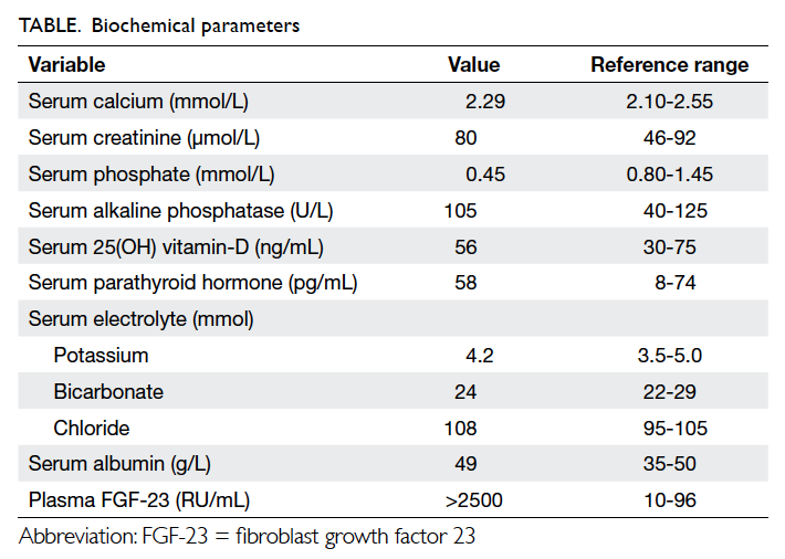
Table. Biochemical parameters
 

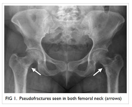
Figure 1. Pseudofractures seen in both femoral neck (arrows)
 

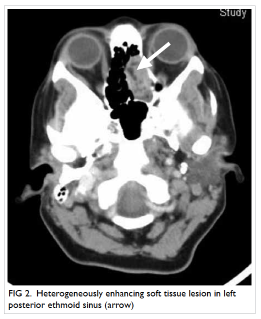
Figure 2. Heterogeneously enhancing soft tissue lesion in left posterior ethmoid sinus (arrow)
 
The patient was treated with phosphate supplements and underwent complete excision of the left ethmoidal sinus mass. Histopathological examination confirmed the diagnosis of a phosphaturic mesenchymal tumour. The patient remained stable after surgery. Two months later she was asymptomatic, by which time her muscle weakness had resolved markedly. Respective serial serum phosphate concentrations were 0.90, 1.00, and 1.35 mmol/L at 1, 2 and 4 weeks after surgery. After surgery, her plasma FGF-23 levels were undetectable.
 
Phosphaturic mesenchymal tumours are rare mesenchymal tumours and mostly comprised of a single histological entity with mixed connective tissue (designated PMT-MCT). Such tumours can occur in the soft tissue or bone.3 Most PMT-MCTs are histologically and clinically benign with rare instances of malignancy. Microscopically, PMT-MCTs are variable in appearance, usually being composed of small, bland round-to-spindle cells embedded in a vascular to myxochondroid matrix with variable amounts of mature adipose tissue (Fig 3). This matrix calcifies in an unusual ‘grungy’ fashion, inciting an osteoclast-rich and fibrohistiocytic response. A very prominent feature of PMT-MCT is its elaborate intrinsic microvasculature. Malignant PMT-MCTs resemble undifferentiated pleomorphic sarcomas or fibrosarcomas.3 After complete excision of the tumour, most patients improve dramatically4 and become symptomatically, biochemically, and radiologically better. Such results should prompt the physicians to search for such treatable and potentially curable causes, whenever they encounter hypophosphataemic osteomalacia.
 

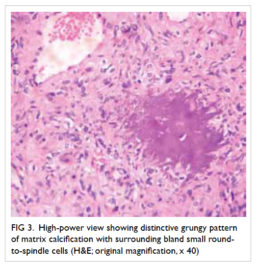
Figure 3. High-power view showing distinctive grungy pattern of matrix calcification with surrounding bland small round-to-spindle cells (H&E; original magnification, x 40)
 
References
1. Chokyu I, Ishibashi K, Goto T, Ohata K. Oncogenic osteomalacia associated with mesenchymal tumor in the middle cranial fossa: a case report. J Med Case Rep 2012;6:181. CrossRef
2. Komínek P, Stárek I, Geierová M, Matoušek P, Zeleník K. Phosphaturic mesenchymal tumour of the sinonasal area: case report and review of the literature. Head Neck Oncol 2011;3:16. CrossRef
3. Folpe AL, Fanburg-Smith JC, Billings SD, et al. Most osteomalacia-associated mesenchymal tumors are a single histopathologic entity: an analysis of 32 cases and a comprehensive review of the literature. Am J Surg Pathol 2004;28:1-30. CrossRef
4. Chong WH, Molinolo AA, Chen CC, Collins MT. Tumor-induced osteomalacia. Endocr Relat Cancer 2011;18:R53-77. CrossRef

Fever with vesicular rash in an adult

Hong Kong Med J 2014;20:264.e3–4 | Number 3, June 2014
DOI: 10.12809/hkmj/134018
© Hong Kong Academy of Medicine. CC BY-NC-ND 4.0
 
PICTORIAL MEDICINE
Fever with vesicular rash in an adult
Prashant Nasa, MD, FNB1; Ankur Gupta, MD, DM2; Shakti Jain, MD3; Kishore Mangal, MD, IDCCM1
1 Department of Critical Care Medicine, Max Super Speciality Hospital, Shalimar Bagh, New Delhi 110088, India
2 Department of Nephrology, Max Super Speciality Hospital, Shalimar Bagh, New Delhi 110088, India
3 Department of Microbiology, Max Super Speciality Hospital, Shalimar Bagh, New Delhi 110088, India
 
Corresponding author: Dr Prashant Nasa (dr.prashantnasa@hotmail.com)
A 30 year-old man was admitted with fever and myalgia for 4 days, rashes over abdomen and forehead for 2 days (Fig 1), and breathing difficulty for 1 day in January 2013. There was a history of similar rashes affecting his wife 7 days earlier. On examination, he was tachypnoeic (respiratory rate, 24 breaths/min), conscious, and oriented. He had a papulo-vesicular rash on the forehead, scalp, and trunk but the extremities were spared. Laboratory investigations revealed thrombocytopenia (platelet counts, 30 x 109/L), deranged liver function tests (serum bilirubin 4.6 mg/dL, direct bilirubin 2.9 mg/dL, aspartate transaminase 1316 IU/L, alanine transaminase 989 IU/L, alkaline phosphates 116 IU/L). The chest X-ray showed no gross abnormality and the smear of skin scrapings showed multinucleated epithelial giant cell (Tzanck cells), abundant erythrocytes, and sporadic leukocytes (Fig 2). Aerobic bacterial culture of a swabbed vesicle fluid was sterile. The history and characteristic pattern of the vesicular rash on the scalp and abdomen with no history of childhood immunisation against chickenpox clinched the diagnosis of chickenpox. The diagnosis is primarily clinical. Confirmation requires either viral culture of the vesicular fluid or direct fluorescent antibody testing of serum,1 but these tests are not readily available. Smear examination showing Tzanck cells in vesicle fluid may be positive in chicken pox, but also occurs in herpes simplex, herpes zoster, cytomegalovirus infection, and even pemphigus vulgaris.1 The differential diagnosis in our case was severe Chicken pox, disseminated herpes simplex infection, disseminated herpes zoster infection, drug eruptions, and pityriasis lichenoides et varioliformis acuta. However the clinical history (including family and vaccination history) favoured severe chickenpox.
 

Figure 1. A rash over the forehead and scalp
 

Figure 2. A Giemsa stain of skin scrapings from the base of a vesicle showing a multinucleated epithelial giant cell (Tzanck cell) [white arrow], abundant erythrocytes (black arrowhead), and sporadic leukocytes (black arrow) [Giemsa stain; original magnification, x 200]
 
Chickenpox is rarely fatal, although it is generally more severe in adults than children; those who are pregnant or immunocompromised have more severe forms of the disease.2 Serious manifestations of varicella-zoster virus (VZV) infection include pneumonia, hepatitis, and encephalitis.2 3 The standard treatment for severe adult VZV infection with or without involvement of vital organs entails early recourse to intravenous antivirals such as acyclovir. Whether severe or not, meticulous skin care, hygiene, and droplet precautions should be implemented to prevent cross-transmission to others till the patient’s lesions desquamate.
 
References
1. Herpesviruses: Varicella zoster virus (VZV). In: Shors T. Understanding viruses. 2nd ed. Burlington, MA: Jones & Bartlett; 2011: 459.
2. Maggi U, Russo R, Conte G, et al. Fulminant multiorgan failure due to varicella zoster virus and HHV6 in an immunocompetent adult patient, and anhepatia. Transplant Proc 2011;43:1184-6. CrossRef
3. Mohsen AH, McKendrick M. Varicella pneumonia in adults. Eur Respir J 2003;21:886-91. CrossRef

Pulmonary tuberculosis complicating asbestosis

Hong Kong Med J 2014;20:265.e3–5 | Number 3, June 2014
DOI: 10.12809/hkmj134019
© Hong Kong Academy of Medicine. CC BY-NC-ND 4.0
 
PICTORIAL MEDICINE
Pulmonary tuberculosis complicating asbestosis
YF Shea, MRCP (UK), FHKAM (Medicine)1; Janice JK Ip, MB, BS, FRCR2
1 Department of Medicine, Queen Mary Hospital, The University of Hong Kong, Pokfulam, Hong Kong
2 Department of Radiology, Queen Mary Hospital, The University of Hong Kong, Pokfulam, Hong Kong
 
Corresponding author: Dr YF Shea (elphashea@gmail.com)
An 87-year-old man who previously worked in shipyard with asbestosis was admitted in November 2012 because of fever of unknown origin. He presented with fever on-and-off for 2 months and cough. On physical examination, there was no cervical lymphadenopathy or hepatosplenomegaly and the chest was clear. Complete blood picture, and liver and renal function tests remained unremarkable. Chest X-ray (CXR) and computed tomography (CT) of the thorax yielded calcified pleural plaques, diaphragmatic calcification, diffuse centrilobular nodules, and interstitial septal thickening (Fig 1). Sputum and urine cultures were negative. Further investigations included smears and cultures for acid-fast bacilli and testing for Mycobacterium tuberculosis (MTB) by polymerase chain reaction of sputum, urine, and bronchoalveolar larvage samples, all of which were negative. Searches for aspergillus antigen, cryptococcal antigen, the Weil-Felix test, the Widal test, nasopharyngeal aspirate for influenza and mycoplasma, urine examination for legionella antigen, automminue profiling, and tests for tumour markers, human immunodeficiency virus, and sputum cytology were all non-contributory. The patient’s C-reactive protein was elevated to 3.39 mg/dL (reference range, <0.76 mg/dL). His fever had persisted on-and-off for 2 months despite multiple courses of broad-spectrum antibiotics. Positron emission tomography (PET)–CT yielded pronounced micronodules, especially over both upper lobes (Fig 1) and patchy ground glass opacities over both lower lobes with increased 18-fluorodeoxyglucose (18FDG) uptake (maximum standardised uptake values of up to 4; Figs 2 and 3). The absence of pulmonary masses, mediastinal or hilar lymphadenopathy or nodular thickening of interlobular septa and any bronchovascular bundle made malignancy or lymphangitis carcinomatosis unlikely. Based on the radiology, the patient was diagnosed to have pulmonary MTB for which anti-MTB treatment was initiated. He developed sudden cardiac arrest 1 day later, and failed resuscitation. Sputum and urine sample culture results available after the patient’s demise grew MTB.
 

Figure 1. (a) Chest X-ray and (b) computed tomography of the thorax showing calcified pleural plaques and diaphragmatic calcifications bilaterally (arrows)
 

Figure 2. Computed tomography thorax (lung window) of the upper lobes taken (a) 2 months earlier and 2 months later are shown; an increase in micronodules (arrows) was evident in (b) as compared with (a)
 

Figure 3. (a) Computed tomography thorax showing a background of calcified pleural plaques and an increase in patchy ground glass opacities over both lower lobes (arrows). (b) Positron emission tomography–computed tomography showing the parenchymal pattern of increased 18fluorodeoxyglucose uptake (maximum standardised uptake values of up to 4) over the both lower lobes (arrows)
 
Asbestosis is caused by the inhalation of asbestos which was once used as an electrical and thermal insulator. Asbestos causes fibrosis of the pleura and lung, as well as malignant mesothelioma, and PET-CT is a well-known means of picking up this complication as a linear area of intense 18FDG uptake surrounding the lungs.1 Fever of unknown origin can be due to infection, malignancy, autoimmune conditions, or drugs. Tuberculosis was one of the most common infectious causes. One of the diagnostic difficulties in our patient was the presence of interstitial lung disease making the interpretation of CXR and CT images challenging. Soussan et al2 has classified the PET appearance of pulmonary MTB into lung and lymphatic patterns and demonstrated improved diagnostic accuracy after taking account of the other specific CT changes such as upper lobe consolidation with cavitations or multiple ill-defined micronodules surrounding a cavity. Findings pertaining to our patient fitted well into previously described lung pattern of increased 18FDG uptake in MTB. These included predominant lung parenchymal involvement and together with an interval excess of micronodules, especially over the both upper lobes, which led us to make a radiological diagnosis of MTB. The increased in 18FDG uptake is caused by local accumulation of inflammatory cells.3 Thus, PET-CT is useful in identifying an active pulmonary tuberculoma in the absence of initial microbiological proof. Monitoring the response to anti-MTB treatment can also provide early evidence of drug-resistant MTB.3 4 The earlier performance of PET-CT and institution of anti-MTB treatment may have changed the clinical outcome of our patient.
 
References
1. Alavi A, Gupta N, Alberini JL, et al. Positron emission tomography imaging in nonmalignant thoracic disorders. Semin Nucl Med 2002;32:293-321. CrossRef
2. Soussan M, Brillet PY, Mekinian A, et al. Patterns of pulmonary tuberculosis on FDG-PET/CT. Eur J Radiol 2012;81:2872-6. CrossRef
3. Kosterink JG. Positron emission tomography in the diagnosis and treatment management of tuberculosis. Curr Pharm Des 2011;17:2875-80. CrossRef
4. Demura Y, Tsuchida T, Uesaka D, et al. Usefulness of 18F-fluorodeoxyglucose positron emission tomography for diagnosing disease activity and monitoring therapeutic response in patients with pulmonary mycobacteriosis. Eur J Nucl Med Mol Imaging 2009;36:632-9. CrossRef

Amelanotic melanoma masquerading as a pyogenic granuloma: caution warranted

Hong Kong Med J 2014;20:265.e1–2 | Number 3, June 2014
DOI: 10.12809/hkmj133987
© Hong Kong Academy of Medicine. CC BY-NC-ND 4.0
 
PICTORIAL MEDICINE
Amelanotic melanoma masquerading as a pyogenic granuloma: caution warranted
Noah LW So, MB, BS; CF Chan, MB, BS, FHKAM (Orthopaedic Surgery); Kenneth WY Ho, MB, BS, FHKAM (Orthopaedic Surgery); YL Lam, MB, ChB, FHKAM (Orthopaedic Surgery)
Department of Orthopaedics and Traumatology, Queen Mary Hospital, 102 Pokfulam Road, Hong Kong
 
Corresponding author: Dr Noah LW So (noahlwso@gmail.com)
Case
In April 2012, a 69-year-old Chinese woman complained of a non-healing raised lesion over the lateral aspect of her left fifth toe which had developed 2 years earlier. It appeared to have started as a minor abrasion after wearing ill-fitting shoes. A painless swelling then developed over that site with occasional friction-induced bleeding. She attended several podiatry treatment sessions and tried applying topical medications such as silver nitrate. The lesion did not resolve, and increased in size and eventually developed into a pink nodule. Apart from hypercholesterolaemia, the patient was an otherwise healthy non-smoker and non-drinker who enjoyed sunbathing on the beach in her youth, often spending half a day under the sun.
 
On physical examination, a 1 cm x 1 cm round and dull red nodule with superficial ulceration was noted on the lateral aspect of the left fifth toe. There were no signs of infection and no contact bleeding was evident (Fig). Given the history of minor trauma, tendency to bleed with minor friction and the appearance of the lesion, pyogenic granuloma was the main differential diagnosis. Owing to failure of repeated conservative treatment, excision of the lesion was performed.
 

Figure. Left fifth toe before excision showing the malignant melanoma (arrow): (a) lateral view and (b) plantar view (oblique angle)
 
Histology revealed it to be a malignant melanoma; no melanin pigment was detected but the tumour cells were positive for melanocytic markers. A staging positron emission tomography–computed tomography showed no metastasis. A wide resection involving ray amputation of the 4th and 5th toes was performed to ensure a clear resection margin.
 
Discussion
Amelanotic melanoma is reported to account for about 1.8% to 8.1% of all melanomas.1 Cheung et al2 defined amelanotic melanoma as being unsuspected clinically and with melanin in less than 5% of the tumour cells. Because the diagnosis was not suspected clinically, he noted that excisional biopsies were seldom performed, and that if undertaken, transection of the melanoma was common. In their database of 1170 patients, McClain et al3 found that red melanomas accounted for 3.9% of all such malignancies and up to 70% of those that were amelanotic. Regarding prognosis, their review found no significant difference between red amelanotic melanomas and pigmented melanomas in terms of disease-free survival, overall survival, metastasis, and recurrence. In other words, the mortality of red amelanotic melanomas appeared comparable to that of pigmented melanoma.
 
In our patient, the most notable risk factor for developing melanoma was prolonged sunlight exposure but such a history may be missed if the diagnosis is not suspected. Amelanotic melanomas appear clinically indistinguishable from pyogenic granulomas. We therefore caution all our readers to the possibility of amelanotic melanoma presenting as seemingly benign red lesions. Whilst uncommon in China (age-standardised incidence of 0.2 per 100 000 population and year),4 melanomas are highly malignant; both observation without investigation and excision without undertaking histology can delay the diagnosis with potentially disastrous consequences. The mnemonic ‘ABCD’ summarises features suggesting melanoma: Asymmetry, irregular Border, uneven Colour, Diameter of >6 mm.4 The mnemonic RRR (Red, Raised lesion, Recent change3) is proposed additionally for the diagnosis of red amelanotic melanomas. We recommend that for such red lesions of uncertain nature, referral to specialists experienced in managing melanomas is warranted.
 
References
1. Koch SE, Lange JR. Amelanotic melanoma: the great masquerader. J Am Acad Dermatol 2000;42:731-4. CrossRef
2. Cheung WL, Patel RR, Leonard A, Firoz B, Meehan SA. Amelanotic melanoma: a detailed morphologic analysis with clinicopathologic correlation of 75 cases. J Cutan Pathol 2012;39;33-9. CrossRef
3. McClain SE, Mayo KB, Shada AL, Smolkin ME, Patterson JW, Slingluff CL Jr. Amelanotic melanomas presenting as red skin lesions: a diagnostic challenge with potentially lethal consequences. Int J Dermatol 2012;51:420-6. CrossRef
4. LeBoit PE, Burg G, Weedon D, Sarasin A, editors. Pathology and genetics of skin tumours (IARC WHO Classification of Tumours). Lyon: IARC Press; 2006.

More than skin deep: Paget’s disease of the perineum

Hong Kong Med J 2014;20:264.e1–2 | Number 3, June 2014
DOI: 10.12809/hkmj133983
© Hong Kong Academy of Medicine. CC BY-NC-ND 4.0
 
PICTORIAL MEDICINE
More than skin deep: Paget’s disease of the perineum
JW Li, MB, BS, MRCP; CM Ng, MRCP, FHKAM
Department of Medicine, Queen Elizabeth Hospital, 30 Gascoigne Road, Kowloon, Hong Kong
 
Corresponding author: Dr JW Li (lijohnwing@gmail.com)
In July 2012, an extensive eczematous lesion was found incidentally over the external genitalia in a 77-year-old man who was being evaluated for lower limb cellulitis. The affected area had been present for 10 years; it was erythematous, macerated, and itchy. He had consulted countless doctors, but the itchiness was intense and the area of involvement was enlarging despite application of topical creams. On physical examination, the perineum was weepy with excoriation, crusts, and a serous discharge. The erythematous skin was indurated and had a well-defined margin. The penis shaft was swollen with palpable groin lymph nodes (Fig 1). Blood tests showed eosinophilia (1.1 x 10⁹/L) and an elevated erythrocyte sedimentation rate (58 mm/h).
 

Figure 1. Clinical picture of extramammary Paget’s disease of the perineum, showing its weepy nature and the sharp margin around the affected area
 
A clinical suspicion of extramammary Paget’s disease (EMPD) was suggested by the dermatologist. Skin biopsy of the right suprapubic region confirmed the diagnosis (Fig 2), while another biopsy of the left groin showed poorly differentiated invasive carcinoma, consistent with invasive EMPD. Urine for cytology, as part of the malignancy screening, showed atypical cells. Computed tomography of the abdomen revealed left ureteric obstruction with enhanced mural thickening of left ureter, of which transitional cell carcinoma could not be excluded. He was referred to a urologist for further assessment. Radiotherapy to the affected skin lesions was given in view of the extensive involvement.
 

Figure 2. The skin biopsy taken from the right suprapubic area shows many nests of cohesive pleomorphic malignant cells invading the epidermis. Immunohistochemical study demonstrates that the tumour cells are cytokeratin (CAM5.2) positive (not shown), confirming their epithelial nature (H&E; original magnification, x 200)
 
Extramammary Paget’s disease is a rare intraepithelial adenocarcinoma of the skin rich in apocrine glands, first described by Crocker in 1888.1 The disease involves the epidermis, but can potentially metastasise via the lymphatic system.2 Its clinical and histological features are similar to Paget’s disease of the nipple. The penis, scrotum, and vulva are frequently involved in sites. The erythematous area has a sharp margin and scaly surface. Intense itchiness is common and may result in erosion, excoriation, and lichenification.2
 
Our patient had a typical presentation with longstanding, distressing, and difficult-to-treat, eczema-like perineal lesion with a well-defined border. Delay in diagnosis, up to a decade,3 is notoriously common, because EMPD often mimics benign dermatological diseases such as eczema, contact dermatitis and fungal infection, and even malignant skin conditions like superficial melanoma, basal cell carcinoma, and Bowen’s disease.
 
A clinical diagnosis should be confirmed by skin biopsy. Immunohistochemistry shows the cells are positive for CEA, CAM 5.2, as well as keratins CK7 and 8. Once EMPD is diagnosed, thorough search for malignancy is mandatory, since up to 37% of patients have associated malignancies, in which case the condition is referred to as secondary EMPD.4 A recent study among Chinese patients showed that 8.3% out of 48 patients with EMPD developed malignancy.3 Location of skin involvement in EMPD predicts the type of cancer; perianal lesions are associated with anal and colorectal cancer, whereas penoscrotal lesions tend to be associated with urogenital malignancies.4 The positivity of Cytokeratin 20 in immunostaining is also highly associated with internal malignancies.5
 
Treatment is mainly surgical resection with reconstruction. Radiotherapy is an adjunctive therapy or reserved for frail elderly patients.2 Photodynamic therapy and topical imiquimod have been used. Despite these treatments, the local recurrence rate still ranges from 21% to 61%,3 with a median time to recurrence of 2 years.
 
Acknowledgement
We would like to thank Dr Shun-chin Ng, Dermatologist, Department of Health, Hong Kong, for his clinical care and manuscript advice. We thank Dr Wing-hung Lau, Department of Pathology, Queen Elizabeth Hospital, for provision of histological diagnosis and picture.
 
References
1. Crocker HR. Paget's disease affecting the scrotum and penis. Trans Pathol Soc Lond 1888;40:187-91.
2. Burns T, Breathnach S, Cox N, Griffiths C, editors. Rook's textbook of dermatology. 8th ed. Oxford, UK: Wiley-Blackwell; 2010.
3. Chan JY, Li GK, Chung JH, Chow VL. Extramammary Paget's disease: 20 years of experience in Chinese population. Int J Surg Oncol 2012;2012:416418.
4. Sarmiento JM, Wolff BG, Burgart LJ, Frizelle FA, Ilstrup DM. Paget's disease of the perianal region—an aggressive disease? Dis Colon Rectum 1997;40:1187-94. CrossRef
5. Liegl B, Liegl S, Gogg-Kamerer M, Tessaro B, Horn LC, Moinfar F. Mammary and extramammary Paget's disease: an immunohistochemical study of 83 cases. Histopathology 2007;50:439-47. CrossRef

Laparoscopic removal of an eroding Mirena coil through the sigmoid colon

ABSTRACT

Hong Kong Med J 2013;19:560.e3–4 | Number 6, December 2013
DOI: 10.12809/hkmj133845
PICTORIAL MEDICINE
Laparoscopic removal of an eroding Mirena coil through the sigmoid colon
A Hussain, K Omar, S El-Hasani
General Surgery Department, Princess Royal University Hospital, Farnborough Common, Orpington, Kent, BR6 8ND, United Kingdom
 
 
No abstract available.
 
View this abstract indexed in MEDLINE:
 

Brunner's gland adenoma: unusual cause of duodenal haemorrhage and obstruction

ABSTRACT

Hong Kong Med J 2013;19:460.e1–2 | Number 5, October 2013
DOI: 10.12809/hkmj133776
PICTORIAL MEDICINE
Brunner's gland adenoma: unusual cause of duodenal haemorrhage and obstruction
TT Cheung, EWK Ip, RTP Poon, N Trendell-Smith
Department of Surgery, The University of Hong Kong, Queen Mary Hospital, Pokfulam, Hong Kong
 
 
No abstract available.
 
View this abstract indexed in MEDLINE:
 

Mounier-Kuhn syndrome: an unusual underlying cause for chronic coughs and recurrent pneumonias

ABSTRACT

Hong Kong Med J 2013;19:365.e3–4 | Number 4, August 2013
DOI: 10.12809/hkmj133760
PICTORIAL MEDICINE
Mounier-Kuhn syndrome: an unusual underlying cause for chronic coughs and recurrent pneumonias
Janice JK Ip, Peter KT Hui, Sonia HY Lam, Wendy WM Lam, MT Chau
Department of Radiology, Queen Mary Hospital, Pokfulam, Hong Kong
 
 
No abstract available.
 
View this abstract indexed in MEDLINE:
 

Pages